User login
How to engage soldiers, veterans in psychiatric treatment
Deployments in places such as Afghanistan and Iraq, and traumatic events such as the Sept. 11, 2001, attacks affect everyone, but military personnel and veterans face unique circumstances that can present challenges to treatment. Much progress has been made in recent years in treating people with posttraumatic stress disorder and helping them recover after traumatic events.

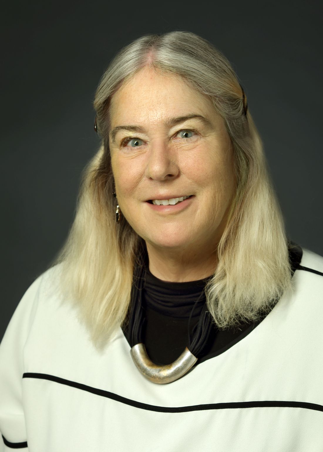
To explore some of those changes and challenges, this news organization interviewed Col. (Ret.) Elspeth Cameron Ritchie, MD, MPH, who retired from the Army in 2010 after assignments and missions that took her to Korea, Somalia, Iraq, and Cuba, about her approaches to treating soldiers and veterans.
Dr. Ritchie is chief of psychiatry at Medstar Washington Hospital Center, and a professor of psychiatry at the Uniformed Services University of the Health Sciences in Bethesda, Md., and at Georgetown University and George Washington University, both in Washington.
She is the author of 250 publications, including the book, “Forensic and Ethical Issues in Military Behavioral Health” (Fort Sam Houston, Tex.: Borden Institute, 2015). In addition, Dr. Ritchie is coeditor of “Post-Traumatic Stress Disorder and Related Diseases in Combat Veterans” (New York: Springer, 2015) and “Psychiatrists in Combat, Clinicians Experience in the War Zone” (New York: Springer, 2017).
Question: What are some of the interventions available in the aftermath of traumatic events?
Answer: What we thought the standard of care should be after a traumatic event was to have what’s called a critical incident stress debriefing (CISD). It was basically getting the members of the group who had been traumatized by a school shooting or plane crash, or the Oklahoma City bombing, getting them all together literally a few hours after the event, and having them tell what happened. And the idea is to get it all out. But what we discovered is that this could actually make people worse, because you’d be hearing not only about your own trauma, but other people’s traumas, and that it was too soon for the event.
So prior to 9/11, we had organized a conference, which was held in October 2001, just a month after 9/11. At that conference, we worked on mass violence and early intervention, which is the name of the book that came out from the (National Institute of Mental Health) as a result. It focused on basic principles of safety and security and communication, and knowing where your family was, rather than reliving the trauma. Now, we did think that sometimes you could have a CISD that would be helpful, but only when it was people who knew each other well, like an ED group who would work with each other or soldiers who served together.
Q: What was your involvement in the aftermath of the Sept. 11 attacks?
A: At the time of 9/11, I was assigned at the Pentagon, but I wasn’t there. When the plane hit, I was actually across the river at the Navy’s Bureau of Medicine and Surgery. And then for the next 3 weeks, all I did was work at the Pentagon. We used some of these principles of early intervention but not focusing on telling us what happened right afterward. We focused on how the service members and their families were coping in the here and now, and how they could support each other.
We knew that soldiers would not come out of their offices to go to a therapist. They are too strong for that. So, we did what was called “therapy by walking around.” We went to the service members’ offices.
There was also a Family Assistance Center. That was for the families of the people who died. And that was very helpful because you had all the services there in one place – medical care, mental health care, therapy dogs, massage, the people who collected the DNA to identify remains. You had it in one place, the Sheraton in Crystal City, Va.. That has become a model now, especially for mass transportation fatalities. There are a lot more in the literature about Family Assistance Centers now, mainly formed by the National Transportation Safety Board.
Right after 9/11, we went to war in Afghanistan, and later in Iraq, and we had a lot of soldiers who developed both PTSD and traumatic brain injury (TBI). One of the good things that the military can do is they can really innovate with both medical treatment and mental health treatment because they don’t have to ask for an insurance company to pay for it. So for some years, starting in about 2004, Congress allocated a large sum of money every year to the Department of Defense to focus on treatment for PTSD and TBI.
And as a result of that, a couple of things happened. One was that the treatments that we had, we were able to study much better, exposure therapy and cognitive-behavioral therapy. We were able to do large trials, and then we continued with the use of medications when necessary. There are only two (Food and Drug Administration)–approved medicines for the treatment of PTSD: sertraline and paroxetine, but many others are used.
We also learned what didn’t work and what soldiers would not take. Most of these medications have sexual side effects. If you’re a young, healthy soldier, you really don’t want to be taking something that causes you erectile dysfunction, or in women a loss of libido. So many people wouldn’t take these therapies. As for exposure therapy, if you got into it and completed the program, usually your PTSD symptoms went down. But many people couldn’t complete it. In the exposure therapy, you’re talking about whatever trauma you’ve been through – maybe your best friend died next to you, and you don’t want to talk about that all the time.
When I talk to patients about this, I say the first bucket is medication, the second bucket is therapy, and the third bucket is everything else. And everything else includes meditation, yoga, exercise, and it also involves working with animals. There are programs where you’re paired with a service dog, who helps calm you down, and you feel protected.
One of my favorites is called Warrior Canine Connection, where a soldier with PTSD trains a puppy to become a service animal. And in the training of the dog, you have to learn to control your emotions, you have to modulate your voice, you have to appear calm. Often soldiers have a background that they’re familiar with animals, especially dogs. So that’s been very successful.
A couple of other (treatments) to mention one is called stellate ganglion block, where a little lidocaine is injected into the back of the cervical spine. It was used initially for pain control, and they found that it was actually very helpful for PTSD. Another thing we’ve learned is that pain and PTSD often go hand in hand, because if you’re in pain, you’ll be feeling awful, you won’t sleep well, you’ll have more nightmares. But if you can control both of them together, then that’s going to help.
Q: One issue that veterans may face is moral injury. Can you talk about that?
A: Moral injury is a term that was first used after Vietnam. Moral injury is not a psychiatric diagnosis. It is feelings of shame and guilt that can be very corrosive and can lead to suicide. It overlaps with PTSD. You feel either you’ve let yourself down, or the government has let you down. And this can be very corrosive. Another thing that could happen is, say, you switched your tour of duty with a buddy, and he got killed and you didn’t. A very common scenario is you’re manning a checkpoint, and a car comes at you and doesn’t stop like it’s supposed to. You do what you’ve been trained to do, which is open fire, and check on the car afterward. And there’s four little kids and their parents in the car all dead. And that is something that even though that was your sort of duty, that it still eats at you because you have kids the same age as the ones who were dead in the car.
You can still have these feelings of shame and guilt, and it will often bleed into your relationships with your family. And that can lead to distance and divorce, which is a further risk factor for suicide.
Q: Are there are any specific treatments that have been designed for moral injury, different from PTSD or other conditions?
A: The Armed Services has set up a number of intensive programs at different places, and each is a little bit different. They usually integrate moral injury in with some of the other treatments. There was one at Fort Bliss, Tex., that had reiki; they had art therapy. And they had the chaplains working on moral injury. So there’s no medical treatment for it, but there certainly is talking about it, and for some people to go to a chaplain can be very helpful.
There’s a Military Health System Centers of Excellence, which is a place by the new Walter Reed on the campus, they have a marvelous wall full of masks. And the masks have been painted by soldiers with usually a combination of PTSD, TBI, and although it’s not an official psychiatric diagnosis, moral injury. They’re able to draw and paint. Another thing that’s been used quite a bit as writing therapy, and journaling, and just writing down how you feel about something, because you can do that without retraumatizing anybody else, except perhaps if you are working with a therapist.
Q: For therapists who are treating soldiers, veterans, are there specific challenges that they should be aware of? Are these patients maybe different from the patients that they might otherwise see? Are there specific pieces of advice as to how to engage them?
A: There are a few things that are different. One is that many people in the military are not used to talking about their feelings. And that’s especially if you’ve got a young man who only grunts and says: “Hooah!” That is going to be hard to break through. And that’s why some of these other ways of reaching somebody is very effective. Also, the military likes to have physical activity; they’re usually not comfortable sitting in a chair. If you’re a civilian psychiatrist, I don’t expect you to go bungee jumping with your patients. But what I’d recommend is that you recommend to your patients that they stay active.
Another thing about veterans is that they like to be self-sufficient. They really don’t like to ask for help, although they might ask for help for their buddy. After the Pentagon and 9/11, when I was working with senior officers, they never needed any help. No, but their buddy over here might, so I could help them in the guise of providing care for their buddy in a group setting. We could work with everybody and enhance cohesion, morale, bonding, “we’re all in this together” type of feeling.
I think one thing that’s really improved is that there is less stigma around PTSD. People are more willing to present for help, and some people have called PTSD the Purple Heart of mental disorders. People don’t feel like it’s as bad as having depression or anxiety. Even though PTSD often has depression and anxiety components to it – they run hand in hand – still, it’s sort of more honorable if you’ve been at war and have gotten PTSD.
Q: How have you been faring yourself, in the face of the 9/11 anniversary and recent events in Afghanistan?
A: (The Sept. 11 weekend) was very sad for me – and a lot of my colleagues [with] the combination of the 20th anniversary of 9/11, and the recent development. Fortunately, I have friends and people I can talk to. I walked with a colleague of mine who was in the Army. I’m following my own rule of the three buckets, so we took a walk around the hospital center for about 45 minutes, and we have five fish ponds here. And we went and looked at the fish, and talked to the fish. At the National Rehab Hospital, they were playing the guitar. So there’s are a variety of things that people can do.
Deployments in places such as Afghanistan and Iraq, and traumatic events such as the Sept. 11, 2001, attacks affect everyone, but military personnel and veterans face unique circumstances that can present challenges to treatment. Much progress has been made in recent years in treating people with posttraumatic stress disorder and helping them recover after traumatic events.
To explore some of those changes and challenges, this news organization interviewed Col. (Ret.) Elspeth Cameron Ritchie, MD, MPH, who retired from the Army in 2010 after assignments and missions that took her to Korea, Somalia, Iraq, and Cuba, about her approaches to treating soldiers and veterans.
Dr. Ritchie is chief of psychiatry at Medstar Washington Hospital Center, and a professor of psychiatry at the Uniformed Services University of the Health Sciences in Bethesda, Md., and at Georgetown University and George Washington University, both in Washington.
She is the author of 250 publications, including the book, “Forensic and Ethical Issues in Military Behavioral Health” (Fort Sam Houston, Tex.: Borden Institute, 2015). In addition, Dr. Ritchie is coeditor of “Post-Traumatic Stress Disorder and Related Diseases in Combat Veterans” (New York: Springer, 2015) and “Psychiatrists in Combat, Clinicians Experience in the War Zone” (New York: Springer, 2017).
Question: What are some of the interventions available in the aftermath of traumatic events?
Answer: What we thought the standard of care should be after a traumatic event was to have what’s called a critical incident stress debriefing (CISD). It was basically getting the members of the group who had been traumatized by a school shooting or plane crash, or the Oklahoma City bombing, getting them all together literally a few hours after the event, and having them tell what happened. And the idea is to get it all out. But what we discovered is that this could actually make people worse, because you’d be hearing not only about your own trauma, but other people’s traumas, and that it was too soon for the event.
So prior to 9/11, we had organized a conference, which was held in October 2001, just a month after 9/11. At that conference, we worked on mass violence and early intervention, which is the name of the book that came out from the (National Institute of Mental Health) as a result. It focused on basic principles of safety and security and communication, and knowing where your family was, rather than reliving the trauma. Now, we did think that sometimes you could have a CISD that would be helpful, but only when it was people who knew each other well, like an ED group who would work with each other or soldiers who served together.
Q: What was your involvement in the aftermath of the Sept. 11 attacks?
A: At the time of 9/11, I was assigned at the Pentagon, but I wasn’t there. When the plane hit, I was actually across the river at the Navy’s Bureau of Medicine and Surgery. And then for the next 3 weeks, all I did was work at the Pentagon. We used some of these principles of early intervention but not focusing on telling us what happened right afterward. We focused on how the service members and their families were coping in the here and now, and how they could support each other.
We knew that soldiers would not come out of their offices to go to a therapist. They are too strong for that. So, we did what was called “therapy by walking around.” We went to the service members’ offices.
There was also a Family Assistance Center. That was for the families of the people who died. And that was very helpful because you had all the services there in one place – medical care, mental health care, therapy dogs, massage, the people who collected the DNA to identify remains. You had it in one place, the Sheraton in Crystal City, Va.. That has become a model now, especially for mass transportation fatalities. There are a lot more in the literature about Family Assistance Centers now, mainly formed by the National Transportation Safety Board.
Right after 9/11, we went to war in Afghanistan, and later in Iraq, and we had a lot of soldiers who developed both PTSD and traumatic brain injury (TBI). One of the good things that the military can do is they can really innovate with both medical treatment and mental health treatment because they don’t have to ask for an insurance company to pay for it. So for some years, starting in about 2004, Congress allocated a large sum of money every year to the Department of Defense to focus on treatment for PTSD and TBI.
And as a result of that, a couple of things happened. One was that the treatments that we had, we were able to study much better, exposure therapy and cognitive-behavioral therapy. We were able to do large trials, and then we continued with the use of medications when necessary. There are only two (Food and Drug Administration)–approved medicines for the treatment of PTSD: sertraline and paroxetine, but many others are used.
We also learned what didn’t work and what soldiers would not take. Most of these medications have sexual side effects. If you’re a young, healthy soldier, you really don’t want to be taking something that causes you erectile dysfunction, or in women a loss of libido. So many people wouldn’t take these therapies. As for exposure therapy, if you got into it and completed the program, usually your PTSD symptoms went down. But many people couldn’t complete it. In the exposure therapy, you’re talking about whatever trauma you’ve been through – maybe your best friend died next to you, and you don’t want to talk about that all the time.
When I talk to patients about this, I say the first bucket is medication, the second bucket is therapy, and the third bucket is everything else. And everything else includes meditation, yoga, exercise, and it also involves working with animals. There are programs where you’re paired with a service dog, who helps calm you down, and you feel protected.
One of my favorites is called Warrior Canine Connection, where a soldier with PTSD trains a puppy to become a service animal. And in the training of the dog, you have to learn to control your emotions, you have to modulate your voice, you have to appear calm. Often soldiers have a background that they’re familiar with animals, especially dogs. So that’s been very successful.
A couple of other (treatments) to mention one is called stellate ganglion block, where a little lidocaine is injected into the back of the cervical spine. It was used initially for pain control, and they found that it was actually very helpful for PTSD. Another thing we’ve learned is that pain and PTSD often go hand in hand, because if you’re in pain, you’ll be feeling awful, you won’t sleep well, you’ll have more nightmares. But if you can control both of them together, then that’s going to help.
Q: One issue that veterans may face is moral injury. Can you talk about that?
A: Moral injury is a term that was first used after Vietnam. Moral injury is not a psychiatric diagnosis. It is feelings of shame and guilt that can be very corrosive and can lead to suicide. It overlaps with PTSD. You feel either you’ve let yourself down, or the government has let you down. And this can be very corrosive. Another thing that could happen is, say, you switched your tour of duty with a buddy, and he got killed and you didn’t. A very common scenario is you’re manning a checkpoint, and a car comes at you and doesn’t stop like it’s supposed to. You do what you’ve been trained to do, which is open fire, and check on the car afterward. And there’s four little kids and their parents in the car all dead. And that is something that even though that was your sort of duty, that it still eats at you because you have kids the same age as the ones who were dead in the car.
You can still have these feelings of shame and guilt, and it will often bleed into your relationships with your family. And that can lead to distance and divorce, which is a further risk factor for suicide.
Q: Are there are any specific treatments that have been designed for moral injury, different from PTSD or other conditions?
A: The Armed Services has set up a number of intensive programs at different places, and each is a little bit different. They usually integrate moral injury in with some of the other treatments. There was one at Fort Bliss, Tex., that had reiki; they had art therapy. And they had the chaplains working on moral injury. So there’s no medical treatment for it, but there certainly is talking about it, and for some people to go to a chaplain can be very helpful.
There’s a Military Health System Centers of Excellence, which is a place by the new Walter Reed on the campus, they have a marvelous wall full of masks. And the masks have been painted by soldiers with usually a combination of PTSD, TBI, and although it’s not an official psychiatric diagnosis, moral injury. They’re able to draw and paint. Another thing that’s been used quite a bit as writing therapy, and journaling, and just writing down how you feel about something, because you can do that without retraumatizing anybody else, except perhaps if you are working with a therapist.
Q: For therapists who are treating soldiers, veterans, are there specific challenges that they should be aware of? Are these patients maybe different from the patients that they might otherwise see? Are there specific pieces of advice as to how to engage them?
A: There are a few things that are different. One is that many people in the military are not used to talking about their feelings. And that’s especially if you’ve got a young man who only grunts and says: “Hooah!” That is going to be hard to break through. And that’s why some of these other ways of reaching somebody is very effective. Also, the military likes to have physical activity; they’re usually not comfortable sitting in a chair. If you’re a civilian psychiatrist, I don’t expect you to go bungee jumping with your patients. But what I’d recommend is that you recommend to your patients that they stay active.
Another thing about veterans is that they like to be self-sufficient. They really don’t like to ask for help, although they might ask for help for their buddy. After the Pentagon and 9/11, when I was working with senior officers, they never needed any help. No, but their buddy over here might, so I could help them in the guise of providing care for their buddy in a group setting. We could work with everybody and enhance cohesion, morale, bonding, “we’re all in this together” type of feeling.
I think one thing that’s really improved is that there is less stigma around PTSD. People are more willing to present for help, and some people have called PTSD the Purple Heart of mental disorders. People don’t feel like it’s as bad as having depression or anxiety. Even though PTSD often has depression and anxiety components to it – they run hand in hand – still, it’s sort of more honorable if you’ve been at war and have gotten PTSD.
Q: How have you been faring yourself, in the face of the 9/11 anniversary and recent events in Afghanistan?
A: (The Sept. 11 weekend) was very sad for me – and a lot of my colleagues [with] the combination of the 20th anniversary of 9/11, and the recent development. Fortunately, I have friends and people I can talk to. I walked with a colleague of mine who was in the Army. I’m following my own rule of the three buckets, so we took a walk around the hospital center for about 45 minutes, and we have five fish ponds here. And we went and looked at the fish, and talked to the fish. At the National Rehab Hospital, they were playing the guitar. So there’s are a variety of things that people can do.
How do alcohol, obesity impact cirrhosis?
Alcohol intake and obesity are independent risk factors for morbidity among patients with cirrhosis, but the two factors do not appear to combine for a stronger effect (supra-additive), according to conclusions from a new analysis of participants in the UK Biobank study published in Hepatology.

The researchers analyzed data from the records of 489,285 individuals in the UK Biobank from May 2006 to July 2010. Researchers defined morbidity as first-time hospitalization for cirrhosis and calculated the cumulative incidence at 10 years among included individuals. The researchers defined obesity as body mass index of at least 30 kg/m2 and healthy BMI as 20-25. Safe drinking was defined as having fewer than 22 units per week for males or fewer than 15 units for females, harmful drinking was defined as more than 50 units per week for males or more than 35 for females, and hazardous drinking was defined as 22-49 units per week for males and 15-35 for females. The researchers assumed 2 units in a pint of beer or cider, 1.5 units in a glass of wine and “other” drinks, and 1 unit per measure of spirits.
The mean age was 57.0 years, and 45.4% were male. Overall, 24.3% of subjects were obese, 76.5% had safe levels of alcohol consumption, 19.7% had hazardous alcohol consumption, and 3.8% were classified as harmful drinkers.
Overall, harmful drinking was associated with 5.0 times the 10-year cumulative incidence of cirrhosis morbidity among harmful versus safe drinkers (1.51% vs. 0.30%). However, among those with a healthy BMI, harmful was associated with an 8.6-fold increase of cirrhosis morbidity, compared with safe drinkers (1.38% vs. 0.16%). On the other hand, obese patients with harmful drinking habits had a 3.6-fold increase over obese safe drinkers (1.99% vs. 0.56%).
When looked at according to BMI, 10-year cumulative incidence was 3.1 times higher in patients who with obesity versus those who with healthy BMI (0.65% vs. 0.21%). This varied strongly with drinking: Safe drinkers who with obesity had 3.7 times the incidence, compared with safe drinkers with healthy BMI (0.56% vs. 0.15%), and harmful drinkers who were obese had a 1.4-fold increased incidence, compared with harmful drinkers of a healthy weight (1.99% vs. 1.38%).
“In contrast to some previous studies, we found little evidence that [obesity and drinking] interacted supra-additively to modulate the risk of cirrhosis morbidity,” the authors wrote. “On the contrary, through a relative risk lens, the association between alcohol intake and cirrhosis morbidity was actually weaker for individuals with obesity than for individuals with a healthy BMI (indicating a sub-additive relationship).”
Fine-Gray regression modelling seemed to confirm that the relationship was sub-additive. After controlling for various factors, researchers found that harmful drinkers had a 6.84-fold increased risk at a healthy BMI, while the risk was only 3.14 times higher in obese patients (P interaction = 3.53 x 10⁻⁶).
The findings contradict previous studies, which suggested that high BMI and harmful drinking combined may produce much higher risk than either factor alone, possibly because obesity might “prime” the liver to be vulnerable to the effects of alcohol.
The authors suggest that the differences in findings may be caused by methodological limitations of the earlier studies, such as reliance on self-reported BMI data; small sample sizes and a relatively small number of liver events among those with obesity and harmful alcohol consumption; and the failure to use a competing risk perspective. The latter is relevant because alcohol and obesity are risk factors for other potentially fatal health conditions.
But the current study is not without its own limitations, according to Nancy Reau, MD, who is a professor of medicine and chair of hepatology at Rush University Medical Center in Chicago, who was asked to comment on the findings. Dr. Reau pointed out that the authors found the highest frequency of complications was observed in people with harmful alcohol intake whose BMI was under 20. That group may be composed of subjects with sarcopenia and end-stage liver disease from alcohol use. “Until you can separate these from the truly healthy BMI but [with harmful alcohol use], you can’t interpret this arm,” said Dr. Reau.
Beyond that, the researchers found increased risks of harm among individuals regardless of BMI, but the risks were highest among those with BMI over 30. Dr. Reau posited that the frequency might have been significantly greater at BMI higher than 35 and 40, but the researchers didn’t report results among these subcategories.
“In no way does this suggest that we need to ignore alcohol use in our patients with NAFLD [nonalcoholic fatty liver disease] or [nonalcoholic steatohepatitis],” said Dr. Reau.
In fact, she pointed to a figure in the paper that showed the highest increase in frequency among those with harmful alcohol use and obesity. “It’s clear that both conditions are much more serious than just obesity alone. It is incredibly important to council our NAFLD patients on appropriate alcohol use, [since] problematic drinking increases their risk. Problematic drinking remains a serious problem and increased awareness and linking to addiction services is important,” she said.
The authors reported no conflicts of interest. Dr. Reau has no relevant financial disclosures.
Alcohol intake and obesity are independent risk factors for morbidity among patients with cirrhosis, but the two factors do not appear to combine for a stronger effect (supra-additive), according to conclusions from a new analysis of participants in the UK Biobank study published in Hepatology.
The researchers analyzed data from the records of 489,285 individuals in the UK Biobank from May 2006 to July 2010. Researchers defined morbidity as first-time hospitalization for cirrhosis and calculated the cumulative incidence at 10 years among included individuals. The researchers defined obesity as body mass index of at least 30 kg/m2 and healthy BMI as 20-25. Safe drinking was defined as having fewer than 22 units per week for males or fewer than 15 units for females, harmful drinking was defined as more than 50 units per week for males or more than 35 for females, and hazardous drinking was defined as 22-49 units per week for males and 15-35 for females. The researchers assumed 2 units in a pint of beer or cider, 1.5 units in a glass of wine and “other” drinks, and 1 unit per measure of spirits.
The mean age was 57.0 years, and 45.4% were male. Overall, 24.3% of subjects were obese, 76.5% had safe levels of alcohol consumption, 19.7% had hazardous alcohol consumption, and 3.8% were classified as harmful drinkers.
Overall, harmful drinking was associated with 5.0 times the 10-year cumulative incidence of cirrhosis morbidity among harmful versus safe drinkers (1.51% vs. 0.30%). However, among those with a healthy BMI, harmful was associated with an 8.6-fold increase of cirrhosis morbidity, compared with safe drinkers (1.38% vs. 0.16%). On the other hand, obese patients with harmful drinking habits had a 3.6-fold increase over obese safe drinkers (1.99% vs. 0.56%).
When looked at according to BMI, 10-year cumulative incidence was 3.1 times higher in patients who with obesity versus those who with healthy BMI (0.65% vs. 0.21%). This varied strongly with drinking: Safe drinkers who with obesity had 3.7 times the incidence, compared with safe drinkers with healthy BMI (0.56% vs. 0.15%), and harmful drinkers who were obese had a 1.4-fold increased incidence, compared with harmful drinkers of a healthy weight (1.99% vs. 1.38%).
“In contrast to some previous studies, we found little evidence that [obesity and drinking] interacted supra-additively to modulate the risk of cirrhosis morbidity,” the authors wrote. “On the contrary, through a relative risk lens, the association between alcohol intake and cirrhosis morbidity was actually weaker for individuals with obesity than for individuals with a healthy BMI (indicating a sub-additive relationship).”
Fine-Gray regression modelling seemed to confirm that the relationship was sub-additive. After controlling for various factors, researchers found that harmful drinkers had a 6.84-fold increased risk at a healthy BMI, while the risk was only 3.14 times higher in obese patients (P interaction = 3.53 x 10⁻⁶).
The findings contradict previous studies, which suggested that high BMI and harmful drinking combined may produce much higher risk than either factor alone, possibly because obesity might “prime” the liver to be vulnerable to the effects of alcohol.
The authors suggest that the differences in findings may be caused by methodological limitations of the earlier studies, such as reliance on self-reported BMI data; small sample sizes and a relatively small number of liver events among those with obesity and harmful alcohol consumption; and the failure to use a competing risk perspective. The latter is relevant because alcohol and obesity are risk factors for other potentially fatal health conditions.
But the current study is not without its own limitations, according to Nancy Reau, MD, who is a professor of medicine and chair of hepatology at Rush University Medical Center in Chicago, who was asked to comment on the findings. Dr. Reau pointed out that the authors found the highest frequency of complications was observed in people with harmful alcohol intake whose BMI was under 20. That group may be composed of subjects with sarcopenia and end-stage liver disease from alcohol use. “Until you can separate these from the truly healthy BMI but [with harmful alcohol use], you can’t interpret this arm,” said Dr. Reau.
Beyond that, the researchers found increased risks of harm among individuals regardless of BMI, but the risks were highest among those with BMI over 30. Dr. Reau posited that the frequency might have been significantly greater at BMI higher than 35 and 40, but the researchers didn’t report results among these subcategories.
“In no way does this suggest that we need to ignore alcohol use in our patients with NAFLD [nonalcoholic fatty liver disease] or [nonalcoholic steatohepatitis],” said Dr. Reau.
In fact, she pointed to a figure in the paper that showed the highest increase in frequency among those with harmful alcohol use and obesity. “It’s clear that both conditions are much more serious than just obesity alone. It is incredibly important to council our NAFLD patients on appropriate alcohol use, [since] problematic drinking increases their risk. Problematic drinking remains a serious problem and increased awareness and linking to addiction services is important,” she said.
The authors reported no conflicts of interest. Dr. Reau has no relevant financial disclosures.
FROM HEPATOLOGY
A new weight loss threshold for T2d remission after bariatric surgery?
Patients with type 2 diabetes who underwent bariatric surgery commonly experienced remission, but there was little increase in rates of remission above a threshold of 20% total weight loss (TWL), according to a retrospective analysis of 5,928 patients with diabetes in an integrated health care system in Southern California.

The findings should reassure physicians and patients that surgery will be beneficial, according to lead author Karen Coleman, PhD, professor of health systems science at Kaiser Permanente Southern California.
Dr. Coleman has heard from many physicians saying they recommend against bariatric surgery because of concerns that patients gain weight back and therefore won’t get a long-term benefit, but this is not supported by the literature. “Hundreds of articles at this point show that this simply is not true. In addition, providers seem to think about bariatric surgery as an ‘all or none’ treatment. Gaining any weight back means that patients ‘fail.’ Weight regain is a normal part of massive weight loss; however, maintaining a certain amount of weight loss still provides benefits for patients, especially those with cardiovascular conditions like diabetes,” said Dr. Coleman.
Most patients lose 20%-30% of their body weight after bariatric surgery, but they don’t have to lose that much to see an improvement in type 2 diabetes (T2D). In addition, if patients lose that much or more, and then gain some weight back, it doesn’t eliminate benefit. “Although we did not measure weight regain, a corollary statement is that patients can regain some of the weight they lose, but if they stay around 20% of their total weight lost, then their diabetes still remits,” said Dr. Coleman.
In the past, some standards to treat severe weight loss and metabolic disease called for 50% or more TWL. More recent standards target a 30% threshold. “We want physicians to understand that they need to have more reasonable expectations of weight loss with surgery and that these reasonable expectations still result in profound improvements in cardiovascular risk, death, and quality of life. A 20% TWL threshold is easier for these patients to get to, and like other patients, they still get the benefit. So even if these patients may not have as much weight loss they can still benefit from the surgery for their diabetes,” Dr. Coleman added.
Physicians have long assumed that the effect of bariatric surgery on T2D remission is tied to weight loss, but this has been tested only recently. Previous studies found a link and suggested that 25% TWL may be the needed threshold, but more data are needed, especially for sleeve gastrectomy.
In the current study, published in Diabetes Care, 73% of patients were female. Mean age was 49.8 years, and mean body mass index was 43.8 kg/m2. Fifty-seven percent underwent Roux-en-Y gastric bypass (RYGB). Follow-up averaged 5.9 years. Overall, 71% of patients had an initial remission of their diabetes (72% RYGB, 70% sleeve). The average time to remission was 1.0 years. The researchers categorized participants by percentage TWL. Compared with the 0%-5% group, each 5% increase in TWL was linked with a greater likelihood of achieving remission: 5%-10%, hazard ratio 1.22 (P = .23); 10%-15%, HR 1.97 (95% confidence interval, 1.47-2.64); 15%-20%, HR 2.33 (95% CI, 1.74-3.11); 20%-25%, HR 2.81 (95% CI, 2.11-3.75); 25%-30%, HR 2.88 (95% CI, 2.16-3.83); >30%, HR, 2.92 (95% CI, 2.19-3.88). Categories above 25% TWL had remission rates similar to those of the 20%-25% group. Those in the over 20% TWL group who were taking insulin at the time of surgery had better odds of T2D remission than did those in the 0%-5% TWL group who were not taking insulin (HR, 2.18; 95% CI, 1.64-2.88).
The study is a useful addition to the literature on the topic, according to W. Timothy Garvey, MD, director of the diabetes research center at the University of Alabama at Birmingham. “This tends to quantify it a little bit more than people might have had before,” he said.

Dr. Garvey noted that there were wide error bars in the outcomes grouped by TWL, and suggested that individual results of surgery may vary widely. “There are plenty of individuals in each of those bins that will require more weight loss for remission or less weight loss. That’s just the average of people in that weight loss category. So if a clinician is going to use this information, they need to take it with a grain of salt and realize that, just because they reach that 20% weight loss threshold, it doesn’t mean that their patient is going to go into remission. As a loose guide, as something to shoot for, I think this is valuable,” he added.
Dr. Coleman recommended that physicians not wait too long to suggest bariatric surgery, since patients are likely to have better outcomes if they are healthier going in. “Bariatric surgery is by far the most effective long-term treatment we have for severe obesity and we should be treating it as a secondary prevention strategy, not a last resort to save people’s lives. Bariatric surgery cannot regrow the cells in the pancreas that make insulin. So if we wait until patients with type 2 diabetes are insulin dependent to offer bariatric surgery, we are compromising the great effect surgery can have for them,” said Dr. Coleman.
Patients with type 2 diabetes who underwent bariatric surgery commonly experienced remission, but there was little increase in rates of remission above a threshold of 20% total weight loss (TWL), according to a retrospective analysis of 5,928 patients with diabetes in an integrated health care system in Southern California.
The findings should reassure physicians and patients that surgery will be beneficial, according to lead author Karen Coleman, PhD, professor of health systems science at Kaiser Permanente Southern California.
Dr. Coleman has heard from many physicians saying they recommend against bariatric surgery because of concerns that patients gain weight back and therefore won’t get a long-term benefit, but this is not supported by the literature. “Hundreds of articles at this point show that this simply is not true. In addition, providers seem to think about bariatric surgery as an ‘all or none’ treatment. Gaining any weight back means that patients ‘fail.’ Weight regain is a normal part of massive weight loss; however, maintaining a certain amount of weight loss still provides benefits for patients, especially those with cardiovascular conditions like diabetes,” said Dr. Coleman.
Most patients lose 20%-30% of their body weight after bariatric surgery, but they don’t have to lose that much to see an improvement in type 2 diabetes (T2D). In addition, if patients lose that much or more, and then gain some weight back, it doesn’t eliminate benefit. “Although we did not measure weight regain, a corollary statement is that patients can regain some of the weight they lose, but if they stay around 20% of their total weight lost, then their diabetes still remits,” said Dr. Coleman.
In the past, some standards to treat severe weight loss and metabolic disease called for 50% or more TWL. More recent standards target a 30% threshold. “We want physicians to understand that they need to have more reasonable expectations of weight loss with surgery and that these reasonable expectations still result in profound improvements in cardiovascular risk, death, and quality of life. A 20% TWL threshold is easier for these patients to get to, and like other patients, they still get the benefit. So even if these patients may not have as much weight loss they can still benefit from the surgery for their diabetes,” Dr. Coleman added.
Physicians have long assumed that the effect of bariatric surgery on T2D remission is tied to weight loss, but this has been tested only recently. Previous studies found a link and suggested that 25% TWL may be the needed threshold, but more data are needed, especially for sleeve gastrectomy.
In the current study, published in Diabetes Care, 73% of patients were female. Mean age was 49.8 years, and mean body mass index was 43.8 kg/m2. Fifty-seven percent underwent Roux-en-Y gastric bypass (RYGB). Follow-up averaged 5.9 years. Overall, 71% of patients had an initial remission of their diabetes (72% RYGB, 70% sleeve). The average time to remission was 1.0 years. The researchers categorized participants by percentage TWL. Compared with the 0%-5% group, each 5% increase in TWL was linked with a greater likelihood of achieving remission: 5%-10%, hazard ratio 1.22 (P = .23); 10%-15%, HR 1.97 (95% confidence interval, 1.47-2.64); 15%-20%, HR 2.33 (95% CI, 1.74-3.11); 20%-25%, HR 2.81 (95% CI, 2.11-3.75); 25%-30%, HR 2.88 (95% CI, 2.16-3.83); >30%, HR, 2.92 (95% CI, 2.19-3.88). Categories above 25% TWL had remission rates similar to those of the 20%-25% group. Those in the over 20% TWL group who were taking insulin at the time of surgery had better odds of T2D remission than did those in the 0%-5% TWL group who were not taking insulin (HR, 2.18; 95% CI, 1.64-2.88).
The study is a useful addition to the literature on the topic, according to W. Timothy Garvey, MD, director of the diabetes research center at the University of Alabama at Birmingham. “This tends to quantify it a little bit more than people might have had before,” he said.
Dr. Garvey noted that there were wide error bars in the outcomes grouped by TWL, and suggested that individual results of surgery may vary widely. “There are plenty of individuals in each of those bins that will require more weight loss for remission or less weight loss. That’s just the average of people in that weight loss category. So if a clinician is going to use this information, they need to take it with a grain of salt and realize that, just because they reach that 20% weight loss threshold, it doesn’t mean that their patient is going to go into remission. As a loose guide, as something to shoot for, I think this is valuable,” he added.
Dr. Coleman recommended that physicians not wait too long to suggest bariatric surgery, since patients are likely to have better outcomes if they are healthier going in. “Bariatric surgery is by far the most effective long-term treatment we have for severe obesity and we should be treating it as a secondary prevention strategy, not a last resort to save people’s lives. Bariatric surgery cannot regrow the cells in the pancreas that make insulin. So if we wait until patients with type 2 diabetes are insulin dependent to offer bariatric surgery, we are compromising the great effect surgery can have for them,” said Dr. Coleman.
FROM DIABETES CARE
Korean siblings face high familial IBD risk
Among Asian-Pacific populations, the first-degree relatives (FDRs) of individuals with inflammatory bowel disease (IBD) have a significantly increased risk for IBD themselves, according to a large analysis of data from South Korea. The greatest risk was found in siblings and for Crohn’s disease (CD).

The analysis of the South Korean Health Insurance Database included a cohort of 21,940,795 individuals from about 12 million families, with data collected between 2002 and 2017.
Previous studies have examined risk of IBD and familial relationships with existing IBD patients, but they have been subject to biases and have been heterogeneous in design, according to the authors, led by co–first authors Hyun Jung Kim, MD, of Korea University in Seoul, South Korea, and Shailja C. Shah, MD, of Vanderbilt University in Nashville, Tenn. There are few true population-based studies that quantify specific risks for family members of IBD patients, and none that were conducted in non-Western populations.
There are concerns about extrapolating familial IBD risk estimates from Western European populations to Asian populations because new data suggest that there are both genetic and nongenetic disease risk factors that reflect geography and ethnicity, the authors noted.
The researchers identified 45,717 individuals with ulcerative colitis (UC) and 17,848 with CD. Mean annual incidence rates were 4.6 cases of UC and 3.2 cases of CD per 100,000 person-years, which was relatively stable across the study period.
In all, 3.8% of UC and 3.1% of CD diagnoses occurred in FDR’s of existing patients. Among those with an FDR with IBD, the incidence of UC and CD was 54.5 and 99.2 per 100,000 person-years, respectively. When compared with individuals who had no FDRs with IBD, subjects who had an FDR with CD were at a more than 20-fold increased risk of CD (incident rate ratio, 22.2; 95% confidence interval, 20.5-24.5), whereas individuals with an FDR with UC were at a little more than a 10-fold risk for UC (IRR, 10.2; 95% CI, 9.39-11.1).
Subjects with an FDR with CD were at higher risk of UC (IRR, 3.56; 95% CI, 2.77-4.50), and those with an FDR with UC were at higher risk of CD (IRR, 2.94; 95% CI, 2.45-3.49). After adjustment for smoking, having an FDR with IBD was associated with an almost eightfold increased risk of UC (IRR, 7.94; 95% CI, 6.98-9.03) and a nearly 20-fold increased risk of CD (IRR, 19.03; 95% CI, 15.58-23.25).
The investigators also performed an analysis based on type of relative, with matching relations with unaffected relatives as the reference for each comparison. The highest risk for incident CD was with twin siblings (IRR, 336.2; 95% CI, 235.0-481.1) followed by nontwin siblings (IRR, 27.6; 95% CI, 24.6-30.9). The risk of CD among offspring of an affected father was 9.40 (95% CI, 6.81-13.0) and 6.54 (95% CI, 4.17-10.3) for offspring of affected mothers. There was a similar pattern for UC, although the magnitude was smaller: 163.7 for twin siblings (95% CI, 105.6-253.9), 13.1 for nontwin siblings (95% CI, 11.4-15.0), 7.11 for offspring of affected fathers (95% CI, 6.10-8.29), and 8.77 for offspring of affected mothers (95% CI, 7.46-10.3).
The researchers found no evidence of a birth cohort effect. Family history and IBD risk is a complicated relationship. Family history includes shared genetics as well as similar environmental exposures, and gene-environment interactions can add another layer of uncertainty. Previous studies have found that asymptomatic family members of IBD patients sometimes have preclinical signs such as changes in intestinal permeability, immune function, the microbiome, and biomarker levels.
IBD has emerged recently among Asian-Pacific populations as a serious health concern, with a recent rapid increase. This may reflect a shift in potentially modifiable environmental triggers. “Precisely quantifying familial risk and patterns might enable more accurate risk counseling and better-targeted clinical surveillance for earlier diagnosis and treatment among FDRs. Moreover, an accurate definition of familial IBD risk across populations also might inform subsequent investigations untangling the various shared environmental and genetic contributions,” the authors wrote.
Although genetic susceptibility is generally accepted as the predominant driver in familial trends for IBD, the authors noted their “study was not designed to determine the contribution of genetic vs. nongenetic determinants to familial IBD risk, and future well-designed dedicated investigations are needed to provide this clarity.”
The study is limited by the relatively short follow-up period, which may not have captured all IBD cases within patients’ families.
The authors have no relevant financial disclosures.

One of the hallmarks of effective IBD management is early disease intervention to modify the natural history. This work will be instrumental in counseling patients’ families on the need to monitor for subclinical red flag or early warning signs, and it will be important to recognize that male and female IBD patients will both need to be counseled equally on the risk of offspring developing IBD. Further work will be needed to understand whether modifiable risk factors can be identified to help prevent the development of IBD in these at-risk individuals and whether specific mutations are responsible for multilineage IBD syndromes affecting several generations or multiple first-degree relatives.
Parambir S. Dulai, MD, is an assistant professor in the division of gastroenterology and hepatology at University of California, San Diego. He has no relevant conflicts of interest.
Among Asian-Pacific populations, the first-degree relatives (FDRs) of individuals with inflammatory bowel disease (IBD) have a significantly increased risk for IBD themselves, according to a large analysis of data from South Korea. The greatest risk was found in siblings and for Crohn’s disease (CD).
The analysis of the South Korean Health Insurance Database included a cohort of 21,940,795 individuals from about 12 million families, with data collected between 2002 and 2017.
Previous studies have examined risk of IBD and familial relationships with existing IBD patients, but they have been subject to biases and have been heterogeneous in design, according to the authors, led by co–first authors Hyun Jung Kim, MD, of Korea University in Seoul, South Korea, and Shailja C. Shah, MD, of Vanderbilt University in Nashville, Tenn. There are few true population-based studies that quantify specific risks for family members of IBD patients, and none that were conducted in non-Western populations.
There are concerns about extrapolating familial IBD risk estimates from Western European populations to Asian populations because new data suggest that there are both genetic and nongenetic disease risk factors that reflect geography and ethnicity, the authors noted.
The researchers identified 45,717 individuals with ulcerative colitis (UC) and 17,848 with CD. Mean annual incidence rates were 4.6 cases of UC and 3.2 cases of CD per 100,000 person-years, which was relatively stable across the study period.
In all, 3.8% of UC and 3.1% of CD diagnoses occurred in FDR’s of existing patients. Among those with an FDR with IBD, the incidence of UC and CD was 54.5 and 99.2 per 100,000 person-years, respectively. When compared with individuals who had no FDRs with IBD, subjects who had an FDR with CD were at a more than 20-fold increased risk of CD (incident rate ratio, 22.2; 95% confidence interval, 20.5-24.5), whereas individuals with an FDR with UC were at a little more than a 10-fold risk for UC (IRR, 10.2; 95% CI, 9.39-11.1).
Subjects with an FDR with CD were at higher risk of UC (IRR, 3.56; 95% CI, 2.77-4.50), and those with an FDR with UC were at higher risk of CD (IRR, 2.94; 95% CI, 2.45-3.49). After adjustment for smoking, having an FDR with IBD was associated with an almost eightfold increased risk of UC (IRR, 7.94; 95% CI, 6.98-9.03) and a nearly 20-fold increased risk of CD (IRR, 19.03; 95% CI, 15.58-23.25).
The investigators also performed an analysis based on type of relative, with matching relations with unaffected relatives as the reference for each comparison. The highest risk for incident CD was with twin siblings (IRR, 336.2; 95% CI, 235.0-481.1) followed by nontwin siblings (IRR, 27.6; 95% CI, 24.6-30.9). The risk of CD among offspring of an affected father was 9.40 (95% CI, 6.81-13.0) and 6.54 (95% CI, 4.17-10.3) for offspring of affected mothers. There was a similar pattern for UC, although the magnitude was smaller: 163.7 for twin siblings (95% CI, 105.6-253.9), 13.1 for nontwin siblings (95% CI, 11.4-15.0), 7.11 for offspring of affected fathers (95% CI, 6.10-8.29), and 8.77 for offspring of affected mothers (95% CI, 7.46-10.3).
The researchers found no evidence of a birth cohort effect. Family history and IBD risk is a complicated relationship. Family history includes shared genetics as well as similar environmental exposures, and gene-environment interactions can add another layer of uncertainty. Previous studies have found that asymptomatic family members of IBD patients sometimes have preclinical signs such as changes in intestinal permeability, immune function, the microbiome, and biomarker levels.
IBD has emerged recently among Asian-Pacific populations as a serious health concern, with a recent rapid increase. This may reflect a shift in potentially modifiable environmental triggers. “Precisely quantifying familial risk and patterns might enable more accurate risk counseling and better-targeted clinical surveillance for earlier diagnosis and treatment among FDRs. Moreover, an accurate definition of familial IBD risk across populations also might inform subsequent investigations untangling the various shared environmental and genetic contributions,” the authors wrote.
Although genetic susceptibility is generally accepted as the predominant driver in familial trends for IBD, the authors noted their “study was not designed to determine the contribution of genetic vs. nongenetic determinants to familial IBD risk, and future well-designed dedicated investigations are needed to provide this clarity.”
The study is limited by the relatively short follow-up period, which may not have captured all IBD cases within patients’ families.
The authors have no relevant financial disclosures.
FROM CLINICAL GASTROENTEROLOGY AND HEPATOLOGY
Opioid overdoses tied to lasting cognitive impairment
Opioid overdoses usually aren’t fatal, but a new review of numerous studies, mostly case reports and case series, suggests that they can have long-lasting effects on cognition, possibly because of hypoxia resulting from respiratory depression.
Erin L. Winstanley, PhD, MA, and associates noted in the review that opioids cause about 80% of worldwide deaths from illicit drug use, and the Centers for Disease Control and Prevention’s provisional August 2021 number of more than 88,000 opioid-caused deaths in the United States is the highest ever recorded – a 27% increase over what was reported last December. That number suggests that the opioid epidemic continues to rage, but the study results also show that the neurological consequences of nonfatal overdoses are an important public health problem.

And that’s something that may be overlooked, according to Mark S. Gold, MD, who was not involved with the study and was asked to comment on the review, which was published in the Journal of Addiction Science.
“Assuming that an overdose has no effect on the brain, mood, and behavior is not supported by experience or the literature. He is a University of Florida, Gainesville, Emeritus Eminent Scholar, adjunct professor of psychiatry at Washington University in St. Louis, and a member of the clinical council of Washington University’s Public Health Institute.
A common pattern among patients with opioid use disorder (OUD) is that they undergo treatment with medication-assisted therapy (MAT), only to drop out of treatment and then repeat the treatment at a later date. That suggests that physicians should take a harder look at the limitations of MAT and other treatments, Dr. Gold said.
Although the review found some associations between neurocognitive deficits and opioid overdose, the authors point out that it is difficult to make direct comparisons because of biases and differences in methodology among the included studies. They were not able to reach conclusions about the prevalence of brain injuries following nonfatal opioid overdoses. Few included studies controlled for confounding factors that might contribute to or explain neurocognitive impairments, reported Dr. Winstanley, associate professor in the department of behavioral medicine and psychiatry at the University of West Virginia, Morgantown, and associates.
Still, distinct patterns emerged from the analysis of almost 3,500 subjects in 79 studies in 21 countries. Twenty-nine studies reported diagnoses of leukoencephalopathy, which affects white matter. Spongiform leukoencephalopathy is known to occur secondarily after exposure to a variety of toxic agents, including carbon monoxide poisoning and drugs of abuse. The damage can lead to erosion of higher cerebral function. The condition can occur from 2 to 180 days after a hypoxic brain injury, potentially complicating efforts to attribute it specifically to an opioid overdose. Amnestic syndrome was also reported in some studies. One study found that about 39% of people seeking buprenorphine treatment suffered from neurocognitive impairment.
Dr. Gold called the study’s findings novel and of public health importance. “Each overdose takes a toll on the body, and especially the brain,” he said.
Better documentation needed
The variability in symptoms, as well as their timing, present challenges to initial treatment, which often occur before a patient reaches the hospital. This is a vital window because the length of time of inadequate respiration because of opioid overdose is likely to predict the extent of brain injury. The duration of inadequate respiration may not be captured in electronic medical records, and emergency departments don’t typically collect toxicology information, which may lead health care providers to attribute neurocognitive impairments to ongoing drug use rather than an acute anoxic or hypoxic episode. Further neurocognitive damage may have a delayed onset, and better documentation of these events could help physicians determine whether those symptoms stem from the acute event.
Dr. Winstanley and associates called for more research, including prospective case-control studies to identify brain changes following opioid-related overdose.
The authors also suggested that physicians might want to consider screening patients who experience prolonged anoxia or hypoxia for neurocognitive impairments and brain injuries. Dr. Gold agreed.
“Clinicians working with OUD patients should take these data to heart and take a comprehensive history of previous overdoses, loss of consciousness, head trauma, and following up on the history with neuropsychological and other tests of brain function,” Dr. Gold said. “After an assessment, rehabilitation and treatment might then be more personalized and effective.”
Dr. Gold had no relevant financial disclosures.
Opioid overdoses usually aren’t fatal, but a new review of numerous studies, mostly case reports and case series, suggests that they can have long-lasting effects on cognition, possibly because of hypoxia resulting from respiratory depression.
Erin L. Winstanley, PhD, MA, and associates noted in the review that opioids cause about 80% of worldwide deaths from illicit drug use, and the Centers for Disease Control and Prevention’s provisional August 2021 number of more than 88,000 opioid-caused deaths in the United States is the highest ever recorded – a 27% increase over what was reported last December. That number suggests that the opioid epidemic continues to rage, but the study results also show that the neurological consequences of nonfatal overdoses are an important public health problem.
And that’s something that may be overlooked, according to Mark S. Gold, MD, who was not involved with the study and was asked to comment on the review, which was published in the Journal of Addiction Science.
“Assuming that an overdose has no effect on the brain, mood, and behavior is not supported by experience or the literature. He is a University of Florida, Gainesville, Emeritus Eminent Scholar, adjunct professor of psychiatry at Washington University in St. Louis, and a member of the clinical council of Washington University’s Public Health Institute.
A common pattern among patients with opioid use disorder (OUD) is that they undergo treatment with medication-assisted therapy (MAT), only to drop out of treatment and then repeat the treatment at a later date. That suggests that physicians should take a harder look at the limitations of MAT and other treatments, Dr. Gold said.
Although the review found some associations between neurocognitive deficits and opioid overdose, the authors point out that it is difficult to make direct comparisons because of biases and differences in methodology among the included studies. They were not able to reach conclusions about the prevalence of brain injuries following nonfatal opioid overdoses. Few included studies controlled for confounding factors that might contribute to or explain neurocognitive impairments, reported Dr. Winstanley, associate professor in the department of behavioral medicine and psychiatry at the University of West Virginia, Morgantown, and associates.
Still, distinct patterns emerged from the analysis of almost 3,500 subjects in 79 studies in 21 countries. Twenty-nine studies reported diagnoses of leukoencephalopathy, which affects white matter. Spongiform leukoencephalopathy is known to occur secondarily after exposure to a variety of toxic agents, including carbon monoxide poisoning and drugs of abuse. The damage can lead to erosion of higher cerebral function. The condition can occur from 2 to 180 days after a hypoxic brain injury, potentially complicating efforts to attribute it specifically to an opioid overdose. Amnestic syndrome was also reported in some studies. One study found that about 39% of people seeking buprenorphine treatment suffered from neurocognitive impairment.
Dr. Gold called the study’s findings novel and of public health importance. “Each overdose takes a toll on the body, and especially the brain,” he said.
Better documentation needed
The variability in symptoms, as well as their timing, present challenges to initial treatment, which often occur before a patient reaches the hospital. This is a vital window because the length of time of inadequate respiration because of opioid overdose is likely to predict the extent of brain injury. The duration of inadequate respiration may not be captured in electronic medical records, and emergency departments don’t typically collect toxicology information, which may lead health care providers to attribute neurocognitive impairments to ongoing drug use rather than an acute anoxic or hypoxic episode. Further neurocognitive damage may have a delayed onset, and better documentation of these events could help physicians determine whether those symptoms stem from the acute event.
Dr. Winstanley and associates called for more research, including prospective case-control studies to identify brain changes following opioid-related overdose.
The authors also suggested that physicians might want to consider screening patients who experience prolonged anoxia or hypoxia for neurocognitive impairments and brain injuries. Dr. Gold agreed.
“Clinicians working with OUD patients should take these data to heart and take a comprehensive history of previous overdoses, loss of consciousness, head trauma, and following up on the history with neuropsychological and other tests of brain function,” Dr. Gold said. “After an assessment, rehabilitation and treatment might then be more personalized and effective.”
Dr. Gold had no relevant financial disclosures.
FROM THE JOURNAL OF ADDICTION SCIENCE
Weight-loss surgery linked to fewer cardiovascular events, more so with RYGB

Those are the key findings of a retrospective analysis of a large group of patients who received care at the Cleveland Clinic between 1998 and 2017. MACE is defined as first occurrence of coronary artery events, cerebrovascular events, heart failure, nephropathy, atrial fibrillation, and all-cause mortality.
“I think what it tells us is that, in making these choices and in counseling patients about the potential advantages of undergoing bariatric surgery for their obesity and diabetes, that they should know that they’re more likely to be protected by a Roux-en-Y gastric bypass, although certainly sleeve gastrectomy is effective,” said study coauthor Steven E. Nissen, MD, who is the chief academic officer of the Heart and Vascular Institute at the Cleveland Clinic.
Previous studies have shown a benefit to metabolic surgery in patients with type 2 diabetes and obesity, improving diabetes control and altering cardiometabolic risk factors. Others have shown a link between surgery and reduced mortality. Most studies examined the impact of RYGB. SG is a newer procedure, but its relative simplicity and lower complication rate have helped it become the most commonly performed metabolic surgery in the world.
“There was no study to compare gastric bypass and sleeve gastrectomy head to head in terms of reduction in risk of cardiovascular disease. There are studies comparing these two procedures for diabetes control and weight loss, but not specifically in terms of effects on their risk of developing cardiovascular disease. That’s the unique feature of this study,” said lead author Ali Aminian, MD, who is director of the Bariatric and Metabolic Institute at the Cleveland Clinic.

The researchers included 2,287 adults with type 2 diabetes and a body mass index of at least 30 kg/m2, with no history of solid organ transplant, severe heart failure, or active cancer. 1,362 underwent RYGB, and 693 SG. Outcomes were compared with 11,435 matched nonsurgical patients.
At 5 years, 13.7% of the RYGB group experienced a MACE (95% confidence interval, 11.4-15.9), compared with 24.7% of the SG group for a relative reduction of 33% (95% CI, 19.0-30.0; adjusted hazard ratio, 0.77; P = .035). The nonsurgical group had a 5-year MACE incidence of 30.4% (95% CI, 29.4-31.5). Compared with usual care, the risk of MACE was lower in both the RYGB group (HR, 0.53; P < .001) and the SG group (HR, 0.69; P < .001). The researchers also analyzed the cumulative incidence of all-cause mortality, myocardial infarction, and ischemic stroke (three-component MACE) at 5 years. The cumulative incidence of three-component MACE at 5 years was 15.5% in the usual care group, 6.4% in the RYGB group (HR, 0.53 versus usual care; P < .001) and 11.8% in the SG group (HR vs. usual care, 0.65; P = .006).
The RYGB group had less nephropathy at 5 years (2.8% vs. 8.3%; HR, 0.47; P = .005), and experienced a greater reduction in weight, glycated hemoglobin, and diabetes and cardiovascular medication use. At 5 years, RYGB was associated with a higher frequency of upper endoscopy (45.8% vs. 35.6%, P < .001) and abdominal surgical procedures (10.8% vs. 5.4%, P = .001), compared with SG.
“Both procedures are extremely safe and extremely effective,” said Dr. Aminian. He pointed out the need to consider multiple factors when choosing between the procedures, including overall health, weight, comorbidities, and the patient’s values and goals.
A few factors may be contraindicated for one procedure or another. The sleeve may worsen severe reflux disease, while the gastric bypass may interfere more with absorption of psychiatric medications. Some patients may have multiple comorbidities that could point to a less risky procedure. “Decision-making should not be solely based on findings of this study. All these conditions need to be considered when patients and surgeons make a final decision about the most appropriate procedure,” said Dr. Aminian.
Dr. Nissen noted that the associations were wide ranging, including classic outcomes like death, stroke, and heart failure, but also extending to heart failure, coronary events, cerebral vascular events, nephropathy, and atrial fibrillation. “I found the nephropathy results to be amongst the most striking, that Roux-en-Y really dramatically reduced the risk of neuropathy,” he added. That’s a particularly important point because end-stage renal disease is a common cause of diabetes mortality.
Dr. Nissen acknowledged the limitations of the retrospective nature of the study, though he feels confident that the relationships are causal. “Bariatric surgery desperately needs a randomized, controlled trial, where both groups get intensive dietary and lifestyle counseling, but one group gets metabolic surgery and the other doesn’t. Given the dramatic effects in diabetic patients of reducing their hemoglobin A1c in a sustained way, reducing their body weight. We think these are very strong data to suggest that we have a major reduction in all the endpoints. If we’re right about this, the randomized controlled trial will show that dramatic effect, and will convince even the skeptics that metabolic surgery is the best way to go.”
Those are the key findings of a retrospective analysis of a large group of patients who received care at the Cleveland Clinic between 1998 and 2017. MACE is defined as first occurrence of coronary artery events, cerebrovascular events, heart failure, nephropathy, atrial fibrillation, and all-cause mortality.
“I think what it tells us is that, in making these choices and in counseling patients about the potential advantages of undergoing bariatric surgery for their obesity and diabetes, that they should know that they’re more likely to be protected by a Roux-en-Y gastric bypass, although certainly sleeve gastrectomy is effective,” said study coauthor Steven E. Nissen, MD, who is the chief academic officer of the Heart and Vascular Institute at the Cleveland Clinic.
Previous studies have shown a benefit to metabolic surgery in patients with type 2 diabetes and obesity, improving diabetes control and altering cardiometabolic risk factors. Others have shown a link between surgery and reduced mortality. Most studies examined the impact of RYGB. SG is a newer procedure, but its relative simplicity and lower complication rate have helped it become the most commonly performed metabolic surgery in the world.
“There was no study to compare gastric bypass and sleeve gastrectomy head to head in terms of reduction in risk of cardiovascular disease. There are studies comparing these two procedures for diabetes control and weight loss, but not specifically in terms of effects on their risk of developing cardiovascular disease. That’s the unique feature of this study,” said lead author Ali Aminian, MD, who is director of the Bariatric and Metabolic Institute at the Cleveland Clinic.
The researchers included 2,287 adults with type 2 diabetes and a body mass index of at least 30 kg/m2, with no history of solid organ transplant, severe heart failure, or active cancer. 1,362 underwent RYGB, and 693 SG. Outcomes were compared with 11,435 matched nonsurgical patients.
At 5 years, 13.7% of the RYGB group experienced a MACE (95% confidence interval, 11.4-15.9), compared with 24.7% of the SG group for a relative reduction of 33% (95% CI, 19.0-30.0; adjusted hazard ratio, 0.77; P = .035). The nonsurgical group had a 5-year MACE incidence of 30.4% (95% CI, 29.4-31.5). Compared with usual care, the risk of MACE was lower in both the RYGB group (HR, 0.53; P < .001) and the SG group (HR, 0.69; P < .001). The researchers also analyzed the cumulative incidence of all-cause mortality, myocardial infarction, and ischemic stroke (three-component MACE) at 5 years. The cumulative incidence of three-component MACE at 5 years was 15.5% in the usual care group, 6.4% in the RYGB group (HR, 0.53 versus usual care; P < .001) and 11.8% in the SG group (HR vs. usual care, 0.65; P = .006).
The RYGB group had less nephropathy at 5 years (2.8% vs. 8.3%; HR, 0.47; P = .005), and experienced a greater reduction in weight, glycated hemoglobin, and diabetes and cardiovascular medication use. At 5 years, RYGB was associated with a higher frequency of upper endoscopy (45.8% vs. 35.6%, P < .001) and abdominal surgical procedures (10.8% vs. 5.4%, P = .001), compared with SG.
“Both procedures are extremely safe and extremely effective,” said Dr. Aminian. He pointed out the need to consider multiple factors when choosing between the procedures, including overall health, weight, comorbidities, and the patient’s values and goals.
A few factors may be contraindicated for one procedure or another. The sleeve may worsen severe reflux disease, while the gastric bypass may interfere more with absorption of psychiatric medications. Some patients may have multiple comorbidities that could point to a less risky procedure. “Decision-making should not be solely based on findings of this study. All these conditions need to be considered when patients and surgeons make a final decision about the most appropriate procedure,” said Dr. Aminian.
Dr. Nissen noted that the associations were wide ranging, including classic outcomes like death, stroke, and heart failure, but also extending to heart failure, coronary events, cerebral vascular events, nephropathy, and atrial fibrillation. “I found the nephropathy results to be amongst the most striking, that Roux-en-Y really dramatically reduced the risk of neuropathy,” he added. That’s a particularly important point because end-stage renal disease is a common cause of diabetes mortality.
Dr. Nissen acknowledged the limitations of the retrospective nature of the study, though he feels confident that the relationships are causal. “Bariatric surgery desperately needs a randomized, controlled trial, where both groups get intensive dietary and lifestyle counseling, but one group gets metabolic surgery and the other doesn’t. Given the dramatic effects in diabetic patients of reducing their hemoglobin A1c in a sustained way, reducing their body weight. We think these are very strong data to suggest that we have a major reduction in all the endpoints. If we’re right about this, the randomized controlled trial will show that dramatic effect, and will convince even the skeptics that metabolic surgery is the best way to go.”
FROM DIABETES CARE
Climate change demands ‘green’ endoscopy
Climate change is a global threat, and it presents a dual problem to health care: The system must address health threats that may be caused or exacerbated by climate change, while at the same time minimizing its environmental impact, according to the authors of a paper in Techniques and Innovations in Gastrointestinal Endoscopy.

Because of how often it is performed, endoscopy may have one of the highest environmental impacts of any health care procedure. Waste produced by endoscopy is the third largest source in a typical hospital, equivalent yearly to burning 39 million pounds of coal or 13,500 tons of plastic. That makes endoscopy a key target in reducing the environmental footprint of health care, according to the authors, who were led by Rosemary Haddock, MBChB, MRCP, of Ninewells Hospital in Dundee, Scotland.
Climate change has direct impacts on health, ranging from the effects of wildfire smoke and pollution on respiratory and cardiac health to food insecurity, heat stroke, and alterations to the geographic ranges of vector-borne diseases. It also raises the risk of future pandemics like COVID-19. “Climate change is a major threat to health and threatens to undermine the last 50 years of public health gains,” the authors wrote.
Although the effects of climate change on gastrointestinal diseases has not been studied as extensively as other organ systems, there are known impacts. These include more gastrointestinal infections at higher temperatures, the risk of enteric pathogens and viral hepatitis as a result of flooding and higher water temperatures, and malnutrition caused by the disruption of food crops and distribution. “It seems a little unlikely that the organs which we are interested in as gastroenterologists and hepatologists are largely exempt from the direct effects of hotter temperatures, when every other human organ system appears to be affected almost without exception,” the authors wrote.
Those issues put an onus on health care to address climate change, not only in health care delivery but also to find ways to reduce emissions as an industry. Hospitals and other large facilities can act as “anchor institutions” that set an example within the community and influence others since they procure goods and services and own assets and land. To date, few institutions have adopted this stance.
A key question is how health care institutions can reduce resource use while maintaining quality of care. One approach is to identify areas of medical overuse, where wasteful practices have no patient benefit. The authors believe that a reduction in endoscopic procedures could have one of the largest impacts on carbon emissions. They emphasized that reduced numbers of procedures would likely have greater effect than making procedures “greener.”
Some endoscopic procedures offer little value to the patient. The approach of screening to combat disease, introduced in 1968, should be challenged in some patient groups because it can lead to unnecessary procedures.
The American Gastroenterological Association has identified some procedures as commonly overused, including screening colonoscopy in average-risk individuals, surveillance colonoscopy for low-risk polyps, and surveillance esophagogastroduodenoscopy in Barrett’s esophagus. The authors note that performing fewer endoscopies will require shifts in behavior, referral patterns, education, and culture, all of which will take time.
In the meantime, endoscopists can take some steps to reduce the footprint of existing procedures: source supplies through sustainable means, which is important because supply chain emissions account for more than half of health care emissions; seek out sources of renewable energy; use their institution’s status as an “anchor institution” to pressure suppliers into using sustainable practices; evaluate less invasive procedures, such as Cytosponge or fecal immunochemical test; employ reusable or recyclable equipment; minimize the use of nitrous oxide, which is a key greenhouse gas; segregate infectious waste; and develop multiple recycling streams.
The authors have no relevant financial disclosures.
Endoscopy has a large environmental impact and generates the third-highest amount of waste in health care facilities. Its annual CO2 emissions in the United States are equivalent to 39 million pounds of coal. Mining of elements used in endoscopy equipment such as molybdenum and titanium is destructive to the environment.
The authors state that this puts an onus on health care to address climate change, not only in health care delivery but also to find ways to reduce emissions as an industry.
A key question posed by the authors is how health care institutions can reduce use of resources while maintaining quality of care. They emphasize the importance of both local/individual changes and the support of national bodies and institutions to achieve these goals. There is an urgent need to incentivize development of greener endoscopy units, to increase focus on studies tackling medical excess, and for behavioral change to reduce unnecessary procedures.
Kaveh Sharzehi, MD, MS, is the medical director of endoscopy and associate professor of medicine, division of gastroenterology & hepatology, at Oregon Health & Science University. He has no conflicts.
Climate change is a global threat, and it presents a dual problem to health care: The system must address health threats that may be caused or exacerbated by climate change, while at the same time minimizing its environmental impact, according to the authors of a paper in Techniques and Innovations in Gastrointestinal Endoscopy.
Because of how often it is performed, endoscopy may have one of the highest environmental impacts of any health care procedure. Waste produced by endoscopy is the third largest source in a typical hospital, equivalent yearly to burning 39 million pounds of coal or 13,500 tons of plastic. That makes endoscopy a key target in reducing the environmental footprint of health care, according to the authors, who were led by Rosemary Haddock, MBChB, MRCP, of Ninewells Hospital in Dundee, Scotland.
Climate change has direct impacts on health, ranging from the effects of wildfire smoke and pollution on respiratory and cardiac health to food insecurity, heat stroke, and alterations to the geographic ranges of vector-borne diseases. It also raises the risk of future pandemics like COVID-19. “Climate change is a major threat to health and threatens to undermine the last 50 years of public health gains,” the authors wrote.
Although the effects of climate change on gastrointestinal diseases has not been studied as extensively as other organ systems, there are known impacts. These include more gastrointestinal infections at higher temperatures, the risk of enteric pathogens and viral hepatitis as a result of flooding and higher water temperatures, and malnutrition caused by the disruption of food crops and distribution. “It seems a little unlikely that the organs which we are interested in as gastroenterologists and hepatologists are largely exempt from the direct effects of hotter temperatures, when every other human organ system appears to be affected almost without exception,” the authors wrote.
Those issues put an onus on health care to address climate change, not only in health care delivery but also to find ways to reduce emissions as an industry. Hospitals and other large facilities can act as “anchor institutions” that set an example within the community and influence others since they procure goods and services and own assets and land. To date, few institutions have adopted this stance.
A key question is how health care institutions can reduce resource use while maintaining quality of care. One approach is to identify areas of medical overuse, where wasteful practices have no patient benefit. The authors believe that a reduction in endoscopic procedures could have one of the largest impacts on carbon emissions. They emphasized that reduced numbers of procedures would likely have greater effect than making procedures “greener.”
Some endoscopic procedures offer little value to the patient. The approach of screening to combat disease, introduced in 1968, should be challenged in some patient groups because it can lead to unnecessary procedures.
The American Gastroenterological Association has identified some procedures as commonly overused, including screening colonoscopy in average-risk individuals, surveillance colonoscopy for low-risk polyps, and surveillance esophagogastroduodenoscopy in Barrett’s esophagus. The authors note that performing fewer endoscopies will require shifts in behavior, referral patterns, education, and culture, all of which will take time.
In the meantime, endoscopists can take some steps to reduce the footprint of existing procedures: source supplies through sustainable means, which is important because supply chain emissions account for more than half of health care emissions; seek out sources of renewable energy; use their institution’s status as an “anchor institution” to pressure suppliers into using sustainable practices; evaluate less invasive procedures, such as Cytosponge or fecal immunochemical test; employ reusable or recyclable equipment; minimize the use of nitrous oxide, which is a key greenhouse gas; segregate infectious waste; and develop multiple recycling streams.
The authors have no relevant financial disclosures.
FROM TECHNIQUES AND INNOVATIONS IN GASTROINTESTINAL ENDOSCOPY
Korean siblings face high familial IBD risk
Among Asian-Pacific populations, the first-degree relatives (FDRs) of individuals with inflammatory bowel disease (IBD) have a significantly increased risk for IBD themselves, according to a large analysis of data from South Korea. The greatest risk was found in siblings and for Crohn’s disease (CD).
The analysis of the South Korean Health Insurance Database included a cohort of 21,940,795 individuals from about 12 million families, with data collected between 2002 and 2017.
Previous studies have examined risk of IBD and familial relationships with existing IBD patients, but they have been subject to biases and have been heterogeneous in design, according to the authors, led by co–first authors Hyun Jung Kim, MD, of Korea University in Seoul, South Korea, and Shailja C. Shah, MD, of Vanderbilt University in Nashville, Tenn. There are few true population-based studies that quantify specific risks for family members of IBD patients, and none that were conducted in non-Western populations.
There are concerns about extrapolating familial IBD risk estimates from Western European populations to Asian populations because new data suggest that there are both genetic and nongenetic disease risk factors that reflect geography and ethnicity, the authors noted.
The researchers identified 45,717 individuals with ulcerative colitis (UC) and 17,848 with CD. Mean annual incidence rates were 4.6 cases of UC and 3.2 cases of CD per 100,000 person-years, which was relatively stable across the study period.
In all, 3.8% of UC and 3.1% of CD diagnoses occurred in FDR’s of existing patients. Among those with an FDR with IBD, the incidence of UC and CD was 54.5 and 99.2 per 100,000 person-years, respectively. When compared with individuals who had no FDRs with IBD, subjects who had an FDR with CD were at a more than 20-fold increased risk of CD (incident rate ratio, 22.2; 95% confidence interval, 20.5-24.5), whereas individuals with an FDR with UC were at a little more than a 10-fold risk for UC (IRR, 10.2; 95% CI, 9.39-11.1).
Subjects with an FDR with CD were at higher risk of UC (IRR, 3.56; 95% CI, 2.77-4.50), and those with an FDR with UC were at higher risk of CD (IRR, 2.94; 95% CI, 2.45-3.49). After adjustment for smoking, having an FDR with IBD was associated with an almost eightfold increased risk of UC (IRR, 7.94; 95% CI, 6.98-9.03) and a nearly 20-fold increased risk of CD (IRR, 19.03; 95% CI, 15.58-23.25).
The investigators also performed an analysis based on type of relative, with matching relations with unaffected relatives as the reference for each comparison. The highest risk for incident CD was with twin siblings (IRR, 336.2; 95% CI, 235.0-481.1) followed by nontwin siblings (IRR, 27.6; 95% CI, 24.6-30.9). The risk of CD among offspring of an affected father was 9.40 (95% CI, 6.81-13.0) and 6.54 (95% CI, 4.17-10.3) for offspring of affected mothers. There was a similar pattern for UC, although the magnitude was smaller: 163.7 for twin siblings (95% CI, 105.6-253.9), 13.1 for nontwin siblings (95% CI, 11.4-15.0), 7.11 for offspring of affected fathers (95% CI, 6.10-8.29), and 8.77 for offspring of affected mothers (95% CI, 7.46-10.3).
The researchers found no evidence of a birth cohort effect. Family history and IBD risk is a complicated relationship. Family history includes shared genetics as well as similar environmental exposures, and gene-environment interactions can add another layer of uncertainty. Previous studies have found that asymptomatic family members of IBD patients sometimes have preclinical signs such as changes in intestinal permeability, immune function, the microbiome, and biomarker levels.
IBD has emerged recently among Asian-Pacific populations as a serious health concern, with a recent rapid increase. This may reflect a shift in potentially modifiable environmental triggers. “Precisely quantifying familial risk and patterns might enable more accurate risk counseling and better-targeted clinical surveillance for earlier diagnosis and treatment among FDRs. Moreover, an accurate definition of familial IBD risk across populations also might inform subsequent investigations untangling the various shared environmental and genetic contributions,” the authors wrote.
Although genetic susceptibility is generally accepted as the predominant driver in familial trends for IBD, the authors noted their “study was not designed to determine the contribution of genetic vs. nongenetic determinants to familial IBD risk, and future well-designed dedicated investigations are needed to provide this clarity.”
The study is limited by the relatively short follow-up period, which may not have captured all IBD cases within patients’ families.
The authors have no relevant financial disclosures.
FROM CLINICAL GASTROENTEROLOGY AND HEPATOLOGY
VARSITY: Better histologic outcomes with vedolizumab than adalimumab in UC
In patients with moderate to severe ulcerative colitis (UC), treatment with vedolizumab leads to better histologic outcomes than treatment with adalimumab, according to findings from the VARSITY trial.
The findings come from an analysis in Gastroenterology of prespecified histologic exploratory endpoints from the phase 3, multicenter, randomized, controlled VARSITY trial, which was the first head-to-head comparison of two biologics in the treatment of UC. VARSITY demonstrated improved rates of clinical remission and endoscopic improvement at week 52 with vedolizumab.
The authors, led by Laurent Peyrin-Biroulet of the department of gastroenterology at Nancy (France) University Hospital, noted that there is general consensus that endoscopic improvement is considered the best endpoint for demonstrating effective maintenance therapy in UC. However, they added that “endoscopic changes do not necessarily reflect quiescent microscopic disease, and complete resolution of mucosal inflammation can only be confirmed by histologic assessment.” Still, histologic outcomes are not currently recommended as a goal of therapy in clinical practice, possibly due to a lack of standardized and validated scoring systems suitable for routine clinical use. Nevertheless, histologic outcomes have been shown to predict hospitalization, corticosteroid use, exacerbation, and the risk of advanced colorectal neoplasia.
To assess histologic outcomes in the two treatment regimens, the researchers included the Geboes Index score and the Robarts Histopathology Index (RHI) as two validated scoring systems.
During the 52-week study, 769 patients were assigned to vedolizumab (300 mg IV) or adalimumab (40 mg subcutaneously).
At week 14 and week 52, more patients in the vedolizumab group achieved histologic remission as determined by Geboes Index score less than 2 (week 52, 29.2% vs. 8.3%; difference, 20.9%; 95% confidence interval, 15.6%-26.2%; P < .0001) and RHI score of 2 or less (week 52, 37.6% vs. 19.9%; difference, 17.6%; 95% CI, 11.3%-23.8%; P < .0001).
At week 52, more patients in the vedolizumab group than in the adalimumab group achieved minimum histologic disease activity as determined by Geboes Index score of 3.1 or less (45.7% vs. 30.8%; difference, 14.8%; 95% CI, 8.0%-21.5%; P < .0001) and RHI score of 4 or less(42.3% vs. 25.6%; difference, 16.6%; 95% CI, 10.0%-23.1%; P < .0001).
The investigators performed post hoc analyses of mucosal healing, defined as a composite of the histologic and endoscopic outcomes, with the latter defined as Mayo endoscopic subscore of 1 or less. A greater proportion of patients treated with vedolizumab than with adalimumab met the composite of histologic remission on each score plus endoscopic improvement (Geboes, 35.0% vs. 20.2%; RHI, 33.7% vs. 18.1%), with similar findings for minimal histologic disease activity plus endoscopic improvement (Geboes, 35.0% vs. 20.2%; RHI, 33.7% vs. 18.1%).
The authors noted that the RHI scoring system revealed greater associations between histologic outcomes and endoscopic improvement than did the Geboes Index score, which is an important finding considering the European Crohn’s and Colitis Organisation’s stance recommending consideration of mucosal healing based on findings from endoscopy and histology.
Some study limitations included how the study design precluded dose escalation and a lack of long-term follow-up among these patients.
The researchers believe that the RHI score may be a better choice than the Geboes score for comparing efficacy in clinical trials because RHI is more reproducible, more sensitive to change, and is comparatively easy to interpret.
The study was funded by Takeda, which makes vedolizumab. The authors disclosed several relationships with industry, including some having stock options with or being employed by Takeda.

Another important element of this study was the exploration of association between endoscopic and histologic outcomes using two validated histologic indices (the Geboes Index score and the Robarts Histopathology Index). While both indices showed moderate agreement overall between histologic activity and endoscopic improvement, the Robarts score correlated better with endoscopic improvement. Therefore, the authors propose that the Robarts scoring system may be the better index for assessing histologic outcomes. This is important because standardized scoring systems would be needed to translate histologic outcomes as a goal in real clinical practice.
The landscape continues to evolve for treatment goals in UC. Symptom control is the tip of the iceberg and endoscopic along with histologic control may lead to a more durable remission.
Robin Dalal, MD, is an assistant professor of medicine at Vanderbilt University Medical Center, Nashville, Tenn. She has nothing to disclose.
In patients with moderate to severe ulcerative colitis (UC), treatment with vedolizumab leads to better histologic outcomes than treatment with adalimumab, according to findings from the VARSITY trial.
The findings come from an analysis in Gastroenterology of prespecified histologic exploratory endpoints from the phase 3, multicenter, randomized, controlled VARSITY trial, which was the first head-to-head comparison of two biologics in the treatment of UC. VARSITY demonstrated improved rates of clinical remission and endoscopic improvement at week 52 with vedolizumab.
The authors, led by Laurent Peyrin-Biroulet of the department of gastroenterology at Nancy (France) University Hospital, noted that there is general consensus that endoscopic improvement is considered the best endpoint for demonstrating effective maintenance therapy in UC. However, they added that “endoscopic changes do not necessarily reflect quiescent microscopic disease, and complete resolution of mucosal inflammation can only be confirmed by histologic assessment.” Still, histologic outcomes are not currently recommended as a goal of therapy in clinical practice, possibly due to a lack of standardized and validated scoring systems suitable for routine clinical use. Nevertheless, histologic outcomes have been shown to predict hospitalization, corticosteroid use, exacerbation, and the risk of advanced colorectal neoplasia.
To assess histologic outcomes in the two treatment regimens, the researchers included the Geboes Index score and the Robarts Histopathology Index (RHI) as two validated scoring systems.
During the 52-week study, 769 patients were assigned to vedolizumab (300 mg IV) or adalimumab (40 mg subcutaneously).
At week 14 and week 52, more patients in the vedolizumab group achieved histologic remission as determined by Geboes Index score less than 2 (week 52, 29.2% vs. 8.3%; difference, 20.9%; 95% confidence interval, 15.6%-26.2%; P < .0001) and RHI score of 2 or less (week 52, 37.6% vs. 19.9%; difference, 17.6%; 95% CI, 11.3%-23.8%; P < .0001).
At week 52, more patients in the vedolizumab group than in the adalimumab group achieved minimum histologic disease activity as determined by Geboes Index score of 3.1 or less (45.7% vs. 30.8%; difference, 14.8%; 95% CI, 8.0%-21.5%; P < .0001) and RHI score of 4 or less(42.3% vs. 25.6%; difference, 16.6%; 95% CI, 10.0%-23.1%; P < .0001).
The investigators performed post hoc analyses of mucosal healing, defined as a composite of the histologic and endoscopic outcomes, with the latter defined as Mayo endoscopic subscore of 1 or less. A greater proportion of patients treated with vedolizumab than with adalimumab met the composite of histologic remission on each score plus endoscopic improvement (Geboes, 35.0% vs. 20.2%; RHI, 33.7% vs. 18.1%), with similar findings for minimal histologic disease activity plus endoscopic improvement (Geboes, 35.0% vs. 20.2%; RHI, 33.7% vs. 18.1%).
The authors noted that the RHI scoring system revealed greater associations between histologic outcomes and endoscopic improvement than did the Geboes Index score, which is an important finding considering the European Crohn’s and Colitis Organisation’s stance recommending consideration of mucosal healing based on findings from endoscopy and histology.
Some study limitations included how the study design precluded dose escalation and a lack of long-term follow-up among these patients.
The researchers believe that the RHI score may be a better choice than the Geboes score for comparing efficacy in clinical trials because RHI is more reproducible, more sensitive to change, and is comparatively easy to interpret.
The study was funded by Takeda, which makes vedolizumab. The authors disclosed several relationships with industry, including some having stock options with or being employed by Takeda.
FROM GASTROENTEROLOGY
Climate change demands ‘green’ endoscopy
Climate change is a global threat, and it presents a dual problem to health care: The system must address health threats that may be caused or exacerbated by climate change, while at the same time minimizing its environmental impact, according to the authors of a paper in Techniques and Innovations in Gastrointestinal Endoscopy.
Because of how often it is performed, endoscopy may have one of the highest environmental impacts of any health care procedure. Waste produced by endoscopy is the third largest source in a typical hospital, equivalent yearly to burning 39 million pounds of coal or 13,500 tons of plastic. That makes endoscopy a key target in reducing the environmental footprint of health care, according to the authors, who were led by Rosemary Haddock, MBChB, MRCP, of Ninewells Hospital in Dundee, Scotland.
Climate change has direct impacts on health, ranging from the effects of wildfire smoke and pollution on respiratory and cardiac health to food insecurity, heat stroke, and alterations to the geographic ranges of vector-borne diseases. It also raises the risk of future pandemics like COVID-19. “Climate change is a major threat to health and threatens to undermine the last 50 years of public health gains,” the authors wrote.
Although the effects of climate change on gastrointestinal diseases has not been studied as extensively as other organ systems, there are known impacts. These include more gastrointestinal infections at higher temperatures, the risk of enteric pathogens and viral hepatitis as a result of flooding and higher water temperatures, and malnutrition caused by the disruption of food crops and distribution. “It seems a little unlikely that the organs which we are interested in as gastroenterologists and hepatologists are largely exempt from the direct effects of hotter temperatures, when every other human organ system appears to be affected almost without exception,” the authors wrote.
Those issues put an onus on health care to address climate change, not only in health care delivery but also to find ways to reduce emissions as an industry. Hospitals and other large facilities can act as “anchor institutions” that set an example within the community and influence others since they procure goods and services and own assets and land. To date, few institutions have adopted this stance.
A key question is how health care institutions can reduce resource use while maintaining quality of care. One approach is to identify areas of medical overuse, where wasteful practices have no patient benefit. The authors believe that a reduction in endoscopic procedures could have one of the largest impacts on carbon emissions. They emphasized that reduced numbers of procedures would likely have greater effect than making procedures “greener.”
Some endoscopic procedures offer little value to the patient. The approach of screening to combat disease, introduced in 1968, should be challenged in some patient groups because it can lead to unnecessary procedures.
The American Gastroenterological Association has identified some procedures as commonly overused, including screening colonoscopy in average-risk individuals, surveillance colonoscopy for low-risk polyps, and surveillance esophagogastroduodenoscopy in Barrett’s esophagus. The authors note that performing fewer endoscopies will require shifts in behavior, referral patterns, education, and culture, all of which will take time.
In the meantime, endoscopists can take some steps to reduce the footprint of existing procedures: source supplies through sustainable means, which is important because supply chain emissions account for more than half of health care emissions; seek out sources of renewable energy; use their institution’s status as an “anchor institution” to pressure suppliers into using sustainable practices; evaluate less invasive procedures, such as Cytosponge or fecal immunochemical test; employ reusable or recyclable equipment; minimize the use of nitrous oxide, which is a key greenhouse gas; segregate infectious waste; and develop multiple recycling streams.
The authors have no relevant financial disclosures.
FROM TECHNIQUES AND INNOVATIONS IN GASTROINTESTINAL ENDOSCOPY