User login
M. Alexander Otto began his reporting career early in 1999 covering the pharmaceutical industry for a national pharmacists' magazine and freelancing for the Washington Post and other newspapers. He then joined BNA, now part of Bloomberg News, covering health law and the protection of people and animals in medical research. Alex next worked for the McClatchy Company. Based on his work, Alex won a year-long Knight Science Journalism Fellowship to MIT in 2008-2009. He joined the company shortly thereafter. Alex has a newspaper journalism degree from Syracuse (N.Y.) University and a master's degree in medical science -- a physician assistant degree -- from George Washington University. Alex is based in Seattle.
Epicondylitis, ACL repair helped by platelet-rich plasma
Most evidence supports the effectiveness of platelet-rich plasma therapy for elbow tendinitis and anterior cruciate ligament reconstruction, raising the possibility that insurance companies might one day cover the procedure for those problems, according to a literature review in the Journal of the American Academy of Orthopaedic Surgeons.
"The clinical evidence suggests that local injection of PRP [platelet-rich plasma] containing [white blood cells] may be beneficial to patients with chronic elbow epicondylitis refractory to standard nonsurgical treatment. However, the results of PRP treatment of other chronic tendinopathies are not as clear," concluded lead author Dr. Wellington Hsu, an orthopedic surgeon at Northwestern University in Chicago, and his coauthors, also orthopedic surgeons. They also determined that "although no significant difference in clinical outcomes has been found, preliminary clinical evidence suggests that PRP may be beneficial during the ligamentization and maturation processes of [anterior cruciate ligament] graft healing as well as that of the patellar tendon graft harvest sites."
However, for rotator cuff and Achilles tendon repairs, "the results of clinical studies are equivocal, and further study is needed before definitive conclusions can be drawn and recommendations can be made." Similarly, "further study is required before conclusions can be made regarding the efficacy of PRP in the management of osteochondral lesions and knee osteoarthritis," they wrote.
"Limited clinical evidence exists demonstrating any beneficial effects from the use of PRP in bone-healing applications. The available evidence indicates that PRP is not efficacious either alone or as an adjunct to local bone graft[s]," the authors wrote. The review included more than 60 PRP studies and publications (J. Am. Acad. Orthop. Surg. 2013;21:739-48).
PRP is created by spinning down a patient’s blood sample to isolate and concentrate platelets; the resulting solution is then injected into their joint spaces, tendon sheaths, or other areas. It’s rich in growth factors and other substances thought to aid tissue healing and regeneration.
PRP was first used in the 1950s for dermatology and oromaxillofacial conditions; "interest in PRP jumped way ahead of the research" during the last 5 years partly because celebrity athletes have been using it to recover from injuries. "The hype around PRP definitely came before the science," which is why insurance companies don’t cover it, Dr. Hsu said in a statement.
Instead, patients sometimes pay more than $1,000 for just one of several injections during a typical treatment course. As evidence builds for some indications, "insurance companies hopefully will consider coverage," he said.
Success varies depending on the preparation method and composition. With more than 40 commercial PRP systems on the market, both preparation method and composition vary from one study to the next, as do protocols. In addition, "the dose-response curve is not linear, and a saturation effect has been described in which an inhibitory cascade ensues once a sufficiently high concentration of platelets is reached," the authors wrote.
"Because platelets can exert the greatest influence on healing during or immediately after the inflammatory phase of injury, some authors have postulated that the timing of the administration of PRP has a greater impact on healing than does the number of platelets," they wrote.
Valued at $45 million in 2009, the PRP market is expected to grow to $126 million by 2016.
Dr. Hsu and most of the other seven authors reported financial ties to companies that make PRP equipment or products, including Medtronic Sofamor Danek, Stryker, Terumo Medical, Zimmer, Baxter, Biomet, ThermoGenesis, BioParadox, Smith & Nephew, and DePuy.
Rheumatologist Norman Gaylis said he isn’t surprised by the findings; he’s had success with platelet-rich plasma at his own practice.
"We’ve seen fairly significant clinical responses in tendinitis of the elbow, Achilles tendinitis, osteoarthritis of the knee, and rotator cuff tears. We’ve seen it definitely improve symptoms and function and reduce loss of joint space in" knee osteoarthritis, he said.
 |
|
Dr. Gaylis isn’t so sure, though, that insurance companies will ever cover the procedure. The large blinded trials it would take to convince them will probably never be done because there’s no way for a company to patent a natural patient-derived product like PRP.
Most people turn to PRP after failing steroid shots, NSAIDs, and other things their insurance will pay for. For those who can afford it earlier in the disease process, "we get better results," he said.
It takes about 35-60 mL of blood to get 3-6 mL of PRP. The injection volume depends on the area treated; a knee joint might get all 6 mL. Dr. Gaylis said he injects under ultrasound guidance, and limits joint motion afterward with, for instance, an orthopedic boot.
Patients should know it can take 4-6 weeks to notice a response. After that time, Dr. Gaylis might reinject partial responders, but likely skip a second shot in nonresponders and full responders.
"We don’t use any NSAIDs immediately before or for a couple weeks after" an injection. "They may counteract the inflammatory response you are trying to generate," he said.
Dr. Gaylis made his remarks in an interview with this newspaper. He has a private rheumatology practice in Aventura, Fla. He said he has no commercial interests in PRP outside of his own practice.
Rheumatologist Norman Gaylis said he isn’t surprised by the findings; he’s had success with platelet-rich plasma at his own practice.
"We’ve seen fairly significant clinical responses in tendinitis of the elbow, Achilles tendinitis, osteoarthritis of the knee, and rotator cuff tears. We’ve seen it definitely improve symptoms and function and reduce loss of joint space in" knee osteoarthritis, he said.
|
|
Dr. Gaylis isn’t so sure, though, that insurance companies will ever cover the procedure. The large blinded trials it would take to convince them will probably never be done because there’s no way for a company to patent a natural patient-derived product like PRP.
Most people turn to PRP after failing steroid shots, NSAIDs, and other things their insurance will pay for. For those who can afford it earlier in the disease process, "we get better results," he said.
It takes about 35-60 mL of blood to get 3-6 mL of PRP. The injection volume depends on the area treated; a knee joint might get all 6 mL. Dr. Gaylis said he injects under ultrasound guidance, and limits joint motion afterward with, for instance, an orthopedic boot.
Patients should know it can take 4-6 weeks to notice a response. After that time, Dr. Gaylis might reinject partial responders, but likely skip a second shot in nonresponders and full responders.
"We don’t use any NSAIDs immediately before or for a couple weeks after" an injection. "They may counteract the inflammatory response you are trying to generate," he said.
Dr. Gaylis made his remarks in an interview with this newspaper. He has a private rheumatology practice in Aventura, Fla. He said he has no commercial interests in PRP outside of his own practice.
Most evidence supports the effectiveness of platelet-rich plasma therapy for elbow tendinitis and anterior cruciate ligament reconstruction, raising the possibility that insurance companies might one day cover the procedure for those problems, according to a literature review in the Journal of the American Academy of Orthopaedic Surgeons.
"The clinical evidence suggests that local injection of PRP [platelet-rich plasma] containing [white blood cells] may be beneficial to patients with chronic elbow epicondylitis refractory to standard nonsurgical treatment. However, the results of PRP treatment of other chronic tendinopathies are not as clear," concluded lead author Dr. Wellington Hsu, an orthopedic surgeon at Northwestern University in Chicago, and his coauthors, also orthopedic surgeons. They also determined that "although no significant difference in clinical outcomes has been found, preliminary clinical evidence suggests that PRP may be beneficial during the ligamentization and maturation processes of [anterior cruciate ligament] graft healing as well as that of the patellar tendon graft harvest sites."
However, for rotator cuff and Achilles tendon repairs, "the results of clinical studies are equivocal, and further study is needed before definitive conclusions can be drawn and recommendations can be made." Similarly, "further study is required before conclusions can be made regarding the efficacy of PRP in the management of osteochondral lesions and knee osteoarthritis," they wrote.
"Limited clinical evidence exists demonstrating any beneficial effects from the use of PRP in bone-healing applications. The available evidence indicates that PRP is not efficacious either alone or as an adjunct to local bone graft[s]," the authors wrote. The review included more than 60 PRP studies and publications (J. Am. Acad. Orthop. Surg. 2013;21:739-48).
PRP is created by spinning down a patient’s blood sample to isolate and concentrate platelets; the resulting solution is then injected into their joint spaces, tendon sheaths, or other areas. It’s rich in growth factors and other substances thought to aid tissue healing and regeneration.
PRP was first used in the 1950s for dermatology and oromaxillofacial conditions; "interest in PRP jumped way ahead of the research" during the last 5 years partly because celebrity athletes have been using it to recover from injuries. "The hype around PRP definitely came before the science," which is why insurance companies don’t cover it, Dr. Hsu said in a statement.
Instead, patients sometimes pay more than $1,000 for just one of several injections during a typical treatment course. As evidence builds for some indications, "insurance companies hopefully will consider coverage," he said.
Success varies depending on the preparation method and composition. With more than 40 commercial PRP systems on the market, both preparation method and composition vary from one study to the next, as do protocols. In addition, "the dose-response curve is not linear, and a saturation effect has been described in which an inhibitory cascade ensues once a sufficiently high concentration of platelets is reached," the authors wrote.
"Because platelets can exert the greatest influence on healing during or immediately after the inflammatory phase of injury, some authors have postulated that the timing of the administration of PRP has a greater impact on healing than does the number of platelets," they wrote.
Valued at $45 million in 2009, the PRP market is expected to grow to $126 million by 2016.
Dr. Hsu and most of the other seven authors reported financial ties to companies that make PRP equipment or products, including Medtronic Sofamor Danek, Stryker, Terumo Medical, Zimmer, Baxter, Biomet, ThermoGenesis, BioParadox, Smith & Nephew, and DePuy.
FROM THE JOURNAL OF THE AMERICAN ACADEMY OF ORTHOPAEDIC SURGEONS
Palm lines predict worse outcomes in atopic dermatitis
LAS VEGAS – Hyperlinearity in the palms of atopic dermatitis patients may indicate filaggrin deficiency, which increases the likelihood of more severe and persistent disease and associated problems, according to Dr. Jacob Thyssen of the National Allergy Research Centre at Copenhagen University.
"If you can’t genotype" the filaggrin gene to check for mutations that decrease or eliminate the protective skin barrier protein, "check the palms of your patients and their parents" for deeper, more numerous, and slightly erythematous lines, said Dr. Thyssen. "I do that routinely. It’s harder in children, but it gets easier as they age," he said at the Skin Disease Education Foundation’s annual Las Vegas Dermatology Seminar.
When such lines are present, it’s probably a good idea to tell patients they may have a more severe case of atopic dermatitis and need to follow treatments and advice carefully, he said.
Filaggrin mutations were first identified in 2006 as the cause of ichthyosis vulgaris – a related condition associated with palm and plantar hyperlinearity – plus keratosis pilaris and fissured, dry, and scaly skin. The mutations have since been identified in about half of all severe atopic dermatitis patients, and risk of the disease increases 150-fold in homozygous mutation carriers who produce no filaggrin at all. Perhaps 10% of people of European ancestry carry filaggrin mutations, and it is less common in people of Asian or African descent, Dr. Thyssen noted.
"The filaggrin deficient group needs emollients," perhaps more so than other atopic dermatitis patients, and may benefit less from anti-inflammatory treatments, he said. "Barrier restoration [with hypoallergenic emollients] should be the main issue," he emphasized.
Moisturizers with ceramides and filaggrin breakdown products haven’t been heavily promoted in Europe. Although they may be effective, at this point "we just try to use very lipid-rich emollients," Dr. Thyssen said.
Filaggrin mutations in atopic dermatitis patients have been associated with allergic rhinitis and asthma, Dr. Thyssen said. Deficient patients also may be more susceptible to sun sensitivity, skin dehydration, skin infections, food allergies, and. contact dermatitis. The reason for these associations remains unclear; perhaps allergens have an easier time penetrating the skin and engaging the immune system when filaggrin is lacking, he said. Patients also seem to self-select for careers that avoid skin irritants such as nickel, he added.
Research is ongoing to devise treatments that directly correct the deficiency. However, it is likely that atopic dermatitis represents "many diseases with one face," said Dr. Thyssen. "Today, we treat all cases the same, but we are beginning to dissect" the various factors at play in individuals, and may one day be able to individualize treatment approaches, he said.
Dr. Thyssen had no relevant disclosures.
SDEF and this news organization are owned by Frontline Medical Communications.
AT SDEF LAS VEGAS DERMATOLOGY SEMINAR

Keep atropine in reserve for pediatric rapid-sequence intubation
SEATTLE – Routine atropine is "optional, not standard" for preventing reflex bradycardia in children during rapid-sequence intubation, according to Dr. Marianne Gausche-Hill, associate professor of emergency medicine at the University of California, Los Angeles.
A common-sense guideline is to have atropine available in case you need it, she said. If bradycardia occurs, "the first thing I do ... is stop the intubation and begin bag-mask ventilation, and then, if they don’t respond, atropine is a reasonable choice," she said at the annual meeting of the American College of Emergency Physicians (ACEP).

Dr. Gausche-Hill uses a high-dose nasal cannula during the apneic period to prevent hypoxia – 5 L/min in neonates and young children, and 15 L/min in older children – and correct hypovolemia, when possible, before intubation. She also uses suction to deal with secretions.
When atropine is needed during rapid-sequence intubation (RSI), Dr. Gausche-Hill opts for a dose of 0.01-0.02 mg/kg for infants under 5 kg, based on research that found that dose to be both safe and effective (Pediatrics 2011;127:783-4).
"Atropine can prevent bradycardia associated with the administration of succinylcholine, but we don’t know how effective that is in the face of hypoxia. It appears that hypoxia is really the issue here, and not forgoing pretreatment with atropine," she said, echoing a meta-analysis of the issue (Emerg. Med. J. 2007;24:361-2).
Increasingly, emergency departments are not routinely using atropine in pediatric RSI, but the approach hasn’t been adopted everywhere, according to an informal poll taken during Dr. Gausche-Hill’s presentation at the ACEP Scientific Assembly.
In addition, Advanced Pediatric Life Support (APLS) guidelines recommend routine use of atropine in infants under 1 year old, in children 1-5 years old who receive succinylcholine, and in older children who receive a second dose of succinylcholine during RSI, she said.
The notion that children need atropine to prevent reflex bradycardia is based on pediatric surgery studies using inhaled anesthetics and succinylcholine. Researchers "felt that with those inhalation anesthetics, atropine would decrease bradycardia associated with use of high-dose succinylcholine, [but] we are not using those inhalation agents, which may in themselves have resulted in mild hypoxia and bradycardia," she said.
In one study, atropine made no difference in pediatric RSI. Among 68 young children who got atropine, 3 became bradycardic, as did 3 of 75 other children who didn’t get atropine (Pediatr. Emerg. Care 2004;20:651-5).
It appears hypoxia, not forgoing pretreatment with atropine, is a strong predictor of patients who will develop reflex bradycardia, she said.
Further, atropine masks the ability to monitor heart rate changes; increases the risk of ventricular arrhythmias; relaxes the lower esophageal sphincter, possibly increasing the risk of aspiration; and increases temperature and the risk of malignant hyperthermia.
Dr. Gausche-Hill had no relevant disclosures.
SEATTLE – Routine atropine is "optional, not standard" for preventing reflex bradycardia in children during rapid-sequence intubation, according to Dr. Marianne Gausche-Hill, associate professor of emergency medicine at the University of California, Los Angeles.
A common-sense guideline is to have atropine available in case you need it, she said. If bradycardia occurs, "the first thing I do ... is stop the intubation and begin bag-mask ventilation, and then, if they don’t respond, atropine is a reasonable choice," she said at the annual meeting of the American College of Emergency Physicians (ACEP).
Dr. Gausche-Hill uses a high-dose nasal cannula during the apneic period to prevent hypoxia – 5 L/min in neonates and young children, and 15 L/min in older children – and correct hypovolemia, when possible, before intubation. She also uses suction to deal with secretions.
When atropine is needed during rapid-sequence intubation (RSI), Dr. Gausche-Hill opts for a dose of 0.01-0.02 mg/kg for infants under 5 kg, based on research that found that dose to be both safe and effective (Pediatrics 2011;127:783-4).
"Atropine can prevent bradycardia associated with the administration of succinylcholine, but we don’t know how effective that is in the face of hypoxia. It appears that hypoxia is really the issue here, and not forgoing pretreatment with atropine," she said, echoing a meta-analysis of the issue (Emerg. Med. J. 2007;24:361-2).
Increasingly, emergency departments are not routinely using atropine in pediatric RSI, but the approach hasn’t been adopted everywhere, according to an informal poll taken during Dr. Gausche-Hill’s presentation at the ACEP Scientific Assembly.
In addition, Advanced Pediatric Life Support (APLS) guidelines recommend routine use of atropine in infants under 1 year old, in children 1-5 years old who receive succinylcholine, and in older children who receive a second dose of succinylcholine during RSI, she said.
The notion that children need atropine to prevent reflex bradycardia is based on pediatric surgery studies using inhaled anesthetics and succinylcholine. Researchers "felt that with those inhalation anesthetics, atropine would decrease bradycardia associated with use of high-dose succinylcholine, [but] we are not using those inhalation agents, which may in themselves have resulted in mild hypoxia and bradycardia," she said.
In one study, atropine made no difference in pediatric RSI. Among 68 young children who got atropine, 3 became bradycardic, as did 3 of 75 other children who didn’t get atropine (Pediatr. Emerg. Care 2004;20:651-5).
It appears hypoxia, not forgoing pretreatment with atropine, is a strong predictor of patients who will develop reflex bradycardia, she said.
Further, atropine masks the ability to monitor heart rate changes; increases the risk of ventricular arrhythmias; relaxes the lower esophageal sphincter, possibly increasing the risk of aspiration; and increases temperature and the risk of malignant hyperthermia.
Dr. Gausche-Hill had no relevant disclosures.
EXPERT ANALYSIS AT THE ACEP SCIENTIFIC ASSEMBLY 2013
Passive leg raise may predict fluid responsiveness in sepsis
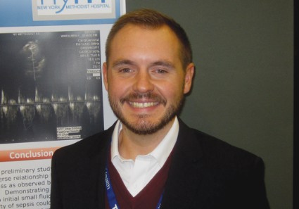
SEATTLE – Septic patients are more likely to respond to fluid therapy if their velocity time integral – a Doppler ultrasound measurement of blood flow across the left ventricular outflow tract – increases by 15% or more with a passive single-leg raise, according to a preliminary, observational study of 32 patients at New York Methodist Hospital in Brooklyn.
A passive leg raise to 45 degrees simulates a 250- to 500-cc fluid bolus. "We have found that people who don’t respond with a VTI greater than 15% have higher repeat lactate levels. Instead of giving them 2 L [of fluid] and then reassessing, maybe they’re patients you want to start on pressors right away," Dr. Andrew Balk said at the annual meeting of the American College of Emergency Physicians.
Echocardiogram machines can automatically calculate VTI. The measurement, which Dr. Balk and his associates obtained from the apical five-chamber view, is a surrogate for, and can be used to calculate, cardiac output. Poor response to fluid challenge indicates that fluids are less likely to increase cardiac output and more likely to cause fluid overload, said Dr. Balk, associate director of the clinical ultrasound division at the hospital.
The patients’ mean age was 68 years, and those with valvular pathology and atrial fibrillation were excluded from the study.
The group’s mean baseline VTI was 22 cm (range, 15-29 cm), which leg raises raised to a mean of 26 cm (18-34 cm), an increase of about 18% (4%-36%). A subsequent 2-L normal saline challenge increased VTI to a mean of 33 cm.
The mean baseline lactate level was 3.2 mmol/L (1.2-5.2 mmol/L), and 2 mmol/L (1-3 mmol/L) after the 2-L challenge. The percent change in VTI correlated significantly with the percent change in serum lactate levels. "Below-average responsiveness to the initial small fluid bolus was associated with a higher repeat lactate value, ... which suggests an inverse relationship between a patient’s fluid responsiveness as observed by the change in VTI and the severity of sepsis," the researchers concluded.
The VTI/leg-raise approach looks promising as a possible quick bedside marker that identifies patients who need aggressive treatment, without the need for central line measurements, Dr. Balk said. "The quickest initial fluid bolus you can get is a passive leg raise. You can watch for changes" in real time, and don’t have to move the probe from the point of maximum impact.
Dr. Balk reported having no disclosures.

Dr. Steven Q. Simpson commented: The search for noninvasive measures of or predictors for volume responsiveness in septic patients continues. VTI is the integral of velocity and time, i.e., the distance a small blood bolus travels. When multiplied by cross-sectional area of the aortic outflow tract, this would result in stroke volume. Since one would not expect the cross-sectional area to change significantly after a fluid bolus, alterations in VTI should reflect alterations in stroke volume. While promising, this technique is not as easy as the authors make it sound
and is operator dependent, even though the machine does the calculating. The incident angle of the probe must remain constant during the leg raise (at least 90 seconds). The user must know whether valve pathology or LV impairment are present and, if so, the degree. Massively volume depleted patients may fail to respond adequately to a passive leg raise.
One would be remiss to rely on this small study, which does not report sensitivity or specificity, to establish a reliable percent increase for predicting lactate response or to guide fluid therapy. However, this research is certainly aimed in the right direction.
Dr. Steven Q. Simpson is professor of medicine and director of fellowship training in the pulmonary disease and critical care medicine division at the University of Kansas Medical Center, Kansas City.
SEATTLE – Septic patients are more likely to respond to fluid therapy if their velocity time integral – a Doppler ultrasound measurement of blood flow across the left ventricular outflow tract – increases by 15% or more with a passive single-leg raise, according to a preliminary, observational study of 32 patients at New York Methodist Hospital in Brooklyn.
A passive leg raise to 45 degrees simulates a 250- to 500-cc fluid bolus. "We have found that people who don’t respond with a VTI greater than 15% have higher repeat lactate levels. Instead of giving them 2 L [of fluid] and then reassessing, maybe they’re patients you want to start on pressors right away," Dr. Andrew Balk said at the annual meeting of the American College of Emergency Physicians.
Echocardiogram machines can automatically calculate VTI. The measurement, which Dr. Balk and his associates obtained from the apical five-chamber view, is a surrogate for, and can be used to calculate, cardiac output. Poor response to fluid challenge indicates that fluids are less likely to increase cardiac output and more likely to cause fluid overload, said Dr. Balk, associate director of the clinical ultrasound division at the hospital.
The patients’ mean age was 68 years, and those with valvular pathology and atrial fibrillation were excluded from the study.
The group’s mean baseline VTI was 22 cm (range, 15-29 cm), which leg raises raised to a mean of 26 cm (18-34 cm), an increase of about 18% (4%-36%). A subsequent 2-L normal saline challenge increased VTI to a mean of 33 cm.
The mean baseline lactate level was 3.2 mmol/L (1.2-5.2 mmol/L), and 2 mmol/L (1-3 mmol/L) after the 2-L challenge. The percent change in VTI correlated significantly with the percent change in serum lactate levels. "Below-average responsiveness to the initial small fluid bolus was associated with a higher repeat lactate value, ... which suggests an inverse relationship between a patient’s fluid responsiveness as observed by the change in VTI and the severity of sepsis," the researchers concluded.
The VTI/leg-raise approach looks promising as a possible quick bedside marker that identifies patients who need aggressive treatment, without the need for central line measurements, Dr. Balk said. "The quickest initial fluid bolus you can get is a passive leg raise. You can watch for changes" in real time, and don’t have to move the probe from the point of maximum impact.
Dr. Balk reported having no disclosures.
Dr. Steven Q. Simpson commented: The search for noninvasive measures of or predictors for volume responsiveness in septic patients continues. VTI is the integral of velocity and time, i.e., the distance a small blood bolus travels. When multiplied by cross-sectional area of the aortic outflow tract, this would result in stroke volume. Since one would not expect the cross-sectional area to change significantly after a fluid bolus, alterations in VTI should reflect alterations in stroke volume. While promising, this technique is not as easy as the authors make it sound
and is operator dependent, even though the machine does the calculating. The incident angle of the probe must remain constant during the leg raise (at least 90 seconds). The user must know whether valve pathology or LV impairment are present and, if so, the degree. Massively volume depleted patients may fail to respond adequately to a passive leg raise.
One would be remiss to rely on this small study, which does not report sensitivity or specificity, to establish a reliable percent increase for predicting lactate response or to guide fluid therapy. However, this research is certainly aimed in the right direction.
Dr. Steven Q. Simpson is professor of medicine and director of fellowship training in the pulmonary disease and critical care medicine division at the University of Kansas Medical Center, Kansas City.
AT THE ACEP SCIENTIFIC ASSEMBLY 2013
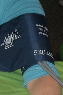
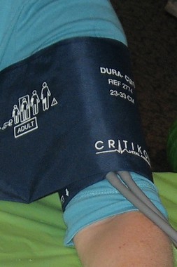
Blood pressure control tied to decline in stroke mortality over past 50 years
Antihypertensive therapy is probably the main reason why stroke fatalities have dropped dramatically in the United States over the past 50 years, according to an American Heart Association study published Dec. 5 in Stroke.
Despite an aging and heavier population, "the accelerated decline in stroke mortality that began in the 1970s is consistent with the aggressive hypertension treatment and control strategies implemented in that period. ... The decrease in blood pressure with drug therapy ... appears to be the major determinant of reduction in the risk of stroke and stroke deaths," wrote lead author Dr. Daniel Lackland, professor of epidemiology at the Medical University of South Carolina, Charleston, and his colleagues (Stroke 2013 Dec. 5 [doi:10.1161/01.str.0000437068.30550.cf]).
Statins, diabetes drugs, public health efforts, increased research, improved imaging, and quicker and better stroke treatment, among other things, have helped, too. People have also begun to lose weight, cut salt from their diets, and eat and smoke less.
But when it comes to accounting for the stroke mortality rate reduction from 88/100,000 in 1950 to 23/100,000 in 2010 – a reduction that holds across racial, age, and gender lines – the evidence is strongest for hypertension control, the authors said.
"Although the decline in stroke mortality in the United States started at the beginning of the 20th century, decades before hypertension treatment, the slope of the decline in mortality accelerated significantly after the introduction of tolerable antihypertensive drug therapy in the 1960s," they wrote. Stroke is now the fourth leading cause of death in the United States, instead of the third, and both recurrent and first-time strokes are down. Europe has had a similar decline.
Although great racial disparities still exist in stroke mortality, particularly for blacks, "the decline in stroke mortality for all racial/ethnic groups has reduced the magnitude of the racial/ethnic gap in stroke mortality risks and likewise the variation in stroke mortality by geographic area, with particular emphasis in the Stroke Belt," Dr. Lackland and his colleagues wrote.
"The decline is real, not a statistical fluke or the result of more people dying of lung disease, the third leading cause of death," Dr. Lackland said in a statement. It’s also not due to changes in billing codes, diagnostic improvements, death certificate causes of death, or other factors the team considered. Instead, it is "one of the major public health successes of the past 50 years," the authors wrote.
The findings are based on literature reviews, morbidity and mortality reports, clinical and public health guidelines, expert opinion, and other sources.
The authors noted that "increased application of advanced neuroimaging ... might improve the diagnosis of milder, less-fatal strokes over time. This would result in an apparent decline in the stroke case-fatality rate, solely as a result of improved detection. However, this should not result in a change in stroke mortality over time unless technological advances improve the diagnosis of more severe, fatal strokes also, which seems unlikely."
Dr. Lackland said he had no relevant financial disclosures. One of the 13 authors on the paper is a speaker and consultant for Allergan and a consultant for Reata.
FROM STROKE
IV antihypertensives are overused, single-center ED chart audit finds
SEATTLE – About a third of the patients who received intravenous bolus antihypertensives in the emergency department at the Detroit Receiving Hospital did not need them, according to the results of a retrospective review at the 340-bed level 1 trauma center.
The findings were presented at the annual meeting of the American College of Emergency Physicians.

Of the 295 patients in the study, all had blood pressure of at least 180/110 mm Hg, but 95 did not have signs and symptoms of end-organ damage and had failed on oral therapy. These patients received no further workup or diagnosis related to acute hypertension after being given at least one IV push of, most commonly, labetalol. Atropine was needed to correct subsequent bradycardia in 1 of those 95 patients, and vasopressors in 1 of 2 patients who became hypotensive.
"A lot of physicians jump the gun and treat the number, not the patient," but in the absence of hypertensive emergencies, patients "can get away with oral therapy. You really don’t want to drop their blood pressure too quickly; you just want to bring them down slowly to their normal," which may only be a bit below their presenting pressure, said Suprat Saely, Pharm. D., an emergency department pharmacist at the hospital.
For the study, pharmacy orders were matched to patient medical records. There were no statistical differences in survival to discharge and 30-day revisits to the ED between the 200 patients treated appropriately and the 95 who were unnecessarily given IV antihypertensives.
These days, "as soon as I get these orders, I assess whether or not they are appropriate. Usually they are, but if not, I’ll go ask the physician if they really meant to order it," said Dr. Saely, who had no disclosures.
SEATTLE – About a third of the patients who received intravenous bolus antihypertensives in the emergency department at the Detroit Receiving Hospital did not need them, according to the results of a retrospective review at the 340-bed level 1 trauma center.
The findings were presented at the annual meeting of the American College of Emergency Physicians.
Of the 295 patients in the study, all had blood pressure of at least 180/110 mm Hg, but 95 did not have signs and symptoms of end-organ damage and had failed on oral therapy. These patients received no further workup or diagnosis related to acute hypertension after being given at least one IV push of, most commonly, labetalol. Atropine was needed to correct subsequent bradycardia in 1 of those 95 patients, and vasopressors in 1 of 2 patients who became hypotensive.
"A lot of physicians jump the gun and treat the number, not the patient," but in the absence of hypertensive emergencies, patients "can get away with oral therapy. You really don’t want to drop their blood pressure too quickly; you just want to bring them down slowly to their normal," which may only be a bit below their presenting pressure, said Suprat Saely, Pharm. D., an emergency department pharmacist at the hospital.
For the study, pharmacy orders were matched to patient medical records. There were no statistical differences in survival to discharge and 30-day revisits to the ED between the 200 patients treated appropriately and the 95 who were unnecessarily given IV antihypertensives.
These days, "as soon as I get these orders, I assess whether or not they are appropriate. Usually they are, but if not, I’ll go ask the physician if they really meant to order it," said Dr. Saely, who had no disclosures.
AT THE ACEP SCIENTIFIC ASSEMBLY 2013
Emergency department electronic record systems fell short in Boston bombings
The massive number of injured patients from the April 2013 Boston Marathon bombings overwhelmed electronic medical records and information systems in some Boston emergency departments.
Their experiences offer an important lesson at a time when most hospitals have switched to electronic systems and providers are soon to face financial penalties for not doing so.
During mass-casualty events, paper and pen might be best, said Dr. Andrew Ulrich, executive vice chair of emergency medicine at the Boston Medical Center (BMC).

"The ability to get names into the system fast enough to order tests and establish a medical record limited us" on April 15, he noted. "There were so many patients coming through so quickly [23 at BMC right after the bombs went off] that it was difficult to know exactly who had what done and where they were going. You couldn’t leave the patient to" go to a computer, log on, get an update, and place orders, Dr. Ulrich said.
Other hospitals "had similar experiences. Communication was one of the more difficult components of this event," he said.
At Brigham and Women’s Hospital, "the first issue was our naming convention. We found that when every patient is ‘unidentified’ with a string of numbers, there’s a lot of confusion and many near misses. [Also,] if your system is inundated with multiple patients at once, it takes more time to register them so you can" go to work, said Dr. Eric Goralnick, the hospital’s medical director of emergency preparedness, who helped care for the 19 blast victims his ED received in the first half hour, and more later.

Now, at Brigham and Women’s, unknown patients are identified with a color, state, or other quickly recognized word.
Since the bombings, the EDs at Boston Medical Center and Brigham and Women’s have stocked up on old-school paper-and-pen trauma packets – an envelope with a wrist band, presigned orders, and other care documents all prestamped with a unique identifier – so that they are ready for the next mass-casualty event.
"We are much more flexible, much more adaptable if we go back to basics on this one, and that’s paper and pen. We are preparing to do that" for the next disaster, so "we can pull a bracelet out of the packet, throw it on the wrist," and start to work. It’s "much faster than getting [patients] onto the [electronic] medical record," Dr. Ulrich said.
Also from the old school department: "We [may also] have a clipboard attached to the patient’s bed, so when the patient rolls somewhere else, whoever gets them just has to look at the paperwork" to know what’s going on "instead of separating information from the patient" with the computer system, he said. "In a situation where there’s so much going on, it’s a clearer way of exchanging information."
Electronic record problems aren’t uncommon in EDs.
"There are many places where the computer system is a barrier to care. If you are trying to handle [an event] like this with a system that’s optimized for internal medicine clinics, it’s not going to work," said Dr. Larry Nathanson, who helped tend to the 22 bombing victims Beth Israel Deaconess Medical Center received in the first hour, almost half of whom were in critical condition.
Things went more smoothly at Beth Israel, where the electronic information system worked well. Part of the reason is that the hospital uses a home-grown system designed by Dr. Nathanson, a computer programmer as well as an emergency physician. The system is quick and provides patient information with few clicks. To identify unknown patients, the system uses a convention similar to the one used to name hurricanes (Annie, Bruce, Candace, etc.); the record can be easily updated once a patient’s identity is known.
The system had been tested prior to the bombings via simulation including 500 patients. "We made sure beforehand that [it] could handle the influx and quickly register large volumes of critical patients," said Dr. Nathanson, who serves as the ED’s director of emergency medical informatics.
"The reason it worked well in a disaster is because it works well every day," he said.
Based on feedback from his colleagues, he’s since added an interface that pops up when two or more mass-casualty victims are in the ED. Pressing it displays them all with a summary of their situations, so providers can home in on them.
Dr. Ulrich and Dr. Goralnick have no relevant disclosures. Dr. Nathanson owns stock in Forerun Inc., the commercialized version of the system he developed for Beth Israel.
The massive number of injured patients from the April 2013 Boston Marathon bombings overwhelmed electronic medical records and information systems in some Boston emergency departments.
Their experiences offer an important lesson at a time when most hospitals have switched to electronic systems and providers are soon to face financial penalties for not doing so.
During mass-casualty events, paper and pen might be best, said Dr. Andrew Ulrich, executive vice chair of emergency medicine at the Boston Medical Center (BMC).
"The ability to get names into the system fast enough to order tests and establish a medical record limited us" on April 15, he noted. "There were so many patients coming through so quickly [23 at BMC right after the bombs went off] that it was difficult to know exactly who had what done and where they were going. You couldn’t leave the patient to" go to a computer, log on, get an update, and place orders, Dr. Ulrich said.
Other hospitals "had similar experiences. Communication was one of the more difficult components of this event," he said.
At Brigham and Women’s Hospital, "the first issue was our naming convention. We found that when every patient is ‘unidentified’ with a string of numbers, there’s a lot of confusion and many near misses. [Also,] if your system is inundated with multiple patients at once, it takes more time to register them so you can" go to work, said Dr. Eric Goralnick, the hospital’s medical director of emergency preparedness, who helped care for the 19 blast victims his ED received in the first half hour, and more later.
Now, at Brigham and Women’s, unknown patients are identified with a color, state, or other quickly recognized word.
Since the bombings, the EDs at Boston Medical Center and Brigham and Women’s have stocked up on old-school paper-and-pen trauma packets – an envelope with a wrist band, presigned orders, and other care documents all prestamped with a unique identifier – so that they are ready for the next mass-casualty event.
"We are much more flexible, much more adaptable if we go back to basics on this one, and that’s paper and pen. We are preparing to do that" for the next disaster, so "we can pull a bracelet out of the packet, throw it on the wrist," and start to work. It’s "much faster than getting [patients] onto the [electronic] medical record," Dr. Ulrich said.
Also from the old school department: "We [may also] have a clipboard attached to the patient’s bed, so when the patient rolls somewhere else, whoever gets them just has to look at the paperwork" to know what’s going on "instead of separating information from the patient" with the computer system, he said. "In a situation where there’s so much going on, it’s a clearer way of exchanging information."
Electronic record problems aren’t uncommon in EDs.
"There are many places where the computer system is a barrier to care. If you are trying to handle [an event] like this with a system that’s optimized for internal medicine clinics, it’s not going to work," said Dr. Larry Nathanson, who helped tend to the 22 bombing victims Beth Israel Deaconess Medical Center received in the first hour, almost half of whom were in critical condition.
Things went more smoothly at Beth Israel, where the electronic information system worked well. Part of the reason is that the hospital uses a home-grown system designed by Dr. Nathanson, a computer programmer as well as an emergency physician. The system is quick and provides patient information with few clicks. To identify unknown patients, the system uses a convention similar to the one used to name hurricanes (Annie, Bruce, Candace, etc.); the record can be easily updated once a patient’s identity is known.
The system had been tested prior to the bombings via simulation including 500 patients. "We made sure beforehand that [it] could handle the influx and quickly register large volumes of critical patients," said Dr. Nathanson, who serves as the ED’s director of emergency medical informatics.
"The reason it worked well in a disaster is because it works well every day," he said.
Based on feedback from his colleagues, he’s since added an interface that pops up when two or more mass-casualty victims are in the ED. Pressing it displays them all with a summary of their situations, so providers can home in on them.
Dr. Ulrich and Dr. Goralnick have no relevant disclosures. Dr. Nathanson owns stock in Forerun Inc., the commercialized version of the system he developed for Beth Israel.
Replace Narcotics for Skin Pain
LAS VEGAS – A topical mixture of amitriptyline and ketamine controls pain in a variety of skin conditions, even when more traditional options fail, according to Dr. Mark Davis, chair of the division of clinical dermatology at the Mayo Clinic in Rochester, Minn.
"We are getting patients off systemic narcotics with these mixtures," he said at Skin Disease Education Foundation’s annual Las Vegas dermatology seminar.
Lidocaine patches are allowing leg ulcer patients to drop narcotics, too, and are greatly helping those with erythromelalgia, an often-misdiagnosed condition involving intermittent and excruciating, burning pain in the feet, hands, and sometimes ears. "To have something topical [for such pain] is terrific," Dr. Davis said.

The Mayo Clinic uses topical amitriptyline and ketamine for brachioradial pruritus, erythromelalgia, rectogenital and perineal pain, and other localized skin pain recalcitrant to oral medications and other standard approaches. Dr. Davis and his colleagues recently published several reviews and retrospective studies on the use of topical pain relievers, including a recent article showing that more than half the patients who used topical amitriptyline-ketamine for pain reported substantial or complete relief (Pain Physician 2012;15:485-8).
Pharmaceutical companies are working to bring the combination to market, but it’s not yet available commercially, so Mayo Clinic dermatologists have their pharmacists compound it in two strengths – 2% amitriptyline and either 0.5% or 5% ketamine – using Lipoderm cream as the base, according to Dr. Davis. Patients apply the mixture three times daily. Why it works isn’t clear; the drugs have different and perhaps synergistic effects on skin pain.
"I try to get people to use it [the product] on small parts of the body, such as the hands and feet, and, in patients with brachioradial pruritus, both arms," said Dr. Davis. "I’ve never used it any more extensively than that; I am afraid patients would absorb too much ketamine," he said.
Of more than 1,000 Mayo Clinic patients who have tried the combination, "I’d say less than 1% has told me that they’ve ever had a side effect," he said. "I’ve had just two or three patients tell me they’ve gotten nightmares," a known effect of ketamine. "This is a product that has great promise," he noted.
Meanwhile, some erythromelalgia patients at the Mayo Clinic have been able to walk again after applying lidocaine patches to their feet, and the clinicians there consider the patches first-line treatment for the condition. "Many patients are well controlled just by using [the patches], and I don’t think I’ve come across any side effects in the hundreds of patients I’ve treated," Dr. Davis said. In the inherited form of the disease, which accounts for perhaps 5% of cases, erythromelalgia is caused by a genetic sodium channel glitch that keeps nerves in the skin firing once they are stimulated.
Lidocaine patches also are "really useful for patients with bad, painful ulcers; I use [them] over whatever wound care I am using," Dr. Davis noted. Sometimes these patients can discontinue, or at least reduce, their use of narcotics, he said.
Lidocaine helps with debridement, too. "For leg ulcers, our nurses take 4 x 4-inch gauze and soak it in lidocaine 4% solution, and leave it on the ulcer for about 20 minutes. We are usually able to debride those ulcers [after that] even if they were very painful to start," he said.
Dr. Davis has no relevant disclosures. SDEF and this news organization are owned by Frontline Medical Communications.
LAS VEGAS – A topical mixture of amitriptyline and ketamine controls pain in a variety of skin conditions, even when more traditional options fail, according to Dr. Mark Davis, chair of the division of clinical dermatology at the Mayo Clinic in Rochester, Minn.
"We are getting patients off systemic narcotics with these mixtures," he said at Skin Disease Education Foundation’s annual Las Vegas dermatology seminar.
Lidocaine patches are allowing leg ulcer patients to drop narcotics, too, and are greatly helping those with erythromelalgia, an often-misdiagnosed condition involving intermittent and excruciating, burning pain in the feet, hands, and sometimes ears. "To have something topical [for such pain] is terrific," Dr. Davis said.
The Mayo Clinic uses topical amitriptyline and ketamine for brachioradial pruritus, erythromelalgia, rectogenital and perineal pain, and other localized skin pain recalcitrant to oral medications and other standard approaches. Dr. Davis and his colleagues recently published several reviews and retrospective studies on the use of topical pain relievers, including a recent article showing that more than half the patients who used topical amitriptyline-ketamine for pain reported substantial or complete relief (Pain Physician 2012;15:485-8).
Pharmaceutical companies are working to bring the combination to market, but it’s not yet available commercially, so Mayo Clinic dermatologists have their pharmacists compound it in two strengths – 2% amitriptyline and either 0.5% or 5% ketamine – using Lipoderm cream as the base, according to Dr. Davis. Patients apply the mixture three times daily. Why it works isn’t clear; the drugs have different and perhaps synergistic effects on skin pain.
"I try to get people to use it [the product] on small parts of the body, such as the hands and feet, and, in patients with brachioradial pruritus, both arms," said Dr. Davis. "I’ve never used it any more extensively than that; I am afraid patients would absorb too much ketamine," he said.
Of more than 1,000 Mayo Clinic patients who have tried the combination, "I’d say less than 1% has told me that they’ve ever had a side effect," he said. "I’ve had just two or three patients tell me they’ve gotten nightmares," a known effect of ketamine. "This is a product that has great promise," he noted.
Meanwhile, some erythromelalgia patients at the Mayo Clinic have been able to walk again after applying lidocaine patches to their feet, and the clinicians there consider the patches first-line treatment for the condition. "Many patients are well controlled just by using [the patches], and I don’t think I’ve come across any side effects in the hundreds of patients I’ve treated," Dr. Davis said. In the inherited form of the disease, which accounts for perhaps 5% of cases, erythromelalgia is caused by a genetic sodium channel glitch that keeps nerves in the skin firing once they are stimulated.
Lidocaine patches also are "really useful for patients with bad, painful ulcers; I use [them] over whatever wound care I am using," Dr. Davis noted. Sometimes these patients can discontinue, or at least reduce, their use of narcotics, he said.
Lidocaine helps with debridement, too. "For leg ulcers, our nurses take 4 x 4-inch gauze and soak it in lidocaine 4% solution, and leave it on the ulcer for about 20 minutes. We are usually able to debride those ulcers [after that] even if they were very painful to start," he said.
Dr. Davis has no relevant disclosures. SDEF and this news organization are owned by Frontline Medical Communications.
EXPERT ANALYSIS FROM THE SDEF LAS VEGAS DERMATOLOGY SEMINAR
Creams and patches can replace narcotics for skin pain
LAS VEGAS – A topical mixture of amitriptyline and ketamine controls pain in a variety of skin conditions, even when more traditional options fail, according to Dr. Mark Davis, chair of the division of clinical dermatology at the Mayo Clinic in Rochester, Minn.
"We are getting patients off systemic narcotics with these mixtures," he said at Skin Disease Education Foundation’s annual Las Vegas dermatology seminar.
Lidocaine patches are allowing leg ulcer patients to drop narcotics, too, and are greatly helping those with erythromelalgia, an often-misdiagnosed condition involving intermittent and excruciating, burning pain in the feet, hands, and sometimes ears. "To have something topical [for such pain] is terrific," Dr. Davis said.

The Mayo Clinic uses topical amitriptyline and ketamine for brachioradial pruritus, erythromelalgia, rectogenital and perineal pain, and other localized skin pain recalcitrant to oral medications and other standard approaches. Dr. Davis and his colleagues recently published several reviews and retrospective studies on the use of topical pain relievers, including a recent article showing that more than half the patients who used topical amitriptyline-ketamine for pain reported substantial or complete relief (Pain Physician 2012;15:485-8).
Pharmaceutical companies are working to bring the combination to market, but it’s not yet available commercially, so Mayo Clinic dermatologists have their pharmacists compound it in two strengths – 2% amitriptyline and either 0.5% or 5% ketamine – using Lipoderm cream as the base, according to Dr. Davis. Patients apply the mixture three times daily. Why it works isn’t clear; the drugs have different and perhaps synergistic effects on skin pain.
"I try to get people to use it [the product] on small parts of the body, such as the hands and feet, and, in patients with brachioradial pruritus, both arms," said Dr. Davis. "I’ve never used it any more extensively than that; I am afraid patients would absorb too much ketamine," he said.
Of more than 1,000 Mayo Clinic patients who have tried the combination, "I’d say less than 1% has told me that they’ve ever had a side effect," he said. "I’ve had just two or three patients tell me they’ve gotten nightmares," a known effect of ketamine. "This is a product that has great promise," he noted.
Meanwhile, some erythromelalgia patients at the Mayo Clinic have been able to walk again after applying lidocaine patches to their feet, and the clinicians there consider the patches first-line treatment for the condition. "Many patients are well controlled just by using [the patches], and I don’t think I’ve come across any side effects in the hundreds of patients I’ve treated," Dr. Davis said. In the inherited form of the disease, which accounts for perhaps 5% of cases, erythromelalgia is caused by a genetic sodium channel glitch that keeps nerves in the skin firing once they are stimulated.
Lidocaine patches also are "really useful for patients with bad, painful ulcers; I use [them] over whatever wound care I am using," Dr. Davis noted. Sometimes these patients can discontinue, or at least reduce, their use of narcotics, he said.
Lidocaine helps with debridement, too. "For leg ulcers, our nurses take 4 x 4-inch gauze and soak it in lidocaine 4% solution, and leave it on the ulcer for about 20 minutes. We are usually able to debride those ulcers [after that] even if they were very painful to start," he said.
Dr. Davis has no relevant disclosures. SDEF and this news organization are owned by Frontline Medical Communications.
LAS VEGAS – A topical mixture of amitriptyline and ketamine controls pain in a variety of skin conditions, even when more traditional options fail, according to Dr. Mark Davis, chair of the division of clinical dermatology at the Mayo Clinic in Rochester, Minn.
"We are getting patients off systemic narcotics with these mixtures," he said at Skin Disease Education Foundation’s annual Las Vegas dermatology seminar.
Lidocaine patches are allowing leg ulcer patients to drop narcotics, too, and are greatly helping those with erythromelalgia, an often-misdiagnosed condition involving intermittent and excruciating, burning pain in the feet, hands, and sometimes ears. "To have something topical [for such pain] is terrific," Dr. Davis said.
The Mayo Clinic uses topical amitriptyline and ketamine for brachioradial pruritus, erythromelalgia, rectogenital and perineal pain, and other localized skin pain recalcitrant to oral medications and other standard approaches. Dr. Davis and his colleagues recently published several reviews and retrospective studies on the use of topical pain relievers, including a recent article showing that more than half the patients who used topical amitriptyline-ketamine for pain reported substantial or complete relief (Pain Physician 2012;15:485-8).
Pharmaceutical companies are working to bring the combination to market, but it’s not yet available commercially, so Mayo Clinic dermatologists have their pharmacists compound it in two strengths – 2% amitriptyline and either 0.5% or 5% ketamine – using Lipoderm cream as the base, according to Dr. Davis. Patients apply the mixture three times daily. Why it works isn’t clear; the drugs have different and perhaps synergistic effects on skin pain.
"I try to get people to use it [the product] on small parts of the body, such as the hands and feet, and, in patients with brachioradial pruritus, both arms," said Dr. Davis. "I’ve never used it any more extensively than that; I am afraid patients would absorb too much ketamine," he said.
Of more than 1,000 Mayo Clinic patients who have tried the combination, "I’d say less than 1% has told me that they’ve ever had a side effect," he said. "I’ve had just two or three patients tell me they’ve gotten nightmares," a known effect of ketamine. "This is a product that has great promise," he noted.
Meanwhile, some erythromelalgia patients at the Mayo Clinic have been able to walk again after applying lidocaine patches to their feet, and the clinicians there consider the patches first-line treatment for the condition. "Many patients are well controlled just by using [the patches], and I don’t think I’ve come across any side effects in the hundreds of patients I’ve treated," Dr. Davis said. In the inherited form of the disease, which accounts for perhaps 5% of cases, erythromelalgia is caused by a genetic sodium channel glitch that keeps nerves in the skin firing once they are stimulated.
Lidocaine patches also are "really useful for patients with bad, painful ulcers; I use [them] over whatever wound care I am using," Dr. Davis noted. Sometimes these patients can discontinue, or at least reduce, their use of narcotics, he said.
Lidocaine helps with debridement, too. "For leg ulcers, our nurses take 4 x 4-inch gauze and soak it in lidocaine 4% solution, and leave it on the ulcer for about 20 minutes. We are usually able to debride those ulcers [after that] even if they were very painful to start," he said.
Dr. Davis has no relevant disclosures. SDEF and this news organization are owned by Frontline Medical Communications.
EXPERT ANALYSIS FROM THE SDEF LAS VEGAS DERMATOLOGY SEMINAR

Know the standards to protect against laser lawsuits
With the rapid evolution of the field of laser dermatology, it’s important for practitioners to understand "standard of practice" to avoid malpractice suits, according to Dr. David Goldberg, an attorney and director of laser research at Mount Sinai Hospital in New York.
To win a negligence case, plaintiffs must prove that physicians both had a duty to patients and breached that duty, and that doing so caused damages and that the damages were real and substantial.
Meeting practice standards is a key defense against breach of duty arguments. The idea is to demonstrate that laser skin surgery was done "in accordance with the standard of care. The standard of care is the way in which the majority of the physicians in a similar medical community would practice." Physician extenders are held to the same standard, Dr. Goldberg said (Sem. Cut. Med. Surg. 2013;32:205-208).
Doctors don’t need to demonstrate that they’re the best in the field, only that they "used the care and skill ordinarily possessed by a specialist in that field in the same or similar locality under similar circumstances," Dr. Goldberg noted. "Even if one method turns out to be less effective than another, a physician does not fall below the standard of care" if the method was an acceptable alternative, he said. Similarly, an "unfavorable result due to an error in judgment ... is not in and of itself a violation of the standard of care if the physician acted appropriately" before making the judgment call, he added.
A rock solid expert witness is key; "the standard of care is defined by some as whatever an expert witness says it is and what a jury will believe," said Dr. Goldberg. "If the jury believes an expert who testifies for a defendant doctor, then the standard of care in that particular case has been met." Expert witnesses need to back up their testimony by citing their own and others’ practices, plus journal articles, practice guidelines, and statutes, among other things, he emphasized.
It’s important that experts practice like other physicians do; otherwise these witnesses "will have a difficult time explaining why the majority of the medical community does not practice according to their way," Dr. Goldberg said.
Clinical practice guidelines can help prove that laser dermatologists followed professional customs, but an expert is still needed to "introduce the standard and establish its sources and relevancy," said Dr. Goldberg. That’s harder when, as is often the case, guidelines "contain disclaimers stating that they are not intended to displace physician discretion," he said.
The standards test doesn’t necessarily rule out innovation. "Laser surgeons often view themselves as artists in addition to scientists, customizing a treatment for a particular condition," Dr. Goldberg noted. Most clinical innovations fall short of rank experimentation. If a procedure wasn’t vetted by an institutional review board and wasn’t subject to other federal human research requirements, plaintiffs are going to have a hard time proving that "an actual given laser procedure is experimental," he said.
For instance, although "most would suggest that the safest technique for the removal of unwanted hair is by use of the Nd:YAG laser ... a physician using a non-Nd:YAG laser or light source that is also approved by the FDA for the treatment of unwanted hair in darker skin types may be performing laser treatment within the standard of care," said Dr. Goldberg.
Practice location matters, too. Dermatology laser centers aren’t often in hospitals, but when they are "a plaintiff may seek hospital committee proceeding minutes about the allegedly negligent physician," he said.
These individuals are not likely to get anywhere, however; courts have ruled that the "immunity of committee proceedings protects certain communications and encourages the quality review process," Dr. Goldberg noted. Incident reports, however, are a different matter, and less likely to be protected because they are "often more directly related to a single claim for malpractice," he said.
Dr. Goldberg said he had no relevant financial conflicts to disclose.
FROM SEMINARS IN CUTANEOUS MEDICINE AND SURGERY