User login
Obesity might be targetable driver of psoriatic arthritis progression
MADRID – Two sets of data presented at the European Congress of Rheumatology support the potential for weight loss to be a valuable adjunctive strategy for improving outcomes in patients with psoriatic arthritis (PsA).

One set, drawn from the ongoing PsABio observational study, correlated increasing body mass index with greater disease activity and greater disability. Another, based on patients followed for 12 months, showed that a weight loss of about 15% is associated with a significant reduction in PsA activity.
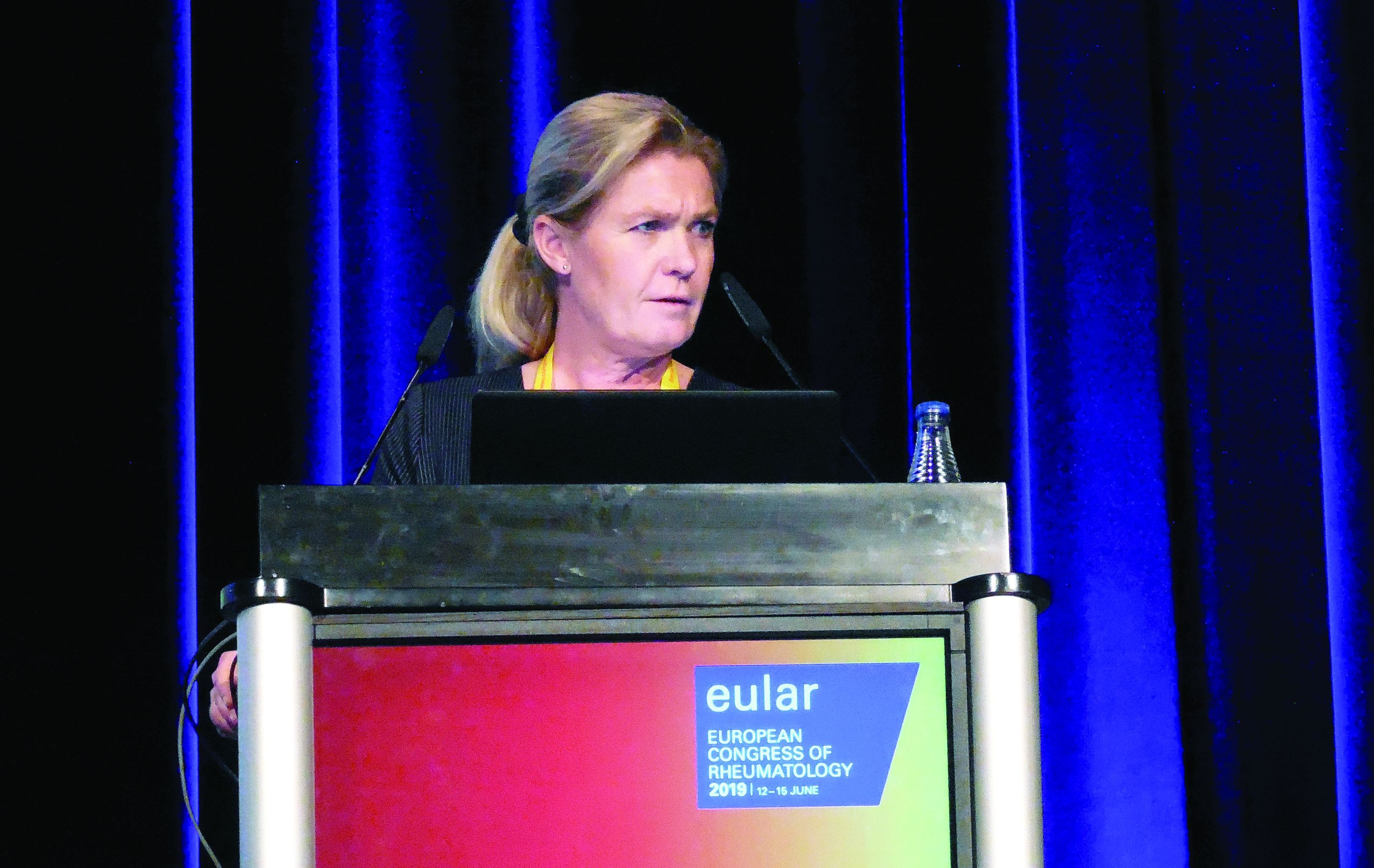
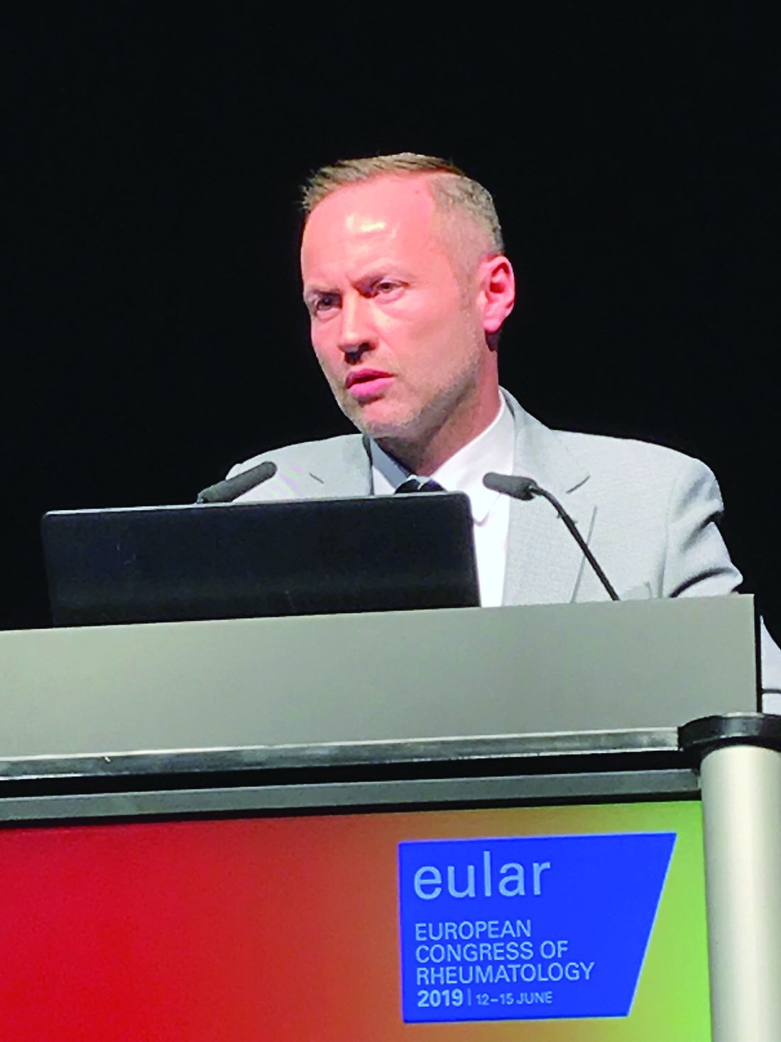
“As clinicians, we largely focus on drugs in the treatment of PsA, but these data draw attention to obesity as a potential target for improving outcomes in PsA,” said Stefan Siebert, MD, a rheumatologist at the Institute of Infection, Immunity, and Inflammation at the University of Glasgow (Scotland).
Dr. Siebert cautioned that his data show association, not causation, but he said these data add to a growing body of evidence that provide compelling support for trials to test the premise that weight loss improves outcomes.
Although not a trial, a study by Eva Klingberg, MD, PhD, of the Sahlgrenska Academy at the University of Gothenburg (Sweden) and her associates tested this premise and showed weight loss was associated with improvement in multiple PsA activity parameters 6 and 12 months after a significant weight loss program.
“This is just one study, so we need more data, but we are already using weight loss to manage PsA in obese patients in Sweden,” said Dr. Klingberg, speaking about her work in advance of the presentation. Like Dr. Siebert, she agreed that weight loss is an important potential treatment strategy in PsA.
In the observational PsABio study, which is following patients with PsA at rheumatology centers in eight European countries, the goal of its analysis was to evaluate disease activity and outcomes in relationship to baseline weight for patients starting a biologic therapy as part of standard clinical practice. Of the 917 patients evaluated, 450 started ustekinumab (Stelara) and 467 started a tumor necrosis factor inhibitor (TNFi). The researchers had weight data for 827 of these patients.
At the time of enrollment, 40% were overweight as defined by a body mass index (BMI) ranging from 25 to 29 kg/m2, and 30.4% were obese as defined by a BMI greater than 30 kg/m2. The mean baseline BMI was 28.1 kg/m2. The mean age of the study population was 49.7 years. Slightly more than half were female.
Relative to a BMI of 30 kg/m2 or less, higher BMI at baseline is shown in multiple regression analysis to be independently and significantly linked to disease activity assessed by the clinical Disease Activity Index for Psoriatic Arthritis (cDAPSA; P = .026), to patient perception of disease impact as measured by Psoriatic Arthritis Impact of Disease (PsAID-12; P less than .0001), and to greater disability as measured with Health Assessment Questionnaire Disability Index (HAQ-DI; P less than .0001).
“There are multiple sets of data that show obesity predicts who develops PsA. Our data further show that, of patients with PsA who are candidates for a biologic, those with obesity have greater disease activity,” Dr. Siebert said. “We are using all of these expensive drugs, but I think there is now a need to also focus on lifestyle interventions, in addition to drug therapy, to reduce disease activity and improve outcomes in PsA.”
The data to be presented by Dr. Klingberg provide a step in that direction. In this study, 46 PsA patients participated in a weight-loss treatment that restricted calorie intake to 640 kcal/day, and the researchers followed 39 of these patients for 1 year. The participants averaged 56 years old, and almost two-thirds were women. All enrolled patients had to have a BMI of at least 33 kg/m2, and the actual average BMI was 35 kg/m2. The median weight loss among the 39 patients followed for 1 year after the start of a 12- to 16-week weight-loss treatment was 16.1 kg, representing about 16% of their body weight at entry.
Dr. Klingberg showed that disease activity in those who achieved and maintained weight loss after the program was significant at 6 and 12 months when measured with the Psoriatic Arthritis Response Criteria (PsARC) or the American College of Rheumatology (ACR) 20, 50, and 70 criteria. In the 39 patients followed for 12 months, 36% fulfilled PsARC, and 54%, 36%, and 15% fulfilled the ACR 20, 50, and 70 responses, respectively.
“In Sweden, any obese individual can be referred for a weight loss program because of the multiple health benefits that are associated with weight reduction,” Dr. Klingberg explained. “We were able to look at patients with PsA and show that this substantially reduces the burden of their joint disease in addition to the other health advantages of losing weight.”
An improvement in symptoms is a logical expectation from reducing the mechanical strain imposed by obesity on inflamed joints, but Dr. Klingberg is more impressed by the potential for weight loss to reduce the proinflammatory signaling generated by adipose tissue. In PsA, there is evidence that weight loss reduces disease activity in the skin, as well as the joints, which supports this link.
“We need more data to document the benefits from weight loss in patients with PsA, but I think management of the comorbidities of PsA, including obesity, is something that should already be routinely discussed with patients,” Dr. Klingberg said.
Dr. Siebert has been a consultant to or speaker on behalf of AbbVie, Boehringer Ingelheim, Celgene, Janssen, Novartis, and UCB, and he has received research funding from Boehringer Ingelheim, Bristol-Myers Squibb, Celgene, GlaxoSmithKline, Janssen, Novartis, Pfizer, and UCB. Dr. Klingberg has been an advisor to Novartis, a speaker on behalf of Lilly, and has receive research funding from Roche.
Mitchel L. Zoler contributed to this report.
SOURCE: Siebert S et al. Ann Rheum Dis. Jun 2019;78(suppl 2):69. Abstract OP0007. doi: 10.1136/annrheumdis-2019-eular.5841; Klingberg E et al. Ann Rheum Dis. Jun 2019;78(suppl 2):69-70. Abstract OP0008. doi: 10.1136/annrheumdis-2019-eular.5551.
MADRID – Two sets of data presented at the European Congress of Rheumatology support the potential for weight loss to be a valuable adjunctive strategy for improving outcomes in patients with psoriatic arthritis (PsA).
One set, drawn from the ongoing PsABio observational study, correlated increasing body mass index with greater disease activity and greater disability. Another, based on patients followed for 12 months, showed that a weight loss of about 15% is associated with a significant reduction in PsA activity.
“As clinicians, we largely focus on drugs in the treatment of PsA, but these data draw attention to obesity as a potential target for improving outcomes in PsA,” said Stefan Siebert, MD, a rheumatologist at the Institute of Infection, Immunity, and Inflammation at the University of Glasgow (Scotland).
Dr. Siebert cautioned that his data show association, not causation, but he said these data add to a growing body of evidence that provide compelling support for trials to test the premise that weight loss improves outcomes.
Although not a trial, a study by Eva Klingberg, MD, PhD, of the Sahlgrenska Academy at the University of Gothenburg (Sweden) and her associates tested this premise and showed weight loss was associated with improvement in multiple PsA activity parameters 6 and 12 months after a significant weight loss program.
“This is just one study, so we need more data, but we are already using weight loss to manage PsA in obese patients in Sweden,” said Dr. Klingberg, speaking about her work in advance of the presentation. Like Dr. Siebert, she agreed that weight loss is an important potential treatment strategy in PsA.
In the observational PsABio study, which is following patients with PsA at rheumatology centers in eight European countries, the goal of its analysis was to evaluate disease activity and outcomes in relationship to baseline weight for patients starting a biologic therapy as part of standard clinical practice. Of the 917 patients evaluated, 450 started ustekinumab (Stelara) and 467 started a tumor necrosis factor inhibitor (TNFi). The researchers had weight data for 827 of these patients.
At the time of enrollment, 40% were overweight as defined by a body mass index (BMI) ranging from 25 to 29 kg/m2, and 30.4% were obese as defined by a BMI greater than 30 kg/m2. The mean baseline BMI was 28.1 kg/m2. The mean age of the study population was 49.7 years. Slightly more than half were female.
Relative to a BMI of 30 kg/m2 or less, higher BMI at baseline is shown in multiple regression analysis to be independently and significantly linked to disease activity assessed by the clinical Disease Activity Index for Psoriatic Arthritis (cDAPSA; P = .026), to patient perception of disease impact as measured by Psoriatic Arthritis Impact of Disease (PsAID-12; P less than .0001), and to greater disability as measured with Health Assessment Questionnaire Disability Index (HAQ-DI; P less than .0001).
“There are multiple sets of data that show obesity predicts who develops PsA. Our data further show that, of patients with PsA who are candidates for a biologic, those with obesity have greater disease activity,” Dr. Siebert said. “We are using all of these expensive drugs, but I think there is now a need to also focus on lifestyle interventions, in addition to drug therapy, to reduce disease activity and improve outcomes in PsA.”
The data to be presented by Dr. Klingberg provide a step in that direction. In this study, 46 PsA patients participated in a weight-loss treatment that restricted calorie intake to 640 kcal/day, and the researchers followed 39 of these patients for 1 year. The participants averaged 56 years old, and almost two-thirds were women. All enrolled patients had to have a BMI of at least 33 kg/m2, and the actual average BMI was 35 kg/m2. The median weight loss among the 39 patients followed for 1 year after the start of a 12- to 16-week weight-loss treatment was 16.1 kg, representing about 16% of their body weight at entry.
Dr. Klingberg showed that disease activity in those who achieved and maintained weight loss after the program was significant at 6 and 12 months when measured with the Psoriatic Arthritis Response Criteria (PsARC) or the American College of Rheumatology (ACR) 20, 50, and 70 criteria. In the 39 patients followed for 12 months, 36% fulfilled PsARC, and 54%, 36%, and 15% fulfilled the ACR 20, 50, and 70 responses, respectively.
“In Sweden, any obese individual can be referred for a weight loss program because of the multiple health benefits that are associated with weight reduction,” Dr. Klingberg explained. “We were able to look at patients with PsA and show that this substantially reduces the burden of their joint disease in addition to the other health advantages of losing weight.”
An improvement in symptoms is a logical expectation from reducing the mechanical strain imposed by obesity on inflamed joints, but Dr. Klingberg is more impressed by the potential for weight loss to reduce the proinflammatory signaling generated by adipose tissue. In PsA, there is evidence that weight loss reduces disease activity in the skin, as well as the joints, which supports this link.
“We need more data to document the benefits from weight loss in patients with PsA, but I think management of the comorbidities of PsA, including obesity, is something that should already be routinely discussed with patients,” Dr. Klingberg said.
Dr. Siebert has been a consultant to or speaker on behalf of AbbVie, Boehringer Ingelheim, Celgene, Janssen, Novartis, and UCB, and he has received research funding from Boehringer Ingelheim, Bristol-Myers Squibb, Celgene, GlaxoSmithKline, Janssen, Novartis, Pfizer, and UCB. Dr. Klingberg has been an advisor to Novartis, a speaker on behalf of Lilly, and has receive research funding from Roche.
Mitchel L. Zoler contributed to this report.
SOURCE: Siebert S et al. Ann Rheum Dis. Jun 2019;78(suppl 2):69. Abstract OP0007. doi: 10.1136/annrheumdis-2019-eular.5841; Klingberg E et al. Ann Rheum Dis. Jun 2019;78(suppl 2):69-70. Abstract OP0008. doi: 10.1136/annrheumdis-2019-eular.5551.
REPORTING FROM EULAR 2019 CONGRESS
Leflunomide added to glucocorticoids reduces relapse in IgG4-related disease
MADRID – The addition of leflunomide to standard glucocorticoids (GCs) in the treatment of IgG4-related disease increases the median duration of response, reduces the proportion of patients with relapse within 12 months, and permits GCs to be tapered, according to results of a randomized trial presented at the European Congress of Rheumatology.

“The rate of adverse events with the addition of leflunomide was numerically higher, but there were no significant differences in risks of any specific adverse event,” reported Feng Huang, MD, of the department of rheumatology at Chinese People’s Liberation Army General Hospital in Beijing.
GCs are highly effective in IgG4-related disease, which is an autoimmune process driven by elevated concentrations of the antibody IgG4 in the tissue of affected organs and in the serum. It has been described in a broad array of sites, including the heart, lung, kidneys, and meninges. It has been widely recognized only in the last 10 years, according to Dr. Huang. Although most patients respond to GCs, he said the problem is that about 50% of patients relapse within 12 months and more than 90% within 3 years.
This randomized, controlled study was conducted after positive results were observed with leflunomide in a small, uncontrolled pilot study published several years ago (Intern Med J. 2017 Jun;47[6]:680-9. doi: 10.1111/imj.13430). In this randomized trial, the objectives were to confirm that leflunomide extends the relapse-free period and has acceptable safety relative to GC alone.
Patients with confirmed IgG4-related disease were enrolled. Patients randomized to GC were started on 0.5 to 0.8 mg/kg per day. A predefined taper regimen was employed in those with symptom control. Those randomized to the experimental arm received GC in the same dose and schedule plus 20 mg/day of leflunomide.
The 33 patients in each group were well matched at baseline for age, comorbidities, and disease severity.
At the end of 12 months, 50% of those treated with GC alone versus 21.2% of those treated with GC plus leflunomide had relapse. That translated into a significantly higher hazard ratio (HR) for relapse in the GC monotherapy group (HR, 1.75; P = .034).
The mean duration of remission was 7 months on the combination versus 3 months on GC alone. Dr. Huang also reported a significantly higher proportion of complete responses in the group receiving the combination.
In addition, “more patients on the combination therapy were able to adhere to the steroid-tapering schedule without relapse,” Dr. Huang reported. The rate of 54.5% of patients on combination therapy who were able to reach a daily GC dose of 5 mg/day or less proved significantly higher than the 18.2% rate seen with GC alone (P = .002).
Adverse events were reported by 54% of those on the combination versus 42% of those on monotherapy, but this difference did not reach statistical significance. The biggest differences in adverse events were the proportions of patients with infections (18.2% vs. 12.1%) and elevated liver enzymes (12.1% vs. 3.0%), both of which were more common in the combination therapy group. Neither of these differences was statistically significant.
Of patients with relapses, the most common organs involved were the salivary gland, the pancreas, and the bile ducts, each accounting for relapse in five patients. Other organs in which relapse occurred included the lacrimal gland and the skin. There were three cases of relapse characterized by retroperitoneal fibrosis.
Over the course of follow-up, new-onset diabetes mellitus occurred in 21.2% and 27.3% of the combination and GC-only groups, respectively. This difference also did not reach statistical significance.
Although this study was small with an open-label design, Dr. Huang said the data strongly suggest that a combination of leflunomide and GC is superior to GC alone. Based on these results, he said a starting dose of 20 mg/day of leflunomide is a reasonable standard in this setting.
Dr. Huang and colleagues reported no potential conflicts of interest.
SOURCE: Wang Y et al. Ann Rheum Dis. Jun 2019;78(Suppl 2):157. Abstract OPO164, doi: 10.1136/annrheumdis-2019-eular.5717
MADRID – The addition of leflunomide to standard glucocorticoids (GCs) in the treatment of IgG4-related disease increases the median duration of response, reduces the proportion of patients with relapse within 12 months, and permits GCs to be tapered, according to results of a randomized trial presented at the European Congress of Rheumatology.
“The rate of adverse events with the addition of leflunomide was numerically higher, but there were no significant differences in risks of any specific adverse event,” reported Feng Huang, MD, of the department of rheumatology at Chinese People’s Liberation Army General Hospital in Beijing.
GCs are highly effective in IgG4-related disease, which is an autoimmune process driven by elevated concentrations of the antibody IgG4 in the tissue of affected organs and in the serum. It has been described in a broad array of sites, including the heart, lung, kidneys, and meninges. It has been widely recognized only in the last 10 years, according to Dr. Huang. Although most patients respond to GCs, he said the problem is that about 50% of patients relapse within 12 months and more than 90% within 3 years.
This randomized, controlled study was conducted after positive results were observed with leflunomide in a small, uncontrolled pilot study published several years ago (Intern Med J. 2017 Jun;47[6]:680-9. doi: 10.1111/imj.13430). In this randomized trial, the objectives were to confirm that leflunomide extends the relapse-free period and has acceptable safety relative to GC alone.
Patients with confirmed IgG4-related disease were enrolled. Patients randomized to GC were started on 0.5 to 0.8 mg/kg per day. A predefined taper regimen was employed in those with symptom control. Those randomized to the experimental arm received GC in the same dose and schedule plus 20 mg/day of leflunomide.
The 33 patients in each group were well matched at baseline for age, comorbidities, and disease severity.
At the end of 12 months, 50% of those treated with GC alone versus 21.2% of those treated with GC plus leflunomide had relapse. That translated into a significantly higher hazard ratio (HR) for relapse in the GC monotherapy group (HR, 1.75; P = .034).
The mean duration of remission was 7 months on the combination versus 3 months on GC alone. Dr. Huang also reported a significantly higher proportion of complete responses in the group receiving the combination.
In addition, “more patients on the combination therapy were able to adhere to the steroid-tapering schedule without relapse,” Dr. Huang reported. The rate of 54.5% of patients on combination therapy who were able to reach a daily GC dose of 5 mg/day or less proved significantly higher than the 18.2% rate seen with GC alone (P = .002).
Adverse events were reported by 54% of those on the combination versus 42% of those on monotherapy, but this difference did not reach statistical significance. The biggest differences in adverse events were the proportions of patients with infections (18.2% vs. 12.1%) and elevated liver enzymes (12.1% vs. 3.0%), both of which were more common in the combination therapy group. Neither of these differences was statistically significant.
Of patients with relapses, the most common organs involved were the salivary gland, the pancreas, and the bile ducts, each accounting for relapse in five patients. Other organs in which relapse occurred included the lacrimal gland and the skin. There were three cases of relapse characterized by retroperitoneal fibrosis.
Over the course of follow-up, new-onset diabetes mellitus occurred in 21.2% and 27.3% of the combination and GC-only groups, respectively. This difference also did not reach statistical significance.
Although this study was small with an open-label design, Dr. Huang said the data strongly suggest that a combination of leflunomide and GC is superior to GC alone. Based on these results, he said a starting dose of 20 mg/day of leflunomide is a reasonable standard in this setting.
Dr. Huang and colleagues reported no potential conflicts of interest.
SOURCE: Wang Y et al. Ann Rheum Dis. Jun 2019;78(Suppl 2):157. Abstract OPO164, doi: 10.1136/annrheumdis-2019-eular.5717
REPORTING FROM EULAR 2019 CONGRESS
Despite advances, imaging of axSpA remains an adjunctive tool
MADRID – Evidence for always using imaging in an adjunctive role to clinical findings in the diagnosis and assessment of axial spondyloarthritis (axSpA) continues to grow, two experts agreed in a scientific session at the European Congress of Rheumatology.

“Imaging has to be understood in the context of other findings. With the patient history, the physical examination, and the laboratory results, the value of imaging improves substantially. Therefore, before an image is ordered it is important to ask how likely is it that a patient has axial spondylitis,” said Floris A. van Gaalen, MD, PhD, of Leiden (Netherlands) University Medical Center.
As one of the experts who participated in the scientific session, Dr. van Gaalen focused specifically on the value of x-ray and MRI in the diagnosis of axSpA, emphasizing their limited value if interpreted without clinical context. He explained that even highly experienced radiologists are fooled, particularly at early stages of disease.
Although the quality of imaging has been increasing steadily, “there is no cookbook approach with which you can guarantee a diagnosis of spondyloarthritis. Imaging can be valuable, but there is a risk of false positives because features on imaging, such as bone marrow edema, are shared with other sources of back pain,” Dr. van Gaalen said.
Considering the importance of context, Dr. van Gaalen advised clinicians against reading the radiology report without evaluating the images themselves. He said the features on imaging make more sense when they are considered at the same time as the patient’s history, symptoms, and laboratory reports.
Order imaging relevant to treatment decisions
Assigned to discuss the value of imaging for assessing progression, Xenofon Baraliakos, MD, a rheumatologist and clinical researcher at Rheumazentrum Ruhrgebiet, Ruhr-University Bochum, Herne, Germany, offered the same message.
“It is important to consider all of the clinical information available, not just the features on imaging,” Dr. Baraliakos said. Often, MRI findings provide corroboration for other objective measures of disease status, but Dr. Baraliakos advised that imaging should be ordered only when it has the potential to alter therapy.
“What we can learn from imaging might be interesting, but the question to ask is whether it is useful,” Dr. Baraliakos said. Rather than incurring the costs of imaging for reassurance, Dr. Baraliakos recommended ordering these studies with specific objectives relevant to treatment decisions.
Neither Dr. van Gaalen nor Dr. Baraliakos denied the value of imaging, particularly MRI, to increase confidence in the diagnosis of axSpA or to guide therapy. Rather, their point was that imaging should not be considered a reliable stand-alone axSpA assessment strategy.
Clinical and imaging findings better then imaging alone
Data from a blinded radiology study presented during the same scientific session reinforced this conclusion. Led by Dr. Baraliakos and presented separately from his discussion about the adjunctive nature of imaging data in axSpA, the study showed that rheumatologists with access to both clinical and imaging data can detect a greater proportion of axSpA than radiologists working from imaging data alone.
In this study, 300 consecutive patients suspected of axSpA were enrolled. All had chronic back pain of more than 3 months’ duration. While highly experienced radiologists were asked to diagnose or rule out a diagnosis of axSpA on the basis of the MRI blinded to other clinical information, experienced rheumatologists evaluated the patients with access to all clinical, laboratory, and imaging data.
A diagnosis of axSpA was reached in 131 patients by the rheumatologists. The remaining 169 were determined not to have axSpA. Although the radiologists agreed on those with or without axSpA in 86.3% of cases, there were 31 cases (28.1%) in which rheumatologists diagnosed axSpA but radiologists did not.
In an analysis of which MRI features were considered critical by radiologists when there was agreement, they identified bone marrow edema in seven cases (7.2%). In 30 cases (30.9%), the radiologists considered the presence of chronic lesions to be critical to their diagnosis. In the remaining 69.9% of cases, radiologists were confident in their diagnosis only when both bone edema and chronic lesions were present.
Not surprisingly, the presence of chronic lesions and more pronounced bone marrow edema permitted both radiologists and rheumatologists to increase their confidence when discriminating between axSpA and non-axSpA patients.
“The combination of structural changes and bone marrow edema as assessed by MRI performed best in the process of diagnosing or ruling out axSpA in this real-life setting at our center,” Dr. Baraliakos said.
However, when only one or two features are considered, trade-offs of lower sensitivity for higher specificity or higher sensitivity for lower specificity occur. For example, although the specificity for a diagnosis of axSpA reached 99.4% when both bone marrow edema and ankylosis are present, the sensitivity of this finding was only 5.3%, according to data provided by Dr. Baraliakos. Conversely, the presence of sclerosis had a sensitivity of 81.7% but a specificity of only 43.2%.
One lesson from this analysis is that there is “increasing insecurity of only including bone marrow edema of the sacroiliac joint as the major criterion for diagnosing axSpA,” Dr. Baraliakos said. However, the larger point in the context of the earlier expert comments is that MRI findings should be considered important but insufficient for the evaluation of axSpA.
SOURCE: Baraliakos X et al. Ann Rheum Dis. Jun 2019;78(Suppl 2):255-6. Abstract OPO344, doi: 10.1136/annrheumdis-2019-eular.5027
MADRID – Evidence for always using imaging in an adjunctive role to clinical findings in the diagnosis and assessment of axial spondyloarthritis (axSpA) continues to grow, two experts agreed in a scientific session at the European Congress of Rheumatology.
“Imaging has to be understood in the context of other findings. With the patient history, the physical examination, and the laboratory results, the value of imaging improves substantially. Therefore, before an image is ordered it is important to ask how likely is it that a patient has axial spondylitis,” said Floris A. van Gaalen, MD, PhD, of Leiden (Netherlands) University Medical Center.
As one of the experts who participated in the scientific session, Dr. van Gaalen focused specifically on the value of x-ray and MRI in the diagnosis of axSpA, emphasizing their limited value if interpreted without clinical context. He explained that even highly experienced radiologists are fooled, particularly at early stages of disease.
Although the quality of imaging has been increasing steadily, “there is no cookbook approach with which you can guarantee a diagnosis of spondyloarthritis. Imaging can be valuable, but there is a risk of false positives because features on imaging, such as bone marrow edema, are shared with other sources of back pain,” Dr. van Gaalen said.
Considering the importance of context, Dr. van Gaalen advised clinicians against reading the radiology report without evaluating the images themselves. He said the features on imaging make more sense when they are considered at the same time as the patient’s history, symptoms, and laboratory reports.
Order imaging relevant to treatment decisions
Assigned to discuss the value of imaging for assessing progression, Xenofon Baraliakos, MD, a rheumatologist and clinical researcher at Rheumazentrum Ruhrgebiet, Ruhr-University Bochum, Herne, Germany, offered the same message.
“It is important to consider all of the clinical information available, not just the features on imaging,” Dr. Baraliakos said. Often, MRI findings provide corroboration for other objective measures of disease status, but Dr. Baraliakos advised that imaging should be ordered only when it has the potential to alter therapy.
“What we can learn from imaging might be interesting, but the question to ask is whether it is useful,” Dr. Baraliakos said. Rather than incurring the costs of imaging for reassurance, Dr. Baraliakos recommended ordering these studies with specific objectives relevant to treatment decisions.
Neither Dr. van Gaalen nor Dr. Baraliakos denied the value of imaging, particularly MRI, to increase confidence in the diagnosis of axSpA or to guide therapy. Rather, their point was that imaging should not be considered a reliable stand-alone axSpA assessment strategy.
Clinical and imaging findings better then imaging alone
Data from a blinded radiology study presented during the same scientific session reinforced this conclusion. Led by Dr. Baraliakos and presented separately from his discussion about the adjunctive nature of imaging data in axSpA, the study showed that rheumatologists with access to both clinical and imaging data can detect a greater proportion of axSpA than radiologists working from imaging data alone.
In this study, 300 consecutive patients suspected of axSpA were enrolled. All had chronic back pain of more than 3 months’ duration. While highly experienced radiologists were asked to diagnose or rule out a diagnosis of axSpA on the basis of the MRI blinded to other clinical information, experienced rheumatologists evaluated the patients with access to all clinical, laboratory, and imaging data.
A diagnosis of axSpA was reached in 131 patients by the rheumatologists. The remaining 169 were determined not to have axSpA. Although the radiologists agreed on those with or without axSpA in 86.3% of cases, there were 31 cases (28.1%) in which rheumatologists diagnosed axSpA but radiologists did not.
In an analysis of which MRI features were considered critical by radiologists when there was agreement, they identified bone marrow edema in seven cases (7.2%). In 30 cases (30.9%), the radiologists considered the presence of chronic lesions to be critical to their diagnosis. In the remaining 69.9% of cases, radiologists were confident in their diagnosis only when both bone edema and chronic lesions were present.
Not surprisingly, the presence of chronic lesions and more pronounced bone marrow edema permitted both radiologists and rheumatologists to increase their confidence when discriminating between axSpA and non-axSpA patients.
“The combination of structural changes and bone marrow edema as assessed by MRI performed best in the process of diagnosing or ruling out axSpA in this real-life setting at our center,” Dr. Baraliakos said.
However, when only one or two features are considered, trade-offs of lower sensitivity for higher specificity or higher sensitivity for lower specificity occur. For example, although the specificity for a diagnosis of axSpA reached 99.4% when both bone marrow edema and ankylosis are present, the sensitivity of this finding was only 5.3%, according to data provided by Dr. Baraliakos. Conversely, the presence of sclerosis had a sensitivity of 81.7% but a specificity of only 43.2%.
One lesson from this analysis is that there is “increasing insecurity of only including bone marrow edema of the sacroiliac joint as the major criterion for diagnosing axSpA,” Dr. Baraliakos said. However, the larger point in the context of the earlier expert comments is that MRI findings should be considered important but insufficient for the evaluation of axSpA.
SOURCE: Baraliakos X et al. Ann Rheum Dis. Jun 2019;78(Suppl 2):255-6. Abstract OPO344, doi: 10.1136/annrheumdis-2019-eular.5027
REPORTING FROM EULAR 2019 CONGRESS
Experts agree on routine lung disease screening in systemic sclerosis
MADRID – The for early detection, monitoring, and, when warranted, treatment, Anna-Maria Hoffmann-Vold, MD, PhD, reported at the European Congress of Rheumatology.

“Everyone with systemic sclerosis needs to be screened because this is the most important risk factor for ILD,” said Dr. Hoffmann-Vold, a clinical scientist in the division of rheumatology at the University of Oslo and head of scleroderma research at Oslo University Hospital.
Although the frequency of screening is not specified based on the opinion that this should be based on risk factors and other clinical characteristics, there was unanimous agreement that lung function tests do not represent an adequate screening tool or method for assessing ILD severity. Rather, the recommendations make clear that lung function studies are adjunctive to high-resolution computed tomography (HRCT).
“HRCT is the primary tool for evaluating ILD, but there was 100% agreement that assessment should include more than one measure, including lung function tests and clinical assessment,” Dr. Hoffmann-Vold reported.
There was a strong opinion that the numerous potential biomarkers described for ILD, although promising, are not yet ready for clinical use.
In developing these new recommendations, 95 potential statements were considered by the panel of 27 rheumatologists, pulmonologists, and others with experience in this field. A Delphi process was used for members of the panel to identify areas of agreement to produce consensus statements.
The result has been more than 50 statements issued in six major domains. These include statements on risk factors, appropriate methodology for diagnosis and severity assessment, when to initiate therapy, and when and how to initiate treatment escalation.
“We want to increase clinician awareness and provide standardized guidance for evaluating patients for the presence and medical management of ILD-SSc,” Dr. Hoffmann-Vold explained.
ILD occurs in about half of all patients with systemic sclerosis. Among these, approximately one out of three will experience lung disease progression. Although these high prevalence rates are well recognized and associated with high morbidity and mortality, Dr. Hoffmann-Vold said that there has been uncertainty about how to screen systemic sclerosis patients for ILD and what steps to take when it was found. It is this uncertainty that prompted the present initiative.
The consensus recommendations are an initial step to guide clinicians, but Dr. Hoffmann-Vold noted that the many statements are based on expert opinion, suggesting more studies are needed to compare strategies for objective severity grading and prediction of which patients are most at risk for ILD progression.
“There are still huge knowledge gaps we need to fill,” she stated. Still, she believes these recommendations represent progress in this field. While they are likely “to increase the standard of care” for those who develop ILD-SSc, they also have identified where to concentrate further research.
Dr. Hoffmann-Vold reported financial relationships with Actelion, Boehringer Ingelheim, and GlaxoSmithKline.
SOURCE: Hoffmann-Vold A-M et al. Ann Rheum Dis. Jun 2019;78(Suppl 2):104, Abstract OPO064, doi: 10.1136/annrheumdis-2019-eular.3225.
MADRID – The for early detection, monitoring, and, when warranted, treatment, Anna-Maria Hoffmann-Vold, MD, PhD, reported at the European Congress of Rheumatology.
“Everyone with systemic sclerosis needs to be screened because this is the most important risk factor for ILD,” said Dr. Hoffmann-Vold, a clinical scientist in the division of rheumatology at the University of Oslo and head of scleroderma research at Oslo University Hospital.
Although the frequency of screening is not specified based on the opinion that this should be based on risk factors and other clinical characteristics, there was unanimous agreement that lung function tests do not represent an adequate screening tool or method for assessing ILD severity. Rather, the recommendations make clear that lung function studies are adjunctive to high-resolution computed tomography (HRCT).
“HRCT is the primary tool for evaluating ILD, but there was 100% agreement that assessment should include more than one measure, including lung function tests and clinical assessment,” Dr. Hoffmann-Vold reported.
There was a strong opinion that the numerous potential biomarkers described for ILD, although promising, are not yet ready for clinical use.
In developing these new recommendations, 95 potential statements were considered by the panel of 27 rheumatologists, pulmonologists, and others with experience in this field. A Delphi process was used for members of the panel to identify areas of agreement to produce consensus statements.
The result has been more than 50 statements issued in six major domains. These include statements on risk factors, appropriate methodology for diagnosis and severity assessment, when to initiate therapy, and when and how to initiate treatment escalation.
“We want to increase clinician awareness and provide standardized guidance for evaluating patients for the presence and medical management of ILD-SSc,” Dr. Hoffmann-Vold explained.
ILD occurs in about half of all patients with systemic sclerosis. Among these, approximately one out of three will experience lung disease progression. Although these high prevalence rates are well recognized and associated with high morbidity and mortality, Dr. Hoffmann-Vold said that there has been uncertainty about how to screen systemic sclerosis patients for ILD and what steps to take when it was found. It is this uncertainty that prompted the present initiative.
The consensus recommendations are an initial step to guide clinicians, but Dr. Hoffmann-Vold noted that the many statements are based on expert opinion, suggesting more studies are needed to compare strategies for objective severity grading and prediction of which patients are most at risk for ILD progression.
“There are still huge knowledge gaps we need to fill,” she stated. Still, she believes these recommendations represent progress in this field. While they are likely “to increase the standard of care” for those who develop ILD-SSc, they also have identified where to concentrate further research.
Dr. Hoffmann-Vold reported financial relationships with Actelion, Boehringer Ingelheim, and GlaxoSmithKline.
SOURCE: Hoffmann-Vold A-M et al. Ann Rheum Dis. Jun 2019;78(Suppl 2):104, Abstract OPO064, doi: 10.1136/annrheumdis-2019-eular.3225.
REPORTING FROM EULAR 2019 Congress
Checkpoint inhibitor–induced rheumatic complications have unique features
MADRID – The musculoskeletal complications of checkpoint inhibitors therapy are sometimes described as RA like, but a detailed analysis of a consecutive series of patients presented at the European Congress of Rheumatology produced the conclusion that the phenotypic expression is unique.

“These manifestations do not necessarily include synovial involvement, so their description as a rheumatoid arthritis–like presentation is not accurate. Rather, our findings suggest the pathology is something completely different and completely new,” said Alexandra Filippopoulou, MD, a rheumatology resident at the University of Patras (Greece).
This comment was based on a prospective study evaluating musculoskeletal complications in patients treated with checkpoint inhibitors over a recent 2-year period. Of the 130 consecutive patients who received a checkpoint inhibitor in the study period, 10 (7.7%) complained of joint pain and were determined to have an inflammatory complication.
The median time to development of musculoskeletal symptoms in this mostly male patient series was 2.5 months. The site of cancer was lung in four, bladder in three, kidney in two, and skin in one. Nivolumab (Opdivo) was the most common checkpoint inhibitor used, but others were represented.
MRI studies were conducted in 8 of the 10 patients. Overall, the MRI studies showed more myofascial than synovial involvement, but Dr. Filippopoulou described three distinct patterns.
In four patients, there was prominent periarticular involvement marked by diffuse swelling in the hands, feet, knees, or a combination of these joints. Synovitis, when observed, was mild, but myositis and fasciitis were common in adjacent tissues.
In three patients with a chief complaint of knee pain, myofasciitis was prominent in the surrounding muscles. Again, synovitis, when observed, was mild. It was unclear whether a partial tear of the quadriceps tendon observed in one patient was checkpoint inhibitor related.
In a third pattern, shared by three other patients, synovitis was prominent, but so was myositis in adjacent muscles. In two of these patients, the inflammatory activity was confined to the hands; in the third, both the knees and the ankle were also involved.
Regardless of these patterns of inflammation, “almost all of these patients continued to show good range of motion, which is not something that is commonly seen in patients with rheumatoid arthritis,” Dr. Filippopoulou observed.
Overall, the joint pain tended to be mild to moderate. They all responded well to low-dose glucocorticoids or analgesics without need to discontinue the anticancer therapy, Dr. Filippopoulou reported.
Not least interesting of the findings, 50% of the patients with musculoskeletal adverse events had a favorable response to the checkpoint inhibitor therapy, compared with just 12.5% of patients without these complaints, a difference that reached statistical significance (P = .0016), according to Dr. Filippopoulou. This observation is consistent with a study published last year that also associated immune-related adverse events with a greater likelihood of an anticancer response (Ann Rheumatic Dis. 2018;77:393-8).
“This is an interesting finding, but the theory that musculoskeletal adverse events predict a better response to checkpoint inhibitor therapy needs to be proven,” she said.
A larger case series is needed to better characterize joint inflammation associated with checkpoint inhibitors, but Dr. Filippopoulou concluded from her series that these adverse events are not accurately described as RA like. Rather, the phenotypic expression appears to be unique, not fully resembling any other joint pathology.
Dr. Filippopoulou reported no potential conflicts of interest.
SOURCE: Filippopoulou A et al. Ann Rheum Dis. Jun 2019;78 (Suppl 2):251. Abstract OP0335. doi: 10.1136/annrheumdis-2019-eular.5029.
MADRID – The musculoskeletal complications of checkpoint inhibitors therapy are sometimes described as RA like, but a detailed analysis of a consecutive series of patients presented at the European Congress of Rheumatology produced the conclusion that the phenotypic expression is unique.
“These manifestations do not necessarily include synovial involvement, so their description as a rheumatoid arthritis–like presentation is not accurate. Rather, our findings suggest the pathology is something completely different and completely new,” said Alexandra Filippopoulou, MD, a rheumatology resident at the University of Patras (Greece).
This comment was based on a prospective study evaluating musculoskeletal complications in patients treated with checkpoint inhibitors over a recent 2-year period. Of the 130 consecutive patients who received a checkpoint inhibitor in the study period, 10 (7.7%) complained of joint pain and were determined to have an inflammatory complication.
The median time to development of musculoskeletal symptoms in this mostly male patient series was 2.5 months. The site of cancer was lung in four, bladder in three, kidney in two, and skin in one. Nivolumab (Opdivo) was the most common checkpoint inhibitor used, but others were represented.
MRI studies were conducted in 8 of the 10 patients. Overall, the MRI studies showed more myofascial than synovial involvement, but Dr. Filippopoulou described three distinct patterns.
In four patients, there was prominent periarticular involvement marked by diffuse swelling in the hands, feet, knees, or a combination of these joints. Synovitis, when observed, was mild, but myositis and fasciitis were common in adjacent tissues.
In three patients with a chief complaint of knee pain, myofasciitis was prominent in the surrounding muscles. Again, synovitis, when observed, was mild. It was unclear whether a partial tear of the quadriceps tendon observed in one patient was checkpoint inhibitor related.
In a third pattern, shared by three other patients, synovitis was prominent, but so was myositis in adjacent muscles. In two of these patients, the inflammatory activity was confined to the hands; in the third, both the knees and the ankle were also involved.
Regardless of these patterns of inflammation, “almost all of these patients continued to show good range of motion, which is not something that is commonly seen in patients with rheumatoid arthritis,” Dr. Filippopoulou observed.
Overall, the joint pain tended to be mild to moderate. They all responded well to low-dose glucocorticoids or analgesics without need to discontinue the anticancer therapy, Dr. Filippopoulou reported.
Not least interesting of the findings, 50% of the patients with musculoskeletal adverse events had a favorable response to the checkpoint inhibitor therapy, compared with just 12.5% of patients without these complaints, a difference that reached statistical significance (P = .0016), according to Dr. Filippopoulou. This observation is consistent with a study published last year that also associated immune-related adverse events with a greater likelihood of an anticancer response (Ann Rheumatic Dis. 2018;77:393-8).
“This is an interesting finding, but the theory that musculoskeletal adverse events predict a better response to checkpoint inhibitor therapy needs to be proven,” she said.
A larger case series is needed to better characterize joint inflammation associated with checkpoint inhibitors, but Dr. Filippopoulou concluded from her series that these adverse events are not accurately described as RA like. Rather, the phenotypic expression appears to be unique, not fully resembling any other joint pathology.
Dr. Filippopoulou reported no potential conflicts of interest.
SOURCE: Filippopoulou A et al. Ann Rheum Dis. Jun 2019;78 (Suppl 2):251. Abstract OP0335. doi: 10.1136/annrheumdis-2019-eular.5029.
REPORTING FROM EULAR 2019 CONGRESS
Booster vaccines found largely safe in children on immunosuppressive drugs
MADRID – Administration of live attenuated booster of the MMR vaccine with or without varicella (MMR/V) was not associated with serious adverse events in children on immunosuppressive therapy for a rheumatic disease, according to data presented at the European Congress of Rheumatology.
“The study implies that patients can receive booster vaccinations regardless of age, diagnosis, or therapy,” reported Veronica Bergonzo Moshe, MD, a pediatric rheumatologist at Meir Medical Center, Kfar Saba, Israel.
In the absence of safety data, the vaccination of children with rheumatic diseases taking immunosuppressive therapies has been controversial. Although these children face communicable and sometimes life-threatening diseases without vaccination, many clinicians are not offering this protection because they fear adverse consequences.
Current Paediatric Rheumatology European Society (PReS) guidelines have been equivocal, recommending that vaccines be considered on a “case-by-case basis” in children with a rheumatic disease if they are taking high doses of disease-modifying antirheumatic drugs (DMARDs), glucocorticoids, or any dose of biologics.
“The fear is that a state of immune suppression might decrease response to the vaccine or lead to a flare of the rheumatologic disease,” Dr. Moshe said.
In the retrospective study presented by Dr. Moshe, data were collected on 234 children with rheumatic diseases who received a live attenuated MMR/V booster. The children were drawn from 12 pediatric rheumatology centers in 10 countries.
In this relatively large series, 82% of the children had oligoarticular or polyarticular juvenile idiopathic arthritis (JIA). A range of other rheumatic diseases, including juvenile dermatomyositis, localized scleroderma, and isolated idiopathic uveitis were represented among the remaining patients. All were taking medication, and 48% were in remission.
When broken down by therapy, there were three localized reactions in 110 (2.7%) children who received the booster while on methotrexate. No other adverse events, including disease flare, were observed.
Similarly, six of the seven adverse events observed in 76 (8%) patients who were taking methotrexate plus a tumor necrosis factor (TNF) inhibitor biologic at the time of vaccination were local reactions. Fever was reported in one patient. All of these events were transient.
In the 39 patients taking a TNF inhibitor alone, there was a single case of transient fever. There were no adverse events reported in the three patients vaccinated while on tocilizumab, seven patients while on anakinra, or five patients while on canakinumab.
Following vaccination, there were no signs of symptoms of the diseases that the vaccines are designed to prevent. In the minority of patients who did develop localized reactions or fever in this series, there was no apparent relationship with disease activity, age, or sex when compared to those who did not develop an adverse event.
These retrospective data are not definitive, but they are reassuring, according to Dr. Moshe. A larger prospective study by the PReS vaccination study group is now planned. The issue of leaving children unvaccinated is topical due to the recent outbreaks of measles in the United States.
“We must have clear guidelines on how to deal with the administration of live vaccines in this patient population so that we can provide the safest and most effective practice,” Dr. Moshe said.
These data are a first step.
“This large retrospective study demonstrates that live attenuated booster vaccine is probably safe in children with rheumatic diseases,” said Dr. Moshe, but she deferred to the PReS guidelines in suggesting that the decision to vaccinate still might best be performed on a case-by-case basis.
REPORTING FROM EULAR 2019 CONGRESS
Ultra low-dose rituximab retains promise in rheumatoid arthritis
MADRID – Retreatment of rheumatoid arthritis (RA) with an ultra low-dose of rituximab failed to meet the predefined noninferiority endpoint relative to a higher dose in a double-blind randomized trial, but the investigators still think this strategy may be viable in selected patients.
Lise M. Verhoef, MSc, a researcher in rheumatology at the Sint Maartenskliniek in Nijmegen, the Netherlands, who presented the data as a late-breaker at the European Congress of Rheumatology, explains in a video interview why the negative trial still might support an ultra low-dose strategy.
This trial, called REDO, was conceived after it was observed that most patients with RA are well controlled on a single injection of 1,000 mg of rituximab even though this is half the standard dose of two 1,000 mg doses given 15 days apart. The study was designed to determine whether even lower doses could be used.
The study enrolled 142 patients with RA who were being retreated with rituximab after responding previously to this therapy. In a 1:2:2 ratio, patients were randomized to single rituximab injections of 1,000 mg, 500 mg, or 200 mg. Outcome then were compared at the end of 6 months.
Noninferiority was defined as 0.5 difference in DAS28-CRP score (disease activity score using C-reactive protein instead of erythrocyte sedimentation rate) score adjusted for baseline disease status and use of conventional disease-modifying antirheumatic drugs (DMARDs).
Although noninferior at 3 months, the 500 mg dose did not meet the noninferiority criteria at 6 months. Due to a hierarchical design, evaluation of the 200 mg dose was precluded by the negative result with the 500 mg dose.
However, the majority of patients did respond to both the 500 mg and 200 mg dose. The failure to meet noninferiority was due to a limited number of patients who required rescue therapy for a flare. As a result, the investigators believe a trial of ultra low-dose therapy still might be reasonable.
In this interview, Ms. Verhoef explains that at her center patients who are well controlled on a 1,000 mg dose of rituximab now are being offered a 500 mg dose for retreatment. If they continue to respond, further retreatment with a 200 mg dose is considered.
Ms. Verhoef had no relevant financial disclosures.
REPORTING FROM EULAR 2019 Congress

Retention rates comparable for biosimilars, original drug in spondyloarthritis
MADRID – judging from data drawn from registries in five Scandinavian countries in a study that evaluated retention rates after 1 year of therapy.
Bente Glintborg, MD, PhD, from the Center for Rheumatology and Spine Diseases, Rigshospitalet, Glostrup, Denmark, explains in a video interview that the indication provided to biosimilars for spondyloarthritis was extended from comparisons conducted in rheumatoid arthritis (RA).
In the absence of a randomized trial in spondyloarthritis, she suggested that this comparison might be the best opportunity to evaluate whether biosimilars perform as well as their biologic originator. This is an important aim based on the theoretical possibility that equivalence in RA does not translate into equivalence in other rheumatic conditions where biologics are indicated.
As she explains, 1,015 biologic-naïve patients initiating etanercept, a tumor necrosis factor (TNF) inhibitor, or a biosimilar were assessed at baseline and at the end of 1 year of therapy. The patients were enrolled in biologic registries maintained in Denmark, Finland, Iceland, Norway, or Sweden.
Retention rates at 1 year were numerically lower on etanercept than the biosimilars, but the difference was not significant (66% vs. 73%; P = 0.18). There also were no significant differences between the biosimilars and etanercept when disease activity was compared at 6 months.
Retention rates are a reasonable surrogate for both efficacy and tolerability based on the expectation that more patients would switch or discontinue agents in the event of lack of efficacy or unacceptable side effects, Dr. Glintborg said at the European Congress of Rheumatology.
In this interview, she notes that a similar study from the Nordic registries led by a coinvestigator also showed equivalent retention rates among spondyloarthritis patients when biosimilars and infliximab were compared at 2 years.
Dr. Glintborg received research support from Biogen, Pfizer, and Abbievie.
REPORTING FROM EULAR 2019 Congress

Video program engages patients in treat-to-target concept
MADRID – according to data generated from a randomized trial.
One of the major goals of the video program is to inform patients about the treat-to-target concept of RA management, explained Maria I. Danila, MD, a rheumatologist at the University of Alabama at Birmingham.
Although physicians know this to be a guiding tenet for RA management, she explains in this video interview that 50% or more of patients are unaware of this therapeutic goal.
For patients who resist treatment escalation for fear of side effects, this lack of awareness might provide one explanation for failing to adhere to guideline-recommended therapy, Dr. Danila said at the European Congress of Rheumatology. She believes that patients need more information about the potential for treatment escalation to improve function.
To address this issue, a short video was developed to explain the treat-to-target concept. It was then tested in a randomized trial. Those who viewed the video expressed greater willingness to change intervention on the advice of their rheumatologist relative to those who did not (P = 0.01).
Further studies are planned, including studies to test whether willingness to escalate treatment results in better outcomes and whether linking patient behavioral goals such as being able to play golf again will enhance treatment adherence. Dr. Danila envisions wide distribution of this video if further studies demonstrate that it helps patients cooperate with treatment escalation when needed.
Dr. Danila received research support from Pfizer.
REPORTING FROM EULAR 2019 Congress
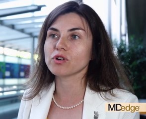
EULAR issues guidelines on managing rheumatic complications of cancer immunotherapies
MADRID – EULAR has issued recommendations to help rheumatologists address the increasingly common clinical issue of diagnosing and managing rheumatic-related adverse events associated with cancer immunotherapy.

“The rheumatic adverse events associated with immunotherapy represent a spectrum of new clinical entities, and they are challenging because they can be difficult to control while attempting to preserve the antitumor effects of oncological drugs,” Marie Kostine, MD, of the Centre Universitaire Hospitalier, Bordeaux, France, explained at the European Congress of Rheumatology.
The recommendations were drawn from the deliberations of an expert task force that identified the clinical issues to address and then developed a consensus about best practice recommendations. In addition to rheumatologists with expertise in this field, the task force included oncologists, allied health personnel, and two patient representatives.
The recommendations include four overarching principles and 10 recommendations.
“One of the overarching principles regards the importance of shared decision making between rheumatologists, oncologists, and patients,” Dr. Kostine said. Because of the expertise of rheumatologists in employing immunomodulatory therapies as they pertain to inflammation of the joints, the recommendations emphasize the value of their collaboration in clinical decisions.
The recommendations address patient referral, the assessment of preexisting rheumatic conditions, diagnosis, and therapeutic strategies.
“Rheumatologists should make themselves aware of the wide spectrum of potential clinical presentations of rheumatic adverse events following the initiation of immunotherapy,” Dr. Kostine said. While rheumatoid arthritis–like symptoms are common, the immune activation produced by checkpoint inhibitors and other immunotherapies can affect nearly every organ in the body, which includes diverse involvement of joint tissues.
In addition to joint pain, which has occurred in up to 40% of patients receiving a checkpoint inhibitor in some series, rheumatology-related events can include vasculitis, systemic sclerosis, and lupus. When associated with immunotherapy, these events sometimes develop in the absence of inflammatory markers or autoantibodies.
The new consensus guidelines emphasize that glucocorticoids can be “considered” to control rheumatic-related adverse events despite their immunosuppressive effect. However, because of their potential to attenuate the benefit of immune activation for treatment of the oncologic disease, such drugs, if used, “should be tapered to the lowest effective dose.”
The consensus recommendations were based on an extensive literature review, but Dr. Kostine acknowledged that prospective studies regarding the best practices for managing rheumatic-related adverse events of immunotherapies remain limited. She suggested that this knowledge gap was one reason for creating an expert task force.
“There has been an immunotherapy revolution, such that rheumatologists who have not yet seen these adverse events soon will,” said Dr. Kostine, noting that the number of approved immunotherapies and their clinical indications have been increasing rapidly.
The EULAR recommendations were created specifically for rheumatologists. In addition to guiding them toward best practice, the report from the task force provides background on the clinical issues raised by therapies that cause inflammatory side effects while stimulating immune function to treat malignancy.
MADRID – EULAR has issued recommendations to help rheumatologists address the increasingly common clinical issue of diagnosing and managing rheumatic-related adverse events associated with cancer immunotherapy.
“The rheumatic adverse events associated with immunotherapy represent a spectrum of new clinical entities, and they are challenging because they can be difficult to control while attempting to preserve the antitumor effects of oncological drugs,” Marie Kostine, MD, of the Centre Universitaire Hospitalier, Bordeaux, France, explained at the European Congress of Rheumatology.
The recommendations were drawn from the deliberations of an expert task force that identified the clinical issues to address and then developed a consensus about best practice recommendations. In addition to rheumatologists with expertise in this field, the task force included oncologists, allied health personnel, and two patient representatives.
The recommendations include four overarching principles and 10 recommendations.
“One of the overarching principles regards the importance of shared decision making between rheumatologists, oncologists, and patients,” Dr. Kostine said. Because of the expertise of rheumatologists in employing immunomodulatory therapies as they pertain to inflammation of the joints, the recommendations emphasize the value of their collaboration in clinical decisions.
The recommendations address patient referral, the assessment of preexisting rheumatic conditions, diagnosis, and therapeutic strategies.
“Rheumatologists should make themselves aware of the wide spectrum of potential clinical presentations of rheumatic adverse events following the initiation of immunotherapy,” Dr. Kostine said. While rheumatoid arthritis–like symptoms are common, the immune activation produced by checkpoint inhibitors and other immunotherapies can affect nearly every organ in the body, which includes diverse involvement of joint tissues.
In addition to joint pain, which has occurred in up to 40% of patients receiving a checkpoint inhibitor in some series, rheumatology-related events can include vasculitis, systemic sclerosis, and lupus. When associated with immunotherapy, these events sometimes develop in the absence of inflammatory markers or autoantibodies.
The new consensus guidelines emphasize that glucocorticoids can be “considered” to control rheumatic-related adverse events despite their immunosuppressive effect. However, because of their potential to attenuate the benefit of immune activation for treatment of the oncologic disease, such drugs, if used, “should be tapered to the lowest effective dose.”
The consensus recommendations were based on an extensive literature review, but Dr. Kostine acknowledged that prospective studies regarding the best practices for managing rheumatic-related adverse events of immunotherapies remain limited. She suggested that this knowledge gap was one reason for creating an expert task force.
“There has been an immunotherapy revolution, such that rheumatologists who have not yet seen these adverse events soon will,” said Dr. Kostine, noting that the number of approved immunotherapies and their clinical indications have been increasing rapidly.
The EULAR recommendations were created specifically for rheumatologists. In addition to guiding them toward best practice, the report from the task force provides background on the clinical issues raised by therapies that cause inflammatory side effects while stimulating immune function to treat malignancy.
REPORTING FROM EULAR 2019 CONGRESS