User login
GOTHENBURG, Sweden – Consider rapidly growing nontuberculous mycobacteria infection as a distinct possibility in patients who present with a rash or other skin lesions with a new tattoo.
That's the advice of Mayo Clinic dermatologist Dr. Lisa A. Drage, who encountered six affected patients in relatively short order, all with tattoos done by the same artist at a single Rochester, Minn., tattoo parlor.
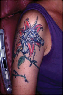
The likely source of this outbreak of Mycobacterium chelonae infection was the tap water the tattoo artist used to dilute black ink to create shades of gray for the popular gray wash technique, which creates a subtle photographic effect, she explained at the annual congress of the European Academy of Dermatology and Venereology.
The tipoff that the likely contaminant was tap water was that the skin lesions, although quite varied in appearance, were concentrated in the gray areas of the tattoos, with the deep black areas and clear-skin uninked portions largely spared, Dr. Drage said.
More broadly, she suggested keeping M. chelonae and the other rapidly growing mycobacteria (RGM) in mind when diagnosing any patient with a postprocedure skin infection that's not responding to appropriate antibiotic therapy.
Diagnosis
"RGMs are underrecognized, underreported, and we think they're increasing in prevalence," the dermatologist said.
"I think 'staph' first because it's so much more common. But if an infection isn’t responding within a couple of weeks and tests show it's not methicillin-resistant staph – and especially if there's been a procedure – think RGM," Dr. Drage said.
The reason RGM infections are underdiagnosed is that the culture techniques required to confirm the diagnosis are completely different from those utilized in conventional bacterial cultures.
The techniques for nontuberculous mycobacteria (NTM) are quite involved, with multiple cultures needing to be started at a variety of temperatures, since some RGM will grow only at 28 degrees C to 30 degrees C while others require 35 degrees C to 37 degrees C. Unlike the case in M. tuberculosis infections, where polymerase chain reaction or other rapid diagnostic systems can be applied directly to the clinical specimen, the NTM organisms must first be grown out on solid culture media before DNA sequencing can be utilized to identify the species.
"Most physicians who send the specimen to the laboratory fail to order the appropriate test. If you just send it for bacteriologic testing, they won't do the specific techniques for mycobacteria, and you'll get a negative result. It's important to obtain cultures specifically for mycobacteria, along with susceptibility studies. I find it most helpful to speak with the lab so they know what I'm thinking and what to do," she said.
The RGM, which include M. chelonae and M. fortuitum, are the most common NTM involved in community-acquired skin and soft-tissue infections. M. fortuitum alone is believed to account for more than 60% of such infections.
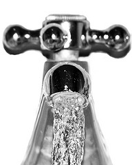
These are environmental microorganisms; there is no person-to-person spread. Tap water is the most common source of contamination. The NTM reside in the biofilm. They are resistant to sterilizers, disinfectants, and antiseptics. Numerous infections have come from hospital water systems.
Cutaneous infections with RGM have been documented in association with a variety of procedures, including Mohs surgery, acupuncture, punch biopsy, liposuction, laser resurfacing and other cosmetic procedures, and tattooing. In one California study, 29 of 30 pedicure foot baths in 18 salons in five counties contained NTM.
The skin lesions typically appear within a couple of weeks after the procedure, but lengthy delay in diagnosis is common. Patients may initially shrug off the skin lesions as bug bites or something else not warranting medical attention. And once they visit a physician, further diagnostic delay often ensues.
In the six Minnesota patients with M. chelonae infections in tattoos, for example, the skin lesions appeared 1-2 weeks after the tattoo was placed, and all patients sought medical care promptly, but they were treated for a diagnosis other than NTM infection. The median time between tattoo placement and the correct diagnosis was 18 weeks.
Timely diagnosis of RGM infections is made more challenging by the highly variable clinical appearance of the skin lesions. Painful red nodules that ulcerate and drain are one of the classic pictures. Pink or purple papules, granulomatous papules, pustules, plaques, folliculitis, cellulitis, and nonhealing ulcers can also be encountered.
"I've recently seen three patients referred for nonresponsive pyoderma gangrenosum that was actually due to NTM," Dr. Drage recalled.
The histopathologic findings can vary widely as well. A tuberculoid granulomatous infiltrate is most helpful, but some patients may instead have a lymphohistiocytic infiltrate, palisading or sarcoidal granulomas, a mixed inflammatory infiltrate, or other nonspecific findings. Acid-fast bacilli staining typically produces negative results.
Treatment
Treatment of NTM skin infections is based entirely on susceptibility test results. There are no clearcut treatment guidelines. Many infections will be resistant to the more common antimicrobials, and it's quite common for resistance to emerge during the required lengthy course of treatment. How lengthy? The typical treatment duration required for a localized infection is about 4 months, climbing to 6 months for more severe or disseminated infection.
Oral clarithromycin at 500 mg twice daily is a good option to start with while awaiting the susceptibility test results, as it covers some of the more common NTM. Combination therapy is often employed to prevent or treat resistance. Surgical removal of isolated foci of infection is often helpful, according to Dr. Drage.
As for the M. chelonae infection outbreak in tattoos, once Dr. Drage and her colleagues recognized the common denominator in the cases, she decided it was time to meet the artist.
She found the tattoo artists were dismayed at the rash of complications and highly receptive to her suggestion that they switch from tap water to sterile water for their work. They're eager to learn how to have safer, better procedures, she said, but reliable information can be hard for them to come by because of the lack of professional standards.
"In the United States, people who cut hair are more regulated than people who do tattoos," Dr. Drage noted.
She reported having no relevant financial conflicts.
GOTHENBURG, Sweden – Consider rapidly growing nontuberculous mycobacteria infection as a distinct possibility in patients who present with a rash or other skin lesions with a new tattoo.
That's the advice of Mayo Clinic dermatologist Dr. Lisa A. Drage, who encountered six affected patients in relatively short order, all with tattoos done by the same artist at a single Rochester, Minn., tattoo parlor.
The likely source of this outbreak of Mycobacterium chelonae infection was the tap water the tattoo artist used to dilute black ink to create shades of gray for the popular gray wash technique, which creates a subtle photographic effect, she explained at the annual congress of the European Academy of Dermatology and Venereology.
The tipoff that the likely contaminant was tap water was that the skin lesions, although quite varied in appearance, were concentrated in the gray areas of the tattoos, with the deep black areas and clear-skin uninked portions largely spared, Dr. Drage said.
More broadly, she suggested keeping M. chelonae and the other rapidly growing mycobacteria (RGM) in mind when diagnosing any patient with a postprocedure skin infection that's not responding to appropriate antibiotic therapy.
Diagnosis
"RGMs are underrecognized, underreported, and we think they're increasing in prevalence," the dermatologist said.
"I think 'staph' first because it's so much more common. But if an infection isn’t responding within a couple of weeks and tests show it's not methicillin-resistant staph – and especially if there's been a procedure – think RGM," Dr. Drage said.
The reason RGM infections are underdiagnosed is that the culture techniques required to confirm the diagnosis are completely different from those utilized in conventional bacterial cultures.
The techniques for nontuberculous mycobacteria (NTM) are quite involved, with multiple cultures needing to be started at a variety of temperatures, since some RGM will grow only at 28 degrees C to 30 degrees C while others require 35 degrees C to 37 degrees C. Unlike the case in M. tuberculosis infections, where polymerase chain reaction or other rapid diagnostic systems can be applied directly to the clinical specimen, the NTM organisms must first be grown out on solid culture media before DNA sequencing can be utilized to identify the species.
"Most physicians who send the specimen to the laboratory fail to order the appropriate test. If you just send it for bacteriologic testing, they won't do the specific techniques for mycobacteria, and you'll get a negative result. It's important to obtain cultures specifically for mycobacteria, along with susceptibility studies. I find it most helpful to speak with the lab so they know what I'm thinking and what to do," she said.
The RGM, which include M. chelonae and M. fortuitum, are the most common NTM involved in community-acquired skin and soft-tissue infections. M. fortuitum alone is believed to account for more than 60% of such infections.
These are environmental microorganisms; there is no person-to-person spread. Tap water is the most common source of contamination. The NTM reside in the biofilm. They are resistant to sterilizers, disinfectants, and antiseptics. Numerous infections have come from hospital water systems.
Cutaneous infections with RGM have been documented in association with a variety of procedures, including Mohs surgery, acupuncture, punch biopsy, liposuction, laser resurfacing and other cosmetic procedures, and tattooing. In one California study, 29 of 30 pedicure foot baths in 18 salons in five counties contained NTM.
The skin lesions typically appear within a couple of weeks after the procedure, but lengthy delay in diagnosis is common. Patients may initially shrug off the skin lesions as bug bites or something else not warranting medical attention. And once they visit a physician, further diagnostic delay often ensues.
In the six Minnesota patients with M. chelonae infections in tattoos, for example, the skin lesions appeared 1-2 weeks after the tattoo was placed, and all patients sought medical care promptly, but they were treated for a diagnosis other than NTM infection. The median time between tattoo placement and the correct diagnosis was 18 weeks.
Timely diagnosis of RGM infections is made more challenging by the highly variable clinical appearance of the skin lesions. Painful red nodules that ulcerate and drain are one of the classic pictures. Pink or purple papules, granulomatous papules, pustules, plaques, folliculitis, cellulitis, and nonhealing ulcers can also be encountered.
"I've recently seen three patients referred for nonresponsive pyoderma gangrenosum that was actually due to NTM," Dr. Drage recalled.
The histopathologic findings can vary widely as well. A tuberculoid granulomatous infiltrate is most helpful, but some patients may instead have a lymphohistiocytic infiltrate, palisading or sarcoidal granulomas, a mixed inflammatory infiltrate, or other nonspecific findings. Acid-fast bacilli staining typically produces negative results.
Treatment
Treatment of NTM skin infections is based entirely on susceptibility test results. There are no clearcut treatment guidelines. Many infections will be resistant to the more common antimicrobials, and it's quite common for resistance to emerge during the required lengthy course of treatment. How lengthy? The typical treatment duration required for a localized infection is about 4 months, climbing to 6 months for more severe or disseminated infection.
Oral clarithromycin at 500 mg twice daily is a good option to start with while awaiting the susceptibility test results, as it covers some of the more common NTM. Combination therapy is often employed to prevent or treat resistance. Surgical removal of isolated foci of infection is often helpful, according to Dr. Drage.
As for the M. chelonae infection outbreak in tattoos, once Dr. Drage and her colleagues recognized the common denominator in the cases, she decided it was time to meet the artist.
She found the tattoo artists were dismayed at the rash of complications and highly receptive to her suggestion that they switch from tap water to sterile water for their work. They're eager to learn how to have safer, better procedures, she said, but reliable information can be hard for them to come by because of the lack of professional standards.
"In the United States, people who cut hair are more regulated than people who do tattoos," Dr. Drage noted.
She reported having no relevant financial conflicts.
EXPERT ANALYSIS FROM THE ANNUAL CONGRESS OF THE EUROPEAN ACADEMY OF DERMATOLOGY AND VENEREOLOGY