User login
Bank could help docs identify optimal AML treatment, team says

Photo courtesy of
University Hospital Ulm
Research published in Nature Genetics suggests a knowledge bank
can reveal the optimal treatment for patients with acute
myeloid leukemia (AML), although more research is needed before such
banks can be used in the clinic.
Researchers built a knowledge
bank using data from 1540 AML patients enrolled in clinical
trials in Germany and Austria.
The bank includes information on genetic features, treatment, and outcomes for each patient.
The researchers used this information to develop models that could predict a patient’s likelihood of remission, relapse, and mortality.
The team then validated those results using data from patients in The Cancer Genome Atlas.
The researchers estimate that up to 1 in 3 AML patients would be prescribed a different treatment regimen if physicians used the knowledge bank approach rather than current practice.
“The knowledge bank approach makes far more detailed and accurate predictions about the likely future course of a patient with AML than what we can make in the clinic at the moment,” said study author Peter Campbell, PhD, of the Wellcome Trust Sanger Institute in Hinxton, UK.
“Current guides use a simple set of rules based on only a few genetic findings. For any given patient, using the new tool, we can compare the likely future outcomes under a transplant route versus a standard chemotherapy route. This means that we can make a treatment choice that is personally tailored to the unique features of that particular patient.”
However, the researchers said the knowledge bank approach requires further testing before it can be used to prescribe treatment in AML clinics.
“Our analysis reveals that knowledge banks of up to 10,000 patients would be needed to obtain the precision needed for routine clinical application,” said study author Moritz Gerstung, PhD, of the European Bioinformatics Institute in Hinxton, UK.
“Building knowledge banks is not easy,” added author Hartmut Döhner, MD, of the University of Ulm in Germany. “To get accurate treatment predictions, you need data from thousands of patients and all tumor types.”
“Furthermore, such knowledge banks will need continuous updating as new therapies become approved and available. As genetic testing enters routine clinical practice, there is an opportunity to learn from patients undergoing care in our health systems. Our paper gives the first real evidence that the approach is worthwhile, how it could be used, and what the scale needs to be.” 
Photo courtesy of
University Hospital Ulm
Research published in Nature Genetics suggests a knowledge bank
can reveal the optimal treatment for patients with acute
myeloid leukemia (AML), although more research is needed before such
banks can be used in the clinic.
Researchers built a knowledge
bank using data from 1540 AML patients enrolled in clinical
trials in Germany and Austria.
The bank includes information on genetic features, treatment, and outcomes for each patient.
The researchers used this information to develop models that could predict a patient’s likelihood of remission, relapse, and mortality.
The team then validated those results using data from patients in The Cancer Genome Atlas.
The researchers estimate that up to 1 in 3 AML patients would be prescribed a different treatment regimen if physicians used the knowledge bank approach rather than current practice.
“The knowledge bank approach makes far more detailed and accurate predictions about the likely future course of a patient with AML than what we can make in the clinic at the moment,” said study author Peter Campbell, PhD, of the Wellcome Trust Sanger Institute in Hinxton, UK.
“Current guides use a simple set of rules based on only a few genetic findings. For any given patient, using the new tool, we can compare the likely future outcomes under a transplant route versus a standard chemotherapy route. This means that we can make a treatment choice that is personally tailored to the unique features of that particular patient.”
However, the researchers said the knowledge bank approach requires further testing before it can be used to prescribe treatment in AML clinics.
“Our analysis reveals that knowledge banks of up to 10,000 patients would be needed to obtain the precision needed for routine clinical application,” said study author Moritz Gerstung, PhD, of the European Bioinformatics Institute in Hinxton, UK.
“Building knowledge banks is not easy,” added author Hartmut Döhner, MD, of the University of Ulm in Germany. “To get accurate treatment predictions, you need data from thousands of patients and all tumor types.”
“Furthermore, such knowledge banks will need continuous updating as new therapies become approved and available. As genetic testing enters routine clinical practice, there is an opportunity to learn from patients undergoing care in our health systems. Our paper gives the first real evidence that the approach is worthwhile, how it could be used, and what the scale needs to be.”
Photo courtesy of
University Hospital Ulm
Research published in Nature Genetics suggests a knowledge bank
can reveal the optimal treatment for patients with acute
myeloid leukemia (AML), although more research is needed before such
banks can be used in the clinic.
Researchers built a knowledge
bank using data from 1540 AML patients enrolled in clinical
trials in Germany and Austria.
The bank includes information on genetic features, treatment, and outcomes for each patient.
The researchers used this information to develop models that could predict a patient’s likelihood of remission, relapse, and mortality.
The team then validated those results using data from patients in The Cancer Genome Atlas.
The researchers estimate that up to 1 in 3 AML patients would be prescribed a different treatment regimen if physicians used the knowledge bank approach rather than current practice.
“The knowledge bank approach makes far more detailed and accurate predictions about the likely future course of a patient with AML than what we can make in the clinic at the moment,” said study author Peter Campbell, PhD, of the Wellcome Trust Sanger Institute in Hinxton, UK.
“Current guides use a simple set of rules based on only a few genetic findings. For any given patient, using the new tool, we can compare the likely future outcomes under a transplant route versus a standard chemotherapy route. This means that we can make a treatment choice that is personally tailored to the unique features of that particular patient.”
However, the researchers said the knowledge bank approach requires further testing before it can be used to prescribe treatment in AML clinics.
“Our analysis reveals that knowledge banks of up to 10,000 patients would be needed to obtain the precision needed for routine clinical application,” said study author Moritz Gerstung, PhD, of the European Bioinformatics Institute in Hinxton, UK.
“Building knowledge banks is not easy,” added author Hartmut Döhner, MD, of the University of Ulm in Germany. “To get accurate treatment predictions, you need data from thousands of patients and all tumor types.”
“Furthermore, such knowledge banks will need continuous updating as new therapies become approved and available. As genetic testing enters routine clinical practice, there is an opportunity to learn from patients undergoing care in our health systems. Our paper gives the first real evidence that the approach is worthwhile, how it could be used, and what the scale needs to be.”
Predicting response to cytarabine in AML patients
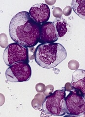
New research suggests the protein SAMHD1 could be used to predict which
patients with acute myeloid leukemia (AML) will respond to treatment
with cytarabine.
Researchers found that response to cytarabine
was inversely correlated with SAMHD1 expression in AML cell lines, mouse
models of the disease, and adult patients with AML.
Jindrich Cinatl, PhD, of the University of Frankfurt in Germany, and his colleagues reported these findings in Nature Medicine.
The researchers first analyzed 13 AML cell lines and found that SAMHD1 reduces the cytotoxic effect of cytarabine. When the team depleted SAMHD1 in these cell lines, they were “markedly sensitized” to cytarabine.
The researchers also cultivated cytarabine-resistant AML cell lines and found that SAMHD1 levels increased along with cytarabine resistance. However, depleting SAMHD1 resensitized the cells to cytarabine.
Investigation revealed that SAMHD1 removes the phosphate residues from the active form of cytarabine, Ara-CTP, and converts the drug to its inactive form, Ara-C.
The researchers then evaluated the role of SAMHD1 in AML in vivo. They transplanted SAMHD1-knockout AML cells and wild-type SAMHD1 AML cells into mice and treated the mice with cytarabine or phosphate-buffered saline.
Mice that received SAMHD1-knockout AML cells and cytarabine had significantly longer survival than mice that received wild-type SAMHD1 AML cells and cytarabine or either AML cell type plus phosphate-buffered saline.
Next, the researchers tested blasts isolated from the bone marrow of patients with therapy-naive AML.
The team found that basal SAMHD1 expression was significantly correlated with cytarabine IC50 values. And depleting SAMHD1 diminished cytarabine IC50 values by 3- to 15-fold.
Lastly, the researchers assessed whether SAMHD1 expression might be used to predict response to cytarabine-based therapy in patients with AML.
The team analyzed a cohort of 150 adult AML patients who had received 1 to 2 courses of induction therapy including cytarabine—either 2 cycles of 7+3 or 7+3 plus high-dose cytarabine in combination with mitoxantrone.
Analysis revealed that SAMHD1 expression was “markedly increased” among patients who did not achieve a complete remission (CR) at the end of induction.
Of the 112 patients who achieved a CR, 90 were scored as “SAMHD1 low,” and 22 were scored as “SAMHD1 high.” The CR rate was 44% in the SAMHD1-high cohort and 90% in the SAMHD1-low cohort.
In addition, the researchers found the level of SAMHD1 expression in blasts at patients’ initial diagnosis was predictive of event-free survival, relapse-free survival, and overall survival.
The team said these results suggest SAMHD1 could be used to guide treatment with cytarabine-based therapies in patients with AML.
New research suggests the protein SAMHD1 could be used to predict which
patients with acute myeloid leukemia (AML) will respond to treatment
with cytarabine.
Researchers found that response to cytarabine
was inversely correlated with SAMHD1 expression in AML cell lines, mouse
models of the disease, and adult patients with AML.
Jindrich Cinatl, PhD, of the University of Frankfurt in Germany, and his colleagues reported these findings in Nature Medicine.
The researchers first analyzed 13 AML cell lines and found that SAMHD1 reduces the cytotoxic effect of cytarabine. When the team depleted SAMHD1 in these cell lines, they were “markedly sensitized” to cytarabine.
The researchers also cultivated cytarabine-resistant AML cell lines and found that SAMHD1 levels increased along with cytarabine resistance. However, depleting SAMHD1 resensitized the cells to cytarabine.
Investigation revealed that SAMHD1 removes the phosphate residues from the active form of cytarabine, Ara-CTP, and converts the drug to its inactive form, Ara-C.
The researchers then evaluated the role of SAMHD1 in AML in vivo. They transplanted SAMHD1-knockout AML cells and wild-type SAMHD1 AML cells into mice and treated the mice with cytarabine or phosphate-buffered saline.
Mice that received SAMHD1-knockout AML cells and cytarabine had significantly longer survival than mice that received wild-type SAMHD1 AML cells and cytarabine or either AML cell type plus phosphate-buffered saline.
Next, the researchers tested blasts isolated from the bone marrow of patients with therapy-naive AML.
The team found that basal SAMHD1 expression was significantly correlated with cytarabine IC50 values. And depleting SAMHD1 diminished cytarabine IC50 values by 3- to 15-fold.
Lastly, the researchers assessed whether SAMHD1 expression might be used to predict response to cytarabine-based therapy in patients with AML.
The team analyzed a cohort of 150 adult AML patients who had received 1 to 2 courses of induction therapy including cytarabine—either 2 cycles of 7+3 or 7+3 plus high-dose cytarabine in combination with mitoxantrone.
Analysis revealed that SAMHD1 expression was “markedly increased” among patients who did not achieve a complete remission (CR) at the end of induction.
Of the 112 patients who achieved a CR, 90 were scored as “SAMHD1 low,” and 22 were scored as “SAMHD1 high.” The CR rate was 44% in the SAMHD1-high cohort and 90% in the SAMHD1-low cohort.
In addition, the researchers found the level of SAMHD1 expression in blasts at patients’ initial diagnosis was predictive of event-free survival, relapse-free survival, and overall survival.
The team said these results suggest SAMHD1 could be used to guide treatment with cytarabine-based therapies in patients with AML.
New research suggests the protein SAMHD1 could be used to predict which
patients with acute myeloid leukemia (AML) will respond to treatment
with cytarabine.
Researchers found that response to cytarabine
was inversely correlated with SAMHD1 expression in AML cell lines, mouse
models of the disease, and adult patients with AML.
Jindrich Cinatl, PhD, of the University of Frankfurt in Germany, and his colleagues reported these findings in Nature Medicine.
The researchers first analyzed 13 AML cell lines and found that SAMHD1 reduces the cytotoxic effect of cytarabine. When the team depleted SAMHD1 in these cell lines, they were “markedly sensitized” to cytarabine.
The researchers also cultivated cytarabine-resistant AML cell lines and found that SAMHD1 levels increased along with cytarabine resistance. However, depleting SAMHD1 resensitized the cells to cytarabine.
Investigation revealed that SAMHD1 removes the phosphate residues from the active form of cytarabine, Ara-CTP, and converts the drug to its inactive form, Ara-C.
The researchers then evaluated the role of SAMHD1 in AML in vivo. They transplanted SAMHD1-knockout AML cells and wild-type SAMHD1 AML cells into mice and treated the mice with cytarabine or phosphate-buffered saline.
Mice that received SAMHD1-knockout AML cells and cytarabine had significantly longer survival than mice that received wild-type SAMHD1 AML cells and cytarabine or either AML cell type plus phosphate-buffered saline.
Next, the researchers tested blasts isolated from the bone marrow of patients with therapy-naive AML.
The team found that basal SAMHD1 expression was significantly correlated with cytarabine IC50 values. And depleting SAMHD1 diminished cytarabine IC50 values by 3- to 15-fold.
Lastly, the researchers assessed whether SAMHD1 expression might be used to predict response to cytarabine-based therapy in patients with AML.
The team analyzed a cohort of 150 adult AML patients who had received 1 to 2 courses of induction therapy including cytarabine—either 2 cycles of 7+3 or 7+3 plus high-dose cytarabine in combination with mitoxantrone.
Analysis revealed that SAMHD1 expression was “markedly increased” among patients who did not achieve a complete remission (CR) at the end of induction.
Of the 112 patients who achieved a CR, 90 were scored as “SAMHD1 low,” and 22 were scored as “SAMHD1 high.” The CR rate was 44% in the SAMHD1-high cohort and 90% in the SAMHD1-low cohort.
In addition, the researchers found the level of SAMHD1 expression in blasts at patients’ initial diagnosis was predictive of event-free survival, relapse-free survival, and overall survival.
The team said these results suggest SAMHD1 could be used to guide treatment with cytarabine-based therapies in patients with AML.
Obesity-associated protein linked to AML

Photo courtesy of
University of Cincinnati
Preclinical research indicates that a protein associated with obesity is also involved in the development of acute myeloid leukemia (AML) and may affect AML patients’ response to treatment.
Researchers found evidence to suggest that the fat mass- and obesity-associated protein (FTO) regulates the expression of a set of genes through a mechanism involving RNA modification, thereby increasing the reproduction of leukemia cells and prohibiting drug response.
Jianjun Chen, PhD, of the University of Cincinnati in Ohio, and his colleagues conducted this research and reported the findings in Cancer Cell.
The team noted that N6-methyladenosine (m6A) RNA methylation is the most prevalent internal modification in messenger RNAs (mRNAs) in genes. And they found that FTO, an m6A demethylase, plays a critical oncogenic role in AML.
The researchers made this discovery by analyzing 2 microarray datasets of samples from AML as well as samples from control subjects.
The team found that FTO was highly expressed in AMLs with t(11q23)/MLL rearrangements, t(15;17)/PML-RARA, FLT3-ITD, and/or NPM1 mutations.
The high level of FTO expression contributed to leukemia cells multiplying and surviving and also promoted the development of AML in animal models and the non-response of AML cells to therapeutic agents.
Additionally, the researchers found that genes like ASB2 and RARA, which were reported to inhibit leukemia cell growth and/or mediate the response of leukemia cells to therapeutic agents, were suppressed in the AML samples with higher FTO expression.
The suppression of these genes was attributed to FTO-controlled decreased stability of their mRNA and was connected to FTO’s m6A demethylase activity.
“Our study shows, for the first time, the functional importance of the m6A modification machinery in leukemia,” Dr Chen said. “In addition, given the functional importance of FTO in the formation of leukemia and drug response, targeting FTO signaling may present a new therapeutic strategy to treat leukemia.”
“As FTO may also play a cancer-promoting role in various types of solid tumors, besides leukemia, our discoveries may have a broad impact in cancer biology and cancer therapy. Further studies are needed to advance our understanding of the critical role of FTO in various types of cancers and to develop more effective novel therapeutic strategies based on such understanding to treat cancers.”
Photo courtesy of
University of Cincinnati
Preclinical research indicates that a protein associated with obesity is also involved in the development of acute myeloid leukemia (AML) and may affect AML patients’ response to treatment.
Researchers found evidence to suggest that the fat mass- and obesity-associated protein (FTO) regulates the expression of a set of genes through a mechanism involving RNA modification, thereby increasing the reproduction of leukemia cells and prohibiting drug response.
Jianjun Chen, PhD, of the University of Cincinnati in Ohio, and his colleagues conducted this research and reported the findings in Cancer Cell.
The team noted that N6-methyladenosine (m6A) RNA methylation is the most prevalent internal modification in messenger RNAs (mRNAs) in genes. And they found that FTO, an m6A demethylase, plays a critical oncogenic role in AML.
The researchers made this discovery by analyzing 2 microarray datasets of samples from AML as well as samples from control subjects.
The team found that FTO was highly expressed in AMLs with t(11q23)/MLL rearrangements, t(15;17)/PML-RARA, FLT3-ITD, and/or NPM1 mutations.
The high level of FTO expression contributed to leukemia cells multiplying and surviving and also promoted the development of AML in animal models and the non-response of AML cells to therapeutic agents.
Additionally, the researchers found that genes like ASB2 and RARA, which were reported to inhibit leukemia cell growth and/or mediate the response of leukemia cells to therapeutic agents, were suppressed in the AML samples with higher FTO expression.
The suppression of these genes was attributed to FTO-controlled decreased stability of their mRNA and was connected to FTO’s m6A demethylase activity.
“Our study shows, for the first time, the functional importance of the m6A modification machinery in leukemia,” Dr Chen said. “In addition, given the functional importance of FTO in the formation of leukemia and drug response, targeting FTO signaling may present a new therapeutic strategy to treat leukemia.”
“As FTO may also play a cancer-promoting role in various types of solid tumors, besides leukemia, our discoveries may have a broad impact in cancer biology and cancer therapy. Further studies are needed to advance our understanding of the critical role of FTO in various types of cancers and to develop more effective novel therapeutic strategies based on such understanding to treat cancers.”
Photo courtesy of
University of Cincinnati
Preclinical research indicates that a protein associated with obesity is also involved in the development of acute myeloid leukemia (AML) and may affect AML patients’ response to treatment.
Researchers found evidence to suggest that the fat mass- and obesity-associated protein (FTO) regulates the expression of a set of genes through a mechanism involving RNA modification, thereby increasing the reproduction of leukemia cells and prohibiting drug response.
Jianjun Chen, PhD, of the University of Cincinnati in Ohio, and his colleagues conducted this research and reported the findings in Cancer Cell.
The team noted that N6-methyladenosine (m6A) RNA methylation is the most prevalent internal modification in messenger RNAs (mRNAs) in genes. And they found that FTO, an m6A demethylase, plays a critical oncogenic role in AML.
The researchers made this discovery by analyzing 2 microarray datasets of samples from AML as well as samples from control subjects.
The team found that FTO was highly expressed in AMLs with t(11q23)/MLL rearrangements, t(15;17)/PML-RARA, FLT3-ITD, and/or NPM1 mutations.
The high level of FTO expression contributed to leukemia cells multiplying and surviving and also promoted the development of AML in animal models and the non-response of AML cells to therapeutic agents.
Additionally, the researchers found that genes like ASB2 and RARA, which were reported to inhibit leukemia cell growth and/or mediate the response of leukemia cells to therapeutic agents, were suppressed in the AML samples with higher FTO expression.
The suppression of these genes was attributed to FTO-controlled decreased stability of their mRNA and was connected to FTO’s m6A demethylase activity.
“Our study shows, for the first time, the functional importance of the m6A modification machinery in leukemia,” Dr Chen said. “In addition, given the functional importance of FTO in the formation of leukemia and drug response, targeting FTO signaling may present a new therapeutic strategy to treat leukemia.”
“As FTO may also play a cancer-promoting role in various types of solid tumors, besides leukemia, our discoveries may have a broad impact in cancer biology and cancer therapy. Further studies are needed to advance our understanding of the critical role of FTO in various types of cancers and to develop more effective novel therapeutic strategies based on such understanding to treat cancers.”
Infection in AML patient prompts discovery

Photo courtesy of
Janice Carr/CDC
The quest to understand a prolonged infection in an infant with acute myeloid leukemia (AML) has led to the discovery of a mutation that allows bacteria to tolerate antibiotic therapy.
Researchers described this discovery in the journal mBio.
“These findings detail a ‘perfect storm’ for development of antibiotic tolerance by bacteria that already pose a clinical challenge,” said study author Jason Rosch, PhD, of St. Jude Children’s Research Hospital in Memphis, Tennessee.
“The same conditions may be present in other patients with immune systems that have been compromised by chemotherapy or disease,” added co-author Joshua Wolf, MBBS, also of St. Jude.
The “perfect storm” involved a patient who was 6 weeks old when she was diagnosed with AML. The treatment wiped out her white blood cells, and, despite infection-control measures, she developed a bloodstream infection with vancomycin-resistant Enterococcus faecium (VRE).
The infection persisted for 26 days and only resolved after her immune system recovered. She then successfully completed AML treatment.
In-depth DNA sequencing of 22 VRE samples collected during the patient’s infection helped researchers link the prolonged infection to a point mutation in the relA gene of VRE.
The mutation inappropriately activated the stringent response pathway, which bacteria use to survive under stress and to tolerate antibiotics.
The mutation resulted in elevated levels of the signaling molecule alarmone, and this likely primed the bacteria to survive exposure to multiple antibiotics, the researchers said.
The team also noted that relA-mutant VRE was susceptible to the antibiotics linezolid and daptomycin in minimum inhibitory concentration testing and during planktonic growth.
However, when growing in biofilm, relA-mutant VRE could tolerate high doses of both antibiotics.
“This mutation has particular clinical significance because the antibiotics involved, linezolid and daptomycin, are the last line of defense against VRE infection,” Dr Wolf said.
Among the compounds in development for the treatment of bacterial biofilms is the experimental antibiotic ADEP-4. In this study, ADEP-4 killed relA-mutant and non-mutant VRE growing in biofilm in the lab.
“In the future, compounds like ADEP-4 may provide a new approach to resolving persistent infections,” Dr Wolf said.
Dr Rosch noted that evidence gleaned from tracking the evolution of VRE throughout the infection suggested the patient’s immune-compromised state was essential to survival of the mutant VRE.
Gene transcription was altered significantly in relA-mutant VRE and produced biofilms that were less robust and possibly unlikely to otherwise survive.
“The case expands our understanding of the role of the stringent response in susceptibility and tolerance to a wide range of antibiotics, especially in biofilms,” Dr Rosch said. “It also demonstrates that these mutations can develop and gain a foothold during a human infection.”
Photo courtesy of
Janice Carr/CDC
The quest to understand a prolonged infection in an infant with acute myeloid leukemia (AML) has led to the discovery of a mutation that allows bacteria to tolerate antibiotic therapy.
Researchers described this discovery in the journal mBio.
“These findings detail a ‘perfect storm’ for development of antibiotic tolerance by bacteria that already pose a clinical challenge,” said study author Jason Rosch, PhD, of St. Jude Children’s Research Hospital in Memphis, Tennessee.
“The same conditions may be present in other patients with immune systems that have been compromised by chemotherapy or disease,” added co-author Joshua Wolf, MBBS, also of St. Jude.
The “perfect storm” involved a patient who was 6 weeks old when she was diagnosed with AML. The treatment wiped out her white blood cells, and, despite infection-control measures, she developed a bloodstream infection with vancomycin-resistant Enterococcus faecium (VRE).
The infection persisted for 26 days and only resolved after her immune system recovered. She then successfully completed AML treatment.
In-depth DNA sequencing of 22 VRE samples collected during the patient’s infection helped researchers link the prolonged infection to a point mutation in the relA gene of VRE.
The mutation inappropriately activated the stringent response pathway, which bacteria use to survive under stress and to tolerate antibiotics.
The mutation resulted in elevated levels of the signaling molecule alarmone, and this likely primed the bacteria to survive exposure to multiple antibiotics, the researchers said.
The team also noted that relA-mutant VRE was susceptible to the antibiotics linezolid and daptomycin in minimum inhibitory concentration testing and during planktonic growth.
However, when growing in biofilm, relA-mutant VRE could tolerate high doses of both antibiotics.
“This mutation has particular clinical significance because the antibiotics involved, linezolid and daptomycin, are the last line of defense against VRE infection,” Dr Wolf said.
Among the compounds in development for the treatment of bacterial biofilms is the experimental antibiotic ADEP-4. In this study, ADEP-4 killed relA-mutant and non-mutant VRE growing in biofilm in the lab.
“In the future, compounds like ADEP-4 may provide a new approach to resolving persistent infections,” Dr Wolf said.
Dr Rosch noted that evidence gleaned from tracking the evolution of VRE throughout the infection suggested the patient’s immune-compromised state was essential to survival of the mutant VRE.
Gene transcription was altered significantly in relA-mutant VRE and produced biofilms that were less robust and possibly unlikely to otherwise survive.
“The case expands our understanding of the role of the stringent response in susceptibility and tolerance to a wide range of antibiotics, especially in biofilms,” Dr Rosch said. “It also demonstrates that these mutations can develop and gain a foothold during a human infection.”
Photo courtesy of
Janice Carr/CDC
The quest to understand a prolonged infection in an infant with acute myeloid leukemia (AML) has led to the discovery of a mutation that allows bacteria to tolerate antibiotic therapy.
Researchers described this discovery in the journal mBio.
“These findings detail a ‘perfect storm’ for development of antibiotic tolerance by bacteria that already pose a clinical challenge,” said study author Jason Rosch, PhD, of St. Jude Children’s Research Hospital in Memphis, Tennessee.
“The same conditions may be present in other patients with immune systems that have been compromised by chemotherapy or disease,” added co-author Joshua Wolf, MBBS, also of St. Jude.
The “perfect storm” involved a patient who was 6 weeks old when she was diagnosed with AML. The treatment wiped out her white blood cells, and, despite infection-control measures, she developed a bloodstream infection with vancomycin-resistant Enterococcus faecium (VRE).
The infection persisted for 26 days and only resolved after her immune system recovered. She then successfully completed AML treatment.
In-depth DNA sequencing of 22 VRE samples collected during the patient’s infection helped researchers link the prolonged infection to a point mutation in the relA gene of VRE.
The mutation inappropriately activated the stringent response pathway, which bacteria use to survive under stress and to tolerate antibiotics.
The mutation resulted in elevated levels of the signaling molecule alarmone, and this likely primed the bacteria to survive exposure to multiple antibiotics, the researchers said.
The team also noted that relA-mutant VRE was susceptible to the antibiotics linezolid and daptomycin in minimum inhibitory concentration testing and during planktonic growth.
However, when growing in biofilm, relA-mutant VRE could tolerate high doses of both antibiotics.
“This mutation has particular clinical significance because the antibiotics involved, linezolid and daptomycin, are the last line of defense against VRE infection,” Dr Wolf said.
Among the compounds in development for the treatment of bacterial biofilms is the experimental antibiotic ADEP-4. In this study, ADEP-4 killed relA-mutant and non-mutant VRE growing in biofilm in the lab.
“In the future, compounds like ADEP-4 may provide a new approach to resolving persistent infections,” Dr Wolf said.
Dr Rosch noted that evidence gleaned from tracking the evolution of VRE throughout the infection suggested the patient’s immune-compromised state was essential to survival of the mutant VRE.
Gene transcription was altered significantly in relA-mutant VRE and produced biofilms that were less robust and possibly unlikely to otherwise survive.
“The case expands our understanding of the role of the stringent response in susceptibility and tolerance to a wide range of antibiotics, especially in biofilms,” Dr Rosch said. “It also demonstrates that these mutations can develop and gain a foothold during a human infection.”
Drugs may be effective against hematologic, other cancers
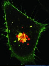
Image courtesy of PNAS
A diabetes medication and an antihypertensive drug may prove effective in the treatment of hematologic malignancies and other cancers, according to preclinical research published in Science Advances.
Past research has shown that metformin, a drug used to treat type 2 diabetes, has anticancer properties.
However, the usual therapeutic dose is too low to effectively fight cancer, and higher doses of metformin could be too toxic.
With the current study, researchers found that the antihypertensive drug syrosingopine enhances the anticancer efficacy of metformin without harming normal blood cells.
The team screened over a thousand drugs to find one that could boost metformin’s efficacy against cancers.
They identified syrosingopine and tested it in combination with metformin—at concentrations substantially below the drugs’ therapeutic thresholds—on a range of cancer cell lines and in mouse models of liver cancer.
Thirty-five of the 43 cell lines tested were susceptible to both syrosingopine and metformin. This included leukemia, lymphoma, and multiple myeloma cell lines.
In addition, the mice given a short course of syrosingopine and metformin experienced a reduction in the number of visible liver tumors.
The researchers also tested syrosingopine and metformin in peripheral blasts from 12 patients with acute myeloid leukemia and a patient with blast crisis chronic myeloid leukemia. All 13 samples responded to the treatment.
On the other hand, syrosingopine and metformin did not affect peripheral blood cells from healthy subjects.
“[A]lmost all tumor cells were killed by this cocktail and at doses that are actually not toxic to normal cells,” said study author Don Benjamin, of the University of Basel in Switzerland.
“And the effect was exclusively confined to cancer cells, as the blood cells from healthy donors were insensitive to the treatment.”
The researchers believe metformin functions by lowering blood glucose levels for cancer cells, starving them of essential nutrients needed for their survival. However, it is not clear how syrosingopine works in conjunction with metformin.
The team emphasized the need for more research evaluating the drugs in combination.
“We have been able to show that the 2 known drugs lead to more profound effects on cancer cell proliferation than each drug alone,” Dr Benjamin said. “The data from this study support the development of combination approaches for the treatment of cancer patients.”
Image courtesy of PNAS
A diabetes medication and an antihypertensive drug may prove effective in the treatment of hematologic malignancies and other cancers, according to preclinical research published in Science Advances.
Past research has shown that metformin, a drug used to treat type 2 diabetes, has anticancer properties.
However, the usual therapeutic dose is too low to effectively fight cancer, and higher doses of metformin could be too toxic.
With the current study, researchers found that the antihypertensive drug syrosingopine enhances the anticancer efficacy of metformin without harming normal blood cells.
The team screened over a thousand drugs to find one that could boost metformin’s efficacy against cancers.
They identified syrosingopine and tested it in combination with metformin—at concentrations substantially below the drugs’ therapeutic thresholds—on a range of cancer cell lines and in mouse models of liver cancer.
Thirty-five of the 43 cell lines tested were susceptible to both syrosingopine and metformin. This included leukemia, lymphoma, and multiple myeloma cell lines.
In addition, the mice given a short course of syrosingopine and metformin experienced a reduction in the number of visible liver tumors.
The researchers also tested syrosingopine and metformin in peripheral blasts from 12 patients with acute myeloid leukemia and a patient with blast crisis chronic myeloid leukemia. All 13 samples responded to the treatment.
On the other hand, syrosingopine and metformin did not affect peripheral blood cells from healthy subjects.
“[A]lmost all tumor cells were killed by this cocktail and at doses that are actually not toxic to normal cells,” said study author Don Benjamin, of the University of Basel in Switzerland.
“And the effect was exclusively confined to cancer cells, as the blood cells from healthy donors were insensitive to the treatment.”
The researchers believe metformin functions by lowering blood glucose levels for cancer cells, starving them of essential nutrients needed for their survival. However, it is not clear how syrosingopine works in conjunction with metformin.
The team emphasized the need for more research evaluating the drugs in combination.
“We have been able to show that the 2 known drugs lead to more profound effects on cancer cell proliferation than each drug alone,” Dr Benjamin said. “The data from this study support the development of combination approaches for the treatment of cancer patients.”
Image courtesy of PNAS
A diabetes medication and an antihypertensive drug may prove effective in the treatment of hematologic malignancies and other cancers, according to preclinical research published in Science Advances.
Past research has shown that metformin, a drug used to treat type 2 diabetes, has anticancer properties.
However, the usual therapeutic dose is too low to effectively fight cancer, and higher doses of metformin could be too toxic.
With the current study, researchers found that the antihypertensive drug syrosingopine enhances the anticancer efficacy of metformin without harming normal blood cells.
The team screened over a thousand drugs to find one that could boost metformin’s efficacy against cancers.
They identified syrosingopine and tested it in combination with metformin—at concentrations substantially below the drugs’ therapeutic thresholds—on a range of cancer cell lines and in mouse models of liver cancer.
Thirty-five of the 43 cell lines tested were susceptible to both syrosingopine and metformin. This included leukemia, lymphoma, and multiple myeloma cell lines.
In addition, the mice given a short course of syrosingopine and metformin experienced a reduction in the number of visible liver tumors.
The researchers also tested syrosingopine and metformin in peripheral blasts from 12 patients with acute myeloid leukemia and a patient with blast crisis chronic myeloid leukemia. All 13 samples responded to the treatment.
On the other hand, syrosingopine and metformin did not affect peripheral blood cells from healthy subjects.
“[A]lmost all tumor cells were killed by this cocktail and at doses that are actually not toxic to normal cells,” said study author Don Benjamin, of the University of Basel in Switzerland.
“And the effect was exclusively confined to cancer cells, as the blood cells from healthy donors were insensitive to the treatment.”
The researchers believe metformin functions by lowering blood glucose levels for cancer cells, starving them of essential nutrients needed for their survival. However, it is not clear how syrosingopine works in conjunction with metformin.
The team emphasized the need for more research evaluating the drugs in combination.
“We have been able to show that the 2 known drugs lead to more profound effects on cancer cell proliferation than each drug alone,” Dr Benjamin said. “The data from this study support the development of combination approaches for the treatment of cancer patients.”
Combos prove no better than 7+3 for AML

Photo courtesy of
MD Anderson Cancer Center
SAN DIEGO—Neither a 2-drug combination nor a 3-drug combination is superior to 7+3 chemotherapy in younger patients with previously untreated acute myeloid leukemia (AML), according to a phase 3 trial.
Treatment
with idarubicin and high-dose cytarabine (IA), with or without
vorinostat (V), was no more effective than standard cytarabine plus
daunorubicin (7+3) in this trial.
In fact, among patients with favorable cytogenetics, outcomes with IA or IA+V were inferior to outcomes with 7+3.
Guillermo Garcia-Manero, MD, of The University of Texas MD Anderson Cancer Center in Houston, presented these results at the 2016 ASH Annual Meeting (abstract 901*).
In a phase 2 trial, Dr Garcia-Manero and his colleagues found that IA+V produced a high response rate (85%) in patients with previously untreated AML or high-risk myelodysplastic syndromes.
So the researchers conducted a phase 3 study (SWOG S1203) to determine if IA or IA+V could improve outcomes for younger AML patients when compared to 7+3.
Treatment
Induction therapy was as follows:
- 7+3 arm—daunorubicin** at 90 mg/m2 once daily on days 1 to 3 with cytarabine at 100 mg/m2 once daily on days 1 to 7.
- IA arm—idarubicin at 12 mg/m2 once daily on days 1 to 3 with cytarabine at 1.5 gm/m2 once daily on days 1 to 4.
- IA+V arm—vorinostat at 500 mg orally 3 times a day on days 1 to 3, idarubicin at 12 mg/m2 once daily on days 4 to 6, and cytarabine at 1.5 gm/m2 once daily on days 4 to 7.
Consolidation was as follows:
- 7+3 arm—standard high-dose cytarabine at 3 gm/m2 over 3 hours every 12 hours x 6 doses for 1 to 4 cycles, depending on transplant availability.
- IA arm—idarubicin at 8 mg/m2 once daily on days 1 to 2 with cytarabine at 0.75 mg/m2 for 3 days on days 1 to 3 for 4 cycles.
- IA+V arm—vorinostat at 500 mg orally 3 times a day on days 1 to 3, idarubicin at 8 mg/m2 once daily on days 4 to 5, and cytarabine at 0.75 gm/m2 once daily on days 4 to 6.
The number of consolidation cycles varied depending on transplant indication. In all, 43% of patients (n=317) proceeded to allogeneic transplant. (Details on these patients were presented at ASH as abstract 1166.)
Patients in the IA+V arm also received maintenance with vorinostat at 300 mg 3 times a day for 14 days every 28 days.
**There was a shortage of daunorubicin during this trial. So if daunorubicin was not available, patients received idarubicin at 12 mg/m2 once daily on days 1 to 3. Dr Garcia-Manero could not provide data on how many patients assigned to daunorubicin actually received idarubicin.
Patients
There were a total of 738 eligible patients—261 in the 7+3 arm, 261 in the IA arm, and 216 in the IA+V arm. Dr Garcia-Manero said baseline characteristics were well balanced among the arms.
Overall, the median age was 49 (range, 18-60), 49% of patients were female, and 13% had a performance status of 2-3.
Thirteen percent of patients had favorable cytogenetics, 22% had high-risk cytogenetics, 16% had FLT3-ITD, and 21% had mutated NPM1.
Results
The complete response rates were 62% overall, 63% for 7+3, 64% for IA, and 60% for IA+V (P=0.58).
The rates of complete response with incomplete count recovery were 15%, 13%, 16%, and 17%, respectively. The failure rates were 23%, 25%, 21%, and 23%, respectively.
The rate of mortality within 30 days was 4% overall, 3% for 7+3, 6% for IA, and 4% for IA+V (P=0.013). The rate of mortality within 60 days was 7%, 5%, 9%, and 9%, respectively (P=0.097).
The rate of event-free survival was 42% overall, 43% for 7+3, 43% for IA, and 40% for IA+V.
There was no significant difference in event-free survival between IA+V and IA (P=0.66), IA+V and 7+3 (P=0.91), or IA and 7+3 (P=0.76).
The rate of overall survival (OS) was 62% overall, 62% for 7+3, 63% for IA, and 59% for IA+V.
There was no significant difference in OS between IA+V and IA (P=0.6), IA+V and 7+3 (P=0.67), or IA and 7+3 (P=0.92).
Among patients with favorable cytogenetics, there was no significant difference in OS between IA and IA+V (P=0.8). However, patients who received IA (P=0.011) or IA+V (P=0.012) had significantly better OS than patients who received 7+3.
There were more grade 5 adverse events (AEs) in the IA (n=19) and IA+V arms (n=16) than in the 7+3 arm (n=6).
Grade 5 AEs in the 7+3 arm were classified as follows: cardiac disorder (n=1), gastrointestinal disorder (n=1), general disorders (n=2), hepatobiliary disorder (n=1), and respiratory/thoracic/mediastinal disorder (n=1).
Grade 5 AEs in the IA arm included cardiac disorders (n=3), gastrointestinal disorder (n=1), general disorders (n=2), infections and infestations (n=7), nervous system disorder (n=1), respiratory/thoracic/mediastinal disorders (n=4), and vascular disorder (n=1).
Grade 5 AEs in the IA+V arm included cardiac disorder (n=1), general disorders (n=2), infections and infestations (n=7), nervous system disorder (n=1), and respiratory/thoracic/mediastinal disorders (n=5).
“In newly diagnosed adults with AML ages 18 to 60, neither IA [plus] vorinostat nor IA were superior to standard 7+3,” Dr Garcia-Manero said in closing.
“Indeed, 7+3 was superior to IA and IA [plus] vorinostat for those patients with favorable cytogenetics, reinforcing the need for high-dose ara-C during the consolidation phase. Newer studies with other combinations, including, perhaps, nucleoside analogues, monoclonal antibodies, or targeted agents are needed.”
*Some data in the abstract differ from the presentation.
Photo courtesy of
MD Anderson Cancer Center
SAN DIEGO—Neither a 2-drug combination nor a 3-drug combination is superior to 7+3 chemotherapy in younger patients with previously untreated acute myeloid leukemia (AML), according to a phase 3 trial.
Treatment
with idarubicin and high-dose cytarabine (IA), with or without
vorinostat (V), was no more effective than standard cytarabine plus
daunorubicin (7+3) in this trial.
In fact, among patients with favorable cytogenetics, outcomes with IA or IA+V were inferior to outcomes with 7+3.
Guillermo Garcia-Manero, MD, of The University of Texas MD Anderson Cancer Center in Houston, presented these results at the 2016 ASH Annual Meeting (abstract 901*).
In a phase 2 trial, Dr Garcia-Manero and his colleagues found that IA+V produced a high response rate (85%) in patients with previously untreated AML or high-risk myelodysplastic syndromes.
So the researchers conducted a phase 3 study (SWOG S1203) to determine if IA or IA+V could improve outcomes for younger AML patients when compared to 7+3.
Treatment
Induction therapy was as follows:
- 7+3 arm—daunorubicin** at 90 mg/m2 once daily on days 1 to 3 with cytarabine at 100 mg/m2 once daily on days 1 to 7.
- IA arm—idarubicin at 12 mg/m2 once daily on days 1 to 3 with cytarabine at 1.5 gm/m2 once daily on days 1 to 4.
- IA+V arm—vorinostat at 500 mg orally 3 times a day on days 1 to 3, idarubicin at 12 mg/m2 once daily on days 4 to 6, and cytarabine at 1.5 gm/m2 once daily on days 4 to 7.
Consolidation was as follows:
- 7+3 arm—standard high-dose cytarabine at 3 gm/m2 over 3 hours every 12 hours x 6 doses for 1 to 4 cycles, depending on transplant availability.
- IA arm—idarubicin at 8 mg/m2 once daily on days 1 to 2 with cytarabine at 0.75 mg/m2 for 3 days on days 1 to 3 for 4 cycles.
- IA+V arm—vorinostat at 500 mg orally 3 times a day on days 1 to 3, idarubicin at 8 mg/m2 once daily on days 4 to 5, and cytarabine at 0.75 gm/m2 once daily on days 4 to 6.
The number of consolidation cycles varied depending on transplant indication. In all, 43% of patients (n=317) proceeded to allogeneic transplant. (Details on these patients were presented at ASH as abstract 1166.)
Patients in the IA+V arm also received maintenance with vorinostat at 300 mg 3 times a day for 14 days every 28 days.
**There was a shortage of daunorubicin during this trial. So if daunorubicin was not available, patients received idarubicin at 12 mg/m2 once daily on days 1 to 3. Dr Garcia-Manero could not provide data on how many patients assigned to daunorubicin actually received idarubicin.
Patients
There were a total of 738 eligible patients—261 in the 7+3 arm, 261 in the IA arm, and 216 in the IA+V arm. Dr Garcia-Manero said baseline characteristics were well balanced among the arms.
Overall, the median age was 49 (range, 18-60), 49% of patients were female, and 13% had a performance status of 2-3.
Thirteen percent of patients had favorable cytogenetics, 22% had high-risk cytogenetics, 16% had FLT3-ITD, and 21% had mutated NPM1.
Results
The complete response rates were 62% overall, 63% for 7+3, 64% for IA, and 60% for IA+V (P=0.58).
The rates of complete response with incomplete count recovery were 15%, 13%, 16%, and 17%, respectively. The failure rates were 23%, 25%, 21%, and 23%, respectively.
The rate of mortality within 30 days was 4% overall, 3% for 7+3, 6% for IA, and 4% for IA+V (P=0.013). The rate of mortality within 60 days was 7%, 5%, 9%, and 9%, respectively (P=0.097).
The rate of event-free survival was 42% overall, 43% for 7+3, 43% for IA, and 40% for IA+V.
There was no significant difference in event-free survival between IA+V and IA (P=0.66), IA+V and 7+3 (P=0.91), or IA and 7+3 (P=0.76).
The rate of overall survival (OS) was 62% overall, 62% for 7+3, 63% for IA, and 59% for IA+V.
There was no significant difference in OS between IA+V and IA (P=0.6), IA+V and 7+3 (P=0.67), or IA and 7+3 (P=0.92).
Among patients with favorable cytogenetics, there was no significant difference in OS between IA and IA+V (P=0.8). However, patients who received IA (P=0.011) or IA+V (P=0.012) had significantly better OS than patients who received 7+3.
There were more grade 5 adverse events (AEs) in the IA (n=19) and IA+V arms (n=16) than in the 7+3 arm (n=6).
Grade 5 AEs in the 7+3 arm were classified as follows: cardiac disorder (n=1), gastrointestinal disorder (n=1), general disorders (n=2), hepatobiliary disorder (n=1), and respiratory/thoracic/mediastinal disorder (n=1).
Grade 5 AEs in the IA arm included cardiac disorders (n=3), gastrointestinal disorder (n=1), general disorders (n=2), infections and infestations (n=7), nervous system disorder (n=1), respiratory/thoracic/mediastinal disorders (n=4), and vascular disorder (n=1).
Grade 5 AEs in the IA+V arm included cardiac disorder (n=1), general disorders (n=2), infections and infestations (n=7), nervous system disorder (n=1), and respiratory/thoracic/mediastinal disorders (n=5).
“In newly diagnosed adults with AML ages 18 to 60, neither IA [plus] vorinostat nor IA were superior to standard 7+3,” Dr Garcia-Manero said in closing.
“Indeed, 7+3 was superior to IA and IA [plus] vorinostat for those patients with favorable cytogenetics, reinforcing the need for high-dose ara-C during the consolidation phase. Newer studies with other combinations, including, perhaps, nucleoside analogues, monoclonal antibodies, or targeted agents are needed.”
*Some data in the abstract differ from the presentation.
Photo courtesy of
MD Anderson Cancer Center
SAN DIEGO—Neither a 2-drug combination nor a 3-drug combination is superior to 7+3 chemotherapy in younger patients with previously untreated acute myeloid leukemia (AML), according to a phase 3 trial.
Treatment
with idarubicin and high-dose cytarabine (IA), with or without
vorinostat (V), was no more effective than standard cytarabine plus
daunorubicin (7+3) in this trial.
In fact, among patients with favorable cytogenetics, outcomes with IA or IA+V were inferior to outcomes with 7+3.
Guillermo Garcia-Manero, MD, of The University of Texas MD Anderson Cancer Center in Houston, presented these results at the 2016 ASH Annual Meeting (abstract 901*).
In a phase 2 trial, Dr Garcia-Manero and his colleagues found that IA+V produced a high response rate (85%) in patients with previously untreated AML or high-risk myelodysplastic syndromes.
So the researchers conducted a phase 3 study (SWOG S1203) to determine if IA or IA+V could improve outcomes for younger AML patients when compared to 7+3.
Treatment
Induction therapy was as follows:
- 7+3 arm—daunorubicin** at 90 mg/m2 once daily on days 1 to 3 with cytarabine at 100 mg/m2 once daily on days 1 to 7.
- IA arm—idarubicin at 12 mg/m2 once daily on days 1 to 3 with cytarabine at 1.5 gm/m2 once daily on days 1 to 4.
- IA+V arm—vorinostat at 500 mg orally 3 times a day on days 1 to 3, idarubicin at 12 mg/m2 once daily on days 4 to 6, and cytarabine at 1.5 gm/m2 once daily on days 4 to 7.
Consolidation was as follows:
- 7+3 arm—standard high-dose cytarabine at 3 gm/m2 over 3 hours every 12 hours x 6 doses for 1 to 4 cycles, depending on transplant availability.
- IA arm—idarubicin at 8 mg/m2 once daily on days 1 to 2 with cytarabine at 0.75 mg/m2 for 3 days on days 1 to 3 for 4 cycles.
- IA+V arm—vorinostat at 500 mg orally 3 times a day on days 1 to 3, idarubicin at 8 mg/m2 once daily on days 4 to 5, and cytarabine at 0.75 gm/m2 once daily on days 4 to 6.
The number of consolidation cycles varied depending on transplant indication. In all, 43% of patients (n=317) proceeded to allogeneic transplant. (Details on these patients were presented at ASH as abstract 1166.)
Patients in the IA+V arm also received maintenance with vorinostat at 300 mg 3 times a day for 14 days every 28 days.
**There was a shortage of daunorubicin during this trial. So if daunorubicin was not available, patients received idarubicin at 12 mg/m2 once daily on days 1 to 3. Dr Garcia-Manero could not provide data on how many patients assigned to daunorubicin actually received idarubicin.
Patients
There were a total of 738 eligible patients—261 in the 7+3 arm, 261 in the IA arm, and 216 in the IA+V arm. Dr Garcia-Manero said baseline characteristics were well balanced among the arms.
Overall, the median age was 49 (range, 18-60), 49% of patients were female, and 13% had a performance status of 2-3.
Thirteen percent of patients had favorable cytogenetics, 22% had high-risk cytogenetics, 16% had FLT3-ITD, and 21% had mutated NPM1.
Results
The complete response rates were 62% overall, 63% for 7+3, 64% for IA, and 60% for IA+V (P=0.58).
The rates of complete response with incomplete count recovery were 15%, 13%, 16%, and 17%, respectively. The failure rates were 23%, 25%, 21%, and 23%, respectively.
The rate of mortality within 30 days was 4% overall, 3% for 7+3, 6% for IA, and 4% for IA+V (P=0.013). The rate of mortality within 60 days was 7%, 5%, 9%, and 9%, respectively (P=0.097).
The rate of event-free survival was 42% overall, 43% for 7+3, 43% for IA, and 40% for IA+V.
There was no significant difference in event-free survival between IA+V and IA (P=0.66), IA+V and 7+3 (P=0.91), or IA and 7+3 (P=0.76).
The rate of overall survival (OS) was 62% overall, 62% for 7+3, 63% for IA, and 59% for IA+V.
There was no significant difference in OS between IA+V and IA (P=0.6), IA+V and 7+3 (P=0.67), or IA and 7+3 (P=0.92).
Among patients with favorable cytogenetics, there was no significant difference in OS between IA and IA+V (P=0.8). However, patients who received IA (P=0.011) or IA+V (P=0.012) had significantly better OS than patients who received 7+3.
There were more grade 5 adverse events (AEs) in the IA (n=19) and IA+V arms (n=16) than in the 7+3 arm (n=6).
Grade 5 AEs in the 7+3 arm were classified as follows: cardiac disorder (n=1), gastrointestinal disorder (n=1), general disorders (n=2), hepatobiliary disorder (n=1), and respiratory/thoracic/mediastinal disorder (n=1).
Grade 5 AEs in the IA arm included cardiac disorders (n=3), gastrointestinal disorder (n=1), general disorders (n=2), infections and infestations (n=7), nervous system disorder (n=1), respiratory/thoracic/mediastinal disorders (n=4), and vascular disorder (n=1).
Grade 5 AEs in the IA+V arm included cardiac disorder (n=1), general disorders (n=2), infections and infestations (n=7), nervous system disorder (n=1), and respiratory/thoracic/mediastinal disorders (n=5).
“In newly diagnosed adults with AML ages 18 to 60, neither IA [plus] vorinostat nor IA were superior to standard 7+3,” Dr Garcia-Manero said in closing.
“Indeed, 7+3 was superior to IA and IA [plus] vorinostat for those patients with favorable cytogenetics, reinforcing the need for high-dose ara-C during the consolidation phase. Newer studies with other combinations, including, perhaps, nucleoside analogues, monoclonal antibodies, or targeted agents are needed.”
*Some data in the abstract differ from the presentation.
FDA places AML trials on full, partial clinical hold
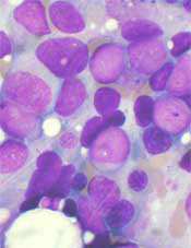
The US Food and Drug Administration (FDA) has placed holds on 3 early stage trials of vadastuximab talirine (SGN-CD33A) in acute myeloid leukemia (AML).
A phase 1/2 trial of vadastuximab talirine monotherapy in pre- and post-allogeneic transplant patients has been placed on full clinical hold.
This means no new subjects can be enrolled on the trial, and there can be no further dosing of subjects who are already enrolled.
Two phase 1 trials of vadastuximab talirine have been placed on partial clinical hold. This means no new subjects can be enrolled, but existing patients may continue treatment with re-consent.
In one of the trials on partial hold, researchers are investigating vadastuximab talirine alone and in combination with hypomethylating agents in AML patients who either relapsed after induction/consolidation or declined treatment with high-dose induction/consolidation.
In the other trial on partial hold, researchers are testing vadastuximab talirine in combination with 7+3 chemotherapy in newly diagnosed AML patients. Results from this trial were presented at the 2016 ASH Annual Meeting.
All 3 clinical holds were initiated to evaluate the potential risk of hepatotoxicity in patients who were treated with vadastuximab talirine and received allogeneic stem cell transplant either before or after treatment.
There have been 6 patients with hepatotoxicity, including several cases of veno-occlusive disease, with 4 fatal events.
Seattle Genetics, Inc., the company developing vadastuximab talirine, said it is working with the FDA to determine whether there is any association between hepatotoxicity and treatment with vadastuximab talirine to identify appropriate protocol amendments for patient safety and to enable continuation of these trials.
No new studies of vadastuximab talirine will be initiated until the clinical holds are lifted.
Seattle Genetics’ other ongoing trials of vadastuximab talirine, including the phase 3 CASCADE trial in older AML patients and phase 1/2 trial in patients with myelodysplastic syndrome (MDS), are proceeding with enrollment.
Overall, more than 300 patients have been treated with vadastuximab talirine in clinical trials across multiple treatment settings.
Vadastuximab talirine is an investigational antibody-drug conjugate (ADC) targeted to CD33, which is expressed on most AML and MDS blast cells. The CD33 engineered cysteine antibody is stably linked to a DNA binding agent called a pyrrolobenzodiazepine (PBD) dimer via site-specific conjugation technology (EC-mAb).
PBD dimers are said to be significantly more potent than systemic chemotherapeutic drugs, and the EC-mAb technology allows uniform drug-loading onto an ADC. The ADC is designed to be stable in the bloodstream and to release its PBD agent upon internalization into CD33-expressing cells.
The US Food and Drug Administration (FDA) has placed holds on 3 early stage trials of vadastuximab talirine (SGN-CD33A) in acute myeloid leukemia (AML).
A phase 1/2 trial of vadastuximab talirine monotherapy in pre- and post-allogeneic transplant patients has been placed on full clinical hold.
This means no new subjects can be enrolled on the trial, and there can be no further dosing of subjects who are already enrolled.
Two phase 1 trials of vadastuximab talirine have been placed on partial clinical hold. This means no new subjects can be enrolled, but existing patients may continue treatment with re-consent.
In one of the trials on partial hold, researchers are investigating vadastuximab talirine alone and in combination with hypomethylating agents in AML patients who either relapsed after induction/consolidation or declined treatment with high-dose induction/consolidation.
In the other trial on partial hold, researchers are testing vadastuximab talirine in combination with 7+3 chemotherapy in newly diagnosed AML patients. Results from this trial were presented at the 2016 ASH Annual Meeting.
All 3 clinical holds were initiated to evaluate the potential risk of hepatotoxicity in patients who were treated with vadastuximab talirine and received allogeneic stem cell transplant either before or after treatment.
There have been 6 patients with hepatotoxicity, including several cases of veno-occlusive disease, with 4 fatal events.
Seattle Genetics, Inc., the company developing vadastuximab talirine, said it is working with the FDA to determine whether there is any association between hepatotoxicity and treatment with vadastuximab talirine to identify appropriate protocol amendments for patient safety and to enable continuation of these trials.
No new studies of vadastuximab talirine will be initiated until the clinical holds are lifted.
Seattle Genetics’ other ongoing trials of vadastuximab talirine, including the phase 3 CASCADE trial in older AML patients and phase 1/2 trial in patients with myelodysplastic syndrome (MDS), are proceeding with enrollment.
Overall, more than 300 patients have been treated with vadastuximab talirine in clinical trials across multiple treatment settings.
Vadastuximab talirine is an investigational antibody-drug conjugate (ADC) targeted to CD33, which is expressed on most AML and MDS blast cells. The CD33 engineered cysteine antibody is stably linked to a DNA binding agent called a pyrrolobenzodiazepine (PBD) dimer via site-specific conjugation technology (EC-mAb).
PBD dimers are said to be significantly more potent than systemic chemotherapeutic drugs, and the EC-mAb technology allows uniform drug-loading onto an ADC. The ADC is designed to be stable in the bloodstream and to release its PBD agent upon internalization into CD33-expressing cells.
The US Food and Drug Administration (FDA) has placed holds on 3 early stage trials of vadastuximab talirine (SGN-CD33A) in acute myeloid leukemia (AML).
A phase 1/2 trial of vadastuximab talirine monotherapy in pre- and post-allogeneic transplant patients has been placed on full clinical hold.
This means no new subjects can be enrolled on the trial, and there can be no further dosing of subjects who are already enrolled.
Two phase 1 trials of vadastuximab talirine have been placed on partial clinical hold. This means no new subjects can be enrolled, but existing patients may continue treatment with re-consent.
In one of the trials on partial hold, researchers are investigating vadastuximab talirine alone and in combination with hypomethylating agents in AML patients who either relapsed after induction/consolidation or declined treatment with high-dose induction/consolidation.
In the other trial on partial hold, researchers are testing vadastuximab talirine in combination with 7+3 chemotherapy in newly diagnosed AML patients. Results from this trial were presented at the 2016 ASH Annual Meeting.
All 3 clinical holds were initiated to evaluate the potential risk of hepatotoxicity in patients who were treated with vadastuximab talirine and received allogeneic stem cell transplant either before or after treatment.
There have been 6 patients with hepatotoxicity, including several cases of veno-occlusive disease, with 4 fatal events.
Seattle Genetics, Inc., the company developing vadastuximab talirine, said it is working with the FDA to determine whether there is any association between hepatotoxicity and treatment with vadastuximab talirine to identify appropriate protocol amendments for patient safety and to enable continuation of these trials.
No new studies of vadastuximab talirine will be initiated until the clinical holds are lifted.
Seattle Genetics’ other ongoing trials of vadastuximab talirine, including the phase 3 CASCADE trial in older AML patients and phase 1/2 trial in patients with myelodysplastic syndrome (MDS), are proceeding with enrollment.
Overall, more than 300 patients have been treated with vadastuximab talirine in clinical trials across multiple treatment settings.
Vadastuximab talirine is an investigational antibody-drug conjugate (ADC) targeted to CD33, which is expressed on most AML and MDS blast cells. The CD33 engineered cysteine antibody is stably linked to a DNA binding agent called a pyrrolobenzodiazepine (PBD) dimer via site-specific conjugation technology (EC-mAb).
PBD dimers are said to be significantly more potent than systemic chemotherapeutic drugs, and the EC-mAb technology allows uniform drug-loading onto an ADC. The ADC is designed to be stable in the bloodstream and to release its PBD agent upon internalization into CD33-expressing cells.
FOUND IN TRANSLATION Minimal nomenclature and maximum sensitivity complicate MRD measures
In hematologic malignancies, there is a deep and direct connection between each individual patient, that patient’s symptoms, the visible cells that cause the disease, and the direct measurements and assessments of those cells. The totality of these factors helps to determine the diagnosis and treatment plan. As a butterfly needle often is sufficient for obtaining a diagnostic tumor biopsy, it is not surprising that these same diagnostic techniques are now standardly being used to monitor disease response.
The techniques differ in their limits of detection, however. With sequencing depths able to reliably detect variant allele frequencies of less than 10%, even when patients’ overt leukemia may no longer be detectable, clinicians may be left to ponder what to do with persistent “preleukemic” or “rising clones.”1-3
These patients, now more appropriately stratified for risk of recurrence, are in desperate need of better care algorithms. Standard MRD assessment by flow cytometric analysis is able to detect less than 1 x 10⁻⁴ cells. While it can be applied to most patients, its sensitivity will likely be surpassed by new and emerging genomic assays. Real time quantitative polymerase chain reaction (RT-qPCR) and next generation sequencing (NGS) require a leukemia-specific abnormality but have the potential for far greater sensitivity with deeper sequencing techniques.
Long-term follow up data in acute promyelocytic leukemia (APL) provides the illustrative example where morphologic remission is not durable in the setting of a persistent PML-RARa transcript and therapeutic goals for PCR negativity irrespective of morphology are standard. Pathologic fusion proteins are ideal for marker-driven therapy, but are found in only about 50% of patients, mainly those with APL and Philadelphia chromosome-positive leukemias.
With driver mutations identified in the majority of patients, we can be hopeful that NGS negativity may be a useful clinical endpoint. In work presented at ASH 2016 by Bartlomiej M Getta, MBBS, of Memorial Sloan Kettering Cancer Center, New York, and his colleagues, patients with concordant MRD positivity by flow cytometry and NGS had inferior outcomes, even after allogeneic transplant, compared to patients with MRD positivity on one assay but not both.5 Nonetheless, caution should be taken in early adoption of NGS as a independent marker of MRD status for two main reasons: 1) False positives and lack of standardization make current interpretation difficult. 2) The presence of “preleukemic” clones remains enigmatic – and no matter the nomenclature used, can a DNMT3A or IDH-mutant clone really be deemed “clonal hematopoiesis of indeterminate potential” when a patient has already had clonal transformation?
Conversely, not all patients reported in the work by Klco2 and Getta ultimately relapse. Thus, while it would be preferred to clear all mutant clones, as a therapeutic goal this likely would subject many patients to unnecessary toxicity. One half of the patients reported by Getta were disease free at a year with concordant flow and NGS positive MRD while patients with NGS positivity alone had outcomes equivalent to those of MRD-negative patients, highlighting that certain persistent clones in NGS-only, MRD-positive patients might be amenable to immunotherapy, either with checkpoint inhibitors or allogeneic transplant. Insight into which clones remain quiescent and which are more sinister will require more investigation, but there does seem to be an additive role to NGS-positivity, whereby all MRD is not created equal and the precision and clinical utility of MRD status will likely take on nuanced nomenclature.
References
1. Jan, M. et al. Clonal evolution of preleukemic hematopoietic stem cells precedes human acute myeloid leukemia. Science Translational Medicine 4, 149ra118, doi: 10.1126/scitranslmed.3004315 (2012).
2. Klco, J. M. et al. Association Between Mutation Clearance After Induction Therapy and Outcomes in Acute Myeloid Leukemia. JAMA 2015;314:811-22. doi: 10.1001/jama.2015.9643.
3. Wong, T. N. et al. Rapid expansion of preexisting nonleukemic hematopoietic clones frequently follows induction therapy for de novo AML. Blood 2016;127:893-7. doi: 10.1182/blood-2015-10-677021 (2016).
4. Lane, A. A. et al. Results from Ongoing Phase II Trial of SL-401 As Consolidation Therapy in Patients with Acute Myeloid Leukemia (AML) in Remission with High Relapse Risk Including Minimal Residual Disease (MRD), Abstract 215, ASH 2016.
5. Getta, B. M. et al. Multicolor Flow Cytometry and Multi-Gene Next Generation Sequencing Are Complimentary and Highly Predictive for Relapse in Acute Myeloid Leukemia Following Allogeneic Hematopoietic Stem Cell Transplant, Abstract 834, ASH 2016.
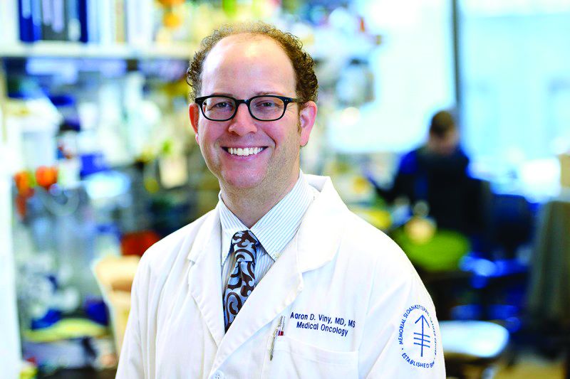
Dr. Viny is with the Memorial Sloan-Kettering Cancer Center, New York, where he is a clinical instructor, on the staff of the leukemia service, and a clinical researcher in The Ross Levine Lab. Contact Dr. Viny at vinya@mskcc.org.
In hematologic malignancies, there is a deep and direct connection between each individual patient, that patient’s symptoms, the visible cells that cause the disease, and the direct measurements and assessments of those cells. The totality of these factors helps to determine the diagnosis and treatment plan. As a butterfly needle often is sufficient for obtaining a diagnostic tumor biopsy, it is not surprising that these same diagnostic techniques are now standardly being used to monitor disease response.
The techniques differ in their limits of detection, however. With sequencing depths able to reliably detect variant allele frequencies of less than 10%, even when patients’ overt leukemia may no longer be detectable, clinicians may be left to ponder what to do with persistent “preleukemic” or “rising clones.”1-3
These patients, now more appropriately stratified for risk of recurrence, are in desperate need of better care algorithms. Standard MRD assessment by flow cytometric analysis is able to detect less than 1 x 10⁻⁴ cells. While it can be applied to most patients, its sensitivity will likely be surpassed by new and emerging genomic assays. Real time quantitative polymerase chain reaction (RT-qPCR) and next generation sequencing (NGS) require a leukemia-specific abnormality but have the potential for far greater sensitivity with deeper sequencing techniques.
Long-term follow up data in acute promyelocytic leukemia (APL) provides the illustrative example where morphologic remission is not durable in the setting of a persistent PML-RARa transcript and therapeutic goals for PCR negativity irrespective of morphology are standard. Pathologic fusion proteins are ideal for marker-driven therapy, but are found in only about 50% of patients, mainly those with APL and Philadelphia chromosome-positive leukemias.
With driver mutations identified in the majority of patients, we can be hopeful that NGS negativity may be a useful clinical endpoint. In work presented at ASH 2016 by Bartlomiej M Getta, MBBS, of Memorial Sloan Kettering Cancer Center, New York, and his colleagues, patients with concordant MRD positivity by flow cytometry and NGS had inferior outcomes, even after allogeneic transplant, compared to patients with MRD positivity on one assay but not both.5 Nonetheless, caution should be taken in early adoption of NGS as a independent marker of MRD status for two main reasons: 1) False positives and lack of standardization make current interpretation difficult. 2) The presence of “preleukemic” clones remains enigmatic – and no matter the nomenclature used, can a DNMT3A or IDH-mutant clone really be deemed “clonal hematopoiesis of indeterminate potential” when a patient has already had clonal transformation?
Conversely, not all patients reported in the work by Klco2 and Getta ultimately relapse. Thus, while it would be preferred to clear all mutant clones, as a therapeutic goal this likely would subject many patients to unnecessary toxicity. One half of the patients reported by Getta were disease free at a year with concordant flow and NGS positive MRD while patients with NGS positivity alone had outcomes equivalent to those of MRD-negative patients, highlighting that certain persistent clones in NGS-only, MRD-positive patients might be amenable to immunotherapy, either with checkpoint inhibitors or allogeneic transplant. Insight into which clones remain quiescent and which are more sinister will require more investigation, but there does seem to be an additive role to NGS-positivity, whereby all MRD is not created equal and the precision and clinical utility of MRD status will likely take on nuanced nomenclature.
References
1. Jan, M. et al. Clonal evolution of preleukemic hematopoietic stem cells precedes human acute myeloid leukemia. Science Translational Medicine 4, 149ra118, doi: 10.1126/scitranslmed.3004315 (2012).
2. Klco, J. M. et al. Association Between Mutation Clearance After Induction Therapy and Outcomes in Acute Myeloid Leukemia. JAMA 2015;314:811-22. doi: 10.1001/jama.2015.9643.
3. Wong, T. N. et al. Rapid expansion of preexisting nonleukemic hematopoietic clones frequently follows induction therapy for de novo AML. Blood 2016;127:893-7. doi: 10.1182/blood-2015-10-677021 (2016).
4. Lane, A. A. et al. Results from Ongoing Phase II Trial of SL-401 As Consolidation Therapy in Patients with Acute Myeloid Leukemia (AML) in Remission with High Relapse Risk Including Minimal Residual Disease (MRD), Abstract 215, ASH 2016.
5. Getta, B. M. et al. Multicolor Flow Cytometry and Multi-Gene Next Generation Sequencing Are Complimentary and Highly Predictive for Relapse in Acute Myeloid Leukemia Following Allogeneic Hematopoietic Stem Cell Transplant, Abstract 834, ASH 2016.
Dr. Viny is with the Memorial Sloan-Kettering Cancer Center, New York, where he is a clinical instructor, on the staff of the leukemia service, and a clinical researcher in The Ross Levine Lab. Contact Dr. Viny at vinya@mskcc.org.
In hematologic malignancies, there is a deep and direct connection between each individual patient, that patient’s symptoms, the visible cells that cause the disease, and the direct measurements and assessments of those cells. The totality of these factors helps to determine the diagnosis and treatment plan. As a butterfly needle often is sufficient for obtaining a diagnostic tumor biopsy, it is not surprising that these same diagnostic techniques are now standardly being used to monitor disease response.
The techniques differ in their limits of detection, however. With sequencing depths able to reliably detect variant allele frequencies of less than 10%, even when patients’ overt leukemia may no longer be detectable, clinicians may be left to ponder what to do with persistent “preleukemic” or “rising clones.”1-3
These patients, now more appropriately stratified for risk of recurrence, are in desperate need of better care algorithms. Standard MRD assessment by flow cytometric analysis is able to detect less than 1 x 10⁻⁴ cells. While it can be applied to most patients, its sensitivity will likely be surpassed by new and emerging genomic assays. Real time quantitative polymerase chain reaction (RT-qPCR) and next generation sequencing (NGS) require a leukemia-specific abnormality but have the potential for far greater sensitivity with deeper sequencing techniques.
Long-term follow up data in acute promyelocytic leukemia (APL) provides the illustrative example where morphologic remission is not durable in the setting of a persistent PML-RARa transcript and therapeutic goals for PCR negativity irrespective of morphology are standard. Pathologic fusion proteins are ideal for marker-driven therapy, but are found in only about 50% of patients, mainly those with APL and Philadelphia chromosome-positive leukemias.
With driver mutations identified in the majority of patients, we can be hopeful that NGS negativity may be a useful clinical endpoint. In work presented at ASH 2016 by Bartlomiej M Getta, MBBS, of Memorial Sloan Kettering Cancer Center, New York, and his colleagues, patients with concordant MRD positivity by flow cytometry and NGS had inferior outcomes, even after allogeneic transplant, compared to patients with MRD positivity on one assay but not both.5 Nonetheless, caution should be taken in early adoption of NGS as a independent marker of MRD status for two main reasons: 1) False positives and lack of standardization make current interpretation difficult. 2) The presence of “preleukemic” clones remains enigmatic – and no matter the nomenclature used, can a DNMT3A or IDH-mutant clone really be deemed “clonal hematopoiesis of indeterminate potential” when a patient has already had clonal transformation?
Conversely, not all patients reported in the work by Klco2 and Getta ultimately relapse. Thus, while it would be preferred to clear all mutant clones, as a therapeutic goal this likely would subject many patients to unnecessary toxicity. One half of the patients reported by Getta were disease free at a year with concordant flow and NGS positive MRD while patients with NGS positivity alone had outcomes equivalent to those of MRD-negative patients, highlighting that certain persistent clones in NGS-only, MRD-positive patients might be amenable to immunotherapy, either with checkpoint inhibitors or allogeneic transplant. Insight into which clones remain quiescent and which are more sinister will require more investigation, but there does seem to be an additive role to NGS-positivity, whereby all MRD is not created equal and the precision and clinical utility of MRD status will likely take on nuanced nomenclature.
References
1. Jan, M. et al. Clonal evolution of preleukemic hematopoietic stem cells precedes human acute myeloid leukemia. Science Translational Medicine 4, 149ra118, doi: 10.1126/scitranslmed.3004315 (2012).
2. Klco, J. M. et al. Association Between Mutation Clearance After Induction Therapy and Outcomes in Acute Myeloid Leukemia. JAMA 2015;314:811-22. doi: 10.1001/jama.2015.9643.
3. Wong, T. N. et al. Rapid expansion of preexisting nonleukemic hematopoietic clones frequently follows induction therapy for de novo AML. Blood 2016;127:893-7. doi: 10.1182/blood-2015-10-677021 (2016).
4. Lane, A. A. et al. Results from Ongoing Phase II Trial of SL-401 As Consolidation Therapy in Patients with Acute Myeloid Leukemia (AML) in Remission with High Relapse Risk Including Minimal Residual Disease (MRD), Abstract 215, ASH 2016.
5. Getta, B. M. et al. Multicolor Flow Cytometry and Multi-Gene Next Generation Sequencing Are Complimentary and Highly Predictive for Relapse in Acute Myeloid Leukemia Following Allogeneic Hematopoietic Stem Cell Transplant, Abstract 834, ASH 2016.
Dr. Viny is with the Memorial Sloan-Kettering Cancer Center, New York, where he is a clinical instructor, on the staff of the leukemia service, and a clinical researcher in The Ross Levine Lab. Contact Dr. Viny at vinya@mskcc.org.
Intermittent fasting fights ALL, not AML, in mice

Photo by Steve Berger
Intermittent fasting inhibits the development and progression of acute lymphoblastic leukemia (ALL), according to preclinical research published in Nature Medicine.
Fasting had an inhibitory effect in mouse models of T-cell and B-cell ALL but not acute myeloid leukemia (AML).
“This study using mouse models indicates that the effects of fasting on blood cancers are type-dependent and provides a platform for identifying new targets for leukemia treatments,” said study author Chengcheng “Alec” Zhang, PhD, of UT Southwestern Medical Center in Dallas, Texas.
“We also identified a mechanism responsible for the differing response to the fasting treatment.”
For this study, Dr Zhang and his colleagues created mouse models of acute leukemia—N-Myc B-ALL, activated Notch1 T-ALL, MLL-AF9 AML, and AML driven by the AML1-Eto9a oncogene—and tested the effects of various dietary restriction plans.
The team used green or yellow florescent proteins to mark and trace the leukemia cells so they could determine if the cells’ levels rose or fell in response to the fasting treatment.
“Strikingly, we found that, in models of ALL, a regimen consisting of 6 cycles of 1 day of fasting followed by 1 day of feeding completely inhibited cancer development,” Dr Zhang said.
At the end of 7 weeks, fasted mice with B-ALL had virtually no detectible cancerous cells—an average of 0.48%—compared to an average of 67.68% of cells found to be cancerous in the test areas of the non-fasted B-ALL mice.
Dr Zhang noted that, compared to B-ALL mice that ate normally, the mice on alternate-day fasting had dramatic reductions in the percentage of ALL cells in the bone marrow and spleen, as well as reduced numbers of white blood cells.
In addition, the spleens and lymph nodes in the fasted mice with B-ALL were similar in size to those of normal mice.
“Although initially cancerous, the few fluorescent cells that remained in the fasted mice after 7 weeks appeared to behave like normal cells,” Dr Zhang said. “Mice in the [B-ALL] model group that ate normally died within 59 days, while 75% of the fasted mice survived more than 120 days without signs of leukemia.”
Dr Zhang and his colleagues said they observed similar results in the T-ALL model but not the AML models. There was no decrease in leukemia cells among fasted mice with AML. And fasting actually shortened survival time in these mice.
Identifying the mechanism
Fasting is known to reduce the level of leptin, a cell signaling molecule created by fat tissue. In addition, previous studies have shown weakened activity by leptin receptors in humans with ALL. For those reasons, the researchers studied both leptin levels and leptin receptors in the mouse models.
The team found that mice with ALL showed reduced leptin receptor activity that increased with intermittent fasting.
“We found that fasting decreased the levels of leptin circulating in the bloodstream as well as decreased the leptin levels in the bone marrow,” Dr Zhang said. “These effects became more pronounced with repeated cycles of fasting. After fasting, the rate at which the leptin levels recovered seemed to correspond to the rate at which the cancerous ALL cells were cleared from the blood.”
The researchers also found that AML was associated with higher levels of leptin receptors that were unaffected by fasting, which could help explain why the fasting treatment was ineffective against this type of leukemia.
It also suggests a mechanism—the leptin receptor pathway—by which fasting exerts its effects in ALL, Dr Zhang said.
“It will be important to determine whether ALL cells can become resistant to the effects of fasting,” he noted. “It also will be interesting to investigate whether we can find alternative ways that mimic fasting to block ALL development.”
Given that this study did not involve drug treatment, researchers are discussing with clinicians whether the tested regimen might be able to move forward quickly to clinical trials.
Photo by Steve Berger
Intermittent fasting inhibits the development and progression of acute lymphoblastic leukemia (ALL), according to preclinical research published in Nature Medicine.
Fasting had an inhibitory effect in mouse models of T-cell and B-cell ALL but not acute myeloid leukemia (AML).
“This study using mouse models indicates that the effects of fasting on blood cancers are type-dependent and provides a platform for identifying new targets for leukemia treatments,” said study author Chengcheng “Alec” Zhang, PhD, of UT Southwestern Medical Center in Dallas, Texas.
“We also identified a mechanism responsible for the differing response to the fasting treatment.”
For this study, Dr Zhang and his colleagues created mouse models of acute leukemia—N-Myc B-ALL, activated Notch1 T-ALL, MLL-AF9 AML, and AML driven by the AML1-Eto9a oncogene—and tested the effects of various dietary restriction plans.
The team used green or yellow florescent proteins to mark and trace the leukemia cells so they could determine if the cells’ levels rose or fell in response to the fasting treatment.
“Strikingly, we found that, in models of ALL, a regimen consisting of 6 cycles of 1 day of fasting followed by 1 day of feeding completely inhibited cancer development,” Dr Zhang said.
At the end of 7 weeks, fasted mice with B-ALL had virtually no detectible cancerous cells—an average of 0.48%—compared to an average of 67.68% of cells found to be cancerous in the test areas of the non-fasted B-ALL mice.
Dr Zhang noted that, compared to B-ALL mice that ate normally, the mice on alternate-day fasting had dramatic reductions in the percentage of ALL cells in the bone marrow and spleen, as well as reduced numbers of white blood cells.
In addition, the spleens and lymph nodes in the fasted mice with B-ALL were similar in size to those of normal mice.
“Although initially cancerous, the few fluorescent cells that remained in the fasted mice after 7 weeks appeared to behave like normal cells,” Dr Zhang said. “Mice in the [B-ALL] model group that ate normally died within 59 days, while 75% of the fasted mice survived more than 120 days without signs of leukemia.”
Dr Zhang and his colleagues said they observed similar results in the T-ALL model but not the AML models. There was no decrease in leukemia cells among fasted mice with AML. And fasting actually shortened survival time in these mice.
Identifying the mechanism
Fasting is known to reduce the level of leptin, a cell signaling molecule created by fat tissue. In addition, previous studies have shown weakened activity by leptin receptors in humans with ALL. For those reasons, the researchers studied both leptin levels and leptin receptors in the mouse models.
The team found that mice with ALL showed reduced leptin receptor activity that increased with intermittent fasting.
“We found that fasting decreased the levels of leptin circulating in the bloodstream as well as decreased the leptin levels in the bone marrow,” Dr Zhang said. “These effects became more pronounced with repeated cycles of fasting. After fasting, the rate at which the leptin levels recovered seemed to correspond to the rate at which the cancerous ALL cells were cleared from the blood.”
The researchers also found that AML was associated with higher levels of leptin receptors that were unaffected by fasting, which could help explain why the fasting treatment was ineffective against this type of leukemia.
It also suggests a mechanism—the leptin receptor pathway—by which fasting exerts its effects in ALL, Dr Zhang said.
“It will be important to determine whether ALL cells can become resistant to the effects of fasting,” he noted. “It also will be interesting to investigate whether we can find alternative ways that mimic fasting to block ALL development.”
Given that this study did not involve drug treatment, researchers are discussing with clinicians whether the tested regimen might be able to move forward quickly to clinical trials.
Photo by Steve Berger
Intermittent fasting inhibits the development and progression of acute lymphoblastic leukemia (ALL), according to preclinical research published in Nature Medicine.
Fasting had an inhibitory effect in mouse models of T-cell and B-cell ALL but not acute myeloid leukemia (AML).
“This study using mouse models indicates that the effects of fasting on blood cancers are type-dependent and provides a platform for identifying new targets for leukemia treatments,” said study author Chengcheng “Alec” Zhang, PhD, of UT Southwestern Medical Center in Dallas, Texas.
“We also identified a mechanism responsible for the differing response to the fasting treatment.”
For this study, Dr Zhang and his colleagues created mouse models of acute leukemia—N-Myc B-ALL, activated Notch1 T-ALL, MLL-AF9 AML, and AML driven by the AML1-Eto9a oncogene—and tested the effects of various dietary restriction plans.
The team used green or yellow florescent proteins to mark and trace the leukemia cells so they could determine if the cells’ levels rose or fell in response to the fasting treatment.
“Strikingly, we found that, in models of ALL, a regimen consisting of 6 cycles of 1 day of fasting followed by 1 day of feeding completely inhibited cancer development,” Dr Zhang said.
At the end of 7 weeks, fasted mice with B-ALL had virtually no detectible cancerous cells—an average of 0.48%—compared to an average of 67.68% of cells found to be cancerous in the test areas of the non-fasted B-ALL mice.
Dr Zhang noted that, compared to B-ALL mice that ate normally, the mice on alternate-day fasting had dramatic reductions in the percentage of ALL cells in the bone marrow and spleen, as well as reduced numbers of white blood cells.
In addition, the spleens and lymph nodes in the fasted mice with B-ALL were similar in size to those of normal mice.
“Although initially cancerous, the few fluorescent cells that remained in the fasted mice after 7 weeks appeared to behave like normal cells,” Dr Zhang said. “Mice in the [B-ALL] model group that ate normally died within 59 days, while 75% of the fasted mice survived more than 120 days without signs of leukemia.”
Dr Zhang and his colleagues said they observed similar results in the T-ALL model but not the AML models. There was no decrease in leukemia cells among fasted mice with AML. And fasting actually shortened survival time in these mice.
Identifying the mechanism
Fasting is known to reduce the level of leptin, a cell signaling molecule created by fat tissue. In addition, previous studies have shown weakened activity by leptin receptors in humans with ALL. For those reasons, the researchers studied both leptin levels and leptin receptors in the mouse models.
The team found that mice with ALL showed reduced leptin receptor activity that increased with intermittent fasting.
“We found that fasting decreased the levels of leptin circulating in the bloodstream as well as decreased the leptin levels in the bone marrow,” Dr Zhang said. “These effects became more pronounced with repeated cycles of fasting. After fasting, the rate at which the leptin levels recovered seemed to correspond to the rate at which the cancerous ALL cells were cleared from the blood.”
The researchers also found that AML was associated with higher levels of leptin receptors that were unaffected by fasting, which could help explain why the fasting treatment was ineffective against this type of leukemia.
It also suggests a mechanism—the leptin receptor pathway—by which fasting exerts its effects in ALL, Dr Zhang said.
“It will be important to determine whether ALL cells can become resistant to the effects of fasting,” he noted. “It also will be interesting to investigate whether we can find alternative ways that mimic fasting to block ALL development.”
Given that this study did not involve drug treatment, researchers are discussing with clinicians whether the tested regimen might be able to move forward quickly to clinical trials.
Novel trial aims to BEAT AML
SAN DIEGO – A multi-arm clinical trial aims to transform the treatment of acute myeloid leukemia, a deadly blood cancer whose standard of care has remained essentially unchanged for 4 decades.
Launched in October 2016, the multicenter BEAT AML Master Trial is based on a simple but radical goal – turn around genomic tests of bone marrow biopsies within 7 days to allow targeted therapy, said lead investigator Brian Druker, MD, of Oregon Health and Science University Knight Cancer Institute in Portland.

Speaking at a press conference at the annual meeting of the American Society of Hematology, he emphasized that rapid, accurate genomic testing is the only way to prescribe targeted agents for AML in time for them to help patients. “It really is about matching the right patient with the right drug,” he said. He also spoke about AML in a video interview at the conference.
That is a major departure from the current approach to treating AML, in which patients receive standard chemotherapy regimens that are toxic and largely ineffective. “Patients themselves call this barbaric therapy,” said John Byrd, MD, who is co-leading the trial on behalf of the Ohio State University Wexner Medical Center in Columbus. “In this trial, we’re going to move away from toxic therapy that is not potentially curative to give more targeted medicine instead.”
In addition to Dr. Druker’s and Dr. Byrd’s centers, Memorial Sloan Kettering Cancer Center, New York, and Dana-Farber Cancer Institute and Massachusetts General Hospital, both in Boston, are onboard for the study. The lead investigators hope to add another six centers to the study group and to have 10 arms of the study underway by mid-2017.
Older patients with AML find chemotherapy especially hard to tolerate and typically respond poorly. Accordingly, the trial will enroll those aged 60 years and up regardless of their genomic profile, the researchers said. Patients lacking targetable markers will be offered investigational therapies showing broad activity in AML.
Another complexity of AML is that any patient can have a variety of mutations, including some affecting only a small subset of leukemia cells, Dr. Byrd noted. Targeting those mutations cannot eradicate disease, but past trials did not rank or choose therapies based on mutation prevalence. Thus, this trial is the first to ask “which mutation is in all of the cells, which gives you the opportunity to get rid of all the disease,” he emphasized. Again, patients – not individual markers or agents – are the priority.
The study also is meant to be nimble – arms can be quickly opened or closed if bench or clinical data are promising or lackluster. This design does not preclude FDA approvals, said Louis J. DeGennaro, PhD, of the Leukemia and Lymphoma Society, which is sponsoring the trial. “We have worked closely with FDA to design a unique protocol that we believe will change the paradigm of AML treatment and future clinical trials,” he added. “This is an unprecedented collaboration.”
Dr. Druker agreed. “If we do this correctly, we can potentially see large effects, and that can become the impetus for rapid FDA approval of these drugs for the right patients,” he said. “That one of the things this trial is designed to do.”
Dr. DeGennaro is president and chief executive officer of the Leukemia and Lymphoma Society, which is sponsoring the BEAT AML Master trial. Dr. Druker disclosed ties to a number of pharmaceutical companies. Dr. Byrd had no relevant disclosures.
The video associated with this article is no longer available on this site. Please view all of our videos on the MDedge YouTube channel
SAN DIEGO – A multi-arm clinical trial aims to transform the treatment of acute myeloid leukemia, a deadly blood cancer whose standard of care has remained essentially unchanged for 4 decades.
Launched in October 2016, the multicenter BEAT AML Master Trial is based on a simple but radical goal – turn around genomic tests of bone marrow biopsies within 7 days to allow targeted therapy, said lead investigator Brian Druker, MD, of Oregon Health and Science University Knight Cancer Institute in Portland.
Speaking at a press conference at the annual meeting of the American Society of Hematology, he emphasized that rapid, accurate genomic testing is the only way to prescribe targeted agents for AML in time for them to help patients. “It really is about matching the right patient with the right drug,” he said. He also spoke about AML in a video interview at the conference.
That is a major departure from the current approach to treating AML, in which patients receive standard chemotherapy regimens that are toxic and largely ineffective. “Patients themselves call this barbaric therapy,” said John Byrd, MD, who is co-leading the trial on behalf of the Ohio State University Wexner Medical Center in Columbus. “In this trial, we’re going to move away from toxic therapy that is not potentially curative to give more targeted medicine instead.”
In addition to Dr. Druker’s and Dr. Byrd’s centers, Memorial Sloan Kettering Cancer Center, New York, and Dana-Farber Cancer Institute and Massachusetts General Hospital, both in Boston, are onboard for the study. The lead investigators hope to add another six centers to the study group and to have 10 arms of the study underway by mid-2017.
Older patients with AML find chemotherapy especially hard to tolerate and typically respond poorly. Accordingly, the trial will enroll those aged 60 years and up regardless of their genomic profile, the researchers said. Patients lacking targetable markers will be offered investigational therapies showing broad activity in AML.
Another complexity of AML is that any patient can have a variety of mutations, including some affecting only a small subset of leukemia cells, Dr. Byrd noted. Targeting those mutations cannot eradicate disease, but past trials did not rank or choose therapies based on mutation prevalence. Thus, this trial is the first to ask “which mutation is in all of the cells, which gives you the opportunity to get rid of all the disease,” he emphasized. Again, patients – not individual markers or agents – are the priority.
The study also is meant to be nimble – arms can be quickly opened or closed if bench or clinical data are promising or lackluster. This design does not preclude FDA approvals, said Louis J. DeGennaro, PhD, of the Leukemia and Lymphoma Society, which is sponsoring the trial. “We have worked closely with FDA to design a unique protocol that we believe will change the paradigm of AML treatment and future clinical trials,” he added. “This is an unprecedented collaboration.”
Dr. Druker agreed. “If we do this correctly, we can potentially see large effects, and that can become the impetus for rapid FDA approval of these drugs for the right patients,” he said. “That one of the things this trial is designed to do.”
Dr. DeGennaro is president and chief executive officer of the Leukemia and Lymphoma Society, which is sponsoring the BEAT AML Master trial. Dr. Druker disclosed ties to a number of pharmaceutical companies. Dr. Byrd had no relevant disclosures.
The video associated with this article is no longer available on this site. Please view all of our videos on the MDedge YouTube channel
SAN DIEGO – A multi-arm clinical trial aims to transform the treatment of acute myeloid leukemia, a deadly blood cancer whose standard of care has remained essentially unchanged for 4 decades.
Launched in October 2016, the multicenter BEAT AML Master Trial is based on a simple but radical goal – turn around genomic tests of bone marrow biopsies within 7 days to allow targeted therapy, said lead investigator Brian Druker, MD, of Oregon Health and Science University Knight Cancer Institute in Portland.
Speaking at a press conference at the annual meeting of the American Society of Hematology, he emphasized that rapid, accurate genomic testing is the only way to prescribe targeted agents for AML in time for them to help patients. “It really is about matching the right patient with the right drug,” he said. He also spoke about AML in a video interview at the conference.
That is a major departure from the current approach to treating AML, in which patients receive standard chemotherapy regimens that are toxic and largely ineffective. “Patients themselves call this barbaric therapy,” said John Byrd, MD, who is co-leading the trial on behalf of the Ohio State University Wexner Medical Center in Columbus. “In this trial, we’re going to move away from toxic therapy that is not potentially curative to give more targeted medicine instead.”
In addition to Dr. Druker’s and Dr. Byrd’s centers, Memorial Sloan Kettering Cancer Center, New York, and Dana-Farber Cancer Institute and Massachusetts General Hospital, both in Boston, are onboard for the study. The lead investigators hope to add another six centers to the study group and to have 10 arms of the study underway by mid-2017.
Older patients with AML find chemotherapy especially hard to tolerate and typically respond poorly. Accordingly, the trial will enroll those aged 60 years and up regardless of their genomic profile, the researchers said. Patients lacking targetable markers will be offered investigational therapies showing broad activity in AML.
Another complexity of AML is that any patient can have a variety of mutations, including some affecting only a small subset of leukemia cells, Dr. Byrd noted. Targeting those mutations cannot eradicate disease, but past trials did not rank or choose therapies based on mutation prevalence. Thus, this trial is the first to ask “which mutation is in all of the cells, which gives you the opportunity to get rid of all the disease,” he emphasized. Again, patients – not individual markers or agents – are the priority.
The study also is meant to be nimble – arms can be quickly opened or closed if bench or clinical data are promising or lackluster. This design does not preclude FDA approvals, said Louis J. DeGennaro, PhD, of the Leukemia and Lymphoma Society, which is sponsoring the trial. “We have worked closely with FDA to design a unique protocol that we believe will change the paradigm of AML treatment and future clinical trials,” he added. “This is an unprecedented collaboration.”
Dr. Druker agreed. “If we do this correctly, we can potentially see large effects, and that can become the impetus for rapid FDA approval of these drugs for the right patients,” he said. “That one of the things this trial is designed to do.”
Dr. DeGennaro is president and chief executive officer of the Leukemia and Lymphoma Society, which is sponsoring the BEAT AML Master trial. Dr. Druker disclosed ties to a number of pharmaceutical companies. Dr. Byrd had no relevant disclosures.
The video associated with this article is no longer available on this site. Please view all of our videos on the MDedge YouTube channel
AT ASH 2016