User login
Abrocitinib emerges as a potential treatment option for moderate-to-severe atopic dermatitis
Key clinical point: Preliminary evidence from a meta-analysis suggests that abrocitinib was significantly beneficial with a tolerable adverse event profile in patients with moderate-to-severe atopic dermatitis (AD).
Major finding: At 12 weeks, patients treated with abrocitinib achieved a significantly higher Investigator’s Global Assessment response vs those treated with placebo (risk ratio [RR], 3.52; P less than .00001). Treatment-emergent adverse effects were mostly mild and manageable, with a higher risk in the abrocitinib vs placebo group (RR, 1.17; P = .002).
Study details: Findings are from a meta-analysis of 4 randomized controlled trials, which assessed clinical outcomes in 1,175 patients with moderate-to-severe AD with inadequate response to topical medications who received abrocitinib 100 or 200 mg and 334 control participants who received placebo.
Disclosures: The study did not receive any funding. No conflict of interests was reported.
Source: Meher BR et al. J Dermatol Treat. 2021 Jul 27. doi: 10.1080/09546634.2021.1961997.
Key clinical point: Preliminary evidence from a meta-analysis suggests that abrocitinib was significantly beneficial with a tolerable adverse event profile in patients with moderate-to-severe atopic dermatitis (AD).
Major finding: At 12 weeks, patients treated with abrocitinib achieved a significantly higher Investigator’s Global Assessment response vs those treated with placebo (risk ratio [RR], 3.52; P less than .00001). Treatment-emergent adverse effects were mostly mild and manageable, with a higher risk in the abrocitinib vs placebo group (RR, 1.17; P = .002).
Study details: Findings are from a meta-analysis of 4 randomized controlled trials, which assessed clinical outcomes in 1,175 patients with moderate-to-severe AD with inadequate response to topical medications who received abrocitinib 100 or 200 mg and 334 control participants who received placebo.
Disclosures: The study did not receive any funding. No conflict of interests was reported.
Source: Meher BR et al. J Dermatol Treat. 2021 Jul 27. doi: 10.1080/09546634.2021.1961997.
Key clinical point: Preliminary evidence from a meta-analysis suggests that abrocitinib was significantly beneficial with a tolerable adverse event profile in patients with moderate-to-severe atopic dermatitis (AD).
Major finding: At 12 weeks, patients treated with abrocitinib achieved a significantly higher Investigator’s Global Assessment response vs those treated with placebo (risk ratio [RR], 3.52; P less than .00001). Treatment-emergent adverse effects were mostly mild and manageable, with a higher risk in the abrocitinib vs placebo group (RR, 1.17; P = .002).
Study details: Findings are from a meta-analysis of 4 randomized controlled trials, which assessed clinical outcomes in 1,175 patients with moderate-to-severe AD with inadequate response to topical medications who received abrocitinib 100 or 200 mg and 334 control participants who received placebo.
Disclosures: The study did not receive any funding. No conflict of interests was reported.
Source: Meher BR et al. J Dermatol Treat. 2021 Jul 27. doi: 10.1080/09546634.2021.1961997.
Atopic dermatitis tied to increased risk for hypertension
Key clinical point: Patients with atopic dermatitis (AD), particularly those with a moderate-to-severe form of the disease, had significantly higher odds of hypertension than healthy controls.
Major finding: Overall, odds of hypertension was significantly higher in patients with AD than healthy controls (odds ratio [OR], 1.16; 95% confidence interval [CI], 1.04-1.30), particularly in those with moderate-to-severe AD (OR, 2.33; 95% CI, 1.10-4.94). Hypertension was mainly reported as an adverse event from cyclosporine A (pooled prevalence, 7.8%).
Study details: Findings are from a meta-analysis of 19 studies involving 269,861 adults with AD and 718,873 healthy controls, including 52,530 children with AD and 340,356 children as healthy controls.
Disclosures: The study did not report any source of funding. No conflict of interests was reported.
Source: Yousaf M et al. Br J Dermatol. 2021 Jul 28. doi: 10.1111/bjd.20661.
Key clinical point: Patients with atopic dermatitis (AD), particularly those with a moderate-to-severe form of the disease, had significantly higher odds of hypertension than healthy controls.
Major finding: Overall, odds of hypertension was significantly higher in patients with AD than healthy controls (odds ratio [OR], 1.16; 95% confidence interval [CI], 1.04-1.30), particularly in those with moderate-to-severe AD (OR, 2.33; 95% CI, 1.10-4.94). Hypertension was mainly reported as an adverse event from cyclosporine A (pooled prevalence, 7.8%).
Study details: Findings are from a meta-analysis of 19 studies involving 269,861 adults with AD and 718,873 healthy controls, including 52,530 children with AD and 340,356 children as healthy controls.
Disclosures: The study did not report any source of funding. No conflict of interests was reported.
Source: Yousaf M et al. Br J Dermatol. 2021 Jul 28. doi: 10.1111/bjd.20661.
Key clinical point: Patients with atopic dermatitis (AD), particularly those with a moderate-to-severe form of the disease, had significantly higher odds of hypertension than healthy controls.
Major finding: Overall, odds of hypertension was significantly higher in patients with AD than healthy controls (odds ratio [OR], 1.16; 95% confidence interval [CI], 1.04-1.30), particularly in those with moderate-to-severe AD (OR, 2.33; 95% CI, 1.10-4.94). Hypertension was mainly reported as an adverse event from cyclosporine A (pooled prevalence, 7.8%).
Study details: Findings are from a meta-analysis of 19 studies involving 269,861 adults with AD and 718,873 healthy controls, including 52,530 children with AD and 340,356 children as healthy controls.
Disclosures: The study did not report any source of funding. No conflict of interests was reported.
Source: Yousaf M et al. Br J Dermatol. 2021 Jul 28. doi: 10.1111/bjd.20661.
Comorbidities account for higher incidence of hospitalization in patients with AD
Key clinical point: Over 20 years of evidence indicates an increased incidence of hospitalizations among patients with atopic dermatitis (AD) in the United States, mainly attributed to increased burden of medical comorbidities rather than AD itself, highlighting the need for increased screening and management of comorbidities.
Major finding: The number of hospitalizations among patients with AD increased from 1.0 to 2.3 per 100,000 persons from 1998 to 2018 (adjusted P-trend less than .0001). However, the proportion of hospitalizations with AD as the principal diagnosis reduced from 11.5% in 1998 to 3.7% in 2018 (adjusted P-trend = .001). A higher proportion of patients reported Charlson Comorbidity Index score of 3 or higher in 2018 vs 1998 (27.8% vs 10.5%; adjusted P-trend less than .0001).
Study details: Findings are from a longitudinal study including 23,410 adults hospitalized with any form of AD between 1998 and 2018.
Disclosures: The study did not report any source of funding. No conflict of interests was reported.
Source: Edigin E et al. J Am Acad Dermatol. 2021 Jul 9. doi: 10.1016/j.jaad.2021.06.882.
Key clinical point: Over 20 years of evidence indicates an increased incidence of hospitalizations among patients with atopic dermatitis (AD) in the United States, mainly attributed to increased burden of medical comorbidities rather than AD itself, highlighting the need for increased screening and management of comorbidities.
Major finding: The number of hospitalizations among patients with AD increased from 1.0 to 2.3 per 100,000 persons from 1998 to 2018 (adjusted P-trend less than .0001). However, the proportion of hospitalizations with AD as the principal diagnosis reduced from 11.5% in 1998 to 3.7% in 2018 (adjusted P-trend = .001). A higher proportion of patients reported Charlson Comorbidity Index score of 3 or higher in 2018 vs 1998 (27.8% vs 10.5%; adjusted P-trend less than .0001).
Study details: Findings are from a longitudinal study including 23,410 adults hospitalized with any form of AD between 1998 and 2018.
Disclosures: The study did not report any source of funding. No conflict of interests was reported.
Source: Edigin E et al. J Am Acad Dermatol. 2021 Jul 9. doi: 10.1016/j.jaad.2021.06.882.
Key clinical point: Over 20 years of evidence indicates an increased incidence of hospitalizations among patients with atopic dermatitis (AD) in the United States, mainly attributed to increased burden of medical comorbidities rather than AD itself, highlighting the need for increased screening and management of comorbidities.
Major finding: The number of hospitalizations among patients with AD increased from 1.0 to 2.3 per 100,000 persons from 1998 to 2018 (adjusted P-trend less than .0001). However, the proportion of hospitalizations with AD as the principal diagnosis reduced from 11.5% in 1998 to 3.7% in 2018 (adjusted P-trend = .001). A higher proportion of patients reported Charlson Comorbidity Index score of 3 or higher in 2018 vs 1998 (27.8% vs 10.5%; adjusted P-trend less than .0001).
Study details: Findings are from a longitudinal study including 23,410 adults hospitalized with any form of AD between 1998 and 2018.
Disclosures: The study did not report any source of funding. No conflict of interests was reported.
Source: Edigin E et al. J Am Acad Dermatol. 2021 Jul 9. doi: 10.1016/j.jaad.2021.06.882.
Methylation on golli-MBP locus serves as an indicator of atopic dermatitis severity in children
Key clinical point: Reduced DNA methylation of golli-myelin basic protein (MBP) locus could serve as an important biomarker to determine the severity of atopic dermatitis (AD) in pediatric patients.
Major finding: Loss of DNA methylation and higher golli-MBP mRNA expression were observed in pediatric patients with AD (both P less than .0001). The score in the intensity of symptoms increased with decreasing methylation levels in cg27400313, the differentially methylated CpG cluster of MBP gene (P = .012).
Study details: The study used a discovery cohort of 24 pediatric patients with AD and 24 control participants to screen for DNA methylation. The results were further validated in an additional cohort of 224 pediatric patients with AD and 44 control participants.
Disclosures: The study was supported by grants from Ministry of Health and Welfare and Chang Gung Memorial Hospital. The authors declared no conflict of interests.
Source: Chen KD et al. J Invest Dermatol. 2021 Jul 19. doi: 10.1016/j.jid.2021.06.025.
Key clinical point: Reduced DNA methylation of golli-myelin basic protein (MBP) locus could serve as an important biomarker to determine the severity of atopic dermatitis (AD) in pediatric patients.
Major finding: Loss of DNA methylation and higher golli-MBP mRNA expression were observed in pediatric patients with AD (both P less than .0001). The score in the intensity of symptoms increased with decreasing methylation levels in cg27400313, the differentially methylated CpG cluster of MBP gene (P = .012).
Study details: The study used a discovery cohort of 24 pediatric patients with AD and 24 control participants to screen for DNA methylation. The results were further validated in an additional cohort of 224 pediatric patients with AD and 44 control participants.
Disclosures: The study was supported by grants from Ministry of Health and Welfare and Chang Gung Memorial Hospital. The authors declared no conflict of interests.
Source: Chen KD et al. J Invest Dermatol. 2021 Jul 19. doi: 10.1016/j.jid.2021.06.025.
Key clinical point: Reduced DNA methylation of golli-myelin basic protein (MBP) locus could serve as an important biomarker to determine the severity of atopic dermatitis (AD) in pediatric patients.
Major finding: Loss of DNA methylation and higher golli-MBP mRNA expression were observed in pediatric patients with AD (both P less than .0001). The score in the intensity of symptoms increased with decreasing methylation levels in cg27400313, the differentially methylated CpG cluster of MBP gene (P = .012).
Study details: The study used a discovery cohort of 24 pediatric patients with AD and 24 control participants to screen for DNA methylation. The results were further validated in an additional cohort of 224 pediatric patients with AD and 44 control participants.
Disclosures: The study was supported by grants from Ministry of Health and Welfare and Chang Gung Memorial Hospital. The authors declared no conflict of interests.
Source: Chen KD et al. J Invest Dermatol. 2021 Jul 19. doi: 10.1016/j.jid.2021.06.025.
Sleep disturbance and geriatric age in atopic dermatitis: Is there a link?
Key clinical point: Geriatric patients experienced more profound sleep disturbance (SD) despite having similar severity of atopic dermatitis (AD) as younger adult patients with AD.
Major finding: Geriatric age was not associated with severity of AD as measured by Eczema Area and Severity Index score (adjusted odds ratio [aOR], 1.47; P = .3269). However, geriatric patients with AD spent an increased number of nights with SD from eczema (aOR, 2.14; P = .0142), experienced fatigue (aOR, 1.81; P = .0313), and had trouble staying asleep (aOR, 2.26; P = .0030).
Study details: Findings are from a cross-sectional, dermatology practice-based study conducted between 2014 and 2019 and included adults diagnosed with AD.
Disclosures: This study was supported by Agency for Healthcare Research and Quality, the Dermatology Foundation, and an unrestricted research grant from Galderma. The authors declared no conflict of interests.
Source: Manjunath J and Silverberg JI. J Am Acad Dermatol. 2021 Jul 29. doi: 10.1016/j.jaad.2021.07.039.
Key clinical point: Geriatric patients experienced more profound sleep disturbance (SD) despite having similar severity of atopic dermatitis (AD) as younger adult patients with AD.
Major finding: Geriatric age was not associated with severity of AD as measured by Eczema Area and Severity Index score (adjusted odds ratio [aOR], 1.47; P = .3269). However, geriatric patients with AD spent an increased number of nights with SD from eczema (aOR, 2.14; P = .0142), experienced fatigue (aOR, 1.81; P = .0313), and had trouble staying asleep (aOR, 2.26; P = .0030).
Study details: Findings are from a cross-sectional, dermatology practice-based study conducted between 2014 and 2019 and included adults diagnosed with AD.
Disclosures: This study was supported by Agency for Healthcare Research and Quality, the Dermatology Foundation, and an unrestricted research grant from Galderma. The authors declared no conflict of interests.
Source: Manjunath J and Silverberg JI. J Am Acad Dermatol. 2021 Jul 29. doi: 10.1016/j.jaad.2021.07.039.
Key clinical point: Geriatric patients experienced more profound sleep disturbance (SD) despite having similar severity of atopic dermatitis (AD) as younger adult patients with AD.
Major finding: Geriatric age was not associated with severity of AD as measured by Eczema Area and Severity Index score (adjusted odds ratio [aOR], 1.47; P = .3269). However, geriatric patients with AD spent an increased number of nights with SD from eczema (aOR, 2.14; P = .0142), experienced fatigue (aOR, 1.81; P = .0313), and had trouble staying asleep (aOR, 2.26; P = .0030).
Study details: Findings are from a cross-sectional, dermatology practice-based study conducted between 2014 and 2019 and included adults diagnosed with AD.
Disclosures: This study was supported by Agency for Healthcare Research and Quality, the Dermatology Foundation, and an unrestricted research grant from Galderma. The authors declared no conflict of interests.
Source: Manjunath J and Silverberg JI. J Am Acad Dermatol. 2021 Jul 29. doi: 10.1016/j.jaad.2021.07.039.
Cold atmospheric plasma alleviates AD severity without any safety issues
Key clinical point: Cold atmospheric plasma (CAP) can potentially improve the clinical severity of atopic dermatitis (AD) by recovering the diversity of skin microbiome and promoting wound healing for damaged skin barriers without any safety issues.
Major finding: At the end of treatment, reduction in Staphylococcus aureus count was significantly higher for the CAP vs sham group (10.14% vs 15.29%; P = .047). In the CAP group, mean modified AD antecubital severity score reduced significantly at week 4 vs baseline (13.12 vs 33.73; P less than .001), whereas reduction in the sham group was not statistically significant (P = .114). No severe adverse events were reported.
Study details: Findings are from a prospective analysis of 22 adults with mild-to-moderate AD having symmetric lesions. For each patient, the symmetric lesions were randomly assigned to either CAP or sham treatment.
Disclosures: The study did not report any source of funding. No conflicts of interest were reported.
Source: Kim YJ et al. Sci Rep. 2021 Jul 14. doi: 10.1038/s41598-021-93941-y.
Key clinical point: Cold atmospheric plasma (CAP) can potentially improve the clinical severity of atopic dermatitis (AD) by recovering the diversity of skin microbiome and promoting wound healing for damaged skin barriers without any safety issues.
Major finding: At the end of treatment, reduction in Staphylococcus aureus count was significantly higher for the CAP vs sham group (10.14% vs 15.29%; P = .047). In the CAP group, mean modified AD antecubital severity score reduced significantly at week 4 vs baseline (13.12 vs 33.73; P less than .001), whereas reduction in the sham group was not statistically significant (P = .114). No severe adverse events were reported.
Study details: Findings are from a prospective analysis of 22 adults with mild-to-moderate AD having symmetric lesions. For each patient, the symmetric lesions were randomly assigned to either CAP or sham treatment.
Disclosures: The study did not report any source of funding. No conflicts of interest were reported.
Source: Kim YJ et al. Sci Rep. 2021 Jul 14. doi: 10.1038/s41598-021-93941-y.
Key clinical point: Cold atmospheric plasma (CAP) can potentially improve the clinical severity of atopic dermatitis (AD) by recovering the diversity of skin microbiome and promoting wound healing for damaged skin barriers without any safety issues.
Major finding: At the end of treatment, reduction in Staphylococcus aureus count was significantly higher for the CAP vs sham group (10.14% vs 15.29%; P = .047). In the CAP group, mean modified AD antecubital severity score reduced significantly at week 4 vs baseline (13.12 vs 33.73; P less than .001), whereas reduction in the sham group was not statistically significant (P = .114). No severe adverse events were reported.
Study details: Findings are from a prospective analysis of 22 adults with mild-to-moderate AD having symmetric lesions. For each patient, the symmetric lesions were randomly assigned to either CAP or sham treatment.
Disclosures: The study did not report any source of funding. No conflicts of interest were reported.
Source: Kim YJ et al. Sci Rep. 2021 Jul 14. doi: 10.1038/s41598-021-93941-y.
Difamilast ointment shows promise for pediatric atopic dermatitis in phase 3
Key clinical point: Difamilast 0.3% and 1% ointments demonstrated superiority over vehicle along with a favorable safety profile when applied twice daily for up to 4 weeks in pediatric patients with atopic dermatitis (AD).
Major finding: At week 4, the success rate in investigator global assessment score was significantly higher with difamilast 0.3% (44.6%; P = .0005) and 1% (47.1%; P less than .0001) vs vehicle (18.1%) group. Treatment-emergent adverse effects were mostly mild or moderate in severity, with adverse event profiles similar between treatment and vehicle groups.
Study details: Findings are from a double-blind phase 3 trial including 251 patients aged 2-14 years with mild-to-moderate AD who were randomly assigned to receive difamilast 0.3%, difamilast 1%, or vehicle ointment twice daily for 4 weeks.
Disclosures: This study was funded by Otsuka Pharmaceutical Co., Ltd. Dr. Saeki and Dr. Baba declared receiving consultation fees from Otsuka Pharmaceutical Co., Ltd. Dr. Ito, Dr. Yokota, and Dr. Tsubouchi declared being employees of Otsuka Pharmaceutical Co., Ltd.
Source: Saeki H et al. Br J Dermatol. 2021 Jul 21. doi: 10.1111/bjd.20655.
Key clinical point: Difamilast 0.3% and 1% ointments demonstrated superiority over vehicle along with a favorable safety profile when applied twice daily for up to 4 weeks in pediatric patients with atopic dermatitis (AD).
Major finding: At week 4, the success rate in investigator global assessment score was significantly higher with difamilast 0.3% (44.6%; P = .0005) and 1% (47.1%; P less than .0001) vs vehicle (18.1%) group. Treatment-emergent adverse effects were mostly mild or moderate in severity, with adverse event profiles similar between treatment and vehicle groups.
Study details: Findings are from a double-blind phase 3 trial including 251 patients aged 2-14 years with mild-to-moderate AD who were randomly assigned to receive difamilast 0.3%, difamilast 1%, or vehicle ointment twice daily for 4 weeks.
Disclosures: This study was funded by Otsuka Pharmaceutical Co., Ltd. Dr. Saeki and Dr. Baba declared receiving consultation fees from Otsuka Pharmaceutical Co., Ltd. Dr. Ito, Dr. Yokota, and Dr. Tsubouchi declared being employees of Otsuka Pharmaceutical Co., Ltd.
Source: Saeki H et al. Br J Dermatol. 2021 Jul 21. doi: 10.1111/bjd.20655.
Key clinical point: Difamilast 0.3% and 1% ointments demonstrated superiority over vehicle along with a favorable safety profile when applied twice daily for up to 4 weeks in pediatric patients with atopic dermatitis (AD).
Major finding: At week 4, the success rate in investigator global assessment score was significantly higher with difamilast 0.3% (44.6%; P = .0005) and 1% (47.1%; P less than .0001) vs vehicle (18.1%) group. Treatment-emergent adverse effects were mostly mild or moderate in severity, with adverse event profiles similar between treatment and vehicle groups.
Study details: Findings are from a double-blind phase 3 trial including 251 patients aged 2-14 years with mild-to-moderate AD who were randomly assigned to receive difamilast 0.3%, difamilast 1%, or vehicle ointment twice daily for 4 weeks.
Disclosures: This study was funded by Otsuka Pharmaceutical Co., Ltd. Dr. Saeki and Dr. Baba declared receiving consultation fees from Otsuka Pharmaceutical Co., Ltd. Dr. Ito, Dr. Yokota, and Dr. Tsubouchi declared being employees of Otsuka Pharmaceutical Co., Ltd.
Source: Saeki H et al. Br J Dermatol. 2021 Jul 21. doi: 10.1111/bjd.20655.
Upadacitinib shows superior efficacy over dupilumab for moderate-to-severe atopic dermatitis in phase 3b
Key clinical point: Upadacitinib demonstrated superior efficacy over dupilumab for treatment of adults with moderate-to-severe atopic dermatitis (AD) with no new safety signals identified.
Major finding: At week 16, a higher proportion of patients achieved 75% improvement in Eczema Area and Severity Index with upadacitinib vs dupilumab (71.0% vs 61.1%; P = .006). Rates of serious treatment-emergent adverse events leading to drug discontinuation in upadacitinib vs dupilumab were 2.9% vs 1.2%, with no new safety risks observed for upadacitinib.
Study details: Findings are 24-week results of Heads up, a phase 3b trial including 692 adults with moderate-to-severe AD who were randomly assigned to receive oral upadacitinib, 30 mg once daily, or subcutaneous dupilumab, 300 mg every alternate week.
Disclosures: This study was funded by AbbVie. The authors declared serving as speaker, consultant, scientific advisor, clinical study investigator, and/or receiving personal fees and grants from various sources including AbbVie. Four authors declared being employees and/or shareholders of AbbVie.
Source: Blauvelt A et al. JAMA Dermatol. 2021 Aug 4. doi: 10.1001/jamadermatol.2021.3023.
Key clinical point: Upadacitinib demonstrated superior efficacy over dupilumab for treatment of adults with moderate-to-severe atopic dermatitis (AD) with no new safety signals identified.
Major finding: At week 16, a higher proportion of patients achieved 75% improvement in Eczema Area and Severity Index with upadacitinib vs dupilumab (71.0% vs 61.1%; P = .006). Rates of serious treatment-emergent adverse events leading to drug discontinuation in upadacitinib vs dupilumab were 2.9% vs 1.2%, with no new safety risks observed for upadacitinib.
Study details: Findings are 24-week results of Heads up, a phase 3b trial including 692 adults with moderate-to-severe AD who were randomly assigned to receive oral upadacitinib, 30 mg once daily, or subcutaneous dupilumab, 300 mg every alternate week.
Disclosures: This study was funded by AbbVie. The authors declared serving as speaker, consultant, scientific advisor, clinical study investigator, and/or receiving personal fees and grants from various sources including AbbVie. Four authors declared being employees and/or shareholders of AbbVie.
Source: Blauvelt A et al. JAMA Dermatol. 2021 Aug 4. doi: 10.1001/jamadermatol.2021.3023.
Key clinical point: Upadacitinib demonstrated superior efficacy over dupilumab for treatment of adults with moderate-to-severe atopic dermatitis (AD) with no new safety signals identified.
Major finding: At week 16, a higher proportion of patients achieved 75% improvement in Eczema Area and Severity Index with upadacitinib vs dupilumab (71.0% vs 61.1%; P = .006). Rates of serious treatment-emergent adverse events leading to drug discontinuation in upadacitinib vs dupilumab were 2.9% vs 1.2%, with no new safety risks observed for upadacitinib.
Study details: Findings are 24-week results of Heads up, a phase 3b trial including 692 adults with moderate-to-severe AD who were randomly assigned to receive oral upadacitinib, 30 mg once daily, or subcutaneous dupilumab, 300 mg every alternate week.
Disclosures: This study was funded by AbbVie. The authors declared serving as speaker, consultant, scientific advisor, clinical study investigator, and/or receiving personal fees and grants from various sources including AbbVie. Four authors declared being employees and/or shareholders of AbbVie.
Source: Blauvelt A et al. JAMA Dermatol. 2021 Aug 4. doi: 10.1001/jamadermatol.2021.3023.
Atopic Dermatitis in Adults
Inflamed skin lesions on legs
Superinfected eczema is an inflammation of the skin accompanied by itchy, weeping, oozing lesions. Secondary infection of the open skin can occur as a result of scratching. In this case, the infection was a consequence of sensitivity to wearing shin pads.
Although superinfected eczema is a complication of eczema, it can produce separate challenges. With damaged protective skin function and disturbance of quantity and quality of skin lipids, patients with eczema may have secondary bacterial infections. Staphylococcus aureus organisms are the most common etiologic agents; up to 90% of patients with eczema have staphylococcal colonization. Streptococcus is less commonly a cause. The progression from colonization to infection often is associated with a flare of eczema, and increased severity of the eczema is associated with more colonization. More erythema may be noted when the infection begins; then, the affected areas become encrusted or show a serous drainage. A clinical clue to superinfection of any kind is when patients stop responding to therapies that they are normally responsive to (eg, topical steroids).
With the recent increase in methicillin-resistant S. aureus (MRSA), treatment of a secondary infection of eczema with these organisms can be tricky. Patients with eczema and secondary bacterial skin infections should be treated with topical steroids or other anti-inflammatory medications and moisturizers to repair the skin barrier. The level of skin colonization with S. aureus is decreased with use of these agents alone. Topical or systemic antibiotics appropriate for specific or community-based sensitivities may be needed in severe infections.
Because of the damaged protective skin function, cutaneous inoculation of herpes simplex virus (HSV) can occur. Eczema herpeticum, or HSV-associated Kaposi varicelliform eruption, describes eczema secondarily infected with HSV (type 1 or type 2). The eczema may become more erythematous; then, vesicles develop that are arranged in a grouped pattern. Accompanying symptoms include fever, malaise, and lymphadenopathy. The condition can be diagnosed by a Tzanck smear (seeking multinucleated giant cells), a fluorescent antibody smear, or culture of a vesicular lesion. Patients with eczema herpeticum should be treated with acyclovir. More severe involvement may require hospitalization and use of systemic antivirals. In addition, topical steroids and moisturizers should be continued to repair the skin barrier.
Children with eczema are more likely than adults to acquire molluscum contagiosum infection, and the disease tends to be widespread. The lesions are generally smooth papules, sometimes with umbilicated skin. The lesions can spread by auto-inoculation to surrounding areas. Molluscum dermatitis accompanies 10% of molluscum lesions, and the dermatitis can be difficult to distinguish from eczema lesions. Untreated, molluscum lesions may resolve on their own; if necessary, the lesions can be treated with cantharidin, liquid nitrogen, or curettage.
Molluscum dermatitis is treated with topical steroids. Early recognition of infections associated with a diagnosis of eczema is critical for timely initiation of appropriate treatment.
Brian S. Kim, MD, Associate Professor, Department of Medicine, Division of Dermatology, Washington University School of Medicine, St. Louis, Missouri
Brian S. Kim, MD, has disclosed no relevant financial relationships.
Superinfected eczema is an inflammation of the skin accompanied by itchy, weeping, oozing lesions. Secondary infection of the open skin can occur as a result of scratching. In this case, the infection was a consequence of sensitivity to wearing shin pads.
Although superinfected eczema is a complication of eczema, it can produce separate challenges. With damaged protective skin function and disturbance of quantity and quality of skin lipids, patients with eczema may have secondary bacterial infections. Staphylococcus aureus organisms are the most common etiologic agents; up to 90% of patients with eczema have staphylococcal colonization. Streptococcus is less commonly a cause. The progression from colonization to infection often is associated with a flare of eczema, and increased severity of the eczema is associated with more colonization. More erythema may be noted when the infection begins; then, the affected areas become encrusted or show a serous drainage. A clinical clue to superinfection of any kind is when patients stop responding to therapies that they are normally responsive to (eg, topical steroids).
With the recent increase in methicillin-resistant S. aureus (MRSA), treatment of a secondary infection of eczema with these organisms can be tricky. Patients with eczema and secondary bacterial skin infections should be treated with topical steroids or other anti-inflammatory medications and moisturizers to repair the skin barrier. The level of skin colonization with S. aureus is decreased with use of these agents alone. Topical or systemic antibiotics appropriate for specific or community-based sensitivities may be needed in severe infections.
Because of the damaged protective skin function, cutaneous inoculation of herpes simplex virus (HSV) can occur. Eczema herpeticum, or HSV-associated Kaposi varicelliform eruption, describes eczema secondarily infected with HSV (type 1 or type 2). The eczema may become more erythematous; then, vesicles develop that are arranged in a grouped pattern. Accompanying symptoms include fever, malaise, and lymphadenopathy. The condition can be diagnosed by a Tzanck smear (seeking multinucleated giant cells), a fluorescent antibody smear, or culture of a vesicular lesion. Patients with eczema herpeticum should be treated with acyclovir. More severe involvement may require hospitalization and use of systemic antivirals. In addition, topical steroids and moisturizers should be continued to repair the skin barrier.
Children with eczema are more likely than adults to acquire molluscum contagiosum infection, and the disease tends to be widespread. The lesions are generally smooth papules, sometimes with umbilicated skin. The lesions can spread by auto-inoculation to surrounding areas. Molluscum dermatitis accompanies 10% of molluscum lesions, and the dermatitis can be difficult to distinguish from eczema lesions. Untreated, molluscum lesions may resolve on their own; if necessary, the lesions can be treated with cantharidin, liquid nitrogen, or curettage.
Molluscum dermatitis is treated with topical steroids. Early recognition of infections associated with a diagnosis of eczema is critical for timely initiation of appropriate treatment.
Brian S. Kim, MD, Associate Professor, Department of Medicine, Division of Dermatology, Washington University School of Medicine, St. Louis, Missouri
Brian S. Kim, MD, has disclosed no relevant financial relationships.
Superinfected eczema is an inflammation of the skin accompanied by itchy, weeping, oozing lesions. Secondary infection of the open skin can occur as a result of scratching. In this case, the infection was a consequence of sensitivity to wearing shin pads.
Although superinfected eczema is a complication of eczema, it can produce separate challenges. With damaged protective skin function and disturbance of quantity and quality of skin lipids, patients with eczema may have secondary bacterial infections. Staphylococcus aureus organisms are the most common etiologic agents; up to 90% of patients with eczema have staphylococcal colonization. Streptococcus is less commonly a cause. The progression from colonization to infection often is associated with a flare of eczema, and increased severity of the eczema is associated with more colonization. More erythema may be noted when the infection begins; then, the affected areas become encrusted or show a serous drainage. A clinical clue to superinfection of any kind is when patients stop responding to therapies that they are normally responsive to (eg, topical steroids).
With the recent increase in methicillin-resistant S. aureus (MRSA), treatment of a secondary infection of eczema with these organisms can be tricky. Patients with eczema and secondary bacterial skin infections should be treated with topical steroids or other anti-inflammatory medications and moisturizers to repair the skin barrier. The level of skin colonization with S. aureus is decreased with use of these agents alone. Topical or systemic antibiotics appropriate for specific or community-based sensitivities may be needed in severe infections.
Because of the damaged protective skin function, cutaneous inoculation of herpes simplex virus (HSV) can occur. Eczema herpeticum, or HSV-associated Kaposi varicelliform eruption, describes eczema secondarily infected with HSV (type 1 or type 2). The eczema may become more erythematous; then, vesicles develop that are arranged in a grouped pattern. Accompanying symptoms include fever, malaise, and lymphadenopathy. The condition can be diagnosed by a Tzanck smear (seeking multinucleated giant cells), a fluorescent antibody smear, or culture of a vesicular lesion. Patients with eczema herpeticum should be treated with acyclovir. More severe involvement may require hospitalization and use of systemic antivirals. In addition, topical steroids and moisturizers should be continued to repair the skin barrier.
Children with eczema are more likely than adults to acquire molluscum contagiosum infection, and the disease tends to be widespread. The lesions are generally smooth papules, sometimes with umbilicated skin. The lesions can spread by auto-inoculation to surrounding areas. Molluscum dermatitis accompanies 10% of molluscum lesions, and the dermatitis can be difficult to distinguish from eczema lesions. Untreated, molluscum lesions may resolve on their own; if necessary, the lesions can be treated with cantharidin, liquid nitrogen, or curettage.
Molluscum dermatitis is treated with topical steroids. Early recognition of infections associated with a diagnosis of eczema is critical for timely initiation of appropriate treatment.
Brian S. Kim, MD, Associate Professor, Department of Medicine, Division of Dermatology, Washington University School of Medicine, St. Louis, Missouri
Brian S. Kim, MD, has disclosed no relevant financial relationships.
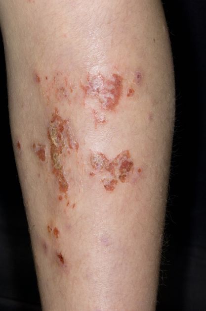
An 11-year-old boy presents with diffuse, inflamed lesions on both shins. His mother first noticed the abrasions when he was scratching them to the point where they bled. He doesn't remember any insect bites, but as a catcher on his Little League baseball team, he noticed that the lesions are always worse after a game and after practice. He is generally healthy and of normal weight for his height. He has no history of allergies or asthma.