User login
ASTRO: Combined radioimmunotherapy proves promising in metastatic melanoma
SAN ANTONIO – The combination of the checkpoint inhibitor ipilimumab with palliative doses of radiation therapy was not associated with any unexpected toxicities, and half of the patients with metastatic melanoma who were treated showed a promising clinical response in a phase II study.
The trial included 22 patients with stage IV disease aged a median of 62 years, of whom 11 (50%) responded to the immunotherapy.

So far 3 (14%) of the 11 patients who responded have achieved a complete response (CR) at a median 55 weeks’ follow-up, and 3 (14%) had a partial response at a median 40 weeks’ follow-up, reported the lead study investigator, Dr. Susan Hiniker of Stanford University, during a press briefing at the annual meeting of the American Society for Radiation Oncology. The remaining five (23%) patients who responded had stable disease at a median 39 weeks’ follow-up.
The trial is one of the first prospective studies to report on the combined use of radiation with systemic immunotherapy in this patient population, she observed, noting that the idea behind the combination was that radiation might enhance the immunogenicity of tumors, that may then in turn make systemic immunotherapy more potent.
“Multiple trials have shown that there is still a minority of patients who respond to ipilimumab, with response rates of approximately 15%”, Dr. Hiniker said.
“As such, new methods of potentiating ipilimumab-induced responses are needed,” she added. “Local irradiation can modulate the local tumor environment such as to promote immune responses in a number of ways,” and other preliminary data have suggested that it may be able to increase overall response rates to ipilimumab.
Patients were recruited for the trial if they had stage IV melanoma and were aged between 18 and 65 years. Treatment consisted of palliative radiation delivered to one or two metastatic sites and at least one nonmetastatic site within 5 days of initial immunotherapy treatment with ipilimumab at a dose of 3 mg/kg every 3 weeks for a total of four treatment cycles. The dose and fractionation schedule was at the discretion of the treating physician.
Tumors were assessed for response using Response Evaluation Criteria in Solid Tumors (RECIST) and Immune Response Criteria (IRC) at baseline, 2-4 weeks after the last dose of ipilimumab was given, and then every 3 months thereafter until disease progression.
This being a phase II trial, the primary objective was to look at the safety of the approach; no additional or unexpected toxicities were seen.
“We saw approximately a 14% rate of grade 3-4 toxicity, which is really on par with what has been reported for ipilimumab monotherapy – on the order of 20%,” Dr. Hiniker said in an interview. “But that being said, we don’t believe that radiation itself caused any additional toxicity.”
One of the patients did have a perforated colon, she acknowledged, and for this reason future research may look at the combination of radiation therapy with the PD-1 (programmed T-cell death) inhibitors.
“I think right now everyone is so excited about the PD-1 inhibitors, and think that they’re safer,” Dr. Hiniker suggested. “It’s hard to give someone ipilimumab alone as the first line at this point; although many patients may not experience toxicity, [in] the ones that do, it can be severe.”
Dr. Hiniker also reported that IRC (independent review committee) analysis results suggested that several proinflammatory cytokines were elevated in some patients who responded to treatment. Higher levels of interleukin 2 at baseline, and at two time points during the study, for example, were associated with better response rates than lower levels. The findings also suggested that there might be a relationship between an increased number of elevated CD8-positive T cells and response.
“I think that we will find biomarkers, but we are not there yet, and it would need to be confirmed in a longer, larger study,” Dr. Hiniker said.
From a practical perspective, if a patient is already being treated with ipilimumab, and a physician is considering radiotherapy, then these results suggest that there will be no additional toxicity, she suggested.
“This combination approach does appear to be promising,” she said. “So for a subset of carefully selected patients – and the problem is we don’t totally know how to select them yet – this can be extremely promising.”
Dr. Hiniker had no disclosures to report.
SAN ANTONIO – The combination of the checkpoint inhibitor ipilimumab with palliative doses of radiation therapy was not associated with any unexpected toxicities, and half of the patients with metastatic melanoma who were treated showed a promising clinical response in a phase II study.
The trial included 22 patients with stage IV disease aged a median of 62 years, of whom 11 (50%) responded to the immunotherapy.
So far 3 (14%) of the 11 patients who responded have achieved a complete response (CR) at a median 55 weeks’ follow-up, and 3 (14%) had a partial response at a median 40 weeks’ follow-up, reported the lead study investigator, Dr. Susan Hiniker of Stanford University, during a press briefing at the annual meeting of the American Society for Radiation Oncology. The remaining five (23%) patients who responded had stable disease at a median 39 weeks’ follow-up.
The trial is one of the first prospective studies to report on the combined use of radiation with systemic immunotherapy in this patient population, she observed, noting that the idea behind the combination was that radiation might enhance the immunogenicity of tumors, that may then in turn make systemic immunotherapy more potent.
“Multiple trials have shown that there is still a minority of patients who respond to ipilimumab, with response rates of approximately 15%”, Dr. Hiniker said.
“As such, new methods of potentiating ipilimumab-induced responses are needed,” she added. “Local irradiation can modulate the local tumor environment such as to promote immune responses in a number of ways,” and other preliminary data have suggested that it may be able to increase overall response rates to ipilimumab.
Patients were recruited for the trial if they had stage IV melanoma and were aged between 18 and 65 years. Treatment consisted of palliative radiation delivered to one or two metastatic sites and at least one nonmetastatic site within 5 days of initial immunotherapy treatment with ipilimumab at a dose of 3 mg/kg every 3 weeks for a total of four treatment cycles. The dose and fractionation schedule was at the discretion of the treating physician.
Tumors were assessed for response using Response Evaluation Criteria in Solid Tumors (RECIST) and Immune Response Criteria (IRC) at baseline, 2-4 weeks after the last dose of ipilimumab was given, and then every 3 months thereafter until disease progression.
This being a phase II trial, the primary objective was to look at the safety of the approach; no additional or unexpected toxicities were seen.
“We saw approximately a 14% rate of grade 3-4 toxicity, which is really on par with what has been reported for ipilimumab monotherapy – on the order of 20%,” Dr. Hiniker said in an interview. “But that being said, we don’t believe that radiation itself caused any additional toxicity.”
One of the patients did have a perforated colon, she acknowledged, and for this reason future research may look at the combination of radiation therapy with the PD-1 (programmed T-cell death) inhibitors.
“I think right now everyone is so excited about the PD-1 inhibitors, and think that they’re safer,” Dr. Hiniker suggested. “It’s hard to give someone ipilimumab alone as the first line at this point; although many patients may not experience toxicity, [in] the ones that do, it can be severe.”
Dr. Hiniker also reported that IRC (independent review committee) analysis results suggested that several proinflammatory cytokines were elevated in some patients who responded to treatment. Higher levels of interleukin 2 at baseline, and at two time points during the study, for example, were associated with better response rates than lower levels. The findings also suggested that there might be a relationship between an increased number of elevated CD8-positive T cells and response.
“I think that we will find biomarkers, but we are not there yet, and it would need to be confirmed in a longer, larger study,” Dr. Hiniker said.
From a practical perspective, if a patient is already being treated with ipilimumab, and a physician is considering radiotherapy, then these results suggest that there will be no additional toxicity, she suggested.
“This combination approach does appear to be promising,” she said. “So for a subset of carefully selected patients – and the problem is we don’t totally know how to select them yet – this can be extremely promising.”
Dr. Hiniker had no disclosures to report.
SAN ANTONIO – The combination of the checkpoint inhibitor ipilimumab with palliative doses of radiation therapy was not associated with any unexpected toxicities, and half of the patients with metastatic melanoma who were treated showed a promising clinical response in a phase II study.
The trial included 22 patients with stage IV disease aged a median of 62 years, of whom 11 (50%) responded to the immunotherapy.
So far 3 (14%) of the 11 patients who responded have achieved a complete response (CR) at a median 55 weeks’ follow-up, and 3 (14%) had a partial response at a median 40 weeks’ follow-up, reported the lead study investigator, Dr. Susan Hiniker of Stanford University, during a press briefing at the annual meeting of the American Society for Radiation Oncology. The remaining five (23%) patients who responded had stable disease at a median 39 weeks’ follow-up.
The trial is one of the first prospective studies to report on the combined use of radiation with systemic immunotherapy in this patient population, she observed, noting that the idea behind the combination was that radiation might enhance the immunogenicity of tumors, that may then in turn make systemic immunotherapy more potent.
“Multiple trials have shown that there is still a minority of patients who respond to ipilimumab, with response rates of approximately 15%”, Dr. Hiniker said.
“As such, new methods of potentiating ipilimumab-induced responses are needed,” she added. “Local irradiation can modulate the local tumor environment such as to promote immune responses in a number of ways,” and other preliminary data have suggested that it may be able to increase overall response rates to ipilimumab.
Patients were recruited for the trial if they had stage IV melanoma and were aged between 18 and 65 years. Treatment consisted of palliative radiation delivered to one or two metastatic sites and at least one nonmetastatic site within 5 days of initial immunotherapy treatment with ipilimumab at a dose of 3 mg/kg every 3 weeks for a total of four treatment cycles. The dose and fractionation schedule was at the discretion of the treating physician.
Tumors were assessed for response using Response Evaluation Criteria in Solid Tumors (RECIST) and Immune Response Criteria (IRC) at baseline, 2-4 weeks after the last dose of ipilimumab was given, and then every 3 months thereafter until disease progression.
This being a phase II trial, the primary objective was to look at the safety of the approach; no additional or unexpected toxicities were seen.
“We saw approximately a 14% rate of grade 3-4 toxicity, which is really on par with what has been reported for ipilimumab monotherapy – on the order of 20%,” Dr. Hiniker said in an interview. “But that being said, we don’t believe that radiation itself caused any additional toxicity.”
One of the patients did have a perforated colon, she acknowledged, and for this reason future research may look at the combination of radiation therapy with the PD-1 (programmed T-cell death) inhibitors.
“I think right now everyone is so excited about the PD-1 inhibitors, and think that they’re safer,” Dr. Hiniker suggested. “It’s hard to give someone ipilimumab alone as the first line at this point; although many patients may not experience toxicity, [in] the ones that do, it can be severe.”
Dr. Hiniker also reported that IRC (independent review committee) analysis results suggested that several proinflammatory cytokines were elevated in some patients who responded to treatment. Higher levels of interleukin 2 at baseline, and at two time points during the study, for example, were associated with better response rates than lower levels. The findings also suggested that there might be a relationship between an increased number of elevated CD8-positive T cells and response.
“I think that we will find biomarkers, but we are not there yet, and it would need to be confirmed in a longer, larger study,” Dr. Hiniker said.
From a practical perspective, if a patient is already being treated with ipilimumab, and a physician is considering radiotherapy, then these results suggest that there will be no additional toxicity, she suggested.
“This combination approach does appear to be promising,” she said. “So for a subset of carefully selected patients – and the problem is we don’t totally know how to select them yet – this can be extremely promising.”
Dr. Hiniker had no disclosures to report.
AT THE ASTRO ANNUAL MEETING
Key clinical point: Some patients with metastatic melanoma may benefit from combined use of novel immunotherapies and radiation treatment.
Major finding: No additional toxicity was seen and 50% of patients had a complete or partial response to the treatment regimen of combined radioimmunotherapy.
Data source: Prospective phase II clinical trial of 22 patients with melanoma treated with ipilimumab and palliative radiation therapy.
Disclosures: Dr. Hiniker had no disclosures to report.

Ipilimumab approved as adjuvant treatment for resected metastatic melanoma
Ipilimumab is now approved as an adjuvant treatment for patients who have undergone a complete resection of metastatic cutaneous melanoma.
The approval is based on data from a large study showing that ipilimumab, marketed as Yervoy by Bristol-Myers Squibb, improved recurrence-free survival, compared with placebo.

“Today’s approval of Yervoy extends its use to patients who are at high risk of developing recurrence of melanoma after surgery,” Dr. Richard Pazdur, director of the office of hematology and oncology products in Food and Drug Administration’s Center for Drug Evaluation and Research, said in the FDA statement announcing the approval on Oct. 28. “This new use of the drug in earlier stages of the disease builds on our understanding of the immune system’s interaction with cancer.”
Ipilimumab, a monoclonal antibody that blocks cytotoxic T-lymphocyte antigen 4 (CTLA-4), was initially approved in 2011 for the treatment of unresectable or metastatic melanoma.
The new indication is for the adjuvant treatment of patients “with cutaneous melanoma with pathologic involvement of regional lymph nodes of more than 1 mm who have undergone complete resection, including total lymphadenectomy.”
In the study of 951 patients, with resected Stage IIIA, IIIB, and IIIC cutaneous melanoma, the median recurrence-free survival was 26 months in the ipilimumab group vs. 17 months in the placebo group; 3-year recurrence-free survival was 46.5% vs. 34.8% (Lancet Oncol. 2015; 16[5]:522-30). The data on overall survival have not yet been analyzed.
The recommended dose and schedule for ipilimumab for adjuvant treatment is 10 mg/kg administered intravenously over 90 minutes every 3 weeks for four doses, followed by 10 mg/kg every 12 weeks for up to 3 years. Doses are omitted, not delayed, if toxicity occurs.
In the study, 52% of those treated with ipilimumab had to discontinue the drug because of adverse reactions. In addition, 41% experienced grade 3-5 immune mediated adverse reactions, including enterocolitis (16%), hepatitis (11%), endocrinopathy (8%), dermatitis (4%), and neuropathy (1.7%).
There were five treatment-related deaths in the study, from immune-mediated adverse reactions, which were enterocolitis (three), Guillain-Barré syndrome (one) and myocarditis (one).The prescribing information includes a boxed warning about the risk of immune-mediated adverse reactions.
In a statement, FDA officials cautioned physicians to carefully monitor patients taking the drug for signs of enterocolitis, dermatitis, neuropathy, and endocrinopathy. Liver function, adrenocorticotropic hormone level, and thyroid function should be measured at baseline and before each dose.
Among the most common reactions were rash, pruritus, diarrhea, nausea, colitis, vomiting, weight loss, fatigue, pyrexia, headache, decreased appetite, and insomnia.
CTLA-4 “may play a role in slowing down or turning off the body’s immune system, and affects its ability to fight off cancerous cells. Yervoy may work by allowing the body’s immune system to recognize, target and attack cells in melanoma tumors,” according to the FDA statement.
Serious adverse events associated with ipilimumab should be reported to the FDA’s MedWatch program or at 800-332-1088.
Ipilimumab is now approved as an adjuvant treatment for patients who have undergone a complete resection of metastatic cutaneous melanoma.
The approval is based on data from a large study showing that ipilimumab, marketed as Yervoy by Bristol-Myers Squibb, improved recurrence-free survival, compared with placebo.
“Today’s approval of Yervoy extends its use to patients who are at high risk of developing recurrence of melanoma after surgery,” Dr. Richard Pazdur, director of the office of hematology and oncology products in Food and Drug Administration’s Center for Drug Evaluation and Research, said in the FDA statement announcing the approval on Oct. 28. “This new use of the drug in earlier stages of the disease builds on our understanding of the immune system’s interaction with cancer.”
Ipilimumab, a monoclonal antibody that blocks cytotoxic T-lymphocyte antigen 4 (CTLA-4), was initially approved in 2011 for the treatment of unresectable or metastatic melanoma.
The new indication is for the adjuvant treatment of patients “with cutaneous melanoma with pathologic involvement of regional lymph nodes of more than 1 mm who have undergone complete resection, including total lymphadenectomy.”
In the study of 951 patients, with resected Stage IIIA, IIIB, and IIIC cutaneous melanoma, the median recurrence-free survival was 26 months in the ipilimumab group vs. 17 months in the placebo group; 3-year recurrence-free survival was 46.5% vs. 34.8% (Lancet Oncol. 2015; 16[5]:522-30). The data on overall survival have not yet been analyzed.
The recommended dose and schedule for ipilimumab for adjuvant treatment is 10 mg/kg administered intravenously over 90 minutes every 3 weeks for four doses, followed by 10 mg/kg every 12 weeks for up to 3 years. Doses are omitted, not delayed, if toxicity occurs.
In the study, 52% of those treated with ipilimumab had to discontinue the drug because of adverse reactions. In addition, 41% experienced grade 3-5 immune mediated adverse reactions, including enterocolitis (16%), hepatitis (11%), endocrinopathy (8%), dermatitis (4%), and neuropathy (1.7%).
There were five treatment-related deaths in the study, from immune-mediated adverse reactions, which were enterocolitis (three), Guillain-Barré syndrome (one) and myocarditis (one).The prescribing information includes a boxed warning about the risk of immune-mediated adverse reactions.
In a statement, FDA officials cautioned physicians to carefully monitor patients taking the drug for signs of enterocolitis, dermatitis, neuropathy, and endocrinopathy. Liver function, adrenocorticotropic hormone level, and thyroid function should be measured at baseline and before each dose.
Among the most common reactions were rash, pruritus, diarrhea, nausea, colitis, vomiting, weight loss, fatigue, pyrexia, headache, decreased appetite, and insomnia.
CTLA-4 “may play a role in slowing down or turning off the body’s immune system, and affects its ability to fight off cancerous cells. Yervoy may work by allowing the body’s immune system to recognize, target and attack cells in melanoma tumors,” according to the FDA statement.
Serious adverse events associated with ipilimumab should be reported to the FDA’s MedWatch program or at 800-332-1088.
Ipilimumab is now approved as an adjuvant treatment for patients who have undergone a complete resection of metastatic cutaneous melanoma.
The approval is based on data from a large study showing that ipilimumab, marketed as Yervoy by Bristol-Myers Squibb, improved recurrence-free survival, compared with placebo.
“Today’s approval of Yervoy extends its use to patients who are at high risk of developing recurrence of melanoma after surgery,” Dr. Richard Pazdur, director of the office of hematology and oncology products in Food and Drug Administration’s Center for Drug Evaluation and Research, said in the FDA statement announcing the approval on Oct. 28. “This new use of the drug in earlier stages of the disease builds on our understanding of the immune system’s interaction with cancer.”
Ipilimumab, a monoclonal antibody that blocks cytotoxic T-lymphocyte antigen 4 (CTLA-4), was initially approved in 2011 for the treatment of unresectable or metastatic melanoma.
The new indication is for the adjuvant treatment of patients “with cutaneous melanoma with pathologic involvement of regional lymph nodes of more than 1 mm who have undergone complete resection, including total lymphadenectomy.”
In the study of 951 patients, with resected Stage IIIA, IIIB, and IIIC cutaneous melanoma, the median recurrence-free survival was 26 months in the ipilimumab group vs. 17 months in the placebo group; 3-year recurrence-free survival was 46.5% vs. 34.8% (Lancet Oncol. 2015; 16[5]:522-30). The data on overall survival have not yet been analyzed.
The recommended dose and schedule for ipilimumab for adjuvant treatment is 10 mg/kg administered intravenously over 90 minutes every 3 weeks for four doses, followed by 10 mg/kg every 12 weeks for up to 3 years. Doses are omitted, not delayed, if toxicity occurs.
In the study, 52% of those treated with ipilimumab had to discontinue the drug because of adverse reactions. In addition, 41% experienced grade 3-5 immune mediated adverse reactions, including enterocolitis (16%), hepatitis (11%), endocrinopathy (8%), dermatitis (4%), and neuropathy (1.7%).
There were five treatment-related deaths in the study, from immune-mediated adverse reactions, which were enterocolitis (three), Guillain-Barré syndrome (one) and myocarditis (one).The prescribing information includes a boxed warning about the risk of immune-mediated adverse reactions.
In a statement, FDA officials cautioned physicians to carefully monitor patients taking the drug for signs of enterocolitis, dermatitis, neuropathy, and endocrinopathy. Liver function, adrenocorticotropic hormone level, and thyroid function should be measured at baseline and before each dose.
Among the most common reactions were rash, pruritus, diarrhea, nausea, colitis, vomiting, weight loss, fatigue, pyrexia, headache, decreased appetite, and insomnia.
CTLA-4 “may play a role in slowing down or turning off the body’s immune system, and affects its ability to fight off cancerous cells. Yervoy may work by allowing the body’s immune system to recognize, target and attack cells in melanoma tumors,” according to the FDA statement.
Serious adverse events associated with ipilimumab should be reported to the FDA’s MedWatch program or at 800-332-1088.

Biopsy-site photography an easy winner on all counts
CHICAGO – Biopsy-site photography appears to reduce the risk of potential wrong-site surgery and can easily be incorporated into dermatology practice, according to Dr. Jeremy Etzkorn.
When Dr. Etzkorn took on this quality improvement initiative on his own, only 5 of 239 routine biopsy-site photographs evaluated were inadequate. The biopsy site was not clearly marked in two photos with multiple suspicious lesions, and anatomic landmarks were absent in three.
“Almost 98% of the time, the photograph was adequate, which just shows it doesn’t require much training or time to get images of the skin,” the Mohs surgeon said at the annual meeting of the American Society for Dermatologic Surgery.
Biopsy-site photos were taken primarily by medical assistants, as well as nurses, who received minimal, informal training on digital photography and were guided to take at least one photograph with anatomic landmarks present.

Dr. Etzkorn of the University of Pennsylvania Health System, Philadelphia, conducted a prospective, observational cohort study of 329 patients/tumors referred for Mohs micrographic surgery or standard excision to the dermatologic surgery unit at Penn Dermatology. Patients were asked to identify their biopsy site, indicate whether they remembered a photo being taken, and quantify on a 10-point scale their level of confidence that the originally biopsied site was treated on the day of surgery.
Dr. Etzkorn identified the biopsy site before consulting the medical record for a biopsy-site photograph. If the photo was absent and he and the patient agreed on the biopsy site, they proceeded to surgery. If there was any disagreement, surgery was postponed and the referring physician consulted.
Overall, 239 patients (73%) had biopsy-site photographs, and 90 patients (27%), referred to the practice before photography was implemented, did not.
In 12.5% of cases, the patient misidentified the biopsy site, and in 6.7% of cases the physician did, which is similar to what has been reported in the literature, Dr. Etzkorn said.
Biopsy-site photography prevented wrong-site surgery in 3 of the 239 cases (1.25%) where these photographs were available. “Without the photo I would normally have done surgery on that site because the patient was confident it was the right site; I was confident it was the right site,” he said.
Importantly, all three lesions were biopsied, and all were squamous cell carcinoma in situ. So while it was the wrong site, the surgery would not have been inappropriate, Dr. Etzkorn noted.
Surgery was postponed to consult the referring physician in 3% of cases (10/329).
Complete patient confidence (10 of 10 points) that the correct site was treated was achieved in 95% of cases, with most of the remaining patients at 9 of 10 points, he said.
Risk factors for patient biopsy-site misidentification were the inability to see the site without a mirror (odds ratio, 3.95; P = .002) and time between the biopsy and surgery (OR, 2.19; P = .028). Prior studies have also shown that difficult-to-visualize sites are associated with biopsy-site misidentification, he noted.
For Dr. Etzkorn, the risk of biopsy-site misidentification quadrupled if there were multiple simultaneous biopsies from different locations (OR, 4.39; P = .003) and tripled with longer time, defined as longer than a 6-week delay vs. a delay of less than 6 weeks between biopsy and surgery (OR, 3.68; P = .007).
A biopsy-site photograph significantly increased the odds that a patient was completely confident the correct site was treated (OR, 5.48; P = .001), as did the use of Mohs surgery vs. excision (OR, 4.87; P = .017).
Once again, time between the biopsy and surgery was a significant risk factor for postponing surgery (OR, 3.52; P = .035), whereas the presence of a biopsy-site photograph cut that risk by almost 13-fold (OR, 12.5: P less than .001), Dr. Etzkorn reported.
“Biopsy-site photography is associated with increased patient confidence that the correct site is treated, decreases in surgical postponement, and the ability to identify wrong-site surgery and prevent it,” he concluded.
Dr. Etzkorn and his coauthor reported having no relevant financial disclosures.
CHICAGO – Biopsy-site photography appears to reduce the risk of potential wrong-site surgery and can easily be incorporated into dermatology practice, according to Dr. Jeremy Etzkorn.
When Dr. Etzkorn took on this quality improvement initiative on his own, only 5 of 239 routine biopsy-site photographs evaluated were inadequate. The biopsy site was not clearly marked in two photos with multiple suspicious lesions, and anatomic landmarks were absent in three.
“Almost 98% of the time, the photograph was adequate, which just shows it doesn’t require much training or time to get images of the skin,” the Mohs surgeon said at the annual meeting of the American Society for Dermatologic Surgery.
Biopsy-site photos were taken primarily by medical assistants, as well as nurses, who received minimal, informal training on digital photography and were guided to take at least one photograph with anatomic landmarks present.
Dr. Etzkorn of the University of Pennsylvania Health System, Philadelphia, conducted a prospective, observational cohort study of 329 patients/tumors referred for Mohs micrographic surgery or standard excision to the dermatologic surgery unit at Penn Dermatology. Patients were asked to identify their biopsy site, indicate whether they remembered a photo being taken, and quantify on a 10-point scale their level of confidence that the originally biopsied site was treated on the day of surgery.
Dr. Etzkorn identified the biopsy site before consulting the medical record for a biopsy-site photograph. If the photo was absent and he and the patient agreed on the biopsy site, they proceeded to surgery. If there was any disagreement, surgery was postponed and the referring physician consulted.
Overall, 239 patients (73%) had biopsy-site photographs, and 90 patients (27%), referred to the practice before photography was implemented, did not.
In 12.5% of cases, the patient misidentified the biopsy site, and in 6.7% of cases the physician did, which is similar to what has been reported in the literature, Dr. Etzkorn said.
Biopsy-site photography prevented wrong-site surgery in 3 of the 239 cases (1.25%) where these photographs were available. “Without the photo I would normally have done surgery on that site because the patient was confident it was the right site; I was confident it was the right site,” he said.
Importantly, all three lesions were biopsied, and all were squamous cell carcinoma in situ. So while it was the wrong site, the surgery would not have been inappropriate, Dr. Etzkorn noted.
Surgery was postponed to consult the referring physician in 3% of cases (10/329).
Complete patient confidence (10 of 10 points) that the correct site was treated was achieved in 95% of cases, with most of the remaining patients at 9 of 10 points, he said.
Risk factors for patient biopsy-site misidentification were the inability to see the site without a mirror (odds ratio, 3.95; P = .002) and time between the biopsy and surgery (OR, 2.19; P = .028). Prior studies have also shown that difficult-to-visualize sites are associated with biopsy-site misidentification, he noted.
For Dr. Etzkorn, the risk of biopsy-site misidentification quadrupled if there were multiple simultaneous biopsies from different locations (OR, 4.39; P = .003) and tripled with longer time, defined as longer than a 6-week delay vs. a delay of less than 6 weeks between biopsy and surgery (OR, 3.68; P = .007).
A biopsy-site photograph significantly increased the odds that a patient was completely confident the correct site was treated (OR, 5.48; P = .001), as did the use of Mohs surgery vs. excision (OR, 4.87; P = .017).
Once again, time between the biopsy and surgery was a significant risk factor for postponing surgery (OR, 3.52; P = .035), whereas the presence of a biopsy-site photograph cut that risk by almost 13-fold (OR, 12.5: P less than .001), Dr. Etzkorn reported.
“Biopsy-site photography is associated with increased patient confidence that the correct site is treated, decreases in surgical postponement, and the ability to identify wrong-site surgery and prevent it,” he concluded.
Dr. Etzkorn and his coauthor reported having no relevant financial disclosures.
CHICAGO – Biopsy-site photography appears to reduce the risk of potential wrong-site surgery and can easily be incorporated into dermatology practice, according to Dr. Jeremy Etzkorn.
When Dr. Etzkorn took on this quality improvement initiative on his own, only 5 of 239 routine biopsy-site photographs evaluated were inadequate. The biopsy site was not clearly marked in two photos with multiple suspicious lesions, and anatomic landmarks were absent in three.
“Almost 98% of the time, the photograph was adequate, which just shows it doesn’t require much training or time to get images of the skin,” the Mohs surgeon said at the annual meeting of the American Society for Dermatologic Surgery.
Biopsy-site photos were taken primarily by medical assistants, as well as nurses, who received minimal, informal training on digital photography and were guided to take at least one photograph with anatomic landmarks present.
Dr. Etzkorn of the University of Pennsylvania Health System, Philadelphia, conducted a prospective, observational cohort study of 329 patients/tumors referred for Mohs micrographic surgery or standard excision to the dermatologic surgery unit at Penn Dermatology. Patients were asked to identify their biopsy site, indicate whether they remembered a photo being taken, and quantify on a 10-point scale their level of confidence that the originally biopsied site was treated on the day of surgery.
Dr. Etzkorn identified the biopsy site before consulting the medical record for a biopsy-site photograph. If the photo was absent and he and the patient agreed on the biopsy site, they proceeded to surgery. If there was any disagreement, surgery was postponed and the referring physician consulted.
Overall, 239 patients (73%) had biopsy-site photographs, and 90 patients (27%), referred to the practice before photography was implemented, did not.
In 12.5% of cases, the patient misidentified the biopsy site, and in 6.7% of cases the physician did, which is similar to what has been reported in the literature, Dr. Etzkorn said.
Biopsy-site photography prevented wrong-site surgery in 3 of the 239 cases (1.25%) where these photographs were available. “Without the photo I would normally have done surgery on that site because the patient was confident it was the right site; I was confident it was the right site,” he said.
Importantly, all three lesions were biopsied, and all were squamous cell carcinoma in situ. So while it was the wrong site, the surgery would not have been inappropriate, Dr. Etzkorn noted.
Surgery was postponed to consult the referring physician in 3% of cases (10/329).
Complete patient confidence (10 of 10 points) that the correct site was treated was achieved in 95% of cases, with most of the remaining patients at 9 of 10 points, he said.
Risk factors for patient biopsy-site misidentification were the inability to see the site without a mirror (odds ratio, 3.95; P = .002) and time between the biopsy and surgery (OR, 2.19; P = .028). Prior studies have also shown that difficult-to-visualize sites are associated with biopsy-site misidentification, he noted.
For Dr. Etzkorn, the risk of biopsy-site misidentification quadrupled if there were multiple simultaneous biopsies from different locations (OR, 4.39; P = .003) and tripled with longer time, defined as longer than a 6-week delay vs. a delay of less than 6 weeks between biopsy and surgery (OR, 3.68; P = .007).
A biopsy-site photograph significantly increased the odds that a patient was completely confident the correct site was treated (OR, 5.48; P = .001), as did the use of Mohs surgery vs. excision (OR, 4.87; P = .017).
Once again, time between the biopsy and surgery was a significant risk factor for postponing surgery (OR, 3.52; P = .035), whereas the presence of a biopsy-site photograph cut that risk by almost 13-fold (OR, 12.5: P less than .001), Dr. Etzkorn reported.
“Biopsy-site photography is associated with increased patient confidence that the correct site is treated, decreases in surgical postponement, and the ability to identify wrong-site surgery and prevent it,” he concluded.
Dr. Etzkorn and his coauthor reported having no relevant financial disclosures.
AT THE ASDS ANNUAL MEETING
Key clinical point: Biopsy-site photography reduces surgical delays, identifies potential wrong-site surgery, and increases patient confidence.
Major finding: Having biopsy-site photos can help prevent wrong-site surgery.
Data source: A prospective, observational cohort study of 329 patients referred for Mohs surgery or standard excision.
Disclosures: Dr. Etzkorn and his coauthor reported having no relevant financial disclosures.

FDA approves T-VEC for metastatic melanoma
The Food and Drug Administration has approved talimogene laherparepvec (T-VEC), an oncolytic immunotherapy, for the local treatment of unresectable cutaneous, subcutaneous, and nodal lesions in patients with melanoma that has recurred after initial surgery, the manufacturer Amgen announced today.
The approval officially makes T-VEC (Imlygic) the first virus-based cancer treatment ever approved, as it is derived from the herpes simplex virus. The FDA’s approval comes just months after an FDA advisory panel voiced their support for T-VEC’s approval in a 22-1 vote, based on a phase III study published in the Journal of Clinical Oncology (2015;33 [25]2780-8).

The study looked at 436 individuals with either stage IIIB/C or stage IV (with limited visceral disease burden considered unresectable) and found that patients treated with T-VEC had a significantly higher overall response rate (26.4%) and complete response rate (10.8%) than did those who were treated with subcutaneous granulocyte macrophage colony-stimulating factor (GM-CSF) (5.7% and 0.7%, respectively).
The durable response rate was also significantly higher among T-VEC–treated patients (16.3%) than among those on GM-CSF (2.1%). T-VEC patients also experienced a median overall survival time of 23.3 months, compared with 18.9 months for those on GM-CSF. In the study, cellulitis was noted in about 2% of patients on T-VEC. Fifty-four percent of T-VEC patients who responded to the treatment initially had disease progression before response began.
“The evidence of local and systemic immune responses with T-VEC supports combinations with other immunotherapies as a rational approach. A phase 1b/2 study of T-VEC and ipilimumab is evaluating the safety and efficacy of this combination,” wrote the investigators of the phase III study, which was led by Dr. Robert Andtbacka of the Huntsman Cancer Institute at the University of Utah, Salt Lake City.
T-VEC was also scrutinized in five other studies, all of which reported that the most common adverse events related to the drug were fatigue, chills, and pyrexia and flulike symptoms; severity of these symptoms ranged from mild to moderate.
The manufacturer intends to make the therapy available to patients almost immediately and anticipates its cost to average $65,000, according to a statement.
The Food and Drug Administration has approved talimogene laherparepvec (T-VEC), an oncolytic immunotherapy, for the local treatment of unresectable cutaneous, subcutaneous, and nodal lesions in patients with melanoma that has recurred after initial surgery, the manufacturer Amgen announced today.
The approval officially makes T-VEC (Imlygic) the first virus-based cancer treatment ever approved, as it is derived from the herpes simplex virus. The FDA’s approval comes just months after an FDA advisory panel voiced their support for T-VEC’s approval in a 22-1 vote, based on a phase III study published in the Journal of Clinical Oncology (2015;33 [25]2780-8).
The study looked at 436 individuals with either stage IIIB/C or stage IV (with limited visceral disease burden considered unresectable) and found that patients treated with T-VEC had a significantly higher overall response rate (26.4%) and complete response rate (10.8%) than did those who were treated with subcutaneous granulocyte macrophage colony-stimulating factor (GM-CSF) (5.7% and 0.7%, respectively).
The durable response rate was also significantly higher among T-VEC–treated patients (16.3%) than among those on GM-CSF (2.1%). T-VEC patients also experienced a median overall survival time of 23.3 months, compared with 18.9 months for those on GM-CSF. In the study, cellulitis was noted in about 2% of patients on T-VEC. Fifty-four percent of T-VEC patients who responded to the treatment initially had disease progression before response began.
“The evidence of local and systemic immune responses with T-VEC supports combinations with other immunotherapies as a rational approach. A phase 1b/2 study of T-VEC and ipilimumab is evaluating the safety and efficacy of this combination,” wrote the investigators of the phase III study, which was led by Dr. Robert Andtbacka of the Huntsman Cancer Institute at the University of Utah, Salt Lake City.
T-VEC was also scrutinized in five other studies, all of which reported that the most common adverse events related to the drug were fatigue, chills, and pyrexia and flulike symptoms; severity of these symptoms ranged from mild to moderate.
The manufacturer intends to make the therapy available to patients almost immediately and anticipates its cost to average $65,000, according to a statement.
The Food and Drug Administration has approved talimogene laherparepvec (T-VEC), an oncolytic immunotherapy, for the local treatment of unresectable cutaneous, subcutaneous, and nodal lesions in patients with melanoma that has recurred after initial surgery, the manufacturer Amgen announced today.
The approval officially makes T-VEC (Imlygic) the first virus-based cancer treatment ever approved, as it is derived from the herpes simplex virus. The FDA’s approval comes just months after an FDA advisory panel voiced their support for T-VEC’s approval in a 22-1 vote, based on a phase III study published in the Journal of Clinical Oncology (2015;33 [25]2780-8).
The study looked at 436 individuals with either stage IIIB/C or stage IV (with limited visceral disease burden considered unresectable) and found that patients treated with T-VEC had a significantly higher overall response rate (26.4%) and complete response rate (10.8%) than did those who were treated with subcutaneous granulocyte macrophage colony-stimulating factor (GM-CSF) (5.7% and 0.7%, respectively).
The durable response rate was also significantly higher among T-VEC–treated patients (16.3%) than among those on GM-CSF (2.1%). T-VEC patients also experienced a median overall survival time of 23.3 months, compared with 18.9 months for those on GM-CSF. In the study, cellulitis was noted in about 2% of patients on T-VEC. Fifty-four percent of T-VEC patients who responded to the treatment initially had disease progression before response began.
“The evidence of local and systemic immune responses with T-VEC supports combinations with other immunotherapies as a rational approach. A phase 1b/2 study of T-VEC and ipilimumab is evaluating the safety and efficacy of this combination,” wrote the investigators of the phase III study, which was led by Dr. Robert Andtbacka of the Huntsman Cancer Institute at the University of Utah, Salt Lake City.
T-VEC was also scrutinized in five other studies, all of which reported that the most common adverse events related to the drug were fatigue, chills, and pyrexia and flulike symptoms; severity of these symptoms ranged from mild to moderate.
The manufacturer intends to make the therapy available to patients almost immediately and anticipates its cost to average $65,000, according to a statement.
FROM THE FDA

Spitzoid melanoma mortality on par with conventional melanoma
CHICAGO – One in four patients with an invasive spitzoid melanoma died from melanoma within 10 years of diagnosis, according to a large SEER database analysis.
The risk of death due to melanoma increased significantly with tumor thickness, metastatic disease, head and neck lesions, male sex, and older age, Dr. Sultan Mirza reported at the annual meeting of the American Society for Dermatologic Surgery.
Spitzoid tumors represent a group of melanocytic neoplasms composed of distinct epithelioid and/or spindle cells and are important because of their mimicry of malignant melanoma.
Although there is a paucity of data, several smaller studies have suggested spitzoid melanomas are less aggressive and have a lower mortality rate when compared with conventional malignant melanoma, he said.
“Our study found spitzoid melanoma–specific mortality to be quite high and is likely to be more on par with the mortality of the conventional malignant melanoma,” Dr. Mirza of Mayo Clinic, Rochester, Minn., said in an interview.
“Given a lack of broadly accepted and well-defined diagnostic criteria, it is our suspicion that many cases of these tumors go unrecognized or are classified under various other types of neoplastic proliferations at the time of diagnosis. This, therefore, hinders our knowledge of the true prognosis of the spitzoid tumors.”
Using the National Cancer Institute’s SEER (Surveillance, Epidemiology, and End Results) database, the investigators identified 2,025 patients who were diagnosed between 1992 and 2010 with an epithelioid/spindle cell melanoma, epithelial cell melanoma, spindle cell melanoma not otherwise specified, or spindle cell melanoma type A or type B. After excluding patients with missing data including microscopic confirmation of malignancy or cause of death, 1,997 patients remained.
Most were white (96%), male (61%), and had invasive disease (98%). Their average age at diagnosis was 64 years, with 21 patients younger than 18 years of age. Median follow-up was 5.6 years.
Of the 1,997 patients, 773 (39%) were deceased at last follow-up. Of these, 331 (43%) died due to metastatic melanoma, Dr. Mirza said. The median time to death was 1.8 years after diagnosis.
Among the 1,956 patients with invasive disease, melanoma cause-specific survival at years 1, 5, 10, and 15 was 95%, 81%, 76%, and 74%, respectively.
Among 41 patients with in situ spitzoid melanoma, the melanoma cause-specific survival was 100% at years 1, 5, and 10.In survival analyses, a Breslow tumor depth of more than 4 mm carried a five times higher risk of melanoma-specific death than a depth of less than 1 mm (hazard ratio, 5.0; P less than .001), Dr. Mirza said.
The risk of death from melanoma increased dramatically as the stage of melanoma advanced from localized (referent) to regional (HR, 4.3; P = .001) and then to distant (HR, 19.4; P = .001).
A similar pattern was observed as patient age increased from 52 years or younger (referent) to 53-66 years (HR, 1.7; P = .001), 67- 77 years (HR, 1.7; P = .001), and finally to age 78 years and older (HR, 2.6; P = .001).
Men were twice as likely to die from melanoma as women (HR, 1.9; P = .001), he said.
The worst anatomic site for a lesion was on the head and scalp, which carried more than twice the risk of melanoma-specific death as lesions on the face and ears (HR, 2.3; P = .001).Limitations of the study were the lack of fluorescence in situ hybridization data, an inability to review and categorize tumor pathology, and confusing terminology regarding spitzoid tumors in the database, resulting in possible inclusion of other types of melanomas, Dr. Mirza said.
This variance of terminology emphasizes the need for standardized nomenclature and once accomplished, will hopefully provide further clarity regarding types of spitzoid growths, so we can know the true prognosis and treatment, he added.
CHICAGO – One in four patients with an invasive spitzoid melanoma died from melanoma within 10 years of diagnosis, according to a large SEER database analysis.
The risk of death due to melanoma increased significantly with tumor thickness, metastatic disease, head and neck lesions, male sex, and older age, Dr. Sultan Mirza reported at the annual meeting of the American Society for Dermatologic Surgery.
Spitzoid tumors represent a group of melanocytic neoplasms composed of distinct epithelioid and/or spindle cells and are important because of their mimicry of malignant melanoma.
Although there is a paucity of data, several smaller studies have suggested spitzoid melanomas are less aggressive and have a lower mortality rate when compared with conventional malignant melanoma, he said.
“Our study found spitzoid melanoma–specific mortality to be quite high and is likely to be more on par with the mortality of the conventional malignant melanoma,” Dr. Mirza of Mayo Clinic, Rochester, Minn., said in an interview.
“Given a lack of broadly accepted and well-defined diagnostic criteria, it is our suspicion that many cases of these tumors go unrecognized or are classified under various other types of neoplastic proliferations at the time of diagnosis. This, therefore, hinders our knowledge of the true prognosis of the spitzoid tumors.”
Using the National Cancer Institute’s SEER (Surveillance, Epidemiology, and End Results) database, the investigators identified 2,025 patients who were diagnosed between 1992 and 2010 with an epithelioid/spindle cell melanoma, epithelial cell melanoma, spindle cell melanoma not otherwise specified, or spindle cell melanoma type A or type B. After excluding patients with missing data including microscopic confirmation of malignancy or cause of death, 1,997 patients remained.
Most were white (96%), male (61%), and had invasive disease (98%). Their average age at diagnosis was 64 years, with 21 patients younger than 18 years of age. Median follow-up was 5.6 years.
Of the 1,997 patients, 773 (39%) were deceased at last follow-up. Of these, 331 (43%) died due to metastatic melanoma, Dr. Mirza said. The median time to death was 1.8 years after diagnosis.
Among the 1,956 patients with invasive disease, melanoma cause-specific survival at years 1, 5, 10, and 15 was 95%, 81%, 76%, and 74%, respectively.
Among 41 patients with in situ spitzoid melanoma, the melanoma cause-specific survival was 100% at years 1, 5, and 10.In survival analyses, a Breslow tumor depth of more than 4 mm carried a five times higher risk of melanoma-specific death than a depth of less than 1 mm (hazard ratio, 5.0; P less than .001), Dr. Mirza said.
The risk of death from melanoma increased dramatically as the stage of melanoma advanced from localized (referent) to regional (HR, 4.3; P = .001) and then to distant (HR, 19.4; P = .001).
A similar pattern was observed as patient age increased from 52 years or younger (referent) to 53-66 years (HR, 1.7; P = .001), 67- 77 years (HR, 1.7; P = .001), and finally to age 78 years and older (HR, 2.6; P = .001).
Men were twice as likely to die from melanoma as women (HR, 1.9; P = .001), he said.
The worst anatomic site for a lesion was on the head and scalp, which carried more than twice the risk of melanoma-specific death as lesions on the face and ears (HR, 2.3; P = .001).Limitations of the study were the lack of fluorescence in situ hybridization data, an inability to review and categorize tumor pathology, and confusing terminology regarding spitzoid tumors in the database, resulting in possible inclusion of other types of melanomas, Dr. Mirza said.
This variance of terminology emphasizes the need for standardized nomenclature and once accomplished, will hopefully provide further clarity regarding types of spitzoid growths, so we can know the true prognosis and treatment, he added.
CHICAGO – One in four patients with an invasive spitzoid melanoma died from melanoma within 10 years of diagnosis, according to a large SEER database analysis.
The risk of death due to melanoma increased significantly with tumor thickness, metastatic disease, head and neck lesions, male sex, and older age, Dr. Sultan Mirza reported at the annual meeting of the American Society for Dermatologic Surgery.
Spitzoid tumors represent a group of melanocytic neoplasms composed of distinct epithelioid and/or spindle cells and are important because of their mimicry of malignant melanoma.
Although there is a paucity of data, several smaller studies have suggested spitzoid melanomas are less aggressive and have a lower mortality rate when compared with conventional malignant melanoma, he said.
“Our study found spitzoid melanoma–specific mortality to be quite high and is likely to be more on par with the mortality of the conventional malignant melanoma,” Dr. Mirza of Mayo Clinic, Rochester, Minn., said in an interview.
“Given a lack of broadly accepted and well-defined diagnostic criteria, it is our suspicion that many cases of these tumors go unrecognized or are classified under various other types of neoplastic proliferations at the time of diagnosis. This, therefore, hinders our knowledge of the true prognosis of the spitzoid tumors.”
Using the National Cancer Institute’s SEER (Surveillance, Epidemiology, and End Results) database, the investigators identified 2,025 patients who were diagnosed between 1992 and 2010 with an epithelioid/spindle cell melanoma, epithelial cell melanoma, spindle cell melanoma not otherwise specified, or spindle cell melanoma type A or type B. After excluding patients with missing data including microscopic confirmation of malignancy or cause of death, 1,997 patients remained.
Most were white (96%), male (61%), and had invasive disease (98%). Their average age at diagnosis was 64 years, with 21 patients younger than 18 years of age. Median follow-up was 5.6 years.
Of the 1,997 patients, 773 (39%) were deceased at last follow-up. Of these, 331 (43%) died due to metastatic melanoma, Dr. Mirza said. The median time to death was 1.8 years after diagnosis.
Among the 1,956 patients with invasive disease, melanoma cause-specific survival at years 1, 5, 10, and 15 was 95%, 81%, 76%, and 74%, respectively.
Among 41 patients with in situ spitzoid melanoma, the melanoma cause-specific survival was 100% at years 1, 5, and 10.In survival analyses, a Breslow tumor depth of more than 4 mm carried a five times higher risk of melanoma-specific death than a depth of less than 1 mm (hazard ratio, 5.0; P less than .001), Dr. Mirza said.
The risk of death from melanoma increased dramatically as the stage of melanoma advanced from localized (referent) to regional (HR, 4.3; P = .001) and then to distant (HR, 19.4; P = .001).
A similar pattern was observed as patient age increased from 52 years or younger (referent) to 53-66 years (HR, 1.7; P = .001), 67- 77 years (HR, 1.7; P = .001), and finally to age 78 years and older (HR, 2.6; P = .001).
Men were twice as likely to die from melanoma as women (HR, 1.9; P = .001), he said.
The worst anatomic site for a lesion was on the head and scalp, which carried more than twice the risk of melanoma-specific death as lesions on the face and ears (HR, 2.3; P = .001).Limitations of the study were the lack of fluorescence in situ hybridization data, an inability to review and categorize tumor pathology, and confusing terminology regarding spitzoid tumors in the database, resulting in possible inclusion of other types of melanomas, Dr. Mirza said.
This variance of terminology emphasizes the need for standardized nomenclature and once accomplished, will hopefully provide further clarity regarding types of spitzoid growths, so we can know the true prognosis and treatment, he added.
AT THE ASDS ANNUAL MEETING
Key clinical point: One-fourth of patients with invasive spitzoid melanoma died from their disease within 10 years of diagnosis.
Major finding: Melanoma-specific mortality was 24% at 10 years.
Data source: SEER database analysis of 1,997 patients with Spitz cutaneous melanoma.
Disclosures: The authors reported having no financial disclosures.
Intermittent interferon falls short for stage III melanoma patients
For patients with resected stage III melanoma, intermittent intravenous high-dose IFN–alpha-2b (iHDI) is not superior to conventional 1-year high-dose IFN–alpha-2b therapy, according to results from a phase III study. Relapse-free survival was significantly inferior with the intermittent regimen.
“These data preclude speculation about a possible equivalence of the regimens. Taken together, the approved schedules for adjuvant IFN [interferon] treatment of patients with melanoma are likely to remain the standard of care until new approaches, such as with ipilimumab or other drugs, show durable improvements in RFS or OS and acceptable safety profiles,” wrote Dr. Peter Mohr of Elbe-Klinikum Buxtehude, Germany, and his colleagues (J Clin. Onc. 2015 Oct. 26. doi: 10.1200/JCO.2015.59.6932).
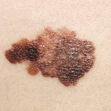
The prospective, multicenter, phase III randomized trial included 649 patients with stage III melanoma who received either three courses of iHDI or the conventional HDI 1-year regimen.After a median follow-up time of 55 months, distant metastasis-free survival and overall survival for the two treatment schedules were similar. The 5-year distant metastasis-free survival for iHDI, compared with HDI, was 49.2% vs. 53.1%; 5-year overall survival was 62.9% vs. 64.1%, respectively. However, relapse-free survival was significantly worse for iHDI than for HDI (hazard ratio, 1.27; 95% confidence interval, 1.02-1.59; P = .03).
Adverse events (AEs) were similar for the two arms, except the HDI group experienced more anemia than the iHDI group (48.1% vs. 30.9%; P less than .001) and neutropenia of grade 3 or higher was more frequent in the iHDI group (38.9% vs. 30.1%; P = .004). The intermittent schedule resulted in fewer cumulative AEs, particularly grade 3 or 4 fatigue. Fatigue is an expected but often debilitating AE of HDI therapy, the investigators noted.
Reduced fatigue was associated with 40% less early discontinuation with iHDI than with standard HDI. In the iHDI group, quality of life measures returned to or exceeded baseline values within 4 weeks of administration. However, improvements in quality of life should be weighed against potential loss of efficacy: at least 50% of patients would tolerate moderate or severe IFN–alpha-2b toxicity for improvement in 5-year, disease-free survival, the investigators said.
For patients with resected stage III melanoma, intermittent intravenous high-dose IFN–alpha-2b (iHDI) is not superior to conventional 1-year high-dose IFN–alpha-2b therapy, according to results from a phase III study. Relapse-free survival was significantly inferior with the intermittent regimen.
“These data preclude speculation about a possible equivalence of the regimens. Taken together, the approved schedules for adjuvant IFN [interferon] treatment of patients with melanoma are likely to remain the standard of care until new approaches, such as with ipilimumab or other drugs, show durable improvements in RFS or OS and acceptable safety profiles,” wrote Dr. Peter Mohr of Elbe-Klinikum Buxtehude, Germany, and his colleagues (J Clin. Onc. 2015 Oct. 26. doi: 10.1200/JCO.2015.59.6932).
The prospective, multicenter, phase III randomized trial included 649 patients with stage III melanoma who received either three courses of iHDI or the conventional HDI 1-year regimen.After a median follow-up time of 55 months, distant metastasis-free survival and overall survival for the two treatment schedules were similar. The 5-year distant metastasis-free survival for iHDI, compared with HDI, was 49.2% vs. 53.1%; 5-year overall survival was 62.9% vs. 64.1%, respectively. However, relapse-free survival was significantly worse for iHDI than for HDI (hazard ratio, 1.27; 95% confidence interval, 1.02-1.59; P = .03).
Adverse events (AEs) were similar for the two arms, except the HDI group experienced more anemia than the iHDI group (48.1% vs. 30.9%; P less than .001) and neutropenia of grade 3 or higher was more frequent in the iHDI group (38.9% vs. 30.1%; P = .004). The intermittent schedule resulted in fewer cumulative AEs, particularly grade 3 or 4 fatigue. Fatigue is an expected but often debilitating AE of HDI therapy, the investigators noted.
Reduced fatigue was associated with 40% less early discontinuation with iHDI than with standard HDI. In the iHDI group, quality of life measures returned to or exceeded baseline values within 4 weeks of administration. However, improvements in quality of life should be weighed against potential loss of efficacy: at least 50% of patients would tolerate moderate or severe IFN–alpha-2b toxicity for improvement in 5-year, disease-free survival, the investigators said.
For patients with resected stage III melanoma, intermittent intravenous high-dose IFN–alpha-2b (iHDI) is not superior to conventional 1-year high-dose IFN–alpha-2b therapy, according to results from a phase III study. Relapse-free survival was significantly inferior with the intermittent regimen.
“These data preclude speculation about a possible equivalence of the regimens. Taken together, the approved schedules for adjuvant IFN [interferon] treatment of patients with melanoma are likely to remain the standard of care until new approaches, such as with ipilimumab or other drugs, show durable improvements in RFS or OS and acceptable safety profiles,” wrote Dr. Peter Mohr of Elbe-Klinikum Buxtehude, Germany, and his colleagues (J Clin. Onc. 2015 Oct. 26. doi: 10.1200/JCO.2015.59.6932).
The prospective, multicenter, phase III randomized trial included 649 patients with stage III melanoma who received either three courses of iHDI or the conventional HDI 1-year regimen.After a median follow-up time of 55 months, distant metastasis-free survival and overall survival for the two treatment schedules were similar. The 5-year distant metastasis-free survival for iHDI, compared with HDI, was 49.2% vs. 53.1%; 5-year overall survival was 62.9% vs. 64.1%, respectively. However, relapse-free survival was significantly worse for iHDI than for HDI (hazard ratio, 1.27; 95% confidence interval, 1.02-1.59; P = .03).
Adverse events (AEs) were similar for the two arms, except the HDI group experienced more anemia than the iHDI group (48.1% vs. 30.9%; P less than .001) and neutropenia of grade 3 or higher was more frequent in the iHDI group (38.9% vs. 30.1%; P = .004). The intermittent schedule resulted in fewer cumulative AEs, particularly grade 3 or 4 fatigue. Fatigue is an expected but often debilitating AE of HDI therapy, the investigators noted.
Reduced fatigue was associated with 40% less early discontinuation with iHDI than with standard HDI. In the iHDI group, quality of life measures returned to or exceeded baseline values within 4 weeks of administration. However, improvements in quality of life should be weighed against potential loss of efficacy: at least 50% of patients would tolerate moderate or severe IFN–alpha-2b toxicity for improvement in 5-year, disease-free survival, the investigators said.
Key clinical point: Distant metastasis-free survival and overall survival were similar in patients with stage III melanoma treated with intermittent high-dose intravenous IFN–alpha-2b (iHDI) vs. standard 1-year high-dose IFN–alpha-2b (HDI), but the difference for relapse-free survival was significant (HR, 1.27; P = .03), favoring standard HDI.
Major finding: The 5-year distant metastasis-free survival for iHDI, compared with HDI was 49.2% vs. 53.1%; 5-year overall survival was 62.9% vs. 64.1%, respectively. Relapse-free survival was significantly worse for iHDI than HDI (HR, 1.27; 95% CI, 1.02-1.59; P = .03).
Data source: The prospective, multicenter, phase III trial randomized 649 patients with stage III melanoma to receive either three courses of iHDI or the conventional HDI 1-year regimen.
Disclosures: Dr. Mohr reported financial relationships with Merck, Bristol-Myers Squibb, Roche, GlaxoSmithKline, LEO Pharma, Novartis, Merck Sharp & Dohme, and Roche. Several of his coauthors reported having ties to industry sources.
Nonmelanoma skin cancer initially misdiagnosed in 36% of small cohort
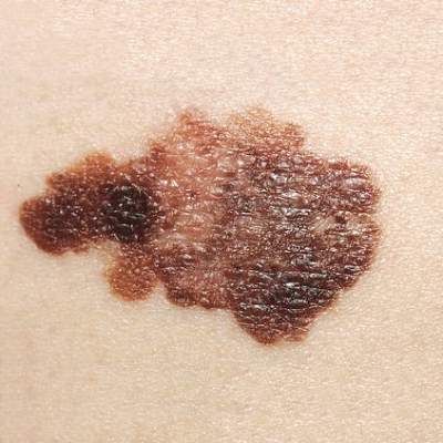
In a small sample study, 10 (36%) of 28 children and young adults diagnosed with nonmelanoma skin cancer were given a misdiagnosis initially, suggesting that young patients with NMSC risk factors may require heightened monitoring from health care providers, according to a study published in Journal of the American Academy of Dermatology (2015 doi: 10.1016/j.jaad.2015.08.007).
In their efforts to identify potential risk factors and gaps in care associated with NMSC in pediatric populations, lead author Hasan Khosravi of Harvard Medical School, Boston, and his associates examined records from 28 patients and 182 occurrences of NMSC, collected from Boston Children’s Hospital between 1993 and 2014.
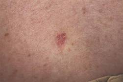
Thirteen (46%) of the 28 pediatric NMSC patients had a history of prolonged immunosuppression, radiation therapy, chemotherapy, voriconazole use, or a combination of these. Among these 28 patients, 19 were diagnosed with basal cell carcinoma (BCC), 7 were diagnosed with squamous cell carcinoma (SCC), and 2 were diagnosed with both BCC and SCC.
The authors noted significant delays in the initial diagnosis in both types of cancer; the mean number of days from time of lesion onset to diagnosis was 667 for SCC and 1,176 for BCC. When misdiagnosed, carcinomas were incorrectly identified as viral wart and graft-versus-host disease for SCC, and psoriasis, acrochordon, wart, nevus, and atypical nevus for BCC.
Even so, interventions were effective in the pediatric population, the authors noted.
“Although most of our patients developed subsequent NMSC after their initial diagnosis, the majority of cases were treated successfully with surgical excision, without recurrence or spread of disease. This suggests that interventions in children and young adults that involve prevention of subsequent disease may be most impactful,” they wrote.
The researchers had no conflicts to declare.
In a small sample study, 10 (36%) of 28 children and young adults diagnosed with nonmelanoma skin cancer were given a misdiagnosis initially, suggesting that young patients with NMSC risk factors may require heightened monitoring from health care providers, according to a study published in Journal of the American Academy of Dermatology (2015 doi: 10.1016/j.jaad.2015.08.007).
In their efforts to identify potential risk factors and gaps in care associated with NMSC in pediatric populations, lead author Hasan Khosravi of Harvard Medical School, Boston, and his associates examined records from 28 patients and 182 occurrences of NMSC, collected from Boston Children’s Hospital between 1993 and 2014.
Thirteen (46%) of the 28 pediatric NMSC patients had a history of prolonged immunosuppression, radiation therapy, chemotherapy, voriconazole use, or a combination of these. Among these 28 patients, 19 were diagnosed with basal cell carcinoma (BCC), 7 were diagnosed with squamous cell carcinoma (SCC), and 2 were diagnosed with both BCC and SCC.
The authors noted significant delays in the initial diagnosis in both types of cancer; the mean number of days from time of lesion onset to diagnosis was 667 for SCC and 1,176 for BCC. When misdiagnosed, carcinomas were incorrectly identified as viral wart and graft-versus-host disease for SCC, and psoriasis, acrochordon, wart, nevus, and atypical nevus for BCC.
Even so, interventions were effective in the pediatric population, the authors noted.
“Although most of our patients developed subsequent NMSC after their initial diagnosis, the majority of cases were treated successfully with surgical excision, without recurrence or spread of disease. This suggests that interventions in children and young adults that involve prevention of subsequent disease may be most impactful,” they wrote.
The researchers had no conflicts to declare.
In a small sample study, 10 (36%) of 28 children and young adults diagnosed with nonmelanoma skin cancer were given a misdiagnosis initially, suggesting that young patients with NMSC risk factors may require heightened monitoring from health care providers, according to a study published in Journal of the American Academy of Dermatology (2015 doi: 10.1016/j.jaad.2015.08.007).
In their efforts to identify potential risk factors and gaps in care associated with NMSC in pediatric populations, lead author Hasan Khosravi of Harvard Medical School, Boston, and his associates examined records from 28 patients and 182 occurrences of NMSC, collected from Boston Children’s Hospital between 1993 and 2014.
Thirteen (46%) of the 28 pediatric NMSC patients had a history of prolonged immunosuppression, radiation therapy, chemotherapy, voriconazole use, or a combination of these. Among these 28 patients, 19 were diagnosed with basal cell carcinoma (BCC), 7 were diagnosed with squamous cell carcinoma (SCC), and 2 were diagnosed with both BCC and SCC.
The authors noted significant delays in the initial diagnosis in both types of cancer; the mean number of days from time of lesion onset to diagnosis was 667 for SCC and 1,176 for BCC. When misdiagnosed, carcinomas were incorrectly identified as viral wart and graft-versus-host disease for SCC, and psoriasis, acrochordon, wart, nevus, and atypical nevus for BCC.
Even so, interventions were effective in the pediatric population, the authors noted.
“Although most of our patients developed subsequent NMSC after their initial diagnosis, the majority of cases were treated successfully with surgical excision, without recurrence or spread of disease. This suggests that interventions in children and young adults that involve prevention of subsequent disease may be most impactful,” they wrote.
The researchers had no conflicts to declare.
FROM JOURNAL OF THE AMERICAN ACADEMY OF DERMATOLOGY
New systemic therapies altering management of melanoma brain metastases
SEATTLE – Targeted therapies for melanoma are improving outcomes for patients with brain metastases and changing management of this dreaded complication, according to Dr. John A. Thompson, codirector of the Melanoma Clinic at the Seattle Cancer Care Alliance and a professor in the medical oncology division at the University of Washington, both in Seattle.

“We are in a new era of treatment of brain metastasis. It’s no longer just surgery and radiation therapy, but a multidisciplinary approach involving medical oncology, radiation oncology, and surgery,” he told attendees of the World Cutaneous Malignancies Congress.
Without question, surgery and radiation therapy techniques have advanced over time, and these modalities remain helpful in achieving local control of disease. Additionally, greater experience with them has improved patient selection.
For example, research has identified several predictors of benefit from stereotactic radiosurgery, such as fewer brain metastases and better performance status (J Neurosurg. 2011;114:769-79). But overall survival was fairly poor given disease elsewhere in the body. “Here’s where I think the new developments in systemic therapy may hopefully be able to start improving this curve,” Dr. Thompson said.
Toxicity of some forms of radiation therapy also remains problematic. For example, a recent randomized phase III trial in patients with up to three brain metastases showed that addition of whole-brain radiation therapy to radiosurgery improved intracranial tumor control, but at the expense of more rapid cognitive decline (ASCO 2015. Abstract LBA4). And there was no gain in overall survival.
“The changes that have happened in systemic therapy over the past 5-10 years have been very exciting,” Dr. Thompson said. By way of example, he pointed to a phase II trial that tested ipilimumab (Yervoy), an antibody to the T-cell receptor cytotoxic T lymphocyte–associated antigen 4 (CTLA4), as induction therapy and then maintenance therapy among patients with melanoma who had brain metastases (Lancet Oncol. 2012;13:459-65).
The subset of patients who were neurologically stable and not on systemic steroids had a disease control rate of 24%, with some having objective responses, including ones in the CNS; the value was lower, at 10%, among patients who were neurologically symptomatic and on steroids, indicating more advanced disease. The 2-year overall survival rate was 26%.
“I think this is encouraging if you compare [these patients] to historical control patients with brain metastases,” said Dr. Thompson, who disclosed that he performs contracted research with Agensys, BMS, Merck, Novartis, and Pfizer.
Another phase II trial, CheckMate 204, which is currently enrolling patients, is taking the concept further, testing the combination of ipilimumab and nivolumab (Opdivo), an antibody to the cell surface receptor programmed death-1 (PD-1), as induction therapy followed by nivolumab maintenance therapy among patients with melanoma who have brain metastases. “This is I think a high-priority trial,” he asserted.
Discussing some cases from his own practice, Dr. Thompson described patient-tailored integration of the new systemic therapies with surgery and radiation therapy.
Oncologists must often deal with toxicities of these therapies as well, such as the colitis and hypophysitis related to ipilimumab therapy, and the development of resistance over time, Dr. Thompson acknowledged.
Nonetheless, the multimodality approach has led to regression and elimination of brain metastases, and even allowed some patients to achieve remission, he reported.
SEATTLE – Targeted therapies for melanoma are improving outcomes for patients with brain metastases and changing management of this dreaded complication, according to Dr. John A. Thompson, codirector of the Melanoma Clinic at the Seattle Cancer Care Alliance and a professor in the medical oncology division at the University of Washington, both in Seattle.
“We are in a new era of treatment of brain metastasis. It’s no longer just surgery and radiation therapy, but a multidisciplinary approach involving medical oncology, radiation oncology, and surgery,” he told attendees of the World Cutaneous Malignancies Congress.
Without question, surgery and radiation therapy techniques have advanced over time, and these modalities remain helpful in achieving local control of disease. Additionally, greater experience with them has improved patient selection.
For example, research has identified several predictors of benefit from stereotactic radiosurgery, such as fewer brain metastases and better performance status (J Neurosurg. 2011;114:769-79). But overall survival was fairly poor given disease elsewhere in the body. “Here’s where I think the new developments in systemic therapy may hopefully be able to start improving this curve,” Dr. Thompson said.
Toxicity of some forms of radiation therapy also remains problematic. For example, a recent randomized phase III trial in patients with up to three brain metastases showed that addition of whole-brain radiation therapy to radiosurgery improved intracranial tumor control, but at the expense of more rapid cognitive decline (ASCO 2015. Abstract LBA4). And there was no gain in overall survival.
“The changes that have happened in systemic therapy over the past 5-10 years have been very exciting,” Dr. Thompson said. By way of example, he pointed to a phase II trial that tested ipilimumab (Yervoy), an antibody to the T-cell receptor cytotoxic T lymphocyte–associated antigen 4 (CTLA4), as induction therapy and then maintenance therapy among patients with melanoma who had brain metastases (Lancet Oncol. 2012;13:459-65).
The subset of patients who were neurologically stable and not on systemic steroids had a disease control rate of 24%, with some having objective responses, including ones in the CNS; the value was lower, at 10%, among patients who were neurologically symptomatic and on steroids, indicating more advanced disease. The 2-year overall survival rate was 26%.
“I think this is encouraging if you compare [these patients] to historical control patients with brain metastases,” said Dr. Thompson, who disclosed that he performs contracted research with Agensys, BMS, Merck, Novartis, and Pfizer.
Another phase II trial, CheckMate 204, which is currently enrolling patients, is taking the concept further, testing the combination of ipilimumab and nivolumab (Opdivo), an antibody to the cell surface receptor programmed death-1 (PD-1), as induction therapy followed by nivolumab maintenance therapy among patients with melanoma who have brain metastases. “This is I think a high-priority trial,” he asserted.
Discussing some cases from his own practice, Dr. Thompson described patient-tailored integration of the new systemic therapies with surgery and radiation therapy.
Oncologists must often deal with toxicities of these therapies as well, such as the colitis and hypophysitis related to ipilimumab therapy, and the development of resistance over time, Dr. Thompson acknowledged.
Nonetheless, the multimodality approach has led to regression and elimination of brain metastases, and even allowed some patients to achieve remission, he reported.
SEATTLE – Targeted therapies for melanoma are improving outcomes for patients with brain metastases and changing management of this dreaded complication, according to Dr. John A. Thompson, codirector of the Melanoma Clinic at the Seattle Cancer Care Alliance and a professor in the medical oncology division at the University of Washington, both in Seattle.
“We are in a new era of treatment of brain metastasis. It’s no longer just surgery and radiation therapy, but a multidisciplinary approach involving medical oncology, radiation oncology, and surgery,” he told attendees of the World Cutaneous Malignancies Congress.
Without question, surgery and radiation therapy techniques have advanced over time, and these modalities remain helpful in achieving local control of disease. Additionally, greater experience with them has improved patient selection.
For example, research has identified several predictors of benefit from stereotactic radiosurgery, such as fewer brain metastases and better performance status (J Neurosurg. 2011;114:769-79). But overall survival was fairly poor given disease elsewhere in the body. “Here’s where I think the new developments in systemic therapy may hopefully be able to start improving this curve,” Dr. Thompson said.
Toxicity of some forms of radiation therapy also remains problematic. For example, a recent randomized phase III trial in patients with up to three brain metastases showed that addition of whole-brain radiation therapy to radiosurgery improved intracranial tumor control, but at the expense of more rapid cognitive decline (ASCO 2015. Abstract LBA4). And there was no gain in overall survival.
“The changes that have happened in systemic therapy over the past 5-10 years have been very exciting,” Dr. Thompson said. By way of example, he pointed to a phase II trial that tested ipilimumab (Yervoy), an antibody to the T-cell receptor cytotoxic T lymphocyte–associated antigen 4 (CTLA4), as induction therapy and then maintenance therapy among patients with melanoma who had brain metastases (Lancet Oncol. 2012;13:459-65).
The subset of patients who were neurologically stable and not on systemic steroids had a disease control rate of 24%, with some having objective responses, including ones in the CNS; the value was lower, at 10%, among patients who were neurologically symptomatic and on steroids, indicating more advanced disease. The 2-year overall survival rate was 26%.
“I think this is encouraging if you compare [these patients] to historical control patients with brain metastases,” said Dr. Thompson, who disclosed that he performs contracted research with Agensys, BMS, Merck, Novartis, and Pfizer.
Another phase II trial, CheckMate 204, which is currently enrolling patients, is taking the concept further, testing the combination of ipilimumab and nivolumab (Opdivo), an antibody to the cell surface receptor programmed death-1 (PD-1), as induction therapy followed by nivolumab maintenance therapy among patients with melanoma who have brain metastases. “This is I think a high-priority trial,” he asserted.
Discussing some cases from his own practice, Dr. Thompson described patient-tailored integration of the new systemic therapies with surgery and radiation therapy.
Oncologists must often deal with toxicities of these therapies as well, such as the colitis and hypophysitis related to ipilimumab therapy, and the development of resistance over time, Dr. Thompson acknowledged.
Nonetheless, the multimodality approach has led to regression and elimination of brain metastases, and even allowed some patients to achieve remission, he reported.
EXPERT ANALYSIS FROM THE WORLD CUTANEOUS MALIGNANCIES CONGRESS

EADV: Prophylactic photodynamic therapy benefits transplant recipients
COPENHAGEN – Twice-yearly prophylactic photodynamic therapy for primary prevention of actinic keratoses and squamous cell carcinomas is a novel and effective strategy that addresses the problem of accelerated photocarcinogenesis in organ transplant recipients, according to an interim analysis of a multinational, randomized, controlled trial.
“The overall aim is to prevent squamous cell carcinoma development. Photodynamic therapy is well established for secondary prevention of further AKs, and these very early data show that it can also be used for primary prevention in very high-risk patients,” Dr. Katrine Togsverd-Bo said at the annual congress of the European Academy of Dermatology and Venereology.

Accelerated carcinogenesis on sun-exposed skin is a major concern in organ transplant recipients (OTRs). They experience early onset of multiple AKs, with field cancerization and up to a 100-fold increased risk of squamous cell carcinomas (SCCs). Moreover, their SCCs are at substantially greater risk of metastasis than SCCs occurring in the general population, noted Dr. Togsverd-Bo of Bispebjerg Hospital and the University of Copenhagen.
She presented an interim analysis of an ongoing 5-year prospective randomized trial in 50 renal transplant recipients at academic dermatology centers in Copenhagen, Oslo, and Gothenburg, Sweden. All participants had clinically normal-appearing skin at baseline, with no history of AKs or SCCs. They are undergoing twice-yearly, split-side photodynamic therapy (PDT) on the face, forearm, and hand, with the opposite side serving as the untreated control.
To date, 25 patients have completed 3 years of the study. At 3 years of prospective follow-up by blinded evaluators, 50% of patients had AKs on their untreated side, compared with 26% on the prophylactic PDT side. The collective number of AKs on untreated skin was 43, compared with just 8 AKs on PDT-treated skin. Seven patients had AKs only on their untreated side, six had AKs on both sides, and none had any AKs only on their PDT-treated side.
The twice-yearly prophylactic PDT regimen consists of a 3-hour application of 20% methyl aminolevulinate as a photosensitizer followed by applications of a conventional LED light at 37 J/cm2.
Dr. Togsverd-Bo reported having no financial conflicts regarding her study.
COPENHAGEN – Twice-yearly prophylactic photodynamic therapy for primary prevention of actinic keratoses and squamous cell carcinomas is a novel and effective strategy that addresses the problem of accelerated photocarcinogenesis in organ transplant recipients, according to an interim analysis of a multinational, randomized, controlled trial.
“The overall aim is to prevent squamous cell carcinoma development. Photodynamic therapy is well established for secondary prevention of further AKs, and these very early data show that it can also be used for primary prevention in very high-risk patients,” Dr. Katrine Togsverd-Bo said at the annual congress of the European Academy of Dermatology and Venereology.
Accelerated carcinogenesis on sun-exposed skin is a major concern in organ transplant recipients (OTRs). They experience early onset of multiple AKs, with field cancerization and up to a 100-fold increased risk of squamous cell carcinomas (SCCs). Moreover, their SCCs are at substantially greater risk of metastasis than SCCs occurring in the general population, noted Dr. Togsverd-Bo of Bispebjerg Hospital and the University of Copenhagen.
She presented an interim analysis of an ongoing 5-year prospective randomized trial in 50 renal transplant recipients at academic dermatology centers in Copenhagen, Oslo, and Gothenburg, Sweden. All participants had clinically normal-appearing skin at baseline, with no history of AKs or SCCs. They are undergoing twice-yearly, split-side photodynamic therapy (PDT) on the face, forearm, and hand, with the opposite side serving as the untreated control.
To date, 25 patients have completed 3 years of the study. At 3 years of prospective follow-up by blinded evaluators, 50% of patients had AKs on their untreated side, compared with 26% on the prophylactic PDT side. The collective number of AKs on untreated skin was 43, compared with just 8 AKs on PDT-treated skin. Seven patients had AKs only on their untreated side, six had AKs on both sides, and none had any AKs only on their PDT-treated side.
The twice-yearly prophylactic PDT regimen consists of a 3-hour application of 20% methyl aminolevulinate as a photosensitizer followed by applications of a conventional LED light at 37 J/cm2.
Dr. Togsverd-Bo reported having no financial conflicts regarding her study.
COPENHAGEN – Twice-yearly prophylactic photodynamic therapy for primary prevention of actinic keratoses and squamous cell carcinomas is a novel and effective strategy that addresses the problem of accelerated photocarcinogenesis in organ transplant recipients, according to an interim analysis of a multinational, randomized, controlled trial.
“The overall aim is to prevent squamous cell carcinoma development. Photodynamic therapy is well established for secondary prevention of further AKs, and these very early data show that it can also be used for primary prevention in very high-risk patients,” Dr. Katrine Togsverd-Bo said at the annual congress of the European Academy of Dermatology and Venereology.
Accelerated carcinogenesis on sun-exposed skin is a major concern in organ transplant recipients (OTRs). They experience early onset of multiple AKs, with field cancerization and up to a 100-fold increased risk of squamous cell carcinomas (SCCs). Moreover, their SCCs are at substantially greater risk of metastasis than SCCs occurring in the general population, noted Dr. Togsverd-Bo of Bispebjerg Hospital and the University of Copenhagen.
She presented an interim analysis of an ongoing 5-year prospective randomized trial in 50 renal transplant recipients at academic dermatology centers in Copenhagen, Oslo, and Gothenburg, Sweden. All participants had clinically normal-appearing skin at baseline, with no history of AKs or SCCs. They are undergoing twice-yearly, split-side photodynamic therapy (PDT) on the face, forearm, and hand, with the opposite side serving as the untreated control.
To date, 25 patients have completed 3 years of the study. At 3 years of prospective follow-up by blinded evaluators, 50% of patients had AKs on their untreated side, compared with 26% on the prophylactic PDT side. The collective number of AKs on untreated skin was 43, compared with just 8 AKs on PDT-treated skin. Seven patients had AKs only on their untreated side, six had AKs on both sides, and none had any AKs only on their PDT-treated side.
The twice-yearly prophylactic PDT regimen consists of a 3-hour application of 20% methyl aminolevulinate as a photosensitizer followed by applications of a conventional LED light at 37 J/cm2.
Dr. Togsverd-Bo reported having no financial conflicts regarding her study.
AT THE EADV CONGRESS
Key clinical point: Prophylactic photodynamic therapy is a new and effective strategy for primary prevention of actinic keratoses and squamous cell carcinomas in organ transplant recipients.
Major finding: At 3 years of follow-up, 25 renal transplant recipients collectively had 8 actinic keratoses on the side of their face, forearms, and hands treated with twice-yearly prophylactic photodynamic therapy, compared with 43 AKs on the untreated control side.
Data source: This is an interim 3-year analysis from an ongoing 5-year prospective multinational, randomized, controlled trial involving 50 renal transplant recipients.
Disclosures: The presenter reported having no financial conflicts regarding this ongoing study.

A Novel Method of Skin Closure for Aging or Fragile Skin
Patients who have been on steroids, aspirin, or anticoagulants or who are elderly may have a fragile outer skin layer that is similar to parchment paper, which may be challenging for surgeons. In these patients, the epidermal layer is thin and translucent; when a surgeon cuts through this thin layer, the tissue beneath shows minimal dermis and poor-quality fat with weakened tissue support. When undergoing excisional surgery, there is no strong tissue to help the closure sutures remain intact. Surgeons may struggle with skin tears around the sutures and dehiscence on suture removal.
This article describes a novel approach to skin closure in patients with aging or thin skin using a polyethylene film with an acrylate adhesive in the excision area to aid in maintaining skin integrity throughout the healing process following surgery.
Closure Technique
First, the skin area is cleansed with a sterilizing soap preparation. A sterile marking pen then is used to outline the excision area. A 10×12-cm layer of polyethylene film is then attached to the excision site. Excision of the tumor is performed by cutting through the film in the marked area (Figure 1A), and closure is performed by suturing the wound edges through the polyethylene film while the area is still covered with the film (Figure 1B). The sutures can be left in for 2 weeks or longer if necessary. The patient should be instructed not to remove the film or perform any extensive cleansing of the treatment area. Antibiotics should be administered, as the polyethylene film maintains its sterile integrity for 7 days only. Because sutures are on the surface of the film, they are easily accessed for removal. Figure 1C shows the excision site after removal of the sutures and polyethylene film on the left tibia of a 95-year-old woman. Adhesive butterfly closures can be applied to strengthen the excision area after suture removal and prevent dehiscence.
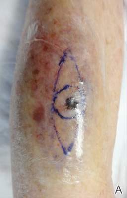
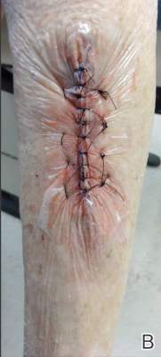
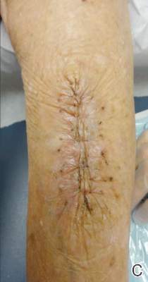
|
Figure 1. The excision site was marked after polyethylene adhesive film was applied to a squamous cell carcinoma on the left tibia of 95-year-old woman (A). Closure was performed by suturing the wound edges through the polyethylene film (B). The excision site appeared to have no dehiscence or signs of infection after removal of the sutures and polyethylene film (C). |
Case Reports
Twelve procedures for skin cancer excision were conducted in 10 patients using polyethylene adhesive film as a surgical aid due to extremely poor quality of the epidermis. The tumors were all squamous cell carcinomas and were located on the arms and legs. Patients were aged 73 to 95 years. Figure 2 demonstrates an example of excision of a squamous cell carcinoma on the left tibia of an 82-year-old man with prior dehiscence and infection after leg surgeries. Good results were achieved using the closure technique described here, along with prophylactic antibiotics.
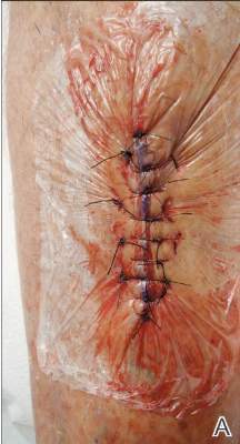
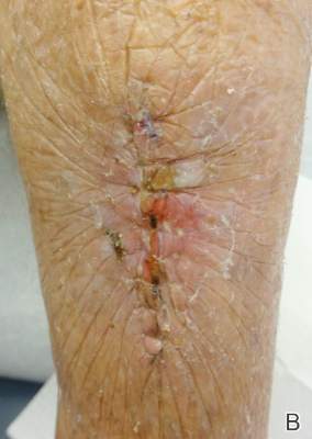
|
Figure 2. A squamous cell carcinoma excision site on the left tibia of an 82-year-old man that had been covered with polyethylene adhesive film prior to excision (A) and 17 days following removal of the sutures and film (B). |
One patient had complications from a Staphylococcus infection because antibiotics were not administered. The patient had prior infections with other surgeries. Antibiotics were given 4 days after surgery. The infection was cleared and the polyethylene film was retained for a total of 12 days.
Sutures were removed after 14 days for excision sites on the arms and 17 days for excision sites on the legs. All excision sites healed without dehiscence with a cosmetically acceptable scar. Figure 3A shows a completed excision on the left hand of a 92-year-old man, and Figure 3B is the result 5 weeks after excision.
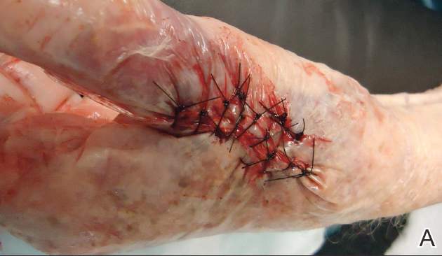
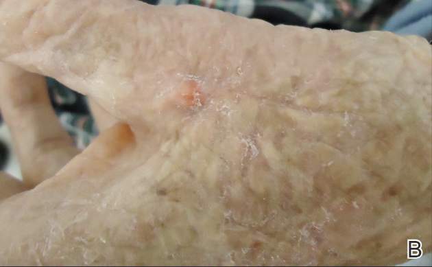
|
Figure 3. A squamous cell carcinoma excision site on the left thumb of a 92-year-old man that had been covered with polyethylene adhesive film prior to exci- sion (A). No visible scarring or dehiscence was noted 5 weeks after excision, following removal of the sutures and film (B). |
None of the patients reported discomfort from the polyethylene film remaining on the skin following surgery, though postoperative care required extra caution when dressing so as not to disturb or compromise the film. Patients were advised about postoperative care and were instructed not to remove the dressing. They were all given antibiotics as a necessary adjunct to maintain a lessened bacteria burden imposed by an impervious layer of acrylate adhesive. Complications resulted from failure to immediately provide antibiotics to 1 patient. The polyethylene film did not hinder healing or postoperative results.
Comment
Various techniques for handling fragile skin during surgery have been described in the literature. Fomon et al1 discussed aging skin as it relates to plastic surgery. Foster and Chan2 described a skin support technique for closing elliptical incisions in patients with fragile skin. Mazzurco and Krach3 discussed the use of a hydrocolloid dressing to aid in the closure of surgical wounds in patients with fragile skin.
The closure method described here was found to be particularly helpful when used as an adjunct to surgery in patients with fragile skin that lacked a suitable dermis. The polyethylene adhesive film helped to hold the sutures more securely. This method is cost-effective and is associated with a high level of patient satisfaction. For the surgeon, this technique may aid in dealing with difficult surgical situations and helps prevent wound complications in elderly patients or those with fragile skin.
1. Fomon S, Bell JW, Schattner A. Aging skin, a surgical challenge. AMA Arch Otolaryngol. 1955;61:554-562.
2. Foster RS, Chan J. The Fixomull skin support method for wound closure in patients with fragile skin. Australas J Dermatol. 2011;52:209-211.
3. Mazzurco JD, Krach KJ. Use of a hydrocolloid dressing to aid in the closure of surgical wounds in patients with fragile skin. J Am Acad Dermatol. 2012;66:335-336.
Patients who have been on steroids, aspirin, or anticoagulants or who are elderly may have a fragile outer skin layer that is similar to parchment paper, which may be challenging for surgeons. In these patients, the epidermal layer is thin and translucent; when a surgeon cuts through this thin layer, the tissue beneath shows minimal dermis and poor-quality fat with weakened tissue support. When undergoing excisional surgery, there is no strong tissue to help the closure sutures remain intact. Surgeons may struggle with skin tears around the sutures and dehiscence on suture removal.
This article describes a novel approach to skin closure in patients with aging or thin skin using a polyethylene film with an acrylate adhesive in the excision area to aid in maintaining skin integrity throughout the healing process following surgery.
Closure Technique
First, the skin area is cleansed with a sterilizing soap preparation. A sterile marking pen then is used to outline the excision area. A 10×12-cm layer of polyethylene film is then attached to the excision site. Excision of the tumor is performed by cutting through the film in the marked area (Figure 1A), and closure is performed by suturing the wound edges through the polyethylene film while the area is still covered with the film (Figure 1B). The sutures can be left in for 2 weeks or longer if necessary. The patient should be instructed not to remove the film or perform any extensive cleansing of the treatment area. Antibiotics should be administered, as the polyethylene film maintains its sterile integrity for 7 days only. Because sutures are on the surface of the film, they are easily accessed for removal. Figure 1C shows the excision site after removal of the sutures and polyethylene film on the left tibia of a 95-year-old woman. Adhesive butterfly closures can be applied to strengthen the excision area after suture removal and prevent dehiscence.
|
|
Figure 1. The excision site was marked after polyethylene adhesive film was applied to a squamous cell carcinoma on the left tibia of 95-year-old woman (A). Closure was performed by suturing the wound edges through the polyethylene film (B). The excision site appeared to have no dehiscence or signs of infection after removal of the sutures and polyethylene film (C). |
Case Reports
Twelve procedures for skin cancer excision were conducted in 10 patients using polyethylene adhesive film as a surgical aid due to extremely poor quality of the epidermis. The tumors were all squamous cell carcinomas and were located on the arms and legs. Patients were aged 73 to 95 years. Figure 2 demonstrates an example of excision of a squamous cell carcinoma on the left tibia of an 82-year-old man with prior dehiscence and infection after leg surgeries. Good results were achieved using the closure technique described here, along with prophylactic antibiotics.
|
|
Figure 2. A squamous cell carcinoma excision site on the left tibia of an 82-year-old man that had been covered with polyethylene adhesive film prior to excision (A) and 17 days following removal of the sutures and film (B). |
One patient had complications from a Staphylococcus infection because antibiotics were not administered. The patient had prior infections with other surgeries. Antibiotics were given 4 days after surgery. The infection was cleared and the polyethylene film was retained for a total of 12 days.
Sutures were removed after 14 days for excision sites on the arms and 17 days for excision sites on the legs. All excision sites healed without dehiscence with a cosmetically acceptable scar. Figure 3A shows a completed excision on the left hand of a 92-year-old man, and Figure 3B is the result 5 weeks after excision.
|
|
Figure 3. A squamous cell carcinoma excision site on the left thumb of a 92-year-old man that had been covered with polyethylene adhesive film prior to exci- sion (A). No visible scarring or dehiscence was noted 5 weeks after excision, following removal of the sutures and film (B). |
None of the patients reported discomfort from the polyethylene film remaining on the skin following surgery, though postoperative care required extra caution when dressing so as not to disturb or compromise the film. Patients were advised about postoperative care and were instructed not to remove the dressing. They were all given antibiotics as a necessary adjunct to maintain a lessened bacteria burden imposed by an impervious layer of acrylate adhesive. Complications resulted from failure to immediately provide antibiotics to 1 patient. The polyethylene film did not hinder healing or postoperative results.
Comment
Various techniques for handling fragile skin during surgery have been described in the literature. Fomon et al1 discussed aging skin as it relates to plastic surgery. Foster and Chan2 described a skin support technique for closing elliptical incisions in patients with fragile skin. Mazzurco and Krach3 discussed the use of a hydrocolloid dressing to aid in the closure of surgical wounds in patients with fragile skin.
The closure method described here was found to be particularly helpful when used as an adjunct to surgery in patients with fragile skin that lacked a suitable dermis. The polyethylene adhesive film helped to hold the sutures more securely. This method is cost-effective and is associated with a high level of patient satisfaction. For the surgeon, this technique may aid in dealing with difficult surgical situations and helps prevent wound complications in elderly patients or those with fragile skin.
Patients who have been on steroids, aspirin, or anticoagulants or who are elderly may have a fragile outer skin layer that is similar to parchment paper, which may be challenging for surgeons. In these patients, the epidermal layer is thin and translucent; when a surgeon cuts through this thin layer, the tissue beneath shows minimal dermis and poor-quality fat with weakened tissue support. When undergoing excisional surgery, there is no strong tissue to help the closure sutures remain intact. Surgeons may struggle with skin tears around the sutures and dehiscence on suture removal.
This article describes a novel approach to skin closure in patients with aging or thin skin using a polyethylene film with an acrylate adhesive in the excision area to aid in maintaining skin integrity throughout the healing process following surgery.
Closure Technique
First, the skin area is cleansed with a sterilizing soap preparation. A sterile marking pen then is used to outline the excision area. A 10×12-cm layer of polyethylene film is then attached to the excision site. Excision of the tumor is performed by cutting through the film in the marked area (Figure 1A), and closure is performed by suturing the wound edges through the polyethylene film while the area is still covered with the film (Figure 1B). The sutures can be left in for 2 weeks or longer if necessary. The patient should be instructed not to remove the film or perform any extensive cleansing of the treatment area. Antibiotics should be administered, as the polyethylene film maintains its sterile integrity for 7 days only. Because sutures are on the surface of the film, they are easily accessed for removal. Figure 1C shows the excision site after removal of the sutures and polyethylene film on the left tibia of a 95-year-old woman. Adhesive butterfly closures can be applied to strengthen the excision area after suture removal and prevent dehiscence.
|
|
Figure 1. The excision site was marked after polyethylene adhesive film was applied to a squamous cell carcinoma on the left tibia of 95-year-old woman (A). Closure was performed by suturing the wound edges through the polyethylene film (B). The excision site appeared to have no dehiscence or signs of infection after removal of the sutures and polyethylene film (C). |
Case Reports
Twelve procedures for skin cancer excision were conducted in 10 patients using polyethylene adhesive film as a surgical aid due to extremely poor quality of the epidermis. The tumors were all squamous cell carcinomas and were located on the arms and legs. Patients were aged 73 to 95 years. Figure 2 demonstrates an example of excision of a squamous cell carcinoma on the left tibia of an 82-year-old man with prior dehiscence and infection after leg surgeries. Good results were achieved using the closure technique described here, along with prophylactic antibiotics.
|
|
Figure 2. A squamous cell carcinoma excision site on the left tibia of an 82-year-old man that had been covered with polyethylene adhesive film prior to excision (A) and 17 days following removal of the sutures and film (B). |
One patient had complications from a Staphylococcus infection because antibiotics were not administered. The patient had prior infections with other surgeries. Antibiotics were given 4 days after surgery. The infection was cleared and the polyethylene film was retained for a total of 12 days.
Sutures were removed after 14 days for excision sites on the arms and 17 days for excision sites on the legs. All excision sites healed without dehiscence with a cosmetically acceptable scar. Figure 3A shows a completed excision on the left hand of a 92-year-old man, and Figure 3B is the result 5 weeks after excision.
|
|
Figure 3. A squamous cell carcinoma excision site on the left thumb of a 92-year-old man that had been covered with polyethylene adhesive film prior to exci- sion (A). No visible scarring or dehiscence was noted 5 weeks after excision, following removal of the sutures and film (B). |
None of the patients reported discomfort from the polyethylene film remaining on the skin following surgery, though postoperative care required extra caution when dressing so as not to disturb or compromise the film. Patients were advised about postoperative care and were instructed not to remove the dressing. They were all given antibiotics as a necessary adjunct to maintain a lessened bacteria burden imposed by an impervious layer of acrylate adhesive. Complications resulted from failure to immediately provide antibiotics to 1 patient. The polyethylene film did not hinder healing or postoperative results.
Comment
Various techniques for handling fragile skin during surgery have been described in the literature. Fomon et al1 discussed aging skin as it relates to plastic surgery. Foster and Chan2 described a skin support technique for closing elliptical incisions in patients with fragile skin. Mazzurco and Krach3 discussed the use of a hydrocolloid dressing to aid in the closure of surgical wounds in patients with fragile skin.
The closure method described here was found to be particularly helpful when used as an adjunct to surgery in patients with fragile skin that lacked a suitable dermis. The polyethylene adhesive film helped to hold the sutures more securely. This method is cost-effective and is associated with a high level of patient satisfaction. For the surgeon, this technique may aid in dealing with difficult surgical situations and helps prevent wound complications in elderly patients or those with fragile skin.
1. Fomon S, Bell JW, Schattner A. Aging skin, a surgical challenge. AMA Arch Otolaryngol. 1955;61:554-562.
2. Foster RS, Chan J. The Fixomull skin support method for wound closure in patients with fragile skin. Australas J Dermatol. 2011;52:209-211.
3. Mazzurco JD, Krach KJ. Use of a hydrocolloid dressing to aid in the closure of surgical wounds in patients with fragile skin. J Am Acad Dermatol. 2012;66:335-336.
1. Fomon S, Bell JW, Schattner A. Aging skin, a surgical challenge. AMA Arch Otolaryngol. 1955;61:554-562.
2. Foster RS, Chan J. The Fixomull skin support method for wound closure in patients with fragile skin. Australas J Dermatol. 2011;52:209-211.
3. Mazzurco JD, Krach KJ. Use of a hydrocolloid dressing to aid in the closure of surgical wounds in patients with fragile skin. J Am Acad Dermatol. 2012;66:335-336.
Practice Points
- A novel method of skin closure using a polyethylene film with an acrylate adhesive can aid in strengthening suture integrity and preventing skin tears.
- Dehiscence of excision sites in patients with aging or fragile skin can be prevented.
- This closure technique promotes healing and efficient scar formation.