User login
Apple Watch algorithm showed 84% positive predictive value for Afib
, investigators reported at the annual meeting of the American College of Cardiology.
This was a single-arm, prospective, open-label, observational study of unprecedented size and speediness of completion. It included nearly 420,000 self-enrolled adults living in the U.S., with 8 months of monitoring. But despite the study’s flashy size and trendy digital health theme, the researchers were careful not to oversell the findings.

“This study was just meant to be a very, very first step in trying to learn if this kind of technology can help us to prevent stroke. It was not a randomized trial of a public health intervention for screening. This is the first half of the first inning. Rigorous investigation of this technology and its potential use in clinical settings will need to happen. But we do think from a trial and operational standpoint the Apple Heart Study provides a solid foundation upon which further research in digital health can be conducted,” according to Mintu Turakhia, MD, co-principal investigator and an electrophysiologist as well as executive director of the Center for Digital Health at Stanford (Calif.) University, which conducted the study.
The study was conducted virtually. Screening, consent, and data gathering were performed electronically by smartphone. Participants had to have an Apple Watch Series 1, 2, or 3, and an Apple iPhone 5 or more recent model in order to join. The majority of subjects were under age 40, and just 6% were age 65 or older, when the risks of atrial fibrillation (AFib) and stroke are higher. All participants self-reported having no history of AFib nor currently being on anticoagulation.
The study algorithm utilized the Apple Watch’s built-in light sensor technology to opportunistically sample the time interval between pulses when the wearer was still. An irregular time interval triggered a cascade of more frequent sampling. If 5 of 6 samples were irregular within a 48-hour period, the wearer received an irregular rhythm notification along with a prompt to contact a participating physician via telemedicine. The physician could then arrange for an ECG patch to be mailed to the participant, who wore it for up to 7 days before mailing it back for analysis.
Among the key findings in the Apple Heart Study: the irregular pulse notification rate was low overall, at 0.5%, ranging from 0.16% in the under-40 group to 3.2% in subjects age 65 or older. As a result, the study population of particular interest nosedived from an initial 419,297 to the less than 2,100 who received an irregular pulse notification. Of the 658 participants who were subsequently sent an ECG patch, 450 returned it for analysis.
An average of 13 days went by between an irregular pulse notification and ECG patch receipt and activation, so it wasn’t particularly surprising that only 34% of the patches were positive for AFib, since early-stage paroxysmal AFib comes and goes. However, of the 86 subjects who received a new notification of an irregular rhythm while they were wearing a patch, 72 simultaneously showed AFib on their patch. That translates to an 84% positive predictive value for an irregular rhythm notification as an indicator of AFib.
Of the 153 subjects with evidence of AFib on their ECG patch, 20% proved to be in AFib for the full week they wore it. Of those with AFib, 89% had a longest episode of at least 1 hour in duration.
Several discussants expressed reservations about this approach to finding individuals with previously undetected AFib. Jeanne E. Poole, MD, an electrophysiologist and professor of medicine at the University of Washington in Seattle, observed that the question of whether patients with asymptomatic AFib should receive oral anticoagulation therapy is as-yet unanswered and is the focus of ongoing randomized trials. The Apple Heart Study approach, she said, “might lead a lot of patients into being treated unnecessarily or prematurely, or flooding doctors’ offices with a lot of young people.”
Co-principal investigator Marco Perez, MD, an electrophysiologist at Stanford, replied, “Stroke is important, and we all worry about it. But it’s also important that there are other things atrial fibrillation is associated with, like cardiomyopathy and heart failure. So finding atrial fibrillation in a young population might be important. Maybe they don’t need anticoagulation, but maybe there’s something else going on.”
Patrick T. O’Gara, MD, professor of medicine at Harvard University, Boston, was concerned about what he called “the signal-to-noise ratio – the noise that will come in when there is an irregularity detected on the watch that could range from anything from ventricular premature beats to atrial fibrillation.” He is also leery of what he considers to be at this point the excessive hype surrounding direct-to-consumer wearable digital health technology.
“I applaud your circumspection,” he told Dr. Turakhia and Dr. Perez. “I understand very directly from you that these are limited observations. But it’s a good step forward.”
Dr. Perez reported receiving research funding from and serving as a consultant to Apple. Dr. Turakhia reported serving as a consultant to AliveCor and Cardiva Medical.
Their presentation was immediately followed by a related panel discussion titled, “Digital Disruption at Our Doorstep – Implications for Clinicians and Patients.” Session moderator John Rumsfeld, MD, chief innovation officer at the ACC and professor of medicine at the University of Colorado, Denver, kicked things off by observing, “Digital health technology certainly exists. There’ve been billions of dollars invested in digital health. Outside of health care there’s been successful digital transformation of almost every other sector of the economy except for health care. But we deliver care pretty much the same as we have for the past 50 or more years.”
Paul Stoeffels, MD, chief scientific officer at Johnson & Johnson, said physicians and payers want to see evidence of benefit before adopting change. Towards that end, he announced that Johnson & Johnson and Apple are collaborating on a randomized controlled trial called the HEARTLINE study. The active intervention arm in the study involves utilization of the Apple Watch’s irregular pulse notification algorithm, with confirmation of AFib to be achieved using the ECG app incorporated in the latest version of the watch, coupled with a medication adherence app developed by Johnson & Johnson. Enrollment of 150,000 U.S. adults age 65 and older is planned to begin this summer. The study, conducted on a digital platform akin to the Apple Heart Study, will look at the intervention’s impact on rates of stroke, MI, and death as well as AF detection.
Maulik Majmudar, MD, a cardiologist and chief health officer for health and wellness at Amazon, declared, “There’s no doubt in my mind that digital solutions will become a mainstay in our care delivery going forward. The question is really not if, but when.”
He predicted that just as the past two decades have seen the birth of new medical specialties, including hospitalists and cardiovascular intensivists, the next 10 years or so will see the creation of a new field within cardiovascular medicine, whose skilled practitioners might be called ‘digitalists’ – experts in collecting, moving, and safeguarding massive quantities of digital health data.
Robert Califf, MD, vice chancellor for health data science at Duke Health in Durham, N.C., addressed the issue of how society is going to pay for a shift to digital health: “It’s very simple. I don’t see any way that fee-for-service medicine can deal with this. It’s just not possible. If you think we’re going to add on more cost to the system by doing virtual visits, I just do not see that happening. Our solution to the payment system is to get rid of fee-for-service medicine and go to pay-for-value. The minute you’re in pay-for-value, virtual visits and digital information will become the way to do it – to move the treatment and the interaction more to home and less of having people wait in doctors’ offices and spending time in hospitals.”
SOURCE: Turakhia M, ACC 19 NCT03335800
, investigators reported at the annual meeting of the American College of Cardiology.
This was a single-arm, prospective, open-label, observational study of unprecedented size and speediness of completion. It included nearly 420,000 self-enrolled adults living in the U.S., with 8 months of monitoring. But despite the study’s flashy size and trendy digital health theme, the researchers were careful not to oversell the findings.
“This study was just meant to be a very, very first step in trying to learn if this kind of technology can help us to prevent stroke. It was not a randomized trial of a public health intervention for screening. This is the first half of the first inning. Rigorous investigation of this technology and its potential use in clinical settings will need to happen. But we do think from a trial and operational standpoint the Apple Heart Study provides a solid foundation upon which further research in digital health can be conducted,” according to Mintu Turakhia, MD, co-principal investigator and an electrophysiologist as well as executive director of the Center for Digital Health at Stanford (Calif.) University, which conducted the study.
The study was conducted virtually. Screening, consent, and data gathering were performed electronically by smartphone. Participants had to have an Apple Watch Series 1, 2, or 3, and an Apple iPhone 5 or more recent model in order to join. The majority of subjects were under age 40, and just 6% were age 65 or older, when the risks of atrial fibrillation (AFib) and stroke are higher. All participants self-reported having no history of AFib nor currently being on anticoagulation.
The study algorithm utilized the Apple Watch’s built-in light sensor technology to opportunistically sample the time interval between pulses when the wearer was still. An irregular time interval triggered a cascade of more frequent sampling. If 5 of 6 samples were irregular within a 48-hour period, the wearer received an irregular rhythm notification along with a prompt to contact a participating physician via telemedicine. The physician could then arrange for an ECG patch to be mailed to the participant, who wore it for up to 7 days before mailing it back for analysis.
Among the key findings in the Apple Heart Study: the irregular pulse notification rate was low overall, at 0.5%, ranging from 0.16% in the under-40 group to 3.2% in subjects age 65 or older. As a result, the study population of particular interest nosedived from an initial 419,297 to the less than 2,100 who received an irregular pulse notification. Of the 658 participants who were subsequently sent an ECG patch, 450 returned it for analysis.
An average of 13 days went by between an irregular pulse notification and ECG patch receipt and activation, so it wasn’t particularly surprising that only 34% of the patches were positive for AFib, since early-stage paroxysmal AFib comes and goes. However, of the 86 subjects who received a new notification of an irregular rhythm while they were wearing a patch, 72 simultaneously showed AFib on their patch. That translates to an 84% positive predictive value for an irregular rhythm notification as an indicator of AFib.
Of the 153 subjects with evidence of AFib on their ECG patch, 20% proved to be in AFib for the full week they wore it. Of those with AFib, 89% had a longest episode of at least 1 hour in duration.
Several discussants expressed reservations about this approach to finding individuals with previously undetected AFib. Jeanne E. Poole, MD, an electrophysiologist and professor of medicine at the University of Washington in Seattle, observed that the question of whether patients with asymptomatic AFib should receive oral anticoagulation therapy is as-yet unanswered and is the focus of ongoing randomized trials. The Apple Heart Study approach, she said, “might lead a lot of patients into being treated unnecessarily or prematurely, or flooding doctors’ offices with a lot of young people.”
Co-principal investigator Marco Perez, MD, an electrophysiologist at Stanford, replied, “Stroke is important, and we all worry about it. But it’s also important that there are other things atrial fibrillation is associated with, like cardiomyopathy and heart failure. So finding atrial fibrillation in a young population might be important. Maybe they don’t need anticoagulation, but maybe there’s something else going on.”
Patrick T. O’Gara, MD, professor of medicine at Harvard University, Boston, was concerned about what he called “the signal-to-noise ratio – the noise that will come in when there is an irregularity detected on the watch that could range from anything from ventricular premature beats to atrial fibrillation.” He is also leery of what he considers to be at this point the excessive hype surrounding direct-to-consumer wearable digital health technology.
“I applaud your circumspection,” he told Dr. Turakhia and Dr. Perez. “I understand very directly from you that these are limited observations. But it’s a good step forward.”
Dr. Perez reported receiving research funding from and serving as a consultant to Apple. Dr. Turakhia reported serving as a consultant to AliveCor and Cardiva Medical.
Their presentation was immediately followed by a related panel discussion titled, “Digital Disruption at Our Doorstep – Implications for Clinicians and Patients.” Session moderator John Rumsfeld, MD, chief innovation officer at the ACC and professor of medicine at the University of Colorado, Denver, kicked things off by observing, “Digital health technology certainly exists. There’ve been billions of dollars invested in digital health. Outside of health care there’s been successful digital transformation of almost every other sector of the economy except for health care. But we deliver care pretty much the same as we have for the past 50 or more years.”
Paul Stoeffels, MD, chief scientific officer at Johnson & Johnson, said physicians and payers want to see evidence of benefit before adopting change. Towards that end, he announced that Johnson & Johnson and Apple are collaborating on a randomized controlled trial called the HEARTLINE study. The active intervention arm in the study involves utilization of the Apple Watch’s irregular pulse notification algorithm, with confirmation of AFib to be achieved using the ECG app incorporated in the latest version of the watch, coupled with a medication adherence app developed by Johnson & Johnson. Enrollment of 150,000 U.S. adults age 65 and older is planned to begin this summer. The study, conducted on a digital platform akin to the Apple Heart Study, will look at the intervention’s impact on rates of stroke, MI, and death as well as AF detection.
Maulik Majmudar, MD, a cardiologist and chief health officer for health and wellness at Amazon, declared, “There’s no doubt in my mind that digital solutions will become a mainstay in our care delivery going forward. The question is really not if, but when.”
He predicted that just as the past two decades have seen the birth of new medical specialties, including hospitalists and cardiovascular intensivists, the next 10 years or so will see the creation of a new field within cardiovascular medicine, whose skilled practitioners might be called ‘digitalists’ – experts in collecting, moving, and safeguarding massive quantities of digital health data.
Robert Califf, MD, vice chancellor for health data science at Duke Health in Durham, N.C., addressed the issue of how society is going to pay for a shift to digital health: “It’s very simple. I don’t see any way that fee-for-service medicine can deal with this. It’s just not possible. If you think we’re going to add on more cost to the system by doing virtual visits, I just do not see that happening. Our solution to the payment system is to get rid of fee-for-service medicine and go to pay-for-value. The minute you’re in pay-for-value, virtual visits and digital information will become the way to do it – to move the treatment and the interaction more to home and less of having people wait in doctors’ offices and spending time in hospitals.”
SOURCE: Turakhia M, ACC 19 NCT03335800
REPORTING FROM ACC 19
Apple Watch algorithm brings wearables closer to clinical practice
NEW ORLEANS – , Matthew W. Martinez, MD, medical director of the Sports Cardiology and Hypertrophic Cardiomyopathy Center at the Lehigh Valley Health Network in Allentown, Pa., said in a video interview.
The Apple Heart Study, presented at the annual meeting of the American College of Cardiology, evaluated a mobile app that uses the watch’s existing light sensor technology to detect subtle changes that might indicate an arrhythmia.
The Apple Watch generates a tachogram, which is a plot of time between heart beats. If an abnormal tachogram occurs five out of six times, they are analyzed by an algorithm and sent to the Apple Watch.
The positive predictive value for the tachogram was 71%, and the positive predictive value for the notification was 84%.
Dr. Martinez, who is lead cardiologist for U.S. Major League Soccer and is also heavily involved with the National Football League, said that the study helps clinicians understand the utility of wearable technology.
His take home from the study is that, when people are notified by their watch, they should notify their health care provider, and the provider should take it seriously.
Dr. Martinez was not involved in the Apple Heart Study, and had no relevant disclosures.
bjancin@mdedge.com
REPORTING FROM ACC 19
Recent trials advance axial spondyloarthritis therapy
MAUI, HAWAII – Arguably the most exciting therapeutic development in axial spondyloarthritis in the past year was the demonstrated efficacy and safety of the investigational oral selective Janus kinase 1 inhibitor filgotinib in the setting of active ankylosing spondylitis, speakers agreed at the 2019 Rheumatology Winter Clinical Symposium.
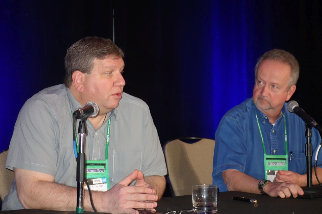
“This is big, big news,” commented symposium director Arthur Kavanaugh, MD. “This is going to be a big deal.”
Other recent clinical trials of note in axial spondyloarthritis (SpA) highlighted by Dr. Kavanaugh and Eric Ruderman, MD, professor of medicine at Northwestern University, Chicago, included a positive phase 3 study of certolizumab pegol (Cimzia) in nonradiographic SpA, two positive phase 3 trials of the interleukin-17A antagonist ixekizumab (Taltz) in radiographic SpA, a positive phase 2b trial of the dual IL-17A/F antagonist bimekizumab, and publication of three surprisingly negative phase 3 trials of the IL-12/23 inhibitor ustekinumab (Stelara).
Filgotinib
TORTUGA was a phase 2b, double-blind, multicenter trial of 116 European patients with active ankylosing spondylitis nonresponsive to NSAIDs who were randomized to oral filgotinib at 200 mg once daily or placebo for 12 weeks. Filgotinib reduced the Ankylosing Spondylitis Disease Activity Score (ASDAS) by a mean of 1.47 points from baseline, a significantly better result for the primary outcome than the 0.57-point decrease in controls (Lancet. 2018 Dec 1;392[10162]:2378-87).
Dr. Ruderman was also favorably impressed with the oral Janus kinase 1 (JAK1) inhibitor’s performance on the secondary outcome measures, including a mean 2.41-point reduction from baseline on the Bath Ankylosing Spondylitis Disease Activity Index, compared with a 1.44-point decrease in controls, with the difference being significant from week 8 onward. The filgotinib group also did significantly better on validated measures of physical function, spinal mobility, physical function, quality of life, peripheral arthritis, fatigue, and spinal and sacroiliac joint inflammation as assessed by MRI.
One patient in the filgotinib group, a smoker, developed pneumonia and another experienced deep venous thrombosis.
The study results are an exciting development because SpA treatments with new mechanisms of action are sorely needed. NSAIDs are considered first-line pharmacotherapy at present, with various tumor necrosis factor (TNF) inhibitors as well as the IL-17 inhibitor secukinumab (Cosentyx) the only approved biologic alternatives.
“This is the most impressive data I’ve seen that JAK inhibitors are effective in ankylosing spondyloarthritis,” commented Paul Emery, MD, professor of rheumatology and director of the University of Leeds (England) Musculoskeletal Biomedical Research Center.
TORTUGA was the first positive phase 2 trial of a selective JAK1 inhibitor in SpA. However, Dr. Kavanaugh noted that while a phase 2 trial of tofacitinib (Xeljanz) failed to meet its “very convoluted” primary endpoint, the JAK1/3 inhibitor was positive for key secondary endpoints, including favorable MRI changes. And a phase 3 trial of tofacitinib in SpA is underway.
A key remaining question pending the outcome of definitive phase 3 trials is whether specificity of JAK enzyme inhibition matters or if a class effect is at work, according to Dr. Kavanaugh, professor of medicine at the University of California, San Diego.
Certolizumab pegol
The TNF inhibitor is already approved for ankylosing spondylitis, among other indications, but it has now demonstrated efficacy and safety in nonradiographic SpA in a phase 3 trial structured with guidance from the Food and Drug Administration.
“It seems likely that UCB will get the indication for this,” according to Dr. Ruderman.
This 317-patient trial was remarkable in that it entailed a full 52 weeks of double-blind therapy with certolizumab at the standard dose of 200 mg every 2 weeks or placebo. The ASDAS Major Improvement rate, defined as at least a 2-point improvement from baseline, was 47% in the active treatment arm, compared with 7% on placebo. The Assessment in Ankylosing Spondylitis International Society 40% (ASAS 40) response rate, a more patient-reported outcome measure, was 57% in the certolizumab group and 16% in controls in this trial, which was recently published (Arthritis Rheumatol. 2019 March 8. doi: 10.1002/art.40866). All participants had to have baseline MRI evidence of sacroiliac joint inflammation and/or an elevated C-reactive protein.
By way of background, Dr. Ruderman explained that the FDA required 52 weeks of double-blind, placebo-controlled therapy because the agency’s advisory committee had formerly expressed reservations about considering an expanded indication for TNF inhibitors in nonradiographic as opposed to radiographic SpA.
“They were very concerned that approval could result in patients with mechanical back pain or fibromyalgia being treated with biologics. And they weren’t sure nonradiographic SpA was a discrete entity. They wondered if it remits on its own,” according to the rheumatologist.
Dr. Ruderman is curious to see how the FDA is going to handle this situation in light of the positive phase 3 certolizumab results. Will the agency require other companies that market TNF inhibitors to mount a similarly rigorous 52-week, double-blind, placebo-controlled trial in order to obtain an expanded indication? That would seem to pose ethical issues now. Or will the companies be able to gain an expanded indication by retrospective analysis of outcomes in patients with nonradiographic SpA in their existing trials databases? Stay tuned.
Ixekizumab
The COAST-V trial was a phase 3, randomized, double-blind, active- and placebo-controlled trial of 341 patients with radiographic SpA who hadn’t previously been treated with a biologic disease-modifying antirheumatic drug. They were assigned to 80 mg of ixekizumab every 2 weeks, 80 mg every 4 weeks, adalimumab (Humira) at 40 mg every 2 weeks, or placebo. The primary endpoint – an ASAS 40 response at week 16 – was achieved in 52% of patients on ixekizumab every 2 weeks, 48% of those who received ixekizumab every 4 weeks, 36% on adalimumab, and 18% on placebo (Lancet. 2018 Dec 8;392[10163]:2441-51).
In contrast, the phase 3 COAST-W trial included 316 patients with radiographic SpA who were inadequate responders or intolerant to one or more prior anti-TNF agents. The 16-week ASAS 40 response rate was 30.6% with ixekizumab every 2 weeks, similar at 25.4% with ixekizumab every 4 weeks, and 12.5% with placebo (Arthritis Rheumatol. 2018 Oct 20. doi: 10.1002/art.40753).
While the COAST-V trial convincingly showed both ixekizumab and adalimumab were more effective than placebo, the patient numbers were way too small to draw any conclusions about the relative efficacy of the two biologics, according to Dr. Ruderman.
“I don’t think these results are surprising,” Dr. Kavanaugh commented. “It would have been surprising if ixekizumab was ineffective, given that secukinumab works. But it’s nice to have the proof.”
Bimekizumab
This investigational dual IL-17A/F inhibitor demonstrated efficacy and safety for SpA in a 297-patient, phase 2b trial presented at the 2018 European Congress of Rheumatology.
“It’s effective, but it doesn’t look like it’s particularly more effective than either of the existing IL-17A inhibitors. We’ll see going forward if there truly is an advantage here to the additional inhibition of IL-17F in this population. I will say that the preclinical and laboratory data on the potential advantages of IL-17F inhibition are mostly in the psoriasis/psoriatic arthritis space. It’s not clear in ankylosing spondyloarthritis specifically whether we should expect to see a difference,” Dr. Ruderman said.
Ustekinumab
The IL-12/23 inhibitor proved no better than placebo in patients with SpA in three separate phase 3, randomized trials recently published as a single summary article (Arthritis Rheumatol. 2019 Feb;71[2]:258-70).
“Ustekinumab was effective in an earlier open study, so I think everybody was surprised by this,” Dr. Kavanaugh said.
He reported serving as a consultant to and/or receiving research funding from a dozen pharmaceutical companies. Dr. Ruderman reported financial relationships with eight companies.
MAUI, HAWAII – Arguably the most exciting therapeutic development in axial spondyloarthritis in the past year was the demonstrated efficacy and safety of the investigational oral selective Janus kinase 1 inhibitor filgotinib in the setting of active ankylosing spondylitis, speakers agreed at the 2019 Rheumatology Winter Clinical Symposium.
“This is big, big news,” commented symposium director Arthur Kavanaugh, MD. “This is going to be a big deal.”
Other recent clinical trials of note in axial spondyloarthritis (SpA) highlighted by Dr. Kavanaugh and Eric Ruderman, MD, professor of medicine at Northwestern University, Chicago, included a positive phase 3 study of certolizumab pegol (Cimzia) in nonradiographic SpA, two positive phase 3 trials of the interleukin-17A antagonist ixekizumab (Taltz) in radiographic SpA, a positive phase 2b trial of the dual IL-17A/F antagonist bimekizumab, and publication of three surprisingly negative phase 3 trials of the IL-12/23 inhibitor ustekinumab (Stelara).
Filgotinib
TORTUGA was a phase 2b, double-blind, multicenter trial of 116 European patients with active ankylosing spondylitis nonresponsive to NSAIDs who were randomized to oral filgotinib at 200 mg once daily or placebo for 12 weeks. Filgotinib reduced the Ankylosing Spondylitis Disease Activity Score (ASDAS) by a mean of 1.47 points from baseline, a significantly better result for the primary outcome than the 0.57-point decrease in controls (Lancet. 2018 Dec 1;392[10162]:2378-87).
Dr. Ruderman was also favorably impressed with the oral Janus kinase 1 (JAK1) inhibitor’s performance on the secondary outcome measures, including a mean 2.41-point reduction from baseline on the Bath Ankylosing Spondylitis Disease Activity Index, compared with a 1.44-point decrease in controls, with the difference being significant from week 8 onward. The filgotinib group also did significantly better on validated measures of physical function, spinal mobility, physical function, quality of life, peripheral arthritis, fatigue, and spinal and sacroiliac joint inflammation as assessed by MRI.
One patient in the filgotinib group, a smoker, developed pneumonia and another experienced deep venous thrombosis.
The study results are an exciting development because SpA treatments with new mechanisms of action are sorely needed. NSAIDs are considered first-line pharmacotherapy at present, with various tumor necrosis factor (TNF) inhibitors as well as the IL-17 inhibitor secukinumab (Cosentyx) the only approved biologic alternatives.
“This is the most impressive data I’ve seen that JAK inhibitors are effective in ankylosing spondyloarthritis,” commented Paul Emery, MD, professor of rheumatology and director of the University of Leeds (England) Musculoskeletal Biomedical Research Center.
TORTUGA was the first positive phase 2 trial of a selective JAK1 inhibitor in SpA. However, Dr. Kavanaugh noted that while a phase 2 trial of tofacitinib (Xeljanz) failed to meet its “very convoluted” primary endpoint, the JAK1/3 inhibitor was positive for key secondary endpoints, including favorable MRI changes. And a phase 3 trial of tofacitinib in SpA is underway.
A key remaining question pending the outcome of definitive phase 3 trials is whether specificity of JAK enzyme inhibition matters or if a class effect is at work, according to Dr. Kavanaugh, professor of medicine at the University of California, San Diego.
Certolizumab pegol
The TNF inhibitor is already approved for ankylosing spondylitis, among other indications, but it has now demonstrated efficacy and safety in nonradiographic SpA in a phase 3 trial structured with guidance from the Food and Drug Administration.
“It seems likely that UCB will get the indication for this,” according to Dr. Ruderman.
This 317-patient trial was remarkable in that it entailed a full 52 weeks of double-blind therapy with certolizumab at the standard dose of 200 mg every 2 weeks or placebo. The ASDAS Major Improvement rate, defined as at least a 2-point improvement from baseline, was 47% in the active treatment arm, compared with 7% on placebo. The Assessment in Ankylosing Spondylitis International Society 40% (ASAS 40) response rate, a more patient-reported outcome measure, was 57% in the certolizumab group and 16% in controls in this trial, which was recently published (Arthritis Rheumatol. 2019 March 8. doi: 10.1002/art.40866). All participants had to have baseline MRI evidence of sacroiliac joint inflammation and/or an elevated C-reactive protein.
By way of background, Dr. Ruderman explained that the FDA required 52 weeks of double-blind, placebo-controlled therapy because the agency’s advisory committee had formerly expressed reservations about considering an expanded indication for TNF inhibitors in nonradiographic as opposed to radiographic SpA.
“They were very concerned that approval could result in patients with mechanical back pain or fibromyalgia being treated with biologics. And they weren’t sure nonradiographic SpA was a discrete entity. They wondered if it remits on its own,” according to the rheumatologist.
Dr. Ruderman is curious to see how the FDA is going to handle this situation in light of the positive phase 3 certolizumab results. Will the agency require other companies that market TNF inhibitors to mount a similarly rigorous 52-week, double-blind, placebo-controlled trial in order to obtain an expanded indication? That would seem to pose ethical issues now. Or will the companies be able to gain an expanded indication by retrospective analysis of outcomes in patients with nonradiographic SpA in their existing trials databases? Stay tuned.
Ixekizumab
The COAST-V trial was a phase 3, randomized, double-blind, active- and placebo-controlled trial of 341 patients with radiographic SpA who hadn’t previously been treated with a biologic disease-modifying antirheumatic drug. They were assigned to 80 mg of ixekizumab every 2 weeks, 80 mg every 4 weeks, adalimumab (Humira) at 40 mg every 2 weeks, or placebo. The primary endpoint – an ASAS 40 response at week 16 – was achieved in 52% of patients on ixekizumab every 2 weeks, 48% of those who received ixekizumab every 4 weeks, 36% on adalimumab, and 18% on placebo (Lancet. 2018 Dec 8;392[10163]:2441-51).
In contrast, the phase 3 COAST-W trial included 316 patients with radiographic SpA who were inadequate responders or intolerant to one or more prior anti-TNF agents. The 16-week ASAS 40 response rate was 30.6% with ixekizumab every 2 weeks, similar at 25.4% with ixekizumab every 4 weeks, and 12.5% with placebo (Arthritis Rheumatol. 2018 Oct 20. doi: 10.1002/art.40753).
While the COAST-V trial convincingly showed both ixekizumab and adalimumab were more effective than placebo, the patient numbers were way too small to draw any conclusions about the relative efficacy of the two biologics, according to Dr. Ruderman.
“I don’t think these results are surprising,” Dr. Kavanaugh commented. “It would have been surprising if ixekizumab was ineffective, given that secukinumab works. But it’s nice to have the proof.”
Bimekizumab
This investigational dual IL-17A/F inhibitor demonstrated efficacy and safety for SpA in a 297-patient, phase 2b trial presented at the 2018 European Congress of Rheumatology.
“It’s effective, but it doesn’t look like it’s particularly more effective than either of the existing IL-17A inhibitors. We’ll see going forward if there truly is an advantage here to the additional inhibition of IL-17F in this population. I will say that the preclinical and laboratory data on the potential advantages of IL-17F inhibition are mostly in the psoriasis/psoriatic arthritis space. It’s not clear in ankylosing spondyloarthritis specifically whether we should expect to see a difference,” Dr. Ruderman said.
Ustekinumab
The IL-12/23 inhibitor proved no better than placebo in patients with SpA in three separate phase 3, randomized trials recently published as a single summary article (Arthritis Rheumatol. 2019 Feb;71[2]:258-70).
“Ustekinumab was effective in an earlier open study, so I think everybody was surprised by this,” Dr. Kavanaugh said.
He reported serving as a consultant to and/or receiving research funding from a dozen pharmaceutical companies. Dr. Ruderman reported financial relationships with eight companies.
REPORTING FROM RWCS 2019
Interventional cardiology pioneer reflects on field’s past, future
SNOWMASS, COLO. – When Spencer B. King III, MD, shared his thoughts about the future of interventional cardiology at the Annual Cardiovascular Conference at Snowmass sponsored by the American College of Cardiology, he felt compelled to offer a cautionary note about his past accuracy as a prognosticator.

It was way back at a poster session during the 1976 annual meeting of the American Heart Association in Miami Beach that he first met Andreas Gruentzig, MD, the father of percutaneous coronary intervention (PCI), who was presenting his initial revolutionary work on what he called “coronary transluminal angioplasty” in dogs.
“I looked at the poster and told him it would never work,” recalled Dr. King, professor emeritus of medicine at Emory University in Atlanta.
He soon changed his mind, however, because, to great acclaim, Dr. Gruentzig performed his successful first in-human coronary angioplasty the next year.
He noted that the Snowmass conference has played a significant role in the development of interventional cardiology in the United States. Dr. Gruentzig attended the conference in 1980, and Dr. King and others took that opportunity to persuade him to leave the bureaucratic confines of Zurich and join him at Emory later that year. The two cardiologists worked closely thereafter, refining angioplasty and conducting clinical trials until Dr. Gruentzig’s death in an airplane crash in Georgia in 1985 at age 46 years.
Turning to the future, Dr. King addressed a number of recent developments in interventional cardiology and rated their chances of significantly improving outcomes in patients with stable ischemic heart disease. He graded the innovations’ potential with use of a four-bar schema, akin to the WiFi signal power rating on a cell phone.
Noninvasive diagnostics to assess anatomy and physiology
“I think coronary CT angiography [CTA] will become the new diagnostic angiogram,” he predicted. “CTA has gotten much better. Outside the United States, in Europe and particularly in Japan and increasingly in China, CTA is becoming extremely common.”
Dr. King cited a recent multicenter study of blinded heart team treatment decision making on the basis of either CTA or conventional invasive angiography in 223 patients with left main or triple-vessel coronary artery disease (CAD). The level of agreement was impressively high: Coronary artery bypass grafting (CABG) was recommended for 28% of patients on the basis of CTA and 26% with conventional angiography, which suggests the feasibility of treatment decision making based solely on noninvasive imaging, history, and clinical examination (Eur Heart J. 2018 Nov 1;39[41]:3689-98).
“The other thing I like about the potential for noninvasive imaging to guide our interventions is that it may [replace] the diagnostic angiogram, which has largely become extinct,” the cardiologist continued. “If you think about it, patients are referred for an angiogram, and as far as informed consent is concerned, the patient is told to pack his bags, go off to some other city, get in the cath lab, and take the family because of what they might do to you. They might put stents in you, they might operate on you. We don’t have any idea because we don’t know what you have. And the patient has to buy into this. With CTA, the potential is there for people to actually know what you’re going to do to them before you do it.”
Coronary artery calcium scoring for primary risk assessment has taken on a prominent role in the latest practice guidelines. “I think it’s mostly helpful in getting people out of the system because they don’t have any calcium,” in Dr. King’s view.
PET and MRI will remain secondary noninvasive technologies. They will be used mostly to diagnose microvascular disease, but that’s information that doesn’t have much influence on whether interventional procedures are performed.
Overall, he gave noninvasive diagnostic tools high marks for their potential to improve outcomes in patients with stable ischemic heart disease.
“I give it a pretty robust three bars. Maybe you could give it four,” he said.
New pharmacologic therapies
Citing in particular the proprotein convertase subtilisin/kexin type 9 (PCSK9) inhibitors and the sodium-glucose cotransporter 2 (SGLT2) inhibitors, Dr. King declared, “It may be that the biggest, newest device in interventional cardiology going forward is not a device at all, it’s medical therapy.”
Interventional cardiologists either need to become expert in advanced medical therapies or else have access to someone in their group who prescribes those medications deftly.
“The future care of our patients will require more than percutaneous coronary intervention,” he emphasized.
So, four bars for the new medical therapies.
PCI and coronary artery bypass surgery
Both get one bar.
“PCI will be a partner of advanced antiatherosclerotic therapies, but will not be replaced by limiting antianginal therapy to medical treatment only,” Dr. King predicted.
Regarding CABG, he highlighted a recent systematic review and pooled analysis of 11,518 patients with stable ischemic heart disease randomized to CABG or PCI using drug-eluting or bare-metal stents in 11 clinical trials. CABG demonstrated a significant mortality benefit over PCI in patients with multivessel disease, particularly among those with diabetes or a higher degree of coronary disease complexity. However, there was no benefit in terms of 5-year all-cause mortality for CABG over PCI in those with left main disease (Lancet. 2018 Mar 10;391[10124]:9399-48).
“CABG will not go away. I predict that about 25% of revascularizations will continue to be done by surgery,” the cardiologist said. “For patients who can have complete revascularization by PCI, it’ll be done with advanced technology, but probably only by a subset of operators. We have a huge number of interventional cardiologists in this country, and some of them do a lot of these kinds of cases and some don’t.”
Endovascular imaging to optimize stent deployment and characterize plaque
Studies suggest that the use of intravascular ultrasound and other endovascular imaging technologies ends up providing better results than when they’re not employed.
“We see greatly increased use of IVUS [intravascular ultrasound], and not so much of optical coherence tomography, because of technical problems. So I give this at least two bars as far as moving practice forward,” according to Dr. King.
Bioresorbable scaffolds
This technology, which he noted “was supposed to solve all of our problems,” has tripped and fallen because of its associated increased risk of scaffold thrombosis. He cited a recent network meta-analysis of 91 randomized, controlled trials comparing bioresorbable scaffolds to current-generation metallic drug-eluting stents in more than 105,000 patients. The bioresorbable scaffolds had a significantly higher rate of scaffold thrombosis in the first 30 days after implantation, as well as from 31 days through 1 year and also beyond 1 year. In fact, there was a rising trend for scaffold thrombosis in the bioresorbable device group after the 1 year mark through a mean 3.7 years of follow-up (EuroIntervention. 2018 Mar 20;13[16]:1904-13).
“The overall impact of bioresorbable scaffolds has been nil. We don’t have them. Bioresorbable scaffolds may become noninferior to the best metal stents, but to become mainstream, they should show superiority,” the cardiologist said.
One bar, based on the uncertain possibility that new bioresorbable scaffolds now in early stages of development ultimately pan out.
Future training needs
PCI operator volumes are low, and that raises a host of issues regarding future training needs. Should fewer interventionalists be trained? Should training in endovascular imaging be a mandatory part of PCI training? Should interventional cardiology be divided into distinct coronary, structural heart, and peripheral vascular subspecialty domains involving different people, a change that is already informally underway in many places? How are operators who are interested in becoming experts in PCI for chronic total occlusion, diffuse disease, left main disease, or other complex cases going to get enough experience to be able to concentrate in those areas?
These are questions that will need to be addressed in the coming years. The answers will surely affect the delivery of interventional cardiology care.
Dr. King reported having no financial conflicts regarding his presentation.
SOURCE: King SB.
SNOWMASS, COLO. – When Spencer B. King III, MD, shared his thoughts about the future of interventional cardiology at the Annual Cardiovascular Conference at Snowmass sponsored by the American College of Cardiology, he felt compelled to offer a cautionary note about his past accuracy as a prognosticator.
It was way back at a poster session during the 1976 annual meeting of the American Heart Association in Miami Beach that he first met Andreas Gruentzig, MD, the father of percutaneous coronary intervention (PCI), who was presenting his initial revolutionary work on what he called “coronary transluminal angioplasty” in dogs.
“I looked at the poster and told him it would never work,” recalled Dr. King, professor emeritus of medicine at Emory University in Atlanta.
He soon changed his mind, however, because, to great acclaim, Dr. Gruentzig performed his successful first in-human coronary angioplasty the next year.
He noted that the Snowmass conference has played a significant role in the development of interventional cardiology in the United States. Dr. Gruentzig attended the conference in 1980, and Dr. King and others took that opportunity to persuade him to leave the bureaucratic confines of Zurich and join him at Emory later that year. The two cardiologists worked closely thereafter, refining angioplasty and conducting clinical trials until Dr. Gruentzig’s death in an airplane crash in Georgia in 1985 at age 46 years.
Turning to the future, Dr. King addressed a number of recent developments in interventional cardiology and rated their chances of significantly improving outcomes in patients with stable ischemic heart disease. He graded the innovations’ potential with use of a four-bar schema, akin to the WiFi signal power rating on a cell phone.
Noninvasive diagnostics to assess anatomy and physiology
“I think coronary CT angiography [CTA] will become the new diagnostic angiogram,” he predicted. “CTA has gotten much better. Outside the United States, in Europe and particularly in Japan and increasingly in China, CTA is becoming extremely common.”
Dr. King cited a recent multicenter study of blinded heart team treatment decision making on the basis of either CTA or conventional invasive angiography in 223 patients with left main or triple-vessel coronary artery disease (CAD). The level of agreement was impressively high: Coronary artery bypass grafting (CABG) was recommended for 28% of patients on the basis of CTA and 26% with conventional angiography, which suggests the feasibility of treatment decision making based solely on noninvasive imaging, history, and clinical examination (Eur Heart J. 2018 Nov 1;39[41]:3689-98).
“The other thing I like about the potential for noninvasive imaging to guide our interventions is that it may [replace] the diagnostic angiogram, which has largely become extinct,” the cardiologist continued. “If you think about it, patients are referred for an angiogram, and as far as informed consent is concerned, the patient is told to pack his bags, go off to some other city, get in the cath lab, and take the family because of what they might do to you. They might put stents in you, they might operate on you. We don’t have any idea because we don’t know what you have. And the patient has to buy into this. With CTA, the potential is there for people to actually know what you’re going to do to them before you do it.”
Coronary artery calcium scoring for primary risk assessment has taken on a prominent role in the latest practice guidelines. “I think it’s mostly helpful in getting people out of the system because they don’t have any calcium,” in Dr. King’s view.
PET and MRI will remain secondary noninvasive technologies. They will be used mostly to diagnose microvascular disease, but that’s information that doesn’t have much influence on whether interventional procedures are performed.
Overall, he gave noninvasive diagnostic tools high marks for their potential to improve outcomes in patients with stable ischemic heart disease.
“I give it a pretty robust three bars. Maybe you could give it four,” he said.
New pharmacologic therapies
Citing in particular the proprotein convertase subtilisin/kexin type 9 (PCSK9) inhibitors and the sodium-glucose cotransporter 2 (SGLT2) inhibitors, Dr. King declared, “It may be that the biggest, newest device in interventional cardiology going forward is not a device at all, it’s medical therapy.”
Interventional cardiologists either need to become expert in advanced medical therapies or else have access to someone in their group who prescribes those medications deftly.
“The future care of our patients will require more than percutaneous coronary intervention,” he emphasized.
So, four bars for the new medical therapies.
PCI and coronary artery bypass surgery
Both get one bar.
“PCI will be a partner of advanced antiatherosclerotic therapies, but will not be replaced by limiting antianginal therapy to medical treatment only,” Dr. King predicted.
Regarding CABG, he highlighted a recent systematic review and pooled analysis of 11,518 patients with stable ischemic heart disease randomized to CABG or PCI using drug-eluting or bare-metal stents in 11 clinical trials. CABG demonstrated a significant mortality benefit over PCI in patients with multivessel disease, particularly among those with diabetes or a higher degree of coronary disease complexity. However, there was no benefit in terms of 5-year all-cause mortality for CABG over PCI in those with left main disease (Lancet. 2018 Mar 10;391[10124]:9399-48).
“CABG will not go away. I predict that about 25% of revascularizations will continue to be done by surgery,” the cardiologist said. “For patients who can have complete revascularization by PCI, it’ll be done with advanced technology, but probably only by a subset of operators. We have a huge number of interventional cardiologists in this country, and some of them do a lot of these kinds of cases and some don’t.”
Endovascular imaging to optimize stent deployment and characterize plaque
Studies suggest that the use of intravascular ultrasound and other endovascular imaging technologies ends up providing better results than when they’re not employed.
“We see greatly increased use of IVUS [intravascular ultrasound], and not so much of optical coherence tomography, because of technical problems. So I give this at least two bars as far as moving practice forward,” according to Dr. King.
Bioresorbable scaffolds
This technology, which he noted “was supposed to solve all of our problems,” has tripped and fallen because of its associated increased risk of scaffold thrombosis. He cited a recent network meta-analysis of 91 randomized, controlled trials comparing bioresorbable scaffolds to current-generation metallic drug-eluting stents in more than 105,000 patients. The bioresorbable scaffolds had a significantly higher rate of scaffold thrombosis in the first 30 days after implantation, as well as from 31 days through 1 year and also beyond 1 year. In fact, there was a rising trend for scaffold thrombosis in the bioresorbable device group after the 1 year mark through a mean 3.7 years of follow-up (EuroIntervention. 2018 Mar 20;13[16]:1904-13).
“The overall impact of bioresorbable scaffolds has been nil. We don’t have them. Bioresorbable scaffolds may become noninferior to the best metal stents, but to become mainstream, they should show superiority,” the cardiologist said.
One bar, based on the uncertain possibility that new bioresorbable scaffolds now in early stages of development ultimately pan out.
Future training needs
PCI operator volumes are low, and that raises a host of issues regarding future training needs. Should fewer interventionalists be trained? Should training in endovascular imaging be a mandatory part of PCI training? Should interventional cardiology be divided into distinct coronary, structural heart, and peripheral vascular subspecialty domains involving different people, a change that is already informally underway in many places? How are operators who are interested in becoming experts in PCI for chronic total occlusion, diffuse disease, left main disease, or other complex cases going to get enough experience to be able to concentrate in those areas?
These are questions that will need to be addressed in the coming years. The answers will surely affect the delivery of interventional cardiology care.
Dr. King reported having no financial conflicts regarding his presentation.
SOURCE: King SB.
EXPERT ANALYSIS FROM ACC SNOWMASS 2019
Eisenmenger syndrome is a minefield for unwary physicians
SNOWMASS, COLO. – Interventions that are simple and straightforward for other patients – such as therapeutic phlebotomy, bronchoscopy, anticoagulation, administration of IV antibiotics – can quickly have catastrophic results in patients with Eisenmenger syndrome, Carole A. Warnes, MD, cautioned at the Annual Cardiovascular Conference at Snowmass sponsored by the American College of Cardiology.
This is why the 2018 ACC/American Heart Association guidelines on the management of patients with adult congenital heart disease recommend as a Class I indication that most patients with ACHD, including all those with Eisenmenger syndrome, be managed in collaboration with a cardiologist at a specialized ACHD center (Circulation. 2018 Aug 16. doi: 10.1161/CIR.0000000000000603), noted Dr. Warnes, professor of medicine at the Mayo Clinic in Rochester, Minn., and director of the Snowmass conference.
“There are lots of mistakes in diagnosis and management because so many physicians are unfamiliar with the kinds of problems these patients face,” the cardiologist said.
She cited as a real-world example a patient with Eisenmenger syndrome admitted for pneumonia. Twenty minutes after being placed on intravenous antibiotics she had a stroke. Why?
“She didn’t have an air filter on her IV line. Any air going into a cyanotic’s blood stream will go to the head and give them a stroke. Something that is simple – routine for other patients – may kill a patient with cyanotic heart disease,” Dr. Warnes said.
Another illustrative case: a patient with Eisenmenger syndrome presents with hemoptysis, an infiltrate in her right lung, a hemoglobin of 19.8 g/dL, and anemia. Should she undergo therapeutic phlebotomy? How about urgent bronchoscopy?
No and no, Dr. Warnes emphasized.
“You do not phlebotomize cyanotic patients unless they have symptoms of hyperviscosity, meaning terrible headache or poor concentration, along with a hemoglobin greater than 20 g/dL. They need that high hemoglobin. They may be blue, and you need those red cells to carry the oxygen around. Otherwise you may make them worse. And if you give them iron for their anemia, you will increase their risk of stroke,” she explained.
Hemoptysis in patients with Eisenmenger syndrome is a life-threatening warning sign. By the time Eisenmenger syndrome patients reach age 40 years, nearly all of them have experienced episodes of hemoptysis. And more and more patients with the syndrome are moving well beyond that milestone and surviving into their 60s and beyond.
“Many Eisenmenger syndrome patients are not dying when they’re 20 or 30. We can get them to a good old age,” according to Dr. Warnes, who founded the ACHD center at the Mayo Clinic.
Most of the time the cause of hemoptysis in patients with Eisenmenger syndrome is an intrapulmonary hemorrhage. Anticoagulation can turn that hemorrhage catastrophic. So can bronchoscopy. Indeed, the cause of death in roughly 30% of patients with this form of congenital heart disease is catastrophic hemoptysis.
“You bar the door from your friendly pulmonologist who wants to bronchoscope these patients. Bronchoscopy never does any good unless the patient is so hypoxic that you need to suck the blood out of their lungs,” Dr. Warnes emphasized.
Anticoagulation is ordinarily to be avoided in patients with Eisenmenger syndrome because of their bleeding risk, even when pulmonary thrombosis is suspected, she added.
So, to summarize: These patients basically need stabilizing, with no anticoagulation, no bronchoscopy, and no phlebotomy, she said.
Another danger zone for patients with Eisenmenger syndrome is pregnancy. The condition is associated with a 30% maternal mortality rate.
Dr. Warnes noted that, even at the Mayo Clinic, which has had a pioneering ACHD center for decades, awareness of key elements of management of affected patients is low among nonspecialists.
“As I’ve done my hospital services in the last couple of months, I sort of felt I was on patrol, snatching these patients from problems,” she recalled.
Dr. Warnes reported having no financial conflicts regarding her presentation.
REPORTING FROM ACC SNOWMASS 2019
Five pitfalls in optimizing medical management of HFrEF
SNOWMASS, COLO. – Many of the abundant missed opportunities to optimize pharmacotherapy for heart failure with reduced ejection fraction revolve around not getting fully on board with the guideline-directed medical therapy shown to be highly effective at improving clinical outcomes, Akshay S. Desai, MD, asserted at the Annual Cardiovascular Conference at Snowmass sponsored by the American College of Cardiology.
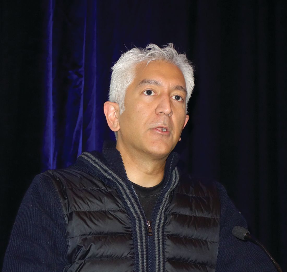
“If you take nothing else away from this talk, is really quite profound,” declared Dr. Desai, director of the cardiomyopathy and heart failure program at Brigham and Women’s Hospital, and a cardiologist at Harvard Medical School, Boston.
He highlighted five common traps or pitfalls for physicians with regard to medical therapy of patients with heart failure with reduced ejection fraction (HFrEF):
Underutilization of guideline-directed medical therapy
The current ACC/American Heart Association/Heart Failure Society of America guidelines on heart failure management (Circulation. 2017 Aug 8;136[6]:e137-61) reflect 20 years of impressive progress in improving heart failure outcomes through the use of increasingly effective guideline-directed medical therapy (GDMT). The magnitude of this improvement was nicely captured in a meta-analysis of 57 randomized controlled trials published during 1987-2015. The meta-analysis showed that, although ACE inhibitor therapy alone had no significant impact on all-cause mortality compared to placebo in patients with HFrEF, the sequential addition of guideline-directed drugs conferred stepwise improvements in survival. This approach culminated in a 56% reduction in all-cause mortality with the combination of an ACE inhibitor, beta-blocker, and mineralocorticoid receptor antagonist (MRA), compared with placebo, and a 63% reduction with an angiotensin receptor-neprilysin inhibitor (ARNI), beta-blocker, and MRA (Circ Heart Fail. 2017 Jan;10(1). pii: e003529).
Moreover, the benefits of contemporary GDMT extend beyond reductions in all-cause mortality, death due to heart failure, and heart failure–related hospitalizations into areas where one wouldn’t necessarily have expected to see much benefit. For example, an analysis of data on more than 40,000 HFrEF patients in 12 clinical trials showed a sharp decline in the rate of sudden death over the years as new agents were incorporated into GDMT. The cumulative incidence of sudden death within 90 days after randomization plunged from 2.4% in the earliest trial to 1.0% in the most recent one (N Engl J Med. 2017 Jul 6;377[1]:41-51).
“We’re at the point where we now question whether routine use of implantable cardioverter-defibrillators in primary prevention patients with nonischemic heart failure is really worthwhile on the backdrop of effective medical therapy,” Dr. Desai observed.
But there’s a problem: “We don’t do a great job with GDMT, even with this incredible evidence base that we have,” the cardiologist said.
He cited a report from the CHAMP-HF registry that scrutinized the use of GDMT in more than 3,500 ambulatory HFrEF patients in 150 U.S. primary care and cardiology practices. It found that 67% of patients deemed eligible for an MRA weren’t on one. Neither were 33% with no contraindications to beta-blocker therapy and 27% who were eligible for an ACE inhibitor, angiotensin receptor blocker (ARB), or ARNI (J Am Coll Cardiol. 2018 Jul 24;72[4]:351-66).
“This highlights a huge opportunity for further guideline-directed optimization of therapy,” he said.
Underdosing of GDMT
The CHAMP-HF registry contained further disappointing news regarding the state of treatment of patients with HFrEF in ambulatory settings: Among those patients who were on GDMT, very few were receiving the recommended target doses of the medications as established in major clinical trials and specified in the guidelines. Only 14% of patients on an ARNI were on the target dose, as were 28% on a beta-blocker, and 17% of those on an ACE inhibitor or ARB. And among patients who were eligible for all classes of GDMT, just 1% were simultaneously on the target doses of an MRA, beta-blocker, and ARNI, ACE inhibitor, or ARB. This despite solid evidence that, although some benefit is derived from initiating these medications, incremental benefit comes from dose titration.
“Even for those of us who feel like we do this quite well, if we examine our practices systematically – and we’ve done this in our own practices at Brigham and Women’s – you see that a lot of eligible patients aren’t on optimal therapy. And you might argue that many of them have contraindications, but even when you do a deep dive into the literature or the electronic medical record and ask the question – Why is this patient with normal renal function and normal potassium with class II HFrEF not on an MRA? – sometimes it’s hard to establish why that’s the case,” said Dr. Desai.
Interrupting GDMT during hospitalizations
This is common practice. But in fact, continuation of GDMT is generally well tolerated in the setting of acute decompensated heart failure in the absence of severe hypotension and cardiogenic shock. Moreover, in-hospital discontinuation or dose reduction is associated with increased risks of readmission and mortality.
And in treatment-naive HFrEF patients, what better place to introduce a medication and assess its tolerability than the hospital? Plus, medications prescribed at discharge are more likely to be continued in the outpatient setting, he noted.
Being seduced by the illusion of stability
The guidelines state that patients with NYHA class II or III HFrEF who tolerate an ACE inhibitor or ARB should be transitioned to an ARNI to further reduce their risk of morbidity and mortality. Yet many physicians wait to make the switch until clinical decompensation occurs. That’s a mistake, as was demonstrated in the landmark PARADIGM-HF trial. Twenty percent of study participants without a prior hospitalization for heart failure experienced cardiovascular death or heart failure hospitalization during the follow-up period. Patients who were clinically stable as defined by no prior heart failure hospitalization or none within 3 months prior to enrollment were as likely to benefit from ARNI therapy with sacubitril/valsartan (Entresto) as were those with a recent decompensation (JACC Heart Fail. 2016 Oct;4[10]:816-22). “A key message is that stability is an illusion in patients with symptomatic heart failure,”said Dr. Desai. “In PARADIGM-HF, the first event for about half of patients was not heralded by a worsening of symptoms or a heart failure hospitalization, it was an abrupt death at home. This may mean that a missed opportunity to optimize treatment may not come back to you down the road, so waiting until patients get worse in order to optimize their therapy may not be the best strategy.”
Inadequate laboratory monitoring
The MRAs, spironolactone and eplerenone (Inspra), are the GDMT drugs for which laboratory surveillance takes on the greatest importance because of their potential to induce hyperkalemia. The guidelines are clear that a potassium level and measurement of renal function should be obtained within a week of initiating therapy with an MRA, again at 4 weeks, and periodically thereafter.
“In general, this is done in clinical practice almost never,” Dr. Desai stressed.
These agents should be avoided in patients with prior hyperkalemia or advanced chronic kidney disease, and used with care in groups known to be at increased risk for hyperkalemia, including the elderly and patients with diabetes.
He considers spironolactone equivalent to eplerenone so long as the dosing is adequate. He generally reserves eplerenone for patients with poorly tolerated antiandrogenic effects on spironolactone.
Dr. Desai reported serving as a paid consultant to more than half a dozen pharmaceutical or medical device companies.
SNOWMASS, COLO. – Many of the abundant missed opportunities to optimize pharmacotherapy for heart failure with reduced ejection fraction revolve around not getting fully on board with the guideline-directed medical therapy shown to be highly effective at improving clinical outcomes, Akshay S. Desai, MD, asserted at the Annual Cardiovascular Conference at Snowmass sponsored by the American College of Cardiology.
“If you take nothing else away from this talk, is really quite profound,” declared Dr. Desai, director of the cardiomyopathy and heart failure program at Brigham and Women’s Hospital, and a cardiologist at Harvard Medical School, Boston.
He highlighted five common traps or pitfalls for physicians with regard to medical therapy of patients with heart failure with reduced ejection fraction (HFrEF):
Underutilization of guideline-directed medical therapy
The current ACC/American Heart Association/Heart Failure Society of America guidelines on heart failure management (Circulation. 2017 Aug 8;136[6]:e137-61) reflect 20 years of impressive progress in improving heart failure outcomes through the use of increasingly effective guideline-directed medical therapy (GDMT). The magnitude of this improvement was nicely captured in a meta-analysis of 57 randomized controlled trials published during 1987-2015. The meta-analysis showed that, although ACE inhibitor therapy alone had no significant impact on all-cause mortality compared to placebo in patients with HFrEF, the sequential addition of guideline-directed drugs conferred stepwise improvements in survival. This approach culminated in a 56% reduction in all-cause mortality with the combination of an ACE inhibitor, beta-blocker, and mineralocorticoid receptor antagonist (MRA), compared with placebo, and a 63% reduction with an angiotensin receptor-neprilysin inhibitor (ARNI), beta-blocker, and MRA (Circ Heart Fail. 2017 Jan;10(1). pii: e003529).
Moreover, the benefits of contemporary GDMT extend beyond reductions in all-cause mortality, death due to heart failure, and heart failure–related hospitalizations into areas where one wouldn’t necessarily have expected to see much benefit. For example, an analysis of data on more than 40,000 HFrEF patients in 12 clinical trials showed a sharp decline in the rate of sudden death over the years as new agents were incorporated into GDMT. The cumulative incidence of sudden death within 90 days after randomization plunged from 2.4% in the earliest trial to 1.0% in the most recent one (N Engl J Med. 2017 Jul 6;377[1]:41-51).
“We’re at the point where we now question whether routine use of implantable cardioverter-defibrillators in primary prevention patients with nonischemic heart failure is really worthwhile on the backdrop of effective medical therapy,” Dr. Desai observed.
But there’s a problem: “We don’t do a great job with GDMT, even with this incredible evidence base that we have,” the cardiologist said.
He cited a report from the CHAMP-HF registry that scrutinized the use of GDMT in more than 3,500 ambulatory HFrEF patients in 150 U.S. primary care and cardiology practices. It found that 67% of patients deemed eligible for an MRA weren’t on one. Neither were 33% with no contraindications to beta-blocker therapy and 27% who were eligible for an ACE inhibitor, angiotensin receptor blocker (ARB), or ARNI (J Am Coll Cardiol. 2018 Jul 24;72[4]:351-66).
“This highlights a huge opportunity for further guideline-directed optimization of therapy,” he said.
Underdosing of GDMT
The CHAMP-HF registry contained further disappointing news regarding the state of treatment of patients with HFrEF in ambulatory settings: Among those patients who were on GDMT, very few were receiving the recommended target doses of the medications as established in major clinical trials and specified in the guidelines. Only 14% of patients on an ARNI were on the target dose, as were 28% on a beta-blocker, and 17% of those on an ACE inhibitor or ARB. And among patients who were eligible for all classes of GDMT, just 1% were simultaneously on the target doses of an MRA, beta-blocker, and ARNI, ACE inhibitor, or ARB. This despite solid evidence that, although some benefit is derived from initiating these medications, incremental benefit comes from dose titration.
“Even for those of us who feel like we do this quite well, if we examine our practices systematically – and we’ve done this in our own practices at Brigham and Women’s – you see that a lot of eligible patients aren’t on optimal therapy. And you might argue that many of them have contraindications, but even when you do a deep dive into the literature or the electronic medical record and ask the question – Why is this patient with normal renal function and normal potassium with class II HFrEF not on an MRA? – sometimes it’s hard to establish why that’s the case,” said Dr. Desai.
Interrupting GDMT during hospitalizations
This is common practice. But in fact, continuation of GDMT is generally well tolerated in the setting of acute decompensated heart failure in the absence of severe hypotension and cardiogenic shock. Moreover, in-hospital discontinuation or dose reduction is associated with increased risks of readmission and mortality.
And in treatment-naive HFrEF patients, what better place to introduce a medication and assess its tolerability than the hospital? Plus, medications prescribed at discharge are more likely to be continued in the outpatient setting, he noted.
Being seduced by the illusion of stability
The guidelines state that patients with NYHA class II or III HFrEF who tolerate an ACE inhibitor or ARB should be transitioned to an ARNI to further reduce their risk of morbidity and mortality. Yet many physicians wait to make the switch until clinical decompensation occurs. That’s a mistake, as was demonstrated in the landmark PARADIGM-HF trial. Twenty percent of study participants without a prior hospitalization for heart failure experienced cardiovascular death or heart failure hospitalization during the follow-up period. Patients who were clinically stable as defined by no prior heart failure hospitalization or none within 3 months prior to enrollment were as likely to benefit from ARNI therapy with sacubitril/valsartan (Entresto) as were those with a recent decompensation (JACC Heart Fail. 2016 Oct;4[10]:816-22). “A key message is that stability is an illusion in patients with symptomatic heart failure,”said Dr. Desai. “In PARADIGM-HF, the first event for about half of patients was not heralded by a worsening of symptoms or a heart failure hospitalization, it was an abrupt death at home. This may mean that a missed opportunity to optimize treatment may not come back to you down the road, so waiting until patients get worse in order to optimize their therapy may not be the best strategy.”
Inadequate laboratory monitoring
The MRAs, spironolactone and eplerenone (Inspra), are the GDMT drugs for which laboratory surveillance takes on the greatest importance because of their potential to induce hyperkalemia. The guidelines are clear that a potassium level and measurement of renal function should be obtained within a week of initiating therapy with an MRA, again at 4 weeks, and periodically thereafter.
“In general, this is done in clinical practice almost never,” Dr. Desai stressed.
These agents should be avoided in patients with prior hyperkalemia or advanced chronic kidney disease, and used with care in groups known to be at increased risk for hyperkalemia, including the elderly and patients with diabetes.
He considers spironolactone equivalent to eplerenone so long as the dosing is adequate. He generally reserves eplerenone for patients with poorly tolerated antiandrogenic effects on spironolactone.
Dr. Desai reported serving as a paid consultant to more than half a dozen pharmaceutical or medical device companies.
REPORTING FROM ACC SNOWMASS 2019
Novel RA strategy: Target first-line biologics for likely methotrexate nonresponders
MAUI, HAWAII – An ongoing proof-of-concept study in the United Kingdom uses T-cell subset analysis to identify those patients with early rheumatoid arthritis who are unlikely to experience remission with methotrexate alone and therefore warrant more potent first-line therapy with a tumor necrosis factor (TNF) inhibitor in combination with methotrexate, Paul Emery, MD, said at the 2019 Rheumatology Winter Clinical Symposium.
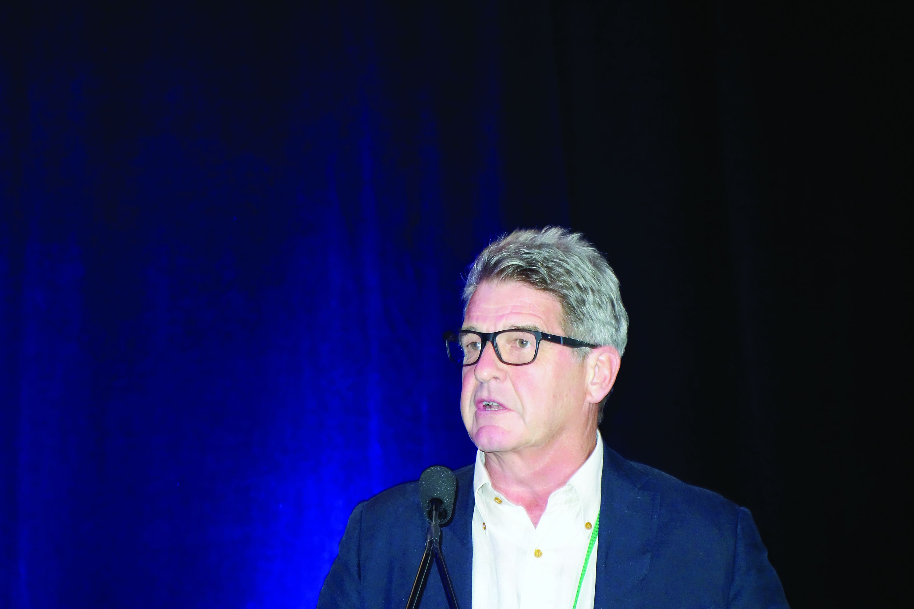
“When we get the readout from this study, if we get the results we expect, we could actually put it to the National Health Service that it would be cost saving to use biologics as first-line therapy in a proportion, which will be about 40% of patients. But we won’t necessarily need to use them for a year, and we’ll get a 70%-plus remission rate, which I think is the sort of level we should be asking for in our patients,” according to Dr. Emery, professor of rheumatology and director of the University of Leeds (England) Musculoskeletal Biomedical Research Center.
This proof-of-concept study capitalizes on earlier work by Dr. Emery and coinvestigators, who showed in 70 patients with early rheumatoid arthritis given methotrexate as their first-ever disease-modifying antirheumatic drug (DMARD) that those with a normal pretreatment frequency of naive CD4+ T cells had an 83% remission rate at 6 months as defined by a Disease Activity Score in 28 joints (DAS28) of less than 2.6. In contrast, only 21% of those with a reduced naive CD4+ T-cell frequency experienced remission when compared with healthy controls. In an analysis adjusted for age and the presence of anti–citrullinated protein antibodies (ACPA), a normal baseline naive CD4+ T-cell frequency was associated with a 5.9-fold increased likelihood of remission on methotrexate (Ann Rheum Dis. 2014 Nov;73[11]:2047-53).
In the new proof-of-concept study, DMARD-naive, ACPA-positive patients with early RA undergo T-cell subset quantification by flow cytometry. Thirty patients with a normal test result are assigned to methotrexate with treat-to-target dose escalation. Based upon the investigators’ earlier work, it’s anticipated that about 80% of these patients will be in remission at 6 months.
Sixty patients with an abnormal baseline T-cell test result are being randomized to methotrexate plus either placebo or biosimilar etanercept. Again based upon the earlier study, the expected remission rate at 6 months in the methotrexate-plus-placebo group is about 20%. In contrast, the anticipated remission rate in the patients on an anti-TNF biologic plus methotrexate as first-line therapy is about 70% on the basis of the results of previous clinical trials, including PRIZE (N Engl J Med. 2014 Nov 6;371[19]:1781-92) and COMET (Ann Rheum Dis. 2012 Jun;71[6]:989-92).
Meanwhile, the price tag for biosimilar TNF inhibitors in the United Kingdom has come down to the point that routine across-the-board use of biologics as first-line therapy is arguably cost effective, a situation Dr. Emery described as hitherto “the unthinkable.”
The cost of biosimilar adalimumab in the coming year will be less than $3,000 annually in the U.K. health care system. So if 100 patients with early RA and no contraindication to biologic therapy are placed on biosimilar adalimumab and methotrexate for 1 year, the total cost for the biologic in this cohort will be less than $300,000, and roughly 70 of the 100 patients will have achieved remission. This approach makes much more sense than current standard practice, which is to reserve biologics as second-line therapy for patients who have failed to achieve remission on nonbiologic DMARDs, thereby allowing their joint damage to advance in the interim to the point that they need to stay on biologic therapy for decades, the rheumatologist argued.
“Once one accepts that remission has become the aim of therapy, then the facts, I think, speak for themselves: There’s absolutely no doubt that the rate of remission is best with the first DMARD. So if our aim is remission, we should use that one opportunity with the best agent first, because we’re not going to get the same response later,” Dr. Emery said.
Also, “there’s no doubt” that the dose of biologics can be halved with no loss of efficacy in patients who achieve remission on full-dose therapy, as previously demonstrated in PRIZE and other trials. This strategy further reduces the overall cost of biologic therapy, he added.
Dr. Emery, who recently received the Order of the British Empire from Queen Elizabeth personally in recognition of his career achievements in rheumatology, reported having no financial conflicts of interest regarding his presentation.
MAUI, HAWAII – An ongoing proof-of-concept study in the United Kingdom uses T-cell subset analysis to identify those patients with early rheumatoid arthritis who are unlikely to experience remission with methotrexate alone and therefore warrant more potent first-line therapy with a tumor necrosis factor (TNF) inhibitor in combination with methotrexate, Paul Emery, MD, said at the 2019 Rheumatology Winter Clinical Symposium.
“When we get the readout from this study, if we get the results we expect, we could actually put it to the National Health Service that it would be cost saving to use biologics as first-line therapy in a proportion, which will be about 40% of patients. But we won’t necessarily need to use them for a year, and we’ll get a 70%-plus remission rate, which I think is the sort of level we should be asking for in our patients,” according to Dr. Emery, professor of rheumatology and director of the University of Leeds (England) Musculoskeletal Biomedical Research Center.
This proof-of-concept study capitalizes on earlier work by Dr. Emery and coinvestigators, who showed in 70 patients with early rheumatoid arthritis given methotrexate as their first-ever disease-modifying antirheumatic drug (DMARD) that those with a normal pretreatment frequency of naive CD4+ T cells had an 83% remission rate at 6 months as defined by a Disease Activity Score in 28 joints (DAS28) of less than 2.6. In contrast, only 21% of those with a reduced naive CD4+ T-cell frequency experienced remission when compared with healthy controls. In an analysis adjusted for age and the presence of anti–citrullinated protein antibodies (ACPA), a normal baseline naive CD4+ T-cell frequency was associated with a 5.9-fold increased likelihood of remission on methotrexate (Ann Rheum Dis. 2014 Nov;73[11]:2047-53).
In the new proof-of-concept study, DMARD-naive, ACPA-positive patients with early RA undergo T-cell subset quantification by flow cytometry. Thirty patients with a normal test result are assigned to methotrexate with treat-to-target dose escalation. Based upon the investigators’ earlier work, it’s anticipated that about 80% of these patients will be in remission at 6 months.
Sixty patients with an abnormal baseline T-cell test result are being randomized to methotrexate plus either placebo or biosimilar etanercept. Again based upon the earlier study, the expected remission rate at 6 months in the methotrexate-plus-placebo group is about 20%. In contrast, the anticipated remission rate in the patients on an anti-TNF biologic plus methotrexate as first-line therapy is about 70% on the basis of the results of previous clinical trials, including PRIZE (N Engl J Med. 2014 Nov 6;371[19]:1781-92) and COMET (Ann Rheum Dis. 2012 Jun;71[6]:989-92).
Meanwhile, the price tag for biosimilar TNF inhibitors in the United Kingdom has come down to the point that routine across-the-board use of biologics as first-line therapy is arguably cost effective, a situation Dr. Emery described as hitherto “the unthinkable.”
The cost of biosimilar adalimumab in the coming year will be less than $3,000 annually in the U.K. health care system. So if 100 patients with early RA and no contraindication to biologic therapy are placed on biosimilar adalimumab and methotrexate for 1 year, the total cost for the biologic in this cohort will be less than $300,000, and roughly 70 of the 100 patients will have achieved remission. This approach makes much more sense than current standard practice, which is to reserve biologics as second-line therapy for patients who have failed to achieve remission on nonbiologic DMARDs, thereby allowing their joint damage to advance in the interim to the point that they need to stay on biologic therapy for decades, the rheumatologist argued.
“Once one accepts that remission has become the aim of therapy, then the facts, I think, speak for themselves: There’s absolutely no doubt that the rate of remission is best with the first DMARD. So if our aim is remission, we should use that one opportunity with the best agent first, because we’re not going to get the same response later,” Dr. Emery said.
Also, “there’s no doubt” that the dose of biologics can be halved with no loss of efficacy in patients who achieve remission on full-dose therapy, as previously demonstrated in PRIZE and other trials. This strategy further reduces the overall cost of biologic therapy, he added.
Dr. Emery, who recently received the Order of the British Empire from Queen Elizabeth personally in recognition of his career achievements in rheumatology, reported having no financial conflicts of interest regarding his presentation.
REPORTING FROM RWCS 2019
Infective endocarditis isn’t what it used to be
SNOWMASS, COLO. – Infective endocarditis in 2019 is very different from the disease most physicians encountered in training, both in terms of epidemiology and clinical presentation, Patrick T. O’Gara, MD, observed at the Annual Cardiovascular Conference at Snowmass sponsored by the American College of Cardiology.
The classic description of infective endocarditis provided by Sir William Osler, MD, was of a subacute bacterial infection characterized by a long latent phase of low-grade fever, back pain, weight loss, and night sweats. It was mainly a right-heart disease of younger individuals with an infected native valve, and the predominant pathogens were streptococci, Dr. O’Gara said.
“I think in the current era endocarditis is more often characterized by an acute illness with toxic features in the context of adults with a high burden of degenerative diseases – for example, patients with rheumatoid arthritis or psoriatic arthritis on immunosuppressive therapy, or diabetes, end-stage renal disease, and risk factors for hospital-acquired infection. Injectable drug use is through the roof, there’s a wider prevalence of cardiac implanted electronic devices, which are a wonderful place for bacteria to hide, and Staphylococcus aureus has certainly become the leading pathogen with regard to endocarditis in the United States, especially MRSA, often multidrug resistant,” said Dr. O’Gara, professor of medicine at Harvard Medical School, Boston.
“Also, no talk about endocarditis is sufficient without paying some attention to the opioid crisis in which we find ourselves. It’s one of the top three causes of death among young men in the United States, along with accidents and gun violence. No region of the country is spared. This has completely inundated our ER and hospitalist services and our inpatient cardiology services with folks who are often repeat offenders when it comes to the difficulty in being able to give up an injectable drug use habit. They have multiple infections and hospitalizations, tricuspid valve involvement, and depending upon the aggressiveness of the Staphylococcus organism, typically they have left-sided disease with multiple complications, including aortic regurgitation and heart failure,” the cardiologist continued.
This description underscored one of Dr. O’Gara’s major points about the challenges posed by infective endocarditis in contemporary practice: “Expect the unexpected,” he advised. “When you’ve seen one case of infective endocarditis, you’ve seen one case of infective endocarditis.”
Outcomes are ‘sobering’
In the current era, outcomes are “sobering,” the cardiologist noted. Infective endocarditis carries a 6-month mortality rate of 20%-25% despite early surgery being performed during the index hospitalization in up to 60% of patients, with a relatively high perioperative mortality rate of about 10%. However, the risk of reinfection occurring in a newly implanted cardiac valve is impressively low at about 2%.
Refer early for multimodality imaging and surgical consultation
Transesophageal echocardiography is valuable in assessment of the infected valve. However, when extravalvular extension of the infection is suspected and the echo assessment is nondiagnostic or indeterminate, it’s time to quickly move on to advanced imaging, such as PET-CT.
The ACC/American Heart Association class I recommendations for early surgery in infected native valves haven’t changed substantially in over a decade. Based largely on observational data, there is an association between early surgery and lower in-hospital mortality (Lancet. 2012 Mar 10;379[9819]:965-975).
Class IIa recommendations for native valve surgery include recurrent emboli and a persistent vegetation despite appropriate antibiotic therapy. A “very controversial” class IIb recommendation for surgery because of weak supporting data is the identification of a mobile vegetation larger than 10 mm, particularly if it’s located on an anterior mitral valve leaflet, he said.
If the decision is made to forgo early surgery, be sure to repeat transesophageal echocardiography on day 7-10 to reassess the size of the patient’s vegetation.
“There is an association between size of vegetation and 1-year mortality, with a cut point of greater than 15 mm. Some would argue this constitutes a reasonable indication for early surgery,” Dr. O’Gara noted.
The embolization rate in patients with infective endocarditis is highest during the day before presentation, the day of presentation, and through the first 2 days afterward. The rate drops precipitously within 2 weeks after initiation of appropriate antibiotic therapy. Thus, to utilize early surgery to maximum effect in order to decrease the risk of embolization, it makes sense to operate within the first several days following presentation, before antibiotics have had sufficient time to catch up with the evolving disease process.
Don’t use half measures when it comes to removal of cardiac implanted electronic devices
The guidelines are clear regarding infected pacemakers, implanted cardioverter-defibrillators, and cardiac resynchronization devices: “It all needs to come out,” Dr. O’Gara emphasized. That includes all leads and the generator in patients with documented infection of only one portion of the device system, as a class I, level of evidence B recommendation. Moreover, complete removal of a pacemaker or defibrillator system is deemed “reasonable” as a class IIa recommendation in all patients with valvular infection caused by S. aureus or fungi even in the absence of evidence of device infection.
“I think we as general cardiologists have become increasingly impressed about how sick and festering these kinds of patients can become, even when we’re not able to prove that the lead is infected. The lead looks okay on transesophageal echo or PET-CT, blood cultures are negative, the valvular heart disease is really not that advanced, but several days go by and the patient is just not responding. We should have a high index of suspicion that there’s an infection we cannot appreciate. But obviously, you make these difficult decisions in consultation with your electrophysiology colleagues,” he added.
Know when the cardiologist should say ‘no’ to early aggressive surgery
While an aggressive early surgical approach often pays off in terms of prevention of embolic sequelae and a reduction in heart failure, the timing of surgery in the 20%-40% of patients with infective endocarditis who present with stroke or other neurologic complications remains controversial. An international group of Canadian and French cardiac surgeons and neurologists developed a useful algorithm regarding the types of neurologic complications for which early cardiac surgery is a poor idea because of the high risk of neurologic exacerbation. For example, a mycotic neuroaneurysm is grounds for postponement of cardiac surgery for at least 4 weeks (Circulation. 2016 Oct 25;134[17]:1280-92).
Dr. O’Gara reported receiving funding from the National Heart, Lung and Blood Institute, the National Institute of Dental and Craniofacial Research, from Medtronic in conjunction with the ongoing pivotal APOLLO transcatheter mitral valve replacement trial, and from Edwards Lifesciences for the ongoing EARLY TAVR trial.
REPORTING FROM ACC SNOWMASS 2019
Rheumatologist involvement often reclassifies interstitial lung disease
MAUI, HAWAII – The latest practice guidelines on the diagnosis of interstitial lung disease issued by the American Thoracic Society and allied organizations recommend as the standard of care a review of all cases by a multidisciplinary team consisting of a pulmonologist, radiologist, and pathologist to ensure accurate diagnosis and classification.

That’s not good enough. A rheumatologist needs to routinely be involved in those multidisciplinary discussions as well, Aryeh Fischer, MD, asserted at the 2019 Rheumatology Winter Clinical Symposium.
Why? Because a rheumatologist’s input often leads to a change in diagnosis. And that change can have important prognostic and therapeutic implications.
“We want to distinguish the IPF [idiopathic pulmonary fibrosis] patients from everybody else. The most important thing with regards to therapy is to identify the IPF patient. The IPF patients are the only ones who are able to be treated with antifibrotic agents: pirfenidone or nintedanib. And we know that immunosuppression can make patients with IPF worse; their risks of hospitalization and mortality are higher on immunosuppression. But everybody else, including anyone with any of the autoimmune diseases along with ILD [interstitial lung disease] or any other causes of ILD, gets treated with immunosuppression,” explained Dr. Fischer, a rheumatologist with a special interest in autoimmune lung disease at the University of Colorado, Denver.
He cited a recent prospective blinded study in which 60 newly diagnosed ILD patients were evaluated separately by a multidisciplinary team comprising a pulmonologist, radiologist, and pathologist and once again with the involvement of a rheumatologist. The rheumatologic assessment reclassified 21% of patients from IPF – that is, lung disease unrelated to connective tissue disease (CTD) or exposure to asbestos, bird droppings, or other triggers – to ILD with connective tissue disease (CTD-ILD). And the number of patients classified as having ILD with autoimmune features without meeting full diagnostic criteria for a major CTD, a category that includes antisynthetase syndrome and IgG4-related ILD, jumped by 77%. Also, the investigators determined that adding a rheumatologist to the multidisciplinary team would have resulted in seven fewer bronchoscopies and one less surgical biopsy among this 60-patient cohort (J Rheumatol. 2018 Nov;45[11]:1509-14).
It’s not at all uncommon to identify a new occult CTD in patients presenting with ILD. Dr. Fischer noted that in a series of 114 consecutive patients evaluated at the interdisciplinary ILD program at Johns Hopkins University, Baltimore, 30% of them had a well-defined CTD, half of whom, or 15% of the total, were newly diagnosed with CTD by virtue of their ILD evaluation (Respir Med. 2009 Aug;103[8]:1152-8).
A CTD-ILD diagnosis has prognostic implications: Survival is significantly better than with IPF, with the exception of ILD with rheumatoid arthritis (RA-ILD), where the prognosis seems to be worse than for other forms of CTD-ILD and is more akin to that of IPF. Indeed, the risk of death is threefold higher in patients with RA-ILD than in those with RA without ILD. While the predominant pattern of lung injury in RA-ILD is usual interstitial pneumonia, marked by fibrosis and honeycombing, the predominant pattern in all other forms of CTD-ILD is nonspecific interstitial pneumonia.
A French study of 778 consecutive patients with ILD highlighted how common autoimmune disease is in the ILD population. Nearly one-third of the ILD patients had autoimmune disease: 22% had CTD-ILD and another 7% had interstitial pneumonia with autoimmune features, or IPAF (Respir Med. 2017 Feb;123:56-62).
Dr. Fischer was lead author of a report by a European Respiratory Society/American Thoracic Society task force that first put forth the term IPAF to describe ILD patients with subtle serologic, clinical, and/or morphologic findings that don’t rise to the level required for formal diagnosis of a defined CTD (Eur Respir J. 2015 Oct;46[4]:976-87).
IPAF is a research construct. Patients who fall into this category are the focus of ongoing prospective studies aimed at better understanding their prognosis and appropriate treatment.
The big three autoimmune diseases comorbid with ILD as reported by Dr. Fischer and coinvestigators in a study of 237 patients with an autoimmune phenotype in a Denver ILD clinic were scleroderma, present in 37%; rheumatoid arthritis, present in 18%; and myositis, with a prevalence of 11%. Another 24% had IPAF.
“There was surprisingly little SLE [3%], and not much Sjögren’s [6%],” he observed.
When pulmonologists come knocking on Dr. Fischer’s door asking if their patients with ILD have occult CTD, he finds it helpful to look for quantifiable specific extrathoracic features suggestive of rheumatologic disease, such as Raynaud’s, sclerodactyly, mechanic hands, and Gottron’s papules.
The ILD practice guidelines recently issued jointly by the American Thoracic Society, European Respiratory Society, Japanese Respiratory Society, and Latin American Thoracic Society recommend that serologic evaluation for a long list of autoantibodies “should be performed even in the absence of signs or symptoms of connective tissue disease” (Am J Respir Crit Care Med. 2018 Sep 1;198[5]:e44-e68). Dr. Fischer indicated he has a problem with that recommendation since nonrheumatologists aren’t typically adept in interpreting the significance of autoantibodies.
“I will just tell you, I see a lot of patients for ILD evaluation, and autoimmune serologies often lead to more questions than answers. They always need to be considered within the clinical context,” the rheumatologist said.
High-resolution CT (HRCT) is the imaging method of choice for detecting ILD and classifying its severity. HRCT also holds clues for detection of CTD-ILD.
“There are no upper lung–predominant ILDs that really make you think CTD. Our diseases like the lower lung zones. We like to see multicompartment involvement. When you hear ‘airways and parenchymal,’ ‘pleural,’ ‘pericardial thickening,’ that sounds a lot like autoimmune disease,” according to Dr. Fischer.
Also, the presence of a dilated esophagus on HRCT is strongly suggestive of scleroderma, he added.
In the event a pathology report is available, it’s important to read the fine print. Secondary histopathologic features of CTD-ILD include extensive pleuritis, lymphoid aggregates with germinal center formation, dense perivascular collagen, and/or prominent plasmacytic infiltration.
While ILD can be the first manifestation of an underlying occult CTD, it’s also common for a patient with an established CTD to subsequently develop ILD.
Rheumatologists are no strangers to ambiguity, so it should come as no surprise that while audible bibasilar crackles on physical examination are strongly predictive of ILD, their absence doesn’t indicate absence of ILD. Similarly, dyspnea in a patient with established CTD can be tough to interpret, and absence of dyspnea doesn’t imply absence of ILD. If a patient with CTD is on immunosuppressive therapy, bronchoalveolar lavage is of value in ruling out infection.
When to order a surgical lung biopsy
“If you’re pretty sure your patient had a CTD and then you get a characteristic HRCT and there are no exposures to account for the imaging findings – if we have a scenario where it all fits – we often don’t biopsy. Biopsy usually means a 2-night hospital stay, and it’s reportedly associated with about a 1% mortality risk. And the clinical reality is the biopsy may not impact treatment. They’re going to give you azathioprine, mycophenolate, cyclophosphamide, or prednisone for the ILD and the extrathoracic disease irrespective of the ILD pattern,” the rheumatologist said.
He reserves surgical biopsy for patients with an atypical HRCT, those with known CTD and a possible alternative etiology for the ILD, such as exposure to asbestos or owning pet birds, and patients where he’s just not sure a CTD is actually present.
Treatment of CTD-ILD
“The available controlled efficacy data are limited to scleroderma-ILD, where cyclophosphamide and mycophenolate work a little bit. And that’s it. We don’t have good data to guide us on which agent for which CTD or ILD pattern, for how long, or what dose,” Dr. Fischer said. “We have good drugs for the joints, nothing for the lungs. Treatment is not evidence based. We initiate with high-dose steroids, switch to a steroid-sparing agent, we evaluate the response to treatment with surveillance every 3-6 months by 6-minute walking, lung function tests, and sometimes imaging, and we treat for a long time. Oftentimes stability equals success.”
That being said, it must be emphasized that not all CTD-ILD warrants treatment of the pulmonary disease. If the patient’s ILD is mild and indolent, clinical surveillance is often appropriate, he continued.
More important than the pharmacotherapy available at present are adjunctive nonpharmacologic approaches: supplemental oxygen, pulmonary rehabilitation, treatment of comorbid GERD, immunizations, and addressing mental health issues related to these devastating diseases.
“We really don’t do these things well,” Dr. Fischer said.
On the horizon
An investigator-driven phase 2 clinical trial of the antifibrotic drug pirfenidone (Esbriet), now approved for IPF, is underway in patients with RA-ILD. Results of a trial of antifibrotic therapy in patients with scleroderma-ILD are due to be presented this year at EULAR. And a small study of antifibrotic therapy in patients with myositis-ILD is ongoing.
As for the biologics, there is a signal in the literature that tumor necrosis factor inhibitors may be associated with increased risk of rapidly progressive lung disease in patients with RA-ILD. An influential report from the British Society for Rheumatology Biologics Register on 299 patients with preexisting RA-ILD treated with anti-TNF therapy and 68 who received traditional disease-modifying antirrheumatic drugs showed that while mortality during follow-up was similar in the two groups, the proportion of deaths attributed to RA-ILD was 21% in the group on anti-TNF therapy, compared with only 7% in those on traditional DMARDs (Ann Rheum Dis. 2010 Jun;69[6]:1086-91).
Paul Emery, MD, rose from the audience at Dr. Fischer’s request to share his extensive experience with anti-TNF therapy and rituximab (Rituxan) in the setting of RA-ILD. When he was involved in several of the pivotal clinical trials of anti-TNF agents for RA he encountered a couple of cases of rapidly progressive ILD in patients on treatment.
“We had never before seen rapidly progressive ILD that didn’t respond to cyclophosphamide. Both patients died,” recalled Dr. Emery, professor of rheumatology and director of the University of Leeds (England) Musculoskeletal Biomedical Research Center.
“Our experience is if you’ve got mild ILD – and if you look hard enough you can find it in many rheumatoids – TNF inhibitor therapy doesn’t affect it. But if there’s any hint of deterioration we move away from anti-TNF therapy. Our preference has been for rituximab,” he said.
Dr. Emery was senior author of the largest study to date of rituximab in patients with RA-ILD (Rheumatology [Oxford]. 2017 Aug 1;56[8]:1348-57). While he and his coinvestigators concluded that rituximab “appears to be an acceptable therapeutic choice for patients with RA-ILD,” Dr. Fischer didn’t find persuasive evidence from this relatively small retrospective observational study in which only 44 patients had lung data available.
“My own conclusion is evidence is lacking to support a role of rituximab for treating ILD in RA,” Dr. Fischer said.
He reported receiving research grants from Boehringer-Ingelheim and Corbus and serving as a consultant to a handful of other pharmaceutical companies.
MAUI, HAWAII – The latest practice guidelines on the diagnosis of interstitial lung disease issued by the American Thoracic Society and allied organizations recommend as the standard of care a review of all cases by a multidisciplinary team consisting of a pulmonologist, radiologist, and pathologist to ensure accurate diagnosis and classification.
That’s not good enough. A rheumatologist needs to routinely be involved in those multidisciplinary discussions as well, Aryeh Fischer, MD, asserted at the 2019 Rheumatology Winter Clinical Symposium.
Why? Because a rheumatologist’s input often leads to a change in diagnosis. And that change can have important prognostic and therapeutic implications.
“We want to distinguish the IPF [idiopathic pulmonary fibrosis] patients from everybody else. The most important thing with regards to therapy is to identify the IPF patient. The IPF patients are the only ones who are able to be treated with antifibrotic agents: pirfenidone or nintedanib. And we know that immunosuppression can make patients with IPF worse; their risks of hospitalization and mortality are higher on immunosuppression. But everybody else, including anyone with any of the autoimmune diseases along with ILD [interstitial lung disease] or any other causes of ILD, gets treated with immunosuppression,” explained Dr. Fischer, a rheumatologist with a special interest in autoimmune lung disease at the University of Colorado, Denver.
He cited a recent prospective blinded study in which 60 newly diagnosed ILD patients were evaluated separately by a multidisciplinary team comprising a pulmonologist, radiologist, and pathologist and once again with the involvement of a rheumatologist. The rheumatologic assessment reclassified 21% of patients from IPF – that is, lung disease unrelated to connective tissue disease (CTD) or exposure to asbestos, bird droppings, or other triggers – to ILD with connective tissue disease (CTD-ILD). And the number of patients classified as having ILD with autoimmune features without meeting full diagnostic criteria for a major CTD, a category that includes antisynthetase syndrome and IgG4-related ILD, jumped by 77%. Also, the investigators determined that adding a rheumatologist to the multidisciplinary team would have resulted in seven fewer bronchoscopies and one less surgical biopsy among this 60-patient cohort (J Rheumatol. 2018 Nov;45[11]:1509-14).
It’s not at all uncommon to identify a new occult CTD in patients presenting with ILD. Dr. Fischer noted that in a series of 114 consecutive patients evaluated at the interdisciplinary ILD program at Johns Hopkins University, Baltimore, 30% of them had a well-defined CTD, half of whom, or 15% of the total, were newly diagnosed with CTD by virtue of their ILD evaluation (Respir Med. 2009 Aug;103[8]:1152-8).
A CTD-ILD diagnosis has prognostic implications: Survival is significantly better than with IPF, with the exception of ILD with rheumatoid arthritis (RA-ILD), where the prognosis seems to be worse than for other forms of CTD-ILD and is more akin to that of IPF. Indeed, the risk of death is threefold higher in patients with RA-ILD than in those with RA without ILD. While the predominant pattern of lung injury in RA-ILD is usual interstitial pneumonia, marked by fibrosis and honeycombing, the predominant pattern in all other forms of CTD-ILD is nonspecific interstitial pneumonia.
A French study of 778 consecutive patients with ILD highlighted how common autoimmune disease is in the ILD population. Nearly one-third of the ILD patients had autoimmune disease: 22% had CTD-ILD and another 7% had interstitial pneumonia with autoimmune features, or IPAF (Respir Med. 2017 Feb;123:56-62).
Dr. Fischer was lead author of a report by a European Respiratory Society/American Thoracic Society task force that first put forth the term IPAF to describe ILD patients with subtle serologic, clinical, and/or morphologic findings that don’t rise to the level required for formal diagnosis of a defined CTD (Eur Respir J. 2015 Oct;46[4]:976-87).
IPAF is a research construct. Patients who fall into this category are the focus of ongoing prospective studies aimed at better understanding their prognosis and appropriate treatment.
The big three autoimmune diseases comorbid with ILD as reported by Dr. Fischer and coinvestigators in a study of 237 patients with an autoimmune phenotype in a Denver ILD clinic were scleroderma, present in 37%; rheumatoid arthritis, present in 18%; and myositis, with a prevalence of 11%. Another 24% had IPAF.
“There was surprisingly little SLE [3%], and not much Sjögren’s [6%],” he observed.
When pulmonologists come knocking on Dr. Fischer’s door asking if their patients with ILD have occult CTD, he finds it helpful to look for quantifiable specific extrathoracic features suggestive of rheumatologic disease, such as Raynaud’s, sclerodactyly, mechanic hands, and Gottron’s papules.
The ILD practice guidelines recently issued jointly by the American Thoracic Society, European Respiratory Society, Japanese Respiratory Society, and Latin American Thoracic Society recommend that serologic evaluation for a long list of autoantibodies “should be performed even in the absence of signs or symptoms of connective tissue disease” (Am J Respir Crit Care Med. 2018 Sep 1;198[5]:e44-e68). Dr. Fischer indicated he has a problem with that recommendation since nonrheumatologists aren’t typically adept in interpreting the significance of autoantibodies.
“I will just tell you, I see a lot of patients for ILD evaluation, and autoimmune serologies often lead to more questions than answers. They always need to be considered within the clinical context,” the rheumatologist said.
High-resolution CT (HRCT) is the imaging method of choice for detecting ILD and classifying its severity. HRCT also holds clues for detection of CTD-ILD.
“There are no upper lung–predominant ILDs that really make you think CTD. Our diseases like the lower lung zones. We like to see multicompartment involvement. When you hear ‘airways and parenchymal,’ ‘pleural,’ ‘pericardial thickening,’ that sounds a lot like autoimmune disease,” according to Dr. Fischer.
Also, the presence of a dilated esophagus on HRCT is strongly suggestive of scleroderma, he added.
In the event a pathology report is available, it’s important to read the fine print. Secondary histopathologic features of CTD-ILD include extensive pleuritis, lymphoid aggregates with germinal center formation, dense perivascular collagen, and/or prominent plasmacytic infiltration.
While ILD can be the first manifestation of an underlying occult CTD, it’s also common for a patient with an established CTD to subsequently develop ILD.
Rheumatologists are no strangers to ambiguity, so it should come as no surprise that while audible bibasilar crackles on physical examination are strongly predictive of ILD, their absence doesn’t indicate absence of ILD. Similarly, dyspnea in a patient with established CTD can be tough to interpret, and absence of dyspnea doesn’t imply absence of ILD. If a patient with CTD is on immunosuppressive therapy, bronchoalveolar lavage is of value in ruling out infection.
When to order a surgical lung biopsy
“If you’re pretty sure your patient had a CTD and then you get a characteristic HRCT and there are no exposures to account for the imaging findings – if we have a scenario where it all fits – we often don’t biopsy. Biopsy usually means a 2-night hospital stay, and it’s reportedly associated with about a 1% mortality risk. And the clinical reality is the biopsy may not impact treatment. They’re going to give you azathioprine, mycophenolate, cyclophosphamide, or prednisone for the ILD and the extrathoracic disease irrespective of the ILD pattern,” the rheumatologist said.
He reserves surgical biopsy for patients with an atypical HRCT, those with known CTD and a possible alternative etiology for the ILD, such as exposure to asbestos or owning pet birds, and patients where he’s just not sure a CTD is actually present.
Treatment of CTD-ILD
“The available controlled efficacy data are limited to scleroderma-ILD, where cyclophosphamide and mycophenolate work a little bit. And that’s it. We don’t have good data to guide us on which agent for which CTD or ILD pattern, for how long, or what dose,” Dr. Fischer said. “We have good drugs for the joints, nothing for the lungs. Treatment is not evidence based. We initiate with high-dose steroids, switch to a steroid-sparing agent, we evaluate the response to treatment with surveillance every 3-6 months by 6-minute walking, lung function tests, and sometimes imaging, and we treat for a long time. Oftentimes stability equals success.”
That being said, it must be emphasized that not all CTD-ILD warrants treatment of the pulmonary disease. If the patient’s ILD is mild and indolent, clinical surveillance is often appropriate, he continued.
More important than the pharmacotherapy available at present are adjunctive nonpharmacologic approaches: supplemental oxygen, pulmonary rehabilitation, treatment of comorbid GERD, immunizations, and addressing mental health issues related to these devastating diseases.
“We really don’t do these things well,” Dr. Fischer said.
On the horizon
An investigator-driven phase 2 clinical trial of the antifibrotic drug pirfenidone (Esbriet), now approved for IPF, is underway in patients with RA-ILD. Results of a trial of antifibrotic therapy in patients with scleroderma-ILD are due to be presented this year at EULAR. And a small study of antifibrotic therapy in patients with myositis-ILD is ongoing.
As for the biologics, there is a signal in the literature that tumor necrosis factor inhibitors may be associated with increased risk of rapidly progressive lung disease in patients with RA-ILD. An influential report from the British Society for Rheumatology Biologics Register on 299 patients with preexisting RA-ILD treated with anti-TNF therapy and 68 who received traditional disease-modifying antirrheumatic drugs showed that while mortality during follow-up was similar in the two groups, the proportion of deaths attributed to RA-ILD was 21% in the group on anti-TNF therapy, compared with only 7% in those on traditional DMARDs (Ann Rheum Dis. 2010 Jun;69[6]:1086-91).
Paul Emery, MD, rose from the audience at Dr. Fischer’s request to share his extensive experience with anti-TNF therapy and rituximab (Rituxan) in the setting of RA-ILD. When he was involved in several of the pivotal clinical trials of anti-TNF agents for RA he encountered a couple of cases of rapidly progressive ILD in patients on treatment.
“We had never before seen rapidly progressive ILD that didn’t respond to cyclophosphamide. Both patients died,” recalled Dr. Emery, professor of rheumatology and director of the University of Leeds (England) Musculoskeletal Biomedical Research Center.
“Our experience is if you’ve got mild ILD – and if you look hard enough you can find it in many rheumatoids – TNF inhibitor therapy doesn’t affect it. But if there’s any hint of deterioration we move away from anti-TNF therapy. Our preference has been for rituximab,” he said.
Dr. Emery was senior author of the largest study to date of rituximab in patients with RA-ILD (Rheumatology [Oxford]. 2017 Aug 1;56[8]:1348-57). While he and his coinvestigators concluded that rituximab “appears to be an acceptable therapeutic choice for patients with RA-ILD,” Dr. Fischer didn’t find persuasive evidence from this relatively small retrospective observational study in which only 44 patients had lung data available.
“My own conclusion is evidence is lacking to support a role of rituximab for treating ILD in RA,” Dr. Fischer said.
He reported receiving research grants from Boehringer-Ingelheim and Corbus and serving as a consultant to a handful of other pharmaceutical companies.
REPORTING FROM RWCS 2019
Derm MOC reboot welcomed
WAIKOLOA, HAWAII – The and the revised program has received rave reviews in pilot testing, Erik J. Stratman, MD, announced at the Hawaii Dermatology Seminar provided by the Global Academy for Medical Education/Skin Disease Education Foundation.
“Especially if you look at the last 2 years, you’ll see that things in MOC are a lot simpler, they’re quicker, and they’re cheaper than what they’ve ever been before,” according to Dr. Stratman, current president of the American Board of Dermatology (ABD) and chairman of the department of dermatology at the Marshfield (Wisc.) Clinic.
The first version of MOC for dermatology started in 2006 and ran for a decade. Unhappiness abounded. Major changes were introduced during the 2016-2018 pilot program. A second pilot program starts in March 2019, with a full launch expected in 2020.
Dr. Stratman came to the Hawaii Dermatology Seminar to convey the message that dermatologists’ complaining about the first version of MOC “was listened to very intently,” and that the second-generation MOC addresses all four of their major complaints.
“It’s very, very rare that the first iteration of anything is perfect,” he observed.
He also bore a second message about the new MOC: “There are some very, very simple things that every one of us ought to be doing that make it so much easier, cheaper, and so much quicker.”
First off, he recommended logging into the ABD website and clicking on “My MOC” to see a personalized table that provides a snapshot of where the user stands in the MOC process. Of note, the “Patient Safety” component is soon going to be eliminated because the ABD has recognized that patient safety is already embedded in other elements of MOC. So there’s no point in wasting time trying to seek out Patient Safety credits. On the My MOC table, the dermatologist can click a box attesting that he or she possesses an unrestricted medical license and another box attesting to the total CME credits accrued for the year. It’s also possible to securely pay the $150 annual MOC fee electronically on that site.
Why the $150 per year? Traditionally, dermatologists with time-limited certification have had to take the dreaded high-stakes, standalone recertification exam once every 10 years at a cost of $1,500, plus the expense incurred in closing the practice and traveling to a test site. The ABD decided to lessen the financial blow by breaking that expense up into 10 yearly payments of $150 for MOC, with no additional charge for the 10-year exam. All the educational materials required to meet the MOC requirements are included in that $150 per year; dermatologists should not feel obligated to purchase any additional materials.
Also, the ABD is one of seven members of the American Board of Medical Specialties (ABMS) piloting a novel longitudinal method of physician assessment known as CertLink. Instead of taking an anxiety-producing, all-or-nothing recertification exam once every 10 years, CertLink, a web-based platform developed by ABMS, involves longitudinal assessment coupled with education on an ongoing basis.
“It’s something you can do at home or work at your own pace using a your desktop or mobile device,” Dr. Stratman explained.
Participating dermatologists will receive small batches of questions electronically, a total of 40-50 per year. Before submitting their answer, they rate the relevance of the question to their own practice and their confidence in their response. After they answer a question they receive immediate focused feedback and an evidence-based explanation as to why their answer was correct or incorrect. And if they have prospectively rated the question as high relevance and then gotten the answer wrong, they can expect that question to come back later in the process.
Dr. Stratman’s first big time-saving MOC tip was directed to the 98% of practicing dermatologists who are members of the American Academy of Dermatology. Go to the academy website, he urged, and sign up to participate in the academy’s CME transcript program. Then check the box that allows everything on that CME transcript to be auto-updated to the ABD My MOC table.
His second major tip, both a time- and money-saver, is to sign up on the AAD website for the self-assessment question of the week. The question, delivered electronically, typically involves a clinical scenario with a photo and perhaps a pathological finding, followed by a question as to the most likely diagnosis or the therapy of choice. Concise feedback is provided about the medical condition and why the right answer was right and the other answers were wrong. And each question that’s completed earns the dermatologist 1 point of ABD MOC self-assessment credit, regardless of whether the answer provided was right or wrong.
“I’d say this is incredibly important. It’s part of your member benefits. It doesn’t cost you any additional money beyond your AAD membership fee. And it’s a very easy way to get your ABD MOC self-assessment elements [a required 100 points every 3 years] met,” Dr. Stratman noted.
An AAD survey showed that the question of the week is the single most successful AAD educational activity ever, with nearly 300,000 total MOC credits claimed and more than 74,000 total CME credits awarded in the first year and a half of the program.
The survey also showed that dermatologists spent an average of 45 seconds from clicking on the question of the week to submitting an answer. That works out to a mere 22 minutes and 30 seconds per year in order to meet the MOC self-assessment elements, simply by participating in the question of the week.
“Not too shabby,” Dr. Stratman commented.
He is especially pleased by this work-time analysis because one of the four major criticisms repeatedly leveled at the ABD regarding the first iteration of MOC was that it’s too time-consuming.
Another major gripe was that MOC is too expensive. But now every MOC requirement can be completed at no additional cost beyond AAD membership and the MOC participation fee, the dermatologist noted.
The third major criticism of MOC was that traditional CME should be adequate to demonstrate proficiency. Not so, according to Dr. Stratman, who cited two Cochrane reviews that concluded traditional CME activities don’t change physician performance or improve health care outcomes, which are the stated purposes of MOC.
The fourth criticism digested by Dr. Stratman and his fellow board members was that MOC wasn’t relevant to clinical practice. So here, major changes have been made in the ABD-approved Practice Improvement activity component. And to accomplish this, the ABD turned to the specialty of ob.gyn.
“We came up with a concept we borrowed from ACOG [the American College of Obstetricians and Gynecologists]. When you talk to people from the ob.gyn. world, they’re like the one specialty that almost never complains about MOC. It makes you ask, what is it that’s so different that they like what they’re doing? Because we want to have diplomates that are very happy with their MOC as well. And it came down to ob.gyns. being asked to participate in very focused quality improvement projects, where they’re looking at 1 thing to be changed, not 35,” the dermatologist explained.
So now, once every 5 years, dermatologists pick from a menu of roughly 50 focused Practice Improvement modules that address every area of dermatology. Some examples include “contraceptive education prior to starting isotretinoin,” “the importance of completing a delayed patch test reading,” or “choosing a step in the skin biopsy pathway to assess in your office.” And no more than three questions are asked about one patient’s chart. “This is what the majority of people are doing, and they’re really finding it worthwhile, quick, and relevant,” Dr. Stratman said.
Indeed, during the 2-year pilot in 2016-2018, more than 6,000 ABD diplomates completed more than 7,500 Practice Improvement modules. And in anonymous surveys, participants gave the program enthusiastically positive reviews. About 98% reported that their focused Practice Improvement activity was relevant to their practice as a dermatologist, 20% reported a resultant improvement in the care they delivered, 21% indicated at least one patient experienced a better outcome as a result, and 97% said they would recommend these modules to a practice partner or other dermatologists, he noted.
One Hawaii audience member offered a personal testimonial: “I used to rail against the irrelevance of the process. But I took my first recertification exam and I give the ABD a lot of credit for listening to our bitching and moaning and then streamlining things and making everything more pertinent. A big thank you to you guys!”
More about CertLink: The questions will be of three types. There will be core questions about general dermatology and skin conditions that all dermatologists should recognize; concentration-based questions focused on whatever the participant considers areas of individual expertise, such as pediatric or cosmetic dermatology; and article-based questions. The article-based questions are based on input from the editors of the major dermatology journals as to what they consider to be the top five articles of the past year that should have the most influence on practice. The participant clicks on the article, reads it, and then launches the questions; the answers to which are embedded in the body of the article. The questions are answered on an unlimited time basis with the article in front of the participant.
“I think the article-based questions are going to be a very big deal because the purpose of MOC isn’t really about testing you, it’s about trying to keep you as right-now and fresh and on top of your game as possible,” Dr. Stratman said.
He encouraged dermatologists to contact the ABD with any MOC-related questions, addressing them to Pam Zuziak (pzuzuak1@hfhs.org).
Dr. Stratman noted that he and his fellow board members are still studying an extensive report recently submitted to the ABMS on recommendations to reform MOC. It’s possible that the ABD will need to tweak its pilot program in response. So he recommended checking the ABD website often during this period of change.
“If you have an inbox email from the ABD, it’s going to be something that you should open and read. It’s not going to be an advertisement,” he promised.
Dr. Stratman reported having created many MOC-related educational materials during the past dozen years.
SDEF/Global Academy for Medical Education and this news organization are owned by the same parent company.
EXPERT ANALYSIS FROM SDEF HAWAII DERMATOLOGY SEMINAR