User login
Genetic variation influences effect of malaria vaccine candidate
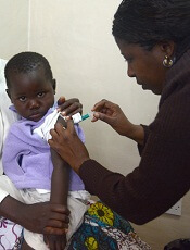
Photo by Caitlin Kleiboer
Results of a genomic sequencing analysis appear to explain why the malaria vaccine candidate RTS,S/AS01 (Mosquirix) is more effective in some children than others.
Researchers sequenced nearly 5000 patient samples and discovered that genetic variation in the protein targeted by RTS,S influences the vaccine’s ability to ward off malaria in young children.
The variation did not appear to affect the vaccine’s efficacy for infants.
Daniel E. Neafsey, PhD, of the Broad Institute in Cambridge, Massachusetts, and his colleagues reported these findings in NEJM.
RTS,S is designed to target a fragment of the protein circumsporozoite (CS), which sits on the surface of the Plasmodium falciparum parasite.
The CS protein is capable of provoking an immune response that can prevent parasites from infecting the liver, where they typically mature and reproduce before dispersing and invading red blood cells, leading to symptomatic malaria.
RTS,S aims to trigger that response as a way to protect against the disease. However, the CS protein is genetically diverse—perhaps due to its evolutionary role in the immune response—and RTS,S includes only one allele of the protein.
With their study, Dr Neafsey and his colleagues sought to test whether alleles of CS that matched the one targeted by RTS,S were linked with better vaccine protection.
The team obtained blood samples from 4985 of the approximately 15,000 infants and children who participated in the vaccine’s phase 3 trial between 2009 and 2013.
The researchers were sent samples when the first symptomatic cases appeared in those vaccinated, as well as samples from all participants at month 14 and month 20 following vaccination.
The team used polymerase chain reaction-based next-generation sequencing of DNA extracted from the samples to survey CS protein polymorphisms. And they set out to determine whether polymorphic positions and haplotypic regions within CS had any effect on the vaccine’s efficacy against first episodes of malaria within a year of vaccination.
The researchers found that RTS,S provided at least partial protection against all strains of P falciparum. However, the vaccine was significantly more effective at preventing malaria in children with matched allele parasites than those with mismatched allele parasites.
Among children who were 5 months to 17 months of age, the 1-year cumulative vaccine efficacy was 50.3% against malaria in which parasites matched the vaccine in the entire CS protein C-terminal, compared to 33.4% against mismatched malaria (P=0.04).
The same effect was not noted in infants. Among infants 6 weeks to 12 weeks of age, there was no evidence of differential allele-specific vaccine efficacy.
Previous genetic studies conducted during RTS,S’s phase 2 trials had not detected an allele-specific effect for this vaccine candidate. The current study had a larger sample size, and recent technological advances made it possible to read the genetic samples with greater sensitivity.
“This is the first study that was big enough and used a methodology that was sufficiently sensitive to detect this phenomenon,” Dr Neafsey said. “Now that we know that it exists, it contributes to our understanding of how RTS,S confers protection and informs future vaccine development efforts.”
RTS,S is the first malaria vaccine candidate to complete phase 3 trials and receive a positive opinion from the European Medicines Agency’s Committee for Medicinal Products for Human Use.
The vaccine was originally designed by scientists at GlaxoSmithKline in 1987. It is now being developed via a public-private partnership between GlaxoSmithKline and PATH Malaria Vaccine Initiative.
The current study was supported by the National Institute of Allergy and Infectious Diseases, the Bill & Melinda Gates Foundation, and the PATH Malaria Vaccine Initiative. ![]()
Radioimmunotherapy shows promise for HL

Copyright 2010 Nephron
Results of a small trial suggest the radiolabeled anti-CD25 antibody 90Y-daclizumab can treat certain patients with relapsed/refractory Hodgkin lymphoma (HL).
90Y-daclizumab produced responses in 50% of patients studied, and most of these were complete responses.
Several patients developed myelodysplastic syndrome (MDS) after receiving 90Y-daclizumab, but researchers found they could prevent this side effect with pretreatment screening.
John Janik, MD, of the National Cancer Institute in Bethesda, Maryland, and his colleagues described this research in PNAS.
The researchers conducted this study to determine if CD25 is a favorable target for systemic radioimmunotherapy in HL. The team noted that although most normal cells don’t express CD25, it is expressed by a minority of Reed–Sternberg cells and by most polyclonal T cells rosetting around Reed–Sternberg cells.
So the researchers tested the anti-CD25 antibody 90Y-daclizumab in 46 patients with relapsed and refractory HL. Patients received 90Y-daclizumab at 10 mCi to 15 mCi every 6 to 10 weeks for up to 7 doses.
The overall response rate was 50%. There were 14 complete responses and 9 partial responses. Fourteen patients had stable disease, and 9 progressed.
The researchers noted that responses occurred in patients whose Reed–Sternberg cells expressed CD25 and in those whose neoplastic cells were CD25-negative, as long as associated rosetting T cells expressed CD25.
Grade 3 or higher toxicities observed in this study included lymphopenia (n=10), neutropenia (n=7), MDS (n=6), thrombocytopenia (n=5), anemia (n=5), and pneumonia (n=2).
At a median follow-up of 9 years, MDS had occurred in 6 patients. They had received a mean of 3.2 doses of 90Y-daclizumab. Prior to 90Y-daclizumab, they had received a mean of 6.2 courses of chemotherapy.
The median time from the initiation of 90Y-daclizumab to MDS diagnosis was 37 months. A retrospective review of 1 patient with MDS revealed that the patient had cytogenetic aberrations on chromosomes 5 and 7 after receiving chemotherapy but before starting 90Y-daclizumab.
So the researchers changed the trial’s entry criteria to require a bone marrow karyotype analysis, and patients who had aberrations were excluded. None of the 16 patients who entered the trial after this change developed MDS.
The researchers said this study is too small to draw meaningful conclusions about the relative roles of chemotherapy and 90Y-daclizumab in the pathogenesis of MDS.
However, this complication is a serious concern, and any subsequent trial should require cytogenetic studies of bone marrow specimens before patients receive systemic radioimmunotherapy. ![]()
‘Room for improvement’ in animal research

Photo by Aaron Logan
A study of animal-based research published over the last 70 years suggests that scientists could have done more to reduce the risk of bias.
A group of researchers examined a few thousand published studies conducted in animals and found that, most of the time, scientists did not employ 4 measures believed to reduce the risk of bias: randomization, blinded assessment of outcome, a conflict of interest statement, and sample size calculation.
“I don’t believe for a moment that scientists set out to do anything other than excellent research, but what this work shows is that there is considerable room for improvement,” said Malcolm Macleod, PhD, of the University of Edinburgh in the UK.
Dr Macleod and his colleagues conducted this research and reported their findings in PLOS Biology.
The team first looked at a random sample of 146 in vivo studies published between 1941 and 2012. Randomization was reported in 20% of the publications in which it would have been appropriate.
Three percent of the publications reported blinding, 10% reported a conflict of interest statement, and none reported a sample size calculation.
Next, the researchers looked at 2671 studies published between 1992 and 2012 that reported drug efficacy in 8 disease models. Randomization was reported in 25% of publications, blinding in 30%, sample size calculation in 0.7%, and a statement of potential conflict of interest in 12%.
However, there were significant increases in 3 of these measures over time. The use of randomization increased from 14% in 1992 to 42% in 2011 (P<0.001), blinding increased from 16% to 39% (P<0.001), and a statement of possible conflict of interest increased from 2% to 35% (P<0.001).
The reporting of a sample size calculation did not change significantly and actually decreased from 2% to 1%.
Dr Macleod and his colleagues also used this dataset to determine whether a journal’s impact factor (a commonly used but disputed measure of journal “quality”)
played a role in the use of the 4 risk-of-bias measures.
The median journal impact factor was significantly higher (P<0.001) for studies reporting a potential conflict of interest, but it was significantly lower in studies reporting randomization (P=0.001). There was no significant difference for the other 2 measures.
Finally, the researchers examined bias in animal studies from the UK’s top 5 universities (according to the
2008 Research Assessment Exercise).
Of 1028 studies published in 2009 and 2010, 14% reported randomization, 17% reported blinding, 10% reported inclusion or exclusion criteria or both, and 1% reported a sample size calculation. Only 1 publication reported using all 4 of these measures.
Dr Macleod and his colleagues concluded that, although this study had its limitations, the results suggest room for improvement in in vivo research. ![]()
HSC finding may have range of implications
Image by Matthias Zepper
Murine research has provided new insight into the functionality of hematopoietic stem cells (HSCs).
Investigators found that a full complement of mini-chromosome maintenance (MCM) proteins is required to preserve HSC functionality and the proper differentiation and maturation of erythrocytes.
Downregulation of the gene MCM3 during embryonic development caused replication stress in hematopoietic progenitors and led to fetal anemia.
In adult mice, downregulation of MCM3 reduced life expectancy and promoted lymphomagenesis.
The investigators therefore believe that therapies designed to modulate replication stress could fight aging, anemia, and hematopoietic malignancies.
This research was published in Nature Communications.
A previous study revealed that replication stress drives the functional decline of HSCs that occurs with age. With the new study, investigators have managed to replicate this phenomenon in mouse embryos.
The team did this by reducing levels of MCM3, one of the components of the MCM complex that is responsible for separating the strands of the DNA double helix during replication. Cells need to maintain high levels of MCM during DNA replication or they experience replication stress.
“When we reduce the levels of the MCM3 gene in the entire organism, we observe that replication stress especially affects the stem cells that give rise to the other blood cells and, in particular, the red blood cell precursors,” explained study author Juan Méndez, PhD, of Centro Nacional de Investigaciones Oncologicas (CNIO) in Madrid, Spain.
“In adult organisms, the production and maturation of red blood cells takes place in the bone marrow, but, during embryonic development, it occurs mainly in the fetal liver. In animals with MCM3 deficiency, the stem cells of the fetal liver are deteriorated, and the embryos develop a severe form of anemia that prevents them from being born.”
“We could say that replication stress turns fetal stem cells, which should be in perfect working order, into very old cells. We have verified this finding in transplantation experiments, where fetal cells with replication stress were unable to adequately reconstitute the blood system in the recipient animals.”
However, the investigators managed to prevent embryonic lethality and reduce anemia by increasing the levels of another gene, CHK1.
“CHK1 is one of the genes responsible for protecting cells from replication stress,” Dr Méndez noted. “It supervises DNA replication. When something goes wrong, CHK1 slows down or halts cell division until the problem has been resolved.”
Embryos that were subjected to replication stress (due to loss of MCM3) but had higher levels of CHK1 showed less pronounced anemia. Four of every 10 embryos developed normally and completed their gestation.
The investigators said an interesting implication of this work is that the type of anemia caused by replication stress is very similar to the aplastic anemia that arises in patients receiving chemotherapy or radiation therapy.
Therefore, the team believes these results could aid the development of novel therapies for aplastic anemia. ![]()
FDA approves factor X concentrate

The US Food and Drug Administration (FDA) has approved a factor X product derived from human plasma (Coagadex) to treat patients with hereditary factor X deficiency who are 12 years of age and older.
Coagadex is approved for on-demand treatment and control of bleeding episodes in these patients as well as for perioperative management of bleeding in patients with mild hereditary factor X deficiency.
Prior to this approval, there was no specific coagulation factor replacement therapy available for patients with hereditary factor X deficiency in the US. The FDA previously granted Coagadex orphan product designation, fast track designation, and priority review.
The FDA based its approval of Coagadex on results from 2 phase 3 trials of patients age 12 and older.
The first trial included 16 patients who received Coagadex for pharmacokinetic evaluation, on-demand treatment and control of bleeding episodes, and/or perioperative management of minor surgical or dental procedures.
Coagadex was used to treat 208 bleeding episodes, and 187 of these episodes (in 15 patients) were evaluated for efficacy. Ninety-eight episodes were major bleeds, 88 were minor bleeds, and 1 was not assessed.
One hundred and fifty-five bleeds (83%) were treated with a single infusion of Coagadex, 28 (15%) were treated with 2 infusions, 3 bleeds (2%) required 3 infusions, and 1 bleed (0.5%) required 4 infusions. Four bleeding episodes in 2 patients were considered treatment failures.
The mean dose of Coagadex per infusion was 25.4 IU/kg, and the mean total dose was 30.4 IU/kg. The recommended dose of 25 IU/kg to treat a bleed was maintained for 14 of the 16 patients. The other 2 patients used doses of up to 30 IU/kg and 33 IU/kg.
There were 176 adverse events in this trial, but only 6 events in 2 patients were considered possibly related to Coagadex. This included 2 reports of infusion site erythema in 1 patient, 2 reports of fatigue in 1 patient, 1 report of back pain, and 1 report of infusion site pain.
The second trial included patients who received Coagadex for perioperative management. Five patients received Coagadex for 7 surgical procedures.
For major surgeries, a median of 13 infusions (range, 2-15) and a median cumulative dose of 181 IU/kg (range, 45-210 IU/kg) were required to maintain hemostasis. For minor surgeries, a median of 2.5 infusions (range, 1-4) and a median cumulative dose of 89 IU/kg (range, 51-127 IU/kg) were required to maintain hemostasis.
There were no adverse events related to Coagadex in this trial.
For more details on these trials, see the Coagadex package insert. Coagadex is manufactured by Bio Products Laboratory Limited in Elstree, Hertfordshire, UK. ![]()
NICE plans to recommend device for SCD patients
Image by Graham Beards
The UK’s National Institute for Health and Care Excellence (NICE) is asking for views on its plans to recommend a device that automatically replaces sickled red blood cells (RBCs) with healthy RBCs in patients with sickle cell disease (SCD).
NICE has issued a draft medical technology guidance supporting use of the Spectra Optia Apheresis System for automated RBC exchange in patients with SCD who need regular transfusions.
The guidance is open for comment until November 16.
The Spectra Optia system is made up of 3 components: the apheresis machine, embedded software, and a single-use disposable blood tubing set. The system is manufactured by Terumo.
NICE said the evidence examined indicates that the Spectra Optia system speeds up the process of RBC exchange compared to manual RBC exchange.
The evidence also suggests that patients require RBC exchange less frequently when using this system than they do with manual RBC exchange.
Furthermore, the Spectra Optia system is estimated to provide cost savings for most patients, when compared to manual RBC exchange or top-up transfusion.
Potential savings depend on the patient’s clinical circumstance and if devices already owned by the National Health Service can also be used to treat SCD.
However, NICE’s draft guidance also highlights a need for additional data on treatment outcomes, as there is limited clinical evidence for some outcomes.
“In particular, the independent Medical Technologies Advisory Committee would like to see long-term data on how manual and automated cell exchange affects the amount of iron in the body and the need to treat this complication,” said Carole Longson, director of the NICE Centre for Health Technology Evaluation. “We welcome comments on the draft guidance as part of this consultation.” ![]()
Drug-resistant malaria could spread to Africa, team says

of Plasmodium falciparum
Image by Mae Melvin/CDC
New research indicates that drug-resistant forms of the malaria parasite Plasmodium falciparum can infect the Anopheles coluzzii mosquito (formerly Anopheles gambiae M form), which is the main transmitter of malaria in Africa.
The discovery suggests that Africa is more at risk for drug-resistant malaria infections than researchers previously thought, and this could further compromise efforts to prevent and eliminate the disease.
Rick Fairhurst, MD, PhD, of the National Institute of Allergy and Infectious Diseases in Rockville, Maryland, and his colleagues reported the discovery in Nature Communications.
The team noted that P falciparum parasites that are resistant to artemisinin, the main drug used to treat malaria, have been rapidly spreading in parts of Southeast Asia.
Malaria experts are concerned that artemisinin-resistant parasites could spread to Africa. However, there were no scientific indications to suggest these parasites could infect A coluzzii mosquitoes—until now.
Dr Fairhurst and his colleagues investigated the transmission potential of these parasites in 3 mosquito species—A coluzzii and the Southeast Asian mosquito species Anopheles dirus and Anopheles minimus.
The researchers evaluated whether these mosquitoes became infected after feeding on blood containing any of 6 artemisinin-resistant parasite strains and 3 artemisinin-sensitive parasite strains previously isolated from malaria patients in Cambodia.
The team found parasites in the mosquitoes’ midguts and salivary glands in almost all cases, showing that the artemisinin-resistant and artemisinin-sensitive Cambodian parasites can infect a variety of mosquito species.
The researchers also discovered a shared genetic background among artemisinin-resistant parasites that may enable them to infect diverse mosquito species by evading the insects’ immune systems.
This study did not show that infected mosquitoes can effectively transmit the disease to humans. However, the results support the idea that artemisinin-resistant parasites could potentially spread beyond Cambodia and to Africa, the researchers said.
They plan to investigate other potential genetic determinants of parasite infection in mosquitoes and further examine which Anopheles species from Cambodia are naturally transmitting artemisinin-resistant parasites in the wild. ![]()
of Plasmodium falciparum
Image by Mae Melvin/CDC
New research indicates that drug-resistant forms of the malaria parasite Plasmodium falciparum can infect the Anopheles coluzzii mosquito (formerly Anopheles gambiae M form), which is the main transmitter of malaria in Africa.
The discovery suggests that Africa is more at risk for drug-resistant malaria infections than researchers previously thought, and this could further compromise efforts to prevent and eliminate the disease.
Rick Fairhurst, MD, PhD, of the National Institute of Allergy and Infectious Diseases in Rockville, Maryland, and his colleagues reported the discovery in Nature Communications.
The team noted that P falciparum parasites that are resistant to artemisinin, the main drug used to treat malaria, have been rapidly spreading in parts of Southeast Asia.
Malaria experts are concerned that artemisinin-resistant parasites could spread to Africa. However, there were no scientific indications to suggest these parasites could infect A coluzzii mosquitoes—until now.
Dr Fairhurst and his colleagues investigated the transmission potential of these parasites in 3 mosquito species—A coluzzii and the Southeast Asian mosquito species Anopheles dirus and Anopheles minimus.
The researchers evaluated whether these mosquitoes became infected after feeding on blood containing any of 6 artemisinin-resistant parasite strains and 3 artemisinin-sensitive parasite strains previously isolated from malaria patients in Cambodia.
The team found parasites in the mosquitoes’ midguts and salivary glands in almost all cases, showing that the artemisinin-resistant and artemisinin-sensitive Cambodian parasites can infect a variety of mosquito species.
The researchers also discovered a shared genetic background among artemisinin-resistant parasites that may enable them to infect diverse mosquito species by evading the insects’ immune systems.
This study did not show that infected mosquitoes can effectively transmit the disease to humans. However, the results support the idea that artemisinin-resistant parasites could potentially spread beyond Cambodia and to Africa, the researchers said.
They plan to investigate other potential genetic determinants of parasite infection in mosquitoes and further examine which Anopheles species from Cambodia are naturally transmitting artemisinin-resistant parasites in the wild.
Older RBCs don’t increase risks, study suggests

Photo by Elise Amendola
Patients undergoing cardiac surgery can safely receive transfusions with older red blood cells (RBCs), according to new research.
In many countries, RBCs can be stored for as long as 6 weeks before transfusion.
But a study published in 2008 suggested that transfusing RBCs stored for more than 2 weeks could increase the risk of serious complications.
Results of subsequent studies both supported and contradicted that finding.
The new study, published in JAMA, adds to the debate. The results suggest the duration of RBC storage does not affect the risk of death or serious complications after transfusion.
“There have literally been hundreds of studies conducted on this topic the past 5 or 6 years, none of which have been able to provide a definitive answer,” said Gustaf Edgren, MD, PhD, of Karolinska Institutet in Stockholm, Sweden.
In an attempt to change that, Dr Edgren and his colleagues conducted a large-scale study of transfusions among cardiac surgery patients in Sweden. National guidelines there require that the oldest available blood unit is allocated first.
The researchers analyzed registry data on patients who underwent coronary artery bypass graft surgery, heart valve surgery, or both between 1997 and 2012.
There were 47,071 patients who received transfusions at 9 different hospitals. Of these patients, 36.6% received RBCs stored for less than 14 days, 26.8%
received RBCs stored 14 to 27 days, 8.9% received RBCs stored 28 to 42
days, and 27.8% received RBCs of mixed age.
The researchers compared these patient groups, looking at the incidence of serious complications at 30 days and mortality at 30 days, 2 years, and 10 years.
They adjusted their analyses for potential confounding factors such as sex, age, blood group, and hospital. And they found no association between RBC storage duration and mortality or serious complications.
| RBC storage duration and adverse outcomes at 30 days | ||||||
| RBCs stored 1-13 days (n=17,224) | 14-27 days (n=12,602) | 28-42 days
(n=4173) |
||||
| Adverse outcome | No. of events | Adjusted
odds ratio (OR) |
No. of events | Adjusted OR | No. of events | Adjusted OR |
| Acute kidney injury | 202 | 1 (reference) | 121 | 0.94 | 38 | 0.97 |
| ARDS/respiratory
failure |
228 | 1 (ref) | 157 | 1.16 | 42 | 1.00 |
| Serious infection | 524 | 1 (ref) | 351 | 0.99 | 133 | 1.13 |
| Stroke | 403 | 1 (ref) | 295 | 1.04 | 106 | 1.13 |
| Thrombosis/embolism | 70 | 1 (ref) | 49 | 1.01 | 17 | 1.09 |
| Composite
adverse outcome (including death) |
1670 | 1 (ref) | 1151 | 1.02 | 371 | 1.03 |
| RBC storage duration and mortality | |||||||
| Storage age | Patient No. | Deaths at
30 days |
Adjusted
hazard ratio (HR) |
Deaths at
2 years |
2-year HR | Deaths at
10 years |
2-year HR |
| 1-13 days | 17,224 | 615 | 1 (ref) | 1593 | 1 (ref) | 5897 | 1 (ref) |
| 14-27 days | 12,602 | 410 | 1.06 | 1074 | 1.02 | 4358 | 1.02 |
| 28-42 days | 4173 | 103 | 0.90 | 325 | 0.98 | 1403 | 0.99 |
| mixed age | 13,072 | 911 | 0.86 | 1954 | 0.94 | 5655 | 0.99 |
“This study is by far the largest investigation focusing on the issue of blood storage in this very sensitive patient group, and we find absolutely no hint of negative health effects associated with stored blood,” said Ulrik Sartipy, MD, PhD, of Karolinska University Hospital.
“[W]e have been able to provide very firm reassurance that the current blood storage practices are safe,” Dr Edgren added.
Antibody can turn AML cells against each other

Image by Joshua Stokes
Scientists say they have discovered an antibody that can transform leukemic cells into immune cells that target their former “siblings.”
This agonist antibody, which is highly specific for the thrombopoietin receptor, can turn acute myeloid leukemia (AML) cells into dendritic cells.
With extended exposure to the antibody, these dendritic cells can then transform into natural killer (NK)-like cells that target AML cells.
The scientists described these discoveries in PNAS.
“It’s a totally new approach to cancer, and we’re working to test it in human patients as soon as possible,” said study author Richard A. Lerner, MD, of The Scripps Research Institute in La Jolla, California.
Unexpected transformation
Dr Lerner’s lab has pioneered techniques to generate and screen large libraries of antibodies to find therapeutic antibodies that bind to a desired target or activate a desired receptor on cells.
During the course of this work, the team discovered a phenomenon they call “receptor pleiotropism,” in which agonist antibodies against known receptors cause cell fates that are extremely different from those induced by the natural agonist to the receptor.
Why this happens is unclear, but the discovery led the scientists to wonder if they could use the method to convert leukemia cells into non-cancerous cells.
To find out, they tested 20 of their recently discovered receptor-activating antibodies against AML cells from patients. This revealed the transformative properties of the agonist antibody that is highly specific for the thrombopoietin receptor.
When this antibody was applied to healthy, immature bone marrow cells, it caused them to mature into megakaryocytes. However, when the antibody was applied to AML cells, they were converted into dendritic cells.
With longer exposures to the antibody and certain other in vitro conditions, the induced dendritic cells morphed into cells that closely resemble NK cells.
“That antibody could have turned those acute myeloid leukemia cells into a lot of other cell types, but, somehow, we were lucky enough to get NK cells,” Dr Lerner said.
The team examined these induced NK cells with electron microscopy and observed that many of the cells had extended tendrils through the outer membranes of neighboring AML cells. In in vitro tests, a modest number of these NK cells wiped out about 15% of the surrounding AML cell population in 24 hours.
The scientists also noted that the induced NK cells’ cancer-killing effects appeared to be purely “fratricidal.” Unrelated breast cancer cells did not die off in large numbers in the presence of the NK cells.
Fratricidin therapy
Why the induced NK cells appear to target only closely related cells isn’t yet clear. However, the finding suggests there may be antibodies—and even small-molecule compounds—that would turn other cancerous cell types into fratricidal NK cells by activating other receptors expressed on those cells.
Such fratricidal therapies, which Dr Lerner calls “fratricidins,” would have several potential advantages. First, especially if they are antibodies, they could be clinically useful with little or no further modification.
Second, their high specificity for their target receptors, and the resulting NK cells’ specificity for related cancer cells, should reduce the likelihood of adverse effects.
Finally, the peculiar dynamics of fratricidin therapy, in which every cancerous cell is potentially convertible to a cancer-killing NK cell, suggests that—if the strategy works—it might not just reduce the targeted cancer-cell population in a patient but eliminate it altogether.
“We’re in discussions with pharmaceutical companies to take this straight into humans after the appropriate preclinical toxicity studies,” Dr Lerner said.
This research was supported by the JPB Foundation and Zebra Biologics.
6PGD inhibitors could treat leukemia, other cancers

Photo by Rhoda Baer
Preclinical research suggests that compounds derived from Himalayan rhubarb could potentially treat leukemias and other cancers.
These compounds, physcion and S3, effectively inhibited 6PGD, a metabolic enzyme that is upregulated in several types of cancer cells.
In addition, physcion and S3 reduced cancer cell viability in vitro and decreased tumor size in vivo—without harming normal cells or exhibiting obvious toxicity.
Jing Chen, PhD, of Emory University School of Medicine in Atlanta, Georgia, and his colleagues described this research in Nature Cell Biology.
The team set out to find 6PGD inhibitors because 6PGD is part of the pentose phosphate pathway, which supplies cellular building blocks for rapid growth. And previous research had revealed increased 6PGD activity in several types of cancer cells.
“This is part of the Warburg effect, the distortion of cancer cells’ metabolism,” Dr Chen said. “We found that 6PGD is an important metabolic branch point in several types of cancer cells.”
Specifically, the investigators found that knocking down 6PGD decreased proliferation in a handful of human cancer cell lines—leukemia (K562), lung cancer (H1299, H157, and H322), and head and neck cancer (212LN).
But 6PGD knockdown did not affect normal proliferating keratinocyte HaCaT cells.
When the team screened for 6PGD inhibitors, they identified physcion—an anthraquinone originally isolated from Himalayan rhubarb—and its derivative, S3.
Experiments showed that physcion decreases cell viability in a range of cancer cell lines—K562, H1299, A549 (lung), and 212LN—in a dose-dependent manner. But the compound did not significantly affect proliferating control cells—human dermal fibroblasts (HDFs) and immortalized human melanocyte PIG1 cells.
Similarly, S3 inhibited the viability of K562 and H1299 cells but not proliferating PIG1 and HDF cells.
S3 also decreased tumor growth in mouse models of leukemia, lung cancer, and head and neck cancer. However, the treatment did not affect body weight, serum chemistry, complete blood counts, or hematopoietic properties in the mice.
Finally, the investigators found that physcion and S3 inhibit 6PGD in human primary leukemia cells (B-cell acute lymphoblastic leukemia), thereby leading to decreased cell viability.
And neither compound affected the viability of mononucleocytes in peripheral blood samples or CD34+ progenitors isolated from the bone marrow samples of healthy donors.