User login
M. Alexander Otto began his reporting career early in 1999 covering the pharmaceutical industry for a national pharmacists' magazine and freelancing for the Washington Post and other newspapers. He then joined BNA, now part of Bloomberg News, covering health law and the protection of people and animals in medical research. Alex next worked for the McClatchy Company. Based on his work, Alex won a year-long Knight Science Journalism Fellowship to MIT in 2008-2009. He joined the company shortly thereafter. Alex has a newspaper journalism degree from Syracuse (N.Y.) University and a master's degree in medical science -- a physician assistant degree -- from George Washington University. Alex is based in Seattle.
Monitor elderly for bone loss after gastric bypass
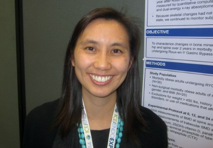
CHICAGO – Older adults who undergo Roux-en-Y gastric bypass are at risk for lessened bone mineral density for at least 2 years after their surgery and should be monitored appropriately for osteoporosis or fragility fractures, according to investigators from Massachusetts General Hospital in Boston.
Two years postoperatively, vertebral bone mineral density (BMD) in 30 patients was about 7% lower on quantitative computed tomography (QCT) and 6% lower on dual-energy x-ray absorptiometry (DXA) – both methods were used to ensure accuracy – when compared with 20 well-matched, morbidly obese controls. Total hip BMD was 6% lower on QCT and 10% lower on DXA. Femoral neck BMD was about 6% lower by both measures.
Biomarkers of bone turnover remained markedly elevated in surgery patients, as well, but unchanged in controls (C-telopeptide 0.65 ng/mL vs. 0.3 ng/mL; amino-terminal propeptide of type I collagen 65 ng/mL vs. 40 ng/mL). The findings were all statistically significant.
The groups started to separate early on BMD, and there’s concern that bone loss will continue for more than 2 years after surgery. Preoperatively, most Roux-en-Y gastric bypass patients have a higher than normal BMD, "so even a loss of 10% over 2 years is not going to put most of them in the osteopenic or osteoporotic range. The caveat now is that we are [offering surgery] to older patients and adolescents. Elderly patients are starting with lower bone mass, so there are concerns about [post-op] skeletal fragility. The oldest patient in our study was 72. She became osteoporotic after surgery because her bone density was low to begin with," said lead investigator Dr. Elaine W. Yu, an endocrinologist at Massachusetts General.
"In adolescents, there are implications for achieving peak bone mass. Even if you have a normal bone density 2 years after surgery, what’s going to happen in 10 years, 20 years?" she asked.
In short, "people should pay attention to bone density and bone loss and discuss this as one of the potential negative effects of bariatric surgery. For patients at risk, you should definitely consider serial bone density monitoring and osteoporosis therapy if needed," she said at the joint meeting of the International Society of Endocrinology and the Endocrine Society.
At baseline, both subjects and controls were about 47 years old and 270 pounds, with a mean a mean body mass index of 45 kg/m2. About 85% were women. The study excluded patients with histories of bone disorders or use of bone-affecting medications.
Surgery patients lost a mean of about 85 pounds in the first 6 months; their weight loss then stabilized, but they continued to lose bone. Controls stayed about the same weight.
The findings can’t be explained by post-op calcium or vitamin D depletion. "These subjects were aggressively supplemented with both," and both groups maintained normal levels throughout the study. Also, there were no statistical differences in parathyroid hormone levels between the groups.
"Our theory is that there are changes in gut hormones after gastric bypass that have direct effects on bone, like ghrelin," Dr. Yu said.
The National Institutes of Health funded the work. Dr. Yu is a consultant for Amgen.
AT ICE/ENDO 2014
Key clinical point: Gastric bypass puts some older patients at risk for osteoporosis.
Major finding: Two years after surgery, spine BMD in 30 patients was about 7% lower on quantitative CT and 6% lower on DXA when compared with 20 well-matched, morbidly obese controls.
Data Source: Retrospective case-control study with 2 years follow-up
Disclosures: The work was funded the National Institutes of Health. Dr. Yu is a consultant for Amgen.
Effects of IV bisphosphonates last 2-3 years in children
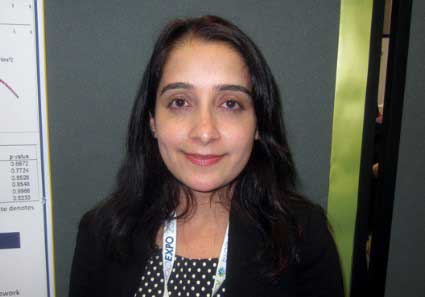
CHICAGO – Serum osteocalcin is the first bone turnover marker to recover after intravenous bisphosphonate therapy in children with primary osteoporosis, secondary osteoporosis, or osteogenesis imperfecta, making it a good indicator that it’s time to consider a second round of treatment, according to investigators from Children’s Mercy Hospitals and Clinics in Kansas City, Mo.
In a review of 104 children followed for up to 5 years, they found that the benefits of IV bisphosphonates – for children, usually two or three 2-day courses about 4 months apart – remain durable for up to 3 years. After an initial decrease, serum osteocalcin starts to return to baseline 1 year after the start of treatment (P = 0.007); improvements in lumbar spine bone mineral density z scores on dual energy x-ray absorptiometry (DXA) start to plateau at about 2 years and then reverse after 3 years (P less than 0.0001). Decreases in urine N-telopeptide, a bone resorption marker, plateau at 2-3 years, then start to increase after 3 years (P less than 0.0001). Serum bone-specific alkaline phosphatase, a marker of bone formation like osteocalcin, continues to decrease for about 4.8-5 years (P less than 0.0001).
Although alkaline phosphatase, urine N-telopeptide, and lumbar z scores had begun to recover by the end of follow-up, they were still significantly improved over baseline. For instance, baseline lumbar z scores, more than two standard deviations below normal before treatment, were 1.31 standard deviations below normal at last follow up (P = 0.002).
However, after initial improvement, there was no statistically significant change in serum osteocalcin from baseline; the mean baseline value was 52.7 ng/mL, and mean value at last follow up was 54.5 ng/mL (P = 0.48).
"Osteocalcin is the first marker to increase within the first year after start of bisphosphonate therapy. The results of the study," perhaps the first to define the drug holiday window for bisphosphonates in children, "provide a framework [for] when to anticipate reinitiation of bisphosphonate therapy," the investigators concluded at the joint meeting of the International Congress of Endocrinology and the Endocrine Society.
"After 3 years, we should consider reinitiation of bisphosphonates in these children," said lead investigator Dr. Naziya Tahseen of the department of pediatric endocrinology at Children’s Mercy Hospitals and Clinics.
The subjects’ mean age was 11.6 years, 61 (58.7%) were girls, 67 (64.4%) were in puberty, and 75 (72.1%) were ambulatory; 59 (56.7%) children had primary or secondary osteoporosis, 26 (25%) had osteopenia, and 19 (18.3%) osteogenesis imperfecta. Lab values were followed every 4 months, with DXA scans repeated every 6-12 months.
Overall, children did better if they were ambulatory, but puberty status and gender had no effect.
The investigators had no relevant disclosures, and no outside funding was used for the project.
AT ICE/ENDO 2014
Key clinical point: In children, osteocalcin is the first bone turnover marker that indicates it’s time to consider another round of IV bisphosphonate treatment.
Major finding: After initial improvement, serum osteocalcin begins to return to baseline about a year later (P = 0.007).
Data Source: Retrospective study of 104 children followed for up to 5 years.
Disclosures: The investigators have no disclosures, and received no outside funding for their work.
VIDEO: Use late-night salivary cortisol to catch recurrent Cushing’s
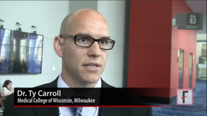
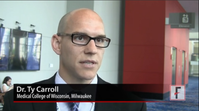
CHICAGO – Late-night salivary cortisol exceeded normal limits in 10 women with recurrent Cushing’s disease a mean of 3.5 years after transsphenoidal surgery, but their urinary free cortisol remained in normal limits, according to a retrospective review from the Medical College of Wisconsin, Milwaukee.
That adds strength to the notion that late-night salivary cortisol (LNSC) catches recurrent Cushing’s that’s missed by urinary free cortisol, even though UFC remains a standard screening approach in some places.
The study is tiny and retrospective, but at the joint meeting of the International Congress of Endocrinology and the Endocrine Society, lead investigator Dr. Ty Carroll explained why the findings still matter, and also why two LNSC measurements are better than one.
The video associated with this article is no longer available on this site. Please view all of our videos on the MDedge YouTube channel
AT ICE/ENDO 2014
VIDEO: Kisspeptin outperforms HCG for early miscarriage prediction
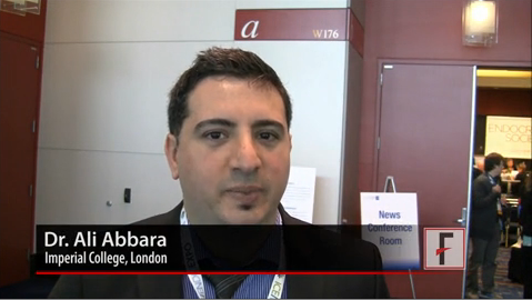
CHICAGO – A one-time measurement of plasma kisspeptin – a family of placental peptides also being studied for fertility treatment – better predicts miscarriage than does serial measurement of human chorionic gonadotropin (HCG), a widely used measure.
British researchers measured both in 993 asymptomatic women at approximately 11 weeks’ gestation, 50 of whom later miscarried. Plasma kisspeptin proved a more accurate predictor of miscarriage than HCG: the area under the receiver operating characteristic (ROC) curve for plasma kisspeptin was 0.899, compared with 0.775 for serum HCG. Plasma kisspeptin above 1,306 pmol/L was associated with a highly significant 87% reduced risk of miscarriage, even after adjusting for age, body mass index, gestational age, smoking, and blood pressure.
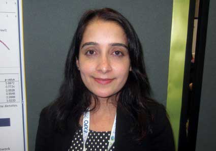
Lead investigator Dr. Ali Abbara of Imperial College London explained why that matters at the joint meeting of the International Congress of Endocrinology and the Endocrine Society.
The video associated with this article is no longer available on this site. Please view all of our videos on the MDedge YouTube channel
AT ICE/ENDO 2014
AAP nomogram means fewer unnecessary heel sticks for newborns
VANCOUVER, B.C. – A phototherapy nomogram from the American Academy of Pediatrics is slightly more reliable than the widely used Bhutani nomogram when it comes to deciding if transcutaneous bilirubin measurements need to be followed by a total serum bilirubin blood draw.
The conclusion is based on a retrospective chart review from the BORN (Better Outcomes Through Research for Newborns) network. The team checked 902 transcutaneous bilirubin (TCB) measurements, an estimate of total serum bilirubin (TSB) from a device that scans the skin, against actual total serum bilirubins drawn no more than 2 hours later. Their subjects were infants a mean of 35 hours old, born after at least 35 weeks’ gestation.
When skin scans indicate that newborns are in the 75th percentile for risk of severe hyperbilirubinemia on the Bhutani nomogram, blood is drawn to check the actual level. That approach missed two of 27 infants who actually had TSB levels that required phototherapy, a false-negative rate of 7.4% (95% confidence interval, 0.9%-24.3%).
When the investigators instead deemed blood draws necessary only when TCB levels were 70% or more of the American Academy of Pediatrics (AAP) threshold for phototherapy, the false-negative rate was 3.4%, with just one of 29 infants missed (95% CI 0.8%-17.8%).
Overall, the team analyzed 7,737 TCB values in a total of 4,993 infants; only a small number led to TSB blood work. They concluded that the Bhutani method would avoid unnecessary TSB blood draws in about 80% of infants, while the AAP nomogram would avoid unnecessary blood draws in about 85%.
"TCB seems like it should be a perfect thing, but a lot of people don’t really do it because they don’t believe the results; we wanted to a do a real-world study in a bunch of nurseries to see how well it really works. Our results indicate that TCB works pretty well," and that "the 70% of phototherapy threshold might be as good if not a little bit better than [Bhutani] in terms of avoiding [unnecessary] blood draws. I’m wondering if we should switch to [it]," said lead investigator Dr. James Taylor, medical director of the University of Washington’s newborn nursery in Seattle.
"The false negatives weren’t very far off. I think the highest TSB level we had was 18 mg/dL," he said at the Academic Pediatric Societies annual meeting.
A larger study is needed to confirm the difference in false negatives, he cautioned.
The study was retrospective, so clinicians used whatever TCB device they wanted. More than 60% of the infants were white, and more than 80% were born after 38 weeks.
Dr. Taylor is also working on a smart phone app to measure TCB.
He said he has no disclosures. The BORN research network is funded by the Academic Pediatric Association.
AT THE PEDIATRIC ACADEMIC SOCIETIES ANNUAL MEETING
Key clinical point: Transcutaneous bilirubins might be more reliably interpreted by using the American Academy of Pediatrics phototherapy nomogram.
Major finding: The Bhutani nomogram avoids unnecessary blood draws in about 80% of newborns and the AAP nomogram avoids unnecessary blood draws in 85%.
Data source: Retrospective review of more than 7,000 TCBs in infants a mean of 35 hours old
Disclosures: The lead investigator has no disclosures. The work was funded by the Academic Pediatric Association.
Recovery is quicker when depressed teens are guided by a treatment manager
VANCOUVER, B.C. – A proactive, collaborative care approach is best for teenage depression, according to Dr. Laura P. Richardson.
She and her team randomized 51 adolescents aged 13-17 years old who had screened positive for depression on the Patient Health Questionnaire 9 and other measures to standard care, and 50 others to a collaborative model overseen by a master’s level depression care manager.
Both groups had a mean baseline score of about 47 on the Children’s Depression Rating Scale (CDRS), but soon separated. At 6 months, 50% of the treatment group, but only 15% of the control group, had a drop of at least 50% in their CDRS score; at 1 year, the difference had grown to 70%, vs. 30%. With the majority of adolescents in each arm still involved in the project, the mean 12-month CDRS score in the collaborative care group was 26, vs. about 35 for the control group. Also at 12 months, the treatment group’s mean score on the Columbia Impairment Scale was 13.2, vs. 17.1 in the control arm. The differences were all statistically significant.
The adolescents in the collaborative arm ended up having more therapy sessions and medication, as well, and reported greater satisfaction with their care.
Often in the United States, when adolescents feeling down see their primary care physician, they walk out of the office with a list of psychotherapists to contact and maybe a short course of antidepressants, after being told to come back in a month. "They often don’t, because they aren’t doing well and aren’t motivated," said lead investigator Dr. Richardson, a specialist in adolescent medicine at Seattle Children’s Hospital.
Instead, the care manager worked with the adolescents and their parents to pick antidepressants, regular cognitive-behavioral therapy sessions, or both. The manager then checked in with them about every week and tracked how the adolescents did in a disease registry. Those who needed more help got it.
As is often the case, the usual care kids were simply encouraged to seek care for depression, Dr. Richardson noted.
The results show that "we can do better" when it comes to depressed teens. "Patient engagement and tracking are critical. Unless we engage youth up front and keep them engaged, we are probably not going to make a difference. A lot of it [comes down to] training people to be proactive, because we haven’t been," she said at the Pediatric Academic Societies annual meeting.
With just a year’s data, it’s unknown if the extra up-front costs save money down the road, though that’s been shown in adults. The model is being tried elsewhere in the country.
The mean age in the study was about 15 years, and about three-quarters of the participants were girls. Most of the adolescents were white.
The investigators excluded adolescents who were already seeing a psychiatrist; those who had developmental delays or previous mental health hospitalizations; and those who had bipolar disorder, were abusing drugs, or were unable to speak English.
Dr. Richardson said she had no relevant financial disclosures. The work was funded by the National Institute of Mental Health.
AT THE PAS ANNUAL MEETING
Key clinical point: Outcomes are better – and, ultimately, costs are probably lower – when a master’s level manager oversees the treatment of depressed teens.
Major finding: After 6 months, 50% of the depressed teens assigned to collaborative care, but just 15% of those assigned to usual care had a drop of at least 50% in their Child Depression Rating Scale score, a significant difference.
Data source: A randomized trial of 101 depressed teenagers.
Disclosures: Dr. Richardson said she had no relevant financial disclosures. The work was funded by the National Institute of Mental Health.
VIDEO: Shedding stigma for advocacy, suicide attempt survivors find a voice
LOS ANGELES – Suicide attempt survivors are overcoming the stigma of talking about their attempts and joining together to demand a voice in suicide prevention efforts. Researchers and leaders in the field are hearing that voice and acknowledging them as sources of insight and advocacy that, for too long, have been largely ignored.
Among the signs of that shift – and due largely to their lobbying – the American Association of Suicidology (AAS) recently formed a division for attempt-survivors and launched a blog on the topic.
"It’s a critical voice. There’s no better advocate for mental well-being than someone who has been on the edge of life and death and chosen a path of recovery," said AAS President William Schmitz Jr., Psy.D.
Asked for an example of how this new voice might help, he noted that "there are a lot of clinicians out there who are not trained in the assessment and management of suicidality. I’ve heard stories from some of our attempt survivors that are truly scary – very negative, hostile encounters with the health care system." People who’ve experienced it firsthand offer "a chance for us to open that dialogue," he said at the annual conference of the American Association of Suicidology.
Cara Anna, a journalist and attempt-survivor who helped spearhead the AAS lobbying campaign, stated her case for us at the meeting. She’s a prominent voice in the attempt-survivor community.
The video associated with this article is no longer available on this site. Please view all of our videos on the MDedge YouTube channel
AT THE ANNUAL AAS CONFERENCE
Late preterm births linked to school-age neurodevelopmental problems
VANCOUVER, B.C. – Late preterm infants have a higher incidence of future neurodevelopmental problems than children born at term, two studies showed.
"Everyone has just assumed that late preterm babies are fine, and there are no long-term problems. We are now finding that the outcomes are probably not as good, and [we] need to find out whether just being born [early] is the problem, or whether it’s being born [early] and having something else going on," said investigator Dr. Elaine Boyle of the University of Leicester (England).
In the first project, a team from the University of Iowa in Iowa City performed structural brain MRIs on 52 children an average of 9.4 years old who were born at 34-36 weeks, and 74 children with a mean age of 10 years born at term. All were singletons.
Among other findings, the mean volume of subcortical tissue was almost a quarter standard deviation lower in late preterm (LPT) children than in children born at term, a difference driven mostly by smaller thalami and hippocampi. "The structural difference on MRI directly related to areas in which the LPT cohort performed more poorly" on a battery of assessments, especially visual short-term memory (Pearson coefficient [r] = 0.3; P = .001) and processing speed (r = 0.2; P = .03), said lead investigator Dr. Jane Brumbaugh of the department of pediatrics at the University of Iowa.
At assessment, LPT children were 36 kg and 138 cm tall on average, with a head circumference of 54 cm. Children born at term were a bit bigger, an average of about 38 kg and 142 cm tall, with a head circumference of 55 cm. The analysis controlled for age, sex, and intracranial volume; there were no statistically significant between-group socioeconomic differences.
Meanwhile, Dr. Boyle’s team found that 16% (101/638) of infants born at 32-36 weeks, but just 10% (77/765) of term infants, had neurodevelopmental disabilities at age 2, according to the children’s parents (RR, 1.4; 95% CI, 1.1-2.0; P = .011).
The finding comes from the PARCA-R (Parent Report of Children’s Abilities–Revised), which the Leicester team administered to parents of 2-year-olds; the investigators did not confirm their findings with clinical assessments. Their results were adjusted for sex, socioeconomic status, and multiple births.
Parents also reported that 1.6% (10) of LPT but 0.3% (2) of term infants have some type of sensory impairment, and that LPT children show a trend toward more communication problems.
Male sex, lower socioeconomic status, prepregnancy hypertension, preeclampsia, and recreational drug use during pregnancy each more than doubled the risk of neurodevelopmental disability in LPT children.
"Late to moderately preterm infants have a 50% increased risk for neurodevelopmental disability at 2 years of age. As we see in the very preterm population, this is largely driven by cognitive problems," said lead investigator Samantha Johnson, Ph.D., of the University of Leicester.
Besides arguing for term birth when possible, intervening in preeclampsia, recreational drug use, and other problems might improve outcomes. Also, it might be a good idea to screen parents of LPT infants to identify children who could benefit from early intervention, she said.
The investigators said they have no relevant financial disclosures. The University of Iowa funded the MRI study, and the U.K. National Institute for Health Research funded the English study.
AT THE PAS ANNUAL MEETING
Key clinical point: Besides arguing for term birth, if possible, the findings suggest a need for early intervention.
Major finding: At age 10, the mean volume of subcortical tissue in late preterm children is almost a quarter standard deviation lower than in children born at term; at age 2, 16% of LPT children have neurodevelopmental disabilities, versus 10% of children born at term, according to their parents.
Data source: An MRI study of 126 2-year-olds and a population cohort study of 1,403 English school children
Disclosures: The investigators said they have no relevant financial disclosures. The MRI study was funded by the University of Iowa, and the English study was funded by the U.K. National Institute for Health Research.
