User login
Mitchel is a reporter for MDedge based in the Philadelphia area. He started with the company in 1992, when it was International Medical News Group (IMNG), and has since covered a range of medical specialties. Mitchel trained as a virologist at Roswell Park Memorial Institute in Buffalo, and then worked briefly as a researcher at Boston Children's Hospital before pivoting to journalism as a AAAS Mass Media Fellow in 1980. His first reporting job was with Science Digest magazine, and from the mid-1980s to early-1990s he was a reporter with Medical World News. @mitchelzoler
Arthritis Self-Management Lags Despite Wide Promotion
Several U.S. public health groups have recently made self-management training for patients with arthritis a priority. But clinicians resist this, despite an evidence-based track record that goes back more than 30 years.
In essence, arthritis self-management consists of arthritis patients using exercise, injury prevention strategies, weight management through healthful eating, disease education, meditation, and other supportive therapies to ease their pain and increase their function.
Both government and rheumatology organizations have been enthusiastic in their support for these measures, as shown by the following:
• The Centers for Disease Control and Prevention has formally supported arthritis self-management training (as well as similar training for patients with other chronic diseases). In addition, the CDC’s arthritis program says that one of its main short-term goals is to "improve and increase self-management attitudes and behaviors among persons with arthritis. ... Without doubt, self-management is a core issue in public health," said Dr. Patience White, a rheumatologist and chief public health officer of the Arthritis Foundation in Atlanta.
• The U.S. Administration on Aging received cash support from the 2009 American Recovery and Reinvestment Act. With that stimulus money, the AoA handed out $27 million in grants to 45 states, Puerto Rico, and the District of Columbia last year to deliver self-management training programs to patients with chronic diseases including those with osteoarthritis (OA) or rheumatoid arthritis (RA).
• In the 2010 report "A National Public Health Agenda for Osteoarthritis," jointly issued by the Arthritis Foundation and the CDC, the first recommendation for action in the report’s 10-item plan is: "Self-management education should be expanded as a community-based intervention for people with symptomatic OA." The American College of Rheumatology has also endorsed self-management training for OA patients in the society’s OA management recommendations (Arthritis Rheum. 2000;43:1905-15), and the Arthritis Foundation has promoted self-management as a key element in managing patients with RA.
• Healthy People 2020, a set of public health targets released in December 2010 by the Department of Health and Human Services, said that one goal for patients with arthritis is to "increase the proportion of adults with doctor-diagnosed arthritis who have had effective, evidence-based arthritis education as an integral part of the management of their condition."
In short, self-management – with its safety and proven modest efficacy – has emerged as an attractive complement to the medical approach for dealing with arthritis.
Data Support Self-Management’s Efficacy
Kate Lorig, Dr.P.H., noted that "we give patients a lot of tools to use so that they can do the things they need to do with less pain."
"What we find is that patients who take the course have statistically significantly less pain," added Dr. Lorig, professor of medicine and director of the Patient Education Research Center at Stanford (Calif.) University in Palo Alto, in an interview. "They have a small to moderate reduction in pain that is similar to what they get from NSAIDs. ... We see people able to do things that they couldn’t before, related to improvements in mobility and depression, and we see improvements in their quality of life."
Dr. Lorig has led a group at Stanford that began developing and testing an arthritis-oriented self-management program in the late 1970s (Arthritis Rheum. 1985;28:680-5). Subsequently the same group developed a more generic chronic disease program (Arthritis Rheum. 2005;53:950-7), and arthritis programs available by mail (Arthritis Rheum. 2009;61:867-75) and over the Internet (Arthritis Rheum. 2008;59:1009-17).
"The individual improvement for each patient can be rather small, but the cost saving [of promoting wide use of self-management training] is high because of the large number of patients," said Dr. White. In addition, although the overall effect size may be small, for a subset of patients the effect is "life changing," she added.
Clinicians’ Indifference
But despite these positive assessments and public health promotions, self-management training for patients with OA or RA remains neglected and unused. Experts widely agree that a scant fraction of U.S. arthritis patients have received self-management training.
According to the Arthritis Foundation’s Dr. White, currently 60,000 U.S. arthritis patients undergo self-management training each year, a number dwarfed by the roughly 50 million Americans who have some form of arthritis. "Very few are in the program, even when it gets megabucks from the government," she said.
The best known and most thoroughly-studied self-management training course for patients with arthritis is the Arthritis Self-Management Program developed by Dr. Lorig and her associates at Stanford University and licensed by Stanford to training sites. The Stanford group also subsequently developed the similar Chronic Disease Self-Management Program. The Stanford programs are the most widely used, and rightly so, said Maura D. Iversen, Sc.D., professor and chairman of the department of physical therapy at Northeastern University in Boston.
The Stanford arthritis program "was the basis for the Arthritis Foundation’s self-help program in the 1980s, and thus many health professionals and patient volunteers used it," Dr. Iversen said in an interview. Writing in a review of self-management last year, Dr. Iversen and her associates said that "self-management programs are now acknowledged as a key element of quality care" for patients with OA, RA, and other chronic diseases. The unaddressed issues today include whether the benefits from self-management training extend long term, and whether any patient attributes are linked with better outcomes following training, they wrote (Ann. Rheum. Dis. 2010;69:955-63).
"There are many ways for patients to get self-help, but from the standpoint of having the data, [the Stanford programs] are the best for both arthritis and chronic disease," said Dr. White.
The Stanford program "is the most popular. It has been well studied and popularized by the Arthritis Foundation and the CDC," said Dr. Daniel H. Solomon, a rheumatologist at Brigham and Women’s Hospital in Boston. But that "most popular" characterization is relative: The program’s popularity drops precipitously inside the offices of many rheumatologists and primary care physicians. Major issues seem to be skepticism about efficacy; questions about the need for formal programs; and a lack of awareness about self-management, time to make a referral, and knowledge about where to refer.
"I doubt many rheumatologists regularly refer patients to such programs. It is hard to argue with such programs, but few rheumatologists view them as beneficial. They are clearly nontoxic, but how beneficial they are can be debated," said Dr. Solomon. Several years ago, he headed a controlled study with 113 patients with OA, RA, or fibromyalgia that failed to find a significant benefit from the arthritis self-management program (J. Rheumatol. 2002;29:362-8). He also coauthored a meta-analysis of 17 other controlled trials of the same program, and found that self-management classes led to small reductions in pain and disability (Arthritis Rheum. 2003;48: 2207-13).
"A self-management course is just one of several tools to promote patient coping," said Dr. Nortin M. Hadler, a rheumatologist and professor of medicine at the University of North Carolina at Chapel Hill. "I believe that almost all rheumatologists are aware that an important part of treating regional joint pain is to have patients exercise and meet with peer groups, but the patient doesn’t need a structured Arthritis Foundation program. They can go the aerobics classes, the YMCA, or a health club."
"As a group, rheumatologists don’t refer their patients to self-management," said rheumatologist Dr. Halsted R. Holman, the Guggenheim Professor of Medicine Emeritus at Stanford and a codeveloper with Dr. Lorig of the Stanford self-management program. In many cases when a rheumatologist or other physician makes the referral, the patient has a hard time finding a nearby program or a program at a convenient time. "It’s mainly access issues," he said.
Another rheumatologist who collaborated on developing the Stanford program agreed. "There are about 7,000 classes given a year in the United States, but the country is big and just because there is a class in a patient’s area doesn’t mean it will be convenient," said Dr. James F. Fries, also a rheumatologist and professor emeritus at Stanford. In addition, "the referral mode has always been lousy." The growing availability of self-management training via the Internet may address the convenience issues and broaden patient uptake, he said in an interview.
Hurdles to Referral
"I’ve never felt any hostility from rheumatologists, and the American College of Rheumatology has never been less than 100% supportive," Dr. Lorig said. "We don’t get a lot of referrals from rheumatologists in most places, but I don’t think it’s the rheumatologists’ fault. We make it exceedingly difficult to refer."
In general, physicians don’t know about self-management programs in their communities – where they’re offered and when – because "the programs have never been closely linked to the medical system. "We have 1,000 license holders [groups that have purchased a license from Stanford to hold arthritis or chronic disease self-management classes] and more than 3,000 trainers," but despite that, doctors don’t know where and when programs are offered. "We all recognize this is a huge problem. We’re now big enough that a national, central referral source is possible, and we will hopefully have one within the next year," she said.
"We also know from our research that people with arthritis who receive a referral or recommendation from their doctor are 18 times more likely to attend a self-management education program," said Teresa J. Brady, Ph.D., a senior behavioral scientist in the CDC’s arthritis program, adding that the No. 1 reason why patients don’t attend a program is that they do not know it exists, and they believe that if it did exist, they would have heard about it from their physician. "As a consequence, we at the CDC’s arthritis program have begun pilot testing a marketing strategy to help health care providers know when and where in their community patients can take training programs."
In recent years, the ways in which patients can take courses has significantly broadened, moving beyond the traditional small-class format to also include courses by mail and the Internet. The National Council on Aging (NCoA) is the technical advisor to the AoA’s grant program for chronic disease self-management training. The NCoA is now developing a Web site where patients and physicians can find all the classes offered in their communities, but Wendy Zenker, vice president of the NCoA’s benefits access group, said she could not provide a target date when the Web site will become operational.
The Arthritis Foundation’s Dr. White agreed that a low referral rate by physicians is a major problem. In general, physicians "don’t refer enough to community services in any form," she said. To help address this, the Arthritis Foundation has recently been working with the American College of Physicians to alert internal medicine physicians about the arthritis self-management program. Yet another issue is the time commitment (2.5 hours a week for 6 weeks) for patients who take a class. "The majority of people don’t complete it," Dr. White said. "The issue is: How do we get people to do it? We don’t have that magic bullet yet."
Dr. White, Dr. Iverson, Dr. Solomon, Dr. Hadler, and Dr. Brady said that they had no disclosures. Dr. Lorig, Dr. Holman, and Dr. Fries said that they receive royalties from the licensing of the Stanford self-management programs and from sales of the teaching texts.
Percutaneous Hepatic Perfusion Boosts Melanoma Response
MIAMI BEACH – A novel percutaneous system for isolating hepatic blood flow allowed clinicians to treat hepatic melanoma metastases with a high concentration of melphalan and then filter out the drug to minimize its entry into the systemic circulation in a study of 93 patients.
This method for percutaneous hepatic perfusion (PHP) produced a more than fourfold increase in the average duration of hepatic progression-free survival in PHP patients compared with control patients in the phase III, randomized controlled trial, Dr. Charles W. Nutting said at ISET 2011, an international symposium on endovascular therapy.
Based on these results, the company that developed the percutaneous catheter system, Delcath Systems, filed a new drug application with the Food and Drug Administration.
"High-dose melphalan, delivered via intra-arterial administration with subsequent hepatic hemofiltration, is effective against hepatic metastases from ocular and cutaneous melanoma and provides improved disease control when compared with standard treatment regimens," said Dr. Nutting, an interventional radiologist at Swedish Medical Center in Denver.
"The FDA told us what result they would want to see" to approve PHP – an average duration of hepatic progression-free survival of 7.7 months in PHP patients and an average 4.0-month survival in control patients, Dr. Nutting said in an interview.
"The results exceeded that," he noted, with an average hepatic progression-free survival of 245 days (8.2 months) in the patients randomized to initial PHP and an average of 49 days (1.6 months) in control patients who received best alternative care. The hazard ratio for the PHP patients compared with the controls for this primary end point was 0.301 (P =.0001.)
PHP is "now one of the primary treatments" for inoperable melanoma liver metastases in patients whose most significant disease is in the liver, he added.
The clear success of PHP also suggested that the method might also help patients with other types of liver tumors, including primary cancers and other malignancies that often metastasize to the liver, such as colon cancer, Dr. Nutting said.
The researchers who developed PHP envisioned it as a potentially easier-to-tolerate variation on isolated hepatic perfusion, a method first used more than a decade ago that involved an open surgical procedure to isolate the hepatic circulation (J. Am. Coll. Surg. 2000;191:519-30). It was so invasive, however, that each patient could tolerate only a single treatment, Dr. Nutting said.
The percutaneous approach, developed as a more tolerable alternative, was applied serially one to six times to each patient in the PHP arm of the current trial with a 4-week interval between treatments.
The procedure involves inserting a double-balloon catheter from the patient’s groin through the hepatic artery and into the inferior vena cava. The superior balloon is then inflated and brought down to occlude the upper hepatic veins, while the second balloon is inflated below the hepatic veins to isolate hepatic venous return. A continuous infusion of melphalan enters the patient via a second catheter into the hepatic artery. After the drug distributes through the liver, the first catheter sucks out the hepatic venous blood (which is blocked by the two balloons from entering the systemic venous flow) via a set of holes and shunts it to an externally placed filter at a rate of 500 mL/min. After the blood is cleansed of the drug, it is returned to the patient.
The total melphalan dose delivered is 3.0 mg/kg ideal body weight of the patient, with the total dose diluted in 500 mL of fluid. The system takes 30 minutes to pump the entire dose into the patient, after which the balloons isolating the hepatic venous system and the filtration mechanism remain in place for another 30 minutes to continue to cleanse the drug from the patient’s liver. The system cuts systemic exposure to the melphalan by 80% compared with the intrahepatic dose delivered, Dr. Nutting said.
The study enrolled 44 patients into the PHP arm and 49 into the control arm at 12 U.S. centers. All patients had metastatic melanoma that primarily affected the liver and was judged too extensive for surgical removal. Patients had a median of 20 hepatic metastases (range, 4-100). In 88% of patients the primary tumor was ocular melanoma, and 12% had cutaneous melanoma. Their average age was 55 years.
Despite the high melphalan dose used, the 40 PHP patients included in the safety analysis had "minimal toxicity that was well within acceptable limits for the doses of medication we gave," he said.
During the 116 total treatments these patients received, grade 3 or 4 neutropenia occurred after 61% of treatments, thrombocytopenia after 74%, and anemia after 47%. Grade 5 neutropenia occurred following two treatments, but these episodes were usually transient and manageable. A total of three patients in the PHP group died, two from neutropenia sepsis and one from hepatic failure.
In a secondary efficacy analysis, no patient in either treatment arm had a complete response. The incidence of patient responses was 34% in the PHP patients and 2% in the controls. The combined rate of partial response or stable disease reached 86% in the PHP-treated patients and 28% in the control patients.
The trial was sponsored in part by Delcath, the company developing the PHP system. Dr. Notting said that he has received research support from, and has spoken on behalf of, Delcath.
FROM ISET 2011, AN INTERNATIONAL SYMPOSIUM ON ENDOVASCULAR THERAPY
Major Finding: A percutaneous method for targeting melphalan to treat liver metastases while filtering it out of patients’ blood before it reached their systemic circulation, led to an average hepatic progression-free survival of 8.2 months in the drug arm and 1.6 months in the standard-treatment control arm (P =.0001).
Data Source: Phase III trial with 44 patients randomized to percutaneous hepatic perfusion and 49 control patients who received standard treatment.
Disclosures: The trial was sponsored in part by Delcath, the company developing the PHP system. Dr. Notting said that he has received research support from, and has spoken on behalf of, Delcath.
tPA Door-to-Needle Time Exceeds 1 Hour for Most U.S. Stroke Patients
LOS ANGELES – Barely more than a quarter of all U.S. acute stroke patients eligible for treatment with intravenous tissue plasminogen activator received the drug within an hour after arriving at the hospital, according to a national registry of more than 1,000 U.S. hospitals dedicated to evidence-based stroke care during 2003-2009.
Among the 641 hospitals in the registry that treated at least 10 stroke patients with intravenous tissue plasminogen activator (tPA) within 3 hours of symptom onset, only 7% of the hospitals achieved a door-to-needle time of 60 minutes or less in a majority of their patients, Dr. Gregg C. Fonarow said Feb. 10 at the Annual International Stroke Conference. Currently, as well as during the study period, national U.S. guidelines called for administering tPA to eligible stroke patients within an hour of their arrival at primary or comprehensive stroke centers (JAMA 2000;283:3102-09)(Circulation 2007;115:e478-e534).
The finding that at least some U.S. hospitals do a good job and get tPA to a majority of appropriate stroke patients within an hour of their arrival "suggests there is a critical need for a targeted campaign tailored to increase the portion of patients with door-to-needle times 60 minutes or less, such as the recently launched Target: Stroke initiative by the American Stroke Association [ASA]," said Dr. Fonarow, professor of medicine and associate chief of cardiology University of California, Los Angeles. The results "identify substantial opportunities for improvement in the speed of tPA therapy."
The analysis also showed that the patients who appropriately received intravenous tPA within an hour of hospitalization had a statistically significant, 1.8% absolute reduction in their rate of in-hospital mortality. In-hospital mortality occurred in 10.4% of patients whose tPA dose was delayed beyond 1 hour, but dropped to to 8.6% in patients whose treatment fell within the first 1-hour.
The 1.8% mortality difference "is a big deal, that is clinically relevant and important," Dr. Fonarow said in an interview. Further analysis of the mortality effect showed that after adjustment for possible confounders, every 15-minute reduction in the door-to-needle time for tPA linked with a statistically significant 5% relative reduction in patient in-hospital mortality.
"These findings demonstrate for the first time that shorter door-to-needle times improve the likelihood that acute ischemic stroke patients will survive," Dr. Fonarow said. When he and his associates saw the rate at 27% "we asked what [was going on]. It made us feel we had to launch an initiative" to improve the rate of faster tPA administration.
Target: Stroke, which was launched a year ago, now has about 1,000 U.S. hospitals enrolled. The program currently promotes best practices culled from published reports and expert opinions, such as having designated stroke teams and codes, and having tPA available at the site of the CT scanner so that it can be delivered to a patient as soon as the brain CT image shows the patient is a candidate for the therapy. As a next step, Dr. Fonarow and his associates will systematically assess the practices of the 7% of hospitals that currently do well in timely tPA delivery to identify more steps that appear to drive their success.
Patients who received intravenous tPA within the first hour of arrival also had a significantly higher rate of discharge to a rehabilitation facility and a reduced rate of needing discharge to a nursing facility or another hospital, and significantly fewer tPA complications, including a significant 0.9% reduction in symptomatic intracranial hemorrhages, from a rate of 5.6% in patients who received tPA beyond 1 hour, compared with a rate of 4.7% in those treated within 1 hour. The reduced hemorrhage rate confirmed the safety of faster tPA administration and likely results from a reduced rate of hemorrhagic transformation because of less ischemic damage.
The analysis used data collected during April 2003-September 2009 on nearly 600,000 patients with an acute ischemic stroke admitted to one of 1,259 U.S. hospitals participating in the Get With the Guidelines stroke registry of the American Heart Association and the ASA.
The analysis excluded more than 465,000 patients who did not arrive at one of these hospitals within 3 hours of their symptom onset. Of the remaining 129,431 patients, 25,504 (20%) received intravenous tPA within 3 hours of their symptom onset at 1,082 hospitals in the registry, and this group constituted the focus of the study. Throughout the 6.5 years of the study, the mean door-to-needle time was 79 minutes, with 6,790 patients (27%) receiving intravenous tPA within an hour. During the study period, mean door-to-needle time improved modestly, from 85 minutes in 2003 to 75* minutes in 2009.
Some notable characteristics of these patients compared with those who received tPA after 1 hour from their arrival included a higher rate of arrival during the hours 7 a.m. to 5 p.m., Monday through Friday; more severe strokes with greater neurologic deficits; and patients who had a more prolonged time reaching the hospital after symptom onset. Patients who were older, blacks, and women were significantly less likely to get timely treatment with tPA.
Hospital factors that significantly linked with tPA treatment within an hour of arrival included sites that treated a higher volume of patients with intravenous tPA, larger hospitals, academic hospitals, and designated primary stroke centers.
Concurrent with Dr. Fonarow’s report, the results were published online (Circulation 2011 Feb. 10 [doi:10.1161/CIRCULATIONAHA.110.974675]).
Dr. Fonarow said that he has been a consultant to Pfizer, Merck, Schering Plough, Bristol Myers Squibb, and Sanofi-Aventis. He is an employee of the University of California, which holds a patent on retriever devices for treating acute stroke.
* The incorrect number of minutes was reported in an earlier version of this story. The error has been corrected.
It’s a mistake to think that there is a one-size-fits-all intervention that will improve and speed up tPA use at all hospitals. Hospitals are heterogeneous, and every hospital has its own problems. I think the big stick approach stands a better chance of working, one that involves regulatory agencies or payers. If there was a stroke DRG (diagnosis-related group) that said stroke patients should receive tPA within 30 minutes, hospitals would figure out how to do it pretty quickly.
The 1.8% improved in-hospital mortality that Dr. Fonarow reports for patients treated with tPA within an hour of arriving at the hospital is significant. But I suspect the mortality improvement did not completely result from faster tPA treatment. Faster tPA use may be a marker for hospitals that are better organized and do a lot of things better for their stroke patients, that together maximize the stroke patients’ survival.
Dr. William J. Meurer is a neurologist and emergency medicine physician at the University of Michigan in Ann Arbor. He reported having no disclosures.
LOS ANGELES – Barely more than a quarter of all U.S. acute stroke patients eligible for treatment with intravenous tissue plasminogen activator received the drug within an hour after arriving at the hospital, according to a national registry of more than 1,000 U.S. hospitals dedicated to evidence-based stroke care during 2003-2009.
Among the 641 hospitals in the registry that treated at least 10 stroke patients with intravenous tissue plasminogen activator (tPA) within 3 hours of symptom onset, only 7% of the hospitals achieved a door-to-needle time of 60 minutes or less in a majority of their patients, Dr. Gregg C. Fonarow said Feb. 10 at the Annual International Stroke Conference. Currently, as well as during the study period, national U.S. guidelines called for administering tPA to eligible stroke patients within an hour of their arrival at primary or comprehensive stroke centers (JAMA 2000;283:3102-09)(Circulation 2007;115:e478-e534).
The finding that at least some U.S. hospitals do a good job and get tPA to a majority of appropriate stroke patients within an hour of their arrival "suggests there is a critical need for a targeted campaign tailored to increase the portion of patients with door-to-needle times 60 minutes or less, such as the recently launched Target: Stroke initiative by the American Stroke Association [ASA]," said Dr. Fonarow, professor of medicine and associate chief of cardiology University of California, Los Angeles. The results "identify substantial opportunities for improvement in the speed of tPA therapy."
The analysis also showed that the patients who appropriately received intravenous tPA within an hour of hospitalization had a statistically significant, 1.8% absolute reduction in their rate of in-hospital mortality. In-hospital mortality occurred in 10.4% of patients whose tPA dose was delayed beyond 1 hour, but dropped to to 8.6% in patients whose treatment fell within the first 1-hour.
The 1.8% mortality difference "is a big deal, that is clinically relevant and important," Dr. Fonarow said in an interview. Further analysis of the mortality effect showed that after adjustment for possible confounders, every 15-minute reduction in the door-to-needle time for tPA linked with a statistically significant 5% relative reduction in patient in-hospital mortality.
"These findings demonstrate for the first time that shorter door-to-needle times improve the likelihood that acute ischemic stroke patients will survive," Dr. Fonarow said. When he and his associates saw the rate at 27% "we asked what [was going on]. It made us feel we had to launch an initiative" to improve the rate of faster tPA administration.
Target: Stroke, which was launched a year ago, now has about 1,000 U.S. hospitals enrolled. The program currently promotes best practices culled from published reports and expert opinions, such as having designated stroke teams and codes, and having tPA available at the site of the CT scanner so that it can be delivered to a patient as soon as the brain CT image shows the patient is a candidate for the therapy. As a next step, Dr. Fonarow and his associates will systematically assess the practices of the 7% of hospitals that currently do well in timely tPA delivery to identify more steps that appear to drive their success.
Patients who received intravenous tPA within the first hour of arrival also had a significantly higher rate of discharge to a rehabilitation facility and a reduced rate of needing discharge to a nursing facility or another hospital, and significantly fewer tPA complications, including a significant 0.9% reduction in symptomatic intracranial hemorrhages, from a rate of 5.6% in patients who received tPA beyond 1 hour, compared with a rate of 4.7% in those treated within 1 hour. The reduced hemorrhage rate confirmed the safety of faster tPA administration and likely results from a reduced rate of hemorrhagic transformation because of less ischemic damage.
The analysis used data collected during April 2003-September 2009 on nearly 600,000 patients with an acute ischemic stroke admitted to one of 1,259 U.S. hospitals participating in the Get With the Guidelines stroke registry of the American Heart Association and the ASA.
The analysis excluded more than 465,000 patients who did not arrive at one of these hospitals within 3 hours of their symptom onset. Of the remaining 129,431 patients, 25,504 (20%) received intravenous tPA within 3 hours of their symptom onset at 1,082 hospitals in the registry, and this group constituted the focus of the study. Throughout the 6.5 years of the study, the mean door-to-needle time was 79 minutes, with 6,790 patients (27%) receiving intravenous tPA within an hour. During the study period, mean door-to-needle time improved modestly, from 85 minutes in 2003 to 75* minutes in 2009.
Some notable characteristics of these patients compared with those who received tPA after 1 hour from their arrival included a higher rate of arrival during the hours 7 a.m. to 5 p.m., Monday through Friday; more severe strokes with greater neurologic deficits; and patients who had a more prolonged time reaching the hospital after symptom onset. Patients who were older, blacks, and women were significantly less likely to get timely treatment with tPA.
Hospital factors that significantly linked with tPA treatment within an hour of arrival included sites that treated a higher volume of patients with intravenous tPA, larger hospitals, academic hospitals, and designated primary stroke centers.
Concurrent with Dr. Fonarow’s report, the results were published online (Circulation 2011 Feb. 10 [doi:10.1161/CIRCULATIONAHA.110.974675]).
Dr. Fonarow said that he has been a consultant to Pfizer, Merck, Schering Plough, Bristol Myers Squibb, and Sanofi-Aventis. He is an employee of the University of California, which holds a patent on retriever devices for treating acute stroke.
* The incorrect number of minutes was reported in an earlier version of this story. The error has been corrected.
FROM THE ANNUAL INTERNATIONAL STROKE CONFERENCE
Major Finding: Among acute stroke patients who presented to a hospital within 3 hours of symptom onset and eligible for intravenous treatment with tPA, 27% received their tPA dose within the first hour after arriving at the hospital.
Data Source: Retrospective analysis of 25,504 acute stroke patients treated with intravenous tPA within 3 hours of their symptom onset during 2003-2009.
Disclosures: Dr. Fonarow reported that he has been a consultant to Pfizer, Merck, Schering Plough, Bristol Myers Squibb, and Sanofi-Aventis. He is an employee of the University of California, which holds a patent on retriever devices for treating acute stroke.
Balloon Occlusion Aids Percutaneous Closure of TAVI Entry Site
MIAMI BEACH – Using a novel balloon occlusion technique, interventional cardiologists at Columbia University have safely performed percutaneous artery closures on 95% of their patients undergoing transfemoral aortic valve implantations.
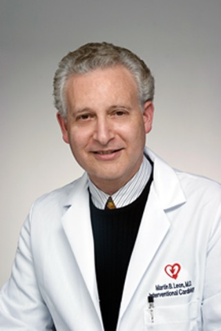
"Open surgery is now rarely needed for femoral artery access-site closure following transfemoral TAVI [transcatheter aortic valve implantation], even with the currently used large sheaths," Dr. Martin B. Leon said at the International Symposium on Endovascular Therapy.
"A percutaneous closure device–enabled technique aided by the crossover balloon occlusion technique (CBOT) after TAVI may be a viable alternative to conventional surgical repair when used in appropriately selected patients," said Dr. Leon, professor of medicine and associate director of the center for interventional vascular therapy at Columbia University in New York.
Dr. Leon and his associates began using the CBOT on patients undergoing TAVI by a transfemoral approach in November 2008, and through last September they had used the CBOT on 56 of their 58 patients (mean age, 85 years). The two exceptions involved patients who underwent elective surgical closures because their femoral-artery anatomy was judged unsuitable for percutaneous closure.
Among the 56 who underwent percutaneous closure using the CBOT, 53 patients (95%) had a successful closure. In two of the unsuccessful patients, the closure attempt produced a perforated external iliac artery, and in the third unsuccessful case the attempt resulted in an occlusive dissection of the external iliac.
The 5% vascular complication rate in this series of 56 TAVI patients compared very favorably with the 16% rate of major vascular complications at 30 days reported by Dr. Leon and his collaborators in the results from the PARTNER (Placement of Aortic Transcatheter Valves) trial published last October, which was the pivotal trial for testing the safety and efficacy of TAVI in patients who were not candidates for valve implantation using open surgery (N. Engl. J. Med. 2010;363:1597-607). Most TAVI patients in PARTNER did not undergo the CBOT.
The CBOT involves three steps, and is a modification of the standard ipsilateral technique for balloon-assisted closure, Dr. Leon explained. First, the operator establishes access into the femoral artery contralateral to the one used for the TAVI catheter, and places a crossover sheath in the external iliac artery that is ipsilateral to the TAVI setup. Next is the insertion of a properly sized balloon with low-pressure inflation to occlude the vessel just above the arteriotomy site. Third, once this balloon has been inflated to decompress the vessel and control bleeding, the operator deploys a two-suture percutaneous closure device. (Dr. Leon uses the ProStar device.)
He recommended then assessing the closure site and reinflating the crossover balloon if the closure does not look perfect. "We don’t allow 70% lesions or minor extravasations," he said. "We’re very aggressive about getting as good a result as we can." He recommended that operators "have great respect for vascular complications during and after TAVI," and that the team "not relax until the [TAVI] sheath is removed and the access site is stable."
He also suggested "meticulous" preprocedure screening of patients, with a special emphasis on CT angiography to identify patients with problematic vascular anatomy. Features to watch for include a deep common femoral artery, a high femoral bifurcation, excessive proximal tortuosity, and atherosclerotic disease at the common femoral puncture site, especially circumferential calcification. Many TAVI candidates are very elderly and hence often have tortuous vessels and a high rate of calcification.
He also suggested that the team have proper training and experience with large-vascular access and suture-mediated percutaneous closure.
Vascular complications and their avoidance represent major issues in the performance of TAVI, Dr. Leon said. The catheters currently used to perform the procedure are large, with outside diameters exceeding 8 mm, and although the catheters come with a hydrophilic coating, this tends to wear off quickly during the procedure, making the catheters sticky and harder to maneuver safely. Smaller catheters will soon appear on the market, but for the time being, the procedure poses a potential risk for access-site closure complications, especially in older patients.
In addition, a vascular complication can have a substantial impact on patient outcome. In the PARTNER trial, patients who underwent TAVI and had a major vascular complication had a 47% mortality rate at 1 year after their procedure, compared with a 28% 1-year mortality rate among the TAVI patients who had no major vascular complication, a statistically significant difference, Dr. Leon said.
Dr. Leon said that he has been an unpaid consultant to Abbott, Boston Scientific, Edwards Lifesciences, and Medtronic. He has been a paid consultant to Angioscore, Neovasc, and Symetis. He also has equity relationships with ATI companies, Claret, Coherex, GDS, Medinol, and Sadra.
MIAMI BEACH – Using a novel balloon occlusion technique, interventional cardiologists at Columbia University have safely performed percutaneous artery closures on 95% of their patients undergoing transfemoral aortic valve implantations.
"Open surgery is now rarely needed for femoral artery access-site closure following transfemoral TAVI [transcatheter aortic valve implantation], even with the currently used large sheaths," Dr. Martin B. Leon said at the International Symposium on Endovascular Therapy.
"A percutaneous closure device–enabled technique aided by the crossover balloon occlusion technique (CBOT) after TAVI may be a viable alternative to conventional surgical repair when used in appropriately selected patients," said Dr. Leon, professor of medicine and associate director of the center for interventional vascular therapy at Columbia University in New York.
Dr. Leon and his associates began using the CBOT on patients undergoing TAVI by a transfemoral approach in November 2008, and through last September they had used the CBOT on 56 of their 58 patients (mean age, 85 years). The two exceptions involved patients who underwent elective surgical closures because their femoral-artery anatomy was judged unsuitable for percutaneous closure.
Among the 56 who underwent percutaneous closure using the CBOT, 53 patients (95%) had a successful closure. In two of the unsuccessful patients, the closure attempt produced a perforated external iliac artery, and in the third unsuccessful case the attempt resulted in an occlusive dissection of the external iliac.
The 5% vascular complication rate in this series of 56 TAVI patients compared very favorably with the 16% rate of major vascular complications at 30 days reported by Dr. Leon and his collaborators in the results from the PARTNER (Placement of Aortic Transcatheter Valves) trial published last October, which was the pivotal trial for testing the safety and efficacy of TAVI in patients who were not candidates for valve implantation using open surgery (N. Engl. J. Med. 2010;363:1597-607). Most TAVI patients in PARTNER did not undergo the CBOT.
The CBOT involves three steps, and is a modification of the standard ipsilateral technique for balloon-assisted closure, Dr. Leon explained. First, the operator establishes access into the femoral artery contralateral to the one used for the TAVI catheter, and places a crossover sheath in the external iliac artery that is ipsilateral to the TAVI setup. Next is the insertion of a properly sized balloon with low-pressure inflation to occlude the vessel just above the arteriotomy site. Third, once this balloon has been inflated to decompress the vessel and control bleeding, the operator deploys a two-suture percutaneous closure device. (Dr. Leon uses the ProStar device.)
He recommended then assessing the closure site and reinflating the crossover balloon if the closure does not look perfect. "We don’t allow 70% lesions or minor extravasations," he said. "We’re very aggressive about getting as good a result as we can." He recommended that operators "have great respect for vascular complications during and after TAVI," and that the team "not relax until the [TAVI] sheath is removed and the access site is stable."
He also suggested "meticulous" preprocedure screening of patients, with a special emphasis on CT angiography to identify patients with problematic vascular anatomy. Features to watch for include a deep common femoral artery, a high femoral bifurcation, excessive proximal tortuosity, and atherosclerotic disease at the common femoral puncture site, especially circumferential calcification. Many TAVI candidates are very elderly and hence often have tortuous vessels and a high rate of calcification.
He also suggested that the team have proper training and experience with large-vascular access and suture-mediated percutaneous closure.
Vascular complications and their avoidance represent major issues in the performance of TAVI, Dr. Leon said. The catheters currently used to perform the procedure are large, with outside diameters exceeding 8 mm, and although the catheters come with a hydrophilic coating, this tends to wear off quickly during the procedure, making the catheters sticky and harder to maneuver safely. Smaller catheters will soon appear on the market, but for the time being, the procedure poses a potential risk for access-site closure complications, especially in older patients.
In addition, a vascular complication can have a substantial impact on patient outcome. In the PARTNER trial, patients who underwent TAVI and had a major vascular complication had a 47% mortality rate at 1 year after their procedure, compared with a 28% 1-year mortality rate among the TAVI patients who had no major vascular complication, a statistically significant difference, Dr. Leon said.
Dr. Leon said that he has been an unpaid consultant to Abbott, Boston Scientific, Edwards Lifesciences, and Medtronic. He has been a paid consultant to Angioscore, Neovasc, and Symetis. He also has equity relationships with ATI companies, Claret, Coherex, GDS, Medinol, and Sadra.
FROM THE INTERNATIONAL SYMPOSIUM ON ENDOVASCULAR THERAPY
Balloon Occlusion Aids Percutaneous Closure of TAVI Entry Site
MIAMI BEACH – Using a novel balloon occlusion technique, interventional cardiologists at Columbia University have safely performed percutaneous artery closures on 95% of their patients undergoing transfemoral aortic valve implantations.
"Open surgery is now rarely needed for femoral artery access-site closure following transfemoral TAVI [transcatheter aortic valve implantation], even with the currently used large sheaths," Dr. Martin B. Leon said at the International Symposium on Endovascular Therapy.
"A percutaneous closure device–enabled technique aided by the crossover balloon occlusion technique (CBOT) after TAVI may be a viable alternative to conventional surgical repair when used in appropriately selected patients," said Dr. Leon, professor of medicine and associate director of the center for interventional vascular therapy at Columbia University in New York.
Dr. Leon and his associates began using the CBOT on patients undergoing TAVI by a transfemoral approach in November 2008, and through last September they had used the CBOT on 56 of their 58 patients (mean age, 85 years). The two exceptions involved patients who underwent elective surgical closures because their femoral-artery anatomy was judged unsuitable for percutaneous closure.
Among the 56 who underwent percutaneous closure using the CBOT, 53 patients (95%) had a successful closure. In two of the unsuccessful patients, the closure attempt produced a perforated external iliac artery, and in the third unsuccessful case the attempt resulted in an occlusive dissection of the external iliac.
The 5% vascular complication rate in this series of 56 TAVI patients compared very favorably with the 16% rate of major vascular complications at 30 days reported by Dr. Leon and his collaborators in the results from the PARTNER (Placement of Aortic Transcatheter Valves) trial published last October, which was the pivotal trial for testing the safety and efficacy of TAVI in patients who were not candidates for valve implantation using open surgery (N. Engl. J. Med. 2010;363:1597-607). Most TAVI patients in PARTNER did not undergo the CBOT.
The CBOT involves three steps, and is a modification of the standard ipsilateral technique for balloon-assisted closure, Dr. Leon explained. First, the operator establishes access into the femoral artery contralateral to the one used for the TAVI catheter, and places a crossover sheath in the external iliac artery that is ipsilateral to the TAVI setup. Next is the insertion of a properly sized balloon with low-pressure inflation to occlude the vessel just above the arteriotomy site. Third, once this balloon has been inflated to decompress the vessel and control bleeding, the operator deploys a two-suture percutaneous closure device. (Dr. Leon uses the ProStar device.)
He recommended then assessing the closure site and reinflating the crossover balloon if the closure does not look perfect. "We don’t allow 70% lesions or minor extravasations," he said. "We’re very aggressive about getting as good a result as we can." He recommended that operators "have great respect for vascular complications during and after TAVI," and that the team "not relax until the [TAVI] sheath is removed and the access site is stable."
He also suggested "meticulous" preprocedure screening of patients, with a special emphasis on CT angiography to identify patients with problematic vascular anatomy. Features to watch for include a deep common femoral artery, a high femoral bifurcation, excessive proximal tortuosity, and atherosclerotic disease at the common femoral puncture site, especially circumferential calcification. Many TAVI candidates are very elderly and hence often have tortuous vessels and a high rate of calcification.
He also suggested that the team have proper training and experience with large-vascular access and suture-mediated percutaneous closure.
Vascular complications and their avoidance represent major issues in the performance of TAVI, Dr. Leon said. The catheters currently used to perform the procedure are large, with outside diameters exceeding 8 mm, and although the catheters come with a hydrophilic coating, this tends to wear off quickly during the procedure, making the catheters sticky and harder to maneuver safely. Smaller catheters will soon appear on the market, but for the time being, the procedure poses a potential risk for access-site closure complications, especially in older patients.
In addition, a vascular complication can have a substantial impact on patient outcome. In the PARTNER trial, patients who underwent TAVI and had a major vascular complication had a 47% mortality rate at 1 year after their procedure, compared with a 28% 1-year mortality rate among the TAVI patients who had no major vascular complication, a statistically significant difference, Dr. Leon said.
Dr. Leon said that he has been an unpaid consultant to Abbott, Boston Scientific, Edwards Lifesciences, and Medtronic. He has been a paid consultant to Angioscore, Neovasc, and Symetis. He also has equity relationships with ATI companies, Claret, Coherex, GDS, Medinol, and Sadra.
FROM THE INTERNATIONAL SYMPOSIUM ON ENDOVASCULAR THERAPY
Major Finding: A series of elderly patients who underwent TAVI with use of a crossover balloon occlusion technique to make a femoral-artery approach had a 5% rate of vascular complications.
Data Source: A single-center series of 56 patients.
Disclosures: Dr. Leon said that he has been an unpaid consultant to Abbott, Boston Scientific, Edwards Lifesciences, and Medtronic. He has been a paid consultant to Angioscore, Neovasc, and Symetis. He also has equity relationships with ATI companies, Claret, Coherex, GDS, Medinol, and Sadra.
Trifurcation Technique Preserves Internal Iliac Flow After EVAR
MIAMI BEACH – A novel, trifurcated, endovascular stenting procedure allowed preservation of blood flow through the internal iliac artery of patients undergoing repair of a thoracoabdominal aortic aneurysm or an aortoiliac aneurysm.
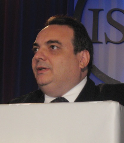
Dr. Armando C. Lobato, who developed the technique, has successfully used it on 34 patients, including 7 patients who required bilateral repairs, he said at the International Symposium on Endovascular Therapy. He performed his first trifurcation procedure – which he also calls the Brazilian Sandwich procedure – in August 2008. He has had 100% technical success using the procedure, with no operative deaths and no need to convert to an open procedure.
During a median follow-up of 5 months (range, 2-24 months), no patient experienced colonic or buttock necrosis, no patient had ischemic colitis, and there were no type III endoleaks. In all, 39 (95%) of the 41 treated internal iliac (hypogastric) arteries maintained primary patency. As a result of the procedure, occlusions also occurred in one contralateral iliac limb and in one ipsilateral external iliac artery. Secondary patency was maintained in 38 of the 41 treated arteries and their associated vessels, said Dr. Lobato, medical director of the Vascular and Endovascular Surgery Institute in São Paulo, Brazil.
About 20%-30% of patients with abdominal aortic aneurysm also have aneurysms in one or both of their common iliac arteries. Endovascular repair in these patients is harder because of the difficulty in finding an adequate landing zone for the stent-graft limbs and because the iliac aneurysm is a potential site for an endoleak. In addition, some patients have short common iliac arteries, an anatomy that is especially prevalent in patients of Asian descent and on the right side of the body. Dr. Lobato said that he developed the trifurcation technique to overcome these anatomical problems so that endovascular aneurysm repair could expand in a safe and cost-effective way via a method that was easy to perform.
Dr. Lobato broke the trifurcation technique into the following five steps:
• First, he inserts the main, bifurcated stent graft using an ipsilateral femoral approach. He positions the graft so that the distal end of the iliac limb is 1 cm above the origin of the internal iliac artery.
• Second, he inserts a long, 5-Fr multipurpose catheter into the ipsilateral internal iliac artery using left brachial access and a 0.035-inch, extra-stiff guide wire with a floppy tip.
• In the third step, he places a covered, self-expanding stent 2 cm into the internal iliac artery with a 6-cm overlap into the iliac limb. He also then positions an iliac limb extension 1 cm below the proximal end of the covered stent. He deploys the iliac limb extension first, followed by deployment of the covered stent.
• Fourth, he models the iliac limb stents with a latex balloon and dilates the covered stent with an angioplasty balloon.
• For the final, fifth step, he deploys the graft within the contralateral iliac limb.
In patients with bilateral aneurysms in their common iliac artery, he repeats steps 2-4 on the second side.
Dr. Lobato said that he had no disclosures.
MIAMI BEACH – A novel, trifurcated, endovascular stenting procedure allowed preservation of blood flow through the internal iliac artery of patients undergoing repair of a thoracoabdominal aortic aneurysm or an aortoiliac aneurysm.
Dr. Armando C. Lobato, who developed the technique, has successfully used it on 34 patients, including 7 patients who required bilateral repairs, he said at the International Symposium on Endovascular Therapy. He performed his first trifurcation procedure – which he also calls the Brazilian Sandwich procedure – in August 2008. He has had 100% technical success using the procedure, with no operative deaths and no need to convert to an open procedure.
During a median follow-up of 5 months (range, 2-24 months), no patient experienced colonic or buttock necrosis, no patient had ischemic colitis, and there were no type III endoleaks. In all, 39 (95%) of the 41 treated internal iliac (hypogastric) arteries maintained primary patency. As a result of the procedure, occlusions also occurred in one contralateral iliac limb and in one ipsilateral external iliac artery. Secondary patency was maintained in 38 of the 41 treated arteries and their associated vessels, said Dr. Lobato, medical director of the Vascular and Endovascular Surgery Institute in São Paulo, Brazil.
About 20%-30% of patients with abdominal aortic aneurysm also have aneurysms in one or both of their common iliac arteries. Endovascular repair in these patients is harder because of the difficulty in finding an adequate landing zone for the stent-graft limbs and because the iliac aneurysm is a potential site for an endoleak. In addition, some patients have short common iliac arteries, an anatomy that is especially prevalent in patients of Asian descent and on the right side of the body. Dr. Lobato said that he developed the trifurcation technique to overcome these anatomical problems so that endovascular aneurysm repair could expand in a safe and cost-effective way via a method that was easy to perform.
Dr. Lobato broke the trifurcation technique into the following five steps:
• First, he inserts the main, bifurcated stent graft using an ipsilateral femoral approach. He positions the graft so that the distal end of the iliac limb is 1 cm above the origin of the internal iliac artery.
• Second, he inserts a long, 5-Fr multipurpose catheter into the ipsilateral internal iliac artery using left brachial access and a 0.035-inch, extra-stiff guide wire with a floppy tip.
• In the third step, he places a covered, self-expanding stent 2 cm into the internal iliac artery with a 6-cm overlap into the iliac limb. He also then positions an iliac limb extension 1 cm below the proximal end of the covered stent. He deploys the iliac limb extension first, followed by deployment of the covered stent.
• Fourth, he models the iliac limb stents with a latex balloon and dilates the covered stent with an angioplasty balloon.
• For the final, fifth step, he deploys the graft within the contralateral iliac limb.
In patients with bilateral aneurysms in their common iliac artery, he repeats steps 2-4 on the second side.
Dr. Lobato said that he had no disclosures.
FROM THE INTERNATIONAL SYMPOSIUM ON ENDOVASCULAR THERAPY
Trifurcation Technique Preserves Internal Iliac Flow After EVAR
MIAMI BEACH – A novel, trifurcated, endovascular stenting procedure allowed preservation of blood flow through the internal iliac artery of patients undergoing repair of a thoracoabdominal aortic aneurysm or an aortoiliac aneurysm.
Dr. Armando C. Lobato, who developed the technique, has successfully used it on 34 patients, including 7 patients who required bilateral repairs, he said at the International Symposium on Endovascular Therapy. He performed his first trifurcation procedure – which he also calls the Brazilian Sandwich procedure – in August 2008. He has had 100% technical success using the procedure, with no operative deaths and no need to convert to an open procedure.
During a median follow-up of 5 months (range, 2-24 months), no patient experienced colonic or buttock necrosis, no patient had ischemic colitis, and there were no type III endoleaks. In all, 39 (95%) of the 41 treated internal iliac (hypogastric) arteries maintained primary patency. As a result of the procedure, occlusions also occurred in one contralateral iliac limb and in one ipsilateral external iliac artery. Secondary patency was maintained in 38 of the 41 treated arteries and their associated vessels, said Dr. Lobato, medical director of the Vascular and Endovascular Surgery Institute in São Paulo, Brazil.
About 20%-30% of patients with abdominal aortic aneurysm also have aneurysms in one or both of their common iliac arteries. Endovascular repair in these patients is harder because of the difficulty in finding an adequate landing zone for the stent-graft limbs and because the iliac aneurysm is a potential site for an endoleak. In addition, some patients have short common iliac arteries, an anatomy that is especially prevalent in patients of Asian descent and on the right side of the body. Dr. Lobato said that he developed the trifurcation technique to overcome these anatomical problems so that endovascular aneurysm repair could expand in a safe and cost-effective way via a method that was easy to perform.
Dr. Lobato broke the trifurcation technique into the following five steps:
• First, he inserts the main, bifurcated stent graft using an ipsilateral femoral approach. He positions the graft so that the distal end of the iliac limb is 1 cm above the origin of the internal iliac artery.
• Second, he inserts a long, 5-Fr multipurpose catheter into the ipsilateral internal iliac artery using left brachial access and a 0.035-inch, extra-stiff guide wire with a floppy tip.
• In the third step, he places a covered, self-expanding stent 2 cm into the internal iliac artery with a 6-cm overlap into the iliac limb. He also then positions an iliac limb extension 1 cm below the proximal end of the covered stent. He deploys the iliac limb extension first, followed by deployment of the covered stent.
• Fourth, he models the iliac limb stents with a latex balloon and dilates the covered stent with an angioplasty balloon.
• For the final, fifth step, he deploys the graft within the contralateral iliac limb.
In patients with bilateral aneurysms in their common iliac artery, he repeats steps 2-4 on the second side.
Dr. Lobato said that he had no disclosures.
FROM THE INTERNATIONAL SYMPOSIUM ON ENDOVASCULAR THERAPY
Major Finding: A novel trifurcation technique safely preserved flow through the internal iliac arteries in patients who underwent an endovascular abdominal aortic aneurysm repair with an aneurysm in one or both common iliac arteries.
Data Source: A single-center series of 34 patients.
Disclosures: Dr. Lobato said that he had no disclosures.
Cardiac Arrhythmia Incidence Hits 29% After Vascular Surgery
MIAMI BEACH – Patients undergoing major vascular surgery showed a surprisingly high incidence of perioperative cardiac arrhythmias in a single-center, prospective study that so far includes 41 patients.

Insertion of a continuous ECG monitor into patients starting about 2 weeks before they underwent major vascular surgery and continuing monitoring of each patient through about 2 months after surgery showed that 12 patients (29%) had some form of new-onset cardiac arrhythmia, Dr. Hence J.M. Verhagen said at ISET 2011, an international symposium on endovascular therapy. The study group included 13 patients (32%) undergoing open repair of an abdominal aortic aneurysm, 18 (44%) patients having an endovascular repair of an abdominal aortic aneurysm, and 10 patients (24%) having peripheral artery bypass surgery.
By using an inserted arrhythmia monitor to track incident arrhythmias for a prolonged period during and after surgery "we detected arrhythmias we were completely unaware of," said Dr. Verhagen, professor of surgery and chief of vascular surgery at Erasmus University Medical Center in Rotterdam, the Netherlands. "Many of these patients are completely asymptomatic," he noted in an interview. The most common arrhythmias were ventricular tachycardias, followed by atrial fibrillations, and supraventricular tachyarrhythmias. Most were paroxysmal.
Prior studies by Dr. Verhagen and his associates showed that vascular surgery patients who develop a cardiac arrhythmia following surgery have significantly worse outcomes. "This is a new opportunity to improve long-term outcomes," he said, but added that his group has not yet examined the impact that early treatment of the arrhythmias has on long-term consequences.
The earlier reports from Dr. Verhagen and his collaborators used externally worn Holter monitors to track cardiac arrhythmias in vascular surgery patients. Because these devices are intrusive for patients, monitoring lasted only 3 days. In one recently reported study, they assessed for 3 days 483 vascular surgery patients with Holter monitors and found new onset tachyarrhythmias in 33 (7%). They reported that the patients with these arrhythmias had a higher rate of reduced left ventricular ejection fraction and myocardial infarctions during the perioperative period. During a 2-year follow-up, their rate of sudden cardiac death was 2.6-fold higher than in the patients who did not have perioperative arrhythmias (J. Vasc. Surg. 2010 [doi:10.1016/j.jvs.2010.09.013]).
In another recent report, they presented findings from 72-hour Holter monitoring in 513 vascular surgery patients and found 55 (11%) with ventricular tachycardia, atrial fibrillation, supraventricular tachycardia, or ventricular fibrillation. Patients who had these arrhythmias had a 2.2-fold increased rate of cardiovascular events during an average follow-up of 1.7 years (Am. Heart J. 2010;159:1108-15).
The study using Holter monitoring included patients who had open or endovascular abdominal aortic aneurysm repair and peripheral artery bypass surgery as well as some patients who underwent carotid endarterectomy or endovascular carotid artery stenting.
The group’s more recent studies using longer-term monitoring with small, inserted devices showed that the incidence of new arrhythmias is much higher than the earlier studies had shown. The arrhythmic episodes seemed to be more common during the first couple of weeks following surgery, but several other episodes occurred several weeks after the vascular operation, Dr. Verhagen said. The pattern of arrhythmia episodes also appeared to have no relationship to whether surgery was done by open or endovascular procedures. His group will continue to collect data using inserted monitors from additional surgery patients, and future studies will need to address the impact that treatment of the arrhythmias has on long-term patient outcomes, Dr. Verhagen said.
Dr. Verhagen said that he has received research funding from and has served as a consultant to Medtronic. The insertable ECG monitors used in his study were Reveal XT models, a device marketed by Medtronic.
MIAMI BEACH – Patients undergoing major vascular surgery showed a surprisingly high incidence of perioperative cardiac arrhythmias in a single-center, prospective study that so far includes 41 patients.
Insertion of a continuous ECG monitor into patients starting about 2 weeks before they underwent major vascular surgery and continuing monitoring of each patient through about 2 months after surgery showed that 12 patients (29%) had some form of new-onset cardiac arrhythmia, Dr. Hence J.M. Verhagen said at ISET 2011, an international symposium on endovascular therapy. The study group included 13 patients (32%) undergoing open repair of an abdominal aortic aneurysm, 18 (44%) patients having an endovascular repair of an abdominal aortic aneurysm, and 10 patients (24%) having peripheral artery bypass surgery.
By using an inserted arrhythmia monitor to track incident arrhythmias for a prolonged period during and after surgery "we detected arrhythmias we were completely unaware of," said Dr. Verhagen, professor of surgery and chief of vascular surgery at Erasmus University Medical Center in Rotterdam, the Netherlands. "Many of these patients are completely asymptomatic," he noted in an interview. The most common arrhythmias were ventricular tachycardias, followed by atrial fibrillations, and supraventricular tachyarrhythmias. Most were paroxysmal.
Prior studies by Dr. Verhagen and his associates showed that vascular surgery patients who develop a cardiac arrhythmia following surgery have significantly worse outcomes. "This is a new opportunity to improve long-term outcomes," he said, but added that his group has not yet examined the impact that early treatment of the arrhythmias has on long-term consequences.
The earlier reports from Dr. Verhagen and his collaborators used externally worn Holter monitors to track cardiac arrhythmias in vascular surgery patients. Because these devices are intrusive for patients, monitoring lasted only 3 days. In one recently reported study, they assessed for 3 days 483 vascular surgery patients with Holter monitors and found new onset tachyarrhythmias in 33 (7%). They reported that the patients with these arrhythmias had a higher rate of reduced left ventricular ejection fraction and myocardial infarctions during the perioperative period. During a 2-year follow-up, their rate of sudden cardiac death was 2.6-fold higher than in the patients who did not have perioperative arrhythmias (J. Vasc. Surg. 2010 [doi:10.1016/j.jvs.2010.09.013]).
In another recent report, they presented findings from 72-hour Holter monitoring in 513 vascular surgery patients and found 55 (11%) with ventricular tachycardia, atrial fibrillation, supraventricular tachycardia, or ventricular fibrillation. Patients who had these arrhythmias had a 2.2-fold increased rate of cardiovascular events during an average follow-up of 1.7 years (Am. Heart J. 2010;159:1108-15).
The study using Holter monitoring included patients who had open or endovascular abdominal aortic aneurysm repair and peripheral artery bypass surgery as well as some patients who underwent carotid endarterectomy or endovascular carotid artery stenting.
The group’s more recent studies using longer-term monitoring with small, inserted devices showed that the incidence of new arrhythmias is much higher than the earlier studies had shown. The arrhythmic episodes seemed to be more common during the first couple of weeks following surgery, but several other episodes occurred several weeks after the vascular operation, Dr. Verhagen said. The pattern of arrhythmia episodes also appeared to have no relationship to whether surgery was done by open or endovascular procedures. His group will continue to collect data using inserted monitors from additional surgery patients, and future studies will need to address the impact that treatment of the arrhythmias has on long-term patient outcomes, Dr. Verhagen said.
Dr. Verhagen said that he has received research funding from and has served as a consultant to Medtronic. The insertable ECG monitors used in his study were Reveal XT models, a device marketed by Medtronic.
FROM THE ANNUAL INTERNATIONAL SYMPOSIUM ON ENDOVASCULAR THERAPY
Cardiac Arrhythmia Incidence Hits 29% After Vascular Surgery
MIAMI BEACH – Patients undergoing major vascular surgery showed a surprisingly high incidence of perioperative cardiac arrhythmias in a single-center, prospective study that so far includes 41 patients.
Insertion of a continuous ECG monitor into patients starting about 2 weeks before they underwent major vascular surgery and continuing monitoring of each patient through about 2 months after surgery showed that 12 patients (29%) had some form of new-onset cardiac arrhythmia, Dr. Hence J.M. Verhagen said at ISET 2011, an international symposium on endovascular therapy. The study group included 13 patients (32%) undergoing open repair of an abdominal aortic aneurysm, 18 (44%) patients having an endovascular repair of an abdominal aortic aneurysm, and 10 patients (24%) having peripheral artery bypass surgery.
By using an inserted arrhythmia monitor to track incident arrhythmias for a prolonged period during and after surgery "we detected arrhythmias we were completely unaware of," said Dr. Verhagen, professor of surgery and chief of vascular surgery at Erasmus University Medical Center in Rotterdam, the Netherlands. "Many of these patients are completely asymptomatic," he noted in an interview. The most common arrhythmias were ventricular tachycardias, followed by atrial fibrillations, and supraventricular tachyarrhythmias. Most were paroxysmal.
Prior studies by Dr. Verhagen and his associates showed that vascular surgery patients who develop a cardiac arrhythmia following surgery have significantly worse outcomes. "This is a new opportunity to improve long-term outcomes," he said, but added that his group has not yet examined the impact that early treatment of the arrhythmias has on long-term consequences.
The earlier reports from Dr. Verhagen and his collaborators used externally worn Holter monitors to track cardiac arrhythmias in vascular surgery patients. Because these devices are intrusive for patients, monitoring lasted only 3 days. In one recently reported study, they assessed for 3 days 483 vascular surgery patients with Holter monitors and found new onset tachyarrhythmias in 33 (7%). They reported that the patients with these arrhythmias had a higher rate of reduced left ventricular ejection fraction and myocardial infarctions during the perioperative period. During a 2-year follow-up, their rate of sudden cardiac death was 2.6-fold higher than in the patients who did not have perioperative arrhythmias (J. Vasc. Surg. 2010 [doi:10.1016/j.jvs.2010.09.013]).
In another recent report, they presented findings from 72-hour Holter monitoring in 513 vascular surgery patients and found 55 (11%) with ventricular tachycardia, atrial fibrillation, supraventricular tachycardia, or ventricular fibrillation. Patients who had these arrhythmias had a 2.2-fold increased rate of cardiovascular events during an average follow-up of 1.7 years (Am. Heart J. 2010;159:1108-15).
The study using Holter monitoring included patients who had open or endovascular abdominal aortic aneurysm repair and peripheral artery bypass surgery as well as some patients who underwent carotid endarterectomy or endovascular carotid artery stenting.
The group’s more recent studies using longer-term monitoring with small, inserted devices showed that the incidence of new arrhythmias is much higher than the earlier studies had shown. The arrhythmic episodes seemed to be more common during the first couple of weeks following surgery, but several other episodes occurred several weeks after the vascular operation, Dr. Verhagen said. The pattern of arrhythmia episodes also appeared to have no relationship to whether surgery was done by open or endovascular procedures. His group will continue to collect data using inserted monitors from additional surgery patients, and future studies will need to address the impact that treatment of the arrhythmias has on long-term patient outcomes, Dr. Verhagen said.
Dr. Verhagen said that he has received research funding from and has served as a consultant to Medtronic. The insertable ECG monitors used in his study were Reveal XT models, a device marketed by Medtronic.
FROM THE ANNUAL INTERNATIONAL SYMPOSIUM ON ENDOVASCULAR THERAPY
Major Finding: During the 2 months following major vascular surgery, patients had a 29% incidence of new-onset cardiac arrhythmias detected by inserted ECG monitors.
Data Source: Single-center series of 41 patients who had undergone open or endovascular repair of an abdominal aortic aneurysm or peripheral artery bypass surgery.
Disclosures: Dr. Verhagen said that he has received research funding from and has served as a consultant to Medtronic. The insertable ECG monitors used in his study were Reveal XT models, a device marketed by Medtronic.
Vanderbilt Uses Genotyping Prior To Catheterization
CHICAGO - Last fall, physicians at Vanderbilt University Medical Center began routinely testing all patients who were scheduled for coronary catheterization with a broad genotyping screen that - among other things - would identify whether they had a problem activating clopidogrel. By mid-November, the program had tested about 300 patients, including 10 found to have a poor-metabolizer genotype in the hepatic-enzyme gene CYP2C19 that would likely blunt the efficacy of a conventional clopidogrel dose. Many of the 10 patients received a doubled dose to compensate, whereas others who weren't aged 75 or older received the pricier, alternative agent prasugrel.
This experience marked the first phase of a new Vanderbilt program that will expand over time to include other patients in line to receive a drug with a pharmacogenetic dimension, Dr. Dan M. Roden said at the meeting. The genotyping program will soon expand to include patients who are scheduled for knee- or hip-replacement surgery, anticipating their need to start on warfarin. Genotype data can also help physicians select the best dosage for starting a warfarin regimen, said Dr. Roden, a cardiologist and assistant vice chancellor for personalized medicine at Vanderbilt in Nashville, Tenn.
Subsequent expansion plans are not yet set, but other candidates for genotyping include patients who are either already on or at an increased risk for soon starting tamoxifen, abacavir, azathioprine, 6-mercaptopurine, codeine, or "virtually any antidepressant or most antipsychotics,” Dr. Roden said in an interview.
"In the long perspective, every 50-year-old” is a good bet to eventually receive at least one drug for which a dosage adjustment based on genotype is warranted, but - stopping short of such global use right now - the Vanderbilt program will instead gradually phase in new groups of patients to the offer of genotyping.
"Implementation is a huge challenge. In my opinion, this will only work with preemptive implementation. Electronic records are not just repositories of information, but are nimble enough to provide support at the time of a prescription,” he said. "The way it ought to work is, a physician prescribes a drug and the electronic system recognizes [that] the drug has a genetic element and goes into the patient's record and finds the genotype information” to decide whether to flash a screen alert about the patient's genotype and the implications.
The program, known as PREDICT (Pharmacogenomic Resource for Enhanced Decisions in Care and Treatment) launched on Sept. 15. It uses a genotyping panel sold on the U.S. market by Illumina that screens for 184 different genetic polymorphisms in 34 genes that affect the absorption, distribution, metabolism, or excretion of various drugs.
Test results get posted into the record within a day of specimen collection.
So far, Vanderbilt itself has completely funded the program, which involved a year of planning and "a huge amount of money,” said Dr. Roden, adding that the program is the first of its kind worldwide. PREDICT is expected to improve patient outcomes and its developers hope to eventually convince payers to cover the cost.
Dr. Roden stated that he is or has been a consultant to several drug companies and has received royalties from Clinical Data Inc.