User login
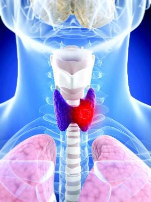
Bone sarcomas are not just for kids
The treatment of bone sarcomas in adults is guided as much by experience as it is by medical evidence, say sarcoma experts who should know.
“[L]arge prospective clinical trials in adults with bone sarcomas are lacking. Although translation of findings in pediatric studies to adults is possible, it is not clear if findings from pediatric studies truly apply to adults,” Dr. Michael J. Wagner and his colleagues from the University of Texas MD Anderson Cancer Center in Houston said.
In a review article in the Journal of Oncology Practice, the investigators outline how they have melded lessons learned from pediatric clinical trials with clinical experience treating adults with bone sarcomas.
Adult osteosarcomas
Although the peak incidence of osteosarcoma occurs in adolescents and young adults, some studies of various chemotherapy regimens have included middle-aged adults and even some patients in their 90s, although many have excluded patients older than 30, the authors noted.
In adults, and in children and adolescents and young adults, chemotherapy and definitive surgical resection are the mainstays of treatment, with the exception of osteosarcomas of the jaw, which are generally treated with resection alone. For the past three decades, adults with high-grade osteosarcomas have been treated with chemotherapy containing doxorubicin, cisplatin, high-dose methotrexate, and ifosfamide.
“However, important differences to consider in the treatment of older patients include, but are not limited to, the increased incidence of histologic variants of osteosarcoma (including secondary osteosarcomas) and medical comorbidities that may limit tolerance of older adults to dose-intense chemotherapy,” wrote Dr. Wagner and his colleagues (J Oncol Pract. 2016 Mar. doi: 10.1200/JOP.2015.009944).
High-grade localized osteosarcoma is treated with neoadjuvant chemotherapy, followed by limb-sparing surgery and adjuvant chemotherapy.
A poor response to neoadjuvant chemotherapy, defined as tumor necrosis of less than 90% at resection, is a marker for poor prognosis.
The authors previously reported that for patients with a poor response following a regimen of doxorubicin intravenous infusion at 90 mg/m2 over 96 hours plus cisplatin infused intra-arterially at 120-160 mg/m2 over 2 to 24 hours, the addition of high-dose methotrexate and ifosfamide resulted in significant improvement in continuous relapse-free survival.
At MD Anderson, adults with osteosarcoma receive preoperative chemotherapy with doxorubicin and cisplatin for three to four cycles and then undergo surgical resection. Those who have a good response go on to receive adjuvant chemotherapy with doxorubicin 75 mg/m2 and ifosfamide 10g/m2 for four cycles. Patients who have a poor response to neoadjuvant chemotherapy receive alternating courses of ifosfamide 14 g/m2, high-dose methotrexate 10 to 12 g/m2, and doxorubicin 75 mg/m2 and ifosfamide 10 g/m2 for up to 12 cycles or until intolerable adverse events.
Patients with metastatic relapsed/refractory osteosarcoma have 5-year overall survival rates ranging from 15% to 20%, the authors noted, adding that chemotherapy has “limited efficacy” in this setting.
Targeted agents, including the tyrosine kinase inhibitor sorafenib (Nexavar) and the mammalian target of rapamycin (mTOR) inhibitor everolimus (Afinitor) have been evaluated in phase II trials.
Ewing sarcoma
Evidence from clinical trials of chemotherapy regimens in Ewing sarcoma show that patients 18 and older tend to have less favorable outcomes than younger patients when treated with dose-dense regimens, the authors noted.
Their practice is to treat adults with Ewing sarcoma with upfront vincristine 2 mg on day 1, doxorubicin 75 to 90 mg/m2 in a continuous 72-hour infusion, and ifosfamide 2.5 g/m2 given over 3 hours daily for four doses, followed by surgery, and adjuvant chemotherapy with an ifosfamide-based regimen.
Giant cell tumors of bone
These rare osteolytic tumors are treated with surgery as the primary therapy if they are resectable. Patients with metastases (which paradoxically, are usually histologically benign) as well as those who have tumors in areas where surgery may cause unacceptable morbidities may be treated with serial embolization, denosumab (Xgeva), interferon, or pegylated interferon.
Chondrosarcoma
The so-called “conventional” sub-type of chondrosarcomas are not generally responsive to cytotoxic chemotherapy and best managed with surgery alone, the authors say. However, patients with mesenchymal chondrosarcoma are treated similarly to patients with Ewing sarcoma, and those with the dedifferentiated subtype are treated with an osteosarcoma regimens.
The authors noted that point mutations in IDH1 and IDH2 have recently been discovered in chondrosarcomas, making them potential targets for an experimental IDH-1 inhibitor.
Dr. Wagner and coauthor Dr. J. Andrew Livingston reported no relevant disclosures. Coauthors Dr. Shreyaskumar R. Patel and Dr. Robert S. Benjamin reported consulting, advisory roles, research funding, or ownership interests in various pharmaceutical companies.
FROM THE JOURNAL OF ONCOLOGY PRACTICE
Key clinical point: Sarcomas of bone have their peak incidence in children and adolescents, but are also seen in adults.
Major finding: Findings from clinical trials in pediatric bone sarcomas may not be applicable to treatment of adults with the same malignancies.
Data source: Review of the medical literature and of clinical experience at the authors’ center.
Disclosures: Dr. Wagner and coauthor Dr. J. Andrew Livingston reported no relevant disclosures. Coauthors Dr. Shreyaskumar R. Patel and Dr. Robert S. Benjamin reported consulting, advisory roles, research funding, or ownership interests in various pharmaceutical companies.
10-year DCIS recurrence risk dwindles with age
BOSTON – Recurrence rates of cancer following breast-conserving surgery for ductal carcinoma in situ differ significantly with patient age, with younger women having a three-fold higher 10-year recurrence rate than older women.
The lower risk for 10-year recurrence among older women was independent of whether they had received adjuvant radiation therapy, reported Dr. Patricia A. Cronin, a surgical fellow at Memorial Sloan Kettering Cancer Center in New York City.

“Women over 80 are at low risk of recurrence, even in the absence of adjuvant therapies, whilst young age is a highly significant risk factor for any recurrence, in particular invasive recurrence, and therefore age is an important factor that should be weighed when considering other treatment options, including surgical options and adjuvant therapy,” she said at the annual Society of Surgical Oncology Cancer Symposium.
Ductal carcinoma in situ (DCIS) now accounts for more than 20% of all breast cancers. It can be treated with breast conserving surgery (BCS) alone or in conjunction with adjuvant therapy, radiation, and endocrine therapy, or by mastectomy.
“All treatment options have excellent survival but there can be markedly different recurrence rates,” Dr. Cronin said.
She noted that age is recognized as a risk factor for recurrence after BCS for DCIS, an observation confirmed in radiotherapy trials, but added that “the full nature of the relationship between age and recurrence and its interaction with other factors is not clear.”
DCIS through the ages
To get a better handle on the effect of age on recurrence, the investigators looked at data on patients who underwent breast-conserving surgery from 1978 through 2010. The women were stratified by age into six categories, beginning with those under age 40 and continuing at 10-year increments to age 80 and older.
Clinical, pathological, and treatment factors included in the analysis were family history, clinical vs. radiologic presentation, nuclear grade, necrosis, number of excision, margin status, radiotherapy, endocrine therapy, and treatment period.
Outcomes included ipsilateral DCIS recurrence, ipsilateral invasive breast cancer, ipsilateral axillary node recurrence, and distant recurrence in the absence of both ipsilateral recurrence and contralateral breast cancer.
A total of 2,996 women were identified, 363 of whom (12%) had local recurrences, either as DCIS in 192 patients, invasive disease in 160, of unknown status in 11.
An analysis of recurrence rates by age showed decreasing risk with each advance in age category, compared with women under age 40, with women aged 40-49 having a hazard ratio (HR) of 0.68 (P = .05), and women age 80 and over having an HR of 0.34 (P = .01), with 10-year recurrence rates ranging from 27% in the youngest cohort to about 8% in the oldest (log rank P less than .0001 for the overall effect of age on recurrence).
Looking at the individual factors associated with age, women under age 40 had a significantly higher proportion of clinical vs. radiologic presentation, with either a mass, nipple discharge, or Paget disease. Younger women also had less endocrine therapy.
Older women less frequently underwent multiple excisions, and more frequently had close or positive surgical margins, as well as lower rates of adjuvant therapy, and less endocrine therapy than women in the middle-age categories.
In a multivariable analysis for recurrence risk by age controlling for presentation, family history, necrosis, excision number, margin status, adjuvant radiation, endocrine therapy, and treatment period, the association between age and risk remained significant beginning in patients aged 50-59 (HR, 0.46; P = .0005) and continuing to dwindle through the decades to those age 80 and older (HR, 0.21; P = .001).
Age was also significantly associated with recurrence both among who received radiation and those who did not.
Women under age 40 were also found to be at greater risk for invasive recurrences, with 15.8% of all patients in this age group having invasive recurrences, and 11.5% having recurrent DCIS. In contrast, among all women aged 80 and older, 3.2% had invasive recurrences, and 3.4% had recurrent DCIS.
Question of mammography
Following Dr. Cronin’s presentation, Dr. Nicholas Tranakas from Broward Health Medical Center in Fort Lauderdale, Fla., asked whether, since the evidence shows that the majority of younger patients do not have indolent disease, they should be screened with mammography at earlier age than current guidelines recommend.
Dr. Cronin noted that although women under 40 don’t routinely undergo screening, “you can see that most patients under 40 present clinically, and therefore it’s hard in this study to make any recommendations in regards to screening.”
Asked how she counsels younger patients, she noted that although the data on recurrence risk in younger women are provocative, overall survival rates are excellent for both breast-conserving procedures and mastectomy, and the decision about which procedure to choose may rely on the patient’s degree of aversion to risk.
“We don’t automatically recommend mastectomy under 40, but I think this data does make us very concerned about recurrence, and particularly invasive recurrence and its potential influence then on distant disease,” she said.
The study funding source was not reported. The authors reported having no disclosures. Dr. Tranakas reported no disclosures.
BOSTON – Recurrence rates of cancer following breast-conserving surgery for ductal carcinoma in situ differ significantly with patient age, with younger women having a three-fold higher 10-year recurrence rate than older women.
The lower risk for 10-year recurrence among older women was independent of whether they had received adjuvant radiation therapy, reported Dr. Patricia A. Cronin, a surgical fellow at Memorial Sloan Kettering Cancer Center in New York City.
“Women over 80 are at low risk of recurrence, even in the absence of adjuvant therapies, whilst young age is a highly significant risk factor for any recurrence, in particular invasive recurrence, and therefore age is an important factor that should be weighed when considering other treatment options, including surgical options and adjuvant therapy,” she said at the annual Society of Surgical Oncology Cancer Symposium.
Ductal carcinoma in situ (DCIS) now accounts for more than 20% of all breast cancers. It can be treated with breast conserving surgery (BCS) alone or in conjunction with adjuvant therapy, radiation, and endocrine therapy, or by mastectomy.
“All treatment options have excellent survival but there can be markedly different recurrence rates,” Dr. Cronin said.
She noted that age is recognized as a risk factor for recurrence after BCS for DCIS, an observation confirmed in radiotherapy trials, but added that “the full nature of the relationship between age and recurrence and its interaction with other factors is not clear.”
DCIS through the ages
To get a better handle on the effect of age on recurrence, the investigators looked at data on patients who underwent breast-conserving surgery from 1978 through 2010. The women were stratified by age into six categories, beginning with those under age 40 and continuing at 10-year increments to age 80 and older.
Clinical, pathological, and treatment factors included in the analysis were family history, clinical vs. radiologic presentation, nuclear grade, necrosis, number of excision, margin status, radiotherapy, endocrine therapy, and treatment period.
Outcomes included ipsilateral DCIS recurrence, ipsilateral invasive breast cancer, ipsilateral axillary node recurrence, and distant recurrence in the absence of both ipsilateral recurrence and contralateral breast cancer.
A total of 2,996 women were identified, 363 of whom (12%) had local recurrences, either as DCIS in 192 patients, invasive disease in 160, of unknown status in 11.
An analysis of recurrence rates by age showed decreasing risk with each advance in age category, compared with women under age 40, with women aged 40-49 having a hazard ratio (HR) of 0.68 (P = .05), and women age 80 and over having an HR of 0.34 (P = .01), with 10-year recurrence rates ranging from 27% in the youngest cohort to about 8% in the oldest (log rank P less than .0001 for the overall effect of age on recurrence).
Looking at the individual factors associated with age, women under age 40 had a significantly higher proportion of clinical vs. radiologic presentation, with either a mass, nipple discharge, or Paget disease. Younger women also had less endocrine therapy.
Older women less frequently underwent multiple excisions, and more frequently had close or positive surgical margins, as well as lower rates of adjuvant therapy, and less endocrine therapy than women in the middle-age categories.
In a multivariable analysis for recurrence risk by age controlling for presentation, family history, necrosis, excision number, margin status, adjuvant radiation, endocrine therapy, and treatment period, the association between age and risk remained significant beginning in patients aged 50-59 (HR, 0.46; P = .0005) and continuing to dwindle through the decades to those age 80 and older (HR, 0.21; P = .001).
Age was also significantly associated with recurrence both among who received radiation and those who did not.
Women under age 40 were also found to be at greater risk for invasive recurrences, with 15.8% of all patients in this age group having invasive recurrences, and 11.5% having recurrent DCIS. In contrast, among all women aged 80 and older, 3.2% had invasive recurrences, and 3.4% had recurrent DCIS.
Question of mammography
Following Dr. Cronin’s presentation, Dr. Nicholas Tranakas from Broward Health Medical Center in Fort Lauderdale, Fla., asked whether, since the evidence shows that the majority of younger patients do not have indolent disease, they should be screened with mammography at earlier age than current guidelines recommend.
Dr. Cronin noted that although women under 40 don’t routinely undergo screening, “you can see that most patients under 40 present clinically, and therefore it’s hard in this study to make any recommendations in regards to screening.”
Asked how she counsels younger patients, she noted that although the data on recurrence risk in younger women are provocative, overall survival rates are excellent for both breast-conserving procedures and mastectomy, and the decision about which procedure to choose may rely on the patient’s degree of aversion to risk.
“We don’t automatically recommend mastectomy under 40, but I think this data does make us very concerned about recurrence, and particularly invasive recurrence and its potential influence then on distant disease,” she said.
The study funding source was not reported. The authors reported having no disclosures. Dr. Tranakas reported no disclosures.
Key clinical point: The risk of 10-year recurrence of ductal carcinoma in situ diminishes significantly with age.
Major finding: The risk for recurrence within 10 years among women younger than age 40 was 27%, compared with approximately 8% for women age 80 and older.
Data source: Retrospective study of 2,996 treated for DCIS from 1978 through 2010.
Disclosures: The study funding source was not reported. The authors reported having no disclosures. Dr. Tranakas reported no disclosures.
Continuity of care can limit HIV deaths
BOSTON – Getting people who are infected with HIV onto early antiretroviral therapy (ART) and keeping them on it may be the best way to reduce HIV-related deaths and simplify the care continuum or “cascade,” said investigators who advocate universal, immediate ART eligibility and access.
A study of HIV deaths and care patterns in four African countries, presented at the Conference on Retroviruses and Opportunistic Infections, suggests that HIV deaths could be reduced significantly if patients have early therapeutic intervention and remain in care.

“We’re all very aware that the provision of antiretroviral therapy has dramatically reduced mortality from HIV in sub-Saharan Africa, but data from population cohort studies in Eastern and Southern Africa show us that mortality rates among HIV positive adults still remain three to six times greater than HIV-negative adults in settings with mature ART programs. So the question that we set out to answer was why do these HIV deaths still continue to occur in settings where ART is widely available?” said coinvestigator Dr. Jeffrey Eaton from Imperial College London.
Dr. Eaton and his colleagues conducted a study to identify the stages in the care cascade where most deaths occur, in the hope of pinpointing targets for interventions that have could have the greatest effect on improving care.
To do this, they designed a study to model the previous HIV care experience of people who died from the infection. They reviewed empirical data from clinical and vital statistic registries from Western Cape, South Africa, and from population cohorts in Uganda, Malawi, and South Africa.
For example, death registration records from South Africa linked to clinical records for 3,161 adult HIV deaths in 2011 showed that 25% of patients were either not diagnosed or not linked to care, 35% never started on ART, 10% had been on ART for less than 6 months, 20% were lost from ART care or had a gap longer than 3 months in the past year, and just 10% were continuously in care.
The investigators also analyzed data from four mathematical models that were calibrated to HIV epidemics and to patterns of care and utilization of treatment in Kenya, Malawi, Rwanda and South Africa. All four models estimated how HIV deaths were distributed across each stage of care, and projected the distribution over the next 10 years, with the assumption that current patterns of HIV care use and retention would be continued. In addition, three of the models simulated what would happen if all patients linked to care were started on immediate ART therapy.
Dr. Eaton and his colleagues examined the care cascade from the time of HIV diagnosis, linkage to care, pre–antiretroviral therapy care, ART initiation, and survival and retention on ART.
‘Dramatic declines in HIV deaths’
“All of the models simulated the dramatic declines in HIV deaths as treatments became available, and the models also indicated that persons who have initiated ART understandably account for an increasing proportion of HIV deaths going into the future. But the projections estimate that 25%-40% of HIV deaths will continue to be among persons never initiated on ART,” Dr. Eaton said.
Looking at HIV death across the care cascade, the models indicated that only 10%-30% of HIV-related deaths would likely occur among patients who are continuously on
“ART for 6 months or more. Deaths among HIV-positive people who were disengaged from care were projected to account for a “substantial” proportion of all HIV deaths in all four models – from 21%-44% of all HIV deaths projected for 2025.
If present conditions were to continue, the models predict, from 9%-22% of HIV deaths from 2016 through 2025 would occur in patients who were linked to care but for one reason or another were never started on ART.
The models project immediate ART initiation could reduce HIV deaths by between 6%-14% during 2016-2025, mostly by removing opportunities for patients to disengage before treatment initiation, rather than by direct prevention or therapeutic benefits of early ART, the investigators stated.
Their findings suggest that “indicators based on monitoring the effectiveness of ART among patients in clinics miss this population [of patients never diagnosed or linked to care], and thus we feel are insufficient for evaluating the overall effectiveness of ART programs,” Dr. Eaton said.
An HIV/AIDS specialist who was not involved in the study commented in an interview that Dr. Eaton’s approach is an innovative way to identify gaps in care that can contribute to HIV-related deaths.
“Some big points that I came away with were that people who know their [HIV] status but are not in care, that’s contributing to mortality, and then as we get people more into care, mortality is going to accrue if and when people fall out of care,” said Dr. Diane Havlir of the University of California, San Francisco.
Dr. Havlir moderated a media briefing where Dr. Eaton discussed his data following his presentation in an oral abstract session.
The study was supported by the Bill and Melinda Gates Foundation. Dr. Eaton and Dr. Havlir reported having no commercial interests relevant to the study.
BOSTON – Getting people who are infected with HIV onto early antiretroviral therapy (ART) and keeping them on it may be the best way to reduce HIV-related deaths and simplify the care continuum or “cascade,” said investigators who advocate universal, immediate ART eligibility and access.
A study of HIV deaths and care patterns in four African countries, presented at the Conference on Retroviruses and Opportunistic Infections, suggests that HIV deaths could be reduced significantly if patients have early therapeutic intervention and remain in care.
“We’re all very aware that the provision of antiretroviral therapy has dramatically reduced mortality from HIV in sub-Saharan Africa, but data from population cohort studies in Eastern and Southern Africa show us that mortality rates among HIV positive adults still remain three to six times greater than HIV-negative adults in settings with mature ART programs. So the question that we set out to answer was why do these HIV deaths still continue to occur in settings where ART is widely available?” said coinvestigator Dr. Jeffrey Eaton from Imperial College London.
Dr. Eaton and his colleagues conducted a study to identify the stages in the care cascade where most deaths occur, in the hope of pinpointing targets for interventions that have could have the greatest effect on improving care.
To do this, they designed a study to model the previous HIV care experience of people who died from the infection. They reviewed empirical data from clinical and vital statistic registries from Western Cape, South Africa, and from population cohorts in Uganda, Malawi, and South Africa.
For example, death registration records from South Africa linked to clinical records for 3,161 adult HIV deaths in 2011 showed that 25% of patients were either not diagnosed or not linked to care, 35% never started on ART, 10% had been on ART for less than 6 months, 20% were lost from ART care or had a gap longer than 3 months in the past year, and just 10% were continuously in care.
The investigators also analyzed data from four mathematical models that were calibrated to HIV epidemics and to patterns of care and utilization of treatment in Kenya, Malawi, Rwanda and South Africa. All four models estimated how HIV deaths were distributed across each stage of care, and projected the distribution over the next 10 years, with the assumption that current patterns of HIV care use and retention would be continued. In addition, three of the models simulated what would happen if all patients linked to care were started on immediate ART therapy.
Dr. Eaton and his colleagues examined the care cascade from the time of HIV diagnosis, linkage to care, pre–antiretroviral therapy care, ART initiation, and survival and retention on ART.
‘Dramatic declines in HIV deaths’
“All of the models simulated the dramatic declines in HIV deaths as treatments became available, and the models also indicated that persons who have initiated ART understandably account for an increasing proportion of HIV deaths going into the future. But the projections estimate that 25%-40% of HIV deaths will continue to be among persons never initiated on ART,” Dr. Eaton said.
Looking at HIV death across the care cascade, the models indicated that only 10%-30% of HIV-related deaths would likely occur among patients who are continuously on
“ART for 6 months or more. Deaths among HIV-positive people who were disengaged from care were projected to account for a “substantial” proportion of all HIV deaths in all four models – from 21%-44% of all HIV deaths projected for 2025.
If present conditions were to continue, the models predict, from 9%-22% of HIV deaths from 2016 through 2025 would occur in patients who were linked to care but for one reason or another were never started on ART.
The models project immediate ART initiation could reduce HIV deaths by between 6%-14% during 2016-2025, mostly by removing opportunities for patients to disengage before treatment initiation, rather than by direct prevention or therapeutic benefits of early ART, the investigators stated.
Their findings suggest that “indicators based on monitoring the effectiveness of ART among patients in clinics miss this population [of patients never diagnosed or linked to care], and thus we feel are insufficient for evaluating the overall effectiveness of ART programs,” Dr. Eaton said.
An HIV/AIDS specialist who was not involved in the study commented in an interview that Dr. Eaton’s approach is an innovative way to identify gaps in care that can contribute to HIV-related deaths.
“Some big points that I came away with were that people who know their [HIV] status but are not in care, that’s contributing to mortality, and then as we get people more into care, mortality is going to accrue if and when people fall out of care,” said Dr. Diane Havlir of the University of California, San Francisco.
Dr. Havlir moderated a media briefing where Dr. Eaton discussed his data following his presentation in an oral abstract session.
The study was supported by the Bill and Melinda Gates Foundation. Dr. Eaton and Dr. Havlir reported having no commercial interests relevant to the study.
AT CROI 2016
Key clinical point: Continuity of care may reduce HIV-related deaths in at-risk populations.
Major finding: HIV deaths could be reduced by 6%-14% if patients are started on ART immediately and remain in care.
Data source: Empirical data, vital statistics, and mathematical modeling.
Disclosures: The study was supported by the Bill and Melinda Gates Foundation. Dr. Eaton and Dr. Havlir reported having no commercial interests relevant to the study.

Breast density pegged as risk factor for contralateral cancer
BOSTON – Extreme breast density on mammography in women with breast cancer is a strong independent risk factor for in situ or invasive cancer in the contralateral breast, investigators in two studies say.
Among 7,684 women with breast cancer followed by the Breast Cancer Surveillance Consortium, those with extremely dense breasts according to Breast Imaging Reporting and Data System (BI-RADS) criteria had a more than twofold risk for cancer in the contralateral breast, compared with women with fatty breasts, reported Dr. Maureen O’Donnell, a surgeon at Johns Hopkins Hospital in Baltimore.
“Mammographic density may identify women whose lifetime contralateral breast cancer risk is high enough to warrant enhanced surveillance with MRI, and may contribute to individualized contralateral breast cancer risk estimations, and help guide surgical decision making,” she said at the annual Society of Surgical Oncology Cancer Symposium.
The findings of Dr. O’Donnell’s study were supported by a separate, nested case-control study in 680 patients showing that patients with extremely dense or heterogeneously dense breasts had a nearly twofold risk for developing a contralateral breast cancer, compared with those with fatty breasts.
“The hormonal receptor status of the contralateral breast cancer was independent of breast density,” noted Dr. Akshara Raghavendra of the University of Texas MD Anderson Cancer Center in Houston.
Breast density is a known risk factor for primary breast cancer. Breasts with greater than 75% density are associated with a fivefold greater risk for cancers than breasts with no dense tissues, according to the Boyd Classification of Breast Density.
To assess whether mammographic density modifies the risk of contralateral in situ or invasive breast cancer after an initial diagnosis, Dr. O’Donnell and colleagues looked at records on 7,684 patients with a screening mammography and diagnosis of breast cancer from 1998 through 2009. In this group, invasive cancers were diagnosed in 70% of patients, ductal carcinoma in situ (DCIS) in 24%, and mixed DCIS/invasive in 6%.
Of these patients, 1,921 developed cancer in the contralateral breast, with a median time from the diagnosis of the primary cancer of 4.8 years. The distribution of contralateral cancers was similar to that of the primary cancers, with more than two-thirds of patients having invasive tumors.
The patients with contralateral cancers were retrospectively matched with three controls each for year of diagnosis of the primary cancer, race, and follow-up time.
In univariate analysis, factors significantly associated with increased risk for contralateral breast cancer were heterogeneous mammographic density (odds ratio, 1.77; P = .016), extreme density (OR, 2.13; P = .004), age 30-40 at diagnosis (OR, 1.38; P = .005), age younger than 30 at diagnosis (OR, 2.27; P = .009), and estrogen receptor–negative status (OR, 1.23; P = .006).
In multivariate analysis controlling for age and antiestrogen therapy, factors associated with increased risk, compared with fatty breasts, were heterogeneous density (OR, 1.78; P = .015), and extreme density (OR, 2.34; P = .004). Women with breasts with scattered fibroglandular density had a borderline but nonsignificantly increased risk.
In the second study, Dr. Raghavendra and colleagues performed a retrospective nested case-control study in which every patient with breast cancer was matched with two controls by year of diagnosis of the primary cancer and by hormone receptor status of the primary tumor.
In multivariate analysis controlling for body mass index, tumor histology, chemotherapy, endocrine therapy, and radiation, they saw that, compared with women with predominantly fatty breasts, women with dense breasts (extreme and heterogeneous densities combined) had a nearly twofold increased risk for contralateral breast cancer (OR, 1.80; P = .03).
Because hormonal therapy is known to influence breast density, the investigators examined the relationship between hormone receptor subtypes of contralateral breast cancers and density, and found that there was no difference between hormone receptor negative or positive cancers as a function of density.
The study by Dr. O’Donnell and her colleagues was supported by the Breast Cancer Surveillance Consortium and by a grant from the National Cancer Institute. Dr. Raghavendra did not disclose a funding source. Both investigators reported having no conflicts of interest.
FROM SSO 2016
Key clinical point: Breast density is a known risk factor for breast cancer.
Major finding: Women with breast cancer in extremely dense breasts had a more than twofold risk for contralateral breast cancer.
Data source: Two retrospective case-control studies.
Disclosures: The study by Dr. O’Donnell and her colleagues was supported by the Breast Cancer Surveillance Consortium and by a grant from the National Cancer Institute. Dr. Raghavendra did not disclose a funding source. Both investigators reported having no conflicts of interest.

Thyroid surgery access and acceptance varies along racial lines
BOSTON – Access to and acceptance of thyroid cancer surgery varies by race, with black patients in particular appearing to be disadvantaged, compared with whites, investigators reported.
A review of data on nearly 138,000 patients diagnosed with thyroid cancer showed that blacks were significantly less likely than were whites to be offered surgery – despite its generally excellent outcomes and low rates of morbidity and mortality, reported Dr. Herbert Castillo Valladares and his colleagues from the department of surgery at the Yale University in New Haven, Conn.

American Indians/Alaskan natives and Asian/Pacific Islanders were significantly more likely to refuse surgery than were whites, the investigators also reported in a poster session at the Society of Surgical Oncology annual cancer symposium.
“In this project, we wanted to focus on the provider-level factors that might be perpetuating these racial disparities, and it appears that we need to educate some providers about the recommendation of surgery or how to educate patients who refuse thyroid cancer surgery,” Dr. Valladares said in an interview.
The investigators noted that although incidence and prevalence rates of thyroid cancer are similar among various racial groups, survival differs by race, and they wanted to find out why. To do so, they polled the Surveillance, Epidemiology, and End Results (SEER) registry to identify 137,483 patients diagnosed with thyroid cancer during 1988-2012. Results were stratified by thyroid cancer type, either papillary, medullary, follicular, or anaplastic.
In all, 82% of the sample were white, 75% were female, 87% had a diagnosis of papillary thyroid cancer, and 95% underwent thyroid cancer surgery.
In logistic regression analysis that controlled for race, the investigators found that blacks, Asian/Pacific Islanders, and persons of unknown race were significantly less likely than whites were to have thyroid cancer surgery (odds ratios, 0.7, 0.82, and 0.34, respectively; P for each less than .0001).
Similarly, surgery was more frequently not recommended for blacks (OR, 1.34; P less than .0001), Asian/Pacific Islanders (OR, 1.2; P = .004) and those of unknown race (OR, 3.06; P less than .0001).
American Indians/Alaskan natives and Asian/Pacific Islanders were also significantly more likely than were whites to refuse surgery (OR, 4.45; P = .0001; OR, 2.96; P less than .0001, respectively).
Compared with whites, blacks – but not other races – had significantly worse 5-year survival (hazard ratio, 1.14; P = .0002).
In an analysis by cancer type, the investigators saw that race was not a predictor for surgery recommendation or refusal of surgery by patients with medullary or anaplastic cancer. However, among patients with papillary thyroid cancer, the most common type, surgery was recommended less often for blacks (OR, 1.2), Asian/Pacific Islanders (OR, 1.3), and patients of unknown race (OR, 3.1; all comparisons significant by 95% confidence interval).
Among patients with follicular histology, patients of unknown race were significantly less likely than were whites to have the surgery recommended (OR, 2.7; significant by 95% CI).
Dr. Valladares explained that the SEER data set does not include information about provider type, such as those in community based versus academic settings, so the next step will be to find a method for analyzing factors at both the patient level and the provider level that might influence recommendations for surgery or patient refusals to accept surgery.
The study was supported by the Paul H. Lavietes, M.D., Summer Research Fellowship of Yale University. The investigators reported no relevant conflicts of interest.
BOSTON – Access to and acceptance of thyroid cancer surgery varies by race, with black patients in particular appearing to be disadvantaged, compared with whites, investigators reported.
A review of data on nearly 138,000 patients diagnosed with thyroid cancer showed that blacks were significantly less likely than were whites to be offered surgery – despite its generally excellent outcomes and low rates of morbidity and mortality, reported Dr. Herbert Castillo Valladares and his colleagues from the department of surgery at the Yale University in New Haven, Conn.
American Indians/Alaskan natives and Asian/Pacific Islanders were significantly more likely to refuse surgery than were whites, the investigators also reported in a poster session at the Society of Surgical Oncology annual cancer symposium.
“In this project, we wanted to focus on the provider-level factors that might be perpetuating these racial disparities, and it appears that we need to educate some providers about the recommendation of surgery or how to educate patients who refuse thyroid cancer surgery,” Dr. Valladares said in an interview.
The investigators noted that although incidence and prevalence rates of thyroid cancer are similar among various racial groups, survival differs by race, and they wanted to find out why. To do so, they polled the Surveillance, Epidemiology, and End Results (SEER) registry to identify 137,483 patients diagnosed with thyroid cancer during 1988-2012. Results were stratified by thyroid cancer type, either papillary, medullary, follicular, or anaplastic.
In all, 82% of the sample were white, 75% were female, 87% had a diagnosis of papillary thyroid cancer, and 95% underwent thyroid cancer surgery.
In logistic regression analysis that controlled for race, the investigators found that blacks, Asian/Pacific Islanders, and persons of unknown race were significantly less likely than whites were to have thyroid cancer surgery (odds ratios, 0.7, 0.82, and 0.34, respectively; P for each less than .0001).
Similarly, surgery was more frequently not recommended for blacks (OR, 1.34; P less than .0001), Asian/Pacific Islanders (OR, 1.2; P = .004) and those of unknown race (OR, 3.06; P less than .0001).
American Indians/Alaskan natives and Asian/Pacific Islanders were also significantly more likely than were whites to refuse surgery (OR, 4.45; P = .0001; OR, 2.96; P less than .0001, respectively).
Compared with whites, blacks – but not other races – had significantly worse 5-year survival (hazard ratio, 1.14; P = .0002).
In an analysis by cancer type, the investigators saw that race was not a predictor for surgery recommendation or refusal of surgery by patients with medullary or anaplastic cancer. However, among patients with papillary thyroid cancer, the most common type, surgery was recommended less often for blacks (OR, 1.2), Asian/Pacific Islanders (OR, 1.3), and patients of unknown race (OR, 3.1; all comparisons significant by 95% confidence interval).
Among patients with follicular histology, patients of unknown race were significantly less likely than were whites to have the surgery recommended (OR, 2.7; significant by 95% CI).
Dr. Valladares explained that the SEER data set does not include information about provider type, such as those in community based versus academic settings, so the next step will be to find a method for analyzing factors at both the patient level and the provider level that might influence recommendations for surgery or patient refusals to accept surgery.
The study was supported by the Paul H. Lavietes, M.D., Summer Research Fellowship of Yale University. The investigators reported no relevant conflicts of interest.
FROM SSO 2016
Key clinical point: Compared with whites, blacks, Asian/Pacific Islanders and persons of unknown race were significantly less likely than were whites to have thyroid cancer surgery.
Major finding: Asian/Pacific Islanders and persons of unknown race were significantly less likely than were whites to have thyroid cancer surgery (OR, 0.7, 0.82, and 0.34, respectively; P for each less than .0001).
Data source: SEER data on 137,483 patients with thyroid cancer during 1988-2012.
Disclosures: The study was supported by a Paul H. Lavietes, M.D., Summer Research Fellowship at Yale University. The investigators reported no relevant conflicts of interest.
Low phosphate linked to postop infection risk
BOSTON – Phosphate levels following colorectal surgery are predictive of risk for intra-abdominal infections, and can be helpful in identifying low-risk patients who may be suitable for early discharge after surgery, investigators say.
“The association between hypophosphatemia and intra-abdominal infections after colorectal resection is a novel finding,” said Dr. Eran Sadot, a visiting surgical oncology fellow at Memorial Sloan Kettering Cancer Center in New York City.

He described an intra-abdominal infection (IAI) risk-prediction tool at the annual Society of Surgical Oncology Cancer Symposium.
An estimated 5%-15% of patients who undergo colorectal surgery develop an IAI, largely from anastomotic leak, fistula, or intra-abdominal abscess. IAIs are associated with prolonged length of stay and higher costs, as well as an estimated 20% increase in short-term mortality, and reduced long-term survival, he said.
“These complications typically become clinically evident beyond postop day 5, and early identification of intra-abdominal infections can potentially lead to early intervention and limit sepsis, “ he said.
Several studies have shown that hypophosphatemia is associated with poor outcomes in various clinical settings – among hospitalized patients in general, in surgical and cardiac intensive care units, following open heart surgery, and after hepatic resections, he noted.
The investigators hypothesized that patients who develop IAIs have an intense acute-phase response accompanied by hypophosphatemia, and that early measurement of postoperative phosphate levels could serve as a marker for systemic response and early IAI. They conducted a retrospective study of data on patients who underwent first colorectal resection at their center from 2005 through 2015. They looked at postoperative hypophosphatemia, defined as serum levels less than 2.5 mg/dL, and they used logistic regression to create a risk model.
The sample included 7,423 patients with a median age of 61 years, including 42% who underwent resection for colon cancer, 26% for rectal cancer, and the remainder for various diagnoses.
In all, 399 patients (5%) developed IAIs, and two of these patients (0.5%) died.
The authors looked at the course of perioperative serum phosphate levels and saw that all patients had a slight rise in phosphate levels on the day of surgery, which then dropped rapidly to a nadir on postoperative day, and began to recover on day 3. They found that patients who did not have IAIs had more rapid recovery of phosphate levels than did patients who developed infections. In addition, they found that hypophosphatemia on postoperative day 3 was associated with a 50% increased risk of IAI (P = .001).
In a multivariable model of the cohort characteristics stratified by IAI, the researchers saw that risk factors independently associated with IAI included body mass index greater than 30 kg.m2 (odds ratio [OR] 1.4, P = .04), combined liver resection (OR 1.9, P less than .001), estimated blood loss greater than 400 mL (OR 1.7, P = .01), hypophosphatemia on postoperative day 3 (OR 1.4, P = .03), and abnormal white blood cell count on postoperative day 3 (OR 1.9, P less than .001).
They then created an IAI risk score assigning 1 point each to the risk factors just mentioned and tested it as a risk prediction tool. A score of 0 or 1 was associated with a low, 5% predicted risk of IAI, giving the score a negative predictive value of 95%. Scores of 2-3 were associated with moderate risk of IAI (9%-15%), and scores of 4 or 5 were associated with high risk (17%-21%) of infection.
The area under the receiver operating characteristic curve was 0.66 (95% confidence interval 0.61-0.7), indicating a test with a good mix of sensitivity and specificity.
“This model may be used in conjunction with the recently developed enhanced recovery or ERAS pathways to safely reduce the hospital stay,” Dr. Sadot concluded.
The study was internally supported. Dr. Sadot and colleagues reported no conflicts of interest.
BOSTON – Phosphate levels following colorectal surgery are predictive of risk for intra-abdominal infections, and can be helpful in identifying low-risk patients who may be suitable for early discharge after surgery, investigators say.
“The association between hypophosphatemia and intra-abdominal infections after colorectal resection is a novel finding,” said Dr. Eran Sadot, a visiting surgical oncology fellow at Memorial Sloan Kettering Cancer Center in New York City.
He described an intra-abdominal infection (IAI) risk-prediction tool at the annual Society of Surgical Oncology Cancer Symposium.
An estimated 5%-15% of patients who undergo colorectal surgery develop an IAI, largely from anastomotic leak, fistula, or intra-abdominal abscess. IAIs are associated with prolonged length of stay and higher costs, as well as an estimated 20% increase in short-term mortality, and reduced long-term survival, he said.
“These complications typically become clinically evident beyond postop day 5, and early identification of intra-abdominal infections can potentially lead to early intervention and limit sepsis, “ he said.
Several studies have shown that hypophosphatemia is associated with poor outcomes in various clinical settings – among hospitalized patients in general, in surgical and cardiac intensive care units, following open heart surgery, and after hepatic resections, he noted.
The investigators hypothesized that patients who develop IAIs have an intense acute-phase response accompanied by hypophosphatemia, and that early measurement of postoperative phosphate levels could serve as a marker for systemic response and early IAI. They conducted a retrospective study of data on patients who underwent first colorectal resection at their center from 2005 through 2015. They looked at postoperative hypophosphatemia, defined as serum levels less than 2.5 mg/dL, and they used logistic regression to create a risk model.
The sample included 7,423 patients with a median age of 61 years, including 42% who underwent resection for colon cancer, 26% for rectal cancer, and the remainder for various diagnoses.
In all, 399 patients (5%) developed IAIs, and two of these patients (0.5%) died.
The authors looked at the course of perioperative serum phosphate levels and saw that all patients had a slight rise in phosphate levels on the day of surgery, which then dropped rapidly to a nadir on postoperative day, and began to recover on day 3. They found that patients who did not have IAIs had more rapid recovery of phosphate levels than did patients who developed infections. In addition, they found that hypophosphatemia on postoperative day 3 was associated with a 50% increased risk of IAI (P = .001).
In a multivariable model of the cohort characteristics stratified by IAI, the researchers saw that risk factors independently associated with IAI included body mass index greater than 30 kg.m2 (odds ratio [OR] 1.4, P = .04), combined liver resection (OR 1.9, P less than .001), estimated blood loss greater than 400 mL (OR 1.7, P = .01), hypophosphatemia on postoperative day 3 (OR 1.4, P = .03), and abnormal white blood cell count on postoperative day 3 (OR 1.9, P less than .001).
They then created an IAI risk score assigning 1 point each to the risk factors just mentioned and tested it as a risk prediction tool. A score of 0 or 1 was associated with a low, 5% predicted risk of IAI, giving the score a negative predictive value of 95%. Scores of 2-3 were associated with moderate risk of IAI (9%-15%), and scores of 4 or 5 were associated with high risk (17%-21%) of infection.
The area under the receiver operating characteristic curve was 0.66 (95% confidence interval 0.61-0.7), indicating a test with a good mix of sensitivity and specificity.
“This model may be used in conjunction with the recently developed enhanced recovery or ERAS pathways to safely reduce the hospital stay,” Dr. Sadot concluded.
The study was internally supported. Dr. Sadot and colleagues reported no conflicts of interest.
Key clinical point: Hypophosphatemia may be a risk marker for intra-abdominal infections (IAI) following colorectal surgery.
Major finding: Hypophosphatemia on postoperative day 3 was associated with a 50% increased risk of IAI.
Data source: Retrospective review of data on 7,423 consecutive colorectal surgery patients.
Disclosures: The study was internally supported. Dr. Sadot and colleagues reported no conflicts of interest.
Serious complications after cancer surgery linked to worse long-term survival
BOSTON – The operation was a success, but the patient died.
It’s an old chestnut for sure, but there is a painful kernel of truth in it, say investigators who found that patients who undergo complex cancer surgery and have serious complications are at significantly increased risk for death for at least 6 months after surgery, compared with patients who undergo the same procedure with few or no complications.
“Our work has important implications for quality assessment. I think in cancer surgery in particular we have to get away from the short-term metrics of survival, and we have to think about the implications of complications for long-term survival, even if at a very high-quality hospital we’re good at salvaging those patients who do experience those complications,” said Dr. Hari Nathan of the University of Michigan, Ann Arbor.
In a retrospective study, results of which were presented at the annual Society of Surgical Oncology Cancer Symposium, Dr. Nathan and colleagues showed that patients who underwent surgery for cancers of the esophagus and lung who had serious complications but survived at least 30 days after surgery had a more than twofold greater risk for death than did patients who had no complications, and patients with serious complications following surgery for cancer of the pancreas had a nearly twofold greater risk.
The effects of serious complications on survival persisted out to at least 180 days after surgery for each of the three procedures.
The findings suggest that just getting the patient through the operation and keeping him or her alive in the ICU is not sufficient cause for celebration by surgeons, Dr. Nathan said.
The investigators conducted the study to examine the incidence of complications following cancer surgery in older patients, the relationship between surgical complications and long-term survival, and whether the effects of complications would diminish or “wash out” over time. They reviewed Surveillance, Epidemiology and End Results–Medicare data on patients aged 65 years and older who underwent surgery with curative intent for esophageal cancer, non–small cell lung cancer, or pancreatic adenocarcinoma from 2005 through 2009.
They defined serious complications as “the appearance of a complication associated with a hospital length of stay greater than the 75th percentile for that procedure.”
The cohort included 965 patients who underwent esophageal surgery, 12,395 who had lung surgery, and 1,966 who underwent pancreatic resection. The proportion of patients over 80 years who underwent the procedures, respectively, were 12%, 18%, and 19%.
Serious complications occurred in 17% of patients with esophageal cancer, 10% of those with lung cancer, and 12% of those with cancer of the pancreas. The respective 30-day mortality rates were 6.%, 3.3%, and 3.9%.
Looking only at those patients with lung cancer who survived at least 30 days after surgery, the investigators found that median survival among those who had no complications was 79 months, compared with 60 months for those who had mild complications, and 33 months for patients who had serious complications (P less than .001)
“And indeed, when we performed adjusted survival analyses looking at all three disease sites, we saw a very consistent story: that those patients who had serious complications had decreased long-term survival for all three malignancies we looked at,” Dr. Nathan said.
Specifically, in survival analyses adjusted for sex, age, and procedure code, hazard ratios for patients with serious complications compared with those who had no complications were 2.55 for esophageal cancer patients, 2.13 for lung cancer patients, and 1.57 for pancreatic cancer patients (all comparisons significant as shown by 95% confidence intervals).
The investigators questioned whether the differences in mortality were due to the late effects of perioperative complications.
“In modern ICUs, we can keep virtually anybody alive for 30 days, and there has been a lot interest in longer-term metrics for perioperative mortality, for example, at 30 or 90 days, so we thought maybe that’s what we were seeing here,” he said. To test this idea, the investigators looked at the effects of complications on patient who survived lung cancer surgery for at least 90 days, and those who lived for at least 180 days after surgery, and they saw that the survival curves were similar to those seen with the 30-day survivors, showing significantly and persistently worse survival for patients with serious complications (P less than .001).
For each of the disease states, patients with serious complications were also significantly less likely than were those with no or mild complications to receive adjuvant chemotherapy, even after adjustment for patient age and cancer stage, two significant determinants of the likelihood of receiving chemotherapy.
And even when the effect of chemotherapy for those who did receive it was added into the survival models, patients with serious complications still had significantly worse overall survival, Dr. Nathan noted.
“Serious complications after these three cancer resections are common and they are associated with dramatically inferior long-term survival. Thirty, 60, 90, and even 180-day measures of mortality do not capture the full impact of complications on long-term survival,” he said.
Asked whether it may be possible to identify those patients at higher risk for serious complications due to comorbidities or other factors, and perhaps suggest withholding surgery from such patients, Dr. Nathan agreed, but added that “the best chance for survival for all of these patients is a high-quality surgical resection, so it’s hard to deny a patient that chance unless you think they have a really high risk of perioperative death.”
The study was internally funded. Dr. Nathan reported no significant disclosures.
AT SSO 2016
Key clinical point: Thirty-day postoperative survival may not be an adequate measure of success of complex cancer surgeries.
Major finding: Patients with serious complications from esophageal, lung, and pancreatic cancer operations had significantly worse survival out to 180 days ,compared with those with mild or no complications.
Data source: Retrospective review of SEER-Medicare data from 2005-2009.
Disclosures: The study was internally funded. Dr. Nathan reported no significant disclosures.
Temsirolimus results in good but short-duration responses in primary CNS lymphoma
Single-agent therapy with temsirolimus was active in patients with relapsed/refractory primary central nervous system lymphoma, but most of the responses were short lived, results of a phase II trial show.
Among 37 patients with primary CNS lymphoma (PCNSL) for whom firstline therapy had failed, there were five complete responses (CR), three CR unconfirmed, and 12 partial responses (PR), for an overall response rate (ORR) of 54%, reported Dr. Agnieszka Korfel from Charité University Medicine Berlin (Germany) and colleagues.

The median progression-free survival (PFS), however, was just 2.1 months, although 1 patient had PFS of 15.8 months duration, and another had a response lasting for more than 44 months, the investigators noted in a study published online in the Journal of Clinical Oncology (doi: 10.1200/JCO.2015.64.9897).
The rationale for trying temsirolimus (Torisel), an inhibitor of the mammalian target of rapamycin (mTOR), came from studies showing the drug’s efficacy against relapsed/refractory mantle-cell lymphoma and against other, more aggressive forms of non-Hodgkin lymphoma. Patients with relapsed/refractory aggressive lymphomas tolerate temsirolimus relatively well, and the drug has the ability to penetrate brain tumor tissue, the authors noted.
They enrolled 37 patients with a median age of 70 years and a median time since their last treatment of 3.9 months into an open-label trial. The patients were all immuncompetent with histologically confirmed primary central nervous system lymphoma for whom high-dose methotrexate-based chemotherapy had failed and for whom high-dose chemotherapy with autologous stem cell transplant had either failed or was not an option.
The first six patients were treated with temsirolimus 25 mg intravenously once weekly, and the remaining 31 were treated with 75 mg IV once weekly until disease progression, intolerable toxicity, patient or physician decision to terminate, or death.
As noted before, ORR, the primary endpoint, was 54%. Median overall survival (OS), a secondary endpoint, was 3.7 months, and 1-year and 2-year OS were 19% and 16.2%, respectively.
The most frequently occurring toxicities include hyperglycemia, myelosuppression, pneumonias and other infections, and fatigue. A total of 28 severe adverse events occurred in 21 patients, including infectious episodes, hospitalizations because of disease progression, deep-vein thromboses, hyperglycemia, and one case each of seizures, grade 4 thrombocytopenia, drug fever, hyponatremia, renal insufficiency, and atrial fibrillation.
“Although most responses were short lived, some patients achieved long-term control. Thus, further evaluation in combination with other drugs seems reasonable. However, one has to be aware of the risk of hematotoxicity and infections necessitating primary antibiotic prophylaxis. Definition of biomarkers allowing identification of potential responders and those who are at particular risk for toxicity would be highly desirable,” the investigators concluded.
Single-agent therapy with temsirolimus was active in patients with relapsed/refractory primary central nervous system lymphoma, but most of the responses were short lived, results of a phase II trial show.
Among 37 patients with primary CNS lymphoma (PCNSL) for whom firstline therapy had failed, there were five complete responses (CR), three CR unconfirmed, and 12 partial responses (PR), for an overall response rate (ORR) of 54%, reported Dr. Agnieszka Korfel from Charité University Medicine Berlin (Germany) and colleagues.
The median progression-free survival (PFS), however, was just 2.1 months, although 1 patient had PFS of 15.8 months duration, and another had a response lasting for more than 44 months, the investigators noted in a study published online in the Journal of Clinical Oncology (doi: 10.1200/JCO.2015.64.9897).
The rationale for trying temsirolimus (Torisel), an inhibitor of the mammalian target of rapamycin (mTOR), came from studies showing the drug’s efficacy against relapsed/refractory mantle-cell lymphoma and against other, more aggressive forms of non-Hodgkin lymphoma. Patients with relapsed/refractory aggressive lymphomas tolerate temsirolimus relatively well, and the drug has the ability to penetrate brain tumor tissue, the authors noted.
They enrolled 37 patients with a median age of 70 years and a median time since their last treatment of 3.9 months into an open-label trial. The patients were all immuncompetent with histologically confirmed primary central nervous system lymphoma for whom high-dose methotrexate-based chemotherapy had failed and for whom high-dose chemotherapy with autologous stem cell transplant had either failed or was not an option.
The first six patients were treated with temsirolimus 25 mg intravenously once weekly, and the remaining 31 were treated with 75 mg IV once weekly until disease progression, intolerable toxicity, patient or physician decision to terminate, or death.
As noted before, ORR, the primary endpoint, was 54%. Median overall survival (OS), a secondary endpoint, was 3.7 months, and 1-year and 2-year OS were 19% and 16.2%, respectively.
The most frequently occurring toxicities include hyperglycemia, myelosuppression, pneumonias and other infections, and fatigue. A total of 28 severe adverse events occurred in 21 patients, including infectious episodes, hospitalizations because of disease progression, deep-vein thromboses, hyperglycemia, and one case each of seizures, grade 4 thrombocytopenia, drug fever, hyponatremia, renal insufficiency, and atrial fibrillation.
“Although most responses were short lived, some patients achieved long-term control. Thus, further evaluation in combination with other drugs seems reasonable. However, one has to be aware of the risk of hematotoxicity and infections necessitating primary antibiotic prophylaxis. Definition of biomarkers allowing identification of potential responders and those who are at particular risk for toxicity would be highly desirable,” the investigators concluded.
FROM JOURNAL OF CLINICAL ONCOLOGY
Key clinical point: Relapsed/refractory primary CNS lymphoma has a poor prognosis and no standard treatment option.
Major finding: The overall response rate to once-weekly temsirolimus was 54%; most responses were short lived.
Data source: Open-label phase 2 study in 37 adults with relapsed/refractory primary CNS lymphoma.
Disclosures: The study was supported by Pfizer Germany. Dr. Korfel and several colleagues disclosed research support from or consulting/advising for the company.
IMWG issues renal impairment recommendations for myeloma patients
The International Myeloma Working Group has issued new recommendations for the diagnosis and management of multiple myeloma–related renal impairment. Depending on whether the condition is defined as elevated serum creatinine or decreased estimated glomerular filtration rate (eGFR), an estimated 20%-50% of patients with multiple myeloma have renal impairment at the time of diagnosis.
The guidelines recommend that all patients with multiple myeloma (MM) at diagnosis and at disease assessment should be tested for serum creatinine, eGFR, electrolytes, and serum free light chain, if available. Additionally, they recommend that all patients have urine electrophoresis of a sample from a 24-hour urine collection. All of the above are grade A recommendations (J Clin Oncol. 2016 Mar 14. doi: 10.1200/JCO.2015.65.0044).
Patients with nonselective proteinuria or significant albuminuria should undergo renal biopsy to determine the cause of the underlying impairment, the IMWG says (grade B recommendation).
For evaluation of eGFR in patients with stabilized serum creatinine, the IMWG favors the Chronic Kidney Disease Epidemiology Collaboration (CKD-EPI) equation, but also acknowledges that eGFR can be assessed with the Modification of Diet in Renal Disease (MDRD) formula (grade A).
“CKD-EPI seems to more accurately reflect GFR than does MDRD, mostly in higher levels of GFR,” the IMWG wrote.
Because the reversibility of renal dysfunction can affect treatment choice, the recommendations noted that for patients on dialysis, achieving independence from dialysis is “strong indication of improvement. For all other patients, IMWG criteria for renal response to therapy are recommended (grade B).
Management
“Acute renal impairment is a myeloma emergency. Diagnosis should be established as fast as possible, and antimyeloma therapy should be started immediately after confirmation of diagnosis to rapidly restore renal function,” working group members wrote.
Supportive care with increased hydration – at least 3 liters per day – is “mandatory” for all with suspected MM-related renal impairment, they add.
The recommendations also noted that antimyeloma therapy should be initiated immediately to reduce the load of toxic serum free light chains, which can help to improve renal function.
“Bortezomib [Velcade]-based regimens remain the cornerstone of the management of myeloma-related renal impairment (grade A). High-dose dexamethasone should be administered at least for the first month of therapy (grade B),” the working group members wrote.
Lenalidomide (Revlimid) can be given, but because it is excreted through the kidneys, the dose must be adjusted according to the degree of renal impairment. In contrast, thalidomide is not excreted and does not require dose modification in this population.
Patients who are eligible for autologous stem cell transplant could receive bortezomib in a three-drug regimen with thalidomide and dexamethasone, or in combination with a conventional chemotherapeutic agent, either doxorubicin or cyclophosphamide. Patients who are not eligible for transplant can be treated with bortezomib, melphalan, and prednisone, the recommendations said, but add that there are no data on the use of this regimen in patients who are on dialysis.
Regarding newer proteasome inhibitors, the guidelines note that carfilzomib (Kyprolis) can be given safely to patients with creatinine clearance above 15 mL/min, and that the recently approved oral agent, ixazomib (Ninlaro), with lenalidomide and dexamethasone can be administered to patients with clearance rates above 30 mL/min (grade A).
FROM JOURNAL OF CLINICAL ONCOLOGY
Key clinical point: All patients diagnosed with multiple myeloma should be evaluated for renal impairment.
Major finding: Bortezomib-based regimens are the standard of care for patients with multiple myeloma.
Data source: Evidence-based clinical recommendations.
Disclosures: Many coauthors disclosed multiple relationships with companies that make antimyeloma therapies and other medications.

Lenalidomide, thalidomide in melphalan regimen yields similar progression-free survival
Swapping lenalidomide for thalidomide in a standard regimen for transplant-ineligible patients with untreated multiple myeloma did not improve efficacy, but the toxicity profile may favor the use of lenalidomide in a maintenance regimen, results of a randomized trial suggest.
Among patients with previously untreated multiple myeloma who were not eligible for autologous stem cell transplant, neither median progression-free survival (PFS) nor overall survival (OS) were significantly different for patients treated with either melphalan, prednisone, and thalidomide (MPT-T) followed by thalidomide maintenance, or with the same regimen with lenalidomide (Revlimid) substituted for thalidomide (MPR-R), reported Dr. Sonja Zweegman of Vrije University Medical Center in Amsterdam.
“MPR-R has no advantage over MPT-T with respect to response rate, PFS, and OS. However, the use of thalidomide as maintenance therapy was associated with a high rate of clinically significant neuropathy and is therefore not preferred for maintenance strategies,” they wrote (Blood 2016;127[9];1109-16).
The investigators randomly assigned 637 transplant-ineligible patients with newly-diagnosed multiple myeloma to receive nine 4-week cycles of either MPT-T (318 patients) or MPR-R (319 patients). At 36 months’ median follow-up, median PFS, the primary endpoint, was 20 months for patients treated with MPT-T, compared with 23 months for those treated with MPR-R. This translated into a hazard ratio (HR) of 0.87, P = .12).
The overall response rates were 81% for MPT-T and 84% for MPR-R. Very good partial responses or better were seen in 47% and 45%, of patients, respectively. The complete response rate with MPT-T was 10%, compared with 13% for MPR-R. Median time to response and time to maximum response were similar between the arms.
OS at 2, 3, and 4 years in the MPT-T and MPR-R arms was 73% vs. 84%, 64% vs 69%, and 52% vs. 56%, respectively. These differences were not statistically significant.
The proportion of patients with one or more grade 3 or 4 adverse events was 81% with thalidomide and 86% with lenalidomide.
The investigators noted, however, that there was a high rate of discontinuation during induction therapy in each arm, with 49% of those starting on MPT-T and 41% of those starting on MPR-R halting therapy. Most of the patients who discontinued were older than age 75. Early treatment deaths (within three cycles) occurred in 13 patients on MPT-T, and 8 on MPR-R.
Among patients who started on maintenance therapy, significantly more patients on thalidomide had to discontinue thalidomide than did patients who started on lenalidomide maintenance (60% vs, 17%, P = .017).
The primary reason for the higher rate of discontinuation of MPT-T maintenance was neuropathy, which occurred in 87% of the discontinuations in this study arm, compared with 3% of those in the lenalidomide arm. Neuropathy of at least grade 3 was 16% in the MPT-T arm vs. 2% in MPR-R, resulting in a significantly shorter duration of maintenance therapy (5 vs. 17 months in MPR-R), irrespective of age.
Hematologic toxicities were higher in the MPR-R group, especially grades 3 and 4 neutropenia (64% vs 27%), but this did not translate into a higher clinical infection rate, and the toxicities were manageable in older patients, the investigators reported.
FROM BLOOD
Key clinical point: Lenalidomide or thalidomide added to a melphalan backbone regimen yielded similar outcomes in transplant-ineligible patients with newly diagnosed multiple myeloma.
Major finding: At 36 months median follow-up, median PFS was 20 months for patients treated with thalidomide, compared with 23 months for those treated with lenalidomide.
Data source: Randomized controlled trial of 637 patients with multiple myeloma who were not candidates for autologous stem cell transplantation.
Disclosures: The Dutch Cancer Society, Norwegian Cancer Society, and Celgene supported the study. Dr. Zweegman and other colleagues reported financial relationships with Celgene.