User login
Tanezumab improves osteoarthritis pain, function in phase 3 trial
MADRID – Tanezumab, an investigational monoclonal antibody directed against nerve growth factor that is under development to treat osteoarthritis pain, met most of the coprimary efficacy endpoints set for the drug in a randomized, double-blind, parallel-group, placebo-controlled phase 3 study.
At the end of a 24-week, double-blind treatment period, Western Ontario and McMaster Universities Osteoarthritis Index (WOMAC) pain and WOMAC physical function subscale scores were significantly improved, compared with placebo in the two tanezumab (2.5 mg and 5 mg) dose groups.
The least squares (ls) mean change from baseline in WOMAC pain scores were –2.24 for placebo, –2.70 for tanezumab 2.5 mg, and –2.85 for tanezumab 5 mg (P less than or equal to .01 and P less than or equal to .001 vs. placebo).
The ls mean change from baseline in WOMAC physical function scores were a respective –2.11, –2.70, and –2.82 (P less than or equal to .001 for both vs. placebo).
The coprimary endpoint of patients’ global assessment of OA (PGA-OA) was also significantly improved with tanezumab 5 mg (–0.90; P less than or equal to .05) but not 2.5 mg (–0.82) versus placebo (–0.72).
As the 2.5-mg dose of tanezumab didn’t meet one of the three coprimary endpoints, further hypothesis testing was not possible, but exploratory findings suggested that tanezumab at 2.5 mg or 5 mg yielded higher proportions of patients with reductions from baseline in WOMAC pain scores when compared against placebo. This was the case for reductions of at least 30% (65.6%, 68.7%, 56.6%, respectively), 50% (45.4%, 47.9%, 33.8%), or 70% (21.3%, 23.2%, 17.8%).
“I think that we have now a lot of studies with tanezumab showing a significant effect on hip and knee OA pain and function, so we have the studies in order to have the drug on the market,” study first author Francis Berenbaum, MD, PhD, of Saint-Antoine Hospital, Sorbonne Université in Paris, said in an interview at the European Congress of Rheumatology.
“Of course, because of the safety issue with rapid progressive osteoarthritis (RPOA), what we are discussing now is: ‘For which patients will there be an optimal benefit-to-risk?’ So, it’s now more a discussion around the population of patients who can benefit the most with the drug,” Dr. Berenbaum added.
A possible link between the use of tanezumab and a risk for developing RPOA was first suggested by preclinical and early clinical trial data, prompting the U.S. Food and Drug Administration to place partial holds on its clinical development in 2010, and again in 2012.
However, Dr. Berenbaum noted that a “mitigation plan” had been put in place for the phase 3 program to try to lower the likelihood of RPOA. This included: lowering the dose of the drug used and delivering it subcutaneously rather than intravenously; not prescribing it with NSAIDs and testing its possible effects and safety in a difficult-to-treat population of patients with no known risk factors for the potentially very serious adverse event.
“Based on this mitigation plan, the risk of rapid progressive osteoarthritis has considerably decreased,” Dr. Berenbaum observed. Indeed, in the phase 3 study he presented at the meeting, he said that around 2% of patients developed RPOA, which is “exactly in line with what has already been shown.” RPOA was reported in none of the placebo-treated patients, in 1.4% of those treated with tanezumab 2.5 mg, and in 2.8% in those treated with tanezumab 5 mg.
However, a “striking” finding of the current study was that despite the small increase in RPOA seen, there was no difference between the tanezumab and placebo groups in the number of patients needing total joint replacement (TJR). The percentages of patients undergoing at least one TJR was 6.7% in the placebo group, 7.8% in the tanezumab 2.5-mg group, and 7.0% in the tanezumab 5-mg group.
The joint safety events seen in the study, including TJRs, were adjudicated as being part of the normal progression of OA in the majority (73.4%) of cases. Other joint events of note were one case of subchondral insufficiency fracture occurring in a patient treated with tanezumab 2.5 mg and one case of primary osteonecrosis in a patient treated with tanezumab 5 mg.
During his presentation of the findings in a late-breaking oral abstract session, Dr. Berenbaum noted that this was a difficult-to-treat population of patients. All 849 patients who had been recruited had moderate to severe OA pain of the knee or hip and had a history of insufficient pain relief or intolerance to treatment with acetaminophen, oral NSAIDs, and tramadol and were also not responding to, or unwilling to take, opioid painkillers. Patients had to have no radiographic evidence of specified bone conditions, including RPOA.
Patients had been treated with subcutaneous tanezumab 2.5 mg (n = 283) or 5 mg (n = 284) or placebo (n = 282) at baseline, week 8, and week 16, with the three coprimary efficacy endpoints assessed at week 24.
Discussing the risk-to-benefit ratio of the drug after his presentation, Dr. Berenbaum said: “You have to keep in mind that, first, it was in very difficult-to-treat patients, compared to the other trials in the field of OA symptoms.”
He added: “Second, is that compared to the other trials, this one was able to include patients with Kellgren-Lawrence grade 4, meaning that this is a more serious population,” and third, “when you look at the responders – WOMAC 30%, 50%, 70% – there is a strong difference in terms of responders.”
Dr. Berenbaum and his coauthors noted on the poster that accompanied the late-breaking oral presentation that “an active-controlled study will provide data to further characterize the risk-benefit of tanezumab in patients with OA.”
The study was sponsored by Pfizer and Eli Lilly. Dr. Berenbaum disclosed receiving research funding through his institution from Pfizer and acting as a consultant to, and speaker for, the company as well as multiple other pharmaceutical companies. Coauthors of the study also disclosed research funding or consultancy agreements with Pfizer or Eli Lilly or were employees of the companies.
SOURCE: Berenbaum F et al. Ann Rheum Dis. Jun 2019;78(Suppl 2):262-4. Abstract LB0007, doi: 10.1136/annrheumdis-2019-eular.8660
REPORTING FROM EULAR 2019 CONGRESS
Tocilizumab preserves lung function in systemic sclerosis
MADRID – , according to a secondary endpoint analysis of the phase 3, double-blind, randomized, controlled focuSSced trial.

After 48 weeks, a significantly lower proportion of patients treated with tocilizumab than placebo experienced any decline in lung function from baseline (50.5% versus 70.3% (P = .015), as defined by the percentage increase in predicted forced vital capacity (%pFVC). When only patients with interstitial lung disease (ILD) were considered, the respective percentages were 51.7% and 75.5% (P = .003).
In SSc-ILD patients, a clinically meaningful decline of 10% or more of the %pFVC in lung function was seen in 24.5% given placebo but in just 8.6% of those treated with tocilizumab.
“ILD is a major complication of scleroderma; it has high morbidity and mortality ... and it’s largely irreversible,” Dinesh Khanna, MD, said at the European Congress of Rheumatology.
“In this day and age, when we treat ILD, we wait for a patient to develop clinical ILD,” added Dr. Khanna, director of the scleroderma program at the University of Michigan, Ann Arbor. Clinical ILD can be defined by symptoms, abnormal pulmonary function tests, and marked abnormalities on high resolution computed tomography (HRCT) scans. He indicated that if improving ILD was not possible, then the next best thing would be to stabilize the disease and ensure there was no worsening in lung function.
As yet, there are no disease-modifying treatments available to treat SSc but there are “ample data that interleukin-6 plays a very important role in the pathogenesis of scleroderma,” Dr. Khanna observed. Tocilizumab is a humanized monoclonal antibody against the interleukin-6 receptor.
Data from the phase 2 faSScinate trial showed initial promise for the drug in SSc where a numerical, but not statistically significant, improvement in skin thickening was seen, and the results had hinted at a possible benefit on lung function (Lancet. 2016 Jun 25;387:2630-40).
However, in the phase 3 focuSSced trial, there was no statistically significant difference in the change from baseline to week 48 modified Rodnan skin score (mRSS) between tocilizumab and placebo, which was the primary endpoint. The least square mean change in mRSS was –6.14 for tocilizumab and –4.41 for placebo (P = .0983).
A total of 205 patients with SSc were studied and randomized, 1:1 in a double-blind fashion, to receive either a once-weekly, subcutaneous dose of 162 mg tocilizumab or a weekly subcutaneous placebo injection for 48 weeks.
For inclusion in the study, patients had to have SSc that met American College of Rheumatology and European League Against Rheumatism (EULAR) criteria and be diagnosed less than 60 months previously. Patients had to have an mRSS of 10-35 units and active disease with one or more of the following: C-reactive protein of 6 mg/L or higher; erythrocyte sedimentation rate of 28 mm/h or higher; and platelet count of330 x 109 L.
“What was astonishing in the trial was that every patient had HRCT at baseline and at the end of the study,” Dr. Khanna reported. These scans showed that 64% of patients had evidence of ILD at baseline and that those treated with tocilizumab had less evidence of fibrosis at week 48 versus placebo, indicating a stabilization rather than worsening of disease.
A time to treatment failure analysis also favored tocilizumab over placebo, but there were no significant changes in patient-reported outcomes.
Dr. Khanna’s slides stated that “given that the primary endpoint for mRSS was not met, all other P values are presented for information purposes only and cannot be considered statistically significant despite the strength of the evidence.” During the Q&A after his presentation, he noted that it was unlikely that the study’s sponsors (Roche/Genentech) will now pursue a license for tocilizumab in SSc.
Nevertheless, Dr. Khanna concluded, “we have the opportunity, based on these data, to treat these patients early on, where you can preserve the lung function, which is a paradigm shift versus waiting for the lung function to decline, become clinically meaningful, significant, and then treat this patient population.”
Roche/Genentech sponsored the study. Dr. Khanna acts as a consultant to Roche/Genentech and eight other pharmaceutical companies. He owns stock in Eicos Sciences.
SOURCE: Khanna D et al. Ann Rheum Dis. Jun 2019;78(Suppl 2):202-3. Abstract OP0245, doi: 10.1136/annrheumdis-2019-eular.2120
MADRID – , according to a secondary endpoint analysis of the phase 3, double-blind, randomized, controlled focuSSced trial.
After 48 weeks, a significantly lower proportion of patients treated with tocilizumab than placebo experienced any decline in lung function from baseline (50.5% versus 70.3% (P = .015), as defined by the percentage increase in predicted forced vital capacity (%pFVC). When only patients with interstitial lung disease (ILD) were considered, the respective percentages were 51.7% and 75.5% (P = .003).
In SSc-ILD patients, a clinically meaningful decline of 10% or more of the %pFVC in lung function was seen in 24.5% given placebo but in just 8.6% of those treated with tocilizumab.
“ILD is a major complication of scleroderma; it has high morbidity and mortality ... and it’s largely irreversible,” Dinesh Khanna, MD, said at the European Congress of Rheumatology.
“In this day and age, when we treat ILD, we wait for a patient to develop clinical ILD,” added Dr. Khanna, director of the scleroderma program at the University of Michigan, Ann Arbor. Clinical ILD can be defined by symptoms, abnormal pulmonary function tests, and marked abnormalities on high resolution computed tomography (HRCT) scans. He indicated that if improving ILD was not possible, then the next best thing would be to stabilize the disease and ensure there was no worsening in lung function.
As yet, there are no disease-modifying treatments available to treat SSc but there are “ample data that interleukin-6 plays a very important role in the pathogenesis of scleroderma,” Dr. Khanna observed. Tocilizumab is a humanized monoclonal antibody against the interleukin-6 receptor.
Data from the phase 2 faSScinate trial showed initial promise for the drug in SSc where a numerical, but not statistically significant, improvement in skin thickening was seen, and the results had hinted at a possible benefit on lung function (Lancet. 2016 Jun 25;387:2630-40).
However, in the phase 3 focuSSced trial, there was no statistically significant difference in the change from baseline to week 48 modified Rodnan skin score (mRSS) between tocilizumab and placebo, which was the primary endpoint. The least square mean change in mRSS was –6.14 for tocilizumab and –4.41 for placebo (P = .0983).
A total of 205 patients with SSc were studied and randomized, 1:1 in a double-blind fashion, to receive either a once-weekly, subcutaneous dose of 162 mg tocilizumab or a weekly subcutaneous placebo injection for 48 weeks.
For inclusion in the study, patients had to have SSc that met American College of Rheumatology and European League Against Rheumatism (EULAR) criteria and be diagnosed less than 60 months previously. Patients had to have an mRSS of 10-35 units and active disease with one or more of the following: C-reactive protein of 6 mg/L or higher; erythrocyte sedimentation rate of 28 mm/h or higher; and platelet count of330 x 109 L.
“What was astonishing in the trial was that every patient had HRCT at baseline and at the end of the study,” Dr. Khanna reported. These scans showed that 64% of patients had evidence of ILD at baseline and that those treated with tocilizumab had less evidence of fibrosis at week 48 versus placebo, indicating a stabilization rather than worsening of disease.
A time to treatment failure analysis also favored tocilizumab over placebo, but there were no significant changes in patient-reported outcomes.
Dr. Khanna’s slides stated that “given that the primary endpoint for mRSS was not met, all other P values are presented for information purposes only and cannot be considered statistically significant despite the strength of the evidence.” During the Q&A after his presentation, he noted that it was unlikely that the study’s sponsors (Roche/Genentech) will now pursue a license for tocilizumab in SSc.
Nevertheless, Dr. Khanna concluded, “we have the opportunity, based on these data, to treat these patients early on, where you can preserve the lung function, which is a paradigm shift versus waiting for the lung function to decline, become clinically meaningful, significant, and then treat this patient population.”
Roche/Genentech sponsored the study. Dr. Khanna acts as a consultant to Roche/Genentech and eight other pharmaceutical companies. He owns stock in Eicos Sciences.
SOURCE: Khanna D et al. Ann Rheum Dis. Jun 2019;78(Suppl 2):202-3. Abstract OP0245, doi: 10.1136/annrheumdis-2019-eular.2120
REPORTING FROM THE EULAR 2019 CONGRESS
Refractory RA responds to vagus nerve stimulation
MADRID –
A minimal clinically important difference in the 28-joint Disease Activity Score using C-reactive protein (DAS28-CRP) and the Clinical Disease Activity Index (CDAI) at 12 weeks was achieved or exceeded by 5 out of 10 patients; with 2 patients achieving DAS28-CRP–defined remission.
The disease activity scores also were paired with MRI scans and showed, in a handful of individuals, that there was improvement in erosions in those with a clinical response. Greater reductions in proinflammatory cytokines – interleukin (IL)-1-beta, IL-6, IL-17, IL-23, and tumor necrosis factor – were seen with neurostimulation, compared with a sham control group.
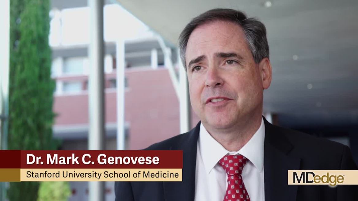
“The goal here was to use electrical stimulation to modify or modulate, and improve the treatment of active rheumatoid arthritis,” Mark C. Genovese, MD, said in an interview at the European Congress of Rheumatology.
“The reason for choosing refractory patients is, one, there’s a clear unmet need, but two, because this was a first-in-human study using a novel microregulatory device stimulating the vagus nerve, we thought the benefits-to-risk ratio was most appropriate for its first trial in patients with refractory disease,” explained Dr. Genovese, professor of medicine and director of the rheumatology clinic in the division of immunology and rheumatology at Stanford (Calif.) University.
He added: “Over time, if the device proves successful for modulating disease, one can see it potentially being used earlier in the disease. Whether it is developed as a stand-alone or used as an adjunct on additional therapy will have to be determined based on both its efficacy and its safety.”
Neurostimulation is a novel concept in rheumatology but has been used with success in other areas of medicine – including epilepsy and depression – using electrical pulses instead of drugs. The idea behind it is that it stimulates the inflammatory reflex that modulates multiple the inflammatory pathways. Essentially, it’s thought that electrically stimulating the vagus nerve sends signals to the spleen where T-lymphocytes then signal to other immune cells, such as macrophages and monocytes, to temper their production of proinflammatory cytokines and other mediators.
“Unlike traditional immunosuppressive biologics that may be specifically targeting one inflammatory process, by suppressing the inflammatory reflex we believe we can suppress a variety of inflammatory cytokines in the region of between 30% and 70%,” Dr. Genovese said at a press briefing.
Data from a 12-week, open-label study (Proc Natl Acad Sci U S A. 2016;113:8284-9) have already shown that the approach works in patients with refractory RA (n = 17). Once-daily electrical vagus nerve stimulation using an existing device made for treating epilepsy showed that clinically meaningful changes in DAS28-CRP could be achieved through TNF suppression. The effects on systemic TNF release lasted for around 24-48 hours after stimulation.
For the current study, a much smaller, leadless, investigational neurostimulation device was used. Called a MicroRegulator (SetPoint Medical), it is about 1 inch long, less than 2 cc in total volume, and is surgically implanted by a neurosurgeon at the top of the vagus nerve. When activated through an iPad app by the health care professional, it sends electrical impulses down the vagus nerve. The device’s battery is charged externally and wirelessly a few minutes each week. Dr. Genovese noted that the device needs to be turned on for only 60 seconds at a time to have an effect and that patients may feel a vibration but this was not reported in the study as an adverse event.
Results of the first in-human study with the device were presented by Dr. Genovese during the late-breaking clinical trials session at the meeting. He described how a total of 14 patients had the device implanted, the first 3 of whom received once-daily, open-label neurostimulation. The remaining 11 patients were randomized to either once-daily or four-times-daily neurostimulation via the device, or to receive sham therapy in which the device was implanted but not switched on. The patients had moderate to severe RA, defined as four or more tender joints, four or more swollen joints, and a CDAI score greater than 10, plus they had radiologically active disease and an insufficient response to at least two biologic or targeted synthetic disease-modifying antirheumatic drugs with differing mechanisms of action.
All patients went through the same schedule of device charging and they did not know if they were in the active or sham groups. At the end of the study, patients had the option to continue in a long-term safety extension phase, have the device switched off, or could have it surgically removed.
“This trial was specifically a pilot trial to assess the MicroRegulator from a safety standpoint,” Dr. Genovese noted, but it also was designed to “help understand whether or not there was going to be clinical efficacy and applicability.”
While “there were no device or treatment-related serious adverse events,” there were some “surgical complications associated with the initial procedure.” One patient experienced paralysis of the left vocal cord during implantation that later resolved, and others experienced the following: Horner’s syndrome, tenderness and swelling at the surgical site, acute postoperative pain, and rash and pruritus. That said, there were no withdrawals from the study due to adverse events.
Commenting in a press release issued by the European League Against Rheumatism, Thomas Dörner, MD, of Charité Universitätsmedizin Berlin, said, “This is a really exciting development. For many patients suffering from rheumatoid arthritis, current treatments don’t work, or aren’t tolerated. These results open the door to a novel approach to treating not only rheumatoid arthritis, but other chronic inflammatory diseases. This is certainly an area for further study.”
The study was sponsored by SetPoint Medical. Dr. Genovese disclosed receiving consulting fees from and having contracts with/grants with the company and acting as a consultant to Galvani and Vorso. He has also received research support from and served as a consultant to Sanofi/Genzyme, Genentech/Roche, and R-Pharm. Dr. Dörner was not involved in the study and commented as part of his role as the chairperson of the EULAR Scientific Program Committee.
SOURCE: Genovese M et al. Ann Rheum Dis. Jun 2019; 78(Suppl 2):264. Abstract LB0009, doi: 10.1136/annrheumdis-2019-eular.8716
MADRID –
A minimal clinically important difference in the 28-joint Disease Activity Score using C-reactive protein (DAS28-CRP) and the Clinical Disease Activity Index (CDAI) at 12 weeks was achieved or exceeded by 5 out of 10 patients; with 2 patients achieving DAS28-CRP–defined remission.
The disease activity scores also were paired with MRI scans and showed, in a handful of individuals, that there was improvement in erosions in those with a clinical response. Greater reductions in proinflammatory cytokines – interleukin (IL)-1-beta, IL-6, IL-17, IL-23, and tumor necrosis factor – were seen with neurostimulation, compared with a sham control group.
“The goal here was to use electrical stimulation to modify or modulate, and improve the treatment of active rheumatoid arthritis,” Mark C. Genovese, MD, said in an interview at the European Congress of Rheumatology.
“The reason for choosing refractory patients is, one, there’s a clear unmet need, but two, because this was a first-in-human study using a novel microregulatory device stimulating the vagus nerve, we thought the benefits-to-risk ratio was most appropriate for its first trial in patients with refractory disease,” explained Dr. Genovese, professor of medicine and director of the rheumatology clinic in the division of immunology and rheumatology at Stanford (Calif.) University.
He added: “Over time, if the device proves successful for modulating disease, one can see it potentially being used earlier in the disease. Whether it is developed as a stand-alone or used as an adjunct on additional therapy will have to be determined based on both its efficacy and its safety.”
Neurostimulation is a novel concept in rheumatology but has been used with success in other areas of medicine – including epilepsy and depression – using electrical pulses instead of drugs. The idea behind it is that it stimulates the inflammatory reflex that modulates multiple the inflammatory pathways. Essentially, it’s thought that electrically stimulating the vagus nerve sends signals to the spleen where T-lymphocytes then signal to other immune cells, such as macrophages and monocytes, to temper their production of proinflammatory cytokines and other mediators.
“Unlike traditional immunosuppressive biologics that may be specifically targeting one inflammatory process, by suppressing the inflammatory reflex we believe we can suppress a variety of inflammatory cytokines in the region of between 30% and 70%,” Dr. Genovese said at a press briefing.
Data from a 12-week, open-label study (Proc Natl Acad Sci U S A. 2016;113:8284-9) have already shown that the approach works in patients with refractory RA (n = 17). Once-daily electrical vagus nerve stimulation using an existing device made for treating epilepsy showed that clinically meaningful changes in DAS28-CRP could be achieved through TNF suppression. The effects on systemic TNF release lasted for around 24-48 hours after stimulation.
For the current study, a much smaller, leadless, investigational neurostimulation device was used. Called a MicroRegulator (SetPoint Medical), it is about 1 inch long, less than 2 cc in total volume, and is surgically implanted by a neurosurgeon at the top of the vagus nerve. When activated through an iPad app by the health care professional, it sends electrical impulses down the vagus nerve. The device’s battery is charged externally and wirelessly a few minutes each week. Dr. Genovese noted that the device needs to be turned on for only 60 seconds at a time to have an effect and that patients may feel a vibration but this was not reported in the study as an adverse event.
Results of the first in-human study with the device were presented by Dr. Genovese during the late-breaking clinical trials session at the meeting. He described how a total of 14 patients had the device implanted, the first 3 of whom received once-daily, open-label neurostimulation. The remaining 11 patients were randomized to either once-daily or four-times-daily neurostimulation via the device, or to receive sham therapy in which the device was implanted but not switched on. The patients had moderate to severe RA, defined as four or more tender joints, four or more swollen joints, and a CDAI score greater than 10, plus they had radiologically active disease and an insufficient response to at least two biologic or targeted synthetic disease-modifying antirheumatic drugs with differing mechanisms of action.
All patients went through the same schedule of device charging and they did not know if they were in the active or sham groups. At the end of the study, patients had the option to continue in a long-term safety extension phase, have the device switched off, or could have it surgically removed.
“This trial was specifically a pilot trial to assess the MicroRegulator from a safety standpoint,” Dr. Genovese noted, but it also was designed to “help understand whether or not there was going to be clinical efficacy and applicability.”
While “there were no device or treatment-related serious adverse events,” there were some “surgical complications associated with the initial procedure.” One patient experienced paralysis of the left vocal cord during implantation that later resolved, and others experienced the following: Horner’s syndrome, tenderness and swelling at the surgical site, acute postoperative pain, and rash and pruritus. That said, there were no withdrawals from the study due to adverse events.
Commenting in a press release issued by the European League Against Rheumatism, Thomas Dörner, MD, of Charité Universitätsmedizin Berlin, said, “This is a really exciting development. For many patients suffering from rheumatoid arthritis, current treatments don’t work, or aren’t tolerated. These results open the door to a novel approach to treating not only rheumatoid arthritis, but other chronic inflammatory diseases. This is certainly an area for further study.”
The study was sponsored by SetPoint Medical. Dr. Genovese disclosed receiving consulting fees from and having contracts with/grants with the company and acting as a consultant to Galvani and Vorso. He has also received research support from and served as a consultant to Sanofi/Genzyme, Genentech/Roche, and R-Pharm. Dr. Dörner was not involved in the study and commented as part of his role as the chairperson of the EULAR Scientific Program Committee.
SOURCE: Genovese M et al. Ann Rheum Dis. Jun 2019; 78(Suppl 2):264. Abstract LB0009, doi: 10.1136/annrheumdis-2019-eular.8716
REPORTING FROM EULAR 2019 CONGRESS
Systemic sclerosis gastrointestinal symptoms helped by gut microbiota transplant
MADRID – compared with control subjects in a 16-week randomized, double-blind, placebo-controlled pilot study presented at the European Congress of Rheumatology.
The effects were most pronounced on lower GI symptoms, including bloating, diarrhea, and fecal incontinence, with improvement reported by three of five of the patients given the gut microbiota transplant, compared with two of the five patients who received placebo.
“We were surprised by the effect the patients reported, as all had longstanding SSc with GI symptoms,” Anna-Maria Hoffmann-Vold, MD, PhD, of Oslo University Hospital, said in an interview ahead of the congress. “We were especially surprised at the strong effect FMT had on fecal incontinence.”“Patients with systemic sclerosis are very prone to having gastrointestinal involvement – up to 90% of patients have GI symptoms, and it’s associated with very high morbidity and mortality,” she observed during her presentation at the Congress. Despite that, there currently are no disease-modifying treatments that specifically addresses GI involvement in SSc.
It’s been known for a while that patients with SSc have a different intestinal microbiota composition, or dysbiosis, compared with healthy controls, and the possibility of permanent modification of the microbiome through fecal microbiota transplant (FMT) from healthy to ill individuals has become a subject of increased attention in the scientific literature in recent years,. Dr. Hoffmann-Vold said.
In particular, FMT has shown promising results in the treatment of Clostridium difficile infections. While the current study did not focus on mechanistic pathways by which FMT might be exerting its effects, such studies are definitely warranted, she said. “One could speculate that there is a mechanistic link between dysmotility and dysbiosis in SSc, and that the manipulation of gut microbiota with FMT primarily affects motility patterns, which in turn leads to improvement of GI symptoms.”
Together with colleagues at the Oslo University Hospital, Dr. Hoffmann-Vold randomly assigned 10 patients – all women – with limited cutaneous SSc either to treatment with a commercially-available gut microbiota preparation known as anaerobic cultivated human intestinal microbiota (ACHIM) or to placebo. Both ACHIM and placebo were given via gastroduodenoscopy. Their aim was to determine the safety of the approach, as well as to obtain preliminary data on its therapeutic potential.
The UCLA GIT 2.0 score questionnaire was used to assess GI symptoms, with patients defined as responders if they met the questionnaire’s definition of a minimally clinically important difference.
Primary endpoints were safety and clinical efficacy on GI symptoms assessed at weeks 4 and 16, and safety was assessed by observation, interviews, and a standardized safety form.
Results showed improvement in GI symptoms (total UCLA GIT score) in three of the five patients who received the gut microbiota transplant versus two of the five placebo-treated patients at 16 weeks. Two patients in the active treatment versus one in the placebo group had unchanged symptoms, and one patient in the placebo group had worsening symptoms.
Adverse events associated with treatment were “transient and mild”. However, one procedure-related serious adverse event occurred in a placebo-treated patient, which was a duodenal perforation.
Concluding her presentation, Dr. Hoffman-Vold said: “FMT of commercially-available ACHIM in patients with SSc appeared safe, had beneficial effects on lower GI symptoms, altered gut microbiota composition – richness and diversity – and appeared to affect the mucosal immune system.”
The research team has just received national funding for a larger randomized clinical trial that will involve 70 SSc patients and should start towards the end of the year.
The study was sponsored by Helse Sør-øst and NKS. Dr. Hoffmann-Vold has received research funding, consulting fees, or other remuneration from Boehringer Ingelheim, GlaxoSmithKline, and Actelion. A coauthor is the owner of the company that provided the gut microbiota.
SOURCE: Hoffmann-Vold AM et al., Ann Rheum Dis. 2019 Jun. doi: 10.1136/annrheumdis-2019-eular.4684 .
REPORTING FROM THE EULAR 2019 CONGRESS

Antibody hierarchy may drive development of SLE vs. antiphospholipid syndrome
according to study findings presented at the European Congress of Rheumatology.

Spanish researchers found that the number of antiphospholipid (aPL) antibodies present was important for the development of antiphospholipid syndrome (APS) and that lupus anticoagulant (LA) was the major aPL antibody linked to systemic lupus erythematosus (SLE)–related organ involvement.
“aPL [antibodies] has been extensively associated with an increased risk of thrombosis and poor pregnancy outcomes, mainly in patients with primary APS,” study investigator Leyre Riancho-Zarrabeitia, MD, PhD, explained in an interview ahead of the congress.
“Moreover, aPL [antibody] positivity in SLE has been proposed to be associated with higher damage accrual and with certain manifestations such as valvular heart disease, pulmonary hypertension, and neuropsychiatric manifestations,” she added.
Anticardiolipin antibodies – notably IgG rather than IgM isotypes – also seemed to play an important role in APS and SLE manifestations, Dr. Riancho-Zarrabeitia, of Hospital Sierrallana, Instituto De Investigación Marqués De Valdecilla, and the University of Cantabria (Spain), noted during her oral presentation.
She reported data on 3,651 patients included in the RELESSER registry between October 2011 and August 2012. This large, multicenter, hospital-based registry retrospectively collects immunologic, clinical and demographic data from unselected adult patients with SLE who are attending 45 Spanish rheumatology services within the country’s national health system.
Over one-third (37.5%) of patients, who had a mean age of 47 years and were mostly (90%) women, were positive for aPL. The most frequent aPL detected was IgG anticardiolipin (aCL) antibodies, seen in 25% of patients, followed by LA in 24%, and IgM aCL in 20%.
Of the aPL-positive patients, 20.6% were positive for only one antibody, 12.1% were positive for two antibodies, and 4.8% were positive for three antibodies.
“All types of aPL were associated with classic APS manifestations,” Dr. Riancho-Zarrabeitia said. The associations were strongest for thrombotic events, such as arterial and venous small-vessel thrombosis and recurrent early pregnancy losses.
aCL antibodies conferred the highest risk for arterial thrombosis, she noted (odds ratio, 5.7), whereas LA conferred the highest risk for venous thrombosis (OR, 4.7). Both IgG and IgM isotypes were associated with thrombotic events, fetal death and recurrent pregnancy loss, but the association was stronger with the IgG isotypes.
Having more than one aPL was particularly associated with a higher risk of these APS manifestations. For example, when one antibody was present the OR for arterial thrombosis was 4.45, but when two or more aPL were detected, the ORs rose to 9.23 and 15.6, respectively.
aCL and LA also were associated with thrombocytopenia and hemolytic anemia, with ORs of around 1-2 and 2-3 respectively. There also were antibody associations with cognitive impairments.
Similar results were seen in patients with SLE. “aPL [antibody] positivity in SLE patients influenced the risk for thrombotic and obstetric manifestations,” Dr. Riancho-Zarrabeitia said. LA and aCL were associated with an increased risk of neuropsychiatric manifestations, and LA was linked to an increased risk for renal disease.
The risk for specific SLE manifestations was again higher with IgG isotypes of aCL, notably an increased risk for cardiac and respiratory events.
While increased antibody numbers generally led to a higher risk of complications, the risk for cutaneous manifestations decreased.
“The load of aPL [antibodies] confers a higher risk for APS,” Dr. Riancho-Zarrabeitia said during her conclusion. “Regarding systemic lupus erythematosus, the number of positive antibodies is directly associated with neurological and ophthalmological manifestations, and inversely associated with cutaneous manifestations.”
What these findings show, said Dr. Riancho-Zarrabeitia in the precongress interview, is that individuals who test positive for aPL antibodies need careful monitoring to prevent and treat severe manifestations. “The next step would be to confirm our findings with a prospective study.”
Dr. Riancho-Zarrabeitia has received travel grants from AbbVie, Pfizer, UCB, Merck, GlaxoSmithKline, Amgen, and Roche.
SOURCE: Riancho-Zarrabeitia L et al. Ann Rheum Dis. Jun 2019;78(Suppl 2):136-7. Abstract OP0124. doi: 10.1136/annrheumdis-2019-eular.2485.
according to study findings presented at the European Congress of Rheumatology.
Spanish researchers found that the number of antiphospholipid (aPL) antibodies present was important for the development of antiphospholipid syndrome (APS) and that lupus anticoagulant (LA) was the major aPL antibody linked to systemic lupus erythematosus (SLE)–related organ involvement.
“aPL [antibodies] has been extensively associated with an increased risk of thrombosis and poor pregnancy outcomes, mainly in patients with primary APS,” study investigator Leyre Riancho-Zarrabeitia, MD, PhD, explained in an interview ahead of the congress.
“Moreover, aPL [antibody] positivity in SLE has been proposed to be associated with higher damage accrual and with certain manifestations such as valvular heart disease, pulmonary hypertension, and neuropsychiatric manifestations,” she added.
Anticardiolipin antibodies – notably IgG rather than IgM isotypes – also seemed to play an important role in APS and SLE manifestations, Dr. Riancho-Zarrabeitia, of Hospital Sierrallana, Instituto De Investigación Marqués De Valdecilla, and the University of Cantabria (Spain), noted during her oral presentation.
She reported data on 3,651 patients included in the RELESSER registry between October 2011 and August 2012. This large, multicenter, hospital-based registry retrospectively collects immunologic, clinical and demographic data from unselected adult patients with SLE who are attending 45 Spanish rheumatology services within the country’s national health system.
Over one-third (37.5%) of patients, who had a mean age of 47 years and were mostly (90%) women, were positive for aPL. The most frequent aPL detected was IgG anticardiolipin (aCL) antibodies, seen in 25% of patients, followed by LA in 24%, and IgM aCL in 20%.
Of the aPL-positive patients, 20.6% were positive for only one antibody, 12.1% were positive for two antibodies, and 4.8% were positive for three antibodies.
“All types of aPL were associated with classic APS manifestations,” Dr. Riancho-Zarrabeitia said. The associations were strongest for thrombotic events, such as arterial and venous small-vessel thrombosis and recurrent early pregnancy losses.
aCL antibodies conferred the highest risk for arterial thrombosis, she noted (odds ratio, 5.7), whereas LA conferred the highest risk for venous thrombosis (OR, 4.7). Both IgG and IgM isotypes were associated with thrombotic events, fetal death and recurrent pregnancy loss, but the association was stronger with the IgG isotypes.
Having more than one aPL was particularly associated with a higher risk of these APS manifestations. For example, when one antibody was present the OR for arterial thrombosis was 4.45, but when two or more aPL were detected, the ORs rose to 9.23 and 15.6, respectively.
aCL and LA also were associated with thrombocytopenia and hemolytic anemia, with ORs of around 1-2 and 2-3 respectively. There also were antibody associations with cognitive impairments.
Similar results were seen in patients with SLE. “aPL [antibody] positivity in SLE patients influenced the risk for thrombotic and obstetric manifestations,” Dr. Riancho-Zarrabeitia said. LA and aCL were associated with an increased risk of neuropsychiatric manifestations, and LA was linked to an increased risk for renal disease.
The risk for specific SLE manifestations was again higher with IgG isotypes of aCL, notably an increased risk for cardiac and respiratory events.
While increased antibody numbers generally led to a higher risk of complications, the risk for cutaneous manifestations decreased.
“The load of aPL [antibodies] confers a higher risk for APS,” Dr. Riancho-Zarrabeitia said during her conclusion. “Regarding systemic lupus erythematosus, the number of positive antibodies is directly associated with neurological and ophthalmological manifestations, and inversely associated with cutaneous manifestations.”
What these findings show, said Dr. Riancho-Zarrabeitia in the precongress interview, is that individuals who test positive for aPL antibodies need careful monitoring to prevent and treat severe manifestations. “The next step would be to confirm our findings with a prospective study.”
Dr. Riancho-Zarrabeitia has received travel grants from AbbVie, Pfizer, UCB, Merck, GlaxoSmithKline, Amgen, and Roche.
SOURCE: Riancho-Zarrabeitia L et al. Ann Rheum Dis. Jun 2019;78(Suppl 2):136-7. Abstract OP0124. doi: 10.1136/annrheumdis-2019-eular.2485.
REPORTING FROM EULAR 2019 CONGRESS
Treat-to-target slowly emerging in axial spondyloarthritis
MADRID – Treating patients with axial spondyloarthritis (axSpA) until a specific target is reached is an emerging concept that has gained a lot of traction in the past few years, Pedro Machado, MD, said at the European Congress of Rheumatology.

“The availability of biologic therapies has improved the clinical outcomes for our patients with axial spondyloarthritis and targeting clinical remission or inactive disease is now an achievable treatment goal in clinical practice,” he observed. “This has trigged the question: Is there a role for ‘treat-to-target’ in axial spondyloarthritis?”
Dr. Machado, an honorary consultant in rheumatology and muscle diseases at University College Hospital and the National Hospital for Neurology and Neurosurgery in London, took a critical look at the treat-to-target approach during a clinical science session at the meeting, organized by the European League Against Rheumatism (EULAR).
The concept of treat-to-target is not new, he acknowledged, having been imported from other chronic conditions where there is a very specific target to achieve – such as lowering glycated hemoglobin in diabetes or hypertension or hyperlipidemia in cardiovascular disease.
“The concept involves changing or escalating therapy according to a predefined target under the assumption that this may lead to a better outcome compared to what we call ‘routine care,’ ” Dr. Machado explained.
Treat-to-target is not only well established in nonrheumatic diseases but also has proved to work in patients with rheumatoid arthritis and psoriatic arthritis with evidence from the TICORA (Tight Control of Rheumatoid Arthritis) and TICOPA (Tight Control in Psoriatic Arthritis) trials.
Whether the approach can also work in axSpA is open to debate, and one of the main arguments against using a treat-to-target in axSpA asks, what exactly is the target? While there is no firm agreement yet, Dr. Machado observed that achieving either clinical remission or inactive disease would be the most likely target.
It could be argued this is already being done to some degree, but “we need to be more ambitious,” Dr. Machado said. Indeed, current Assessment of Spondyloarthritis International Society/EULAR recommendations for the treatment of axSpA (Ann Rheum Dis. 2017;76[6]:978–91) note when patients with high disease activity despite sufficient standard treatment should be escalated to treatment with a biologic disease-modifying antirheumatic drug (bDMARD). High disease activity was defined as an Ankylosing Spondylitis Disease Activity Score (ASDAS) of 2.1 or more or a Bath Ankylosing Spondylitis Disease Activity Index (BASDAI) score of 4 or more.
Another argument against using the approach concerns the evidence base. There are no prospective, randomized trials supporting the use of treat-to-target over routine care. However, there is a lot of observational evidence, Dr. Machado said in an interview. Such studies have shown that achieving inactive disease may improve structural outcomes and stop the development of radiographic damage of the spine. Importantly, these observational studies also show that achieving inactive disease may also help to improve patients’ functional outcomes and quality of life.
Evidence backing a treat-to-target approach in axSpA from a randomized, controlled trial may currently be lacking, but the TiCOSPA (Tight Control in Spondyloarthritis) trial is in progress and should help change that, Dr. Machado said.
“The missing bit is a randomized trial, but I would say that the observational evidence is almost enough to advocate a treat-to-target strategy in axial spondyloarthritis.” This was also the view of an international task force that recently published recommendations and overarching principles for a treat-target strategy in spondyloarthritis, including axSpA (Ann Rheum Dis. 2018;77:3-17).
Of course, a treat-to-target approach may not be without its pitfalls. There are a limited number of drugs currently that could be used to “hit the target” of disease activity, Dr. Machado said in his presentation. The approach might also lead to ‘overtreatment,’ and more treatment is not always better as it could not only lead to more adverse events, but it also may mean the approach is not cost-effective.
Depending on the TiCOSPA study results, which are expected next year, Dr. Machado said that “the feasibility and cost-effectiveness of such a strategy in clinical practice also needs to be tested.”
MADRID – Treating patients with axial spondyloarthritis (axSpA) until a specific target is reached is an emerging concept that has gained a lot of traction in the past few years, Pedro Machado, MD, said at the European Congress of Rheumatology.
“The availability of biologic therapies has improved the clinical outcomes for our patients with axial spondyloarthritis and targeting clinical remission or inactive disease is now an achievable treatment goal in clinical practice,” he observed. “This has trigged the question: Is there a role for ‘treat-to-target’ in axial spondyloarthritis?”
Dr. Machado, an honorary consultant in rheumatology and muscle diseases at University College Hospital and the National Hospital for Neurology and Neurosurgery in London, took a critical look at the treat-to-target approach during a clinical science session at the meeting, organized by the European League Against Rheumatism (EULAR).
The concept of treat-to-target is not new, he acknowledged, having been imported from other chronic conditions where there is a very specific target to achieve – such as lowering glycated hemoglobin in diabetes or hypertension or hyperlipidemia in cardiovascular disease.
“The concept involves changing or escalating therapy according to a predefined target under the assumption that this may lead to a better outcome compared to what we call ‘routine care,’ ” Dr. Machado explained.
Treat-to-target is not only well established in nonrheumatic diseases but also has proved to work in patients with rheumatoid arthritis and psoriatic arthritis with evidence from the TICORA (Tight Control of Rheumatoid Arthritis) and TICOPA (Tight Control in Psoriatic Arthritis) trials.
Whether the approach can also work in axSpA is open to debate, and one of the main arguments against using a treat-to-target in axSpA asks, what exactly is the target? While there is no firm agreement yet, Dr. Machado observed that achieving either clinical remission or inactive disease would be the most likely target.
It could be argued this is already being done to some degree, but “we need to be more ambitious,” Dr. Machado said. Indeed, current Assessment of Spondyloarthritis International Society/EULAR recommendations for the treatment of axSpA (Ann Rheum Dis. 2017;76[6]:978–91) note when patients with high disease activity despite sufficient standard treatment should be escalated to treatment with a biologic disease-modifying antirheumatic drug (bDMARD). High disease activity was defined as an Ankylosing Spondylitis Disease Activity Score (ASDAS) of 2.1 or more or a Bath Ankylosing Spondylitis Disease Activity Index (BASDAI) score of 4 or more.
Another argument against using the approach concerns the evidence base. There are no prospective, randomized trials supporting the use of treat-to-target over routine care. However, there is a lot of observational evidence, Dr. Machado said in an interview. Such studies have shown that achieving inactive disease may improve structural outcomes and stop the development of radiographic damage of the spine. Importantly, these observational studies also show that achieving inactive disease may also help to improve patients’ functional outcomes and quality of life.
Evidence backing a treat-to-target approach in axSpA from a randomized, controlled trial may currently be lacking, but the TiCOSPA (Tight Control in Spondyloarthritis) trial is in progress and should help change that, Dr. Machado said.
“The missing bit is a randomized trial, but I would say that the observational evidence is almost enough to advocate a treat-to-target strategy in axial spondyloarthritis.” This was also the view of an international task force that recently published recommendations and overarching principles for a treat-target strategy in spondyloarthritis, including axSpA (Ann Rheum Dis. 2018;77:3-17).
Of course, a treat-to-target approach may not be without its pitfalls. There are a limited number of drugs currently that could be used to “hit the target” of disease activity, Dr. Machado said in his presentation. The approach might also lead to ‘overtreatment,’ and more treatment is not always better as it could not only lead to more adverse events, but it also may mean the approach is not cost-effective.
Depending on the TiCOSPA study results, which are expected next year, Dr. Machado said that “the feasibility and cost-effectiveness of such a strategy in clinical practice also needs to be tested.”
EXPERT analysis FROM THE EULAR 2019 Congress
Rituximab serious infection risk predicted by immunoglobulin levels
Monitoring immunoglobulin (Ig) levels at baseline and before each cycle of rituximab could reduce the risk of serious infection events (SIEs) in patients needing repeated treatment, according to research published in Arthritis & Rheumatology.

In a large, single-center, longitudinal study conducted at a tertiary referral center, having low IgG (less than 6 g/L) in particular was associated with a higher rate of SIEs, compared with having normal IgG levels (6-16 g/L). Considering 103 of 700 patients who had low levels of IgG before starting treatment with rituximab for various rheumatic and musculoskeletal diseases (RMDs), there were 16.4 SIEs per 100 patient-years. In those who developed low IgG during subsequent cycles of rituximab therapy, the SIE rate was even higher, at 21.3 per 100 patient-years. By comparison, the SIE rate for those with normal IgG levels was 9.7 per 100 patient-years.
“We really have to monitor immunoglobulins at baseline and also before we re-treat the patients, because higher IgG level is protective of serious infections,” study first author Md Yuzaiful Md Yusof, MBChB, PhD, said in an interview.
Low IgG has been linked to a higher risk of SIEs in the first 12 months of rituximab therapy but, until now, there have been limited data on infection predictors during repeated cycles of treatment. While IgG is a consistent marker of SIEs associated with repeated rituximab treatment, IgM and IgA should also be monitored to give a full picture of any hyperglobulinemia that may be present.
“There is no formal guidance on how to safely monitor patients on rituximab,” observed Dr. Md Yusof, who will present these data at the 2019 European Congress of Rheumatology in Madrid. The study’s findings could help to change that, however, as they offer a practical way to help predict and thus prevent SIEs. The study’s findings not only validate previous work, he noted, but also add new insights into why some patients treated with repeat rituximab cycles but not others may experience a higher rate of such infections.
Altogether, the investigators examined data on 700 patients with RMDs treated with rituximab who were consecutively seen during 2012-2017 at Dr. Md Yusof’s institution – the Leeds (England) Institute of Rheumatic and Musculoskeletal Medicine, which is part of the University of Leeds. Their immunoglobulin levels had been measured before starting rituximab therapy and every 4-6 months after each cycle of rituximab treatment.
Patients with any RMD being treated with at least one cycle of rituximab were eligible for inclusion in the retrospective study, with the majority (72%) taking it for rheumatoid arthritis and some for systemic lupus erythematosus (13%) or antineutrophil cytoplasmic antibody (ANCA)–associated vasculitis (7%).
One of the main aims of the study was to look for predictors of SIEs during the first 12 months and during repeated cycles of rituximab. Dr. Md Yusof and his associates also looked at how secondary hypogammaglobulinemia might affect SIE rates and the humoral response to vaccination challenge and its persistence following treatment discontinuation. Their ultimate aim was to see if these findings could then be used to develop a treatment algorithm for rituximab administration in RMDs.
Over a follow-up period encompassing 2,880 patient-years of treatment, 281 SIEs were recorded in 176 patients, giving a rate of 9.8 infections per 100 patient-years. Most (61%) of these were due to lower respiratory tract infections.
The proportion of patients experiencing their first SIE increased with time: 16% within 6 weeks of starting rituximab therapy, 35% at 12 weeks, 72% at 26 weeks, 83% at 38 weeks, and 100% by 1 year of repeated treatment.
Multivariable analysis showed that the presence of several comorbidities at baseline – notably chronic obstructive pulmonary disease, diabetes, heart failure, and prior cancer – raised the risk for SIEs with repeated rituximab therapy. The biggest factor, however, was a history of SIEs – with a sixfold increased risk of further serious infection.
Higher corticosteroid dose and factors specific to rituximab – low IgG, neutropenia, high IgM, and a longer time to retreatment – were also predictive of SIEs.
“Low IgG also results in poor humoral response to vaccination,” Dr. Md Yusof said, noting that the IgG level remains below the lower limit of normal for several years after rituximab is discontinued in most patients.
In the study, 5 of 8 (64%) patients had impaired humoral response to pneumococcal and haemophilus following vaccination challenge and 4 of 11 patients had IgG normalized after switching to another biologic disease-modifying antirheumatic drug (bDMARD).
Cyclophosphamide is commonly used as a first-line agent to induce remission in patients with severe and refractory systemic lupus erythematosus and ANCA-associated vasculitis, with patients switched to rituximab at relapse. The effect of this prior treatment was examined in 20 patients in the study, with a marked decline in almost all immunoglobulin classes seen up to 18 months. Prior treatment with immunosuppressants such as intravenous cyclophosphamide could be behind progressive reductions in Ig levels seen with repeated rituximab treatment rather than entirely because of rituximab, Dr. Md Yusof said.
Dr. Md Yusof, who is a National Institute for Health Research (NIHR) Academic Clinical Lecturer at the University of Leeds, said the value of the study, compared with others, is that hospital data for all patients treated with rituximab with at least 3 months follow-up were included, making it an almost complete data set.
“By carefully reviewing records of every patient to capture all infection episodes in the largest single-center cohort study to date, our findings provide insights on predictors of SIEs as well as a foundation for safety monitoring of rituximab,” he and his coauthors wrote.
They acknowledge reporting a higher rate of SIEs than seen in registry and clinical studies with rituximab, which may reflect a “channeling bias” as the patients comprised those with multiple comorbidities including those that represent a relative contraindication for bDMARD use. That said, the findings clearly show that Ig levels should be monitored before and after each rituximab cycle, especially in those with comorbid diseases and those with low IgG levels to start with.
They conclude that an “individualized benefit-risk assessment” is needed to determine whether rituximab should be repeated in those with low IgG as this is a “consistent predictor” of SIE and may “increase infection profiles when [rituximab] is switched to different bDMARDs.”
The research was supported by Octapharma, the National Institute for Health Research (NIHR), and NIHR Leeds Biomedical Research Centre based at Leeds Teaching Hospitals NHS Trust in England. Dr. Md Yusof had no conflicts of interest. Several coauthors disclosed financial ties to multiple pharmaceutical companies, including Roche.
SOURCE: Md Yusof MY et al. Arthritis Rheumatol. 2019 May 27. doi: 10.1002/art.40937.
Monitoring immunoglobulin (Ig) levels at baseline and before each cycle of rituximab could reduce the risk of serious infection events (SIEs) in patients needing repeated treatment, according to research published in Arthritis & Rheumatology.
In a large, single-center, longitudinal study conducted at a tertiary referral center, having low IgG (less than 6 g/L) in particular was associated with a higher rate of SIEs, compared with having normal IgG levels (6-16 g/L). Considering 103 of 700 patients who had low levels of IgG before starting treatment with rituximab for various rheumatic and musculoskeletal diseases (RMDs), there were 16.4 SIEs per 100 patient-years. In those who developed low IgG during subsequent cycles of rituximab therapy, the SIE rate was even higher, at 21.3 per 100 patient-years. By comparison, the SIE rate for those with normal IgG levels was 9.7 per 100 patient-years.
“We really have to monitor immunoglobulins at baseline and also before we re-treat the patients, because higher IgG level is protective of serious infections,” study first author Md Yuzaiful Md Yusof, MBChB, PhD, said in an interview.
Low IgG has been linked to a higher risk of SIEs in the first 12 months of rituximab therapy but, until now, there have been limited data on infection predictors during repeated cycles of treatment. While IgG is a consistent marker of SIEs associated with repeated rituximab treatment, IgM and IgA should also be monitored to give a full picture of any hyperglobulinemia that may be present.
“There is no formal guidance on how to safely monitor patients on rituximab,” observed Dr. Md Yusof, who will present these data at the 2019 European Congress of Rheumatology in Madrid. The study’s findings could help to change that, however, as they offer a practical way to help predict and thus prevent SIEs. The study’s findings not only validate previous work, he noted, but also add new insights into why some patients treated with repeat rituximab cycles but not others may experience a higher rate of such infections.
Altogether, the investigators examined data on 700 patients with RMDs treated with rituximab who were consecutively seen during 2012-2017 at Dr. Md Yusof’s institution – the Leeds (England) Institute of Rheumatic and Musculoskeletal Medicine, which is part of the University of Leeds. Their immunoglobulin levels had been measured before starting rituximab therapy and every 4-6 months after each cycle of rituximab treatment.
Patients with any RMD being treated with at least one cycle of rituximab were eligible for inclusion in the retrospective study, with the majority (72%) taking it for rheumatoid arthritis and some for systemic lupus erythematosus (13%) or antineutrophil cytoplasmic antibody (ANCA)–associated vasculitis (7%).
One of the main aims of the study was to look for predictors of SIEs during the first 12 months and during repeated cycles of rituximab. Dr. Md Yusof and his associates also looked at how secondary hypogammaglobulinemia might affect SIE rates and the humoral response to vaccination challenge and its persistence following treatment discontinuation. Their ultimate aim was to see if these findings could then be used to develop a treatment algorithm for rituximab administration in RMDs.
Over a follow-up period encompassing 2,880 patient-years of treatment, 281 SIEs were recorded in 176 patients, giving a rate of 9.8 infections per 100 patient-years. Most (61%) of these were due to lower respiratory tract infections.
The proportion of patients experiencing their first SIE increased with time: 16% within 6 weeks of starting rituximab therapy, 35% at 12 weeks, 72% at 26 weeks, 83% at 38 weeks, and 100% by 1 year of repeated treatment.
Multivariable analysis showed that the presence of several comorbidities at baseline – notably chronic obstructive pulmonary disease, diabetes, heart failure, and prior cancer – raised the risk for SIEs with repeated rituximab therapy. The biggest factor, however, was a history of SIEs – with a sixfold increased risk of further serious infection.
Higher corticosteroid dose and factors specific to rituximab – low IgG, neutropenia, high IgM, and a longer time to retreatment – were also predictive of SIEs.
“Low IgG also results in poor humoral response to vaccination,” Dr. Md Yusof said, noting that the IgG level remains below the lower limit of normal for several years after rituximab is discontinued in most patients.
In the study, 5 of 8 (64%) patients had impaired humoral response to pneumococcal and haemophilus following vaccination challenge and 4 of 11 patients had IgG normalized after switching to another biologic disease-modifying antirheumatic drug (bDMARD).
Cyclophosphamide is commonly used as a first-line agent to induce remission in patients with severe and refractory systemic lupus erythematosus and ANCA-associated vasculitis, with patients switched to rituximab at relapse. The effect of this prior treatment was examined in 20 patients in the study, with a marked decline in almost all immunoglobulin classes seen up to 18 months. Prior treatment with immunosuppressants such as intravenous cyclophosphamide could be behind progressive reductions in Ig levels seen with repeated rituximab treatment rather than entirely because of rituximab, Dr. Md Yusof said.
Dr. Md Yusof, who is a National Institute for Health Research (NIHR) Academic Clinical Lecturer at the University of Leeds, said the value of the study, compared with others, is that hospital data for all patients treated with rituximab with at least 3 months follow-up were included, making it an almost complete data set.
“By carefully reviewing records of every patient to capture all infection episodes in the largest single-center cohort study to date, our findings provide insights on predictors of SIEs as well as a foundation for safety monitoring of rituximab,” he and his coauthors wrote.
They acknowledge reporting a higher rate of SIEs than seen in registry and clinical studies with rituximab, which may reflect a “channeling bias” as the patients comprised those with multiple comorbidities including those that represent a relative contraindication for bDMARD use. That said, the findings clearly show that Ig levels should be monitored before and after each rituximab cycle, especially in those with comorbid diseases and those with low IgG levels to start with.
They conclude that an “individualized benefit-risk assessment” is needed to determine whether rituximab should be repeated in those with low IgG as this is a “consistent predictor” of SIE and may “increase infection profiles when [rituximab] is switched to different bDMARDs.”
The research was supported by Octapharma, the National Institute for Health Research (NIHR), and NIHR Leeds Biomedical Research Centre based at Leeds Teaching Hospitals NHS Trust in England. Dr. Md Yusof had no conflicts of interest. Several coauthors disclosed financial ties to multiple pharmaceutical companies, including Roche.
SOURCE: Md Yusof MY et al. Arthritis Rheumatol. 2019 May 27. doi: 10.1002/art.40937.
FROM ARTHRITIS & RHEUMATOLOGY
Key clinical point: Immunoglobulin should be monitored at baseline and before each rituximab cycle to identify patients at risk of serious infection events (SIEs).
Major finding: SIE rates per 100 patient-years were 16.4 and 21.3 in patients with low (less than 6 g/L) IgG at baseline and during rituximab cycles versus 9.7 for patients with normal (6–16 g/L) IgG levels.
Study details: A retrospective, single-center, longitudinal study involving 700 rituximab-treated patients with rheumatoid arthritis and other rheumatic and musculoskeletal diseases.
Disclosures: The research was supported by Octapharma, the National Institute for Health Research (NIHR), and NIHR Leeds Biomedical Research Centre based at Leeds Teaching Hospitals NHS Trust in the United Kingdom. Dr. Md Yusof had no conflicts of interest. Several coauthors disclosed financial ties to multiple pharmaceutical companies, including Roche.
Source: Md Yusof MY et al. Arthritis Rheumatol. 2019 May 27. doi: 10.1002/art.40937.

Could biosimilar switchbacks due to ‘inefficacy’ be subjective?
BIRMINGHAM, ENGLAND – Patients with rheumatoid arthritis (RA) who switch back to an original tumor necrosis factor inhibitor (TNFi) after using a biosimilar product often do so because of ineffectiveness, but this could be largely subjective, research suggests.
Data from the British Society for Rheumatology Biologics Register–Rheumatoid Arthritis (BSRBR-RA), presented at the annual conference of the British Society for Rheumatology, have shown that the majority (81%) of 760 patients who switched from an etanercept originator (ETN-O) product (Enbrel) to an etanercept biosimilar (ETN-B) product (Benepali or Erelzi) remained on the latter at an average of 2 years’ follow-up.
However, of those who switched back to an ETN-O (8%, n = 58), ineffectiveness was the primary reason for doing so in more than half of patients (53%). Of patients who stopped ETN treatment altogether after the biosimilar switch (11%, n = 84), 29% stopped because of inefficacy. Other important reasons for switching back or stopping treatment were adverse events in 28% and 54% of cases, respectively. The switch back to an ETN-O happened within a median time of 4 months (range, 2–6 months).
Patients switching back to an ETN-O tended to be slightly younger than all patients who had switched to an ETN-B (median age 60 vs. 64 years). They were more likely to be female (79% vs. 75%). Median disease activity score in 28 joints (DAS28) at baseline were highest in those who discontinued ETN (3.9 vs. 3.2 for those who switched back and 3.0 for all who had switched to a biosimilar).
It is not known what is driving the purported ineffectiveness, said research assistant Rebecca Davies of the Arthritis Research UK Epidemiology Unit at the University of Manchester (England).
“It could be due to patient factors and a nocebo effect,” she suggested. This is where “negative patient expectations cause treatment to have a more negative effect.” For example, she added, “if a patient is settled on the originator drug and had to mandatorily switch to a biosimilar, it might be that they are more heightened to symptoms.”
Another explanation could be that consultants may be “overcautious” to continuing the biosimilar in patients who had responded well to the originator product, she hypothesized.
“Our next steps are to look at disease activity between 4 and 9 months post switch to determine” the cause of the described ineffectiveness.
Educate patients to reduce switchbacks?
Meanwhile data presented by a team of researchers from Imperial College Healthcare NHS Trust in London, headed by Maresa Carulli, MD, suggested that patient education may be an important factor in tackling why patients want to switch back to biosimilar treatment.
Over a 20-month period among 202 patients who were switched to an ETN-B after they had been established on an ETN-O, 27 (13.4%) switched back.
“The majority of patients who switched back to Enbrel reported subjective worsening of disease symptoms with Benepali,” Dr. Carulli and coauthors said in their poster. Indeed, 78% of patients who switched back after an average of just under 6 months reported that their symptoms had become worse on the biosimilar product.
Analysis of the RA patients (n = 16) who switched back demonstrated that DAS28 scores had increased by more than 1.2 points during the period of the switch, but this was “mainly due to increases in visual analog scores [VAS],” they said.
The average change in DAS28 was an increase of 1.32 points during the switch period, the researchers noted. The average changes in DAS28 components were: +5 and +0.44 points for the tender and swollen joint counts, +9.89 points for the erythrocyte sedimentation rate, and +25.56 points for VAS.
Although further data are need, the results “may suggest that a subjective component contributes toward intolerance” of the biosimilar product, the researchers said.
“Consideration of patient education during initiation of biosimilar treatment could be a significant factor in improving compliance with it.”
The BSR receives restricted income from multiple U.K. pharmaceutical companies, which is used to fund the BSRBR-RA. The pharmaceutical company funders had no role in the design and conduct of the study; collection, management, analysis, and interpretation of the data; preparation, review, or approval of the manuscript; or any decision to submit manuscripts for publication.
Ms. Davies and Dr. Carulli and team declared having no competing interests.
SOURCES: Davies R et al. Rheumatology. 2019;58(suppl 3), Abstract 022; and Dahanayake C et al. Rheumatology. 2019;58(suppl 3), Abstract 102
REPORTING FROM BSR 2019
Proinflammatory diets up rheumatoid arthritis risk
BIRMINGHAM, ENGLAND – Proinflammatory diets are associated with increased C-reactive protein (CRP) and subsequent rheumatoid arthritis (RA), according to combined data from the European Prospective Investigation of Cancer and Nutrition (EPIC) and Norfolk Arthritis Register (NOAR).

“There has always been a debate around this topic,” Max Yates, MBBS, PhD, said at the annual conference of the British Society for Rheumatology. “A quick online search will reveal a plethora of texts claiming to give definitive or the best advice for arthritis” and diet, he said, often from “questionable experts.”
“I think we’re all interested in diet,” observed Dr. Yates, of the University of East Anglia in Norwich (England), and “although the association between diet and arthritis is open to debate, previous studies have shown an association with those who have a lower intake of vitamin C and fiber.” The problem is one of credibility, he noted, so this was something that the NOAR investigators decided to look into with data from the Dietary Inflammatory Index (DII) collected from the EPIC cohort.
The DII is a literature-based, population-derived tool that has been used to determine the inflammatory potential of diet, Dr. Yates explained. Data show that inflammatory diets are is associated with increased levels of inflammatory markers including C-reactive protein (CRP) and interleukin (IL)-6. These diets include items such as trans and saturated fats, and fats from animal protein versus more anti-inflammatory items such as black tea, thyme, turmeric, and saffron.
“We are fortunate that NOAR is in the same geographic location as EPIC,” Dr. Yates said, and the two cohorts have been running alongside each other since the early 1990s. While NOAR has been collecting data on incident inflammatory polyarthritis since 1989, EPIC has been “intensively” collecting information on dietary and lifestyle factors and blood samples from its participants since 1993.
EPIC investigators have been “trailblazers” in recording of dietary data, Dr. Yates said. First using paper-based questionnaires and now smartphone apps that allow people to upload photos of what they are eating. Data are linked to both primary care practice and hospital records.
For the present study, data on 159 patients who participated in both NOAR and EPIC were used. Participants had RA according to the 2010 American College of Rheumatology criteria and had an average disease onset of 7 years after enrollment into NOAR and EPIC.
“Quite pleasingly, the dietary inflammatory index scores were associated with high-sensitivity CRP taken at baseline enrollment in 1993 to 1997, further validating the index again within another population,” Dr. Yates said.
Results showed that there was a significant association between the baseline DII score and subsequent development of RA, with an odds ratio of 1.90 comparing individuals with the highest and lowest DII scores (P less than .01).
When cases were matched by age, sex, and body mass index, however, there was only a trend for an association between inflammatory diets and RA onset. “We hope to identify more patients within EPIC to strengthen this association,” Dr. Yates said.
The results are consistent with data from the Nurses’ Health Study, Dr. Yates noted, adding that future research is needed to address whether dietary modification can demonstrate causality.
“Diet is one of the modifiable risk factors that we can use to tackle RA, and I think it’s about time we as a community take over this area and gave definitive advice.”
Dr. Yates presented the work on behalf of PhD student Ellie Sayer. Neither Dr. Yates nor Ms. Sayer had any conflicts of interests to disclose.
SOURCE: Sayer E et al. Rheumatology. 2019;58(suppl 3), Abstract 014.
BIRMINGHAM, ENGLAND – Proinflammatory diets are associated with increased C-reactive protein (CRP) and subsequent rheumatoid arthritis (RA), according to combined data from the European Prospective Investigation of Cancer and Nutrition (EPIC) and Norfolk Arthritis Register (NOAR).
“There has always been a debate around this topic,” Max Yates, MBBS, PhD, said at the annual conference of the British Society for Rheumatology. “A quick online search will reveal a plethora of texts claiming to give definitive or the best advice for arthritis” and diet, he said, often from “questionable experts.”
“I think we’re all interested in diet,” observed Dr. Yates, of the University of East Anglia in Norwich (England), and “although the association between diet and arthritis is open to debate, previous studies have shown an association with those who have a lower intake of vitamin C and fiber.” The problem is one of credibility, he noted, so this was something that the NOAR investigators decided to look into with data from the Dietary Inflammatory Index (DII) collected from the EPIC cohort.
The DII is a literature-based, population-derived tool that has been used to determine the inflammatory potential of diet, Dr. Yates explained. Data show that inflammatory diets are is associated with increased levels of inflammatory markers including C-reactive protein (CRP) and interleukin (IL)-6. These diets include items such as trans and saturated fats, and fats from animal protein versus more anti-inflammatory items such as black tea, thyme, turmeric, and saffron.
“We are fortunate that NOAR is in the same geographic location as EPIC,” Dr. Yates said, and the two cohorts have been running alongside each other since the early 1990s. While NOAR has been collecting data on incident inflammatory polyarthritis since 1989, EPIC has been “intensively” collecting information on dietary and lifestyle factors and blood samples from its participants since 1993.
EPIC investigators have been “trailblazers” in recording of dietary data, Dr. Yates said. First using paper-based questionnaires and now smartphone apps that allow people to upload photos of what they are eating. Data are linked to both primary care practice and hospital records.
For the present study, data on 159 patients who participated in both NOAR and EPIC were used. Participants had RA according to the 2010 American College of Rheumatology criteria and had an average disease onset of 7 years after enrollment into NOAR and EPIC.
“Quite pleasingly, the dietary inflammatory index scores were associated with high-sensitivity CRP taken at baseline enrollment in 1993 to 1997, further validating the index again within another population,” Dr. Yates said.
Results showed that there was a significant association between the baseline DII score and subsequent development of RA, with an odds ratio of 1.90 comparing individuals with the highest and lowest DII scores (P less than .01).
When cases were matched by age, sex, and body mass index, however, there was only a trend for an association between inflammatory diets and RA onset. “We hope to identify more patients within EPIC to strengthen this association,” Dr. Yates said.
The results are consistent with data from the Nurses’ Health Study, Dr. Yates noted, adding that future research is needed to address whether dietary modification can demonstrate causality.
“Diet is one of the modifiable risk factors that we can use to tackle RA, and I think it’s about time we as a community take over this area and gave definitive advice.”
Dr. Yates presented the work on behalf of PhD student Ellie Sayer. Neither Dr. Yates nor Ms. Sayer had any conflicts of interests to disclose.
SOURCE: Sayer E et al. Rheumatology. 2019;58(suppl 3), Abstract 014.
REPORTING FROM BSR 2019
High multimorbidity in axial spondyloarthritis
BIRMINGHAM, ENGLAND – Almost two-thirds of patients with axial spondyloarthritis (axSpA) have more than one medical condition, and these may cluster together, the results of a single-center, cross-sectional study have shown.
Of 419 patients, 61% had multiple conditions, with a median of at least one other condition in addition to axSpA, but some patients had as many as five. The most common other conditions in patients with axSpA were hypertension (19% of patients), depression (16%), and dyspepsia (11%).
In all, 15 clusters of conditions were found, each dominated by one or two conditions in addition to axSpA. The three largest clusters were hypertension and cardiovascular disease (n = 88), anxiety and depression (n = 50), and dyspepsia (n = 31). In the cohort, 69% of the patients were male; the mean age was 45 years.
“Multimorbidity and comorbidity both essentially mean the same thing but from different perspectives,” Sizheng Steven Zhao, MD, said at the annual conference of the British Society for Rheumatology.
“Many of us will be familiar with comorbidity and this is a concept that has one index disease at the center and we consider all other conditions to be coexisting. This is a model you typically see in hospital settings, so you go to see your rheumatologist for your axSpA and your cardiologist for your cardiovascular disease,” he explained.
“From a patient’s perspective, that’s probably a little less helpful to go from specialist to specialist, each only interested in one condition so, quite rightly, there’s been a push towards providing more holistic care with the patient at the center, and this is really encapsulated by the concept of multimorbidity, which is just defined as the coexistence of any two conditions.” The multimorbidity model is particularly suited to a primary care setting rather than a hospital setting, he added.
“Whatever you want to call it, it’s becoming more of a problem,” Dr. Zhao said. There is increasing multimorbidity as people age, and it’s a common problem in those with rheumatic diseases because of shared risk factors, the consequences of systemic inflammation, and how patients are treated.
Prior studies of multimorbidity in axSpA have either been of individual comorbid diseases or counts. Dr. Zhao, a clinical research fellow at Aintree University Hospital in Liverpool, England, and his associates looked at how coexisting conditions might cluster together to provide insight into how these might be better managed collectively in patients with axSpA.
The investigators used an adapted form of the Radner index for determining multimorbidity in patients consecutively seen at the Aintree University Hospital’s rheumatology clinic between 2010 and 2017. The Radner index was adapted to consider 40 chronic conditions, including fibromyalgia and osteoporosis but excluding rheumatic diseases and their extra-articular manifestations.
Dr. Zhao and his colleagues used regression models adjusted for age, gender, symptom duration, smoking status, body mass index, social deprivation, and NSAID use to see what, if any, effect having one of these clusters of multimorbid conditions might have on quality of life, general health, disease activity, and functional impairment measures. Patients with certain clusters of conditions had worse scores, particularly those with coexisting depression and/or anxiety and those with fibromyalgia, irritable bowel syndrome, or both in addition to axSpA.
“These are important conditions to be aware of in the management of axSpA patients,” Dr. Zhao said.
These data are consistent with what has already been published, with between 50% and 60% of patients having other conditions, particularly depression, said consultant rheumatologist Helena Marzo-Ortega, MD, of Leeds Teaching Hospitals NHS Trust, who chaired the session. “These are really important numbers for us to remember in the clinic,” she said.
None of the investigators reported having relevant disclosures.
SOURCE: Zhao S et al., Rheumatology 2019;58(suppl 3), Abstract 035.
REPORTING FROM BSR 2019