User login
Rheumatoid arthritis treatment less aggressive, not less favorable in older adults
BIRMINGHAM, ENGLAND – Being diagnosed with rheumatoid arthritis at age 75 years or older made it less likely that patients would receive intensive therapy than their younger counterparts, but that did not mean that they were treated any less favorably overall, according to findings derived from the Early RA Network cohort.
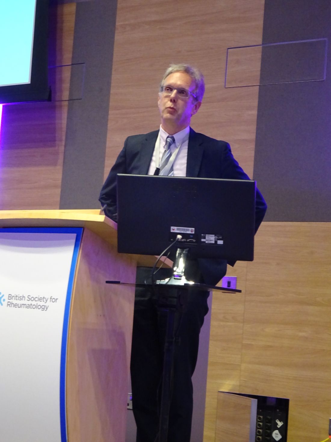
“The claim that the elderly are treated less aggressively isn’t completely true throughout the whole treat-to-target strategy,” said Simone Howard of King’s College London at the British Society for Rheumatology annual conference. While older patients were less likely to receive intensive treatment up to 2 years after their diagnosis, there was a shorter delay between the onset of symptoms and the first outpatient visit to a rheumatology clinic.
When compared against patients who were younger than 65 years, those aged 65-74 years and 75 years and older were 11% (P = .02) and 15% (P = .02) more likely to have their first outpatient visit within 10 months.
Furthermore, no significant differences were seen between any age groups in the time to first initiation of a conventional synthetic disease-modifying antirheumatic drug (csDMARD), which averaged nearly 3 months after symptoms appeared.
Ms. Howard, who has previously worked at the European Medicines Agency, noted that, during her time at the EMA, “there was a real push to incorporate the elderly into the regulatory framework more. In parallel, there were also reports of the elderly being treated less aggressively. So the question was, where was that coming from?”
Similar therapeutic approaches are advocated for older and younger RA patients, and to look for any disparities, Ms. Howard and associates turned to the Early RA Network (ERAN) to “investigate potential treatment bias against the elderly.”
ERAN is a hospital-based inception cohort of 1,236 patients with early RA who were recruited across 23 centers in the United Kingdom and Ireland between 2002 and 2014.
Of 1,131 patients used in the analyses, 9.7% (n = 110) were 75 years or older, 21.5% (n = 243) were aged 65-74 years, and 68.8% (n = 778) were 65 years or younger. The majority (67.7%) of patients were female.
Patients aged 75 years and older were more likely to present with comorbidities than the youngest group, and they had higher health assessment questionnaire scores at baseline. However, they were no more likely to have high disease activity at the first visit, which was defined as a disease activity score in 28 joints of more than 5, and the older patients were 27% less likely to be seropositive (P = .004).
“It’s when we come to pharmacological aspects of care that we are seeing treatment biases,” Ms. Howard noted. Patients over 75 years were significantly more likely than the youngest age group to be treated with glucocorticoids or csDMARD monotherapy at 1 year, and 23% more likely to be on less aggressive therapy (P equal to or less than .0001). Aggressive therapy was defined as the use of a combination of csDMARDs or the use of biologic drugs.
At 2 years, the oldest patients were 46% more likely than those under 65 years to be on less-intensive therapies (P equal to or less than .0001), with those aged 65-74 years 19% more likely to be on glucocorticoid or csDMARD therapy (P = .005).
Factors such as patient choice and tolerance were not considered in the analyses and could be important, Ms. Howard conceded in response to a question after her presentation.
Another point raised was that perhaps the prescribing of aggressive therapy would rationally be different in someone diagnosed with RA at age 85 versus 65 because the duration of time that would be likely to be lived with accumulating joint damage would be shorter at the older age and that would be balanced against the other effects of the therapy. So, there may be important reasons in shared decision making that influenced the treatment choices other than the age of patients.
Ms. Howard agreed, noting that this demonstrated the need to be careful around the language used for defining what constituted aggressive or intensive therapy.
She and her coauthors reported no conflicts of interest.
SOURCE: Howard S et al. Rheumatology. 2019;58(suppl 3), Abstract 011.
REPORTING FROM BSR 2019
Methotrexate does not cause rheumatoid interstitial lung disease
BIRMINGHAM, ENGLAND – Data from two early RA inception cohorts provide reassurance that methotrexate does not cause interstitial lung disease and suggest that treatment with methotrexate might even be protective.
In the Early RA Study (ERAS) and Early RA Network (ERAN), which together include 2,701 patients with RA, 101 (3.7%) had interstitial lung disease (ILD). There were 92 patients with RA-ILD who had information available on exposure to any conventional synthetic disease-modifying antirheumatic drug (csDMARD); of these, 39 (2.5%) had been exposed to methotrexate (n = 1,578) and 53 (4.8%) to other csDMARDs (n = 1,114).
Multivariate analysis showed that methotrexate exposure was associated with a reduced risk of developing ILD, with an odds ratio of 0.48 (P = .004). In a separate analysis that excluded 25 patients who had ILD before they received any csDMARD therapy (n = 67), there was no association between methotrexate use and ILD (OR, 0.85; P = .578). In fact, there was a nonsignificant trend for a delayed onset of ILD in patients who had been treated with methotrexate (OR, 0.54; P = .072).
Methotrexate use is associated with an acute hypersensitivity pneumonitis in patients with RA, explained Patrick Kiely, MBBS, PhD, of St. George’s University Hospitals NHS Foundation Trust in London at the British Society for Rheumatology annual conference. “This is well recognized, it’s very rare [0.43%-1.00%], it’s easy to spot, and usually goes away if you stop methotrexate,” said Dr. Kiely, adding that “it’s not benign, and severe cases can be life threatening.”
Because of the association between methotrexate and pneumonitis, there has been concern that methotrexate may exacerbate or even cause ILD in RA but there are sparse data available to confirm this. The bottom line is that you should not start someone on methotrexate if you think their existing lung capacity is not up to treatment with methotrexate, Dr. Kiely said.
ILD is not always symptomatic in RA, but when it is, it is associated with very poor survival. The lung disease can be present before joint symptoms, Dr. Kiely said. Although less than 10% of cases may be symptomatic, this “is a big deal, because it has a high mortality, with death within 5 years. It’s the second-commonest cause of excess mortality in RA after cardiovascular disease.”
To look at the association between incident RA-ILD and the use of methotrexate, Dr. Kiely and associates analyzed data from ERAS (1986-2001) and ERAN (2002-2013), that together have more than 25 years of follow-up data on patients who were recruited at the first sign of RA symptoms. Patients within these cohorts have been treated according to best practice, and a range of outcomes – including RA-ILD – have been assessed at annual intervals.
In the patients who developed ILD after any csDMARD exposure, older age at RA onset (OR, 1.04; P less than .001) and having ever smoked (OR, 1.91; P = .016) were associated with the development of the lung disease. Incident ILD was also associated with being positive for rheumatoid factor (OR, 2.02; P = .029) at baseline. Being male was also associated with a higher risk for developing ILD, Dr. Kiely reported, as was a longer duration of time between the onset of first RA symptoms and the first secondary care visit. Conversely, the presence of nonrespiratory, major comorbidities at baseline appeared to be protective (OR, 0.62; P = .027).
“We found no association between methotrexate treatment and incident RA-ILD and a possibility that it may be protective,” Dr. Kiely concluded, noting that these data were now published in BMJ Open (2019;9:e028466. doi: 10.1136/bmjopen-2018-028466).
Following Dr. Kiely’s presentation, an audience member asked if the protective effect seen with methotrexate could have been caused by better disease control overall.
Dr. Kiely answered that, up until 2001, the time when ERAS was ongoing, standard practice in the United Kingdom was to use sulfasalazine, but then methotrexate started to be used in higher and higher doses, as seen in ERAN.
The interesting thing is that in ERAN more methotrexate was used in higher doses, but less RA-ILD was seen, Dr. Kiely observed. The overall prevalence of RA-ILD in the later early RA cohort was 3.2% and the median dose of methotrexate used was 20 mg. In ERAS, the prevalence was 4.2% and the median dose of methotrexate used was 10 mg.
There was a suggestion that disease control was slightly better in ERAN than ERAS, but that wasn’t statistically significant, Dr. Kiely said.
So, should a patient with RA and ILD be given methotrexate? There’s no reason not to, Dr. Kiely suggested, based on the evidence shown. Part of the challenge will now be convincing chest physician colleagues that methotrexate is not problematic in terms of causing ILD.
These findings are completely on board with the ILD group’s findings that methotrexate doesn’t cause pulmonary fibrosis in patients with RA, commented Julie Dawson, MD, of St. Helens and Knowsley Teaching Hospitals NHS Trust, St. Helens, England. Her own research, which includes a 10-year follow-up of patients with inflammatory arthritis, has shown that methotrexate does not appear to increase the risk of pulmonary fibrosis.
The study had no specific outside funding. Dr. Kiely reported having no conflicts of interest.
SOURCE: Kiely P et al. Rheumatology. 2019;58(suppl 3), Abstract 009.
REPORTING FROM BSR 2019
Walk-in ultrasound helps to avoid unnecessary steroids for giant cell arteritis
BIRMINGHAM, England – More than half of all patients referred to a fast-track giant cell arteritis (GCA) clinic that offers a walk-in ultrasonography service avoided use of glucocorticoids, according to a report given at the annual conference of the British Society for Rheumatology.
The clinic, an initiative run by the University Hospital Coventry and Warwickshire (UHCW) NHS Trust for the past 6 years, provides same-day diagnosis and treatment for suspected GCA.
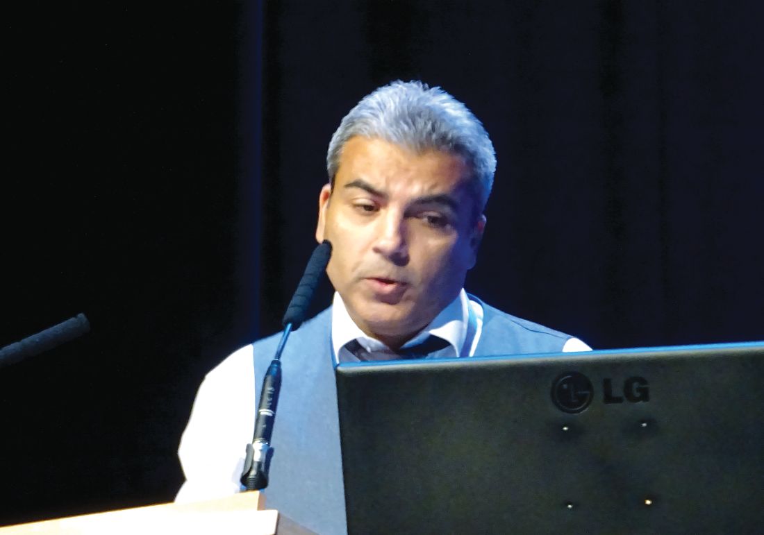
“Walk-in ultrasound helps to avoid steroids completely in a significant proportion of patients,” said study author and presenter Shirish Dubey, MBBS, a consultant rheumatologist at the UHCW NHS Trust. Of 652 patients seen at the UHCW GCA fast-track clinic between 2014 and 2017, 143 (22%) were diagnosed with GCA. Over 400 had not been exposed to glucocorticoids and 369 (57%) were able to avoid unnecessary glucocorticoid use in the cohort, Dr. Dubey reported.
The old NHS paradigm for managing patients with suspected GCA was that when they presented to their primary care physicians, they would be started on immediate glucocorticoid therapy while waiting for an urgent specialist referral. However, that referral could take anywhere from a couple of days to a couple of weeks to happen, Dr. Dubey explained. Patients would then undergo possible temporal artery biopsy (TAB) and only then, following confirmation of a GCA diagnosis, would a management plan be agreed upon.
UCHW introduced its fast-track pathway for the diagnosis of GCA in mid-2013. The pathway called for patients with suspected GCA aged 50 years or older who had two or more features present, such as an abrupt, new-onset headache or facial pain, scalp pain and tenderness, jaw claudication, or visual symptoms. Primary care physicians could make urgent referrals to the service via an on-call rheumatology trainee or ophthalmology senior house officer.
“Patients are normally steroid-naive and seen on the same day,” Dr. Dubey said. Doppler ultrasound of the temporal artery results in around 80% of diagnoses, with TAB still needed in some cases.
One of the downsides of the fast-track process perhaps is the increasing number of referrals. “One thing we find is that we have become a glorified headache service,” Dr. Dubey said. However, many patients do not have GCA and, when there is a low clinical probability and the ultrasound is negative, the patient is usually reassured and discharged with no need for glucocorticoids. Although the number of referrals have increased – 98 patients in 2014, 154 in 2015, 123 in 2016, and 277 in 2017 – the number of those diagnosed with GCA has remained around the same.
To see how ultrasound was faring in real-life practice, the UHCW NHS Trust team compared Doppler ultrasound findings against the final clinical diagnosis for the period 2014 to 2017. A sensitivity of just under 48% and specificity of 98% was recorded. The positive and negative predictive values were 87% and 88%, respectively.
The specificity of ultrasound was lower than that reported previously in the literature, the UHCW NHS Trust team pointed out in its abstract, but it does compare similarly with other real-world studies. The use of glucocorticoids affected the ultrasound results, with better sensitivity (55%) when these drugs were not used prior to the scan.
The use of TAB versus a clinical diagnosis in 100 patients seen over the same time period showed it had a sensitivity of 37% and a specificity of 100%, with positive and negative predictive values of 100% and 62%. The sensitivity of TAB is again low, Dr. Dubey said, but that could be because TAB is performed only when the diagnosis is uncertain.
This was an unselected cohort of patients, but overall there were good positive and negative predictive values. The UHCW NHS Trust team suggested that ultrasound can assist and reassure clinicians trying to diagnose or exclude GCA in their patients.
Regular multidisciplinary team meetings including rheumatology, ophthalmology, and vascular Doppler physiologists are key to the fast-track service, Dr. Dubey pointed out.
Despite the shortcomings of the retrospective study, Dr. Dubey stressed that the team was confident that none of the patients who had been ruled out as having GCA were subsequently diagnosed as having GCA.
Importantly, he said, the use of ultrasound had made a big difference in cost; the group plans to formally evaluate costs of ultrasound versus TAB.
The study received no commercial funding. Dr. Dubey had no conflicts of interest to disclose.
SOURCE: Pinnell J et al. Rheumatology. 2019;58(suppl 3):Abstract 038.
REPORTING FROM BSR 2019
Intradermal etanercept improves discoid lupus
BIRMINGHAM, ENGLAND – Intradermal delivery of a tumor necrosis factor inhibitor (TNFi) could offer patients with discoid lupus erythematosus (DLE) a much-needed additional treatment option, according to results of a phase 2, “proof-of-concept” study.
Overall, 14 (56%) of the 25 patients in the study achieved a 20% or greater reduction in disease activity from baseline to week 12 via intradermal injection of etanercept (Enbrel), which was assessed via the modified limited Score of Activity and Damage in DLE (ML-SADDLE). About half (48%) and one-fifth (20%) also achieved greater reductions of 50% and 70%, respectively.
“Discoid lupus is a chronic form of cutaneous lupus. Usually it occurs in visible areas like the face and scalp, causing scarring, so it’s really disabling and affects patients’ quality of life,” observed the lead study investigator Md Yuzaiful Md Yusof, MBChB, PhD, NIHR Academic Clinical Lecturer at the University of Leeds, England.
“It’s also one of the most resistant manifestations of lupus,” he said during a poster presentation at the annual conference of the British Society for Rheumatology. “Usually, when people have discoid lupus, the dermatologist gives antimalarial treatment, but only 50% of people respond to these drugs. So, what happens to the rest of them?” Basically, it is trial and error, Dr. Md Yusof said; some patients may be given disease-modifying antirheumatic drugs and in some patients this may work well, but in others there may be toxicity that contraindicates treatment.
B-cell therapy with rituximab (Rituxan) has not been successful, he said. In a previous study of 35 patients with refractory discoid lupus, none of the patients responded to rituximab and half of them actually flared after taking the drug.
There is a pathologic case for using anti-TNF therapy in DLE, but the use of TNFis is not without concern. Such treatment can increase antinuclear antibody production and make lupus worse. “In order to overcome this, as the lesion is quite small, we don’t need to use a systemic approach,” Dr. Md Yusof explained in an interview. “If you give directly, it should just be confined to the lesion and not absorbed, that’s the whole idea of thinking outside the box.” He noted that if it worked, such treatment would be for inducing remission and not for maintenance.
The study, “Targeted therapy using intradermal injection of etanercept for remission induction in discoid lupus erythematosus” (TARGET-DLE) was designed to test the validity of using intradermal rather than subcutaneous TNFi therapy in patients with discoid lupus.
Dr. Md Yusof noted that only 25 patients needed to be recruited into the single-arm, prospective trial as a “Simon’s two-stage minimized design” was used (Control Clin Trials. 1989;10[1]:1-10). This involved treating the first few patients to see if a response occurred and if it did, carrying on with treating the others, but if no response occurred in at least two patients, the trial would stop completely.
Adult patients were eligible for inclusion if they had one or more active DLE lesions and had not responded to antimalarial treatment. Stable doses of DMARDs and up to 10 mg of oral prednisolone daily was permitted if already being taken prior to entering the study.
Etanercept was injected intradermally around the most symptomatic lesion once a week for up to 12 weeks. The dosage was determined based on the radius of the selected discoid lesion. Over an 18-month period, all 25 patients were recruited, including 18 women. The median age of patients was 47 years, and six had systemic lupus erythematosus. The median number of prior DMARDs was 5 but ranged from 1 to 16, indicating a very resistant patient population.
The primary endpoint was at least 6 of the 25 patients having at least a 20% reduction in ML-SADDLE at week 12; 14 (56%) patients achieved this.
“We didn’t use CLASI [Cutaneous Lupus Area and Severity Index Activity Score] because that only includes erythema and atrophy,” Dr. Md Yusof explained. “In discoid lupus, induration is quite important as well, so that’s why we used ML-SADDLE. We called it ‘modified limited’ because the original SADDLE score is based on the whole organ score, but we only calculated the one lesion that we wanted to treat.”
In addition to meeting the primary endpoint, several secondary endpoints were met, including significant improvements in scores on visual analog scales as determined by pre- and posttreatment scoring by physicians (53.1 mm vs. 23.2 mm; P less than .001) and patients (56.9 mm vs. 29.7 mm; P = .001). Mean Dermatology Life Quality Index (DLQI) score significantly improved between pre- and post treatment, as did blood perfusion under the skin based on laser Doppler imaging and infrared thermography. However, no difference was seen with optical coherence tomography.
“There were only four grade 3/4 toxicities, and importantly, none of the SLE patients got worse, and none with DLE only converted into SLE,” Dr. Md Yusof reported. Of the four grade 3/4 adverse events, two were chest infections, one was heart failure, and one was a worsening of chilblains.
“It was a full-powered phase 2 trial, and because it was positive, now we can go to phase 3 trial,” he added.
Before conducting a phase 3 trial, however, Dr. Md Yusof wants to refine how the TNFi is delivered. Perhaps an intradermal patch with microneedles could be used. This would be left on the skin for a short amount of time to allow drug delivery and then removed. It could help ensure that all patients comply with treatment and perhaps even self-administer, he noted.
“The median compliance rate was 80%, which is not too bad, but I think when we come to run a phase 3 trial, I’m looking to improve the drug delivery,” he said. Changing the delivery method will need to be validated before a phase 3 trial can be started.
The study was not commercially funded. Dr. Md Yusof had no disclosures. Pfizer provided the study drug free of charge.
SOURCE: Md Yusof MY et al. Rheumatology. 2019;58(suppl 3): Abstract 244. doi: 10.1093/rheumatology/kez107.060.
REPORTING FROM BSR 2019
Experts agree on optimal use of MRI in axSpA
BIRMINGHAM, ENGLAND – An evidence-based approach coupled with expert consensus has been used to determine the best way to use MRI for the diagnosis of axial spondyloarthritis (axSpA).
Working under the auspices of the British Society for Spondyloarthritis (BRITSpA), a task force of nine rheumatologists and nine musculoskeletal radiologists with an interest in axSpA developed a set of seven recommendations that provide guidance on how to best to acquire and then interpret MRI images of the spine and sacroiliac joints.
The recommendations, which were published online (Rheumatology. 2019 May 2. doi: 10.1093/rheumatology/kez173), cover how to perform MRI when axSpA is suspected, such as by imaging both the sacroiliac joints and the spine, and provide guidance on the sequences and order of MRI planes to be used, and what features may increase the diagnostic confidence of axSpA.
The recommendations are as follows:
• When requesting an MRI for suspected axSpA, imaging of both the sacroiliac joints and the spine is recommended.
• T1-weighted and fat-suppressed, fluid-sensitive sequences are recommended for suspected axSpA.
• The minimum protocol when requesting an MRI for suspected axSpA should include sagittal images of the spine with extended lateral coverage and images of the sacroiliac joints that are in an oblique coronal plane to the joint.
• In the sacroiliac joints, the presence of bone marrow edema, fatty infiltration, or erosion is suggestive of the diagnosis of axSpA. The presence of more than one of these features increases the diagnostic confidence of axSpA.
• In the spine, the presence of multiple corner inflammatory lesions and/or multiple corner fatty lesions increases the diagnostic confidence of axSpA.
• In the sacroiliac joints and/or spine, the presence of characteristic new bone formation increases the diagnostic confidence of axSpA.
• The full range and combination of active and structural lesions of the sacroiliac joints and spine should be taken into account when deciding if the MRI scan is suggestive of axSpA or not.
The recommendations “are intended to standardize practice around the use of MRI,” said Alexis Jones, MBBS, MS (Rheumatology), a senior clinical research fellow at University College London Hospitals NHS Foundation Trust. She presented the recommendations on behalf of the expert task force at the annual conference of the British Society for Rheumatology.
“MRI has become an essential tool in axial spondyloarthritis. It facilitates earlier diagnoses and therefore has allowed for earlier initiation of treatment. It can be used to monitor the burden of inflammation and may predict response to therapy,” Dr. Jones said. Despite this, “there is significant inconsistency in the use of MRI” in clinical practice.
For instance, a survey performed in the United Kingdom (J Rheumatol. 2017;44[6]:780-5) highlighted the need for better collaboration between rheumatology and radiology departments to identify axSpA MRI lesions and develop appropriate protocols.
That survey showed that a quarter of radiologists were not aware of the term axSpA, and just 31% and 25%, respectively, were aware of Assessment of Spondyloarthritis international Society (ASAS) criteria for a positive MRI of the sacroiliac joints and spine. Furthermore, 18% of radiologists did not recognize bone marrow edema as a diagnostic feature of axSpA.
The heterogeneity in the performance of MRI in clinical practice could lead to a delay in diagnosis and potentially misdiagnosis, the task force’s lead author and consultant rheumatologist, Pedro Machado, MD, said in an interview.
“I think everyone has been focusing on demonstrating the value of MRI in the condition but then they forgot to look at the standardization aspect,” said Dr. Machado, who works at University College Hospital and the National Hospital for Neurology and Neurosurgery in London.
With that in mind, the BRITSpA-endorsed task force was set up and met to determine the scope of the recommendations. They looked at the evidence for the use of MRI in the diagnosis of axSpA and used two overarching principles to draft the recommendations: 1) the diagnosis of axSpA is based on clinical, laboratory, and imaging features; 2) Some patients with axSpA have isolated inflammation of the sacroiliac joints or spine.
“All of the recommendations were met with a high level of agreement, indicating a strong consensus” among rheumatologists and radiologists, Dr. Jones noted.
“These recommendations can be immediately applied to clinical practice,” said Dr. Machado, who noted that they should standardize practice and decrease heterogeneity around the use of MRI. “This will help ensure a more informed and consistent approach to the diagnosis of axSpA.”
One of the potential impacts of the recommendations, if followed, is that they may actually help to reduce health care costs, Dr. Machado suggested, because an optimized protocol would be used, making MRI more cost effective by not including sequences that do not add value in the condition.
The next task is to share the recommendations more widely and make sure they are applied in clinical practice.
A systematic literature review on which the recommendations were based was published simultaneously with the conference presentation (Rheumatology. 2019 May 2. doi: 10.1093/rheumatology/kez172).
The work was supported by BRITSpA. The authors had no relevant disclosures.
SOURCE: Bray TJP et al. Rheumatology 2019;58(suppl 3): Abstract 033. doi: 10.1093/rheumatology/kez105.032.
REPORTING FROM BSR 2019
Higher infection risk in RA seen with high blood biologic levels
BIRMINGHAM, ENGLAND – Higher blood biologic drug levels in the first year of treatment for rheumatoid arthritis independently increased the risk of any infection by about 50% when compared against low or normal levels in a new observational cohort study, providing support for monitoring biologic drug levels to help to predict infection risk.
Data from the British Society for Rheumatology Biologics Register – Rheumatoid Arthritis (BSRBR-RA) that were presented at the British Society for Rheumatology annual conference showed that the adjusted hazard ratio for any infection occurring within the first year among patients with high drug levels was 1.51, with a 95% confidence interval (CI) of 1.14 to 2.01. The adjustments took into account patients’ age, gender, disease activity score, and use of methotrexate.
There are more than 10 biologics now available for use in rheumatoid arthritis but deciding which to use in a particular patient remains very much “a trial and error approach,” first author Meghna Jani, MBChB, said at the conference.
“From a patient perspective, one of the most important concerns continues to be the risk of serious infections and adverse events,” added Dr. Jani, a National Institute for Health Research Academic Clinical Lecturer in Rheumatology at the University of Manchester (England).
The link between biologic agents and infections, including those that could result in hospitalization or other serious consequences, has been well studied in biologics registries. It is known, for example, that the risk of infections with tumor necrosis factor inhibitor treatment seems to be highest during the first 6-12 months of treatment.
According to Dr. Jani, conventional means of determining risk – such as patient age and the presence of comorbid factors – have limited benefit in terms of deciding which patients could be at heightened risk of infections. “Ideally, we need biomarkers in rheumatology that can be implemented in clinical practice and help us predict efficacy and safety, as well as help us use these medications much more cost-effectively,” she said.
Four years ago, a meta-analysis (Lancet. 2015;386:258-65) suggested that the risk of infection may be linked to using higher doses of anti–tumor necrosis factor drugs, which led the BSRBR-RA team to see if elevated levels of these drugs in the serum could be predictive of the infection risk and thus used as a possible biomarker. There was also prior evidence that serum drug concentrations of biologics were associated with long-term treatment response and that a certain level was needed to determine the likely treatment response.
In the current study, Dr. Jani and colleagues used data on 703 patients with rheumatoid arthritis starting biologic therapy who were simultaneously recruited into the BSRBR-RA, which has been running since 2001, and the Biologics in Rheumatoid Arthritis Genetics and Genomics Study Syndicate (BRAGGSS). The BSRBR-RA did not collect biological samples, but in BRAGGSS serological samples were collected at 3-, 6-, and 12-month intervals after the start of a biologic treatment, along with other assessments. This is the first time two national, U.K.-based, rheumatoid arthritis cohorts have been linked in this way, Dr. Jani said.
Serum samples taken from the patients were assessed via enzyme-linked immunoassay to determine levels of the biologic agent used, with high drug levels defined as more than 4 mcg/mL for etanercept (n = 286), tocilizumab (n = 104), and infliximab (n = 14); more than 8 mcg/mL for adalimumab (n = 179), and 25 mcg/mL or more for certolizumab pegol (n = 120).
In the study, about three-quarters of the patients were women. The mean age was 58 years, and disease duration was just under 6 years. Most patients were starting their first biologic.
The crude rate of all infections at 1 year, including recurrent infections, was 464 per 1,000 patient-years in the high biologic drug level group versus 314 per 1,000 patient-years in the low biologic drug level group. When only the first infections were considered, the crude rate of all infections within the first year were a respective 300 and 229 per 1,000 patient-years, with an adjusted hazard ratio of 1.27, Dr. Jani reported.
As expected, lower respiratory tract infections were the most common type of infection, occurring in 34% of patients with high drug levels versus around 10% in the low drug level group. Upper respiratory tract, urinary tract, and skin infections including shingles were seen in a respective 16%, 15%, and 8% in the high drug level group, with rates less than 5% in the low drug level group.
Of note, there were certain types of infections present in the high but not low drug level groups: bacterial peritonitis, neutropenic sepsis, and herpes zoster.
Crude rates for serious infections at 1 year were 76 and 54 per 1,000 patient-years, respectively, for the high and low drug level groups. The crude rates for the first serious infection within the first year were 44 and 29 per 1,000 patient-years. The adjusted hazard ratio for the risk of serious infection at 1 year was 1.26. Serious infections were rare events, Dr. Jani emphasized, so the power was reduced, but “there was a slightly increased risk.”
Aside from the low statistical power to assess the rarer serious infections, another limitation was that drug levels were not measured at the time of the adverse event.
Concluding, Dr. Jani suggested that perhaps monitoring drug levels could be useful in predicting the risk of infection in patients being treated with biologics for rheumatoid arthritis.
Furthermore, “in patients with remission, dose-tapering guided by therapeutic drug monitoring may help lower infection risk and help us balance safety and efficacy.”
When asked to comment, Tore K. Kvien, MD, PhD, of the department of rheumatology at Diakonhjemmet Hospital in Oslo, supported this conclusion. “Therapeutic drug monitoring [TDM] is widely used among gastroenterologists when treating inflammatory bowel diseases with TNF inhibitors. In recent years, data from several research groups in rheumatology have indicated that TDM may help to optimize drug efficacy. The results from Dr. Jani and her colleagues also support that TDM may be important for safety. The importance of TDM as a ‘new’ hot topic in rheumatology is also supported by the recent establishment of a EULAR [European League Against Rheumatism] task force to further explore the value of TDM when treating patients with inflammatory joint diseases.”
The BSRBR-RA is funded through the BSR, which receives restricted income from several U.K. pharmaceutical companies. These currently include AbbVie, Celltrion, Hospira, Pfizer, UCB, and Roche, and in the past, Swedish Orphan Biovitrum and Merck. The pharmaceutical company funders had no role in the design and conduct of the study; collection, management, analysis, and interpretation of the data; preparation, review, or approval of the manuscript; and decision to submit the manuscript for publication. Dr. Jani has no personal conflicts of interest to disclose.
SOURCE: Jani M et al. Rheumatology, 2019 April;58(Suppl 3):kez105.018.
In this study, the authors use the major British Society for Rheumatology Biologics Register – Rheumatoid Arthritis and examine infections and serious infections across biologics. They define “low/normal” blood levels versus “high” blood levels based on concentration-effect curves. Examining data censored at 1 year versus incidence during 1 year, the results are somewhat inconsistent. With larger numbers available for data censored at 1 year, there is some increased risk using hazard ratios for both all infections and serious infections. With smaller numbers for incident infections during the first year, this hazard ratio does not show an effect.
Daniel E. Furst, MD, is professor of medicine (emeritus) at the University of California, Los Angeles, an adjunct professor at the University of Washington, Seattle, and research professor at the University of Florence (Italy). He is also practices part-time in Los Angeles and Seattle.
BIRMINGHAM, ENGLAND – Higher blood biologic drug levels in the first year of treatment for rheumatoid arthritis independently increased the risk of any infection by about 50% when compared against low or normal levels in a new observational cohort study, providing support for monitoring biologic drug levels to help to predict infection risk.
Data from the British Society for Rheumatology Biologics Register – Rheumatoid Arthritis (BSRBR-RA) that were presented at the British Society for Rheumatology annual conference showed that the adjusted hazard ratio for any infection occurring within the first year among patients with high drug levels was 1.51, with a 95% confidence interval (CI) of 1.14 to 2.01. The adjustments took into account patients’ age, gender, disease activity score, and use of methotrexate.
There are more than 10 biologics now available for use in rheumatoid arthritis but deciding which to use in a particular patient remains very much “a trial and error approach,” first author Meghna Jani, MBChB, said at the conference.
“From a patient perspective, one of the most important concerns continues to be the risk of serious infections and adverse events,” added Dr. Jani, a National Institute for Health Research Academic Clinical Lecturer in Rheumatology at the University of Manchester (England).
The link between biologic agents and infections, including those that could result in hospitalization or other serious consequences, has been well studied in biologics registries. It is known, for example, that the risk of infections with tumor necrosis factor inhibitor treatment seems to be highest during the first 6-12 months of treatment.
According to Dr. Jani, conventional means of determining risk – such as patient age and the presence of comorbid factors – have limited benefit in terms of deciding which patients could be at heightened risk of infections. “Ideally, we need biomarkers in rheumatology that can be implemented in clinical practice and help us predict efficacy and safety, as well as help us use these medications much more cost-effectively,” she said.
Four years ago, a meta-analysis (Lancet. 2015;386:258-65) suggested that the risk of infection may be linked to using higher doses of anti–tumor necrosis factor drugs, which led the BSRBR-RA team to see if elevated levels of these drugs in the serum could be predictive of the infection risk and thus used as a possible biomarker. There was also prior evidence that serum drug concentrations of biologics were associated with long-term treatment response and that a certain level was needed to determine the likely treatment response.
In the current study, Dr. Jani and colleagues used data on 703 patients with rheumatoid arthritis starting biologic therapy who were simultaneously recruited into the BSRBR-RA, which has been running since 2001, and the Biologics in Rheumatoid Arthritis Genetics and Genomics Study Syndicate (BRAGGSS). The BSRBR-RA did not collect biological samples, but in BRAGGSS serological samples were collected at 3-, 6-, and 12-month intervals after the start of a biologic treatment, along with other assessments. This is the first time two national, U.K.-based, rheumatoid arthritis cohorts have been linked in this way, Dr. Jani said.
Serum samples taken from the patients were assessed via enzyme-linked immunoassay to determine levels of the biologic agent used, with high drug levels defined as more than 4 mcg/mL for etanercept (n = 286), tocilizumab (n = 104), and infliximab (n = 14); more than 8 mcg/mL for adalimumab (n = 179), and 25 mcg/mL or more for certolizumab pegol (n = 120).
In the study, about three-quarters of the patients were women. The mean age was 58 years, and disease duration was just under 6 years. Most patients were starting their first biologic.
The crude rate of all infections at 1 year, including recurrent infections, was 464 per 1,000 patient-years in the high biologic drug level group versus 314 per 1,000 patient-years in the low biologic drug level group. When only the first infections were considered, the crude rate of all infections within the first year were a respective 300 and 229 per 1,000 patient-years, with an adjusted hazard ratio of 1.27, Dr. Jani reported.
As expected, lower respiratory tract infections were the most common type of infection, occurring in 34% of patients with high drug levels versus around 10% in the low drug level group. Upper respiratory tract, urinary tract, and skin infections including shingles were seen in a respective 16%, 15%, and 8% in the high drug level group, with rates less than 5% in the low drug level group.
Of note, there were certain types of infections present in the high but not low drug level groups: bacterial peritonitis, neutropenic sepsis, and herpes zoster.
Crude rates for serious infections at 1 year were 76 and 54 per 1,000 patient-years, respectively, for the high and low drug level groups. The crude rates for the first serious infection within the first year were 44 and 29 per 1,000 patient-years. The adjusted hazard ratio for the risk of serious infection at 1 year was 1.26. Serious infections were rare events, Dr. Jani emphasized, so the power was reduced, but “there was a slightly increased risk.”
Aside from the low statistical power to assess the rarer serious infections, another limitation was that drug levels were not measured at the time of the adverse event.
Concluding, Dr. Jani suggested that perhaps monitoring drug levels could be useful in predicting the risk of infection in patients being treated with biologics for rheumatoid arthritis.
Furthermore, “in patients with remission, dose-tapering guided by therapeutic drug monitoring may help lower infection risk and help us balance safety and efficacy.”
When asked to comment, Tore K. Kvien, MD, PhD, of the department of rheumatology at Diakonhjemmet Hospital in Oslo, supported this conclusion. “Therapeutic drug monitoring [TDM] is widely used among gastroenterologists when treating inflammatory bowel diseases with TNF inhibitors. In recent years, data from several research groups in rheumatology have indicated that TDM may help to optimize drug efficacy. The results from Dr. Jani and her colleagues also support that TDM may be important for safety. The importance of TDM as a ‘new’ hot topic in rheumatology is also supported by the recent establishment of a EULAR [European League Against Rheumatism] task force to further explore the value of TDM when treating patients with inflammatory joint diseases.”
The BSRBR-RA is funded through the BSR, which receives restricted income from several U.K. pharmaceutical companies. These currently include AbbVie, Celltrion, Hospira, Pfizer, UCB, and Roche, and in the past, Swedish Orphan Biovitrum and Merck. The pharmaceutical company funders had no role in the design and conduct of the study; collection, management, analysis, and interpretation of the data; preparation, review, or approval of the manuscript; and decision to submit the manuscript for publication. Dr. Jani has no personal conflicts of interest to disclose.
SOURCE: Jani M et al. Rheumatology, 2019 April;58(Suppl 3):kez105.018.
REPORTING FROM BSR 2019
ECHO Rheum widens rheumatology best practice care
according to the results of a qualitative and quantitative study.
Over a 9-year period, ECHO (Extension for Community Healthcare Outcomes) Rheum educated 2,230 primary care clinicians, the majority of whom were physicians (53%) or nurse practitioners (22%), and helped increase the clinicians’ confidence in managing rheumatic conditions locally while still having access to specialist guidance.
Participants in ECHO Rheum accrued a total of 1,958 CME credits between them, and 21 of 30 (70%) clinicians who participated in 2-day miniresidencies obtained online certification from the American College of Rheumatology.
“ECHO Rheum and programs like it have the potential to positively impact the national shortage of rheumatologic care for underserved patients,” Arthur Bankhurst, MD, and his associates from the University of New Mexico, Albuquerque wrote in Arthritis Care & Research. “Empowering the health care workforce by disseminating knowledge of best practice diagnosis and treatment has the potential to reduce suboptimal rheumatologic care and expand access for those suffering from rheumatologic conditions regardless of economic status or location.”
ECHO Rheum is part of the larger Project ECHO (Extension for Community Healthcare Outcomes), a program developed at UNM by gastroenterologist Sanjeev Arora, MD. Dr. Arora came up with the idea for the ECHO model in 2003 to try to improve access to best practice care for patients with hepatitis C. The potential of the model to translate across medical specialties was soon seen, however, and today, there are more than 40 teleECHO programs operating in New Mexico that also involve more than 400 community clinic sites. The ECHO model is also being used nationally, operating in more than 30 states, and globally in 31 countries.
According to information available via the Project ECHO website, “the ECHO model is not traditional ‘telemedicine’ where the specialist assumes care of the patient, but is instead telementoring, a guided practice model where the participating clinician retains responsibility for managing the patient.” The aim is to use medical education and care management to empower “clinicians everywhere to provide better care to more people, right where they live.”
The rheumatology arm of Project ECHO started in 2006 and follows the four main principles of the ECHO model: Using technology to leverage scarce resources, such as by facilitating regular educational sessions between central expert “hubs” at specialist centers and “spokes” at community practices; sharing best practice information to help reduce disparities in the quality of care; presenting and discussion around case histories; and evaluating and monitoring outcomes.
To demonstrate the effectiveness of ECHO Rheum, the team at UNM evaluated data on participants who had attended weekly teleconferencing sessions, lectures, grand rounds, and miniresidency training between June 2006 and June 2014.
The results demonstrated that “participation in ECHO Rheum provides clinicians in underresourced areas access to best practice knowledge and training in rheumatology,” Dr. Bankhurst and his coauthors observed.
Increased knowledge and confidence in managing rheumatic conditions among primary care physicians could help to reduce the ever-widening gap between the demand and supply of appropriate care. Indeed, while the projected population of adults with rheumatic disease is expected to grow to almost 67 million adults in the next 20 years, there is expected to be a shortfall of 4,700 full-time rheumatologists by 2030.
“Regular participation in teleECHO sessions creates a community of practice among rural clinicians,” the UNM team stated. Every week, participants were invited to attend a 90-minute virtual teleconference, either by video or telephone, that consisted of a brief evidence-based lecture by a subject matter expert and presentation of deidentified case histories submitted by the participants. Participants were able to obtain CME credits at no cost and learn how to use ACR-recommended disease activity measures, such as the Disease Activity Score in 28 joints.
Over the 9-year study period, 1,173 cases were presented, the majority of which (85%) reflected the three most common diagnoses seen in rheumatologic practice, namely RA (n = 715), fibromyalgia (n = 241), and systemic lupus erythematosus (n = 54). As might be expected, female cases were presented more often than male cases, as were cases involving patients aged 40-60 years, compared with other age groups.
“ECHO Rheum saw an increase in participation in 2010, when the ACR certification was offered at no cost, suggesting that ACR certification was an incentive for participation,” Dr. Bankhurst and his team observed.
In 2012, however, there was a drop-off in participation, which may have been caused by the end of funding for ACR certification or clinicians becoming “more confident in their abilities to diagnose and initiate treatment appropriately.” Clinical responsibilities may also have prevented clinicians from attending the weekly sessions.
“Although our formal collaboration with the ACR concluded when funds for this initiative came to an end, ECHO Rheum encourages clinicians to obtain certification in rheumatology and provides them with information about opportunities to offset the cost,” Dr. Bankhurst and his colleagues wrote.
Despite a decline in attendance at the weekly teleECHO sessions during the evaluation period, the UNM team believes that its continued use holds value for primary care practice. Since 2014, a further four clinicians have obtained ACR grant funding for certification and a number of the certified participants return to contribute to the weekly teleECHO sessions.
“This continued use of ECHO Rheum as a readily available resource for case presentations and acquisition of new learning about developments in treatment and medication demonstrates the long-term viability of the community of practice created by the program,” the team suggested.
“I think programs like this are going to be very important in addressing the physician shortage in rheumatology,” said Chad Deal, MD, in an interview.
Dr. Deal, who was not involved in the study, noted that such programs may work particularly well in underserved areas such as New Mexico, as shown in this study, as well as other states, such as in Alaska, North Dakota, and South Dakota. Essentially, “places where people are having to drive forever to get to a rheumatologist.”
“I like what they’ve done,” Dr. Deal said. It can make clinicians “feel much more comfortable with diagnosing and even treating inflammatory arthritis. I think that’s really important.”
It would be difficult to have harder outcomes measures, observed Dr. Deal, head of the Center for Osteoporosis and Metabolic Bone Disease and vice chair for quality and outcomes in the department of rheumatology at the Cleveland Clinic. “The goal is obviously early diagnosis and treatment of rheumatoid arthritis, let’s say, and improved outcomes, and that’s really difficult to show in any kind of program like this.”
Dr. Deal added that “maintaining any kind of program like this is difficult; physicians get busy, and like the authors note, sometimes clinicians get to a point where they are comfortable, and they don’t need it as much.”
Perhaps, these programs don’t need to run forever, he suggested. Perhaps a 1- or 2-year program may be sufficient for one group before moving on to focus on a different group of physicians.
Dr. Bankhurst and other authors of the study reported having no conflicts of interest. The work was funded by the Robert Wood Johnson Foundation, the New Mexico legislature, and the New Mexico Department of Health. Dr. Deal reported no conflicts of interest in relation to his comments.
SOURCE: Bankhurst A et al. Arthritis Care Res. 2019 Mar 30. doi: 10.1002/acr.23889.
Although there is controversy regarding a potential physician shortage in the United States, the incontrovertible fact that the number of rheumatologists is declining and the daily experience of difficulty and delay in access to rheumatologists would argue that there surely is a problem. Moreover, the variability in distribution has resulted in many smaller regions of the country having no or few practicing adult rheumatologists. While addressing rheumatologist physician shortage and distribution may be important avenues to pursue, there are many barriers to overcoming the evident lack of access. Thus, changes in the delivery of rheumatologic care are urgently needed, and increasing our ability to use primary care physicians and midlevel practitioners is one immediate avenue to address this problem.
What remains to be done, as the authors themselves note, is measurement of patient and population-level outcomes. It is clear that the demand for rheumatologic services will continue to grow, and disparities in the regional distribution of rheumatologists in the United States will exacerbate the growing limits to access. The use of the ECHO model in this study to facilitate primary care practitioners and other providers in the management of common musculoskeletal conditions, especially in underserved areas, clearly deserves further study.
Calvin R. Brown Jr., MD, is a professor of medicine in the division of rheumatology at Northwestern University, Chicago. He is the director of the rheumatology training program there.
according to the results of a qualitative and quantitative study.
Over a 9-year period, ECHO (Extension for Community Healthcare Outcomes) Rheum educated 2,230 primary care clinicians, the majority of whom were physicians (53%) or nurse practitioners (22%), and helped increase the clinicians’ confidence in managing rheumatic conditions locally while still having access to specialist guidance.
Participants in ECHO Rheum accrued a total of 1,958 CME credits between them, and 21 of 30 (70%) clinicians who participated in 2-day miniresidencies obtained online certification from the American College of Rheumatology.
“ECHO Rheum and programs like it have the potential to positively impact the national shortage of rheumatologic care for underserved patients,” Arthur Bankhurst, MD, and his associates from the University of New Mexico, Albuquerque wrote in Arthritis Care & Research. “Empowering the health care workforce by disseminating knowledge of best practice diagnosis and treatment has the potential to reduce suboptimal rheumatologic care and expand access for those suffering from rheumatologic conditions regardless of economic status or location.”
ECHO Rheum is part of the larger Project ECHO (Extension for Community Healthcare Outcomes), a program developed at UNM by gastroenterologist Sanjeev Arora, MD. Dr. Arora came up with the idea for the ECHO model in 2003 to try to improve access to best practice care for patients with hepatitis C. The potential of the model to translate across medical specialties was soon seen, however, and today, there are more than 40 teleECHO programs operating in New Mexico that also involve more than 400 community clinic sites. The ECHO model is also being used nationally, operating in more than 30 states, and globally in 31 countries.
According to information available via the Project ECHO website, “the ECHO model is not traditional ‘telemedicine’ where the specialist assumes care of the patient, but is instead telementoring, a guided practice model where the participating clinician retains responsibility for managing the patient.” The aim is to use medical education and care management to empower “clinicians everywhere to provide better care to more people, right where they live.”
The rheumatology arm of Project ECHO started in 2006 and follows the four main principles of the ECHO model: Using technology to leverage scarce resources, such as by facilitating regular educational sessions between central expert “hubs” at specialist centers and “spokes” at community practices; sharing best practice information to help reduce disparities in the quality of care; presenting and discussion around case histories; and evaluating and monitoring outcomes.
To demonstrate the effectiveness of ECHO Rheum, the team at UNM evaluated data on participants who had attended weekly teleconferencing sessions, lectures, grand rounds, and miniresidency training between June 2006 and June 2014.
The results demonstrated that “participation in ECHO Rheum provides clinicians in underresourced areas access to best practice knowledge and training in rheumatology,” Dr. Bankhurst and his coauthors observed.
Increased knowledge and confidence in managing rheumatic conditions among primary care physicians could help to reduce the ever-widening gap between the demand and supply of appropriate care. Indeed, while the projected population of adults with rheumatic disease is expected to grow to almost 67 million adults in the next 20 years, there is expected to be a shortfall of 4,700 full-time rheumatologists by 2030.
“Regular participation in teleECHO sessions creates a community of practice among rural clinicians,” the UNM team stated. Every week, participants were invited to attend a 90-minute virtual teleconference, either by video or telephone, that consisted of a brief evidence-based lecture by a subject matter expert and presentation of deidentified case histories submitted by the participants. Participants were able to obtain CME credits at no cost and learn how to use ACR-recommended disease activity measures, such as the Disease Activity Score in 28 joints.
Over the 9-year study period, 1,173 cases were presented, the majority of which (85%) reflected the three most common diagnoses seen in rheumatologic practice, namely RA (n = 715), fibromyalgia (n = 241), and systemic lupus erythematosus (n = 54). As might be expected, female cases were presented more often than male cases, as were cases involving patients aged 40-60 years, compared with other age groups.
“ECHO Rheum saw an increase in participation in 2010, when the ACR certification was offered at no cost, suggesting that ACR certification was an incentive for participation,” Dr. Bankhurst and his team observed.
In 2012, however, there was a drop-off in participation, which may have been caused by the end of funding for ACR certification or clinicians becoming “more confident in their abilities to diagnose and initiate treatment appropriately.” Clinical responsibilities may also have prevented clinicians from attending the weekly sessions.
“Although our formal collaboration with the ACR concluded when funds for this initiative came to an end, ECHO Rheum encourages clinicians to obtain certification in rheumatology and provides them with information about opportunities to offset the cost,” Dr. Bankhurst and his colleagues wrote.
Despite a decline in attendance at the weekly teleECHO sessions during the evaluation period, the UNM team believes that its continued use holds value for primary care practice. Since 2014, a further four clinicians have obtained ACR grant funding for certification and a number of the certified participants return to contribute to the weekly teleECHO sessions.
“This continued use of ECHO Rheum as a readily available resource for case presentations and acquisition of new learning about developments in treatment and medication demonstrates the long-term viability of the community of practice created by the program,” the team suggested.
“I think programs like this are going to be very important in addressing the physician shortage in rheumatology,” said Chad Deal, MD, in an interview.
Dr. Deal, who was not involved in the study, noted that such programs may work particularly well in underserved areas such as New Mexico, as shown in this study, as well as other states, such as in Alaska, North Dakota, and South Dakota. Essentially, “places where people are having to drive forever to get to a rheumatologist.”
“I like what they’ve done,” Dr. Deal said. It can make clinicians “feel much more comfortable with diagnosing and even treating inflammatory arthritis. I think that’s really important.”
It would be difficult to have harder outcomes measures, observed Dr. Deal, head of the Center for Osteoporosis and Metabolic Bone Disease and vice chair for quality and outcomes in the department of rheumatology at the Cleveland Clinic. “The goal is obviously early diagnosis and treatment of rheumatoid arthritis, let’s say, and improved outcomes, and that’s really difficult to show in any kind of program like this.”
Dr. Deal added that “maintaining any kind of program like this is difficult; physicians get busy, and like the authors note, sometimes clinicians get to a point where they are comfortable, and they don’t need it as much.”
Perhaps, these programs don’t need to run forever, he suggested. Perhaps a 1- or 2-year program may be sufficient for one group before moving on to focus on a different group of physicians.
Dr. Bankhurst and other authors of the study reported having no conflicts of interest. The work was funded by the Robert Wood Johnson Foundation, the New Mexico legislature, and the New Mexico Department of Health. Dr. Deal reported no conflicts of interest in relation to his comments.
SOURCE: Bankhurst A et al. Arthritis Care Res. 2019 Mar 30. doi: 10.1002/acr.23889.
FROM Arthritis care & research
Key clinical point: Primary care clinician confidence in managing rheumatic conditions can be increased by the ECHO Rheum program.
Major finding: Over a 9-year period, 2,230 primary care clinicians were educated via the program, with 21 of 30 (70%) participants obtaining American College of Rheumatology online accreditation.
Study details: A qualitative study looking at the success of a telementoring and medical education program in New Mexico between June 2006 and June 2014.
Disclosures: Dr. Bankhurst and other authors of the study reported having no conflicts of interest. The work was funded by the Robert Wood Johnson Foundation, the New Mexico legislature, and the New Mexico Department of Health. Dr. Deal reported no conflicts of interest in relation to his comments.
Source: Bankhurst A et al. Arthritis Care Res. 2019 Mar 30. doi: 10.1002/acr.23889.
Culture change needed to improve gender inequalities in medicine
LONDON –
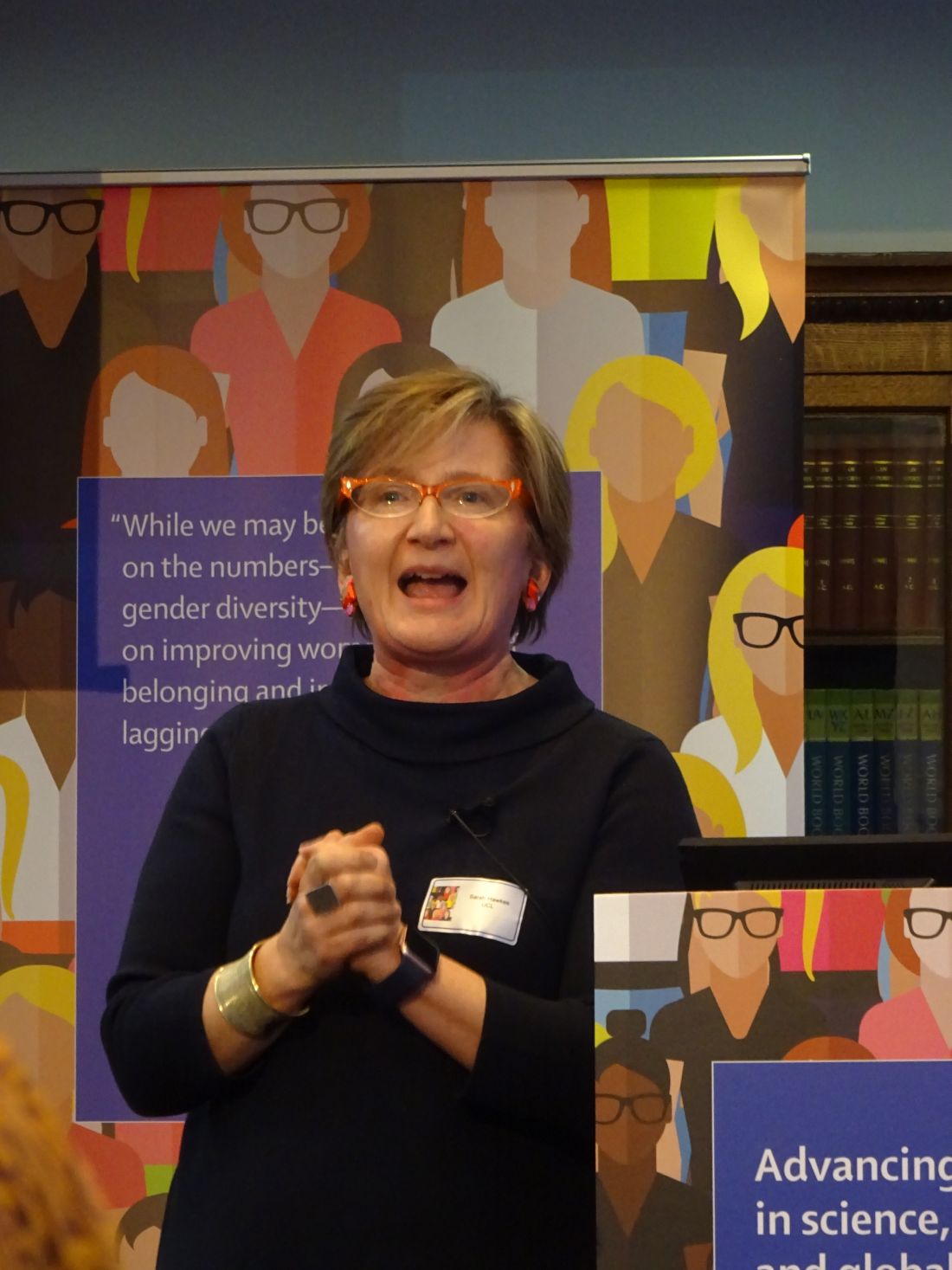
“Gender equality is everyone’s business,” Sarah Hawkes, MBBS, PhD, a professor of global public health at University College London (England), said at the event.
“We’re not talking about women taking over the shop, but women being given an equal opportunity to run the shop. It doesn’t matter where we place ourselves on the gender spectrum as far as advancing equality in science is concerned. What matters is that we all, irrespective of gender, call ourselves feminists.”
For years, women have been “underrepresented in positions of power and leadership, undervalued, and experience discrimination and gender-based violence in scientific and health disciplines across the world,” according to an editorial in the British-based journal (Lancet. 2019;393:493). Such inequalities are compounded and hard to separate from other inequalities, including ethnicity, disability, class, geography, and sexuality.
Despite efforts to readdress the predominantly male culture of medicine, the problem of gender inequality remains “stubbornly persistent,” the editorial said.
“We have spent years being told that the problem lies with us as individuals and that we just need to be better, stronger, more vocal, as women,” Dr. Hawkes observed. “But what really needs to happen is for change to happen in places that hold power.” She further argued: “We don’t need any more individual change; we need organizational and institutional norm change.”
Gender inequality has a long history, and not just in medicine, said British journalist Caroline Criado-Perez OBE, who gave a keynote speech. Ms. Criado-Perez, who is a well-respected feminist campaigner, noted that the world was largely “modeled to fit men.” From architecture to transport, and even crash-test dummies, everything was largely modeled on, or to accommodate, the male rather than the female body.
“I don’t need to tell you that women are 50% more likely to be misdiagnosed following a heart attack” than men. There is no more urgent need to challenge gender inequality than in medicine, Women are dying because of the gender data gap in medicine,” she asserted. “In medical research, in medical education, in medical practice, it needs to be closed as a matter of urgency.”
Original data published in the Advancing Women in Science, Medicine and Global Health special edition of The Lancet found that only 31% of biomedical research papers published in 2016 reported outcomes for both men and women (Lancet. 2019;393:550-9). Reporting of sex differences was somewhat better in clinical or public health-related research papers, at 67% and 69%, respectively. Sex-differences were more likely to be reported if a woman was one of the key authors, Cassidy R. Sugimoto, PhD, associate professor of informatics at Indiana University in Bloomington, and associates, observed in their paper.
That said, women often have to fight to be included as an author on a paper, even when they have done the majority of the work, the event participants highlighted. Women were still less likely than men to be named as the first or last author on a paper, as well as be less likely to receive research funding to enable them to do the work in the first place (Lancet. 2018;393:531-40).
“Was it really you?” was a question sometimes asked of a woman named as a lead or first author, noted Sonia Gandhi, MD, PhD, group leader of the Neurodegeneration Biology Laboratory at the Francis Crick Institute in Cambridge (England). Women network differently to men, Dr. Gandhi observed, and not necessarily in networks that forward careers. Women were also often questioned about their productivity, and regardless of any training on unconscious bias, women were still at a disadvantage if they took a career break to have children.
Women’s credentials and capabilities were often felt to be less respected by male colleagues, and there was talk of being met with microaggressions in the workplace, as in one example given by Nana Odom, MSc, a clinical engineer at the Royal United Hospital Bath (England). She was told “you’re not an engineer, because I have not got a set of screwdrivers and sit at a computer and program.” Such comments can deeply affect a person, Dr. Odom said. “Sometimes I feel that if I don’t go into the workshop and open up a bit of kit that I am not an engineer, but it’s so unconscious, it carries on with you.” These types of stories need to be told so then they can be properly addressed when they do happen, she said.
Female representation is so important, said F. Gigi Osler, MD, head of otolaryngology-head and neck surgery at St. Boniface Hospital in Winnipeg. Dr. Osler is the 2018-2019 president of the Canadian Medical Association, the eighth woman to hold this prestigious position in the organization’s 151 years of operation. She also happens to be the first female surgeon and the first woman of color in the role. “When I stepped into the presidency last August, I thought very long and very hard about how I was going to use my voice and this platform,” Dr. Osler said. “It became very clear to me after I started how important representation was. I can’t tell you how many women, young women, and women of color...have come up to me to say, ‘I’m so excited to have you in this position. I’ve never seen someone who looks like me in that type of leadership position,’ ” she observed.
“As leaders, I think we can advocate for structures and processes,” Dr. Osler added, “I think we set the culture.” Leaders have the responsibility for creating and nurturing and fostering a professional, respectful, and inclusive environment, she said.
“We need more strong leaders, we need more diversity in leadership.” Dr. Osler was keen to point out that greater diversity does not mean only women, but other groups as well. “Look around the room. Who is not here? How can I make it easier for them to get here?”
Another strong female role model at the event was Dame Sally Davies, the Chief Medical Officer for England, a hematologist by training. Not only is she the first female in that role, she will also become the first female Master of Trinity College Cambridge starting in October 2019, a role dominated by men for more than 500 years.
All people, women and men, need to be given fair opportunity, Dame Sally said. Addressing structural issues can help, she said. “We need role models, mentors, and champions,” she said, noting that there were differences between the three. “Mentors give you advice, they get to know you, and they help you think through your issue. Champions may not have time for that, but they know you are good,” and they are putting you forward for opportunities.
“If the system isn’t right, or we are treated badly, we need to call it out,” Dame Sally said. “I do think that we often let things pass that we shouldn’t.” A classic situation is where a woman may suggest something at a meeting and it is ignored, but when a man says the same thing it is taken on board. That kind of behavior needs to stop and be addressed when it happens, by everyone at the table, she said.
Dr. Hawkes observed in her summing up of the day: “Throughout the history of health, change comes about not just through action at the top, but also from action from the bottom up.” She added: “The question is how do we make that change happen?” That’s where the next phase of research needs to take place, she suggested, “we need to actually see, in a very evidence-informed way, what actually works to make and sustain change.”
No financial disclosures were reported by any of the speakers quoted.
SOURCE: Lancet. 2019;393:493–610, e6-e28
FROM A LAUNCH EVENT HELD BY THE LANCET
What makes women leave surgical training?
LONDON – Being unable to take leave and experiencing poor mental health are just two of the reasons uncovered that may help explain why despite having wanted to be a surgeon for many years, a study of women in surgical training has found. The results were presented at a press briefing and published in a special edition of the Lancet.
These factors are in addition to some previously identified, such as the long working hours, fatigue and sleep deprivation, unpredictable lifestyle and its effects on maintaining personal relationships, and the ability to both start and maintain a family life. Then there are the more serious issues of sexism and discrimination, bullying, and sexual harassment and assault that women face in a still male-dominated field that have been noted in prior studies.
“Women are underrepresented in surgery and leave training in higher proportions than men,” study lead Rhea Liang, MBChB, and coauthors wrote (Lancet. 2019;393:541-9). Previous attempts to understand why this is the case “have been confounded by not fully understanding the problem,” they suggested in the briefing. Their research took a more qualitative and feminist approach than other studies, consulting women who had chosen to leave rather than those who continued their surgical training.
Dr. Liang is a consultant general and breast surgeon based at the Gold Coast Hospital and Health Service in Robina, Australia, who personally interviewed women who had decided to leave their surgical training, some as early as 6 months and others up to 4 years after initiation, for reasons other than underperformance.
A “snowball approach” was used to recruit women whereby women who had agreed to participate were asked to refer others. Although only 12 women were interviewed, it’s quality over quantity, Dr. Liang said in a response to a Twitter comment on the study size. “The study is carried out in Australia where about 300 training places are offered across all the specialties annually. About 30% are women; 20% of those women choose to leave. So, if you do the maths, you’ll see that we actually recruited quite well,” she said at the briefing.
According to The Royal College of Practitioners, women made up a very small percentage of consultant surgeons in England in 2016 (11.1%), which didn’t change much by 2018 (12.2%). This is despite a high percentage (58%) of women being accepted onto university courses in medicine and dentistry (58% in 2016). So why so do so few women end up as surgeons?
“Training is a ‘pinch point’ at which women leave surgery,” Tim Dornan, PhD, noted at the launch of the special edition of the Lancet in which the findings appear. Dr. Dornan is professor of medical and interprofessional education at Queen’s University Belfast (Ireland) and one of the coauthors of the research.
This choice to leave surgery deprives society of able surgeons-to-be,” Dr. Dornan said, noting that there was evidence to suggest that women make as good, if not better, surgeons than men. The decision to leave also deprives women of career opportunities and potentially deprives patients of receiving the best surgical care.
“Something very striking about this research is that women who left within an average of 6-18 months after starting surgical training might have wanted to be surgeons from their teenage years, so it seems something happens at that pinch point which makes women to choose to leave.”
Qualitative research is a good way to understand causality in complex social systems, Dr. Dornan explained. Furthermore, “it’s equitable. If you use an open exploratory method, it’s entirely up to the participants to frame the research, it’s not done a priori, and it has the potential for great policy impact.”
Dr. Liang and team found that multiple factors played a role in the decision to leave surgical training, which on their own might be seemingly small, but when stacked on top of each other formed a tower, which was in danger of toppling after a threshold of three or four factors was reached. To exhaustion and lack of opportunity to learn, for example, could be added bullying, and then being denied leave while it is granted for a male colleague for a similar requested reason. The cumulative impact of these factors may all add up to create the impetus to leave.
“Just as a tower of blocks can rebalanced with small adjustments, out study indicates that relatively small interventions (e.g., a cup of tea or a supportive chat) could have been effective in preventing them choosing to leave,” she said.
However, they advocate targeting interventions at all trainees and not just women, to reduce gender differences as focusing on women would be more likely to exaggerate the “otherness” of women further and alienate male trainees. They suggest: “Women might be best helped by interventions that are alert to the possibility of unplanned negative effects, do not unduly focus on gender, and address multiple factors.”
“If you really want to benefit women you should benefit everybody and address the root problem, which is the harsh conditions of training,” Dr. Dornan said. “The prediction would be that, if you do that, then you will actually retain men as well as women.”
The research appears in a special edition of the Lancet that promotes advancing women in science, medicine, and global health.
SOURCE: Liang R et al. Lancet. 2019;393:541-9.
FROM A LAUNCH EVENT HELD BY THE LANCET
Key clinical point: Women leave surgical training for multiple reasons; interventions should focus on surgical training conditions not gender.
Major finding: Six new reasons identified: unavailability of leave; a distinction between valid and invalid reasons for leave; poor mental health; absence of interactions with women in surgery section and other supports; fear of repercussion; lack of pathways for independent and specific support.
Study details: Qualitative study of 12 women who chose to leave their surgical training after 6 months to 4 years.
Disclosures: No financial conflicts of interest were reported.
Source: Liang R et al. Lancet. 2019;393:541-9.
Fund projects, not people to address gender bias in research funding
LONDON – Female investigators are less likely to secure research funding than male investigators, not because their proposed project is of lesser scientific merit, but simply because they are women, according to research published in The Lancet.
Women had a 30% lower chance of success in getting funding for a project than did their male counterparts when the caliber of the principal investigator was considered as an explicit part of the grant application process, with an 8.8% probability of getting funded versus 12.7%, respectively. If the application was considered solely on a project basis, however, the gender bias was less (12.1% vs. 12.9%).
The overall success of grant applications was 15.8% in the analysis, which considered almost 24,000 grant applications from more than 7,000 principal investigators submitted to the Canadian Institutes of Health Research (CIHR) between 2011 and 2016.
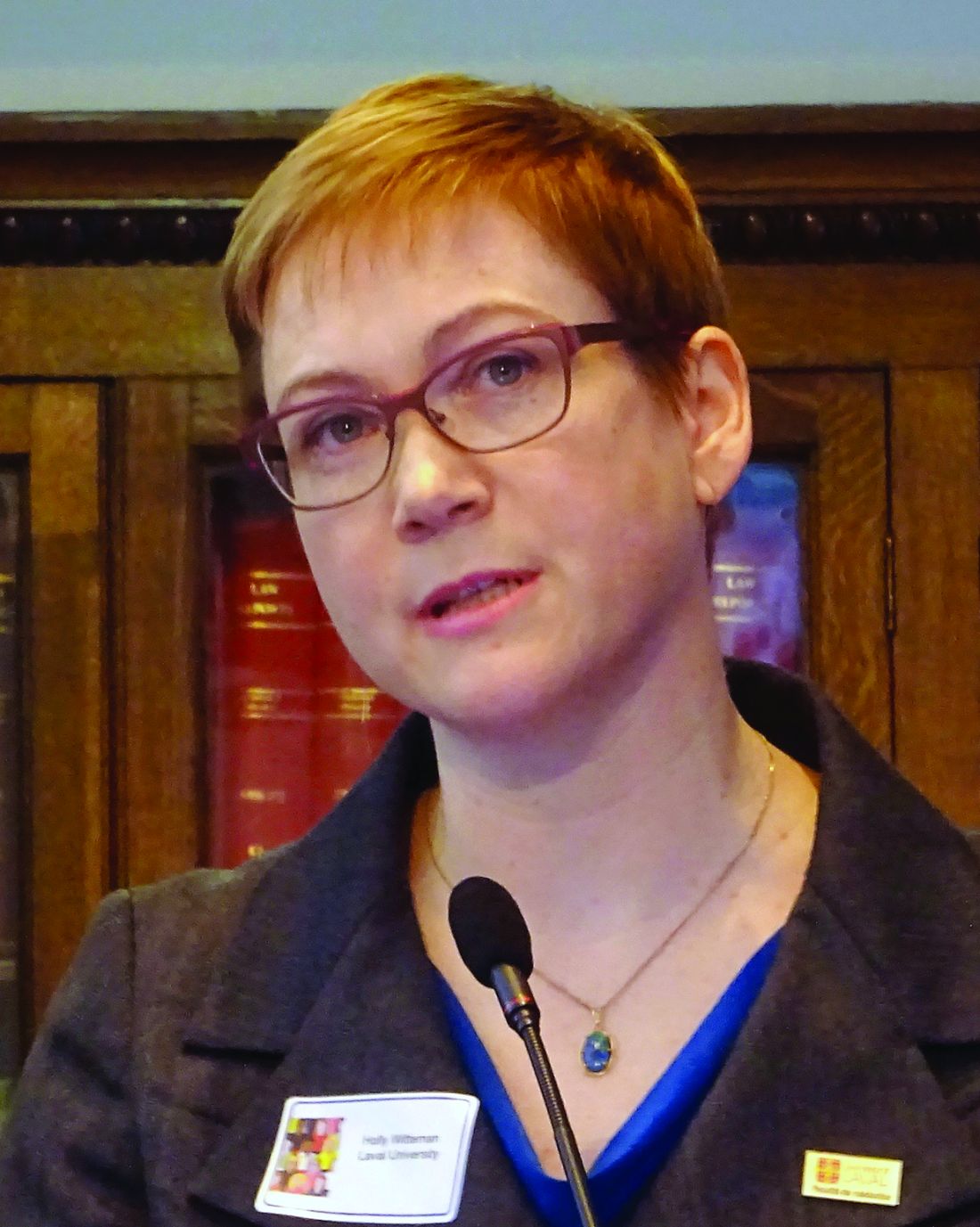
“I see our study as basically one good thwack in a long game of whack-a-mole,” lead study author Holly O. Witteman, PhD, said during an event to launch a special edition of The Lancet focusing on advancing women in science, medicine, and global health.
Dr. Witteman’s research is one of three original articles included in the thematic issue that brings together female authors and commentators to look at gender equity and what needs to be done to address imbalances. The issue is the result of a call for papers that led to more than 300 submissions from more than 40 countries and, according to an editorial from The Lancet, highlights that gender equity in medicine “is not only a matter of justice and rights, it is crucial for producing the best research and providing the best care to patients.”
That there are discrepancies in research funding awarded to female and male investigators has been known for years, Dr. Witteman, associate professor of family and emergency medicine at Laval University, Quebec City, said at the London press conference. To learn how and why, a “quasiexperimental” approach was used to find out what factors might be influencing the gender gap.
“Women are scored lower for competence compared to men with the same publication record,” she said. It’s not that they publish less or do easier research, or that the quality is lower, they are just viewed less favorably overall throughout their careers. Even when you control for confounding factors, “they still don’t advance as quickly,” she said.
“It had been documented for a while that, overall, women tend to get less grant funding and there hasn’t been any evidence to show either way if maybe women’s grant applications weren’t as good,” Dr. Witteman explained.
In 2014, the CIHR changed the way it funded research projects, creating a “natural experiment.” Two new grant application programs were put in place which largely differed by whether or not an explicit review of the principal investigator and their ability to conduct the research was included.
Adjusting for age and type of research, Dr. Witteman and her coauthors found that there was little difference in the success of women in securing research funding when their grant applications were judged solely on a scientific basis; however, when the focus was placed on the principal investigator, women were disadvantaged.
Dr. Witteman said that “this provides robust evidence in support of the idea that women write equally good grant applications but aren’t evaluated as being equally good scientists.”
So how to redress the balance? Dr. Witteman suggested that one way was for funders to collect robust evidence on the success of grant applications and be transparent who is getting funded and how much funding is being awarded. Institutions should invest in and support young investigators, distributing power and flattening traditionally male-led hierarchies. Salaries should be aligned and research support evened out, she said.
Investigators themselves also have a role to play to do the best possible work and try to change the system. “Advocate for others,” she said. That included advocating for others in groups that you may not be part of – which can be easier in some respects than advocating for a group that you are in.
“Funders should evaluate projects, not people,” Jennifer L. Raymond, PhD, and Miriam B. Goodman, PhD, both professors at Stanford (Calif.) University wrote in a comment in The Lancet special issue. They suggested that people-based funding had been gaining popularity but that funders would be better off funding by project to achieve scientific and clinical goals. “Assess the investigator only after double-blind review of the proposed research is complete,” they suggested. “Reduce the assessment of the investigator to a binary judgment of whether or not the investigator has the expertise and resources needed do the proposed research.”
During a panel discussion at The Lancet event, Cassidy R. Sugimoto, PhD, associate professor of informatics at Indiana University in Bloomington and a program director for the Science and Innovation Policy Program at the National Science Foundation (NSF) observed that data on gender equality in research funding were already being collected and will be used to determine how best to adjust funding policies.
“Looking from the 1980s to the present, women make up shy of 20% of the funds given by the National Science Foundation,” Dr. Sugimoto said. “That’s improved over time, and it’s at 28% currently, which is less than their authorship.”
Tammy Clifford, PhD, vice president of research programs at the CIHR observed that data collection was “a critically important step, but of course that’s not the only step,” she said. “We need to look at and analyze the data regularly, and then when you see things that are not on track, you make changes.”
One of the changes the CIHR has made is to train people who are reviewing grant applications on factors that may unconsciously affect their decisions. “There are things to be done, and I don’t think we are quite there yet, but we are committed to continually looking at those data, to making the changes that are required.”
Representing the Wellcome Trust, Ed Whiting, director of policy and chief of staff, said that the funding of projects led by female investigators was moving in the right direction. He noted that there was still a lower rate of applications from women for senior award levels, but that the panels that decide upon the funding were moving toward equal gender representation. The aim was to get to a 50/50 female to male ratio on the panels by 2020, he said; it is was at 46%-52% in 2018.
Dr. Witteman and all other commentators had no financial disclosures.
SOURCE: Witteman HO et al. Lancet. 2019. doi: 10.1016/S0140-6736(18)32611-4
FROM A LAUNCH EVENT HELD BY THE LANCET
Key clinical point: Funding bodies should focus on the science of a research project not on who is conducting the research.
Major finding: Between 2011 and 2016, 8.8% of projects proposed by female researchers and 12.7% of those proposed by male researchers were funded.
Study details: Analysis of nearly 24,000 grant applications from more than 7,000 principal investigators submitted to the Canadian Institutes of Health Research during 2011-2016.
Disclosures: The research was unfunded. Dr. Witteman and all other commentators had no financial disclosures.
Source: Witteman HO et al. Lancet. 2019. doi: 10.1016/S0140-6736(18)32611-4.

