User login
Oral GLP-1 receptor agonist pioneered in T2DM trial
BERLIN – An investigational oral formulation of the glucagonlike peptide–1 receptor agonist (GLP-1 RA) semaglutide reduced glycated hemoglobin (HbA1c) to a greater extent than did placebo at all doses tested in patients with type 2 diabetes mellitus (T2DM) in the phase 3a PIONEER 1 trial.
The estimated mean change in HbA1c from baseline to week 26 – the primary endpoint – using an on-treatment analysis was –0.8% with a once-daily dose of 3 mg, –1.3% with a once-daily dose of 7 mg, and –1.5% with a once-daily dose of 14 mg. The corresponding value for placebo was –0.1%, with all comparisons statistically significant (P less than .001).
The on-treatment analysis evaluated treatment effects for all randomized patients (n = 703) and assumed that all subjects remained on-treatment and excluded the effect of any rescue medication. Results for an intention-to-treat (ITT) analysis provided similar results, however, with estimated mean changes in HbA1c of –0.9%, –1.2%, and –1.4% for the three respective semaglutide doses and –0.3% for placebo.
“There was a very nice dose-dependent decrease in HbA1c, which was superior to placebo for all doses of semaglutide” said study investigator Martin Haluzík, MD, at the annual meeting of the European Association for the Study of Diabetes.
“I think it’s quite important to stress the magnitude of the decrease of HbA1c, because with the highest dose it was –1.5% from a baseline of 8%, which I believe is something that hasn’t ever really been seen with any other oral antidiabetic medication,” added Dr. Haluzík, professor of internal medicine at the 1st Faculty of Medicine at Charles University and deputy head of the Institute for Clinical and Experimental Medicine, both in Prague.
Currently, GLP-1 RAs are available only in a subcutaneous formulation, Dr. Haluzík reminded his audience, adding that oral semaglutide was the first GLP-1 RA to be developed in a tablet formulation and was in the late stages of clinical development.
PIONEER 1 is the first of 10 phase 3a trials with oral semaglutide to be reported. “Additional studies, across the full spectrum of diabetes care, in special populations, comparing it with active comparators with varied trial duration, have been completed or will be completed in 2018,” said coinvestigator for the study Vanita Aroda, MD, during a separate presentation at a dedicated symposium on the PIONEER program.
Dr. Aroda, the director of the diabetes research program at Brigham and Women’s Hospital in Boston, observed that oral semaglutide was being evaluated from early care as monotherapy in the PIONEER 1 study. The other trials, such as PIONEER 2, PIONEER 3, PIONEER 4, and PIONEER 7 were looking at oral semaglutide in combination with oral antidiabetic agents versus various active comparators; PIONEER 5 and PIONEER 6 were in special populations; and PIONEER 8 was looking at its use on top of basal insulin. Two further trials are also part of the study program.
“I think this is the first time that we actually have completed data of an entire program, including cardiovascular data, all within the same year,” Dr. Aroda said. “All of the studies are in the process of data analysis or data reporting.”
PIONEER 1 was a multicenter, randomized, double-blind, placebo-controlled trial examining efficacy and safety of semaglutide versus placebo in 703 adults with drug-naive T2DM who were being treated with diet and exercise only.
Three doses of oral semaglutide – 3 mg, 7 mg, and 14 mg – were assessed and compared with placebo. There was a fixed 4-week dose escalation period, with all patients starting treatment with 3 mg of semaglutide and then increasing to 7 mg by week 4 and 14 mg by week 8. This was to try to reduce the risk of gastrointestinal side effects, which are known to occur with GLP-1 RAs.
Secondary outcome measures were change in body weight, fasting plasma glucose, HbA1c below a target of 7% (53 mmol/mol), which were all measured from baseline to week 26; adverse events, including severe and blood glucose–confirmed symptomatic hypoglycemic episodes, were assessed out to week 31.
The average age of patients in the trial was around 55 years, around half were female, and the starting HbA1c was approximately 8%.The mean body weight ranged from 86.9 kg to 89 kg in the different treatment groups, with a similar body mass index of about 32 kg/m2.
Clinically meaningful weight loss, compared with placebo, was only achieved with the highest dose of oral semaglutide, with a –4.1 kg reduction versus –1.5 kg for placebo from baseline to week 26 (P less than .001, on-treatment analysis). Reductions in body weight for the 3-mg and 7-mg oral semaglutide doses were a respective –1.7 kg and –2.5 kg. ITT results were again similar, with weight losses of –1.5, –2.3, and –3.7 kg for the 3-, 7-, and 14-mg doses of oral semaglutide, respectively, and –1.4 kg for placebo.
More patients treated with oral semaglutide 3, 7, or 14 mg versus placebo achieved an HbA1c of below 7% (59.1%, 71.9%, and 80.3%, respectively, vs. 33.8%) or a body weight loss of 5% or more (21.3%, 28.7%, and 44.3% vs. 15.7%).
Furthermore, more patients treated with oral semaglutide achieved an HbA1c of or below 7% without hypoglycemic episodes or body weight gain than did those given placebo. There were also more patients who achieved an HbA1c reduction of at least 1% and a weight loss of 3% or higher.
“Oral semaglutide demonstrated a safety and tolerability profile consistent with that of [injectable] GLP-1 RAs,” Dr. Haluzík reported. Adverse events were seen in 57.7%, 53.1%, and 56.6%of patients treated with 3, 7, and 14 mg of oral semaglutide and 55.6% of those treated with placebo.
The most common adverse events seen with oral semaglutide affected the gastrointestinal tract, with nausea affecting 8%, 5.1%, and 16% of 3, 7, and 14 mg–treated patients versus 5.6% of placebo-treated patients. Vomiting affected a respective 2.9%, 4.6%, 6.9%, and 2.1%, and diarrhea a respective 8.6%, 5.1%, 5.1%, and 2.2%.
Severe or blood glucose–confirmed, symptomatic hypoglycemia was reported in 2.9%, 1.1%, and 0.6%of those treated with 3, 7, 14 mg of oral semaglutide and 0.6% of placebo-treated patients.
PIONEER 1 represents a “step change in GLP-1 receptor agonist therapy”, said Cliff Bailey, MD, who discussed the trial aa the EASD’s independent commentator during a symposium on the PIONEER program. These data are “leading to a new delivery route for GLP-1 receptor agonists, from injection to oral,” and “this can be done with good metabolic efficacy, with substantial reductions in A1c and body weight, and with a safety profile that’s comparable to the subcutaneous injection.”
Dr. Bailey, who is professor of clinical science at Aston University in Birmingham, England, noted, however, the oral dosing of semaglutide “requires patient commitment because it needs to be taken before breakfast time, and it may also, to some extent, affect the timing of some of the other medications.”
The study was sponsored by Novo Nordisk. Dr. Haluzík disclosed acting as a consultant to AstraZeneca, Eli Lilly, Johnson & Johnson, Mundipharma, Novatin, Novo Nordisk, and Sanofi Aventis. Dr. Aroda was an investigator in the PIONEER 1 study and disclosed acting as a consultant to Novo Nordisk; she also disclosed relationships with multiple other pharmaceutical companies, including AstraZeneca, BMS, Calbra, Eisai, Elcelyx Therapeutics, Janssen, and Sanofi Aventis. Dr. Bailey acknowledged attending advisory boards, undertaking ad hoc consultancy work, and receiving research and travel support from several pharmaceutical companies that included Novo Nordisk.
SOURCES: Haluzík M et al. EASD 2018, Abstract 38; Aroda V. EASD 2018, Session S18 - PIONEER Trial; Bailey C. EASD 2018, Session S18 - PIONEER Trial.
REPORTING FROM EASD 2018
Key clinical point: A novel oral formulation of semaglutide proved better than placebo for diabetes control and weight loss in patients with T2DM.
Major finding: Mean change in hemoglobin A1c (baseline to week 26) – the primary endpoint – using an on-treatment analysis was –0.8% with a once-daily dose of 3 mg, –1.3% with a once-daily dose of 7 mg, and –1.5% with a once-daily dose of 14 mg. The corresponding value for placebo was –0.1%.
Study details: Phase 3a, multicenter, randomized, double-blind, placebo-controlled trial examining efficacy and safety of semaglutide versus placebo in 703 individuals with T2DM being treated with diet and exercise only.
Disclosures: The study was sponsored by Novo Nordisk. Dr. Haluzík disclosed acting as a consultant to AstraZeneca, Eli Lilly, Johnson & Johnson, Mundipharma, Novatin, Novo Nordisk, and Sanofi Aventis. Dr. Aroda was an investigator in the PIONEER 1 study and disclosed acting as a consultant to Novo Nordisk. She also disclosed relationships with multiple other pharmaceutical companies, including AstraZeneca, BMS, Calbra, Eisai, Elcelyx Therapeutics, Janssen, and Sanofi Aventis. Dr. Bailey acknowledged attending advisory boards of, undertaking ad hoc consultancy work for, and receiving research and travel support from several pharmaceutical companies that included Novo Nordisk.
Source: Haluzík M et al. EASD 2018, Abstract 38; Aroda V. EASD 2018, Session S18 – PIONEER Trial; Bailey C. EASD 2018, Session S18 – PIONEER Trial.
Real-world data reveal long-lasting effects achieved with RRMS treatments
BERLIN – Real-world data from six postmarketing surveillance studies suggest that currently available disease-modifying treatments (DMTs) for relapsing-remitting multiple sclerosis (RRMS) have long-lasting effects that are matched by reasonable tolerability.

Long-term efficacy and safety data on natalizumab (Tysabri), fingolimod (Gilenya), alemtuzumab (Lemtrada), dimethyl fumarate (Tecfidera), and teriflunomide (Aubagio) from four Swedish studies, one French study, and one international study were reported during a poster session on long-term treatment monitoring at the annual congress of the European Committee for Treatment and Research in Multiple Sclerosis (ECTRIMS).
The IMSE 1 study with natalizumab
The Immunomodulation and Multiple Sclerosis Epidemiology (IMSE) studies are Swedish postmarketing surveillance studies that were started with the launch of various DMTs in Sweden: natalizumab since 2006 (IMSE 1), fingolimod in 2015 (IMSE 2), alemtuzumab in 2014 (IMSE 3), and dimethyl fumarate in 2014 (IMSE 5).
“Postmarketing surveillance is important for determination of long-term safety and effectiveness in a real-world setting,” Stina Kågström and her associates observed in their poster reporting some findings of the IMSE 1 study with natalizumab (Mult Scler. 2018;24[S2]:699-700, Abstract P1232).
Ms. Kågström of the department of clinical neuroscience at the Karolinska Institute in Stockholm and her colleagues reported that data on 3,108 patients who were seen at 54 Swedish clinics had been collated via the nationwide Swedish Quality Registry for Neurological Care (NEUROreg). NEUROreg started out as an MS register but has since widened its remit to include other neurologic diagnoses.
For the IMSE 1 study, prospectively recorded data regarding natalizumab treatment, adverse events, JC-virus (JCV) status and clinical effectiveness measures were obtained from NEUROreg for 2,225 women and 883 men. Just over one-third (37%, n = 1,150) were still receiving natalizumab at the time of the analysis.
The mean age at which natalizumab was started was 39 years, with treatment primarily given for RRMS (81% of patients) and less often for secondary progressive multiple sclerosis (SPMS, 15%) and rarely for other types of progressive MS. The mean treatment duration was just under 4 years (47.6 months).
JCV testing was introduced in 2011 in Sweden, and this “has led to fewer treated JCV-positive patients,” the IMSE 1 study investigators reported. “This likely explains a reduced incidence of PML [progressive multifocal leukoencephalopathy],” they suggested. There were nine PML cases diagnosed in Sweden from 2008 to the data cut-off point in 2018, one of which was fatal.
JCV status from 2011 onward was available for 1,269 patients, of whom 39% were JCV positive and 61% were JCV negative. The overall drug survival rate was 72% for JCV-negative and 14% for JCV-positive patients. Improved health status was seen, as measured by the Expanded Disability Status Scale (EDSS), the Multiple Sclerosis Severity Score (MSSS), and the physical and psychological Multiple Sclerosis Impact Scale–29 (MSIS-29) components.
A total of 644 of 1,269 patients discontinued treatment with natalizumab at some point, of whom 67% discontinued because of being JCV positive. The main reason for discontinuation in JCV-negative patients was pregnancy or planning a pregnancy (38%), with lack of effect (10%) and adverse events (11%) as other key reasons for stopping natalizumab.
Ms. Kågström and her associates concluded that natalizumab was “generally well tolerated with sustained effectiveness.”
The IMSE 2 study with fingolimod
Data on the long-term safety and efficacy of fingolimod were reported from the IMSE 2 study (Mult Scler. 2018;24[S2]:696-7, Abstract P1228). Lead author Anna Fält, also of the Karolinska Institute, and her associates analyzed data for 1,634 patients who had been treated with fingolimod from June 2015 to September 2018.
Most patients were older than 30 years (79%), and those aged 30 and older were predominantly female (69%), had an RRMS diagnosis (88%), and been treated for a mean of about 3 years (37 months). A total of 829 were being treated with fingolimod at the time of the analysis, with 844 having discontinued treatment at some point. The main reason for discontinuing treatment with fingolimod was a lack of effect (42% of cases) or an adverse effect (34%). The IMSE 2 study authors reported in their abstract that most patients were switched to rituximab after discontinuing fingolimod.
The number of relapses per 1,000 patient-years was reduced by fingolimod treatment from 280 to 82, comparing before and during treatment for all age groups studied. Relapse rate dropped from 694 per 1,000 patient-years before treatment to 138 during treatment in patients aged 20 years or younger, from 454 to 122 in those aged 21-30 years, and from 257 to 72 in those older than 31 years.
After 1 year of treatment, improvements were seen in the health status of patients as measured by various scales, including the EDSS, MSSS, MSIS-29 Physical, and MSIS-29 Psychological. When the researchers analyzed data by age groups, significant improvements were seen in patients aged 21-30 years and older than 30 years.
Ninety nonserious and 62 serious adverse events were reported in fingolimod-treated patients during the time of analysis. Of the latter, 13 serious adverse events involved cardiac disorders, 12 neoplasms, and 10 infections and infestations.
Overall, the IMSE 2 study investigators said that fingolimod was generally tolerable and reduced disease activity in MS.
French experience with fingolimod: The VIRGILE study
Real-world data on the long-term safety and efficacy of fingolimod in France from the VIRGILE study were reported by Christine Lebrun-Frenay, MD, PhD, and her associates (Mult Scler. 2018;24[S2]:698-9, Abstract P1231).
Dr. Lebrun-Frenay of Pasteur 2 Hospital in Nice and her coauthors noted that VIRGILE study included patients starting treatment with fingolimod between January 2014 and February 2016. A total of 1,047 patients were included, and another 330 patients treated with natalizumab were included at the behest of the French health authorities.
The annualized relapse rate after 2 years of follow-up was 0.30 in the fingolimod group. Dr. Lebrun-Frenay and her colleagues noted: “The 3-year data from this interim analysis provide evidence for sustained efficacy of fingolimod.” Indeed, they report that almost 60% of patients did not relapse and 64% had no worsening of disease. On average, EDSS was stable during the 3-year follow-up period.
“Safety and tolerability profiles of fingolimod were in line with previous clinical experience, with lymphopenia being the most frequent AE [adverse event] reported,” they added.
The IMSE 3 study with alemtuzumab
Long-term experience with alemtuzumab as a treatment for RRMS in the real-world setting is more limited as it only became available for use for this indication in 2014, but some insight is provided by the results of the IMSE 3 study (Mult Scler. 2018;24[S2]:706-7, Abstract P1240).
In total, there were 113 patients treated with alemtuzumab; the vast majority (94%) had RRMS and were aged a mean of 34 years at the start of treatment. Treatment was for more than 12 months in 101 patients, more than 24 months in 86 patients, and more than 36 months in 36 patients.
“In patients treated for at least 12 months, significant improvements were seen in several clinical parameters,” Dr. Fält and her associates observed in their poster at ECTRIMS. The mean baseline and 12-month values for the EDSS were 2.0 and 1.6, and for the MSSS they were 3.46 and 2.61. The mean baseline and 12-month values for the MSIS-29 Psychological subscale were 35.1 and 30.8, respectively, and for MSIS-29 Physical they were 22.7 and 17.7.
Overall, there were 14 nonserious and 11 serious adverse events, the most common of which were infections and infestations, metabolism and nutrition disorders, and immune system disorders.
“A longer follow-up period is needed to assess the real-world effectiveness and safety of alemtuzumab,” the IMSE 3 study authors noted.
The IMSE 5 study with dimethyl fumarate
Similarly, the authors of the IMSE 5 study (Mult Scler. 2018;24[S2]:701-2, Abstract 1234) concluded that a longer follow-up period is need to assess the real-world effectiveness of dimethyl fumarate. Selin Safer Demirbüker, also of the Karolinska Institute, and her associates looked at data on 2,108 patients treated with dimethyl fumarate between March 2014 and April 2018, of whom 1,150 were still receiving treatment at the time of their assessment.
The mean age of patients at the start of treatment was 41 years, 91% had RRMS, and 73% were female. The mean treatment duration was 22.3 months. The majority of patients (n = 867) had been previously treated with interferon and glatiramer acetate (Copaxone) prior to dimethyl fumarate, with 538 being naive to treatment.
“Dimethyl fumarate seems to have a positive effect for patients remaining on treatment,” wrote Ms. Safer Demirbüker and her colleagues. The overall 1-year drug survival reported in their abstract was 74%. Their poster showed a lower 2-year drug survival rate of 63.5% for men and 56.4% for women.
“Swedish patients show cognitive, psychological, and physical benefits after 2 or more years of treatment,” the IMSE 5 study authors further noted. Mean EDSS, MSSS, and MSIS-29 Psychological values all fell from baseline to 2 years.
Overall, 958 (47%) of patients discontinued treatment with dimethyl fumarate at some point, primarily (in 52% of cases) because of adverse events or lack of effect (29% of cases). Most patients (39%) switched to rituximab (15% had no new treatment registered), but 35% of patients continued treatment for 3 or more years.
Twelve-year follow-up of teriflunomide shows continued efficacy, safety
Mark Freedman, MD, of the University of Ottawa and the Ottawa Hospital Research Institute and his associates reported long-term follow-up data on the efficacy and safety of teriflunomide (Aubagio) in relapsing forms of MS (Mult Scler. 2018;24[S2]:700-1, Abstract P1233). After up to 12 years’ follow up, teriflunomide 14 mg was associated with an overall annualized relapse rate of 0.228.
Yearly annualized relapse rates were “low and stable,” Dr. Freedman and his coauthors from the United States, Spain, Italy, France, Germany, England, the Republic of Korea, and Australia noted in their poster.
“As of August 2018, over 93,000 patients were being treated with teriflunomide,” the authors stated. This represented a real-world exposure of approximately 186,000 patient-years up to December 2017, they added.
For the analysis, data from one phase 2 study and three phase 3 studies (TEMSO, TOWER, and TENERE) and their long-term extension studies were pooled. In all, there were 1,696 patients treated with 14 mg of teriflunomide in these studies.
Annualized relapse rates ranged from 0.321 in the first year of follow-up in the studies to 0.080 by the 12th year. The proportions of patients remaining relapse free “were high and stable (ranging from 0.75 in year 1 to 0.93 in years 8 and 9).” EDSS scores were 2.57 at baseline and 2.27 at year 12.
Importantly, no new safety signals were reported, Dr. Freedman and his colleagues wrote, adding that most adverse events were mild to moderate in severity.
Taken together, “these data demonstrate the long-term efficacy and safety of teriflunomide,” they concluded.
Study and author disclosures
The teriflunomide analysis was supported by Sanofi. Dr. Freedman disclosed receiving research or educational grant support from Bayer and Genzyme; honoraria/consulting fees from Bayer, Biogen, EMD Canada, Novartis, Sanofi, and Teva; and membership on company advisory boards/boards of directors/other similar groups for Bayer, Biogen, Chugai, Merck Serono, Novartis, Opexa Therapeutics, Sanofi, and Teva.
The IMSE 1 and 5 studies were supported by Biogen and the IMSE 2 and 3 studies by Novartis. The lead study authors for the IMSE studies – Dr. Kågström, Dr. Fält, and Dr. Safer Demirbüker – had nothing personal to disclose. Other authors included employees of the sponsoring companies or those who had received research funding or honoraria for consultancy work from the companies.
The VIRGILE study was supported by Novartis Pharma AG, Switzerland. Dr. Lebrun-Frenay disclosed receiving consultancy fees from Merck, Novartis, Biogen, MedDay, Roche, Teva, and Genzyme. Coauthors included Novartis employees.
BERLIN – Real-world data from six postmarketing surveillance studies suggest that currently available disease-modifying treatments (DMTs) for relapsing-remitting multiple sclerosis (RRMS) have long-lasting effects that are matched by reasonable tolerability.
Long-term efficacy and safety data on natalizumab (Tysabri), fingolimod (Gilenya), alemtuzumab (Lemtrada), dimethyl fumarate (Tecfidera), and teriflunomide (Aubagio) from four Swedish studies, one French study, and one international study were reported during a poster session on long-term treatment monitoring at the annual congress of the European Committee for Treatment and Research in Multiple Sclerosis (ECTRIMS).
The IMSE 1 study with natalizumab
The Immunomodulation and Multiple Sclerosis Epidemiology (IMSE) studies are Swedish postmarketing surveillance studies that were started with the launch of various DMTs in Sweden: natalizumab since 2006 (IMSE 1), fingolimod in 2015 (IMSE 2), alemtuzumab in 2014 (IMSE 3), and dimethyl fumarate in 2014 (IMSE 5).
“Postmarketing surveillance is important for determination of long-term safety and effectiveness in a real-world setting,” Stina Kågström and her associates observed in their poster reporting some findings of the IMSE 1 study with natalizumab (Mult Scler. 2018;24[S2]:699-700, Abstract P1232).
Ms. Kågström of the department of clinical neuroscience at the Karolinska Institute in Stockholm and her colleagues reported that data on 3,108 patients who were seen at 54 Swedish clinics had been collated via the nationwide Swedish Quality Registry for Neurological Care (NEUROreg). NEUROreg started out as an MS register but has since widened its remit to include other neurologic diagnoses.
For the IMSE 1 study, prospectively recorded data regarding natalizumab treatment, adverse events, JC-virus (JCV) status and clinical effectiveness measures were obtained from NEUROreg for 2,225 women and 883 men. Just over one-third (37%, n = 1,150) were still receiving natalizumab at the time of the analysis.
The mean age at which natalizumab was started was 39 years, with treatment primarily given for RRMS (81% of patients) and less often for secondary progressive multiple sclerosis (SPMS, 15%) and rarely for other types of progressive MS. The mean treatment duration was just under 4 years (47.6 months).
JCV testing was introduced in 2011 in Sweden, and this “has led to fewer treated JCV-positive patients,” the IMSE 1 study investigators reported. “This likely explains a reduced incidence of PML [progressive multifocal leukoencephalopathy],” they suggested. There were nine PML cases diagnosed in Sweden from 2008 to the data cut-off point in 2018, one of which was fatal.
JCV status from 2011 onward was available for 1,269 patients, of whom 39% were JCV positive and 61% were JCV negative. The overall drug survival rate was 72% for JCV-negative and 14% for JCV-positive patients. Improved health status was seen, as measured by the Expanded Disability Status Scale (EDSS), the Multiple Sclerosis Severity Score (MSSS), and the physical and psychological Multiple Sclerosis Impact Scale–29 (MSIS-29) components.
A total of 644 of 1,269 patients discontinued treatment with natalizumab at some point, of whom 67% discontinued because of being JCV positive. The main reason for discontinuation in JCV-negative patients was pregnancy or planning a pregnancy (38%), with lack of effect (10%) and adverse events (11%) as other key reasons for stopping natalizumab.
Ms. Kågström and her associates concluded that natalizumab was “generally well tolerated with sustained effectiveness.”
The IMSE 2 study with fingolimod
Data on the long-term safety and efficacy of fingolimod were reported from the IMSE 2 study (Mult Scler. 2018;24[S2]:696-7, Abstract P1228). Lead author Anna Fält, also of the Karolinska Institute, and her associates analyzed data for 1,634 patients who had been treated with fingolimod from June 2015 to September 2018.
Most patients were older than 30 years (79%), and those aged 30 and older were predominantly female (69%), had an RRMS diagnosis (88%), and been treated for a mean of about 3 years (37 months). A total of 829 were being treated with fingolimod at the time of the analysis, with 844 having discontinued treatment at some point. The main reason for discontinuing treatment with fingolimod was a lack of effect (42% of cases) or an adverse effect (34%). The IMSE 2 study authors reported in their abstract that most patients were switched to rituximab after discontinuing fingolimod.
The number of relapses per 1,000 patient-years was reduced by fingolimod treatment from 280 to 82, comparing before and during treatment for all age groups studied. Relapse rate dropped from 694 per 1,000 patient-years before treatment to 138 during treatment in patients aged 20 years or younger, from 454 to 122 in those aged 21-30 years, and from 257 to 72 in those older than 31 years.
After 1 year of treatment, improvements were seen in the health status of patients as measured by various scales, including the EDSS, MSSS, MSIS-29 Physical, and MSIS-29 Psychological. When the researchers analyzed data by age groups, significant improvements were seen in patients aged 21-30 years and older than 30 years.
Ninety nonserious and 62 serious adverse events were reported in fingolimod-treated patients during the time of analysis. Of the latter, 13 serious adverse events involved cardiac disorders, 12 neoplasms, and 10 infections and infestations.
Overall, the IMSE 2 study investigators said that fingolimod was generally tolerable and reduced disease activity in MS.
French experience with fingolimod: The VIRGILE study
Real-world data on the long-term safety and efficacy of fingolimod in France from the VIRGILE study were reported by Christine Lebrun-Frenay, MD, PhD, and her associates (Mult Scler. 2018;24[S2]:698-9, Abstract P1231).
Dr. Lebrun-Frenay of Pasteur 2 Hospital in Nice and her coauthors noted that VIRGILE study included patients starting treatment with fingolimod between January 2014 and February 2016. A total of 1,047 patients were included, and another 330 patients treated with natalizumab were included at the behest of the French health authorities.
The annualized relapse rate after 2 years of follow-up was 0.30 in the fingolimod group. Dr. Lebrun-Frenay and her colleagues noted: “The 3-year data from this interim analysis provide evidence for sustained efficacy of fingolimod.” Indeed, they report that almost 60% of patients did not relapse and 64% had no worsening of disease. On average, EDSS was stable during the 3-year follow-up period.
“Safety and tolerability profiles of fingolimod were in line with previous clinical experience, with lymphopenia being the most frequent AE [adverse event] reported,” they added.
The IMSE 3 study with alemtuzumab
Long-term experience with alemtuzumab as a treatment for RRMS in the real-world setting is more limited as it only became available for use for this indication in 2014, but some insight is provided by the results of the IMSE 3 study (Mult Scler. 2018;24[S2]:706-7, Abstract P1240).
In total, there were 113 patients treated with alemtuzumab; the vast majority (94%) had RRMS and were aged a mean of 34 years at the start of treatment. Treatment was for more than 12 months in 101 patients, more than 24 months in 86 patients, and more than 36 months in 36 patients.
“In patients treated for at least 12 months, significant improvements were seen in several clinical parameters,” Dr. Fält and her associates observed in their poster at ECTRIMS. The mean baseline and 12-month values for the EDSS were 2.0 and 1.6, and for the MSSS they were 3.46 and 2.61. The mean baseline and 12-month values for the MSIS-29 Psychological subscale were 35.1 and 30.8, respectively, and for MSIS-29 Physical they were 22.7 and 17.7.
Overall, there were 14 nonserious and 11 serious adverse events, the most common of which were infections and infestations, metabolism and nutrition disorders, and immune system disorders.
“A longer follow-up period is needed to assess the real-world effectiveness and safety of alemtuzumab,” the IMSE 3 study authors noted.
The IMSE 5 study with dimethyl fumarate
Similarly, the authors of the IMSE 5 study (Mult Scler. 2018;24[S2]:701-2, Abstract 1234) concluded that a longer follow-up period is need to assess the real-world effectiveness of dimethyl fumarate. Selin Safer Demirbüker, also of the Karolinska Institute, and her associates looked at data on 2,108 patients treated with dimethyl fumarate between March 2014 and April 2018, of whom 1,150 were still receiving treatment at the time of their assessment.
The mean age of patients at the start of treatment was 41 years, 91% had RRMS, and 73% were female. The mean treatment duration was 22.3 months. The majority of patients (n = 867) had been previously treated with interferon and glatiramer acetate (Copaxone) prior to dimethyl fumarate, with 538 being naive to treatment.
“Dimethyl fumarate seems to have a positive effect for patients remaining on treatment,” wrote Ms. Safer Demirbüker and her colleagues. The overall 1-year drug survival reported in their abstract was 74%. Their poster showed a lower 2-year drug survival rate of 63.5% for men and 56.4% for women.
“Swedish patients show cognitive, psychological, and physical benefits after 2 or more years of treatment,” the IMSE 5 study authors further noted. Mean EDSS, MSSS, and MSIS-29 Psychological values all fell from baseline to 2 years.
Overall, 958 (47%) of patients discontinued treatment with dimethyl fumarate at some point, primarily (in 52% of cases) because of adverse events or lack of effect (29% of cases). Most patients (39%) switched to rituximab (15% had no new treatment registered), but 35% of patients continued treatment for 3 or more years.
Twelve-year follow-up of teriflunomide shows continued efficacy, safety
Mark Freedman, MD, of the University of Ottawa and the Ottawa Hospital Research Institute and his associates reported long-term follow-up data on the efficacy and safety of teriflunomide (Aubagio) in relapsing forms of MS (Mult Scler. 2018;24[S2]:700-1, Abstract P1233). After up to 12 years’ follow up, teriflunomide 14 mg was associated with an overall annualized relapse rate of 0.228.
Yearly annualized relapse rates were “low and stable,” Dr. Freedman and his coauthors from the United States, Spain, Italy, France, Germany, England, the Republic of Korea, and Australia noted in their poster.
“As of August 2018, over 93,000 patients were being treated with teriflunomide,” the authors stated. This represented a real-world exposure of approximately 186,000 patient-years up to December 2017, they added.
For the analysis, data from one phase 2 study and three phase 3 studies (TEMSO, TOWER, and TENERE) and their long-term extension studies were pooled. In all, there were 1,696 patients treated with 14 mg of teriflunomide in these studies.
Annualized relapse rates ranged from 0.321 in the first year of follow-up in the studies to 0.080 by the 12th year. The proportions of patients remaining relapse free “were high and stable (ranging from 0.75 in year 1 to 0.93 in years 8 and 9).” EDSS scores were 2.57 at baseline and 2.27 at year 12.
Importantly, no new safety signals were reported, Dr. Freedman and his colleagues wrote, adding that most adverse events were mild to moderate in severity.
Taken together, “these data demonstrate the long-term efficacy and safety of teriflunomide,” they concluded.
Study and author disclosures
The teriflunomide analysis was supported by Sanofi. Dr. Freedman disclosed receiving research or educational grant support from Bayer and Genzyme; honoraria/consulting fees from Bayer, Biogen, EMD Canada, Novartis, Sanofi, and Teva; and membership on company advisory boards/boards of directors/other similar groups for Bayer, Biogen, Chugai, Merck Serono, Novartis, Opexa Therapeutics, Sanofi, and Teva.
The IMSE 1 and 5 studies were supported by Biogen and the IMSE 2 and 3 studies by Novartis. The lead study authors for the IMSE studies – Dr. Kågström, Dr. Fält, and Dr. Safer Demirbüker – had nothing personal to disclose. Other authors included employees of the sponsoring companies or those who had received research funding or honoraria for consultancy work from the companies.
The VIRGILE study was supported by Novartis Pharma AG, Switzerland. Dr. Lebrun-Frenay disclosed receiving consultancy fees from Merck, Novartis, Biogen, MedDay, Roche, Teva, and Genzyme. Coauthors included Novartis employees.
REPORTING FROM ECTRIMS 2018
Diuretics linked to diabetic amputations in T2DM
BERLIN – presented at the annual meeting of the European Association for the Study of Diabetes.

A significant and independent increase in the risk of lower limb events, predominantly lower-extremity amputations was seen among patients with type 2 diabetes mellitus (T2DM) who were treated with diuretics versus those who were not. The adjusted hazard ratios in a propensity-matched cohort was 1.60 (95% confidence interval, 1.06-2.42; P = .027) for lower limb events, 2.13 (95% CI, 1.17-3.87; P = .013) for lower limb amputations, and 1.12 (95% CI, 0.70-1.79; P = .6443) for lower limb revascularizations.
“We know diabetes is a leading cause of nontraumatic lower limb amputations in the world,” and thus a very important public health issue, said study investigator Ronan Roussel, MD, PhD, of Hôpital Bichat, Assistance Publique Hôpitaux de Paris. “Many contributing factors are identified, susceptibility to infection, impaired wound healing, peripheral neuropathy; but the most important is the presence of peripheral arterial disease.”
The risk of diabetic amputations is of specific interest because of the recent findings from CANVAS, where treatment with canagliflozin, a sodium-glucose cotransporter 2 (SGLT2) inhibitor, was linked to an almost doubled rate of amputations versus placebo (HR, 1.97; 95% CI, 1.41-2.75) in patients with T2DM.
Conflicting results have been seen in observational studies with other SGLT2 inhibitors, however, and it’s not clear if the risk of amputations is just seen with canagliflozin or if it may be a class effect. The underlying mechanism is unknown, but one theory is that hypovolemia may be involved. If this is the case, Dr. Roussel explained, then diuretics would have a similar safety profile as SGLT2 inhibitors in terms of increasing the risk of amputations.
The aim of the present study was to look at the association between lower limb events and diuretic usage in patients with T2DM. Data on 1,459 subjects with T2DM treated with diuretics and data on lower limb events and amputations were obtained from the single-center SURDIAGENE study. Of these, 670 were and 789 were not taking diuretics.
Baseline differences between diuretic and nondiuretic users were seen, such as diuretic users being older (67 vs. 63 years), having longer diabetes duration (16 vs. 13 years), and being more likely to have cardiovascular disease (32.5% vs. 23.4%). A propensity-score approach was used to even out these differences, leaving a population of 1,074 subjects in the final matched cohort.
Over a median follow-up of 7.2 years, 12.7% of diuretic and 7.2% of nondiuretic users experienced lower limb events (P = .001). In multivariate and sensitivity analyses, lower limb amputations remained significantly higher in patients who had been treated with a diuretic than in those who had not.
These are “hypothesis-generating” data, Dr. Roussel pointed out and “we don’t want to be overconclusive, of course.” However, they may explain the risk signal seen with SGLT2 inhibitors in the CANVAS study. Further studies are needed to explore the role of drug-induced hypovolemia in the association between the use of diuretics and lower limb events.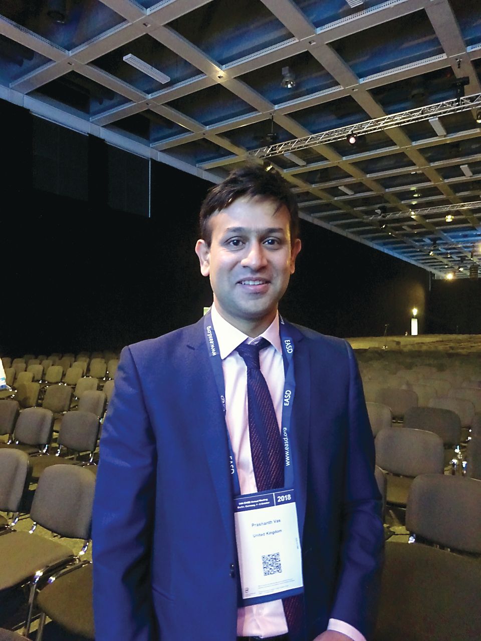
EASD delegate Prashanth Vas, MBBS, MRCP, PhD, noted during the discussion that the use of diuretics was ubiquitous. “Nearly everyone uses diuretics,” he said. The potential risk of lower limb amputation and treatment with SGLT2 inhibitors had “been vexing us for some time since the data from CANVAS came out.”
Dr. Vas, who is a consultant diabetologist and diabetic foot specialist at King’s College Hospital NHS Foundation in London, went on to ask why only canagliflozin was found to be associated with amputations and not the other SGLT2 drugs.
Dr. Roussel responded that data were still needed on the other drugs in this class and that they needed to be treated with caution. The literature is not so clear, he admitted.
“It’s important that you noted it’s a single-center study,” Dr. Vas countered. “It’s very important to have multicenter data. An amputation is a decision made by someone. An amputation in one center may not be an amputation in another center.”
The SURDIAGENE study was supported by grants from the French Ministry of Health, the Association Française des Diabétiques, and the Groupement pour l’Etude des Maladies Métaboliques et Systémiques. Dr. Roussel reported relationships with Janssen, Merck, Sanofi-Aventis, AstraZeneca, and Boehringer Ingelheim. Dr. Vas was not involved in the study or analysis.
SOURCE: Roussel R et al. EASD 2018, Abstract 12.
BERLIN – presented at the annual meeting of the European Association for the Study of Diabetes.
A significant and independent increase in the risk of lower limb events, predominantly lower-extremity amputations was seen among patients with type 2 diabetes mellitus (T2DM) who were treated with diuretics versus those who were not. The adjusted hazard ratios in a propensity-matched cohort was 1.60 (95% confidence interval, 1.06-2.42; P = .027) for lower limb events, 2.13 (95% CI, 1.17-3.87; P = .013) for lower limb amputations, and 1.12 (95% CI, 0.70-1.79; P = .6443) for lower limb revascularizations.
“We know diabetes is a leading cause of nontraumatic lower limb amputations in the world,” and thus a very important public health issue, said study investigator Ronan Roussel, MD, PhD, of Hôpital Bichat, Assistance Publique Hôpitaux de Paris. “Many contributing factors are identified, susceptibility to infection, impaired wound healing, peripheral neuropathy; but the most important is the presence of peripheral arterial disease.”
The risk of diabetic amputations is of specific interest because of the recent findings from CANVAS, where treatment with canagliflozin, a sodium-glucose cotransporter 2 (SGLT2) inhibitor, was linked to an almost doubled rate of amputations versus placebo (HR, 1.97; 95% CI, 1.41-2.75) in patients with T2DM.
Conflicting results have been seen in observational studies with other SGLT2 inhibitors, however, and it’s not clear if the risk of amputations is just seen with canagliflozin or if it may be a class effect. The underlying mechanism is unknown, but one theory is that hypovolemia may be involved. If this is the case, Dr. Roussel explained, then diuretics would have a similar safety profile as SGLT2 inhibitors in terms of increasing the risk of amputations.
The aim of the present study was to look at the association between lower limb events and diuretic usage in patients with T2DM. Data on 1,459 subjects with T2DM treated with diuretics and data on lower limb events and amputations were obtained from the single-center SURDIAGENE study. Of these, 670 were and 789 were not taking diuretics.
Baseline differences between diuretic and nondiuretic users were seen, such as diuretic users being older (67 vs. 63 years), having longer diabetes duration (16 vs. 13 years), and being more likely to have cardiovascular disease (32.5% vs. 23.4%). A propensity-score approach was used to even out these differences, leaving a population of 1,074 subjects in the final matched cohort.
Over a median follow-up of 7.2 years, 12.7% of diuretic and 7.2% of nondiuretic users experienced lower limb events (P = .001). In multivariate and sensitivity analyses, lower limb amputations remained significantly higher in patients who had been treated with a diuretic than in those who had not.
These are “hypothesis-generating” data, Dr. Roussel pointed out and “we don’t want to be overconclusive, of course.” However, they may explain the risk signal seen with SGLT2 inhibitors in the CANVAS study. Further studies are needed to explore the role of drug-induced hypovolemia in the association between the use of diuretics and lower limb events.
EASD delegate Prashanth Vas, MBBS, MRCP, PhD, noted during the discussion that the use of diuretics was ubiquitous. “Nearly everyone uses diuretics,” he said. The potential risk of lower limb amputation and treatment with SGLT2 inhibitors had “been vexing us for some time since the data from CANVAS came out.”
Dr. Vas, who is a consultant diabetologist and diabetic foot specialist at King’s College Hospital NHS Foundation in London, went on to ask why only canagliflozin was found to be associated with amputations and not the other SGLT2 drugs.
Dr. Roussel responded that data were still needed on the other drugs in this class and that they needed to be treated with caution. The literature is not so clear, he admitted.
“It’s important that you noted it’s a single-center study,” Dr. Vas countered. “It’s very important to have multicenter data. An amputation is a decision made by someone. An amputation in one center may not be an amputation in another center.”
The SURDIAGENE study was supported by grants from the French Ministry of Health, the Association Française des Diabétiques, and the Groupement pour l’Etude des Maladies Métaboliques et Systémiques. Dr. Roussel reported relationships with Janssen, Merck, Sanofi-Aventis, AstraZeneca, and Boehringer Ingelheim. Dr. Vas was not involved in the study or analysis.
SOURCE: Roussel R et al. EASD 2018, Abstract 12.
REPORTING FROM EASD 2018
Key clinical point: Diuretics may need to be used cautiously in patients with type 2 diabetes at risk of amputations.
Major finding: The adjusted hazard ratio for lower limb amputations with diuretic versus no diuretic use was 2.13 (95% confidence interval, 1.17-3.87; P = .013).
Study details: The SURDIAGENE trial, a single-center, prospective, observational study including almost 1,500 type 2 diabetes mellitus patients enrolled from 2002 to 2012.
Disclosures: The SURDIAGENE study was supported by grants from the French Ministry of Health, the Association Française des Diabétiques, and the Groupement pour l’Etude des Maladies Métaboliques et Systémiques. Dr. Roussel reported relationships with Janssen, Merck, Sanofi-Aventis, AstraZeneca, and Boehringer Ingelheim. Dr. Vas was not involved in the study or analysis.
Source: Roussel R et al. EASD 2018, Abstract 12.
Particular lesions early after CIS predict long-term MS disability
BERLIN – The presence of infratentorial and deep white matter lesions early in the course of relapse-onset multiple sclerosis was associated with high levels of disability 30 years later in a study looking at MRI predictors.
Univariate predictors of an Expanded Disability Status Scale (EDSS) score of more than 3.5, which is indicative of impaired mobility after 30 years, were the presence of an IT lesion at baseline, with an odds ratio (OR) of 12.4 (95% confidence interval [CI], 3.35-3.46; P less than .001), the presence of a deep white matter (DWM) lesion 1 year after presenting with clinically-isolated syndrome (CIS; OR = 10.65; 95% CI, 2.84-38.84; P less than .001), and the presence of an infratentorial (IT) lesion 1 year post CIS presentation (OR = 11.1; 95% CI, 3.31-37.22; P less than .001). At 5 years after a CIS presentation, the EDSS score, EDSS score change, and a DWM lesion score of more than 5 were indicative of worse disability after 30 years.
There was no significant association with age of onset, gender, CIS type, baseline EDSS, or disease duration, study investigator Karen Chung, MBBS, reported at the annual congress of the European Committee for Treatment and Research in Multiple Sclerosis.
“As we know, approximately 85% of people with MS initially present with a clinically isolated syndrome,” said Dr. Chung, a clinical research associate at the Queen Square Multiple Sclerosis Centre in the UCL Institute of Neurology in London. She added that the long-term prognosis after CIS is very variable, with some patients developing little detectable disability over time, while others may experience considerable decline.
There have been few studies examining whether there are any MRI parameters that might help predict patients’ long-term outcomes, so the aim of the study Dr. Chung presented was to see if there were any MRI parameters that might be predictive of clinical outcome 30 years after the onset of CIS.
Dr. Chung and her coauthors examined data on 120 of 132 individuals from the First London CIS Cohort who were prospectively recruited between 1984 and 1987 and had known outcomes. They looked at prospectively gathered MRI data and EDSS data at baseline, 5, 10, 14, 20, and 30 years. MRI data were obtained for 1 year after the CIS event, and data on the lowest EDSS score after the CIS event were retrospectively determined from patient notes or recall. The cohort was predominantly female (61%), with a mean of 31.5 years at CIS onset. Around half (52%) presented with optic neuritis, 27% with a spinal cord syndrome, and 20% with a brainstem syndrome. The high percentage of patients presenting with optic neuritis may be due to the fact that one of the recruiting centers was a specialist eye hospital, Dr. Chung noted later during discussion.
The 2010 McDonald Criteria were used to determine whether patients had CIS, relapsing-remitting MS (RRMS), secondary progressive MS (SPMS), or death related to MS.
“We looked at all the MRIs available to us and quantified the T2 lesion count for whole brain as well as by location,” Dr. Chung explained. The locations considered were juxtacortical, periventricular, infratentorial, and deep white matter.
“I think it is important to remind ourselves that we have come a long way with MRI technology in the 30 years timespan,” she added, noting that there was “clearly a difference in the quality.”
Clinical outcomes at 30 years were as follows: 80 patients (67%) developed MS, of whom 35 (44%) had RRMS, 26 (33%) had SPMS, 15 (20%) had died as a result of MS, and 3 (4%) had died for unknown or unrelated causes. Of the 40 patients (33%) who remained with CIS, 10 (25%) died without developing MS.
“This is a largely untreated cohort where, within the 80 people with MS, 11 (14%) were treated with a DMT [disease-modifying treatment] at some point,” Dr. Chung reported. All DMTs used were first-line injectable agents, she observed.
EDSS scores could be obtained for 107 patients. At 30 years, people with low EDSS scores were those who remained with CIS or RRMS, and as EDSS scores increased, the severity of MS increased.
“Overall, T2 lesions were better predictors of 30-year outcome than EDSS,” Dr. Chung said. For combinations of predictors, patients who had at least one IT and one DWM lesion within 1 year of a CIS had a higher probability (94%) of having an EDSS score of more than 3.5 at 30 years than when compared with those with neither lesion (13%) and those with one DWM but no IT lesions (49%).
Looking at the best independent predictors up to 5 years, the predicted probability of an EDSS score of more than 3.5 if there were no IT lesions and fewer than five DWM lesions was 18%. But if there were no IT but more than 5 DWM lesions, the probability of disability at 30 years rose to 52%. The probabilities rose even higher to 63% if there was one or more IT and five or less DWM lesions and 90% if there was one or more IT and more than five DWM lesions.
“In this cohort, the presence of early infratentorial and deep white matter lesions following a CIS are associated with higher level of disability 30 years later,” Dr. Chung concluded. “Early predictive models can add information to risk-benefit stratification.”
During discussion, one delegate expressed concerns that these data were “not generalizable to the current situation.” This was a cohort of patients that largely wasn’t treated or if they were, treatment was delayed by more than 10 years. These data were interesting “from a historical perspective,” he argued, “but I don’t understand, how in the absence of contemporary therapies this is applicable in a way that will allow us to use this information to make prognoses for the future.”
Dr. Chung agreed, noting that this was more of a natural history study. “However, I think it is applicable in clinical practice in my opinion.
“When you have a patient presenting with a CIS, at the time of diagnosis, especially now when we can diagnose patients earlier with the new 2017 criteria, it will be helpful for the patient and ourselves to apply some of the information I presented to help perhaps in aiding decisions regarding treatment.”
The study was funded by a grant from the MS Society of Great Britain. Dr. Chung disclosed receiving honoraria from Teva, Biogen, and Roche.
SOURCE: Chung K et al. Mult Scler. 2018;24(S2):58-59, Abstract 157.
REPORTING FROM ECTRIMS 2018
Key clinical point: Early magnetic resonance parameters can provide information that can help risk-stratify patients.
Major finding: Infratentorial and deep white matter lesions early in the course of relapse-onset multiple sclerosis were associated with high levels of disability 30 years later.
Study details: Data on 120 patients with clinically isolated syndrome recruited as part of the First London CIS Cohort between 1984 and 1987.
Disclosures: The MS Society of Great Britain funded the study. Dr. Chung disclosed receiving honoraria from Teva, Biogen, and Roche.
Source: Chung K et al. Mult Scler. 2018;24(S2):58-9, Abstract 157.
Fewer hypos with CGM in routine pediatric T1DM care
BERLIN – A significant reduction in severe hypoglycemia was seen within the first year of using continuous glucose monitoring in a registry study of more than 3,000 children with type 1 diabetes mellitus.
Prior to continuous glucose monitoring (CGM) use, 3.9% of 3,171 individuals, aged a median of 12 years, had hypoglycemia events requiring external help. After 6 months of using CGM, however, the rate of severe hypoglycemia had fallen to 1.2% (P = .10), which remained at 1.2% at 12 months (P = .002). The event rate (events per 100 patient-years) was 10.6 at baseline, 7.8 at 6 months, and 6.1 at 12 months.
Fewer hypoglycemia events leading to coma or convulsion were seen with CGM over time, with 1.3%, 0.6%, and 0.7% of patients reporting at least one event at baseline, 6 months (P = .08), and 12 months (P = .15), respectively. Corresponding event rates were 2.5, 1.6, and 1.7 per 100 patient-years.
“The use of continuous glucose monitoring systems has increased considerably in the past years in individuals with type 1 diabetes,” noted Julia Hermann, a PhD student at Ulm University (Germany), at the annual meeting of the European Association for the Study of Diabetes. “In Germany, for example, CGM use rose considerably when reimbursement by health insurance for CGM started in the summer of 2016.”
Ms. Hermann noted that there were studies showing improved metabolic control with CGM but that clinical studies were often limited by the population of patients studied, restricting treatment to selected patients who may have been adhering better to the use of CGM because they were in a trial.
The aim of the present study was to assess metabolic control and acute complications associated with CGM use in children during its first year of use in a real-world population. Anonymized patient records from the prospective German-Austrian-Luxembourg diabetes patient follow-up (DPV) registry were used.
The DPV database is a standardized, computed-based registry of more than 500,000 adult and pediatric patients with all types of diabetes. It was established in 1995 and receives data from 471 diabetes clinics in Germany, Austria, Luxembourg, and Switzerland.
For the current analysis patients had to be aged under 18 years, have had type 1 diabetes mellitus for at least 1 year, have data available for the months prior to starting CGM, and have at least 1 year of follow-up. For inclusion there also had to be documented use of CMG for at least half of the observation time.
The reduction of severe hypoglycemia observed was not accompanied by any deterioration in metabolic control, Ms. Hermann reported. The median baseline glycosylated hemoglobin (HbA1c) was 7.6% (59 mmol/mol) and this did not noticeably change at either the 6- or 12-month follow-up time point. Furthermore, the percentage of patients achieving a target HbA1c of less than 7.5% was 52% at baseline, 55% at 6 months, and 52% at 12 months.
The rate of diabetic ketoacidosis (DKA), defined as a pH of less than 7.3, also did not change significantly from baseline to the two follow-up points, with event rates of 1.5, 1.4, and 1.1, and the percentage of patients with at least one event being 0.8%, 0.5%, and 0.4%.
“We analyzed an overall well-controlled group of pediatric individuals with type 1 diabetes,” Ms. Hermann said. “We observed a significant reduction of severe hypoglycemia. We observed no improvements in HbA1c, but the overall HbA1c remained stable. We observed no statistically significant change in DKA.”
Future research will try to address some of the limitations that the current data may have, such as looking at a longer follow-up period and other endpoints such as hospitalization, Dr. Hermann suggested. Subgroup analyses are also planned.
“Large databases like the DPV registry ... provide a contemporary picture of diabetes treatment and how the use of diabetes technology has changed over the years and also how diabetes outcomes have improved over the years,” Ms. Hermann said. “It has yet to be seen how most recent changes and advances in diabetes technology will affect these long-term trends.”
The work was partially funded by the German Center for Diabetes Research, the German Diabetes Society, Abbott, and Sanofi. Dr. Hermann reported no personal conflicts of interest.
SOURCE: Hermann J et al. EASD 2018, Abstract 149.
REPORTING FROM EASD 2018
Key clinical point: Continuous glucose monitoring helped to significantly reduce severe hypoglycemia episodes during the first year.
Major finding: A total of 3.9%, 1.2%, and 1.2% of patients experienced hypoglycemia needing external help at baseline, 6 months, and 12 months, respectively.
Study details: More than 3,000 pediatric patients using continuous glucose monitoring in the German-Austrian-Luxembourg diabetes patient follow-up registry.
Disclosures: The work was partially funded by the German Center for Diabetes Research, the German Diabetes Society, Abbott, and Sanofi. The presenting author reported no personal conflicts of interest.
Source: Hermann J et al. EASD 2018, Abstract 149.
Nasal glucagon ‘viable alternative’ to intramuscular administration
BERLIN – A dry-powder nasal formulation of glucagon was as good as intramuscular delivery for the reversal of severe hypoglycemia in patients with type 1 diabetes mellitus (T1DM) in a randomized controlled trial.
A 100% treatment success rate (n = 66) was seen for both nasal glucagon and intramuscular glucagon, defined as an increase in plasma glucose to 70 mg/dL (3.9 mmol/L or greater) or more or an increase of 20 mg/L (1.1 mmol/L) or more from the glucose nadir within 30 minutes of administration.
Furthermore, slightly more than 97% of patients achieved treatment success within 15 minutes in both treatment groups, with mean times of 11.4 minutes for the nasal formulation and 9.8 minutes for intramuscular administration.
Similar glucose responses were observed within 40 minutes of glucagon administration, study investigator Leona Plum-Mörschel, MD, PhD, reported at the annual meeting of the European Association for the Study of Diabetes. Dr. Plum-Mörschel is the CEO of the clinical research organization Profil Mainz (Germany), which helped Eli Lilly conduct the trial.
“We are all aware and fully agree that severe hypoglycemia is a serious and potentially life-threatening complication of diabetes treatment with insulin and the sulfonylureas,” said Dr. Plum-Mörschel. At least one in three patients with diabetes reports one hypoglycemic episode per year, she added. While this is quite prevalent in patients with T1DM, it’s not uncommon for those with type 2 diabetes mellitus (T2DM) to experience hypoglycemic episodes.
“Outside of a hospital or emergency room setting, injectable glucagon is currently the only option to treat severe hypoglycemia,” Dr. Plum-Mörschel reminded delegates. “Thankfully, the available injectable glucagon emergency kits are highly effective for the treatment of hypoglycemic events,” she added.
However, for untrained caregivers they can be “really challenging to use,” according to Dr. Plum-Mörschel. This is because the available kits involve multiple steps, including reconstitution, before being ready to inject. A severe hypoglycemic event is stressful enough without them worrying about getting things right, she suggested.
This is where a nasal formulation would be advantageous, and it is something that’s been touted to be on the horizon for a few years. Studies have previously shown that nasal glucagon is comparable to intramuscular glucagon in both adult and pediatric populations. Turning the formulation used in those studies into a commercial product however, has meant more clinical testing before being licensed by the regulatory authorities.
The nasal formulation consists of a dry power containing 3 mg glucagon that is provided in a single-use, compact, and thus, portable, device. It’s been designed to be stored at room temperature and is ready to use immediately. Pressing the plunger on the device releases the fine powder that does not require inhalation to work, which means it can be given easily to an unconscious patient with severe hypoglycemia.
“The aim of the present study was to compare the efficacy and safety of commercially manufactured nasal glucagon with intramuscular glucagon for the treatment of insulin-induced hypoglycemia in adults with type 1 diabetes,” Dr. Plum-Mörschel said.
It was a randomized, single-dose, crossover study involving 70 adult patients with T1DM with hypoglycemia artificially induced by an intravenous insulin infusion during the two dosing visits. Five minutes after stopping insulin, nasal glucagon (3 mg) or intramuscular (1 mg) was given, with multiple plasma glucose measurements taken for up to 90 minutes.
The mean age of participants was 41.7 years old, 61% were male, the mean duration of diabetes was 19.8 years, with a baseline glycated hemoglobin (HbA1c) of 7.3%.
As for safety, Dr. Plum-Mörschel noted that nasal glucagon had a safety profile that was acceptable for emergency treatment. There was no difference between the nasal or intramuscular glucagon treatment groups in the percentages of patients experiencing treatment-emergent adverse events (at least 5% frequency): nausea was seen in 22% and 29%, vomiting in 10% and 12%, and headache in 12% and 7%, respectively.
Asking patients who had been treated with nasal glucagon about specific symptoms related to nasal administration showed around 63% had watery eyes, 49% nasal itching, 39% nasal congestion, 37% a runny nose, 24% sneezing, 20% itchy eyes, and 12.9% itchy throat. “All of these events were transient, generally mild or moderate in nature, and none were serious. Indeed, there were no deaths in the study and no other serious AEs [adverse events] occurred.”
Dr. Plum-Mörschel concluded: “These results support nasal glucagon as a viable alternative to intramuscular glucagon for the treatment of severe hypoglycemia.
“I personally would expect that, due to its simplicity of use, nasal glucagon will create a greater community who can render quick aid in a rescue situation.”
The study was funded by Eli Lilly. Dr. Plum-Mörschel is an employee of Profil Mainz and has received travel grants and speaker honoraria from Eli Lilly and Novo Nordisk.
SOURCE: Suico J et al. EASD 2018, Abstract 150.
REPORTING FROM EASD 2018
Key clinical point: Nasal glucagon was as good as intramuscular administration for reversing severe hypoglycemia in patients with T1DM.
Major finding: All (100%) of patients achieved treatment success; 97% or greater within 15 minutes in both treatment groups.
Study details: Randomized, single-dose, crossover study of nasal versus intramuscular glucagon in 70 adult patients with T1DM.
Disclosures: The study was funded by Eli Lilly. The presenting investigator Dr. Plum-Mörschel is an employee of Profil Mainz and has received travel grants and speaker honoraria from Eli Lilly and Novo Nordisk.
Source: Suico J et al. EASD 2018, Abstract 150.
MS disease activity returns in one-third after fingolimod withdrawal
BERLIN – At least one-third of patients being treated with fingolimod (Gilenya) for multiple sclerosis (MS) will relapse after stopping the drug, according to data from two “real-world” studies reported at the annual congress of the European Committee for Treatment and Research in Multiple Sclerosis, one of which was undertaken in pregnant women.
In a retrospective analysis of 110 MS patients who discontinued fingolimod from a 433-patient cohort, around 35% experienced a recurrence of disease activity after fingolimod withdrawal. Study investigator Nuria Cerda, MD, of the Neurologic Clinic and Polyclinic at University Hospital Basel (Switzerland), reported that risk factors for increased disease activity were younger rather than older age at MS diagnosis (P = .01) and at discontinuation (P less than .0001), having had a shorter rather than longer disease duration (P = .01), and evidence of MRI-confirmed disease activity occurring while on treatment with fingolimod (P = .02).
“We aimed to describe the frequency of recurrence of disease activity after fingolimod discontinuation in a real-world cohort of MS patients,” Dr. Cerda said. She noted that there were conflicting data on reappearing disease activity after stopping the drug. Case reports and observational data suggest a frequency of 10%-53%, but post hoc analyses of phase 3 trials with fingolimod have not found any higher risk for recurrence versus placebo.
The study also compared “demographic and clinical characteristics in patients with and without recurrence of disease activity, in order to identify possible risk factors for reactivation of the disease.”
The patients included in the study had been treated with fingolimod for a median of 24 months (interquartile range, 10-43 months) and experienced 118 discontinuation events between them. The majority (n = 81) had relapsing-remitting MS, and the remainder (n = 19) were in transition from RRMS to secondary progressive MS.
The mean age at discontinuation was 40 years and, while 60% of patients switched to another disease-modifying therapy (DMT), 40% did not receive further DMT. The reasons for discontinuation were classified as being from disease activity in 44%, side effects in 21%, pregnancy or childbearing preferences in 16%, noncompliance in 2%, and other reasons in 17%.
Recurrence of disease activity was defined as clinical symptoms, gadolinium-enhancing (Gd+) lesions on MRI, or both, within 6 months of stopping fingolimod. This was seen in 38 patients with 41 events, giving a rate of recurrence of 35%. Of these: “almost 60% had clinical and MRI activity, almost 30% had clinical activity only without Gd+ lesions on MRI, and 12% had MRI activity only without ever having a relapse,” Dr. Cerda said. In those who relapsed, the recurrence took a median 8 weeks to happen after fingolimod discontinuation.
A further definition of severe recurrence of disease activity also was used, defined as either a worsening in the Expanded Disability Status Scale (EDSS) score of more than 2 points (signifying a severe relapse) with or without pronounced MRI activity with at least five cerebral Gd+ lesions within 6 months of fingolimod withdrawal. Thirteen severe events were seen in 11 patients, giving a frequency of approximately 11%. Dr. Cerda reported that patients with severe recurrence were significantly younger at MS diagnosis than were those who did not experience recurrence (P = .003). A trend toward a higher annualized relapse rate (ARR) was seen in patients with severely active versus inactive disease (0.84 vs. 0.54), but this was not statistically significant. In addition, “confirmed disability worsening of 1 EDSS point or more was observed in about 3%.”
Relapse rates coming off fingolimod before or during pregnancy
In another study, which used German registry data on 129 pregnancies that occurred around or after the time of fingolimod withdrawal, up to 56% of women who stopped fingolimod before pregnancy experienced a relapse during pregnancy versus 29% of those who stopped fingolimod upon a positive pregnancy test.
In a comparison of women who stopped fingolimod 1 year to 61 days before their last menstrual period against those who stopped less than 60 days prior to or after their last menstrual period, relapse rates were higher during the first (25.8% for early withdrawal vs. 12.5% with late withdrawal) and second trimesters (32.3% vs. 11.5%) than in the third (9.7% vs. 10.4%), Spalmai Hemat, MD, of the department of neurology at St. Josef Hospital, Ruhr University of Bochum (Germany) reported at the congress.
Relapse rates at 6 months postpartum proved to be similar at 36.7% for women who stopped fingolimod before their pregnancy and 38% in those who stopped later. Difference in the percentage of women experiencing disability progression during pregnancy did not prove to be statistically significant for stopping fingolimod before pregnancy (22.7%) vs. stopping later (11.1%; P = .283), while the 23.8% rate of disability progression after pregnancy among women who stopped the drug later versus 13.6% seen with stopping before was also not statistically significant (P = .31). Relapse during pregnancy was the only significant predictor for relapses postpartum.
“The large majority of women did not experience permanent disability,” Dr. Hemat said, but noted that as many as 10%-20% could experience substantial EDSS worsening (2 or more points) at 6 months postpartum.
Dr. Hemat concluded that more data were needed to see if reintroduction of fingolimod very soon after birth, such as within the first 2 weeks, could reduce the postpartum relapse risk.
Dr. Hemat and Dr. Cerda had no relevant disclosures.
SOURCES: Cerda N et al. Mult Scler. 2018;24(S2):73-4, Abstract 206; Hemat S et al. Mult Scler. 2018;24(S2):74-5, Abstract 207.
REPORTING FROM ECTRIMS 2018
Key clinical point:
Major finding: A higher risk of recurrent disease activity was seen in younger MS patients with shorter disease duration and evidence of disease activity while on fingolimod treatment. Pregnant women who stopped more than 2 months before a planned pregnancy also had a higher risk versus those who stopped after a positive pregnancy test.
Study details: A retrospective analysis of 110 MS patients who discontinued fingolimod from a 433-patient cohort, and German registry data on 129 pregnancies that occurred around or after the time of fingolimod withdrawal.
Disclosures: Dr. Spalmai Hemat and Dr. Nuria Cerda had no relevant disclosures.
Sources: Cerda N et al. Mult Scler. 2018;24(S2):73-4, Abstract 206; Hemat S et al. Mult Scler. 2018;24(S2):74-5, Abstract 207.
Alemtuzumab switch linked to good MS outcomes
BERLIN – Switching to alemtuzumab from fingolimod is associated with improved disease activity in patients with relapsing-remitting multiple sclerosis (RRMS), according to the results of a real-world study reported at the annual congress of the European Committee for Treatment and Research in Multiple Sclerosis.
Jessica Frau, MD, of the University of Cagliari (Italy) reported that a switch from fingolimod (Gilenya) to alemtuzumab (Lemtrada) in 77 patients treated at 11 Italian centers was able to “reduce dramatically disease activity in patients who did not respond to fingolimod.”
Dr. Frau reported: “When we compared in our cohort the last year of fingolimod with the first year after the first course of alemtuzumab, we found a significant decrease in the annualized relapse rate [ARR].” The ARRs were 0.60 for fingolimod and 0.20 after 1 year of alemtuzumab treatment.
“We found also a trend towards an improvement in the EDSS [Expanded Disability Status Scale] score (P = .23), and less evidence of disease activity on MRI, both in terms of new T2 lesions and gadolinium-enhancing (Gd+) lesions.”
The last MRI during fingolimod treatment showed new T2 and Gd+ enhancing lesions in 69.2% and 58.6% of patients, respectively. Corresponding figures for the first MRI during alemtuzumab treatment were 10.4% and 2.2% of patients.
The beneficial effects of switching to fingolimod in the Italian study “was not influenced by a shorter washout [period] or a low lymphocyte count when alemtuzumab was started,” Dr. Frau said. A shorter washout period has been hypothesized to account for recent accounts of disease flares seen when switching from fingolimod to alemtuzumab, she explained.
Indeed, Dr. Frau noted that there had been a few studies that reported MS disease reactivation soon after the switch to alemtuzumab was made, which could be because lymphocytes remain in the lymph nodes when alemtuzumab is administered, this means that potentially they could repopulate the central nervous system and reactivate the disease.
However, “when alemtuzumab is started after fingolimod it is not a risk factor for reactivation of the disease,” Dr. Frau said, based on the current study’s findings.
As expected, the frequency of relapses increased during the washout period after stopping fingolimod, going from 12.7% of patients with relapse in the first month, 18.2% at 2 months, and 22.2% at 3 months. The time to first relapse from the start of alemtuzumab treatment was 6 months for 2.9% of patients, 9 months for 10.5% of patients, and 1 year for 20.7% of patients.
Asked to comment on when the optimal time to switch from fingolimod to alemtuzumab might be, Dr. Frau said: “The optimal time could be 1 month when the lymphocyte count is not too low.” However, lymphocyte counts were not measured in the entire cohort, so “these data perhaps need to have more strength.”
The switch from natalizumab to alemtuzumab
Other data on switching to alemtuzumab, this time from natalizumab (Tysabri), in the ANSWERS MS study were presented by Paul Gallagher, MBChB, of Queen Elizabeth University Hospital, London, and the University of Glasgow (Scotland).
ANSWERS MS (Alemtuzumab after Natalizumab Switch in Evolving Rapidly Severe MS) is a retrospective, observational analysis of routinely collected data on the use of alemtuzumab by 13 centers the United Kingdom and Ireland. These centers have been collecting data since before alemtuzumab was licensed in 2014 for MS, Dr. Gallagher observed, with some centers having experience of making the switch for more than a decade.
ANSWERS MS addresses a common clinical question: “Is it safe and effective to switch to alemtuzumab if natalizumab fails in highly active MS?” Dr. Gallagher said. “The truth is we don’t really know the answer to this, although it’s becoming an increasingly used switch.”
Alemtuzumab was developed in Cambridge, England, in 1983, originally as an anticancer agent, and first started being used in MS patients in the 1990s. Natalizumab was first licensed in the United Kingdom in 2007.
The aim of the study was mainly to look at safety, but also examine efficacy, and to offer advice on how to best manage the switch. A total of 79 patients formed the safety cohort; 51 of these patients had more than 2 years of follow-up after their first infusion of alemtuzumab and formed the efficacy cohort.
Data were examined in five phases: before natalizumab, during natalizumab, during the switchover period, during alemtuzumab treatment, and after alemtuzumab treatment, with the latter starting 2 years after the first alemtuzumab infusion.
Dr. Gallagher noted that 43% started natalizumab as a first-line therapy, and almost half (49%) of patients stopped taking natalizumab because of breakthrough disease, making this a bit of an unusual cohort with highly active disease, although other cohort characteristics were pretty typical of an MS population.
“The headline is that there are no new safety concerns identified from this cohort,” Dr. Gallagher reported. “Most [61%] patients had infusion reactions with alemtuzumab as expected, but this gradually reduced with subsequent courses.”
Fewer than 20% of patients developed autoimmune thyroid disease, he added, and there were no cases of idiopathic thrombocytopenic purpura.
Infections were seen in nine patients, including three cases of shingles, two urinary tract infections – one of which was classed as a severe adverse event – and one case each of oral thrush, fungal skin infection, tonsillitis, and norovirus.
There was also one cytomegalovirus infection and one death from sepsis unrelated to alemtuzumab; both of these were classed as serious adverse events.
In terms of efficacy, mean ARRs were 2.3 before and 0.8 during natalizumab treatment, decreasing to 0.4 during alemtuzumab treatment and 0.5 post alemtuzumab. A “spike” in relapses was seen, however, during the switch period.
“There was a similar story with MRI imaging,” Dr. Gallagher said. “The profile suggests high disease activity during the switch phase in comparison to everything else.” The mean number of new or worsened MRI lesions was 4.32 per scan per year during the switch period. This fell, however, during alemtuzumab treatment to 0.006 per MRI scan per year and remained low after the end of alemtuzumab treatment at 0.017 per scan per year.
There was no real benefit to switching on the EDSS, with scores increasing from 3.4 in the pre-natalizumab period to 4.7 during the switch period, but then plateauing out to 4.4. and 4.3 after the initiation of alemtuzumab and in the post-alemtuzumab phase.
“These data were based on medical records, often incomplete, and so not all patients had an EDSS in every phase, for example,” Dr. Gallagher noted. He said an analysis was done to try to account for the missing information. This showed that there was an improvement in EDSS while on alemtuzumab, but the effect was not maintained.
It was evident in looking at the switch period that a shorter time between natalizumab and alemtuzumab was associated with the best outcomes, with the optimum time being around 2-4 months. Bridging therapy with fingolimod did not reduce disease activity during the switch, Dr. Gallagher said.
ANSWERS MS was funded by Sanofi-Genzyme. Paul Gallagher disclosed that he had received salary payment and travel funding for educational events from Sanofi-Genzyme and travel funding from Novartis and Biogen.
Dr. Frau disclosed that she serves on scientific advisory boards for Biogen, Merck, and Genzyme and that she has received honoraria for speaking from Merck Serono, Genzyme, Biogen, and Teva.
SOURCE: Frau J et al. Mult Scler. 2018;24(S2):100-1, Abstract 265; Gallagher P et al. Mult Scler. 2018;24(S2):99-100, Abstract 264.
REPORTING FROM ECTRIMS 2018
Key clinical point: Good results can be achieved by switching from fingolimod or natalizumab to alemtuzumab in relapsing-remitting multiple sclerosis (RRMS).
Major finding: Annualized relapse rates were 0.60 for fingolimod at the time of the switch and 0.20 after 1 year of alemtuzumab treatment in one real-world study. In another, alemtuzumab was effective in reducing inflammatory disease activity when natalizumab failed.
Study details: Two real-world, observational studies: one with 77 RRMS patients treated at 11 Italian centers and the other a retrospective analysis of routinely collected data on 79 patients.
Disclosures: Dr. Frau disclosed that she serves on scientific advisory board for Biogen, Merck, and Genzyme and that she has received honoraria for speaking from Merck Serono, Genzyme, Biogen, and Teva. ANSWERS MS was funded by Sanofi-Genzyme. Dr. Gallagher disclosed he had received salary payment and travel funding for educational events from Sanofi-Genzyme and travel funding from Novartis and Biogen.
Source: Frau J et al. Mult Scler. 2018;24(S2):100-1, Abstract 265; Gallagher P et al. Mult Scler. 2018;24(S2):99-100, Abstract 264.
Closed-loop basal insulin delivery improves suboptimal T1DM control
BERLIN – Adults, adolescents, and children with type 1 diabetes mellitus (T1DM) achieved better glycemic control and spent less time in a hypoglycemic state with an investigational closed-loop basal insulin delivery system than with a sensor-augmented insulin pump.
The primary trial endpoint of the proportion of time spent in a target glucose range of 3.9-10 mmol/L at 12 weeks was achieved by 65% of closed-loop users versus 54% of sensor-augmented insulin-pump users (P less than .0001).
In addition, fewer patients had periods of blood glucose less than 3.9 mmol/L at 12 weeks, potentially reducing their risk for hypoglycemic episodes (2.6% vs. 3.9%, P = .0130). The percentages of patients with blood glucose concentrations higher than the target range were also significantly reduced with the closed-loop system.
Glycated hemoglobin (HbA1c) improved in both groups, from a baseline of 8% to 7.4% at 12 weeks in the closed-loop users and from 7.8% to 7.7% in the sensor-augmented insulin-pump users (mean difference –0.036%, P less than .0001). Mean glucose levels also significantly improved, with a mean between-group difference of –0.45 mmol/L (P less than .0001).
“Most of the improvements are overnight,” with a striking difference between the control and closed-loop groups of time in range, said senior study investigator Roman Hovorka, PhD, during a press briefing at the annual meeting of the European Association for the Study of Diabetes.
“Usually what people need to do is titrate insulin based on blood sugar tests, but in our system those tests are done by a monitoring system and the insulin is titrated by a computer algorithm,” study investigator Martin Tauschmann, MD, explained in an interview after the press briefing.
The prototype system consists of a modified insulin pump (Medtronic 640G) and a continuous glucose sensor (Enlite 3) that are linked by the proprietary “Cambridge control” algorithm via an Android phone. The latter calculates the optimum insulin dose, which is then relayed to the insulin pump every 10 minutes to determine how many insulin units are needed.
“The algorithm is based on mathematical modeling that is trying to predict what the glucose levels will be in the future and trying to work out the optimum insulin dose to bring the predicted glucose levels down,” noted Dr. Tauschmann, who is a pediatrician at Cambridge University (England).
It’s not a fully closed-loop system, he added, it’s a hybrid, so users still need to calculate their carbohydrate intake around mealtimes and input that into the algorithm and use bolus insulin.
“A problem with using currently available insulin formulations is that you are always behind, because we are delivering insulin in the subcutaneous tissue and it just takes some time for the insulin to be absorbed, and, in the meantime, the blood sugar levels go up,” Dr. Tauschmann observed. Studies with fully closed–loop systems, so far, have shown glucose peaking after meals. Perhaps when faster-acting insulins are tested in this setting it could work, he suggested, but perhaps simplifying how the users announce their meals is a future development for the hybrid systems.
Closed-loop insulin delivery is not a new concept. There is already one system available in the United States produced by Medtronic (670G), which was approved by the Food and Drug Administration in September 2016. However, there are no systems available in Europe and there are limited trial data on their use in an outpatient setting.
The aim of the current study, which was an open-label, multicenter, multinational, parallel-group, randomized, controlled trial, was to compare closed-loop basal insulin delivery and sensor augmented insulin pump therapy in helping a mixed adult and pediatric population of patients T1DM achieve good glucose control.
In all, 86 adults, adolescents, and children (age 6 years or older) were randomized: 46 to the closed-loop system and 40 to the control arm of sensor-augmented insulin pump therapy. The mean age of participants was 22 years in the closed-loop group and 21 years in the sensor group, with a respective 24% and 30% aged 6-12 years, 24% and 20% aged 13-21 years, 39% and 35% aged 22-40 years, and 13% and 15% aged 41 years or older. The mean duration of diabetes was 13 and 10 years, in each group, respectively.
The study data “provide more evidence that closed-loop insulin delivery, compared with standard pump and sensor, really improves HbA1c, time spent in range, and mean glucose, and it also reduces hypoglycemia,” Dr. Tauschmann said.
“We didn’t have any real safety issues,” noted Dr. Hovorka, who is professor and director of research in the department of pediatrics at the University of Cambridge. There was one episode of diabetic ketoacidosis caused by infusion set failure in the closed-loop group; 16 other adverse events were noted (13 in the closed-loop group, three in the control group) that were not related to treatment.
“We have a number of studies in the pipeline,” Dr. Hovorka said. “Two exciting studies are in development. In one we are recruiting 70 subjects with newly diagnosed type 1 diabetes mellitus who will be treated with a closed-loop system. There is also a study in children aged between 1 and 7 years that is projected to start next year.”
Work is also underway to create a platform that will work with all insulin pumps and create a commercial product.
The study was funded by the Juvenile Diabetes Research Foundation with additional support from the National Institute for Health Research (England) and the Wellcome Trust. Dr. Tauschmann reported receiving speaker honoraria from Medtronic and Novo Nordisk. Dr. Hovorka reported receiving speaker honoraria from Eli Lilly and Novo Nordisk, serving on an advisory panel for Eli Lilly, receiving license fees from B. Braun Medical and Medtronic, and having patents and patent applications.
SOURCES: Tauschmann M et al. EASD 2018, Oral Presentation 19.
REPORTING FROM EASD 2018
Key clinical point: Improved glycemic control and reduced risk of hypoglycemia was seen across a wide age range.
Major finding: Blood glucose was within the target range of 3.9-10.0 mmol/L at 12 weeks (P less than .0001) in 65% of closed-loop versus 54% of sensor-augmented insulin-pump users.
Study details: An open-label, multicenter, parallel-group, randomized, controlled trial comparing closed-loop basal insulin delivery and sensor-augmented insulin pump therapy in 86 children (age 6 years or older) and adults with suboptimally controlled T1DM.
Disclosures: The study was funded by the Juvenile Diabetes Research Foundation with additional support from the National Institute for Health Research (England) and the Wellcome Trust. Dr. Tauschmann reported receiving speaker honoraria from Medtronic and Novo Nordisk. Dr. Hovorka reported receiving speaker honoraria from Eli Lilly and Novo Nordisk, serving on an advisory panel for Eli Lilly, receiving license fees from B. Braun Medical and Medtronic, and having patents and patent applications.
Sources: Tauschmann M et al. EASD 2018, Oral Presentation 19.
Escalate treatment to avoid MS relapses
BERLIN – Escalating treatment to a “highly effective” disease-modifying treatment (DMT) results in fewer relapses in patients with relapsing-remitting multiple sclerosis (RRMS) than does switching to another “moderately effective” therapy, according to data from a Danish cohort study.
The annualized relapse rate (ARR) was 0.23 for patients who switched to a highly effective DMT, defined as either natalizumab (Tysabri) or fingolimod (Gilenya), whereas the ARR was 0.35 in those who were switched to a moderately effective DMT, defined as an interferon-beta, glatiramer acetate (Copaxone), teriflunomide (Aubagio), or dimethyl fumarate (Tecfidera).
This resulted in a relapse-rate ratio of 0.67 (95% confidence interval [CI], 0.55-0.83) comparing high to moderate DMT, or a 33% lower relapse rate in the high DMT group, Thor Ameri Chalmer, MD, PhD, reported at the annual congress of the European Committee for Treatment and Research in Multiple Sclerosis.
Dr. Chalmer, who works at the Danish Multiple Sclerosis Center at Rigshospitalet, the University of Copenhagen, reported that the time to first relapse and the likelihood for having recurrent relapses were all lowered by escalating treatment rather than switching treatment. Indeed, there was a 38% increase in the time to first relapse (hazard ratio [HR] = 0.62; 95% CI, 0.50-0.76) and a 31% reduction in recurrent relapses (HR = 0.69; 95% CI, 0.57-0.83).
However, there were “no clear differences” in the time to 3-month confirmed Expanded Disability Status Scale (EDSS) worsening and improvement, Dr. Chalmer said. The HR for time to first worsening in the 3-month confirmed EDSS was 0.86, and for recurrent worsening it was 0.86, but the 95% CI in both cases crossed over from 1.0 (0.65-1.14 and 0.71-1.05, respectively). There was no difference in the time to first improvement (HR = 1.23; 95% CI, 0.95-1.60).
“The aim of this study was to use data from the [Danish] MS Registry, find the patients who started on what we define as moderately effective DMT for the first time, and on this treatment experienced a disease breakthrough and then either switched to another moderately effective DMT or escalated to a highly effective DMT,” Dr. Chalmer explained.
Disease breakthrough was defined as at least one relapse occurring within 12 months of the treatment switch, or the treating neurologist defined the reason for switching as disease breakthrough. Dr. Chalmer acknowledged that this was one of the limitations of the observational study as patients could have been misclassified and switched treatment for another reason. The severity of the relapse was not recorded.
Data on more than 5,000 patients enrolled in the Danish Multiple Sclerosis Registry (DMSR) were considered, with a final propensity-match population of 788 included in the analysis; half had received highly effective DMT and half moderately effective DMT.
The DMSR contains data on virtually all patients with MS treated in Denmark, Dr. Chalmer observed; treatment with DMT is free of charge and it can be prescribed only in public MS clinics, he explained. Furthermore, all MS clinics are required to register information about treatment response at each clinical visit.
The mean age of patients in the study was 39 years, around 70% were female, and average disease duration was 5 years.
“I don’t see in your study what is really new,” Gilles Edan, MD, observed during a discussion. “We know that using more active, more efficient drugs gives more control on relapse.”
Dr. Edan, professor and head of the neurosciences department of University Hospital Pontchaillou, Rennes (France), added that these data “confirmed what has already been observed in the clinical trials.”
It is important to consider the safety and efficacy concerns on an individual level, Dr. Edan argued, questioning whether the more highly active drugs should be systematically used first-line rather than second-line in all patients with MS. He also noted that of course patients would need to be treated for very long periods.
Dr. Chalmer responded: “I agree some of the randomized trials have touched upon on this as well, but I think it’s really important to not just rely on one or two trials but to keep on doing the trials over and over again so that we show that we have the right results.”
Dr. Chalmer added that the present study looked only at efficacy and that the median follow-up time was 3.2 years. “Safety is important of course, but it was not the aim of this study.” Perhaps longer follow-up might have detected more differences in the EDSS outcomes, he added.
The study was funded by the Danish Multiple Sclerosis Society, the Foundation for Research in Neurology, Ejnar Jonassen, and Gangstedfonden. Dr. Chalmer disclosed he had received support for congress participation from Merck, Biogen, and Roche.
SOURCE: Chalmer T et al. Mult Scler. 2018;24(S2):99. Abstract 263.
REPORTING FROM ECTRIMS 2018