User login
Pregnancy in Turner’s syndrome carries high hypertensive risk
LONDON – Hypertensive complications are seen in more than one-third of pregnant women with Turner’s syndrome undergoing fertility treatment, according to the results of a longitudinal retrospective study performed in Finland, Denmark, and Sweden.
Of 106 women with Turner’s syndrome who conceived after oocyte donation between 1992 and 2011, 35% had hypertensive complications and 20% had preeclampsia. Although there were no maternal deaths, life-threatening complications occurred in four (3.3%) women. This included a case of aortic dissection.
"We think it’s very important to have a centralized cardiovascular assessment before pregnancy," Dr. Anna Hagman of Sahlgrenska Hospital, Gothenburg, Sweden, said on behalf of the study team at the annual meeting of the European Society of Human Reproduction and Embryology. This should include echocardiography and magnetic resonance imaging, looking for specific cardiac abnormalities known to affect women with Turner’s syndrome.
Turner’s syndrome is caused by an aberration of the X chromosome and affects around one in 2,500 women. It is associated with increased cardiovascular (CV) morbidity and mortality, which raises concern for women who may want to try to conceive either spontaneously or via assisted conception methods. Women with the condition typically are of short stature and almost all (95%–99%) are infertile. Oocyte donation may offer one of their best chances for becoming pregnant.
For the study, medical data from fertility clinics, antenatal clinics, and hospitals where women had been treated or delivered their baby between 1992 and 2011 in Finland and Denmark, or between 2003and 2011 in Sweden, were examined.
The 45,X karyotype was observed in 44% of the women and 9% had a known cardiac defect before pregnancy. Just under half (49%) of women had a CV exam within the 2 years prior to becoming pregnant, and 29% had a CV exam during their pregnancy.
Single embryo transfer was performed in 70% of the cases and the multiple birth rate was 7%; 15% of women conceived twice after two oocyte donations during the study period. The median follow-up time was 4 years, but ranged from 0.3 to 19 years.
There were 122 deliveries studied in total and 131 neonates, of which 18 were twins. Among the single births, the rate of preterm delivery, defined as before 37 weeks’ gestation, was 8%, with 9% of children having low birth weight (less than 2,500 g). Perinatal mortality was 0.8%. Congenital defects were seen in 3.8% of the children.
"Neonatal outcomes were reassuring for singletons," Dr. Hagman said at the annual meeting of the European Society of Human Reproduction and Embryology, later noting that the team "recommends single-embryo transfer."
This is because greater risks for mother and child were seen after twin births. Hypertensive complications occurred in 50% of women and around two thirds (67%) of twins were delivered prematurely. Almost three quarters (72%) had a low birth weight and perinatal mortality was approximately 11% for twin births.
Dr. Hagman suggested that pregnancy should be avoided in women with Turner’s syndrome if there is CV disease or other severe health problems exist. Thyroid, renal, and liver tests should be performed, she advised, as well as testing for diabetes.
"All pregnancies must be carefully monitored," Dr. Hagman advised. The blood pressure should be kept below 140/90 mm Hg and echocardiography, MRI, or both performed two to three times during pregnancy.
The study’s findings have recently been published (Hum. Reprod. 2013;28:1598-09).
The study was funded by a variety of sources, including: the Gothenburg Medical Society, grants from the ALF agreement at the Sahlgrenska University Hospital, Gothenburg, Sweden; Hjalmar Svensson Foundation; the Nordic Federation of Societies of Obstetrics and Gynecology Nordic Fund; Finnish Society of Paediatric and Adolescent Gynecology; Liv och hälsa Foundation in Finland; and MSD Finland, Norway, and Denmark. No conflict of interest was reported.
AT ESHRE 2013
Major finding: Hypertensive complications were seen in 35% of women, with preeclampsia in 20%. Life-threatening complications occurred in four (3.3%) women, but there were no maternal deaths.
Data source: Retrospective cohort study of obstetric and neonatal outcomes involving 106 women with Turner’s syndrome who conceived via oocyte donation and gave birth in Finland (1992-2011), Denmark (1992-2011), or Sweden (2003-2011).
Disclosures: The study was funded by a variety of sources, including: GLS, ALF/SU Hospital, Hjalmar Svensson Foundation–Sweden, the Nordic Federation of Societies of Obstetrics and Gynecology, Finnish Society of Paediatric and Adolescent Gynecology, Liv och hälsa, and MSD. No conflict of interest was reported.
Adequate hypothyroid treatment aids assisted fertility success
LONDON – Women with hypothyroidism can achieve good results following fertility treatment if they maintain low levels of thyroid-stimulating hormone before assisted conception, according to findings of a 3-year retrospective study.
"An adequate treatment maintaining serum TSH levels below the threshold value of 2.5 mIU/L seems to fully overcome the detrimental effects of hypothyroidism on the rate of success of IVF [in vitro fertilization] and ICSI [intracytoplasmic sperm injection]," Dr. Andrea Busnelli reported at the annual meeting of the European Society of Human Reproduction and Embryology.
"Therefore, women scheduled for IVF-ICSI with adequately treated hypothyroidism can be reassured regarding the success of the procedure[s]," Dr. Busnelli of IRCCS Ospedale Maggiore Policlinico Mangiagalli e Regina Elena, Milan, Italy, added.
Previous research has shown that women with hypothyroidism have less chance of becoming pregnant after assisted conception than those with normal thyroid function. Subsequent findings conflict on whether levothyroxine treatment may (Hum. Reprod. Update 2013;19:251-8) or may not (Thyroid 2012;22:631-6) improve pregnancy rates.
These studies have been performed in a small number of women and TSH levels achieved with levothyroxine therapy were higher than recently recommended (J. Clin. Endocrinol. Metab. 2012;97:2543-65).
The current study therefore looked at whether "adequate" levothyroxine treatment, meaning that which ensured the level of TSH before conception was 2.5 mIU/L or lower, would be able to compensate for the reduced fertility success reported previously.
A total of 137 women with clinical or subclinical treated hypothyroidism participated in the study. At recruitment, their baseline TSH was between 0.4 and 2.5 mIU/L. Each case was age matched to two women with normal thyroid function as a control (n = 274).
Ultrasound was used to confirm any pregnancy, defined as a vital embryo within an intrauterine gestational sac at 4-5 weeks after embryo transfer.
The average age of cases and controls was approximately 35 years, body mass index in both groups was approximately 22 kg/m2, and there were no significant differences in the number of previous deliveries, serum hormone levels, or the cause of infertility leading to fertility treatment. Preconception TSH levels were approximately 1.5 mIU/L.
Looking at the number of assisted reproduction cycles, Dr. Busnelli noted that the duration of controlled ovarian hyperstimulation (COH) was longer for women with hypothyroidism than for their euthyroid counterparts: COH was 10.9 days in cases and 10.1 days in controls (P = .001).
Hyperthyroid women also had a higher chance of cancelled treatment cycles because of a poor response (3.6% vs. 0.7% in euthyroid women, P = .04). Failure to obtain viable embryos also occurred more frequently in hyperthyroid than in euthyroid women, at 17% and 7%, respectively (P = .006). Fertilization rates were also lower (75% vs. 80%; P = .017).
However, there was no difference between cases and controls in terms of the implantation (28% vs. 22%; P = .11), clinical pregnancy (36% vs. 34%; P = .93), or live delivery rates (30% vs. 25%; P = .5).
The presence of antithyroid antibodies did not influence the rates of implantation or termination. There was also no difference in the results comparing women with overt versus subclinical hypothyroidism.
Dr. Busnelli noted, however, that anti-TPO, anti-TG antibodies were not screened for in control women, so this limits the study findings as some in the control population could have had thyroid autoimmunity.
"Our observations suggest that the level of thyroxine may constitute a functional reserve in patients with positive TPO/TG antibodies which is able to compensate for the increased request typical of controlled ovarian hyperstimulation and pregnancy," Dr. Busnelli said.
The study received no commercial financial support. Dr. Busnelli reported having no relevant financial disclosures.
AT ESHRE 2013
Major finding: Implantation (28% vs. 22%, P = .11), clinical pregnancy (36% vs. 34%, P = .93), and live delivery (30% vs. 25%, P = .5) rates were similar in treated hypothyroid and euthymic women.
Data source: A retrospective study of 137 women with hypothyroidism and 274 euthyroid women undergoing IVF within a 3-year period
Disclosures: The study received no commercial financial support. Dr. Busnelli reported having no relevant financial disclosures.
Earlier biologic use on the rise in juvenile arthritis
MADRID – Biologic agents are increasingly being used in the treatment of juvenile idiopathic arthritis, earlier in the course of the disease and in less severe cases, according to longitudinal data from the Dutch National Arthritis and Biologicals in Children Register.
Etanercept was the most commonly used biologic in nonsystemic cases of JIA, while anakinra was the most commonly prescribed biologic agent for systemic disease over a 12-year evaluation period.
"Treatment strategies in JIA have changed over the past decade, especially since the introduction of biologics," said Janneke Anink, a third-year postgraduate student at Erasmus MC-Sophia Children’s Hospital in Rotterdam, the Netherlands. This has been possible due to a better understanding of the immunologic and biologic mechanisms underlying the inflammatory joint disease.
Etanercept, which blocks tumor necrosis factor (TNF), was the first biologic agent to be registered in Holland in 1999, Ms. Anink noted. Additional anti-TNF therapies, such as infliximab and adalimumab, became available in 2007-2008, followed by the interleukin (IL-1) blockers canakinumab and anakinra in 2009-2010, and, more recently, the IL-6 blocker tocilizumab in 2011.
Alongside the availability of these novel drugs, treatment goals have changed, from the prevention of long-term joint damage and disability to achieving inactive disease through more aggressive and earlier therapy, Ms. Anink said at the annual European Congress of Rheumatology (Ann. Rheum. Dis. 2013;72:154). It’s not known, however, whether the use of these drugs actually leads to better patient outcomes.
Ms. Anink and her associates therefore set out to determine how prescription trends had changed in Holland since biologic agents became available and how such trends might have influenced patient outcomes. The team used data from the Dutch National Arthritis and Biologicals in Children (ABC) Register, which is an ongoing, multicenter, prospective, observational initiative that began in 1999 with the aim of including all patients with JIA who are treated with a biologic agent for their condition.
Upon inclusion in the ABC Register, key patient characteristics are collected, including age, gender, JIA category, age at diagnosis, disease duration, and prior medication use. Patients are also assessed for current medication use, adverse events of treatment, and a host of laboratory and disease activity parameters, which are assessed again 3 and 6 months after inclusion, and then annually.
A total of 429 cases were included in the current analysis, of which 343 patients had nonsystemic and 86 had systemic disease. Patients had started treatment with at least one biologic agent between 1999 and 2010. There were 82 prescriptions for biologic agents in 2010 for both systemic and nonsystemic JIA, compared with only 12 during 1999-2000.
Biologic agents were prescribed after shorter disease durations in 2008-2010, compared with 1999-2001, dropping from 5.3 years to 3.0 years, respectively, in nonsystemic JIA and from 3.5 years to 0.4 years, respectively, for systemic disease, Ms. Anink reported.
Nonsystemic JIA patients with lower disease activity at baseline were also being treated with these drugs. Indeed, the median number of active joints at baseline fell from 18 before biologic therapy was given to 5. The median number of joints with limited motion decreased from 12 to 3, and Childhood Health Assessment Questionnaire (CHAQ) scores fell from 1.8 to 1.1 over the same time periods.
Importantly, the proportion of patients with inactive disease after 3 months of therapy increased dramatically, from 0% in 1999-2001 to 34% during 2008-2010 for nonsystemic disease and from 0% to 64% for systemic disease.
"We saw the threshold for prescription decreased, which was earlier in the disease course and in patients with lower disease activity," Ms. Anink summarized. "With these trends, we say the short-term treatment outcomes improved in all JIA categories."
Similar findings were presented separately at the meeting by a German team. Dr. Kirsten Minden of the German Rheumatism Research Centre, Berlin, and her associates reported that the use of traditional and biologic disease-modifying agents for the treatment of polyarticular JIA rose and occurred earlier over a 12-year period (Ann. Rheum. Dis. 2013;72:731). Improved patient health status, including functional capacity measured by the CHAQ score, disease activity measured by the 10-joint Juvenile Arthritis Disease Activity Score, and pain and overall well-being, coincided with treatment changes.
The ABC Register was financially supported by the Dutch Board of Health Insurances (from 2003 to 2006), Pfizer (formerly Wyeth International, since 2007), and Abbott (since 2010). Ms. Anink had no disclosures to report.
AT THE EULAR CONGRESS 2013
Major finding: The proportion of patients with inactive disease after 3 months of biologic therapy increased dramatically, from 0% in 1999-2001 to 34% in 2008-2010 for nonsystemic disease, and from 0% to 64% for systemic disease.
Data source: Data on 343 nonsystemic and 86 systemic juvenile idiopathic arthritis patients from the Dutch National Arthritis and Biologicals in Children (ABC) Register who were prescribed at least one biologic agent between 1999 and 2010
Disclosures: The ABC Register was financially supported by the Dutch Board of Health Insurances (from 2003 to 2006), Pfizer (formerly Wyeth International, since 2007), and Abbott (since 2010). Ms. Anink had no disclosures to report.
Arimoclomol eased inclusion body myositis in small trial
MADRID – Arimoclomol showed promise as a treatment for the most common type of inflammatory myopathy in adults over age 50 in a 1-year, phase IIa, "proof-of-concept" study.
Not only was the novel oral agent "well tolerated," which was the study’s main objective to assess, but it also showed early signs that it could be effective in the treatment of patients with sporadic inclusion body myositis (IBM). Indeed, there was a trend toward slower deterioration in physical function, muscle strength, and right-hand grip muscle strength for arimoclomol when compared against placebo at 8 months’ follow-up.

"IBM is an enigmatic disease," study investigator Dr. Pedro Machado said at the recent annual European Congress of Rheumatology. "IBM muscle tissue displays [both] inflammatory and degenerative features."
Dr. Machado, a senior clinical research associate at the MRC Centre for Neuromuscular Diseases at University College London (UCL), explained that arimoclomol targets the heat shock response, amplifying the expression of heat shock protein. As such, it potentially targets both the degenerative and inflammatory components of the disease. "Previous studies have only involved agents directly purely at the inflammatory component of IBM pathology, and all were ineffective," the researcher observed.
For the double-blind study, teams based at UCL and the University of Kansas, Kansas City, collaborated to recruit 17 men and 7 women (mean age, 67 years) who had had IBM for an average of about 8 years. These patients were randomized in a 2:1 ratio to receive active therapy with arimoclomol 100 mg three times daily or matching placebo for 4 months, with follow-up lasting for 12 months (Ann. Rheum. Dis. 2013;72:164).
The investigators assessed patients for the development of adverse events, physical function using the IBM functional rating scale (IBMFRS), and muscle strength via manual muscle testing and maximum voluntary isometric contraction testing (MVICT) at 4, 8, and 12 months. They also measured the patients’ fat-free mass percentage with dual-energy x-ray absorptiometry at 4 and 12 months, and took muscle biopsies to assess the levels of heat shock protein 70 in muscle tissue before and after 4 months of treatment.
Fourteen of the 16 patients randomized to arimoclomol completed 4 months of treatment; 1 patient returned for final assessment at 12 months’ follow-up. All eight placebo patients completed 12 months of follow-up.
"The drug was very safe and well tolerated. Compliance was, on average, 99%, and we also performed ophthalmological assessment, and there were no ophthalmological problems," Dr. Machado said.
The most common adverse events were gastrointestinal problems, infections, and falls, although there was no difference between the arimoclomol and placebo groups in terms of the frequency, type, or severity of these or other adverse events. "We have to remind ourselves that this is an elderly population," Dr. Machado said, noting that the infections seen all responded to standard antibiotic therapy.
"There was one serious adverse event," he conceded. This was a case of hypertension requiring prolonged hospitalization in a patient given arimoclomol. "There were also two cases of hyponatremia in the arimoclomol group, but this was mild, transient, and asymptomatic, and it resolved without treatment."
At 4, 8, and 12 months after baseline, scores on the IBMFRS in the arimoclomol versus the placebo arm changed by a respective –0.34 vs. –0.88 (P = .239), –0.68 vs. –2.50 (P = .055), and –2.03 vs. –3.50 (P = .538). These data suggest that less deterioration in physical function occurred with arimoclomol than with placebo, Dr. Machado said.
Muscle strength appeared to improve with active treatment, as did right-hand grip strength based on MVICT results at 8 months that approached significance (1.26 vs. –0.54; P = .064).
"A trend towards a slower deterioration was observed in the arimoclomol group for the IBMFRS, for the [muscle] strength score, and for the quantitative muscle assessment only for the right-hand grip assessment at 8 months," Dr. Machado said.
He concluded: "This shows a preliminary signal for potential therapeutic benefit in patients with IBM, and therefore we believe that these data support further research of arimoclomol in inclusion body myositis."
The study was funded by Arthritis Research UK, a University of Kansas Neurology Ziegler Grant, and a University of Kansas General Clinical Research Center CReFF Grant. Dr. Machado had no disclosures.
MADRID – Arimoclomol showed promise as a treatment for the most common type of inflammatory myopathy in adults over age 50 in a 1-year, phase IIa, "proof-of-concept" study.
Not only was the novel oral agent "well tolerated," which was the study’s main objective to assess, but it also showed early signs that it could be effective in the treatment of patients with sporadic inclusion body myositis (IBM). Indeed, there was a trend toward slower deterioration in physical function, muscle strength, and right-hand grip muscle strength for arimoclomol when compared against placebo at 8 months’ follow-up.
"IBM is an enigmatic disease," study investigator Dr. Pedro Machado said at the recent annual European Congress of Rheumatology. "IBM muscle tissue displays [both] inflammatory and degenerative features."
Dr. Machado, a senior clinical research associate at the MRC Centre for Neuromuscular Diseases at University College London (UCL), explained that arimoclomol targets the heat shock response, amplifying the expression of heat shock protein. As such, it potentially targets both the degenerative and inflammatory components of the disease. "Previous studies have only involved agents directly purely at the inflammatory component of IBM pathology, and all were ineffective," the researcher observed.
For the double-blind study, teams based at UCL and the University of Kansas, Kansas City, collaborated to recruit 17 men and 7 women (mean age, 67 years) who had had IBM for an average of about 8 years. These patients were randomized in a 2:1 ratio to receive active therapy with arimoclomol 100 mg three times daily or matching placebo for 4 months, with follow-up lasting for 12 months (Ann. Rheum. Dis. 2013;72:164).
The investigators assessed patients for the development of adverse events, physical function using the IBM functional rating scale (IBMFRS), and muscle strength via manual muscle testing and maximum voluntary isometric contraction testing (MVICT) at 4, 8, and 12 months. They also measured the patients’ fat-free mass percentage with dual-energy x-ray absorptiometry at 4 and 12 months, and took muscle biopsies to assess the levels of heat shock protein 70 in muscle tissue before and after 4 months of treatment.
Fourteen of the 16 patients randomized to arimoclomol completed 4 months of treatment; 1 patient returned for final assessment at 12 months’ follow-up. All eight placebo patients completed 12 months of follow-up.
"The drug was very safe and well tolerated. Compliance was, on average, 99%, and we also performed ophthalmological assessment, and there were no ophthalmological problems," Dr. Machado said.
The most common adverse events were gastrointestinal problems, infections, and falls, although there was no difference between the arimoclomol and placebo groups in terms of the frequency, type, or severity of these or other adverse events. "We have to remind ourselves that this is an elderly population," Dr. Machado said, noting that the infections seen all responded to standard antibiotic therapy.
"There was one serious adverse event," he conceded. This was a case of hypertension requiring prolonged hospitalization in a patient given arimoclomol. "There were also two cases of hyponatremia in the arimoclomol group, but this was mild, transient, and asymptomatic, and it resolved without treatment."
At 4, 8, and 12 months after baseline, scores on the IBMFRS in the arimoclomol versus the placebo arm changed by a respective –0.34 vs. –0.88 (P = .239), –0.68 vs. –2.50 (P = .055), and –2.03 vs. –3.50 (P = .538). These data suggest that less deterioration in physical function occurred with arimoclomol than with placebo, Dr. Machado said.
Muscle strength appeared to improve with active treatment, as did right-hand grip strength based on MVICT results at 8 months that approached significance (1.26 vs. –0.54; P = .064).
"A trend towards a slower deterioration was observed in the arimoclomol group for the IBMFRS, for the [muscle] strength score, and for the quantitative muscle assessment only for the right-hand grip assessment at 8 months," Dr. Machado said.
He concluded: "This shows a preliminary signal for potential therapeutic benefit in patients with IBM, and therefore we believe that these data support further research of arimoclomol in inclusion body myositis."
The study was funded by Arthritis Research UK, a University of Kansas Neurology Ziegler Grant, and a University of Kansas General Clinical Research Center CReFF Grant. Dr. Machado had no disclosures.
AT THE EULAR CONGRESS 2013
Major finding: Arimoclomol resulted in less physical function deterioration at 8 months than placebo (IBM functional rating scale, 0.68 vs. –2.50; P = .055).
Data source: Randomized, two-center, double-blind, placebo-controlled phase IIa study of arimoclomol in the treatment of 24 adults with sporadic inclusion body myositis.
Disclosures: The study was funded by Arthritis Research UK, a University of Kansas Neurology Ziegler Grant, and a University of Kansas General Clinical Research Center CReFF Grant. Dr. Machado had no disclosures.

Surgery improves pregnancy rates in women with ovarian cysts
LONDON – Improved pregnancy rates after fertility treatment were achieved by the surgical removal of ovarian cysts in a 10-year longitudinal study of women with endometriomas.
After surgery, 63% of women became pregnant after assisted conception methods whereas before surgery only 7% conceived; overall, 46% of women became pregnant at both time points combined.
"Endometrioma itself is associated with significantly reduced reproductive performance in the long term," said study investigator Dr. Saad Amer of the Royal Derby (England) Hospital.
However, "surgery does not seem to compromise the reproductive performance any further," he said at the annual meeting of the European Society of Human Reproduction and Embryology.
The rates of unassisted pregnancy were about the same before (48%) and after surgery (50%), with an overall rate of 65% in the surgical groups combined versus 98% for women without endometriomas but who had endometriosis (P = .001)
The rationale for the study was that evidence suggests that ovarian reserve is reduced after excision surgery (J. Clin. Endocrinol. Metab. 2012;97:3146-54), although the long-term impact of other types of surgery remains to be seen.
Between January 1999 and December 2009, 704 women with ovarian endometriosis who had surgery, of which 153 had surgery for endometriomas, were identified from hospital records.
Women who had endometriomas were sent a questionnaire and asked to attend an interview about their reproductive history. There was a 45% response rate (n = 151), with 68 women with endometriomas actually participating; they were compared with an age- and weight-matched control group of 68 apparently healthy women.
The median ages of women in both the study group and the control group were similar, at 40 and 41 years, respectively. A similar percentage of patients had a smoking history (8% and 10%), with a little more than half of all women having regular menstruation, and 75% and 84% in each group desiring to become pregnant.
Of the 68 women with endometriomas, 33 underwent excision (n = 27) or ablation (n = 6) procedures, with 21 women having oophorectomy and 14 having the cysts drained. The median diameter of the endometriomas was 6 cm. Surgery was performed by 10 consultant gynecologists, with 53% of procedures performed laparoscopically.
If the women who conceived spontaneously and those who became pregnant following fertility treatment were grouped together, the overall rates of pregnancy before and after endometrioma surgery were 50% and 71%, respectively, with a combined overall pregnancy rate of 80%. This compares well with the 98% of women who had spontaneous pregnancy in the control group, Dr. Amer noted.
A similar percentage of patients became pregnant after single (70%, n = 23) or multiple (73%, n = 15) surgical procedures, although the number of women included in the analysis was small.
There was a trend toward a lower pregnancy rate in women who had bilateral versus unilateral surgeries, at 67% vs. 77%, although the number of patients in this analysis was again small. There also was no significant difference in pregnancy rates when the type of surgery was considered.
"These findings are reassuring for patients with endometrioma who are seeking fertility and need surgery for their disease."
Importantly, surgery was not found to alter the timing or age of menopause, Dr. Amer said. Nine (13%) women who had surgery were postmenopausal, compared with six (9%) of those in the control group. The median age of menopause was 48 years in the surgical group overall versus 49 years in the control group.
The main conclusion of the study was that surgery itself does not appear to affect women’s long-term reproductive performance.
Although the findings are limited by the retrospective design of the study and further validation is needed, the results do suggest that endometriomas per se, rather than surgery, appear to be the main reason for reduced long-term reproductive function.
"These findings are reassuring for patients with endometrioma who are seeking fertility and need surgery for their disease," Dr. Amer said.
Dr. Amer reported having no relevant financial disclosures.
AT ESHRE 2013
Major finding: 63% of women became pregnant after assisted conception methods; before surgery only 7% conceived.
Data source: A 10-year, retrospective, longitudinal, observational cohort study of 153 women with endometriomas.
Disclosures: Dr. Amer reported having no relevant financial disclosures.

Decompression for malignant stroke in elderly lowers death, disability
LONDON – Decompressive surgery dramatically increased the survival chances of older adult patients who had massive brain swelling after a middle cerebral artery stroke, according to the results of a randomized clinical trial.
After 1 year of follow-up, 57% of patients aged 61 years or older were still alive if they had undergone hemicraniectomy, compared with 24% (P less than .001) of those who received standard intensive care alone in the DESTINY II trial. The increase in survival did not lead to an increase in severe disability, study investigator Dr. Werner Hacke of the University of Heidelberg, Germany, reported at the annual European Stroke Conference.

"Malignant infarction of the middle cerebral artery [MCA] is the deadliest type of ischemic brain infarction," Dr. Hacke noted. It is associated with rapid neurological deterioration caused by cerebral edema (Postgrad. Med. J. 2010;86:235-42) and is responsible for 70%-80% of in-hospital mortality if treated conservatively using mechanical ventilation and intracranial pressure reduction.
Evidence supporting decompressive surgery
Previous pooled research, including the DESTINY I study (Decompressive Surgery for the Treatment of Malignant Infarction of the Middle Cerebral Artery), showed that relieving the pressure on the brain by temporarily removing part of the skull within the first 48 hours of ischemic injury reduces the risk of death and poor clinical outcome by almost 50% in patients under the age of 60 years (Lancet Neurol. 2007;6:215-22). The DESTINY II data now show that older patients also can benefit from decompressive surgery, perhaps as much as their younger counterparts.
A total of 112 patients with malignant MCA infarction aged 61 years and older were recruited into the study and randomized to receive maximum conservative treatment alone or in addition to hemicraniectomy within 48 hours after symptom onset (Int. J. Stroke 2011;6:79-86). The mean age was 70 years, and 60%-67% had infarctions affecting the nondominant cerebral hemisphere. The mean National Institutes of Health Stroke Scale (NIHSS) score was 20-22 at admission.
The primary endpoint was the proportion of patients achieving a modified Rankin Scale (mRS) score of 0-4 versus 5-6 at 6 months. A score of 6 on this scale signifies death and a score of 5 represents severe disability, such as being bedridden and incontinent and requiring constant nursing care and attention.
Dr. Hacke noted that the trial’s data and safety monitoring board halted the trial at the 6-month assessment after reviewing data on 82 patients. The exact reasons were undisclosed, but part of the study’s protocol was to stop the trial once statistical significance was reached. At the time the trial has halted, a further 30 patients had already been randomized and were included in the intention-to-treat analysis (ITT).
DESTINY II results
Significantly more patients who underwent surgery had a mRS of 0-4 than did those who had standard intensive care treatment (40.5% vs. 18.6%, P = .039) using the DSMB data set. The percentages of surgically and conservatively treated patients with an mRS of 5 or 6 were 59.5% and 81.4%, respectively. Performing an ITT analysis did not change the findings.
The number needed to treat was just 4, Dr. Hacke reported.
At 12 months in the ITT population, he noted that 38% of surgically and 16% of conservatively managed patients with an mRS of 0-4 were alive, as were 62% versus 84% of those, respectively, who had a mRS of 5-6 (P =.009).
An ITT analysis of multiple secondary endpoints at 1 year – which included the NIHSS score, Barthel Activities of Daily Living Index, Short Form-36 physical and mental domains, Hamilton Rating Scale for Depression, and the EuroQoL-5D – was also significantly in favor of early hemicraniectomy. This was perhaps a statistical artifact, Dr. Hacke observed, as the significance disappeared when data were examined in only the patients who had survived to 1 year.
Retrospective consent to surgery
When survivors and their caregivers were asked if they would undergo the same procedure again, given the knowledge of the final outcome, the majority of both surgically treated (77%, n = 27) and conservatively managed survivors (73%, n = 15) said that they would.
This finding shows that most patients, including those with moderately severe disability (mRS of 4) would rather have the procedures than be dead.
Dr. Christine Roffe, professor of medicine at Keele University in Stoke-on-Trent, England, commented that a person’s point of view of disability often changes after having a stroke.
"We’ve always assumed that if you asked someone before they had a stroke, ‘Would you rather have a stroke or be dead?’ that people would say, ‘I’d rather be dead,’ " she said in an interview. It’s not unreasonable that most people would probably say they would rather be dead than be severely incapacitated for the rest of their lives, she added.
However, after having a stroke, people often report that they are able to continue to live their lives reasonably happily despite often being quite disabled and having lost their independence.
The DESTINY II data show that people who had surgery were quite happy despite any disability, and this was at a similar level to those who were treated conservatively but with higher survival rates, Dr. Roffe noted.
The ethical implications of this could be considerable, she suggested. It shows that people who have living wills or who decide that they do not want treatment in the event of a stroke could live just as happy a life after the event as before. It therefore begs the question of whether patients should be allowed to decide on this aspect of their care, she added.
The German Research Foundation and the German Ministry of Science and Education financially supported the study. Dr. Hacke had no disclosures. Dr. Roffe was not involved in the study and had no relevant conflicts of interest.
LONDON – Decompressive surgery dramatically increased the survival chances of older adult patients who had massive brain swelling after a middle cerebral artery stroke, according to the results of a randomized clinical trial.
After 1 year of follow-up, 57% of patients aged 61 years or older were still alive if they had undergone hemicraniectomy, compared with 24% (P less than .001) of those who received standard intensive care alone in the DESTINY II trial. The increase in survival did not lead to an increase in severe disability, study investigator Dr. Werner Hacke of the University of Heidelberg, Germany, reported at the annual European Stroke Conference.
"Malignant infarction of the middle cerebral artery [MCA] is the deadliest type of ischemic brain infarction," Dr. Hacke noted. It is associated with rapid neurological deterioration caused by cerebral edema (Postgrad. Med. J. 2010;86:235-42) and is responsible for 70%-80% of in-hospital mortality if treated conservatively using mechanical ventilation and intracranial pressure reduction.
Evidence supporting decompressive surgery
Previous pooled research, including the DESTINY I study (Decompressive Surgery for the Treatment of Malignant Infarction of the Middle Cerebral Artery), showed that relieving the pressure on the brain by temporarily removing part of the skull within the first 48 hours of ischemic injury reduces the risk of death and poor clinical outcome by almost 50% in patients under the age of 60 years (Lancet Neurol. 2007;6:215-22). The DESTINY II data now show that older patients also can benefit from decompressive surgery, perhaps as much as their younger counterparts.
A total of 112 patients with malignant MCA infarction aged 61 years and older were recruited into the study and randomized to receive maximum conservative treatment alone or in addition to hemicraniectomy within 48 hours after symptom onset (Int. J. Stroke 2011;6:79-86). The mean age was 70 years, and 60%-67% had infarctions affecting the nondominant cerebral hemisphere. The mean National Institutes of Health Stroke Scale (NIHSS) score was 20-22 at admission.
The primary endpoint was the proportion of patients achieving a modified Rankin Scale (mRS) score of 0-4 versus 5-6 at 6 months. A score of 6 on this scale signifies death and a score of 5 represents severe disability, such as being bedridden and incontinent and requiring constant nursing care and attention.
Dr. Hacke noted that the trial’s data and safety monitoring board halted the trial at the 6-month assessment after reviewing data on 82 patients. The exact reasons were undisclosed, but part of the study’s protocol was to stop the trial once statistical significance was reached. At the time the trial has halted, a further 30 patients had already been randomized and were included in the intention-to-treat analysis (ITT).
DESTINY II results
Significantly more patients who underwent surgery had a mRS of 0-4 than did those who had standard intensive care treatment (40.5% vs. 18.6%, P = .039) using the DSMB data set. The percentages of surgically and conservatively treated patients with an mRS of 5 or 6 were 59.5% and 81.4%, respectively. Performing an ITT analysis did not change the findings.
The number needed to treat was just 4, Dr. Hacke reported.
At 12 months in the ITT population, he noted that 38% of surgically and 16% of conservatively managed patients with an mRS of 0-4 were alive, as were 62% versus 84% of those, respectively, who had a mRS of 5-6 (P =.009).
An ITT analysis of multiple secondary endpoints at 1 year – which included the NIHSS score, Barthel Activities of Daily Living Index, Short Form-36 physical and mental domains, Hamilton Rating Scale for Depression, and the EuroQoL-5D – was also significantly in favor of early hemicraniectomy. This was perhaps a statistical artifact, Dr. Hacke observed, as the significance disappeared when data were examined in only the patients who had survived to 1 year.
Retrospective consent to surgery
When survivors and their caregivers were asked if they would undergo the same procedure again, given the knowledge of the final outcome, the majority of both surgically treated (77%, n = 27) and conservatively managed survivors (73%, n = 15) said that they would.
This finding shows that most patients, including those with moderately severe disability (mRS of 4) would rather have the procedures than be dead.
Dr. Christine Roffe, professor of medicine at Keele University in Stoke-on-Trent, England, commented that a person’s point of view of disability often changes after having a stroke.
"We’ve always assumed that if you asked someone before they had a stroke, ‘Would you rather have a stroke or be dead?’ that people would say, ‘I’d rather be dead,’ " she said in an interview. It’s not unreasonable that most people would probably say they would rather be dead than be severely incapacitated for the rest of their lives, she added.
However, after having a stroke, people often report that they are able to continue to live their lives reasonably happily despite often being quite disabled and having lost their independence.
The DESTINY II data show that people who had surgery were quite happy despite any disability, and this was at a similar level to those who were treated conservatively but with higher survival rates, Dr. Roffe noted.
The ethical implications of this could be considerable, she suggested. It shows that people who have living wills or who decide that they do not want treatment in the event of a stroke could live just as happy a life after the event as before. It therefore begs the question of whether patients should be allowed to decide on this aspect of their care, she added.
The German Research Foundation and the German Ministry of Science and Education financially supported the study. Dr. Hacke had no disclosures. Dr. Roffe was not involved in the study and had no relevant conflicts of interest.
AT THE EUROPEAN STROKE CONFERENCE
Major finding: At 1 year, 57% of surgically managed versus 24% of conservatively managed patients were alive (P less than .001).
Data source: DESTINY II, a multicenter, randomized controlled clinical trial of decompressive surgery or conservative intensive care treatment in 112 patients aged 61 years or older with malignant infarction of the middle cerebral artery.
Disclosures: The German Research Foundation and the German Ministry of Science and Education financially supported the study. Dr. Hacke had no disclosures. Dr. Roffe was not involved in the study and had no relevant conflicts of interest.

No increased cancer risk in children born after assisted conception
LONDON – Children born as a result of assisted conception do not appear to be at any greater cancer risk than children born following spontaneous conception, the results of two large longitudinal studies demonstrated.
A U.K.-based study of more than 100,000 children born after assisted conception between 1992 and 2008 found a lower-than-expected number of cases of cancer, with a standardized incidence rate of 0.98.
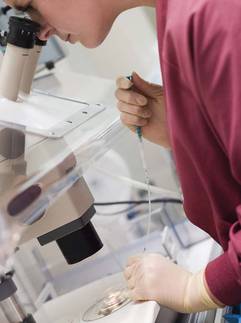
Meanwhile the adjusted odds ratio for total cancer risk in a cohort of more than 92,000 Danish, Finnish, and Swedish children born between 1982 and 2007 was 1.15.
The findings of both studies, presented at the annual meeting of the European Society of Human Reproduction and Embryology, are "reassuring," said Dr. Carrie Williams of the Institute for Child Health at University College London.
"This is fantastic news for the 5 million children born after ART [assisted reproductive technology], their families, fertility specialists, and also for [the] wider society," Dr. Williams, who was part of the U.K. study team, added.
"The main driver for our study was parental concern," Dr. Williams explained. "In a study of several hundred families of children born after ART, over a quarter of [parents] had concerns about the health of their child as a result of their method of conception, and many of these concerns [were] related to the overall cancer risk."
Previously, Dr. Williams and her associates performed a systematic review of 11 studies and found that there was a small but significant increase in cancer risk in children born after ART, although the results of one particular study swayed the overall findings (Arch. Dis. Child. 2011;96:A6). The results of another recent meta-analysis of 25 studies also point to a possible increased risk for cancer in children following assisted conception methods (Fertil. Steril. 2013;100:150-61).
A large, population study was needed to examine the issue further, however, and the U.K. team linked data held by the Human Fertilisation and Embryology Authority (HFEA) on births due to ART with the U.K.’s National Registry of Childhood Tumours (NRCT). Over a 16-year study period, there were 106,381 children born in England, Wales, or Scotland after nondonor ART. Of these, 14,896 (14%) developed cancer before their 15th birthday or before Jan. 1, 2009.
The actual data analysis was performed on 106,013 children because the time at risk for the others could not be determined. Standardized incidence rates (SIRs) were calculated for various childhood cancers by determining the observed versus expected rates of each. Potential effect-moderating factors, such as age, sex, birth weight, multiple births, treatment type, and cause of parental infertility, among others, were considered.
The occurrence of most childhood tumors was lower than expected, with SIRs of 0.94 for renal tumors, 0.91 for leukemia, 0.88 for neuroblastoma, 0.85 for central nervous system tumors, 0.59 for retinoblastoma, and 0.56 for germ cell tumors.
"We did find an increased risk of two very rare types of cancer – hepatoblastoma and rhabdomyosarcoma," Dr. Williams reported. SIRs for these tumor types were 3.27 and 2.62, respectively. Further analysis suggested that a lower birth weight (less than 2,500 g) was associated with a 10-fold increase in the risk for hepatoblastoma. No clear explanation could be found for the increased risk for the connective tissue cancer.
"If there is a risk, then the risk is small," said Dr. Anja Pinborg who presented separate data from the Committee on Nordic ART and Safety (CoNARTaS) study, a prospective study involving all children born after assisted conception in Denmark, Finland, and Sweden (Acta. Obstet. Gynecol. Scand. 2011;90:683-91). Data from medical birth, cancer, patient, and cause of death registries were linked to help determine if ART did indeed carry an increased risk for malignancy.
"We show similar results, perhaps a little bit of an increase in the risk for cancer, but not significantly," added Dr. Pinborg of the University of Copenhagen. A total of 178 children were diagnosed with cancer in the cohort after ART, compared with 643 cancer cases in children who were not artificially conceived, giving respective incidence rates of 1.9% vs. 1.7%. Children born after ART were found to be at increased risk for CNS tumors in the Nordic cohort, but the prevalence was 0.4%.
"It’s very important to underline that the control population of parents differs from the ART population of parents because the mothers are older and they are reproductively healthy," Dr. Pinborg noted. People can be helped to conceive a child, but the underlying cause of the infertility usually cannot be resolved, she said, suggesting subfertility per se might play a role.
The increased risk for rare childhood tumors seen in the U.K. study and in the CoNARTaS study could even be down to chance as the numbers used in the analyses are relatively small.
"When we subdivide by cancer types, our numbers get smaller and smaller, and therefore the role of chance is increased," Dr. Williams commented. "The answer is to keep studying this area, perhaps do another systematic review, putting all of the results together, and then we will have more reliable findings."
Dr. Williams added: "So far, it looks like the results are really positive for the many children born after assisted conception."
Cancer Research U.K. funded the U.K. study. The U.K. National Institute of Health Research provided funding to Dr. Williams. CoNARTaS is supported by ESHRE, Sahlgrenska University Hospital, the University of Copenhagen, the Danish Agency for Science Technology and Innovation, and the Nordic Federation of Societies of Obstetrics and Gynecology. Dr. Williams and Dr. Pinborg said they had no relevant financial disclosures.
LONDON – Children born as a result of assisted conception do not appear to be at any greater cancer risk than children born following spontaneous conception, the results of two large longitudinal studies demonstrated.
A U.K.-based study of more than 100,000 children born after assisted conception between 1992 and 2008 found a lower-than-expected number of cases of cancer, with a standardized incidence rate of 0.98.
Meanwhile the adjusted odds ratio for total cancer risk in a cohort of more than 92,000 Danish, Finnish, and Swedish children born between 1982 and 2007 was 1.15.
The findings of both studies, presented at the annual meeting of the European Society of Human Reproduction and Embryology, are "reassuring," said Dr. Carrie Williams of the Institute for Child Health at University College London.
"This is fantastic news for the 5 million children born after ART [assisted reproductive technology], their families, fertility specialists, and also for [the] wider society," Dr. Williams, who was part of the U.K. study team, added.
"The main driver for our study was parental concern," Dr. Williams explained. "In a study of several hundred families of children born after ART, over a quarter of [parents] had concerns about the health of their child as a result of their method of conception, and many of these concerns [were] related to the overall cancer risk."
Previously, Dr. Williams and her associates performed a systematic review of 11 studies and found that there was a small but significant increase in cancer risk in children born after ART, although the results of one particular study swayed the overall findings (Arch. Dis. Child. 2011;96:A6). The results of another recent meta-analysis of 25 studies also point to a possible increased risk for cancer in children following assisted conception methods (Fertil. Steril. 2013;100:150-61).
A large, population study was needed to examine the issue further, however, and the U.K. team linked data held by the Human Fertilisation and Embryology Authority (HFEA) on births due to ART with the U.K.’s National Registry of Childhood Tumours (NRCT). Over a 16-year study period, there were 106,381 children born in England, Wales, or Scotland after nondonor ART. Of these, 14,896 (14%) developed cancer before their 15th birthday or before Jan. 1, 2009.
The actual data analysis was performed on 106,013 children because the time at risk for the others could not be determined. Standardized incidence rates (SIRs) were calculated for various childhood cancers by determining the observed versus expected rates of each. Potential effect-moderating factors, such as age, sex, birth weight, multiple births, treatment type, and cause of parental infertility, among others, were considered.
The occurrence of most childhood tumors was lower than expected, with SIRs of 0.94 for renal tumors, 0.91 for leukemia, 0.88 for neuroblastoma, 0.85 for central nervous system tumors, 0.59 for retinoblastoma, and 0.56 for germ cell tumors.
"We did find an increased risk of two very rare types of cancer – hepatoblastoma and rhabdomyosarcoma," Dr. Williams reported. SIRs for these tumor types were 3.27 and 2.62, respectively. Further analysis suggested that a lower birth weight (less than 2,500 g) was associated with a 10-fold increase in the risk for hepatoblastoma. No clear explanation could be found for the increased risk for the connective tissue cancer.
"If there is a risk, then the risk is small," said Dr. Anja Pinborg who presented separate data from the Committee on Nordic ART and Safety (CoNARTaS) study, a prospective study involving all children born after assisted conception in Denmark, Finland, and Sweden (Acta. Obstet. Gynecol. Scand. 2011;90:683-91). Data from medical birth, cancer, patient, and cause of death registries were linked to help determine if ART did indeed carry an increased risk for malignancy.
"We show similar results, perhaps a little bit of an increase in the risk for cancer, but not significantly," added Dr. Pinborg of the University of Copenhagen. A total of 178 children were diagnosed with cancer in the cohort after ART, compared with 643 cancer cases in children who were not artificially conceived, giving respective incidence rates of 1.9% vs. 1.7%. Children born after ART were found to be at increased risk for CNS tumors in the Nordic cohort, but the prevalence was 0.4%.
"It’s very important to underline that the control population of parents differs from the ART population of parents because the mothers are older and they are reproductively healthy," Dr. Pinborg noted. People can be helped to conceive a child, but the underlying cause of the infertility usually cannot be resolved, she said, suggesting subfertility per se might play a role.
The increased risk for rare childhood tumors seen in the U.K. study and in the CoNARTaS study could even be down to chance as the numbers used in the analyses are relatively small.
"When we subdivide by cancer types, our numbers get smaller and smaller, and therefore the role of chance is increased," Dr. Williams commented. "The answer is to keep studying this area, perhaps do another systematic review, putting all of the results together, and then we will have more reliable findings."
Dr. Williams added: "So far, it looks like the results are really positive for the many children born after assisted conception."
Cancer Research U.K. funded the U.K. study. The U.K. National Institute of Health Research provided funding to Dr. Williams. CoNARTaS is supported by ESHRE, Sahlgrenska University Hospital, the University of Copenhagen, the Danish Agency for Science Technology and Innovation, and the Nordic Federation of Societies of Obstetrics and Gynecology. Dr. Williams and Dr. Pinborg said they had no relevant financial disclosures.
AT ESHRE 2013
Major finding: The standardized incidence rate for cancer in children from the United Kingdom was 0.98, and the odds ratio for total cancer risk in Nordic children was 1.15.
Data source: Two longitudinal studies: A U.K.-based study of cancer risk in more than 100,000 children born after assisted conception between 1992 and 2008 and CoNARTaS, a cohort of all (more than 92,000) children born after assisted conception in Denmark, Finland, and Sweden between 1982 and 2007.
Disclosures: Cancer Research U.K. funded the U.K. study. The U.K. National Institute of Health Research provided funding to Dr. Williams. CoNARTaS is supported by ESHRE, Sahlgrenska University Hospital, the University of Copenhagen, the Danish Agency for Science Technology and Innovation, and the Nordic Federation of Societies of Obstetrics and Gynecology. Dr. Williams and Dr. Pinborg said they had no relevant financial disclosures.
Poststroke prevention strategies cut dementia risk in half
LONDON – Cognitive dysfunction following a stroke could be reduced significantly if treatment with antihypertensive, antithrombotic, and lipid-lowering drugs were optimized, according to a study looking at the long-term impact of secondary prevention strategies.
The relative risk for cognitive dysfunction was 0.8 with the optimal use of anticoagulants, and 0.9, 0.9, and 0.9 with the optimal use of dual antihypertensive therapy, dual antiplatelet therapy, and lipid-lowering treatment, respectively.
"The combination of antihypertensives, antithrombotics, and lipid-lowering drugs reduced the risk of cognitive impairment by about half," Abdel Douiri, Ph.D., said at the annual European Stroke Conference. The benefit was seen in most stroke subtypes, although not in those with hemorrhagic stroke or in patients with stroke due to atrial fibrillation.
Dr. Douri of King’s College London and coinvestigators looked at whether preventing vascular events could be associated with a protective effect on patients’ overall cognitive function after a stroke. They used the population-based South London Stroke Register to identify 4,413 patients who had experienced a first stroke between 1995 and 2011. Patients were assessed for cognitive function using the Abbreviated Mental Test or Mini-Mental State Examination at about 3 months after their stroke and annually thereafter.
The mean age of patients was 70 years, 49% of the cohort were female, and 70.5% were white. Blacks (21.2%) and other ethnicities made up the remainder of the patient population.
Cognitive impairment rates after stroke vary by study, but have been shown to be relatively consistent over time, affecting up to a quarter of patients overall, Dr. Douri noted (Stroke 2013;44:138-45). Approximately 10% of stroke patients have cognitive impairment prior to their first stroke, 10% develop dementia after a single stroke, and 30% develop dementia after recurrent strokes (Lancet Neurol. 2009;8:1006-18).
Current treatment strategies for preventing cognitive impairment after stroke tend to focus on reducing the risk of recurrent stroke or other vascular events, although most studies to date have had short follow-up or too few patients, with only a small number of these having vascular causes for their cognitive problems.
"The use of recommended therapies after stroke appears to be associated with a protective effect," said Dr. Douiri.
Suboptimal lipid-lowering post stroke
Although optimizing poststroke medications might improve cognitive outcomes, it is not always achieved in routine care. The results of a separate prospective population-based study showed that lipid-lowering targets are not always being achieved.
"The suboptimal lipid control we observed, both preceding and following a stroke or TIA [transient ischemic attack], even where there was established vascular disease or risk factors, highlights the need for improved lipid management in patients who are at risk of stroke or TIA," said Dr. Danielle Ní Chróinín of Mater University Hospital and University College Dublin.
Dr. Chróinín and colleagues assessed patients’ lipid profiles and clinicians’ adherence to evidence-based guidelines for lipid-lowering medications after a stroke or transient ischemic attack using data from the North Dublin Population Stroke Study.
Over the course of the 1-year study, the medical records of 428 patients who had had an ischemic stroke and 188 who had had a TIA were analyzed. The mean age of patients was 71 years.
Lipid measurement at presentation and prescription of statin therapy at discharge were found to be less likely in female patients, those who were older, those with poorer modified Rankin scores before the event, and those with higher National Institutes of Health Stroke Scale scores. There was no difference in the likelihood of measurement or statin treatment based on the type of event or if the patient required hospitalization.
At presentation, only 33.7% of high-risk patients were being treated with lipid-lowering medications. Although 75.5% of patients were discharged on statin therapy, approximately one in four patients who should have been prescribed this medication were not taking a statin at discharge.
Of patients who were on lipid-lowering therapy, less than half (44%-46%) were achieving recommended target levels set by U.S. or European guidelines.
Statin treatment at discharge was more likely in patients who had concomitant diabetes or atherothrombotic stroke.
The study presented by Dr. Chróinín was supported by the Health Research Board, the Irish Heart Foundation, and a Mater University postgraduate research and education grant. Dr. Chróinín and Dr. Douiri said they had no relevant financial disclosures.
AT THE EUROPEAN STROKE CONFERENCE
Major finding: The relative risk for cognitive dysfunction was 0.8-0.9 with the use of recommended antihypertensive, antithrombotic, and lipid-lowering drugs.
Data source: A prospective, population-based South London Stroke Register of 4,413 patients who had a first-ever stroke between 1995 and 2011 and completed a global measure of cognitive function.
Disclosures: The study presented by Dr. Chróinín was supported by the Health Research Board, the Irish Heart Foundation, and a Mater University postgraduate research and education grant. Dr. Chróinín and Dr. Douiri said they had no relevant financial disclosures.
Serum biomarker predicts radiographic progression in spondyloarthritis
MADRID – Elevated serum levels of vascular endothelial growth factor may be predictive of radiographic progression in the spine, according to data from a German study of patients with spondyloarthritis.
The cutoff point appears to be at 600 pg/mL, with the effects particularly strong in patients who also develop syndesmophytes, which are bony growths that develop within ligaments.
"In patients with syndesmophytes, VEGF [vascular endothelial growth factor], as a predictor of radiographic progression, performed better than CRP [C-reactive protein]," reported Dr. Denis Poddubnyy at the annual European Congress of Rheumatology (Ann. Rheum. Dis. 2013;72:125).
"We all know that radiographic progression varies substantially among patients with spondyloarthritis," Dr. Poddubnyy observed. "Until recently there was only one strong predictor of radiographic progression: the presence of syndesmophytes at baseline," he added.

Last year, however, Dr. Poddubnyy of Charité Universitätsmedizin Berlin, Germany, and his associates published the findings of a study involving 210 patients with early axial spondyloarthritis (axSpA) who were recruited from the German Spondyloarthritis Inception Cohort (GESPIC). This study looked at baseline predictors of spinal radiographic progression over 2 years and found that in addition to radiographic damage, elevated CRP levels and cigarette smoking were independently predictive (Arthritis Rheum. 2012;64:1388-98).
The team’s research also suggested that there could be a few serum biomarkers, including VEGF, that could be predictive, so the investigators conducted a larger study in 172 patients with definite (n = 95) or nonradiographic (n = 77) axSpA to look specifically at the possible association.
Radiographs of the spine taken at baseline and at 2 years’ follow-up were reviewed independently by two readers, who used the modified Stoke Ankylosing Spondylitis Spinal Score (mSASSS) to record the extent of radiographic progression. If there was a worsening of 2 units or more in mSASSS and the formation of new or increased growth of syndesmophytes, radiographic progression had occurred.
In total, 22 patients had radiographic progression, including 18 with new formation or growth of syndesmophytes.
Baseline VEGF was measured in the serum at baseline and was significantly higher in patients who developed radiographic progression at 2 years than in those who did not (562 plus or minus 357 pg/mL vs. 402 plus or minus 309 pg/mL; P = .027).
Serum VEGF levels were also significantly higher in the patients who had developed new syndesmophytes at 2 years when compared with those who had not (579 plus or minus 386 pg/mL vs. 404 plus or minus 307 pg/mL; P = .041).
"Patients with elevated VEGF had an odds ratio of 2.9 for [radiographic] progression and 3.1 for syndesmophyte formation," Dr. Poddubnyy reported. This was a little disappointing, he noted, as CRP and the erythrocyte sedimentation rate had similar predictive power.
However, in patients at high risk of progression, namely those with syndesmophytes already present at baseline, VEGF was significantly better than CRP at predicting both radiographic progression and new syndesmophyte formation or growth.
Patients with elevated VEGF at baseline were 36.6 times more likely to have radiographic progression and 13.6 times more likely to have new syndesmophyte formation or growth at 2 years than were those with levels below 600 pg/mL. In comparison, elevated CRP levels increased the risks by only 2.4 and 2.5 times, respectively.
VEGF is an essential mediator of angiogenesis and endochondral ossification, Dr. Poddubnyy observed. It’s been shown to have "a chemoattractive effect on osteoblasts and mesenchymal progenitor cells," he added, and also stimulate osteoblast differentiation and bone turnover.
While the results are very promising, further research is of course required. The possible predictive value of VEGF in relation to spinal radiographic progression in patients treated with tumor necrosis factor–alpha inhibitors remains to be seen, for example, and future studies should perhaps look at this question.
"With VEGF we are probably able to improve our prediction of spinal progression in patients with axSpA," Dr. Poddubnyy said in an interview. This is addition to assessing "classical factors," such as syndesmophytes, CRP, and smoking status.
Dr. Poddubnyy had no disclosures.
MADRID – Elevated serum levels of vascular endothelial growth factor may be predictive of radiographic progression in the spine, according to data from a German study of patients with spondyloarthritis.
The cutoff point appears to be at 600 pg/mL, with the effects particularly strong in patients who also develop syndesmophytes, which are bony growths that develop within ligaments.
"In patients with syndesmophytes, VEGF [vascular endothelial growth factor], as a predictor of radiographic progression, performed better than CRP [C-reactive protein]," reported Dr. Denis Poddubnyy at the annual European Congress of Rheumatology (Ann. Rheum. Dis. 2013;72:125).
"We all know that radiographic progression varies substantially among patients with spondyloarthritis," Dr. Poddubnyy observed. "Until recently there was only one strong predictor of radiographic progression: the presence of syndesmophytes at baseline," he added.
Last year, however, Dr. Poddubnyy of Charité Universitätsmedizin Berlin, Germany, and his associates published the findings of a study involving 210 patients with early axial spondyloarthritis (axSpA) who were recruited from the German Spondyloarthritis Inception Cohort (GESPIC). This study looked at baseline predictors of spinal radiographic progression over 2 years and found that in addition to radiographic damage, elevated CRP levels and cigarette smoking were independently predictive (Arthritis Rheum. 2012;64:1388-98).
The team’s research also suggested that there could be a few serum biomarkers, including VEGF, that could be predictive, so the investigators conducted a larger study in 172 patients with definite (n = 95) or nonradiographic (n = 77) axSpA to look specifically at the possible association.
Radiographs of the spine taken at baseline and at 2 years’ follow-up were reviewed independently by two readers, who used the modified Stoke Ankylosing Spondylitis Spinal Score (mSASSS) to record the extent of radiographic progression. If there was a worsening of 2 units or more in mSASSS and the formation of new or increased growth of syndesmophytes, radiographic progression had occurred.
In total, 22 patients had radiographic progression, including 18 with new formation or growth of syndesmophytes.
Baseline VEGF was measured in the serum at baseline and was significantly higher in patients who developed radiographic progression at 2 years than in those who did not (562 plus or minus 357 pg/mL vs. 402 plus or minus 309 pg/mL; P = .027).
Serum VEGF levels were also significantly higher in the patients who had developed new syndesmophytes at 2 years when compared with those who had not (579 plus or minus 386 pg/mL vs. 404 plus or minus 307 pg/mL; P = .041).
"Patients with elevated VEGF had an odds ratio of 2.9 for [radiographic] progression and 3.1 for syndesmophyte formation," Dr. Poddubnyy reported. This was a little disappointing, he noted, as CRP and the erythrocyte sedimentation rate had similar predictive power.
However, in patients at high risk of progression, namely those with syndesmophytes already present at baseline, VEGF was significantly better than CRP at predicting both radiographic progression and new syndesmophyte formation or growth.
Patients with elevated VEGF at baseline were 36.6 times more likely to have radiographic progression and 13.6 times more likely to have new syndesmophyte formation or growth at 2 years than were those with levels below 600 pg/mL. In comparison, elevated CRP levels increased the risks by only 2.4 and 2.5 times, respectively.
VEGF is an essential mediator of angiogenesis and endochondral ossification, Dr. Poddubnyy observed. It’s been shown to have "a chemoattractive effect on osteoblasts and mesenchymal progenitor cells," he added, and also stimulate osteoblast differentiation and bone turnover.
While the results are very promising, further research is of course required. The possible predictive value of VEGF in relation to spinal radiographic progression in patients treated with tumor necrosis factor–alpha inhibitors remains to be seen, for example, and future studies should perhaps look at this question.
"With VEGF we are probably able to improve our prediction of spinal progression in patients with axSpA," Dr. Poddubnyy said in an interview. This is addition to assessing "classical factors," such as syndesmophytes, CRP, and smoking status.
Dr. Poddubnyy had no disclosures.
AT THE EULAR CONGRESS 2013
Major finding: Baseline serum VEGF levels were 562 plus or minus 357 pg/mL vs. 402 plus or minus 309 pg/mL in patients with and without radiographic progression, respectively, at 2 years (P = .027).
Data source: Study of 95 patients with definite axial spondyloarthritis (axSpA) and 77 with nonradiographic axSpA recruited from the German Spondyloarthritis Inception Cohort (GESPIC).
Disclosures: Dr. Poddubnyy had no disclosures.
