User login
Two studies reveal preneoplastic links between H. pylori and gastric cancer
Molecular pathways linked with CD44 variant 9 (CD44v9), a cell surface glycoprotein tied to aggressive gastric cancer after Helicobacter pylori infection, may open doors to stop cancer before it starts, according to two recent studies.
Findings from the first study suggest that persistent inflammation after eradication therapy may continue to drive cancer risk after infection, while the second study revealed a potential therapeutic target related to preneoplastic changes.
The first study, conducted by lead author Hitoshi Tsugawa, PhD, of Keio University, Tokyo, and colleagues, aimed to determine the origin of CD44v9-positive cancer stem-like cells.
“These cells strongly contribute to the development and recurrence of gastric cancer,” the investigators wrote. Their report is in Cellular and Molecular Gastroenterology and Hepatology. “However, the origin of CD44v9-positive cells is uncertain.”
The association between H. pylori infection and gastric cancer has been documented, along with a high risk of cancer when gastric epithelial cells overexpress capping actin protein of muscle Z-line alpha subunit 1 (CAPZA1), the researchers noted. Although it has also been shown that CAPZA1 overexpression leads to intracellular accumulations of the H. pylori–derived oncoprotein cytotoxin-associated gene A (CagA), just how these phenomena were connected remained unknown.
Through in vitro analyses of human cells, and in vitro and in vivo experiments involving Mongolian gerbils, the investigators uncovered a chain of events between H. pylori infection and CD44v9 expression. First, the investigators showed that expression levels of CD44v9 and CAPZA1 were directly correlated in five human cases of gastric cancer. Next, several experiments revealed that H. pylori–related oxidative stress drives overexpression of CAPZA1, which, in combination with high levels of beta-catenin, ESRP1, and CagA, promotes expression of CD44v9.
Most directly relevant to future therapies, the investigators compared levels of CAPZA1 between five active cases of H. pylori infection versus five cases successfully treated with eradication therapy. After eradication therapy, CAPZA1 overexpression decreased, but not to a significant degree.
“Our findings suggest that CAPZA1-overexpressing cells remaining in the gastric mucosa after eradication therapy increase the risk of metachronous gastric cancer and that reduction of CAPZA1 expression by amelioration of chronic inflammation after eradication therapy is important to prevent the development of gastric cancer,” the investigators concluded.
The second study, by lead author Anne R. Meyer, a graduate student at Vanderbilt University, Nashville, Tenn., and colleagues, evaluated how zymogenic chief cells are reprogrammed into spasmolytic polypeptide-expressing metaplasia (SPEM), a precursor to dysplasia and gastric cancer.
It had been previously shown that reprogramming to SPEM is promoted and maintained by epithelial cell damage, such as that caused by H. pylori infection, but underlying processes remained unclear, until recent studies suggested a link between SPEM transition and upregulation of CD44v9. Knowing that CD44v9 stabilizes the cystine/glutamate antiporter xCT, the investigators homed in on xCT for a closer look, questioning what role it had in chief cell reprogramming. Again, oxidative stress was identified as the inciting pathophysiologic driver.
“The oxidative stress response, including upregulation of nutrient transporters, plays an important role in many biological processes and the pathogenesis of a variety of diseases,” the investigators wrote in their report, published in Cellular and Molecular Gastroenterology and Hepatology. “Perturbations to the CD44v9-xCT system often result in redox imbalance.”
Using a combination of mouse and human cell lines, and a mouse model, the investigators demonstrated that xCT was upregulated during the initial stages of chief cell programming. Blocking xCT with sulfasalazine after acute gastric injury limited SPEM transition by more than 80%, an effect that was further supported by xCT siRNA knockdown and observations in xCT knockout mice. Reduction in chief cell reprogramming was not observed in the presence of sulfasalazine metabolites, suggesting that the anti-inflammatory properties of sulfasalazine were not responsible for downregulation of reprogramming.
“Targeting xCT may prove an effective tool for arresting metaplasia development in the stomach as well as mucous metaplasia in other epithelial tissues for the analysis of cellular plasticity and oxidative stress response,” the investigators concluded.
The study by Tsugawa and colleagues was funded by Grants-in-Aid for Scientific Research; the Yakult Bio-Science Foundation; the Ministry of Education, Culture, Sports, Science and Technology (MEXT)-supported program for the Strategic Research Foundation at Private Universities; and Keio Gijuku Academic Development Funds. Dr. Suzuki disclosed relationships with Daiichi-Sankyo Co, EA Pharma Co, Otsuka Pharmaceutical Co Ltd, and others. The study by Meyer and colleagues was funded by the National Institutes of Health, the American Association of Cancer Research, the Department of Defense, and others, with no relevant conflicts of interest.
SOURCES: Meyer et al. CMGH. 2019 May 6. doi: 10.1016/j.jcmgh.2019.04.015; Tsugawa et al. CMGH. 2019 May 27. doi: 10.1016/j.jcmgh.2019.05.008.
The mechanisms by which injured cells respond to stress rely in part on their ability to reprogram themselves in the setting of injury. This cellular reprogramming involves sensing and regulating intracellular metabolic cues that dictate survival, organization of secretory and degradative machinery, and proliferation. Meyer et al. and Tsugawa et al. illustrate two distinct mechanisms by which gastric epithelial cells handle oxidative stress during injury.
Meyer et al. focus on the xCT subunit of the cystine/glutamate antiporter as a rheostat for intracellular glutathione stores. Pharmacologic inhibition of xCT activity using sulfasalazine hampers the ability of injured gastric epithelial cells to adequately deal with reactive oxygen species. Importantly, these cells do not appropriately reprogram during injury and instead undergo apoptosis. Tsugawa et al. provide mechanistic insight into how oxidative stress may promote precancerous changes in gastric epithelium. Following H. pylori infection, an intracellular oxidative environment that is characterized by an overexpression of the actin filament capping protein CAPZA1, beta-catenin, and the alternative splicing factor ESRP1, promotes expression of CD44 variant 9 (CD44v9), a cell surface glycoprotein that correlates with gastric cancer. Interestingly, this oxidative milieu promotes accumulation of a critical H. pylori virulence factor, CagA, within infected cells.
Taken together, the ability to manage oxidative stress during cellular injury has significant implications for cell fate. It seems likely that the mechanisms for regulating intracellular oxidative stress are not unique to gastric epithelium and instead underlie a conserved injury response that has correlates in other gastrointestinal organs.
José B. Sáenz, MD, PhD, is an investigator and instructor of medicine in the gastroenterology division, John T. Milliken Department of Internal Medicine at the Washington University in St. Louis School of Medicine. He has no conflicts of interest.
Molecular pathways linked with CD44 variant 9 (CD44v9), a cell surface glycoprotein tied to aggressive gastric cancer after Helicobacter pylori infection, may open doors to stop cancer before it starts, according to two recent studies.
Findings from the first study suggest that persistent inflammation after eradication therapy may continue to drive cancer risk after infection, while the second study revealed a potential therapeutic target related to preneoplastic changes.
The first study, conducted by lead author Hitoshi Tsugawa, PhD, of Keio University, Tokyo, and colleagues, aimed to determine the origin of CD44v9-positive cancer stem-like cells.
“These cells strongly contribute to the development and recurrence of gastric cancer,” the investigators wrote. Their report is in Cellular and Molecular Gastroenterology and Hepatology. “However, the origin of CD44v9-positive cells is uncertain.”
The association between H. pylori infection and gastric cancer has been documented, along with a high risk of cancer when gastric epithelial cells overexpress capping actin protein of muscle Z-line alpha subunit 1 (CAPZA1), the researchers noted. Although it has also been shown that CAPZA1 overexpression leads to intracellular accumulations of the H. pylori–derived oncoprotein cytotoxin-associated gene A (CagA), just how these phenomena were connected remained unknown.
Through in vitro analyses of human cells, and in vitro and in vivo experiments involving Mongolian gerbils, the investigators uncovered a chain of events between H. pylori infection and CD44v9 expression. First, the investigators showed that expression levels of CD44v9 and CAPZA1 were directly correlated in five human cases of gastric cancer. Next, several experiments revealed that H. pylori–related oxidative stress drives overexpression of CAPZA1, which, in combination with high levels of beta-catenin, ESRP1, and CagA, promotes expression of CD44v9.
Most directly relevant to future therapies, the investigators compared levels of CAPZA1 between five active cases of H. pylori infection versus five cases successfully treated with eradication therapy. After eradication therapy, CAPZA1 overexpression decreased, but not to a significant degree.
“Our findings suggest that CAPZA1-overexpressing cells remaining in the gastric mucosa after eradication therapy increase the risk of metachronous gastric cancer and that reduction of CAPZA1 expression by amelioration of chronic inflammation after eradication therapy is important to prevent the development of gastric cancer,” the investigators concluded.
The second study, by lead author Anne R. Meyer, a graduate student at Vanderbilt University, Nashville, Tenn., and colleagues, evaluated how zymogenic chief cells are reprogrammed into spasmolytic polypeptide-expressing metaplasia (SPEM), a precursor to dysplasia and gastric cancer.
It had been previously shown that reprogramming to SPEM is promoted and maintained by epithelial cell damage, such as that caused by H. pylori infection, but underlying processes remained unclear, until recent studies suggested a link between SPEM transition and upregulation of CD44v9. Knowing that CD44v9 stabilizes the cystine/glutamate antiporter xCT, the investigators homed in on xCT for a closer look, questioning what role it had in chief cell reprogramming. Again, oxidative stress was identified as the inciting pathophysiologic driver.
“The oxidative stress response, including upregulation of nutrient transporters, plays an important role in many biological processes and the pathogenesis of a variety of diseases,” the investigators wrote in their report, published in Cellular and Molecular Gastroenterology and Hepatology. “Perturbations to the CD44v9-xCT system often result in redox imbalance.”
Using a combination of mouse and human cell lines, and a mouse model, the investigators demonstrated that xCT was upregulated during the initial stages of chief cell programming. Blocking xCT with sulfasalazine after acute gastric injury limited SPEM transition by more than 80%, an effect that was further supported by xCT siRNA knockdown and observations in xCT knockout mice. Reduction in chief cell reprogramming was not observed in the presence of sulfasalazine metabolites, suggesting that the anti-inflammatory properties of sulfasalazine were not responsible for downregulation of reprogramming.
“Targeting xCT may prove an effective tool for arresting metaplasia development in the stomach as well as mucous metaplasia in other epithelial tissues for the analysis of cellular plasticity and oxidative stress response,” the investigators concluded.
The study by Tsugawa and colleagues was funded by Grants-in-Aid for Scientific Research; the Yakult Bio-Science Foundation; the Ministry of Education, Culture, Sports, Science and Technology (MEXT)-supported program for the Strategic Research Foundation at Private Universities; and Keio Gijuku Academic Development Funds. Dr. Suzuki disclosed relationships with Daiichi-Sankyo Co, EA Pharma Co, Otsuka Pharmaceutical Co Ltd, and others. The study by Meyer and colleagues was funded by the National Institutes of Health, the American Association of Cancer Research, the Department of Defense, and others, with no relevant conflicts of interest.
SOURCES: Meyer et al. CMGH. 2019 May 6. doi: 10.1016/j.jcmgh.2019.04.015; Tsugawa et al. CMGH. 2019 May 27. doi: 10.1016/j.jcmgh.2019.05.008.
FROM CELLULAR AND MOLECULAR GASTROENTEROLOGY AND HEPATOLOGY
AGA Clinical Practice Update on the utility of endoscopic submucosal dissection in T1b esophageal cancer: Expert review
Endoscopic submucosal dissection (ESD) is a viable treatment option for patients with submucosal (T1b) esophageal cancer who have a low risk of lymph node metastasis, according to an expert review.
Among patients with T1b esophageal cancer, ideal candidates for ESD have small (less than 2 cm), well-differentiated tumors that do not invade beyond the superficial submucosa (SM1) and lack lymphovascular invasion, reported lead author Mohamed O. Othman, MD, of Baylor College of Medicine in Houston, and colleagues. The literature review was recently commissioned by the American Gastroenterological Association (AGA), because of high clinical relevance.
“[ESD] has been gaining momentum as an alternative to surgery in treating early gastrointestinal neoplasms,” the investigators wrote in Clinical Gastroenterology and Hepatology.
Most patients who undergo surgical resection develop gastroesophageal reflux, the investigators noted, and many others develop serious complications or do not survive the procedure.
“Even a high-volume center such as Mayo Clinic reported a surgical mortality of 4% for T1a esophageal cancer,” the investigators wrote. “Moreover, 34% of patients developed postoperative complications such as anastomotic leaks, anastomotic strictures, cardiopulmonary complications, and feeding jejunostomy leaks. ... Therefore, a less-invasive alternative to esophagectomy would be extremely valuable in the management of early stage [esophageal cancer] if proven effective.”
The investigators reviewed studies evaluating safety and efficacy of surgical and endoscopic techniques, as well as available data for chemoradiation and radiofrequency ablation combinations, which could potentially optimize outcomes of endoscopic resection.
They concluded that most patients with esophageal cancer that does not extend beyond the mucosa (T1a) can be cured with endoscopic resection, based on 5-year survival rates from several Japanese trials. For patients with T1b disease, however, ESD is best suited for those with a low risk of lymph node metastasis. Unfortunately, identifying these candidates can be challenging, according to the investigators.
“The risk of lymph node metastasis depends on the depth of invasion, histologic type, and molecular characterization of the tumor,” the investigators explained, noting that depth of invasion is the trickiest to discern. Although endoscopic ultrasound (EUS) is still recommended for submucosal imaging, the review showed that EUS may overstage cancer in Barrett’s esophagus. The investigators suggested that volume laser endoscopy with infrared light could be a more accurate alternative, but it is not yet a clinical reality.
The review also showed potential for combining ESD with other modalities. For example, a study by Hamada and colleagues involving 66 patients with submucosal (T1b) esophageal squamous cell carcinoma found that a combination of ESD with chemoradiation led to similar 3- and 5-year survival rates as radical esophagectomy. The investigators highlighted the importance of lymph node metastasis in this study, as none of the 30 patients lacking lymph node involvement had metastatic recurrence, compared with 6 of the 36 patients who exhibited lymph node metastasis. According to the investigators, promising data are also anticipated for this combination among those with adenocarcinoma. And for patients with intestinal metaplasia and/or dysplasia, adding radiofrequency ablation after ESD appears to be an effective option; one recent study by Sharmila Subramaniam, BMBS, and colleagues found that this strategy led to clearance rates of 85% and 96% for metaplasia and dysplasia, respectively.
“Additional treatment should be determined by factors such as tumor grade, status of lymphovascular invasion, and depth of tumor, which have a direct influence on metastatic potential,” the investigators wrote.
The investigators suggested that, in the future, better diagnostics will be needed to characterize T1b disease, as this could streamline patient selection. “Future research should focus on novel biological and immunohistochemistry markers that can aid in the prediction of tumor behavior and [lymph node metastasis] in T1b esophageal cancer,” they concluded.
The study was commissioned by the American Gastroenterological Association. The investigators disclosed additional relationships with Boston Scientific, Olympus, Lumendi, and others.
SOURCE: Othman MO et al. CGH. 2019 Jun 4. doi: 10.1016/j.cgh.2019.05.045.
FROM CLINICAL GASTROENTEROLOGY AND HEPATOLOGY
Hormone therapy may do more harm than good for men with low PSA before early salvage radiation
For men with prostate cancer who have a low prostate-specific antigen (PSA) level prior to salvage radiation therapy (SRT), adding an antiandrogen may increase the risk of other-cause mortality by twofold or more, according to investigators.
This finding was drawn from a secondary analysis of the NRG Oncology/RTOG 9601 phase 3 trial, a practice-changing study that showed that 2 years of antiandrogen therapy with bicalutamide improved overall survival when added to SRT, compared with that of SRT alone.
Results from the present analysis paint a more complex picture, revealing that patients with low PSA levels who received hormone therapy had a higher rate of other-cause mortality, primarily because of high-grade cardiac and neurologic events, reported lead author Daniel Spratt, MD, of the University of Michigan Rogel Cancer Center, Ann Arbor, who presented findings at the annual meeting of the American Society for Radiation Oncology.
Dr. Spratt described how treatment paradigms have changed since RTOG 9601 began in 1998, which prompted a revisitation of the trial. “Almost half of the men [in the trial] had a persistently elevated PSA after they underwent their initial surgery,” Dr. Spratt said. “This is uncommonly seen today. Additionally, about 60% of patients received what we call late salvage radiation therapy, where PSA was monitored and continued to rise beyond 0.5 [ng/mL]; again, this is not what is recommended to be used today.”
In the present analysis, the investigators stratified patients by PSA level. Of the 760 men involved, 85% had a PSA of 0.2-1.5 ng/mL. Patients were further subgrouped by those with a PSA of 0.2-0.6 ng/mL and those with a very low PSA, of 0.2-0.3 ng/mL. Multiple endpoints were assessed, including overall survival, other-cause mortality, distant metastasis, and rates of grade 3-5 cardiac or neurologic events.
The analysis showed that men with a PSA greater than 1.5 ng/mL had improved survival (hazard ratio, 0.45; 95% confidence interval, 0.25-0.81) when treated with bicalutamide, but those with PSA of 1.5 ng/mL or less did not (HR, 0.87; 95% CI, 0.66-1.16). Looking more closely at patients with a PSA of 1.5 ng/mL or less, those with a PSA of 0.2-0.6 ng/mL had a twofold increased rate of other-cause mortality (subdistribution HR, 1.94; P = .009). The picture became even more concerning for patients with a PSA of 0.2-0.3 ng/mL who were treated with bicalutamide: They had a fourfold increased risk of other cause mortality (sHR, 4.14). Among the cases with elevated other-cause mortality, grade 3-4 cardiac and neurologic events were likely to be blamed.
“What is likely driving this, and of concern, is that for [patients with a] PSA of less than 1.5 [ng/mL] … there was a three- to four-and-a-half-fold increased risk of high grade cardiac events,” Dr. Spratt said.
“The current guidelines recommend that all men be offered hormone therapy when receiving salvage radiation therapy, but our data demonstrate that men with lower PSA’s are probably more harmed than helped by long-term hormone therapy,” Dr. Spratt concluded. “We now have three randomized trials with over 2,400 men in total, [none of which showed that] short- or long-term hormone therapy improves overall survival in men [with a low PSA level who receive] early salvage radiotherapy. Thus, PSA prior to salvage radiation actually is not only prognostic, it predicts who will benefit most from hormone therapy, and guidelines should now be updated to reflect this finding.”
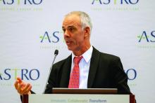
The session moderator, Anthony Zietman, MD, of Massachusetts General Hospital, Boston, helped put the findings in perspective: “This really suggests that we’ve got to hold back a little,” Dr. Zietman said. “There are some people who really benefit [from hormone therapy], some who don’t benefit, and some who just might be harmed, so I think we can be much more thoughtful and cautious in the future. … Thirty thousand men a year are in this situation who could be receiving this treatment. You could fill Fenway Park with that many people. So there are some big downstream implications. From here on out, I’m going to be a lot more cautious with my patients.”
The study was primarily funded by AstraZeneca with additional support from the Korea Institute of Radiological and Medical Sciences. The investigators disclosed relationships with Novartis, Roche, Amgen, and others.
SOURCE: Spratt et al. ASTRO 2019, Abstract LBA1.
FROM ASTRO 2019
PACIFIC: Patterns of lung cancer progression suggest role for local ablative therapy
Most patients with stage III non–small cell lung cancer (NSCLC) who have distant progression on standard therapy typically have one or two new lesions, often in the same organ, which suggests a role for local ablative therapy, according to investigators.
This conclusion was drawn from an exploratory analysis of the phase 3 PACIFIC trial, which previously showed that durvalumab prolonged survival among patients with NSCLC who did not progress after chemoradiotherapy, which turned the trial protocol into a new standard of care.
At the annual meeting of the American Society for Radiation Oncology, coauthor Andreas Rimner, MD, of the Memorial Sloan Kettering Cancer Center in New York presented findings.
“There were always questions regarding detailed patterns of failure and disease progression in [the PACIFIC] trial,” Dr. Rimner said. “This study ... focuses on these patterns of failure, including the type of first progression in the patients on the PACIFIC trial.”
During the trial, 713 patients with NSCLC were randomized in a 2:1 ratio to receive either durvalumab or placebo. After a median follow-up of 25.2 months, the superiority of durvalumab was clear, with a lower rate of progression (45.4% vs. 64.6%).
But the present analysis dug deeper into this finding by dividing patients into three groups based on site or sites of first progression: local (intrathoracic) progression only, distant (extrathoracic) progression only, or simultaneously local and distant progression. Scans were reviewed by an independent radiologist who was not involved in the original PACIFIC trial. In addition to spatial data, the investigators reported times until progression.
Regardless of site, durvalumab was associated with a longer time until progression or death. Although comparative values were not reached for distant or simultaneous spread, median time until local progression or death was reportable, at 25.2 months in the durvalumab group versus with 9.2 months in the placebo group.
These values were available, in part, because local spread was the most common type of progression: It occurred in 80.6% of patients who progressed on durvalumab and 74.5% of progressors in the placebo group.
Durvalumab reduced the rate of progression across the three spatial categories, compared with placebo, including local only (36.6% vs. 48.1%, respectively), distant only (6.9% vs. 13.1%), and simultaneously local and distant (1.9% vs. 3.4%). This means that, at first progression, new distant lesions were found in 8.8% of patients treated with durvalumab, compared with 16.5% of those treated with placebo. Of note, approximately two-thirds of patients with distant progression had only one or two distant lesions, often confined to one organ, most commonly the brain. This pattern of progression was observed in both treatment arms.
According to Dr. Rimner, this finding is clinically relevant because it suggests a potential role for local ablative therapy.
Expert perspective on the analysis was provided by Benjamin Movsas, MD, chair of radiation oncology at the Henry Ford Cancer Institute in Detroit.
“The PACIFIC trial has really transformed the standard of care for patients with locally advanced, inoperable non–small cell lung cancer by adding immunotherapy to the prior standard of care combining chemotherapy and radiation, and this has shown a dramatic improvement in survival,” Dr. Movsas said.
“By adding the immunotherapy durvalumab, you can reduce risk of local failure, you can reduce the risk of distant failure, and interestingly enough, when patients do fail distantly, and this is true in both arms, they tended to fail in only one or two spots, which is encouraging because that suggests maybe a window of opportunity to treat those one or two spots, and we have newer technologies that allow us to consider that. So we really have a new paradigm.”
The study was funded by AstraZeneca. The investigators disclosed additional relationships with Merck, Nanobiotix, Boehringer Ingelheim, and others.
SOURCE: Rimner A et al. ASTRO 2019, Abstract LBA6.
REPORTING FROM ASTRO 2019
Key clinical point: Most patients with stage 3 non–small cell lung cancer (NSCLC) who have distant progression on standard therapy typically have one or two new lesions, often in the same organ, which suggests a role for local ablative therapy.
Major finding: Approximately two-thirds of patients with distant progression had one or two new lesions.
Study details: An exploratory analysis of patterns of progression in the phase 3 PACIFIC trial, which involved 713 patients with stage III NSCLC that had not progressed after chemoradiotherapy.
Disclosures: The study was funded by AstraZeneca. The investigators disclosed additional relationships with Merck, Nanobiotix, Boehringer Ingelheim, and others.
Source: Rimner A et al. ASTRO 2019, Abstract LBA6.
SABR may put immunological brakes on oligometastatic prostate cancer
Stereotactic ablative radiation therapy (SABR) may be able to extend progression-free survival (PFS) among patients with oligometastatic prostate cancer, based on results from the phase 2 ORIOLE trial.
SABR appeared to control disease, in part, by triggering a systemic immune response, reported lead author Ryan Phillips, MD, PhD, chief resident of radiation oncology at Johns Hopkins Sidney Kimmel Cancer Center in Baltimore, at the annual meeting of the American Society for Radiation Oncology. This is a particularly noteworthy finding, since prostate cancer is generally considered to be immunologically cold, Dr. Phillips explained.
The ORIOLE trial also demonstrated how prostate-specific membrane antigen (PSMA)–based PET/CT scans can be used to more accurately predict metastasis by detecting lesions that would otherwise be missed by standard imaging.
“There’s a hypothesis that these first few sites of spread pave the way for additional widespread metastasis down the road,” Dr. Phillips said, “and that if we can treat all detectable disease early enough, we may be able to provide long-term control or in the best-case scenario, be able to cure these patients of early metastatic disease.”
The trial involved 54 men with recurrent, hormone-sensitive prostate cancer who had been previously treated with radiation therapy or surgery. Additional eligibility requirements included one to three lesions that were 5 cm or smaller, detectable by MRI, CT, or bone scan; a prostate-specific antigen (PSA) doubling time of less than 15 months; and an Eastern Cooperative Oncology Group performance status of 2 or less.
Patients were randomized in a 2:1 ratio to receive either SABR or observation, with follow-up every 3 months including physical exam and PSA measurement, and at 6 months, CT and bone scan. The primary endpoint was disease progression at 6 months.
In addition to this protocol, biomarker correlative analyses were performed, including PSMA-based PET/CT, T-cell clonality testing, and circulating tumor DNA (ctDNA) assessment.
Results showed that significantly fewer men treated with SABR had disease progression at 6 months (19% vs. 61%; P = .005). This benefit extended beyond the primary endpoint, as median PFS among patients treated with SABR was not yet reached after more than a year of follow-up, while those in the observation group had a median PFS of 5.8 months (P = .0023).
“Clinically, this is promising,” Dr. Phillips said, “but we also were really interested in learning more about oligometastatic prostate cancer, and something that sets ORIOLE apart are the correlative studies.”
The first of these studies involved PSMA-based PET/CT, which can identify lesions that may be missed or underappreciated with conventional imaging. Within the SABR group, 35 of 36 men had PSMA-based PET/CT performed at baseline and 6 months later. Of these, 19 had all PSMA-based PET/CT–detectable lesions treated with SABR (total consolidation), while 16 had subtotal consolidation. This difference was predictive of outcome, as only 16% of patients with total consolidation developed new lesions 6 months later, compared with 63% of those who had subtotal consolidation (P = .006).
“Not only are we treating the disease we’re detecting, but it seems to be preventing the development of new metastases outside the areas that we treated,” Dr. Phillips said. “This also held when we looked at points beyond 6 months,” he added.
Further testing showed that SABR triggered a systemic adaptive immune response involving expansion of T-cell clones. “There’s a lot more activity and changes within the immune system that were only seen within the SABR arm,” Dr. Phillips said, noting that these changes were “similar in scope to what we see after a vaccination.”
Finally, the investigators assessed ctDNA, which showed that men with at least one high-risk mutation had similar outcomes regardless of treatment group; in contrast, those without any high-risk mutations had significantly better PFS when treated with SABR.
“These are low sample size, hypothesis-generating experiments,” Dr. Phillips said, “but it is promising that there may be measurable baseline factors that will help us decide which patients are likely to benefit from this approach, and which would really be better served with an alternate treatment strategy.”
Session moderator Anthony Zietman, MD, of Massachusetts General Hospital, Boston, suggested that the ORIOLE trial could have a big future. “This is a small study, but it’s a prospective study, and the findings are really provocative,” Dr. Zietman said. “Just imagine, if there was a patient with just two or three metastatic lesions, and by irradiating those, you could liberate proteins from the destroyed metastases, generate an immune response, and suppress the development of new metastases, that will be something extraordinary. So this trial will be followed very closely as it seems to be hinting at something that’s a bit of a Holy Grail in oncology.”
The investigators disclosed relationships with RefleXion Medical, Pfizer, Genentech, and others.
SOURCE: Phillips R et al. ASTRO 2019. Abstract LBA3.
FROM ASTRO 2019
Adding radiation to immunotherapy may extend PFS in progressive lung cancer
For patients with metastatic non–small cell lung cancer (NSCLC) who have disease progression on immunotherapy, adding stereotactic body radiotherapy (SBRT) could improve progression-free survival (PFS), according to investigators.
Patients with more CD8+ T cells in circulation, and those with higher tumor infiltrating lymphocyte (TIL) scores derived the most benefit from SBRT, lead author Allison Campbell, MD, PhD, of Yale Cancer Center in New Haven, Conn., and colleagues, reported at the annual meeting of the American Society for Radiation Oncology.
“In rare cases, adding radiation to immunotherapy has been shown to result in therapeutic synergy,” Dr. Campbell said. “When we give high-dose radiation to patients on immunotherapy, some tumors that were not targeted by the radiation can shrink, and this is called ‘the abscopal effect.’ ”
The investigators designed the phase 2 trial to determine if the abscopal effect would occur if high-dose radiation was delivered to a single site in patients who had progressed on checkpoint inhibitor therapy. Fifty-six patients were enrolled, all with at least two sites of metastatic NSCLC. Of these patients, 6 had already progressed on immunotherapy, while 50 were naive to immunotherapy and began pembrolizumab during the trial, with 16 eventually progressing; collectively, these 22 patients with disease progression were identified as candidates for SBRT. Almost all candidates (21 out of 22) completed SBRT, which was delivered in three or five high-dose fractions. Only one site was treated, while other sites were tracked over time with computed tomography (CT) to assess for the abscopal effect. In addition, blood was analyzed for circulating immune cell composition.
After a median follow-up of 15.2 months, the disease control rate was 57%, with some abscopal responses detected. Two patients (10%) achieved a partial response lasting more than 1 year, and 10 patients (48%) maintained stable disease after SBRT. Although programmed death-ligand 1 (PD-L1) positivity was associated with a trend toward increased PFS, this was not statistically significant. In contrast, TIL score was significantly correlated with PFS; patients with TIL scores of 2-3 had a median PFS of 6.7 months, compared with 2.2 months among those with TIL scores of 1 or less. Similarly, immune-related adverse events predicted outcome, with patients who experienced such events achieving longer median PFS than those who did not (6.5 vs 2.2 months). Furthermore, blood testing revealed that the best responders had more CD8+ killer T cells and fewer CD4+ regulatory T cells in peripheral blood compared with patients who responded poorly.
After Dr. Campbell’s presentation, Benjamin Movsas, MD, chair of radiation oncology at the Henry Ford Cancer Institute in Detroit, offered some expert insight. “[The findings from this study] suggest perhaps that radiation may be able to reinvigorate the immune system,” Dr. Movsas said. “Maybe we can get more mileage out of the immunotherapy with this approach. Could radiation kind of be like an immune vaccine of sorts? There’s a lot of exciting possibilities.”
Dr. Movsas also noted how biomarker findings may be able to guide treatment decisions, highlighting how T cell populations predicted outcomes. “This era of precision medicine is really helping us improve benefits,” he said. “The immune profile really matters.”
The investigators disclosed relationships with Genentech, AstraZeneca, Merck, and others.
SOURCE: Campbell et al. ASTRO 2019. Abstract 74.
REPORTING FROM ASTRO 2019
Closure of women’s health clinics may negatively impact cervical cancer outcomes
.
States with a decreased number of women’s clinics per capita between 2010 and 2013 were found to have less screening for cervical cancer, more advanced stage of cervical cancer at presentation, and higher mortality from cervical cancer than states with no decrease in clinics, reported lead author Amar J. Srivastava, MD, of Washington University in St. Louis, who also noted that these changes occurred within a relatively short time frame.
“We know that women are generally diagnosed through the utilization of Pap smears,” Dr. Srivastava said during a presentation at the annual meeting of the American Society for Radiation Oncology. “These are low-cost tests that are available at multiple low-cost women’s health clinics. Unfortunately ... over the course of the past decade, we’ve seen a significant reduction of these clinics throughout the United States.”
“Between 2010 and 2013, which is the period of interest in this study, we know that about 100 of these women’s health clinics closed,” Dr. Srivastava said. “This was due to a combination of several factors; some of it was due to funding, some of it was due to restructuring of the clinics, and there were also laws passed throughout many states that ultimately led to the closure of many clinics.”
To determine the impact of these closures, the investigators first divided states into those that had women’s clinic closures between 2010 and 2013 and those that did not. Comparisons between these two cohorts involved the use of two databases. The first was the Behavioral Risk Factors Surveillance Study (BRFSS), which provided data from 197,143 cases, enabling assessment of differences between screening availability. The second database was the Surveillance, Epidemiology, and End Results (SEER) registry, which provided data from 10,652 patients, facilitating comparisons of stage at time of diagnosis and mortality rate.
Results were described in terms of relative differences between the two cohorts. For instance, screening rate among women with cervical cancer in states that had a decreased number of clinics was 1.63% lower than in states that did not lose clinics. This disparity was more pronounced in specific demographic subgroups, including Hispanic women (–5.82%), women aged between 21 and 34 years (–5.19%), unmarried women (–4.10%), and uninsured women (–6.88%).
“Historically, these are marginalized, underserved groups, and unfortunately, it comes as no surprise that these were the groups of women who were most dramatically hit by these changes,” Dr. Srivastava said.
Early-stage diagnosis was also significantly less common in states that had a decreased number of clinics, by a margin of 13.2%. Finally, the overall mortality rate among women with cervical cancer was 36% higher in states with clinic closures, a difference that climbed to 40% when comparing only metro residents.
Connecting the dots, Dr. Srivastava suggested that the decreased availability of screening may have led to fewer diagnoses at an early stage, which is more curable than late-stage disease, ultimately translating to a higher mortality rate. After noting that this chain of causality cannot be confirmed, owing to the retrospective nature of the study, Dr. Srivastava finished his presentation with a call to action.
“These findings should really give us some pause,” he said, “as physicians, as people who care about other people, to spend some time, try to figure out what’s going on, and try to address this disparity.”
After the presentation, Geraldine M. Jacobsen, MD, chair of radiation oncology at West Virginia University Cancer Institute, in Morgantown, W.V., echoed Dr. Srivastava’s concern.
“This study really raises broader questions,” Dr. Jacobsen said. “In the United States we’re always engaged in an ongoing dialogue about health care, health care policy, [and] health care costs. But a study like this brings to us the human face of what these dialogues mean. Policy affects people, and if we make changes in health care policy or health care legislation, we’re impacting people’s health and people’s lives.”
The investigators disclosed relationships with Phelps County Regional Medical Center, the Elsa U. Pardee Foundation, the American Society of Clinical Oncology, and ASTRO.
SOURCE: Srivastava AJ et al. ASTRO 2019, Abstract 202.
REPORTING FROM ASTRO 2019
Ovarian function suppression gains support for premenopausal breast cancer
Adding 2 years of ovarian function suppression (OFS) to the standard 5-year regimen of tamoxifen could improve disease-free and overall survival in women with estrogen receptor–positive breast cancer who have been previously treated with chemotherapy and definitive surgery, according to results from the phase 3 ASTRRA trial.
The findings add support to recent results from the similarly designed Suppression of Ovarian Function Trial (SOFT), reported Hyun-Ah Kim, MD, PhD, of Korea Cancer Center Hospital, Seoul, and colleagues.
“Although OFS in breast cancer has been studied for decades and has been used widely in clinical practice, evidence for the benefits of adding OFS to standard adjuvant tamoxifen treatment is insufficient,” the investigators wrote in the Journal of Clinical Oncology.
The ASTRRA trial enrolled 1,483 premenopausal women aged 45 years or younger with estrogen receptor–positive breast cancer who had been previously treated with chemotherapy and definitive surgery. Of those, 1,293 women were randomized to receive either 5 years of tamoxifen, or the same regimen plus 2 years of OFS, at 35 treatment centers in South Korea. In all, 1,282 women were eligible for analysis.
The primary endpoint was disease-free survival, defined as secondary malignancy, invasive contralateral breast cancer, invasive local recurrence, regional recurrence, distant recurrence, or death from any cause. The secondary endpoint was overall survival.
After a median follow-up of 63 months, women who received OFS in addition to tamoxifen had an estimated disease-free survival rate of 91.1%, compared with 87.5% in those who received tamoxifen alone (P = .033). Similarly, adding OFS was associated with a better estimated 5-year overall survival rate, compared with standard monotherapy (99.4% vs. 97.8%; P = .029), Dr. Kim and associates said.
Despite having a shorter follow-up and smaller population size, the results from ASTRRA were similar to those from SOFT, most likely because ASTRRA patients had higher-risk disease, the investigators noted.
“The results of ASTRRA confirm the findings of SOFT, that the addition of OFS to tamoxifen provides survival benefits for women [who are] at sufficient risk for recurrence to receive adjuvant chemotherapy and who remain in a premenopausal state after chemotherapy,” they concluded.
The study was primarily funded by AstraZeneca, with additional support from the Korea Institute of Radiological and Medical Sciences. The investigators disclosed relationships with Novartis, Roche, Amgen, and others.
SOURCE: Kim HA et al. J Clin Oncol. 2019 Sep 16. doi: 10. 1200/JCO.19.00126.
FROM THE JOURNAL OF CLINICAL ONCOLOGY
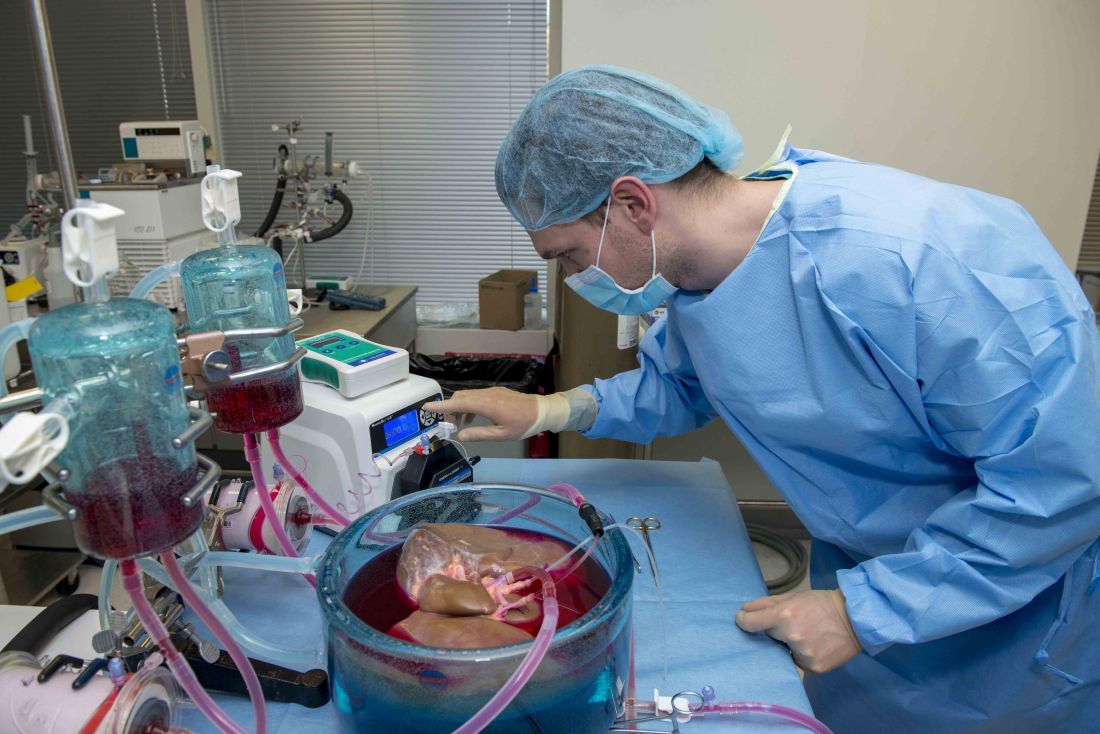
Supercooling extends donor liver viability by 27 hours
Standard cooling to 4°C provides just 12 hours of organ preservation, but laboratory testing showed that supercooling to –4°C added 27 hours of viability, reported lead author Reinier J. de Vries, MD, of Harvard Medical School and Massachusetts General Hospital in Boston, and colleagues.
“The absence of technology to preserve organs for more than a few hours is one of the fundamental causes of the donor organ–shortage crisis,” the investigators wrote in Nature Biotechnology.
Supercooling organs to high-subzero temperatures has been shown to prolong organ life while avoiding ice-mediated injury, but techniques that are successful for rat livers have been difficult to translate to human livers because of their larger size, which increases the risk of ice formation, the investigators explained.
Three strategies were employed to overcome this problem: minimization of air-liquid interfaces, development of a new supercooling-preservation solution, and hypothermic machine perfusion to more evenly distribute preservation solution throughout the liver tissue. For recovery of organs after supercooling, the investigators used subnormothermic machine perfusion, which has been used effectively in rat transplants.
In order to measure the impact of this process on organ viability, the investigators first measured adenylate energy content, both before supercooling and after recovery.
“Adenylate energy content, and, particularly, the organ’s ability to recover it during (re)perfusion, is considered the most representative metric for liver viability,” they wrote.
The difference between pre- and postsupercooling energy charge was less than 20%; in comparison, failed liver transplants in large animals and clinical trials have typically involved an energy-charge loss of 40% or more.
To further test organ viability, the investigators measured pre- and postsupercooling levels of bile production, oxygen uptake, and vascular resistance. All of these parameters have been shown to predict transplant success in rats, and bile production has additional precedent from human studies.
On average, bile production, portal resistance, and arterial resistance were not significantly affected by supercooling. Although portal vein resistance was 20% higher after supercooling, this compared favorably with increases of 100%-150% that have been measured in nonviable livers. Similarly, oxygen uptake increased by a mean of 17%, but this was three times lower than changes that have been observed in livers with impaired viability, at 51%.
Additional measures of hepatocellular injury, including AST and ALT, were also supportive of viability after supercooling. Histopathology confirmed these findings by showing preserved tissue architecture.
“In summary, we find that the human livers tested displayed no substantial difference in viability before and after extended subzero supercooling preservation,” the investigators wrote.
To simulate transplantation, the investigators reperfused the organs with blood at a normal temperature, including platelets, complement, and white blood cells, which are drivers of ischemia reperfusion injury. During this process, energy charge remained stable, which indicates preserved mitochondrial function. While energy charge held steady, lactate metabolism increased with bile and urea production, suggesting increased liver function. Bile pH and HCO3– levels fell within range for viability. Although bile glucose exceeded proposed criteria, the investigators pointed out that levels still fell within parameters for research-quality livers. Lactate levels also rose within the first hour of reperfusion, but the investigators suggested that this finding should be interpreted with appropriate context.
“It should be considered that the livers in this study were initially rejected for transplantation,” they wrote, “and the confidence intervals of the lactate concentration at the end of reperfusion largely overlap with time-matched values reported by others during [normothermic machine perfusion] of rejected human livers.”
Hepatocellular injury and histology also were evaluated during and after simulated transplantation, respectively, with favorable results. Although sites of preexisting hepatic injury were aggravated by the process, and rates of apoptosis increased, the investigators considered these changes were clinically insignificant.
Looking to the future, the investigators suggested that further refinement of the process could facilitate even-lower storage temperatures while better preserving liver viability.
“The use of human livers makes this study clinically relevant and promotes the translation of subzero organ preservation to the clinic,” the investigators concluded. “However, long-term survival experiments of transplanted supercooled livers in swine or an alternative large animal model will be needed before clinical translation.”
The study was funded by the National Institutes of Health and the Department of Defense. Dr. de Vries and four other coauthors have provisional patent applications related to the study, and one coauthor disclosed a financial relationship with Organ Solutions.
SOURCE: de Vries RJ et al. Nature Biotechnol. 2019 Sep 9. doi: 10.1038/s41587-019-0223-y.
FROM NATURE BIOTECHNOLOGY
Nonmyeloablative conditioning carries lowers infection risk in patients with AML
For patients with acute myeloid leukemia (AML) in need of allogeneic hematopoietic cell transplantation (alloHCT), reduced-intensity/nonmyeloablative conditioning (RIC/NMA) offers a lower risk of infection than myeloablative conditioning (MAC), based on a retrospective study involving more than 1,700 patients.
Within 100 days of treatment, patients who underwent MAC were significantly more likely to develop a bacterial infection, and develop it at an earlier date, than patients who had undergone RIC/NMA, reported lead author Celalettin Ustun, MD, of Rush University in Chicago, and colleagues.
“The incidence of infections, a common and often severe complication of alloHCT, is expected to be lower after RIC/NMA compared with MAC and thus contribute to the decreased [nonrelapse mortality],” the investigators wrote in Blood Advances, noting that this hypothesis has previously lacked supporting data, prompting the present study.
The retrospective analysis involved 1,755 patients with AML who were in first complete remission. Data were drawn from the Center for International Blood and Marrow Transplant Research (CIBMTR). The primary end point was incidence of infection within 100 days after T-cell replete alloHCT in patients receiving MAC (n = 978) versus those who underwent RIC/NMA (n = 777). Secondary end points included comparisons of infection types and infection density.
Patients who received RIC/NMA were generally older and more likely to have myelodysplastic syndrome than patients in the MAC group; the groups were otherwise similar, based on comorbidities, cytogenetic risks, and Karnofsky performance scores.
The proportion of patients who developed at least one infection was comparable between groups: 61% of MAC patients versus 58% of RIC/NMA patients (P = .21), but further analysis showed that MAC was in fact associated with some relatively increased risks. For instance, patients in the MAC group tended to develop infections sooner than patients treated with RIC/NMA (21 vs. 15 days), and more patients treated with MAC had at least one bacterial infection by day 100 (46% vs. 37%).
Although the proportion of patients developing at least one viral infection was slightly lower in the MAC group than the RIC/NMA group (34% vs. 39%), overall infection density was higher, which takes into account multiple infections.
The increased bacterial infections after MAC were caused by gram-positive bacteria, while the increased viral infections with RIC/NMA were caused by cytomegalovirus, the investigators reported.
“RIC/NMA alloHCT is associated with a decreased risk of any infection and particularly early bacterial infections,” the investigators wrote. “The risk of viral and fungal infections per days at risk is similar.”
The Center for International Blood and Marrow Transplant Research is supported by grants from the U.S. government and several pharmaceutical companies. The investigators reported having no conflicts of interest.
SOURCE: Ustun C et al. Blood Adv. 2019 Sep 10;3(17):2525-36.
FROM BLOOD ADVANCES
Key clinical point: For patients with acute myeloid leukemia (AML) in need of allogeneic hematopoietic cell transplantation (alloHCT), reduced-intensity/nonmyeloablative conditioning (RIC/NMA) offers a lower risk of infection than myeloablative conditioning (MAC).
Major finding: By day 100, 37% of patients who received RIC/NMA had at least one bacterial infection, compared with 46% of patients who underwent MAC (P = .0004).
Study details: A retrospective study involving 1,755 patients with AML in first complete remission.
Disclosures: The Center for International Blood and Marrow Transplant Research is supported by grants from the U.S. government and several pharmaceutical companies. The investigators reported having no conflicts of interest.
Source: Ustun C et al. Blood Adv. 2019 Sep 3(17):2525-36.


