User login
Intubation for upper GI bleeding may increase cardiopulmonary risk
Prophylactic endotracheal intubation (PEI) prior to endoscopy for upper GI bleeding in critically ill adults may actually increase, rather than decrease, the risk of unplanned cardiopulmonary events, according to results of a retrospective cohort study.
In particular, the study showed a significant increase in risk of patients developing pneumonia, according to study author Umar Hayat, MD, Medicine Institute, Cleveland Clinic, and colleagues.
“The practice of PEI could carry significant risks and might be a factor that leads to this dreaded outcome [pneumonia] in patients presenting with upper GI bleeding, instead of preventing it,” Dr. Hayat and colleagues wrote (Gastrointest Endosc. 2017;86:500-9. doi:10.1016/j.gie.2016.12.008).
The role of PEI in mitigating risk of cardiopulmonary adverse events remains controversial for patients presenting with upper GI bleeding, who can have mortality rates as high as 10% for nonvariceal bleeds and 20% for variceal causes, the investigators said.
Dr. Hayat and colleagues reviewed data for a total of 365 patients who had brisk upper GI bleeding, of whom 144 (39.5%) underwent PEI prior to esophagogastroduodenoscopy (EGD). The average patient age was 59 years, and 64% were male.
The composite primary endpoint of the study, cardiopulmonary unplanned events, was defined as occurrence of pneumonia, pulmonary edema, acute respiratory distress syndrome, shock/hypotension, arrhythmia, myocardial infarction, or cardiac arrest within 48 hours of EGD.
The final analysis included 200 intubated and nonintubated patients matched on a 1:1 basis using propensity score matching.
The researchers found that post-EGD adverse outcomes were more common in patients who had undergone PEI prior to EGD (odds ratio, 3.8; 95% confidence interval, 1.4-10.2), published data show. The rate of unplanned cardiopulmonary events was 20% for intubated patients, compared with 6% for nonintubated patients (P = .008).
Even after adjusting for the presence of esophageal varices, the difference remained significant, Dr. Hayat and colleagues wrote.
Pneumonia in particular was significantly more common in the PEI group: published data show 14% of patients who underwent PEI had pneumonia within 48 hours of EGD, compared with 2% of nonintubated patients (P = .01).
Rates of shock within 48 hours of EGD were also higher in the PEI group (14% vs. 6%), though the finding did not reach statistical significance, the authors added.
Currently, PEI is “variably used” in clinical practice, the authors wrote, and factors that may play into the decision to utilize this strategy include bleeding severity and ongoing hematemesis, among other factors. In survey data cited by Dr. Hayat and associates, 58% of experts said they would consider intubation for patients with ongoing hematemesis, and about one-quarter said they would intubate if they suspected hemodynamic compromise.
Although future prospective, controlled studies are needed to confirm these findings, the authors did advise caution in selecting patients for PEI in critically ill patients presenting with upper GI bleeding.
“The benefits and risks of intubation should be carefully weighed when considering airway protection before an EGD in this group of patients,” they wrote.
The invesigators disclosed no financial relationships relevant to the current study.
FROM GASTROINTESTINAL ENDOSCOPY
Key clinical point: Although many advocate prophylactic endotracheal intubation in critically ill patients presenting with brisk upper GI bleeding, doing so may actually increase, rather than decrease, risk of cardiopulmonary events.
Major finding: The rate of unplanned cardiopulmonary events was 20% for intubated patients, compared with 6% for nonintubated patients (P = .008), with the difference remaining significant even after adjusting for the presence of esophageal varices.
Data source: Retrospective cohort study including data on 365 adult patients who presented with brisk upper GI bleeding at a tertiary care center.
Disclosures: The authors disclosed no financial relationships relevant to the current study.
RBCs from previously pregnant women linked to increased mortality in men
By contrast, all-cause mortality was not higher in female transplant recipients who received transfusions from previously pregnant females and was not higher in male or female recipients of transfusions from never-pregnant female donors.

The results need to be replicated in prospective trials. “Further research is needed to replicate these findings, determine their clinical significance, and identify the underlying mechanism,” wrote Camila Caram-Deelder, MSc, of the Center for Clinical Transfusion Research, Sanquin Research, Leiden, the Netherlands, and her coauthors (JAMA. 2017;318[15]:1471-78).
The association “may be related to either immunologic phenomena or other mechanisms,” the investigators said. The cause of increased mortality also could be some difference in iron status between the previously pregnant female donors and male donors.
Ms. Caram-Deelder and her associates studied first-time transfusion recipients treated between 2005 and 2015 at six hospitals in the Netherlands. A total of 42,132 patients received 106,641 units of red blood cells. Among this full cohort, 31,118 patients (52% female) received 59,320 units of red blood cells exclusively from one of the three types of donor (88% male, 6% ever-pregnant female, and 6% never-pregnant female) and were followed up for a median of 245 days. During that time, 13% of the cohort died.
For the 14,995 male recipients in the no-donor-mixture cohort, 13% died during follow-up. The hazard ratio for death after 1 additional unit of red blood cells from a never-pregnant female donor, compared with a unit from a male donor, was 0.93 (95% CI, 0.81 to 1.06) for male recipients and 1.01 (95% CI, 0.88 to 1.15) for female recipients. This difference was not significant. Alternatively, for male recipients the HR for death after 1 additional unit of red blood cells from an ever-pregnant female donor, compared with a unit from a male donor, was 1.13 (95% CI, 1.01 to 1.26). The difference was statistically significant (P = .03).
Of the 16,123 female recipients in the no-donor-mixture cohort, 12% died during follow-up. Mortality rates for an ever-pregnant female donor vs. a male donor were 74 and 62 per 1,000 person-years (HR, 0.99; 95% CI, 0.87 to 1.13); for a never-pregnant female donor vs. a male donor, mortality rates were 74 and 62 per 1,000 person-years (HR, 1.01; 95% CI, 0.88-1.15). The difference was not significant.
The highest HRs for death after transfusion of red blood cells from ever-pregnant female donors were observed in male recipients 50 years and younger.
Study funding came from the Dutch Ministry of Health, Welfare and Sports. One investigator in the study reported receiving a speaking fee from Vifor Pharma and serving on the advisory councils of Novartis and Amgen Science.
The provocative finding that men have an increased mortality risk if they receive red blood cell transplants from women with a history of pregnancy could have significant practical implications if proven true in subsequent studies.
Because of the complex methodology of the study, alternative explanations for the association should be considered.
The differences in mortality seem to increase 1 year or more following the transfusion, suggesting that the trigger may be an immunologic mechanism based on maternal immunization to paternal antigens, but until prospective studies confirm that a donor’s sex and pregnancy status are associated with mortality after transplant, “appropriate and conservative use of blood products continues to be the gold standard for safe transfusion.”
Ritchard G. Cable, MD, is with American Red Cross Blood Services, Connecticut Region, Farmington. Gustaf Edgren, MD, PhD, is with the department of hematology at Karolinska University Hospital, Stockholm. These comments are based on their accompanying editorial (JAMA. 2017 Oct 17;318[15]:1445-7). They reported no financial conflicts of interest.
By contrast, all-cause mortality was not higher in female transplant recipients who received transfusions from previously pregnant females and was not higher in male or female recipients of transfusions from never-pregnant female donors.
The results need to be replicated in prospective trials. “Further research is needed to replicate these findings, determine their clinical significance, and identify the underlying mechanism,” wrote Camila Caram-Deelder, MSc, of the Center for Clinical Transfusion Research, Sanquin Research, Leiden, the Netherlands, and her coauthors (JAMA. 2017;318[15]:1471-78).
The association “may be related to either immunologic phenomena or other mechanisms,” the investigators said. The cause of increased mortality also could be some difference in iron status between the previously pregnant female donors and male donors.
Ms. Caram-Deelder and her associates studied first-time transfusion recipients treated between 2005 and 2015 at six hospitals in the Netherlands. A total of 42,132 patients received 106,641 units of red blood cells. Among this full cohort, 31,118 patients (52% female) received 59,320 units of red blood cells exclusively from one of the three types of donor (88% male, 6% ever-pregnant female, and 6% never-pregnant female) and were followed up for a median of 245 days. During that time, 13% of the cohort died.
For the 14,995 male recipients in the no-donor-mixture cohort, 13% died during follow-up. The hazard ratio for death after 1 additional unit of red blood cells from a never-pregnant female donor, compared with a unit from a male donor, was 0.93 (95% CI, 0.81 to 1.06) for male recipients and 1.01 (95% CI, 0.88 to 1.15) for female recipients. This difference was not significant. Alternatively, for male recipients the HR for death after 1 additional unit of red blood cells from an ever-pregnant female donor, compared with a unit from a male donor, was 1.13 (95% CI, 1.01 to 1.26). The difference was statistically significant (P = .03).
Of the 16,123 female recipients in the no-donor-mixture cohort, 12% died during follow-up. Mortality rates for an ever-pregnant female donor vs. a male donor were 74 and 62 per 1,000 person-years (HR, 0.99; 95% CI, 0.87 to 1.13); for a never-pregnant female donor vs. a male donor, mortality rates were 74 and 62 per 1,000 person-years (HR, 1.01; 95% CI, 0.88-1.15). The difference was not significant.
The highest HRs for death after transfusion of red blood cells from ever-pregnant female donors were observed in male recipients 50 years and younger.
Study funding came from the Dutch Ministry of Health, Welfare and Sports. One investigator in the study reported receiving a speaking fee from Vifor Pharma and serving on the advisory councils of Novartis and Amgen Science.
FROM JAMA
Key clinical point: Receiving a red blood cell transfusion from a female donor with any history of pregnancy was associated with increased mortality in men, but not in women.
Major finding: Among men, all-cause mortality after transfusion from an ever-pregnant female donor vs. a male donor was 101 vs. 80 deaths per 1000 person-years (hazard ratio, 1.13; 95% confidence interval, 1.01-1.26).
Data source: Retrospective cohort study of 31,118 patients who received red blood cell transfusions exclusively from one of the three types of donor – 88% male, 6% ever-pregnant female, and 6% never-pregnant female – at one of six Dutch hospitals.
Disclosures: Study funding came from the Dutch Ministry of Health, Welfare and Sports. One investigator in the study reported receiving a speaking fee from Vifor Pharma and serving on the advisory councils of Novartis and Amgen Science.
Low risk of bariatric surgery complications in IBD
Bariatric surgery is safe and feasible in patients with inflammatory bowel disease (IBD), with a low risk of postoperative complications vs. controls, according to results of a recent cohort study.
Besides a significantly higher risk of perioperative small-bowel obstruction and a 1-day increase in hospital stay, outcomes were comparable between patients with IBD and controls (Obes Surg. 2017 Oct 10. doi: 10.1007/s11695-017-2955-4).
Limitations of the retrospective study, according to the authors, included a potential underestimation of short-term postoperative complications, since the data set used in the study was limited to in-hospital stays and would not include events occurring after discharge.
Nevertheless, “our data show that it is reasonable to carefully proceed with bariatric interventions in obese IBD patients, especially those who are at higher risk of cardiovascular mortality and drastic need for weight reduction, to accrue benefits of weight loss,” wrote Fateh Bazerbachi, MD, of the Mayo Clinic, Rochester, Minn. and his coauthors.
Bariatric surgery is the “most effective solution” for obesity, and “appropriate candidates should not be deprived of this important, potentially life-saving procedure, if the intervention is deemed acceptably safe,” Dr. Bazerbachi and his colleagues noted.
Their cohort study included data for 314,864 adult patients in the Nationwide Inpatient Sample who underwent bariatric surgery between 2011 and 2013. Of that group, 790 patients had underlying IBD (459 Crohn’s disease, 331 ulcerative colitis). Remaining patients made up the comparator group.
The primary outcomes evaluated in the study included risks of systemic and technical complications. Risk of perioperative small-bowel obstruction was significantly higher in the IBD group (adjusted odds ratio, 4.0; 95% confidence interval, 2.2-7.4). However, the rates of other complications were similar between the two groups, data show.
Secondary outcomes in the study included length of hospital stay and mortality. Mean length of hospital stay was 3.4 days for IBD patients, vs. 2.5 days for the comparison group (P = .01), according to the report. Mortality was 0.25% for controls, while no deaths were reported in the IBD group.
In the future, bariatric surgeons may face increasing demand to treat IBD patients, given the increasing prevalence of obesity in the IBD patient population, Dr. Bazerbachi and his colleagues said.
Some surgeons may believe that bariatric intervention is more challenging in IBD patients, in part because of the underlying inflammatory state that might interfere with healing of wounds and recovery of bowel motility, they said. Bariatric surgery, however, can reduce body mass index, which in turn might make future IBD surgeries less challenging.
Another potential advantage is reduction in cardiovascular risk, which is elevated in IBD patients both due to obesity as well as the IBD condition, they added.
“Further studies are certainly needed to examine long-term outcomes of bariatric surgery on IBD and to determine whether cardiovascular mortality is reduced from these interventions in this susceptible cohort of obese IBD patients,” Dr. Bazerbachi and his colleagues wrote.
The authors declared that they had no conflicts of interest.
FROM OBESITY SURGERY
Key clinical point: Watch for perioperative small-bowel obstruction in IBD patients undergoing bariatric surgery.
Major finding: IBD patients had a higher risk of perioperative small bowel obstruction (adjusted odds ratio, 4.0; 95% confidence interval, 2.2-7.4) and a 1-day increase in hospital stay (P = .01), compared with controls.
Data source: Retrospective cohort study of Nationwide Inpatient Sample data including 790 patients with underlying IBD.
Disclosures: The authors declared that they had no conflicts of interest.
Fungi and bacteria cooperate to form inflammatory gut biofilms
Fungi and bacteria in the gastrointestinal tract collaboratively form biofilms that may exacerbate inflammation in patients with inflammatory bowel disease (IBD), a review article concluded.
In particular, the investigators found compared with healthy family members.
Furthermore, these three organisms “worked together to form robust biofilms capable of exacerbating intestinal inflammation,” wrote authors Christopher L. Hager, MD, and Mahmoud A. Ghannoum, PhD, of the center for medical mycology at Case Western Reserve University, Cleveland, and University Hospitals Cleveland Medical Center.
This “interkingdom interaction” suggests a potential role for antifungals combined with probiotics as a treatment strategy for patients with IBD, they said in their review article discussing both their own studies to date and those by other research groups.
“These studies clearly demonstrate that mycobiome/bacteriome interactions play an important role in the perpetuation of GI inflammation,” they wrote (Dig Liver Dis. 2017 Nov;49[11]:1171-6). “Not only have we shown that fungi are important for overall GI tract health, we have also shown that overgrowth of the fungus due to imbalance has deleterious effects on the gastric mucosa.”
Dr. Ghannoum and his colleagues first highlighted the importance of the mycobiome in a 2010 study that used deep sequencing to characterize the human oral fungal community (PLOS Pathogens. 2010 Jan 8. doi: 10.1371/journal.ppat.1000713). They found that humans were colonized with Candida, as was expected, but also with species including Aspergillus, Cryptococcus, and Fusarium, which was unexpected, Dr. Hager and Dr. Ghannoum wrote in their review.
In their more recent work, Dr. Ghannoum, Dr. Hager, and their coinvestigators compared CD patients with their healthy relatives and found increased levels of E. coli, S. marcescens, and the fungus C. tropicalis in the gastrointestinal tract (mBio. 2016 Sep 20. doi: 10.1128/mBio.01250-16). In vitro, those three organisms cooperate to form biofilms that could activate the host immune response, they said.
“These findings suggest a possible role of these pathogenic organisms in the initiation and perpetuation of chronic intestinal inflammation, such as that observed in patients with inflammatory bowel disease,” they wrote. “Not only has this opened up the possibility of new therapeutic approaches in patients with IBD (i.e., antifungals/antibiotics), it has also paved the way for groundbreaking research on probiotic development aimed at disrupting GI biofilm formation, thus ending a vicious cycle of chronic intestinal inflammation.”
Developing new probiotic therapies that leverage mycobiome-level observations could help overcome some limitations of current treatment options for IBD, such as biologic therapies and antibiotics, the authors wrote.
Biologics can block immune pathways implicated in mucosal inflammation and lead to potentially deleterious secondary infections, they explained. Likewise, antibiotics can be effective in controlling inflammatory symptoms but raise the concern of potentially increasing antibiotic resistance.
Although new probiotic research could provide a new avenue of treatment, development of clinical studies could be limited in part, they said, because probiotics are considered to be food supplements rather than drugs regulated by the Food and Drug Administration.
“Conducting such trials is challenging due to the lack of funding, leaving companies with very little impetus to perform long, expensive, placebo-controlled studies,” the authors wrote.
The authors declared no conflicts of interest.
FROM DIGESTIVE AND LIVER DISEASE
Key clinical point: Fungi and bacteria interact in the gastrointestinal tract to form biofilms that may exacerbate inflammation, suggesting a potential role for antifungals combined with probiotics as a treatment strategy for patients with inflammatory bowel disease.
Major finding: Compared with healthy family members, patients with Crohn’s disease in one key study had higher levels of the fungus Candida tropicalis and of two bacteria, Escherichia coli and Serratia marcescens, all of which worked together to form robust biofilms.
Data source: A review article summarizing the limited number of investigations to date, most published in 2010 or later.
Disclosures: The authors declared no conflicts of interest.
T-VEC improves melanoma response without toxicity increase
In patients with advanced, unresectable melanoma, the combination of talimogene laherparepvec (T-VEC) and ipilimumab yielded a higher objective response rate vs. ipilimumab alone, with a similar rate of severe or life-threatening ipilimumab-related toxicities, according to results of a 198-patient randomized phase II study.
Moreover, the incidence of grade 3/4 toxicities attributed to ipilimumab was similar between the two arms of the study, with no unexpected increases in treatment-related adverse events (AEs), reported Jason A. Chesney, MD, PhD, of the James Graham Brown Cancer Center, University of Louisville (Ky.), and his coinvestigators.
Taken together, the efficacy and safety findings suggest that the combination of T-VEC and ipilimumab “may have significant clinical utility in treatment of advanced melanoma,” Dr. Chesney and his colleagues wrote (J Clin Oncol. 2017 Oct. 5 doi: 10.1200/JCO.2017.73.7379).
The study included patients with unresectable stage IIIB/IV melanoma who had received no more than one previous treatment if BRAF wild type and no more than two treatments if BRAF mutant. Patients randomized to the combination arm received T-VEC starting in week 1 of the study and ipilimumab starting on week 6, while those in the single-agent arm received ipilimumab starting on week 1.
The primary endpoint of the phase II study was objective response rate by immune-related response criteria. Objective responses were seen in 38 of the 98 patients (39%) receiving T-VEC/ipilimumab, vs. 18 of the 100 patients (18%) who received ipilimumab alone (P = 0.002), the investigators said.
The incidence of grade 3 or greater AEs was 45% for the combination arm and 35% for the single-agent arm. There were three fatal AEs in the combination arm, but none was related to treatment, according to the investigators.
“Overall, combination treatment was not associated with unexpected AEs or increase in incidence or severity of AEs, suggesting that the combination therapy is tolerable for patients with advanced melanoma,” Dr. Chesney and his associates wrote.
Median progression-free survival (PFS) was 8.2 months for the combination arm and 6.4 months for ipilimumab alone (P = .35). Although the difference was not statistically significant, investigators remarked that ipilimumab was started later in the combination arm, per study design. Moreover, the 8.2-month median PFS exceeds the 2.8- to 2.9-month median PFS seen in previous ipilimumab studies, they said.
Combination immunotherapy is of great interest now in melanoma research. Ipilimumab is an anticytotoxic T-lymphocyte antigen-4 antibody, while T-VEC is an attenuated herpes simplex 1 virus that expresses the immunostimulatory cytokine granulocyte–macrophage colony-stimulating factor. Some other combinations have shown promise, but with higher rates of toxicity, including the combination of ipilimumab plus nivolumab, which resulted in an increase in clinically significant AEs of grade 3 or greater, Dr. Chesney and his colleagues said.
“Combination regimens with lower toxicity may allow for their use in a broader range of patients,” they added.
The study was funded by Amgen, which manufactures talimogene laherparepvec. Dr. Chesney has a relationship with Amgen that involves consulting or advising; research funding; and travel, accommodation, and expenses. His associates reported financial relationships with Amgen and other companies; three of the investigators are Amgen employees.
FROM THE JOURNAL OF CLINICAL ONCOLOGY
Key clinical point:
Major finding: Of the 98 patients receiving T-VEC/ipilimumab, 38 (39%) had objective responses, vs. 18 of the 100 patients receiving ipilimumab alone (P = .002).
Data source: Analysis of a 198-patient randomized, open-label phase II study of T-VEC/ipilimumab vs. ipilimumab alone.
Disclosures: The study was funded by Amgen, which manufactures talimogene laherparepvec. Dr. Chesney has a relationship with Amgen that involves consulting or advising; research funding; and travel, accommodation, and expenses. His associates reported financial relationships with Amgen and other companies; three of the investigators are Amgen employees.
Ideal intubation position still unknown
In critically ill adults undergoing endotracheal intubation, the ramped position does not significantly improve oxygenation compared with the sniffing position, according to results of a multicenter, randomized trial of 260 patients treated in an intensive care unit.
Moreover, “[ramped] position appeared to worsen glottic view and increase the number of attempts required for successful intubation,” wrote Matthew W. Semler, MD, of Vanderbilt University Medical Center, Nashville, Tenn., and his coauthors (Chest. 2017 Oct. doi: 10.1016/j.chest.2017.03.061).
The ramped and sniffing positions are the two most common patient positions used during emergent intubation, according to investigators. The sniffing position is characterized by supine torso, neck flexed forward, and head extended, while ramped position involves elevating the torso and head.
Some believe the ramped position may offer superior anatomic alignment of the upper airway; however, only a few observational studies suggest it is associated with fewer complications than the sniffing position, the authors wrote.
Accordingly, they conducted a multicenter randomized trial with a primary endpoint of lowest arterial oxygen saturation, hypothesizing that the endpoint would be higher for the ramped position: “Our primary outcome of lowest arterial oxygen saturation is an established endpoint in ICU intubation trials, and is linked to periprocedural cardiac arrest and death,” they wrote.
The investigators instead found that median lowest arterial oxygen saturation was not statistically different between groups, at 93% for the ramped position, and 92% for the sniffing position (P = 0.27), published data show.
Further results showed that the ramped position appeared to be associated with poor glottic view and more difficult intubation. The incidence of grade III (only epiglottis) or grade IV (no visible glottis structures) views were 25.4% for ramped vs. 11.5% for sniffing (P = .01), while the rate of first-attempt intubation was 76.2% for ramped vs 85.4% for sniffing (P = .02).
While the findings are compelling, the authors were forthcoming about the potential limitations of the study and differences compared with earlier investigations. Notably, they said, all prior controlled trials of patient positioning during endotracheal intubation were conducted in the operating room, rather than in the ICU.
Also, the operators’ skill levels may further explain differences in this study’s outcomes from those of similar studies, the researchers noted. Earlier studies included patients intubated by one or two senior anesthesiologists from one center, while this trial involved 30 operators across multiple centers, with the average operator having performed 60 previous intubations. “Thus, our findings may generalize to settings in which airway management is performed by trainees, but whether results would be similar among expert operators remains unknown,” the investigators noted.
The authors reported no potential conflicts of interest. One coauthor reported serving on an advisory board for Avisa Pharma.
Editorialists praised the multicenter, randomized design of this study, and its total recruitment of 260 patients. They also noted several limitations of the study that “could shed some light” on the group’s conclusions (Chest. 2017 Oct. doi: 10.1016/j.chest.2017.06.002).
“The results diverge from [operating room] literature of the past 15 years that suggest that the ramped position is the preferred intubation position for obese patients or those with an anticipated difficult airway.” This may have been caused by shortcomings of this study’s design and differences between it and other research exploring the topic of patient positioning during endotracheal intubation, they wrote.
The study lacked a prespecified algorithm for preoxygenation and the operators had relatively low amounts of experience with intubations. Finally, the beds used in this study could contribute to the divergences between this intensive care unit experience and the operating room literature. The operating room table is narrower, firmer, and more stable, while by contrast, the ICU bed is wider and softer, they noted. This “may make initial positioning, maintenance of positioning, and accessing the patient’s head more difficult.”
Nevertheless, “[this] important study provides ideas for further study of optimal positioning in the ICU and adds valuable data to the sparse literature on the subject in the ICU setting,” they concluded.
James Aaron Scott, DO, Jens Matthias Walz, MD, FCCP, and Stephen O. Heard, MD, FCCP, are in the department of anesthesiology and perioperative medicine, UMass Memorial Medical Center, Worcester, Mass. The authors reported no conflicts of interest. These comments are based on their editorial.
In critically ill adults undergoing endotracheal intubation, the ramped position does not significantly improve oxygenation compared with the sniffing position, according to results of a multicenter, randomized trial of 260 patients treated in an intensive care unit.
Moreover, “[ramped] position appeared to worsen glottic view and increase the number of attempts required for successful intubation,” wrote Matthew W. Semler, MD, of Vanderbilt University Medical Center, Nashville, Tenn., and his coauthors (Chest. 2017 Oct. doi: 10.1016/j.chest.2017.03.061).
The ramped and sniffing positions are the two most common patient positions used during emergent intubation, according to investigators. The sniffing position is characterized by supine torso, neck flexed forward, and head extended, while ramped position involves elevating the torso and head.
Some believe the ramped position may offer superior anatomic alignment of the upper airway; however, only a few observational studies suggest it is associated with fewer complications than the sniffing position, the authors wrote.
Accordingly, they conducted a multicenter randomized trial with a primary endpoint of lowest arterial oxygen saturation, hypothesizing that the endpoint would be higher for the ramped position: “Our primary outcome of lowest arterial oxygen saturation is an established endpoint in ICU intubation trials, and is linked to periprocedural cardiac arrest and death,” they wrote.
The investigators instead found that median lowest arterial oxygen saturation was not statistically different between groups, at 93% for the ramped position, and 92% for the sniffing position (P = 0.27), published data show.
Further results showed that the ramped position appeared to be associated with poor glottic view and more difficult intubation. The incidence of grade III (only epiglottis) or grade IV (no visible glottis structures) views were 25.4% for ramped vs. 11.5% for sniffing (P = .01), while the rate of first-attempt intubation was 76.2% for ramped vs 85.4% for sniffing (P = .02).
While the findings are compelling, the authors were forthcoming about the potential limitations of the study and differences compared with earlier investigations. Notably, they said, all prior controlled trials of patient positioning during endotracheal intubation were conducted in the operating room, rather than in the ICU.
Also, the operators’ skill levels may further explain differences in this study’s outcomes from those of similar studies, the researchers noted. Earlier studies included patients intubated by one or two senior anesthesiologists from one center, while this trial involved 30 operators across multiple centers, with the average operator having performed 60 previous intubations. “Thus, our findings may generalize to settings in which airway management is performed by trainees, but whether results would be similar among expert operators remains unknown,” the investigators noted.
The authors reported no potential conflicts of interest. One coauthor reported serving on an advisory board for Avisa Pharma.
FROM CHEST
Key clinical point: During endotracheal intubation of critically ill adults, use of the ramped position did not significantly improve oxygenation compared with the sniffing position, and it increased the number of attempts needed to achieve successful intubation.
Major finding: The median lowest arterial oxygen saturation was 93% for the ramped position and 92% for the sniffing position (P = .27).
Data source: Multicenter, randomized trial of 260 critically ill adults undergoing endotracheal intubation.
Disclosures: The authors reported no potential conflicts of interest. One coauthor reported serving on an advisory board for Avisa Pharma.
Biomarker predicts CYP17A1-inhibitor response in prostate cancer
In patients with castration-resistant prostate cancer (CRPC), a variant of the HSD3B1 gene predicts sensitivity to the CYP17A1 inhibitor ketoconazole, according to results of a single-center, observational study including 90 men treated between June 1998 and December 2012.
Median progression-free survival (PFS) increased along with the number of variant HSD3B1(1245C) alleles, wrote Nima Almassi, MD, of the Cleveland Clinic, and coauthors (JAMA Oncol. 2017 Oct 12. doi: 10.1001/jamaoncol.2017.3159).
Specifically, median PFS was just 5.0 months for patients with no variant HSD3B1(1245C) alleles, 7.5 months for patients with one allele, and 12.3 months for those with two alleles (P = .03).
“These findings suggest that the variant allele … may be a predictive biomarker of tumor vulnerability to pharmacologic CYP17A1 inhibition with a nonsteroidal drug,” Dr. Almassi and colleagues wrote.
The HSD3B1(1245C) germline variant was already known in the prostate cancer research community as a marker of tumor resistance to androgen deprivation therapy (ADT) and more rapid disease onset. In a separate study published in the same issue (JAMA Oncol. 2017 Oct 12. doi: 10.1001/jamaoncol.2017.3164) investigators provided additional evidence, showing that the genotype was associated with more rapid metastasis development in men with localized prostate cancer who received ADT for biochemical recurrence after radiation treatment.
In that observational study of 218 men treated between 1996 and 2003, median time to metastasis (TTM) decreased according to the number of inherited variant alleles, wrote Jason W. D. Hearn, MD, University of Michigan, Ann Arbor, and coauthors. Those with no variant HSD3B1(1245C) alleles had a TTM of 7.4 months, while those with one allele had a TTM of 5.8 months, and for those with two, TTM was only 4.4 months (P = .03).
However, investigators could not detect any such relationship between variant HSD3B1(1245C) alleles and time to progression (TTP) or overall survival (OS): “It is possible that the high rates of prior ADT exposure and frequent use of androgen receptor (AR) antagonists during salvage ADT modified the impact of genotype with respect to composite TTP and OS,” Dr. Hearn and colleagues wrote. “Nonetheless, the large impact on TTM is statistically and clinically significant.”
While the findings of Dr. Hearn and colleagues bolster existing knowledge that variant HSD3B1(1245C) alleles are associated with poorer outcomes in prostate cancer, the findings from Dr. Almassi and colleagues add new insights regarding the potential role of CYP17A1 inhibitors in patients with this genotype.
“As only a proportion of patients treated with potent CYP17A1 inhibitors or AR antagonists respond clinically, a predictive biomarker for the identification of patients who benefit would undoubtedly have clinical value,” Dr. Almassi and colleagues said.
The findings are somewhat limited, however, because they were focused on ketoconazole, a nonsteroidal CYP17A1 inhibitor that is no longer routinely used in clinic for prostate cancer.
Similar studies of the steroidal CYP17A1 inhibitor abiraterone acetate would be possible, however, “steroidal metabolites of [steroidal CYP17A1 inhibitors] make evaluation more complex,” Dr. Almassi and colleagues wrote.
More and more treatment possibilities for metastatic hormone-sensitive prostate cancer (mHSPC) are expected to become available in the near future; however, we currently do not have a biomarker to predict response and personalize treatment.
The most immediate need is for a biomarker to help select men for treatment with abiraterone vs. docetaxel in newly diagnosed mHSPC.
Therefore, it is intriguing to learn that the inherited HSD3B1(1245C) variant allele can help predict a patient’s response to ketoconazole, a nonsteroidal CYP17A1 inhibitor.
While ketoconazole is not a part of the current armamentarium, this finding raises the possibility that the HSD3B1 variant alleles may predict improved response to androgen axis inhibitors such as abiraterone or enzalutamide.
If this variant allele indeed predicts response to abiraterone or enzalutamide, it would have the potential to become the first biomarker to aid in clinical decision making in men with mHSPC choosing between abiraterone and docetaxel.
Andrew W. Hahn, MD, and Neeraj Agarwal, MD, are in the division of medical oncology, department of internal medicine, Huntsman Cancer Institute, University of Utah, Salt Lake City, and Sumanta K. Pal, MD, is in the department of medical oncology, City of Hope Cancer Center, Duarte, Calif. Dr. Agarwal reported consultancy with Pfizer, Novartis, Merck, Genentech, Eisai, Exelixis, Clovis, and EMD Serono. Dr. Pal reported consultancy with Genentech, Aveo, Eisai, Roche, Pfizer, Novartis, Exelixis, Ipsen, BMS, and Astellas, along with honoraria from Genentech. These remarks are excerpted from their editorial (JAMA Oncol. 2017 Oct 12. doi: 10.1001/jamaoncol.2017.3158).
In patients with castration-resistant prostate cancer (CRPC), a variant of the HSD3B1 gene predicts sensitivity to the CYP17A1 inhibitor ketoconazole, according to results of a single-center, observational study including 90 men treated between June 1998 and December 2012.
Median progression-free survival (PFS) increased along with the number of variant HSD3B1(1245C) alleles, wrote Nima Almassi, MD, of the Cleveland Clinic, and coauthors (JAMA Oncol. 2017 Oct 12. doi: 10.1001/jamaoncol.2017.3159).
Specifically, median PFS was just 5.0 months for patients with no variant HSD3B1(1245C) alleles, 7.5 months for patients with one allele, and 12.3 months for those with two alleles (P = .03).
“These findings suggest that the variant allele … may be a predictive biomarker of tumor vulnerability to pharmacologic CYP17A1 inhibition with a nonsteroidal drug,” Dr. Almassi and colleagues wrote.
The HSD3B1(1245C) germline variant was already known in the prostate cancer research community as a marker of tumor resistance to androgen deprivation therapy (ADT) and more rapid disease onset. In a separate study published in the same issue (JAMA Oncol. 2017 Oct 12. doi: 10.1001/jamaoncol.2017.3164) investigators provided additional evidence, showing that the genotype was associated with more rapid metastasis development in men with localized prostate cancer who received ADT for biochemical recurrence after radiation treatment.
In that observational study of 218 men treated between 1996 and 2003, median time to metastasis (TTM) decreased according to the number of inherited variant alleles, wrote Jason W. D. Hearn, MD, University of Michigan, Ann Arbor, and coauthors. Those with no variant HSD3B1(1245C) alleles had a TTM of 7.4 months, while those with one allele had a TTM of 5.8 months, and for those with two, TTM was only 4.4 months (P = .03).
However, investigators could not detect any such relationship between variant HSD3B1(1245C) alleles and time to progression (TTP) or overall survival (OS): “It is possible that the high rates of prior ADT exposure and frequent use of androgen receptor (AR) antagonists during salvage ADT modified the impact of genotype with respect to composite TTP and OS,” Dr. Hearn and colleagues wrote. “Nonetheless, the large impact on TTM is statistically and clinically significant.”
While the findings of Dr. Hearn and colleagues bolster existing knowledge that variant HSD3B1(1245C) alleles are associated with poorer outcomes in prostate cancer, the findings from Dr. Almassi and colleagues add new insights regarding the potential role of CYP17A1 inhibitors in patients with this genotype.
“As only a proportion of patients treated with potent CYP17A1 inhibitors or AR antagonists respond clinically, a predictive biomarker for the identification of patients who benefit would undoubtedly have clinical value,” Dr. Almassi and colleagues said.
The findings are somewhat limited, however, because they were focused on ketoconazole, a nonsteroidal CYP17A1 inhibitor that is no longer routinely used in clinic for prostate cancer.
Similar studies of the steroidal CYP17A1 inhibitor abiraterone acetate would be possible, however, “steroidal metabolites of [steroidal CYP17A1 inhibitors] make evaluation more complex,” Dr. Almassi and colleagues wrote.
FROM JAMA ONCOLOGY
Key clinical point: .
Major finding: In men with CRPC receiving ketoconazole, median progression-free survival increased from 5.0 months for patients with no variant HSD3B1(1245C) alleles to 7.5 months for one allele, and to 12.3 months for two alleles (P = .03).
Data source: Single-center observational study of men with metastatic CRPC who received ketoconazole between June 1998 and December 2012.
Disclosures: Nima Sharifi, MD, is listed as coinventor on a patent application filed by Cleveland Clinic for treatment of steroid-dependent disease based on HSD3B1.
Many new cancer drugs lack evidence of survival or QoL benefit
Even after several years on the market, only about half of cancer drug indications recently approved by the European Medicines Agency (EMA) had conclusive evidence that they can extend or improve quality of life, according to results of a retrospective cohort study.
With a median of 5.4 years of follow-up, significant improvements in overall survival or quality of life had been published for 35 of 68 (51%) cancer drug indications approved by the EMA, according to the report by Courtney Davis, MD, senior lecturer in the department of global health and social medicine, King’s College London, United Kingdom, and colleagues.
Furthermore, not all survival benefits were clinically meaningful, according to an analysis published in the report.
The dearth of evidence for survival or quality-of-life benefits has “negative implications” for both patients and public health, Dr. Davis and colleagues said in their article (BMJ 2017 Oct 5. doi:10.1136/bmj.j4530).
“When expensive drugs that lack clinically meaningful benefits are approved and paid for within publicly funded healthcare systems, individual patients can be harmed, important societal resources wasted, and the delivery of equitable and affordable care undermined,” they wrote.
Dr. Davis and associates systematically evaluated the evidence base for regulatory and scientific reports on 48 cancer drugs approved for 68 indications by the EMA between 2009-2013. Of those indications, 17 were for hematologic malignancies and 51 were for solid tumors.
Only 18 of 68 indications (26%) were supported by pivotal studies that had a primary outcome of overall survival, according to the investigators. That was an important finding for the investigators, who wrote that that EMA commonly accepts use of surrogate measures of drug benefit despite their own statements that overall survival is the “most persuasive outcome” in studies of new oncology drugs.
“To a large extent, regulatory evidence standards determine the clinical value of … new oncology drugs,” Dr. Davis and co-authors wrote. “Our study suggests these standards are failing to incentivize drug development that best meets the needs of patients, clinicians, and healthcare systems.”
The investigators also assessed the clinical value of reported improvements using the European Society for Medical Oncology-Magnitude of Clinical Benefit Scale (ESMO-MCBS). According to investigators, only 11 of the 23 drugs used to treat solid tumors (48%) reached the threshold for a meaningful survival benefit.
This report in BMJ echoes findings of an earlier study by Chul Kim, MD, and colleagues looking at cancer drugs approved by the U.S. Food and Drug Administration (FDA) between 2008 and 2012 (JAMA Intern Med. 2015;175(12):1992-4).
Dr. Kim, of the medical oncology service, National Cancer Institute, National Institutes of Health, Bethesda, Md., and colleagues found that 36 of 54 FDA approvals (67%) occurred with no evidence of survival or quality of life benefit. After a median of 4.4 years of follow-up, only 5 of those 36 (14%) had additional randomized study data that showed an improvement in overall survival, according to the published report.
The study was supported by Health Action International, which did not have a role in study design or data collection, analysis, or interpretation. The authors did not give financial disclosures.
The expense and toxicity of cancer drugs mean we have an obligation to expose patients to treatment only when they can reasonably expect an improvement in survival or quality of life. The study by Davis and colleagues suggests we may be falling far short of this important benchmark.
Few cancer drugs come to market with good evidence that they improve patient centered outcomes. If they do, they often offer marginal benefits that may be lost in the heterogeneous patients of the real world. Most approvals of cancer drugs are based on flimsy or untested surrogate endpoints, and postmarketing studies rarely validate the efficacy and safety of these drugs on patient centered endpoints.
In the United States, this broken system means huge expenditures on cancer drugs with certain toxicity but uncertain benefit. In Europe, payers yield the stick left unused by lax regulatoers. The National Institute for Health and Care Excellence (NICE) excludes from reimbursement drugs that provide only marginal or uncertain benefits at high cost. Their decisions are continually subjected to political scrutiny and public criticism.
What can be done? The default path to market for all cancer drugs should include rigorous testing against the best standard of care in randomized trials powered to rule in or rule out a clinically meaningful difference in patient centered outcomes in a representative population. The use of uncontrolled study designs or surrogate endpoints should be the exception, not the rule.
Vinay Prasad, MD, MPH, is assistant professor of medicine at Oregon Health and Science University, Portland. He declared a competing interest (royalties from his book Ending Medical Reversal). These comments are from his editorial (BMJ 2017 Oct 5. doi: 10.1136/bmj.j4528 )
Even after several years on the market, only about half of cancer drug indications recently approved by the European Medicines Agency (EMA) had conclusive evidence that they can extend or improve quality of life, according to results of a retrospective cohort study.
With a median of 5.4 years of follow-up, significant improvements in overall survival or quality of life had been published for 35 of 68 (51%) cancer drug indications approved by the EMA, according to the report by Courtney Davis, MD, senior lecturer in the department of global health and social medicine, King’s College London, United Kingdom, and colleagues.
Furthermore, not all survival benefits were clinically meaningful, according to an analysis published in the report.
The dearth of evidence for survival or quality-of-life benefits has “negative implications” for both patients and public health, Dr. Davis and colleagues said in their article (BMJ 2017 Oct 5. doi:10.1136/bmj.j4530).
“When expensive drugs that lack clinically meaningful benefits are approved and paid for within publicly funded healthcare systems, individual patients can be harmed, important societal resources wasted, and the delivery of equitable and affordable care undermined,” they wrote.
Dr. Davis and associates systematically evaluated the evidence base for regulatory and scientific reports on 48 cancer drugs approved for 68 indications by the EMA between 2009-2013. Of those indications, 17 were for hematologic malignancies and 51 were for solid tumors.
Only 18 of 68 indications (26%) were supported by pivotal studies that had a primary outcome of overall survival, according to the investigators. That was an important finding for the investigators, who wrote that that EMA commonly accepts use of surrogate measures of drug benefit despite their own statements that overall survival is the “most persuasive outcome” in studies of new oncology drugs.
“To a large extent, regulatory evidence standards determine the clinical value of … new oncology drugs,” Dr. Davis and co-authors wrote. “Our study suggests these standards are failing to incentivize drug development that best meets the needs of patients, clinicians, and healthcare systems.”
The investigators also assessed the clinical value of reported improvements using the European Society for Medical Oncology-Magnitude of Clinical Benefit Scale (ESMO-MCBS). According to investigators, only 11 of the 23 drugs used to treat solid tumors (48%) reached the threshold for a meaningful survival benefit.
This report in BMJ echoes findings of an earlier study by Chul Kim, MD, and colleagues looking at cancer drugs approved by the U.S. Food and Drug Administration (FDA) between 2008 and 2012 (JAMA Intern Med. 2015;175(12):1992-4).
Dr. Kim, of the medical oncology service, National Cancer Institute, National Institutes of Health, Bethesda, Md., and colleagues found that 36 of 54 FDA approvals (67%) occurred with no evidence of survival or quality of life benefit. After a median of 4.4 years of follow-up, only 5 of those 36 (14%) had additional randomized study data that showed an improvement in overall survival, according to the published report.
The study was supported by Health Action International, which did not have a role in study design or data collection, analysis, or interpretation. The authors did not give financial disclosures.
From BMJ
Key clinical point: Even after several years on the market, only about half of cancer drug indications recently approved by the European Medicines Agency (EMA) lacked conclusive evidence that they can extend or improve quality of life.
Major finding: With a median of 5.4 years of follow-up, significant improvements in overall survival or quality of life had been published for 35 of 68 (51%) cancer drug indications approved by the EMA.
Data source: Retrospective cohort study of regulatory and scientific reports on 48 cancer drugs approved for 68 indications by the EMA between 2009-2013.
Disclosures: The study was supported by Health Action International, which did not have a role in study design or data collection, analysis, or interpretation.
Rare type of MCL mimics Castleman disease
A rare type of mantle cell lymphoma (MCL) has features that are similar to those of Castleman disease, according to a recent report based on three patient cases.
Lymph node biopsies for these patients initially indicated histologic features consistent with those of plasma cell (PC)-type Castleman disease, reported Takuro Igawa, MD, PhD, of Okayama (Japan) University Graduate School of Medicine, Dentistry, and Pharmaceutical Sciences, and his coauthors. Further work-up, including flow cytometric analysis and cyclin D1 immunostaining, showed features consistent with those of MCL.
“This rare type of MCL can mimic Castleman disease in the clinical setting and upon histological examination,” Dr. Igawa and his colleagues wrote (Pathol Res Pract. 2017 Sep 18. pii: S0344-0338[17]30684-2. doi: 10.1016/j.prp.2017.09.015). “These confusing characteristics can make the diagnosis challenging, and careful flow cytometric analysis is recommended when a histopathological diagnosis is made.”
The patients in the study, all male, were 51, 74, and 81 years of age. Each presented with systemic lymphadenopathy, along with abnormal laboratory findings that according to the investigators are not usually associated with B-cell lymphomas such as MCL, including anemia, polyclonal IgG hypergammaglobulinemia, and elevated levels of C-reactive protein.
In lymph node biopsy specimens, the MCL component was “masked by histological features of PC-type Castleman disease” such as interfollicular plasmacytosis and atrophic germinal centers, the researchers wrote.
However, further pathologic investigations revealed features that were “essential to distinguish these 3 cases of MCL from PC-type Castleman disease,” they added.
In particular, an abnormal B-cell population was found using flow cytometric analysis, while subsequent cyclin D1 immunostaining in all three cases showed abnormal B-cells primarily in the mantle zone that were positive for CD20 and CD5, both typically expressed by MCL, along with SOX11, which is an “excellent diagnostic marker for MCL, including atypical MCL,” the investigators wrote.
These case reports also provide some evidence that interleukin-6 (IL-6), which is thought to be a driver of Castleman disease, might also be implicated in the pathogenesis of this rare MCL variant. the researchers found that all three cases had positive IL-6 staining in the interfollicular areas.
If further studies confirm the role of IL-6 in this rare setting, “specific treatments other than chemotherapy could potentially be used for patients with MCL with features of Castleman disease, such as an anti-IL-6 receptor antibody (tocilizumab), which is already used for patients with Castleman disease,” they said.
FROM PATHOLOGY – RESEARCH AND PRACTICE
Key clinical point:
Major finding: Lymph node biopsy revealed histologic features consistent with plasma cell (PC)-type Castleman disease, but cyclin D1 immunostaining and flow cytometric analysis showed features consistent with a diagnosis of MCL.
Data source: A report on three patient cases of MCL with features of PC-type Castleman disease retrieved from surgical pathology consultation files.
Disclosures: The authors reported no conflicts of interest.
VCR regimen showed efficacy in mantle cell and indolent lymphomas
The combination of bortezomib, cladribine, and rituximab (VCR) was an effective treatment regimen for patients with CD20-positive mantle cell lymphoma (MCL) and indolent non-Hodgkin’s lymphoma (iNHL), based on results of a recent phase 2, open-label study.
The overall response rate was 92% in the single-center, 24-patient study. The 2-year progression-free survival (PFS) was 82% and 54%, respectively, for MCL and iNHL patients; PFS was 80% for treatment-naive patients and 57% for those with refractory/recalcitrant disease, according to Soham D. Puvvada, MD, of the University of Arizona Cancer Center in Tucson, and her associates.
Two-year overall survival was 91% for MCL and 69% for iNHL patients. Median time to progression was 34.5 months, and median PFS had not been reached at 2 years, according to the researchers.
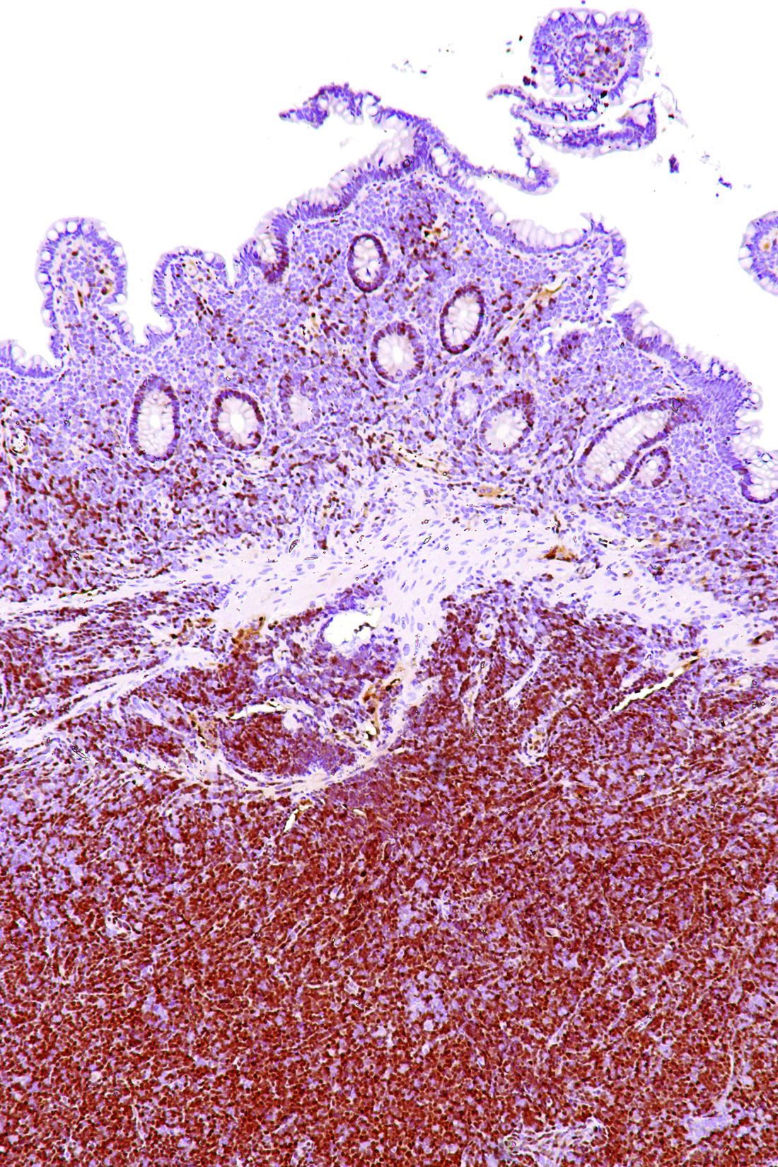
While the study (NCT00980395) was small and limited by its single-center design, the VCR combination “has encouraging activity in both MCL and iNHL and could be compared to standard therapies in future studies,” the researchers wrote. “For MCL in particular, we believe a noninferiority comparison to standard therapies would be justified by our results.”
Adverse events were most commonly hematologic, and three patients experienced febrile neutropenia, data show.
“Although hematological toxicity can be an issue, the regimen provides an alternative option in transplant ineligible relapsed/refractory MCL and iNHL,” wrote Dr. Puvvada and her colleagues. The study was published in Clinical Lymphoma, Myeloma & Leukemia (doi: 10.1016/j.clml.2017.09.001).
The researchers studied the combination of bortezomib, the proteasome inhibitor initially approved for relapsed/refractory MCL, cladribine, which has shown activity and promising response rates in patients with indolent lymphomas, and rituximab in patients with CD20-positive mantle cell or indolent lymphoma.
Patients with follicular lymphomas were eligible to be included in the study if they had received at least one previous line of therapy. All other participants could be treatment naive or have relapsed after previous treatment.
Of the 24 patients enrolled, 11 had MCL, 5 had follicular lymphoma, 4 had marginal zone lymphoma, 3 had lymphoplasmacytic lymphoma, and 1 had small lymphocytic lymphoma.
The VCR regimen, given every 28 days for no more than six cycles, included rituximab at 375 mg/m2 given intravenously on day 1 of each cycle, cladribine 4 mg/m2 given intravenously over 2 hours on days 1 through 5, and bortezomib 1.3 mg/m2 given intravenously on days 1 and 4. Patients received a median of five cycles of therapy.
Adverse events of grade 3 or greater occurred in 14 patients (58%); 8 patients had leukopenia, 6 had thrombocytopenia, 5 had fatigue, and 5 had neutropenia, which included febrile neutropenia in 3 patients.
With a median follow-up of 38.5 months, overall response rate for VCR was 96%. Complete responses occurred in 8 of 23 evaluable patients (35%) and partial responses in 14 more patients (61%).
The combination of bortezomib, cladribine, and rituximab (VCR) was an effective treatment regimen for patients with CD20-positive mantle cell lymphoma (MCL) and indolent non-Hodgkin’s lymphoma (iNHL), based on results of a recent phase 2, open-label study.
The overall response rate was 92% in the single-center, 24-patient study. The 2-year progression-free survival (PFS) was 82% and 54%, respectively, for MCL and iNHL patients; PFS was 80% for treatment-naive patients and 57% for those with refractory/recalcitrant disease, according to Soham D. Puvvada, MD, of the University of Arizona Cancer Center in Tucson, and her associates.
Two-year overall survival was 91% for MCL and 69% for iNHL patients. Median time to progression was 34.5 months, and median PFS had not been reached at 2 years, according to the researchers.
While the study (NCT00980395) was small and limited by its single-center design, the VCR combination “has encouraging activity in both MCL and iNHL and could be compared to standard therapies in future studies,” the researchers wrote. “For MCL in particular, we believe a noninferiority comparison to standard therapies would be justified by our results.”
Adverse events were most commonly hematologic, and three patients experienced febrile neutropenia, data show.
“Although hematological toxicity can be an issue, the regimen provides an alternative option in transplant ineligible relapsed/refractory MCL and iNHL,” wrote Dr. Puvvada and her colleagues. The study was published in Clinical Lymphoma, Myeloma & Leukemia (doi: 10.1016/j.clml.2017.09.001).
The researchers studied the combination of bortezomib, the proteasome inhibitor initially approved for relapsed/refractory MCL, cladribine, which has shown activity and promising response rates in patients with indolent lymphomas, and rituximab in patients with CD20-positive mantle cell or indolent lymphoma.
Patients with follicular lymphomas were eligible to be included in the study if they had received at least one previous line of therapy. All other participants could be treatment naive or have relapsed after previous treatment.
Of the 24 patients enrolled, 11 had MCL, 5 had follicular lymphoma, 4 had marginal zone lymphoma, 3 had lymphoplasmacytic lymphoma, and 1 had small lymphocytic lymphoma.
The VCR regimen, given every 28 days for no more than six cycles, included rituximab at 375 mg/m2 given intravenously on day 1 of each cycle, cladribine 4 mg/m2 given intravenously over 2 hours on days 1 through 5, and bortezomib 1.3 mg/m2 given intravenously on days 1 and 4. Patients received a median of five cycles of therapy.
Adverse events of grade 3 or greater occurred in 14 patients (58%); 8 patients had leukopenia, 6 had thrombocytopenia, 5 had fatigue, and 5 had neutropenia, which included febrile neutropenia in 3 patients.
With a median follow-up of 38.5 months, overall response rate for VCR was 96%. Complete responses occurred in 8 of 23 evaluable patients (35%) and partial responses in 14 more patients (61%).
FROM LYMPHOMA, MYELOMA & LEUKEMIA
Key clinical point:
Major finding: The overall response rate was 92%, with a 2-year PFS of 82% and 54% for patients with mantle cell lymphoma (MCL) and indolent non-Hodgkin’s lymphoma (iNHL), respectively. Adverse events were most commonly hematologic, and three patients experienced febrile neutropenia.
Data source: A phase 2, open-label study including 24 patients with mantle cell or indolent lymphomas.
Disclosures: No disclosures were reported in the accepted manuscript.