User login
Depiction of Cancer in Movies: Not an Accurate Portrayal
This transcript has been edited for clarity.
I’d like to talk about a very different topic from what I normally discuss, which is probably relatively rarely addressed in clinical conversations among clinicians. There was a very provocative commentary that appeared in JCO Oncology Practice, titled “Hollywood’s Take on Oncology: Portrayal of Cancer in Movies, 2010-2020.”
All of us, as we grow up — as kids, adolescents, young adults, adults, and older individuals — watch television and movies. The older of us know that the doctor in everybody’s home that we all wanted was Marcus Welby. Of course, there was Dr. Kildare, ER, Grey’s Anatomy, and St. Elsewhere. There was Love Story and Brian’s Song. We all know about these.
This particular review was fascinating. The authors looked at 100 English-language movies that had cancer included in the storyline over the past decade. They asked some relatively simple questions: How did they discuss it? What were the tumor types they discussed? What were the outcomes?
The question is, what is the public seeing? If you watch these movies and you don’t have family experience or personal experience with cancer, what do you think about cancer? Maybe this is what you know about it. Despite what the National Cancer Institute or the American Society of Clinical Oncology tells you, this may be what you know.
What they showed was really quite interesting.
There is another very interesting phenomenon. What do you think was the most common cancer type when they did define the cancer? It was brain tumors, even though we know that brain tumors are certainly not even within the top 10. They’re obviously very serious cancers, but if you’re talking about common cancers, brain cancer doesn’t rank in the top 10, and it was the most common cancer on these shows.
The authors of this paper made the point of whether this would be an opportunity for filmmakers. Again, with the storyline, they’re trying to sell a product here, but wouldn’t this be the opportunity to provide some information about the reality of cancer? They could emphasize the fact that smokers get lung cancer. In my opinion, they could discuss cervical cancer and comment that if HPV vaccination had been done, maybe this would not have happened.
They noted that the majority of cancers in these movies were incurable, and they commented that that’s not the reality today. Today, obviously, many of our cancers that weren’t curable have become quite curable for a percentage of patients, in addition to which, obviously, with early detection, we have a very high cure rate. How about trying to get that message out, too, that we’ve actually had increasing success?
They commented that there was very rarely, if ever, a conversation about multidisciplinary care, that somehow there are multiple doctors with multiple specialties involved. They noted that this is potentially a very important message to give out. They commented that in 12 of these movies, the patient refused cancer care. Again, that happens, but it’s clearly a rare event today. Maybe this is not really a very accurate depiction of what’s going on.
They commented on the fact that, obviously, we’re going back through the past 10 years, so there were no patients who received immunotherapy or targeted therapy. Again, the goal here is not to sell oncology care but to be accurate, or more accurate, about the state of treatment to the extent you can.
They noted that, in fact, there was essentially very little, if any, comment on palliative care or hospice care. The final point they made is that there was very little conversation in these movies about what we now recognize as financial distress in many of our patients. That’s an unfortunate reality and perhaps that might come in the future.
Again, the point of this was not to tell Hollywood how to make their movies but to have the oncology community recognize that if their patients or the families of their patients are seeing these movies, they are not getting a very accurate picture of what is happening in the oncology world today and that some education may very well be required.
Maurie Markman is Professor, Department of Medical Oncology and Therapeutics Research, City of Hope, Duarte, California, and President of Medicine & Science, City of Hope Atlanta, Chicago, and Phoenix. He disclosed the following relevant financial relationships: income in an amount equal to or greater than $250 from: GlaxoSmithKline; AstraZeneca.
A version of this article first appeared on Medscape.com.
Timing of Blood Pressure Dosing Doesn’t Matter (Again): BedMed and BedMed-Frail
This transcript has been edited for clarity.
Tricia Ward: I’m joined today by Dr. Scott R. Garrison, MD, PhD. He is a professor in the Department of Family Medicine at the University of Alberta in Edmonton, Alberta, Canada, and director of the Pragmatic Trials Collaborative.
You presented two studies at ESC. One is the BedMed study, comparing day vs nighttime dosing of blood pressure therapy. Can you tell us the top-line findings?
BedMed and BedMed-Frail
Dr. Garrison: We were looking to validate an earlier study that suggested a large benefit of taking blood pressure medication at bedtime, as far as reducing major adverse cardiovascular events (MACEs). That was the MAPEC study. They suggested a 60% reduction. The BedMed trial was in hypertensive primary care patients in five Canadian provinces. We randomized well over 3000 patients to bedtime or morning medications. We looked at MACEs — so all-cause death or hospitalizations for acute coronary syndrome, stroke, or heart failure, and a bunch of safety outcomes.
Essentially, . It was safe to take it at bedtime. But it did not convey any extra cardiovascular benefit.
Ms. Ward: And then you did a second study, called BedMed-Frail. Do you want to tell us the reason you did that?
Dr. Garrison: BedMed-Frail took place in a nursing home population. We believed that it was possible that frail, older adults might have very different risks and benefits, and that they would probably be underrepresented, as they normally are in the main trial.
We thought that because bedtime blood pressure medications would be theoretically preferentially lowering night pressure, which is already the lowest pressure of the day, that if you were at risk for hypotensive or ischemic adverse events, that might make it worse. We looked at falls and fractures; worsening cognition in case they had vascular dementia; and whether they developed decubitus ulcers (pressure sores) because you need a certain amount of pressure to get past any obstruction — in this case, it’s the weight of your body if you’re lying in bed all the time.
We also looked at problem behaviors. People who have dementia have what’s called “sundowning,” where agitation and confusion are worse as the evening is going on. We looked at that on the off chance that it had anything to do with blood pressures being lower. And the BedMed-Frail results mirror those of BedMed exactly. So there was no cardiovascular benefit, and in this population, that was largely driven by mortality; one third of these people died every year.
Ms. Ward: The median age was about 88?
Dr. Garrison: Yes, the median age was 88. There was no cardiovascular mortality advantage to bedtime dosing, but neither was there any signal of safety concerns.
Other Complementary and Conflicting Studies
Ms. Ward: These two studies mirror the TIME study from the United Kingdom.
Dr. Garrison: Yes. We found exactly what TIME found. Our point estimate was pretty much the same. The hazard ratio in the main trial was 0.96. Theirs, I believe, was 0.95. Our findings agree completely with those of TIME and differ substantially from the previous trials that suggested a large benefit.
Ms. Ward: Those previous trials were MAPEC and the Hygia Chronotherapy Trial.
Dr. Garrison: MAPEC was the first one. While we were doing our trial, and while the TIME investigators were doing their trial, both of us trying to validate MAPEC, the same group published another study called Hygia, which also reported a large reduction: a 45% reduction in MACE with bedtime dosing.
Ms. Ward: You didn’t present it, but there was also a meta-analysis presented here by somebody independent.
Dr. Garrison: Yes, Ricky Turgeon. I know Ricky. We gave him patient-level data for his meta-analysis, but I was not otherwise involved.
Ms. Ward: And the conclusion is the same.
Dr. Garrison: It’s the same. He only found the same five trials: MAPEC, Hygia, TIME, BedMed, and BedMed-Frail. Combining them all together, the CIs still span 1.0, so it didn’t end up being significant. But he also analyzed TIME and the BedMed trials separately — again suggesting that those trials showed no benefit.
Ms. Ward: There was a TIME substudy of night owls vs early risers or morning people, and there was a hint (or whatever you should say for a subanalysis of a neutral trial) that timing might make a difference there.
Dr. Garrison: They recently published, I guess it is a substudy, where they looked at people’s chronotype according to whether you consider yourself an early bird or a night owl. Their assessment was more detailed. They reported that if people were tending toward being early birds and they took their blood pressure medicine in the morning, or if they were night owls and they took it in the evening, that they tended to have statistically significantly better outcomes than the opposite timing. In that analysis, they were only looking at nonfatal myocardial infarction and nonfatal stroke.
We did ask something that was related. We asked people: “Do we consider yourself more of an early bird or a night owl?” So we do have those data. For what I presented at ESC, we just looked at the primary outcome; we did subgroups according to early bird, night owl, and neither, and that was not statistically significant. It didn’t rule it out. There were some trends in the direction that the TIME group were suggesting. We do intend to do a closer look at that.
But, you know, they call these “late-breaking trials,” and it really was in our case. We didn’t get the last of our data from the last province until the end of June, so we still are finishing up the analysis of the chronotype portion — so more to come in another month or so.
Do What You Like, or Stick to Morning Dosing?
Ms. Ward: For the purposes of people’s take-home message, does this mostly apply to once-daily–dosed antihypertensives?
Dr. Garrison: It was essentially once-daily medicines that were changed. The docs did have the opportunity to consolidate twice-daily meds into once-daily or switch to a different medication. That’s probably the area where adherence was the biggest issue, because it’s largely beta-blockers that were given twice daily at baseline, and they were less likely to want to change.
At 6 months, 83% of once-daily medications were taken per allocation in the bedtime group and 95% per allocation in the morning group, which was actually pretty good. For angiotensin-converting enzyme inhibitors, angiotensin receptor blockers, and calcium-channel blockers, the adherence was excellent. Again, it was beta-blockers taken twice a day where it fell down, and then also diuretics. But if you combine all diuretic medications (ie, pure diuretics and combo agents), still, 75% of them were successful at taking them at bedtime. Only 15% of people switching a diuretic to bedtime dosing actually had problems with nocturia. Most physicians think that they can’t get their patients to take those meds at bedtime, but you can. There’s probably no reason to take it at bedtime, but most people do tolerate it.
Ms. Ward: Is your advice to take it whenever you feel like? I know when TIME came out, Professor George Stergiou, who’s the incoming president of the International Society of Hypertension, said, well, maybe we should stick with the morning, because that’s what most of the trials did.
Dr. Garrison: I think that›s a perfectly valid point of view, and maybe for a lot of people, that could be the default. There are some people, though, who will have a particular reason why one time is better. For instance, most people have no problems with calcium-channel blockers, but some get ankle swelling and you’re more likely to have that happen if you take them in the morning. Or lots of people want to take all their pills at the same time; blood pressure pills are easy ones to switch the timing of if you’re trying to accomplish that, and if that will help adherence. Basically, whatever time of day you can remember to take it the best is probably the right time.
Ms. Ward: Given where we are today, with your trials and TIME, do you think this is now settled science that it doesn’t make a difference?
Dr. Garrison: I’m probably the wrong person to ask, because I clearly have a bias. I think the methods in the TIME trial are really transparent and solid. I hope that when our papers come out, people will feel the same. You just have to look at the different trials. You need people like Dr. Stergiou to wade through the trials to help you with that.
Ms. Ward: Thank you very much for joining me today and discussing this trial.
Scott R. Garrison, MD, PhD, is Professor, Department of Family Medicine, University of Alberta in Edmonton, Alberta, Canada, and Staff Physician, Department of Family Medicine, Kaye Edmonton Clinic, and he has disclosed receiving research grants from Alberta Innovates (the Alberta Provincial Government) and the Canadian Institutes of Health Research (the Canadian Federal Government).
A version of this article first appeared on Medscape.com.
FROM ESC 2024
Prediction, Management of Sjögren-Related Lymphomas Gain Ground With New Studies
, particularly mucosa-associated lymphoid tissue (MALT) lymphoma, based on recent findings that confirmed a key early biomarker and found that a systemic treatment strategy reduced Sjögren disease activity and the risk for lymphoma relapse.
Two European studies published in The Lancet Rheumatology — one a case-control study reporting that rheumatoid factor (RF) was an early and strong predictor of Sjögren disease–related MALT lymphoma and the other a retrospective study that found a combination of chemotherapy and anti-CD20 therapy with rituximab as a first-line treatment for lymphoma was more effective than localized treatment or watch-and-wait approach in minimizing autoimmune activity and treating the lymphoma — potentially shed new light on strategies to manage Sjögren disease–related lymphoma.
A commentary accompanying the studies noted that 5%-10% of patients with Sjögren disease will develop non-Hodgkin B-cell lymphoma, with marginal lymphoma the most common type of low-grade lymphoma. The commentary, led by Suzanne Arends, MD, a rheumatologist at the University of Groningen in Groningen, the Netherlands, found the studies “clinically relevant” but stated that the lack of consistent definitions between the two studies along with their retrospective nature prevent any “definitive conclusions.”
High Lymphoma Risk in Sjögren Disease
“It is the autoimmune disease in which the risk of lymphoma is the highest, a 10- to 20-fold increase of the risk of lymphoma in this disease,” Xavier Mariette, MD, PhD, co-senior author of the retrospective treatment study, said of Sjögren disease.
These lymphomas are predominantly the marginal zone type, specifically MALT occurring in the salivary glands, the same site of the autoimmune disease, said Dr. Mariette, who is the head of Rheumatology and professor at Université Paris-Saclay and Hôpital Bicêtre. Autoimmune B cells become lymphomatous. “So there is a continuity between autoimmunity and lymphoma genesis,” Dr. Mariette told this news organization. Typically, hematologists do not treat the lymphoma if it doesn’t migrate beyond the salivary glands, he said.
Dr. Mariette said his group’s findings make the case for a more aggressive treatment.
“When patients got the systemic treatment, there was a decreased risk of flare of the autoimmune disease of Sjögren’s, but there was no effect on the lymphoma formation,” Dr. Mariette said. “And when these patients have combined therapy, immunotherapy plus chemotherapy, compared to single immunotherapy, they did have improvement of the lymphoma progression-free survival.”
Their multicenter study enrolled 106 patients with Sjögren disease who developed lymphoma, 64% (n = 68) of whom had MALT, 13% (n = 14) of whom had other marginal zone subtypes, and the same percentage with diffuse large B-cell lymphoma. With a median follow-up of 7 years, 32 patients with marginal zone subtypes who had combination chemotherapy and anti-CD20 therapy had a 64% greater chance of lymphoma progression-free survival than 18 of their counterparts who received anti-CD20 monotherapy. Overall, outcomes for Sjögren disease systemic activity or survival were no different between the combination therapy and monotherapy arms.
Patients who had a systemic approach had a 57% reduced risk for new Sjögren disease activity compared with those who had first-line surgery or radiation (16%, n = 13) or underwent watch and wait (23%, n = 19).
The study strengthens the argument for a systemic treatment approach over localized therapy “because patients with Sjögren’s have a higher degree of development of MALT lymphoma of the salivary glands,” Juan Pablo Alderuccio, MD, a hematologist and lymphoma clinical site disease group leader at the Sylvester Comprehensive Cancer Center at the University of Miami Health Systems, Miami, Florida, told this news organization.
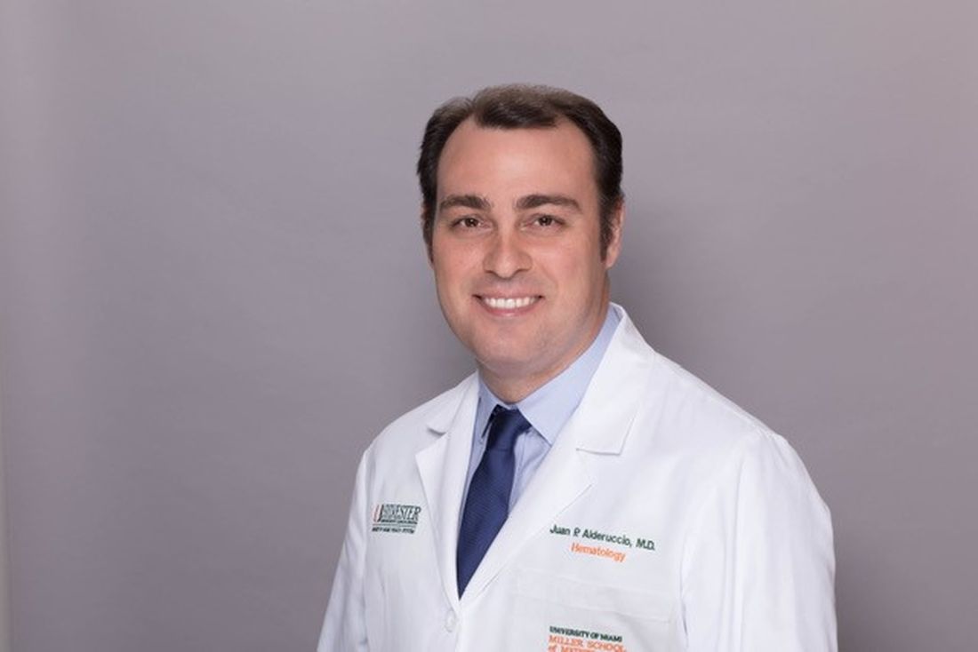
“We already knew that the combination of chemotherapy with rituximab usually achieves a better outcome,” Dr. Alderuccio added, citing a 2017 clinical trial that found combined chemotherapy with chlorambucil plus rituximab improved progression-free survival compared with either therapy alone. The latest retrospective study from France reinforces that, he said.
“The study also shows it’s very important to consider treatment-related specificities — to select the most appropriate treatment for these patients,” Dr. Alderuccio added.
RF Biomarker
The case-control study by researchers in Italy and Greece included 80 patients with Sjögren-related MALT lymphoma matched to controls with Sjögren disease who did not have lymphoma.
“We showed that rheumatoid factor positivity at the time of Sjögren’s disease diagnosis serves as the most reliable and temporally distant independent predictor of MALT lymphoma development,” lead author Andreas Goules, MD, a pathophysiologist at the National and Kapodistrian University of Athens, Athens, Greece, told this news organization.
He added that the study found that specific biomarkers in addition to RF positivity were signs of a high risk for MALT lymphoma and a more advanced stage of Sjögren disease–related lymphomagenesis. They included high systemic disease activity, measured as a European Alliance of Associations for Rheumatology Sjögren’s Syndrome Disease Activity Index ≥ 5, and specific B-cell manifestations, such as cryoglobulinemia, salivary gland enlargement, hypocomplementemia, and palpable purpura.
“Ideally, all patients should be evaluated at the time of diagnosis for the presence of RF and undergo a minor salivary gland biopsy to exclude an underlying ongoing lymphoproliferative process,” Dr. Goules said.
RF-positive patients with Sjögren disease require a closer follow-up to identify an advanced stage of lymphoma development, he added.
“It is well known that Sjögren’s disease is characterized by an increased mortality rate, compared to the general population, mainly due to the related lymphomas,” Dr. Goules added. “Thus, the early diagnosis of MALT lymphoma, which is associated with a better prognosis, is expected to improve the overall clinical outcome of Sjögren’s disease patients.”
Rheumatologists and hematologists should employ a similar strategy for Sjögren disease–related large B-cell lymphomas, he said.
“The pathogenetic mechanisms of these two lymphoma types are vastly different, so it wouldn’t be surprising if an entirely different risk factor emerges,” Dr. Goules said. “However, given the rarity of diffuse large B-cell lymphomas, much larger multinational cohorts will be necessary to obtain clinically and pathogenetically meaningful results.”
Alan Baer, MD, a rheumatologist and founder of the Sjögren’s Disease Clinic at Johns Hopkins University in Baltimore, noted Dr. Goules and colleagues are not the first to identify RF, along with a host of other clinical and laboratory findings, as a risk factor for lymphoma in patients with Sjögren disease. “The current study validates rheumatoid factor as an independent risk factor present at a time that is temporally distant from the time of lymphoma diagnosis,” he said.
However, he cautioned that RF alone isn’t highly predictive of Sjögren-related lymphoma. Up to 60% of patients with Sjögren disease are positive for RF at the time of the diagnosis, Dr. Baer said.
“Thus, the finding of rheumatoid factor alone does not necessarily mandate closer surveillance of this group of patients, with the potential for more frequent clinical exams, imaging, and laboratory testing,” he said. “Such an approach has the risk of subjecting patients to unnecessary testing, including invasive procedures.”
More detailed findings, such as if a certain RF level was more predictive of lymphoma or whether other features in combination with RF heightened the risk, would be helpful, he said.
What Future Studies Should Look At
The studies call for further research into biomarkers for Sjögren disease–related lymphoma and treatment of the disease, both Dr. Mariette and Dr. Goules said.
Dr. Goules said a multicenter prospective study is needed to measure RF positivity and RF titers over time and determine whether higher levels mean an increased risk for lymphoma development or a shorter time interval until lymphoma onset. “Such a study requires a large number of RF-positive Sjögren’s disease patients who would be followed up for a long period of time,” Dr. Goules said.
To further evaluate treatment approaches for Sjögren disease–related lymphoma, Dr. Mariette said, a prospective study should compare the watch-and-wait approach with combination chemotherapy and anti-CD20 therapy. “It would be difficult to run because the primary endpoint would be lymphoma progression–free survival, and the secondary would be Sjögren’s relapse and mortality, but it would take a lot of time,” he said.
He added, “It’s a reason why this retrospective study is important. Maybe if we had another retrospective study reaching the same conclusion, I think it would be very, very strong evidence.”
Funding for the case-control study came from the European Commission–Horizon 2020 program. The retrospective treatment study had no outside funding. Dr. Mariette disclosed financial relationships with AstraZeneca, Bristol-Myers Squibb, Galapagos, GlaxoSmithKline, Novartis, and Pfizer. Dr. Alderuccio, Dr. Goules, and Dr. Baer had no relevant relationships to disclose.
A version of this article first appeared on Medscape.com.
, particularly mucosa-associated lymphoid tissue (MALT) lymphoma, based on recent findings that confirmed a key early biomarker and found that a systemic treatment strategy reduced Sjögren disease activity and the risk for lymphoma relapse.
Two European studies published in The Lancet Rheumatology — one a case-control study reporting that rheumatoid factor (RF) was an early and strong predictor of Sjögren disease–related MALT lymphoma and the other a retrospective study that found a combination of chemotherapy and anti-CD20 therapy with rituximab as a first-line treatment for lymphoma was more effective than localized treatment or watch-and-wait approach in minimizing autoimmune activity and treating the lymphoma — potentially shed new light on strategies to manage Sjögren disease–related lymphoma.
A commentary accompanying the studies noted that 5%-10% of patients with Sjögren disease will develop non-Hodgkin B-cell lymphoma, with marginal lymphoma the most common type of low-grade lymphoma. The commentary, led by Suzanne Arends, MD, a rheumatologist at the University of Groningen in Groningen, the Netherlands, found the studies “clinically relevant” but stated that the lack of consistent definitions between the two studies along with their retrospective nature prevent any “definitive conclusions.”
High Lymphoma Risk in Sjögren Disease
“It is the autoimmune disease in which the risk of lymphoma is the highest, a 10- to 20-fold increase of the risk of lymphoma in this disease,” Xavier Mariette, MD, PhD, co-senior author of the retrospective treatment study, said of Sjögren disease.
These lymphomas are predominantly the marginal zone type, specifically MALT occurring in the salivary glands, the same site of the autoimmune disease, said Dr. Mariette, who is the head of Rheumatology and professor at Université Paris-Saclay and Hôpital Bicêtre. Autoimmune B cells become lymphomatous. “So there is a continuity between autoimmunity and lymphoma genesis,” Dr. Mariette told this news organization. Typically, hematologists do not treat the lymphoma if it doesn’t migrate beyond the salivary glands, he said.
Dr. Mariette said his group’s findings make the case for a more aggressive treatment.
“When patients got the systemic treatment, there was a decreased risk of flare of the autoimmune disease of Sjögren’s, but there was no effect on the lymphoma formation,” Dr. Mariette said. “And when these patients have combined therapy, immunotherapy plus chemotherapy, compared to single immunotherapy, they did have improvement of the lymphoma progression-free survival.”
Their multicenter study enrolled 106 patients with Sjögren disease who developed lymphoma, 64% (n = 68) of whom had MALT, 13% (n = 14) of whom had other marginal zone subtypes, and the same percentage with diffuse large B-cell lymphoma. With a median follow-up of 7 years, 32 patients with marginal zone subtypes who had combination chemotherapy and anti-CD20 therapy had a 64% greater chance of lymphoma progression-free survival than 18 of their counterparts who received anti-CD20 monotherapy. Overall, outcomes for Sjögren disease systemic activity or survival were no different between the combination therapy and monotherapy arms.
Patients who had a systemic approach had a 57% reduced risk for new Sjögren disease activity compared with those who had first-line surgery or radiation (16%, n = 13) or underwent watch and wait (23%, n = 19).
The study strengthens the argument for a systemic treatment approach over localized therapy “because patients with Sjögren’s have a higher degree of development of MALT lymphoma of the salivary glands,” Juan Pablo Alderuccio, MD, a hematologist and lymphoma clinical site disease group leader at the Sylvester Comprehensive Cancer Center at the University of Miami Health Systems, Miami, Florida, told this news organization.
“We already knew that the combination of chemotherapy with rituximab usually achieves a better outcome,” Dr. Alderuccio added, citing a 2017 clinical trial that found combined chemotherapy with chlorambucil plus rituximab improved progression-free survival compared with either therapy alone. The latest retrospective study from France reinforces that, he said.
“The study also shows it’s very important to consider treatment-related specificities — to select the most appropriate treatment for these patients,” Dr. Alderuccio added.
RF Biomarker
The case-control study by researchers in Italy and Greece included 80 patients with Sjögren-related MALT lymphoma matched to controls with Sjögren disease who did not have lymphoma.
“We showed that rheumatoid factor positivity at the time of Sjögren’s disease diagnosis serves as the most reliable and temporally distant independent predictor of MALT lymphoma development,” lead author Andreas Goules, MD, a pathophysiologist at the National and Kapodistrian University of Athens, Athens, Greece, told this news organization.
He added that the study found that specific biomarkers in addition to RF positivity were signs of a high risk for MALT lymphoma and a more advanced stage of Sjögren disease–related lymphomagenesis. They included high systemic disease activity, measured as a European Alliance of Associations for Rheumatology Sjögren’s Syndrome Disease Activity Index ≥ 5, and specific B-cell manifestations, such as cryoglobulinemia, salivary gland enlargement, hypocomplementemia, and palpable purpura.
“Ideally, all patients should be evaluated at the time of diagnosis for the presence of RF and undergo a minor salivary gland biopsy to exclude an underlying ongoing lymphoproliferative process,” Dr. Goules said.
RF-positive patients with Sjögren disease require a closer follow-up to identify an advanced stage of lymphoma development, he added.
“It is well known that Sjögren’s disease is characterized by an increased mortality rate, compared to the general population, mainly due to the related lymphomas,” Dr. Goules added. “Thus, the early diagnosis of MALT lymphoma, which is associated with a better prognosis, is expected to improve the overall clinical outcome of Sjögren’s disease patients.”
Rheumatologists and hematologists should employ a similar strategy for Sjögren disease–related large B-cell lymphomas, he said.
“The pathogenetic mechanisms of these two lymphoma types are vastly different, so it wouldn’t be surprising if an entirely different risk factor emerges,” Dr. Goules said. “However, given the rarity of diffuse large B-cell lymphomas, much larger multinational cohorts will be necessary to obtain clinically and pathogenetically meaningful results.”
Alan Baer, MD, a rheumatologist and founder of the Sjögren’s Disease Clinic at Johns Hopkins University in Baltimore, noted Dr. Goules and colleagues are not the first to identify RF, along with a host of other clinical and laboratory findings, as a risk factor for lymphoma in patients with Sjögren disease. “The current study validates rheumatoid factor as an independent risk factor present at a time that is temporally distant from the time of lymphoma diagnosis,” he said.
However, he cautioned that RF alone isn’t highly predictive of Sjögren-related lymphoma. Up to 60% of patients with Sjögren disease are positive for RF at the time of the diagnosis, Dr. Baer said.
“Thus, the finding of rheumatoid factor alone does not necessarily mandate closer surveillance of this group of patients, with the potential for more frequent clinical exams, imaging, and laboratory testing,” he said. “Such an approach has the risk of subjecting patients to unnecessary testing, including invasive procedures.”
More detailed findings, such as if a certain RF level was more predictive of lymphoma or whether other features in combination with RF heightened the risk, would be helpful, he said.
What Future Studies Should Look At
The studies call for further research into biomarkers for Sjögren disease–related lymphoma and treatment of the disease, both Dr. Mariette and Dr. Goules said.
Dr. Goules said a multicenter prospective study is needed to measure RF positivity and RF titers over time and determine whether higher levels mean an increased risk for lymphoma development or a shorter time interval until lymphoma onset. “Such a study requires a large number of RF-positive Sjögren’s disease patients who would be followed up for a long period of time,” Dr. Goules said.
To further evaluate treatment approaches for Sjögren disease–related lymphoma, Dr. Mariette said, a prospective study should compare the watch-and-wait approach with combination chemotherapy and anti-CD20 therapy. “It would be difficult to run because the primary endpoint would be lymphoma progression–free survival, and the secondary would be Sjögren’s relapse and mortality, but it would take a lot of time,” he said.
He added, “It’s a reason why this retrospective study is important. Maybe if we had another retrospective study reaching the same conclusion, I think it would be very, very strong evidence.”
Funding for the case-control study came from the European Commission–Horizon 2020 program. The retrospective treatment study had no outside funding. Dr. Mariette disclosed financial relationships with AstraZeneca, Bristol-Myers Squibb, Galapagos, GlaxoSmithKline, Novartis, and Pfizer. Dr. Alderuccio, Dr. Goules, and Dr. Baer had no relevant relationships to disclose.
A version of this article first appeared on Medscape.com.
FROM THE LANCET RHEUMATOLOGY
Kidney Disease May Accelerate With Higher Rheumatoid Arthritis Disease Activity
TOPLINE:
Higher rheumatoid arthritis (RA) disease activity is associated with an accelerated kidney function decline and increased risk for chronic kidney disease (CKD) stages G3a and G3b.
METHODOLOGY:
- Researchers analyzed data from the CorEvitas RA registry, a prospective observational cohort in the United States, between 2001 and 2022, to evaluate the longitudinal association between RA disease activity and changes in kidney function.
- They included 31,129 patients with RA (median age, 58 years; 76.3% women) who had a baseline estimated glomerular filtration rate (eGFR) ≥ 60 mL/min per 1.73 m2 and received treatment with disease-modifying antirheumatic drugs (DMARDs).
- The participants were categorized into those in remission (n = 6647) and those with low (n = 10,028), moderate (n = 8548), and high (n = 5906) disease activity based on the time-averaged Clinical Disease Activity Index and followed for a median duration of 3.5 years.
- The primary outcome was a longitudinal change in eGFR, and the secondary outcomes were the development of CKD stage G3a (eGFR < 60 mL/min/1.73 m2) and stage G3b (eGFR < 45 mL/min/1.73 m2).
TAKEAWAY:
- Higher RA disease activity was associated with a faster decline in eGFR, with those having moderate and high RA disease activity experiencing an additional mean annual decline of 0.17 mL/min per 1.73 m2 and 0.18 mL/min per 1.73 m2, respectively, compared with those in remission.
- The decline in annual eGFR was even more accelerated when patients had consistently high disease activity since the time of enrollment (−0.43 mL/min per 1.73 m2).
- Patients with high RA disease activity had a 1.27 times (adjusted hazard ratio, 1.27; 95% CI, 1.05-1.52) higher risk of developing CKD stage G3a and a 1.93 times (aHR, 1.93; 95% CI, 1.16-3.20) higher risk for CKD stage G3b, compared with those in remission.
IN PRACTICE:
“This study suggests that controlling disease activity may potentially contribute to preserving kidney function in patients with RA,” the authors wrote.
SOURCE:
This study was led by Sho Fukui, MD, Division of Rheumatology, Inflammation, and Immunity, Brigham and Women’s Hospital and Harvard Medical School, both in Boston, Massachusetts, and was published online in Annals of the Rheumatic Diseases.
LIMITATIONS:
This study relied on serum creatinine and not cystatin C to estimate kidney function. It also did not collect information on the severity of comorbidities, which may have introduced residual confounding. Further studies are warranted to check the effect of DMARD therapy on renal function.
DISCLOSURES:
The study was funded by the National Institute of Arthritis and Musculoskeletal and Skin Diseases. Some authors reported serving as scientific advisers or consultants, receiving consulting fees or salary support, or having other ties with pharmaceutical companies.
This article was created using several editorial tools, including AI, as part of the process. Human editors reviewed this content before publication. A version of this article first appeared on Medscape.com.
Stroke Risk from Atrial Fibrillation Rises in Presence of Rheumatoid Arthritis
TOPLINE:
Patients with both rheumatoid arthritis (RA) and atrial fibrillation (AF) have a higher risk for ischemic stroke than those with only AF. They are also less likely to receive oral anticoagulant treatment, which may contribute to this increased stroke risk.
METHODOLOGY:
- Researchers conducted a registry-based retrospective cohort study using the Norwegian Cardio-Rheuma Register to evaluate the risk for ischemic stroke following the diagnosis of AF in patients with or without RA.
- They included 163,595 patients with newly diagnosed AF between 2010 and 2017, of whom 2750 had RA. Patients had to be diagnosed with RA before the diagnosis of AF.
- They also assessed whether patients with RA were less likely to receive oral anticoagulants for stroke prevention within 3 months of AF diagnosis than those without RA.
- The median follow-up time was 2.5 years for patients with RA and 3.0 years for those without RA.
- The primary endpoint was ischemic stroke, which was identified through hospital admissions and visits.
TAKEAWAY:
- At 5 years, patients with both RA and AF showed a higher cumulative incidence of ischemic stroke than those with only AF (7.3% vs 5.0%).
- Among patients with AF, the risk of having a stroke was 25% higher in those with RA than in those without RA (adjusted hazard ratio, 1.25; 95% CI, 1.05-1.50).
- Patients with RA were also less likely to receive treatment with oral anticoagulants than those without RA, driven by concerns over potential interactions with RA medications, bleeding risk, or other factors (adjusted odds ratio, 0.88; 95% CI, 0.80-0.97).
IN PRACTICE:
“Our study prompts preventive measures such as meticulous cardiovascular risk factor control among patients with RA and AF and raises the question whether the presence of RA should be taken into account when considering OAC [oral anticoagulant] treatment for AF patients,” the authors wrote.
SOURCE:
This study was led by Anne M. Kerola, MD, PhD, Helsinki University Hospital and University of Helsinki in Finland. It was published online in Rheumatology.
LIMITATIONS:
This study lacked data on smoking, blood pressure measurements, alcohol use, and obesity, which may have affected the comprehensiveness of the findings. The study population was limited to Norway and may not be generalizable to other populations.
DISCLOSURES:
This study was supported by the Olav Thon Foundation, the Research Council of Norway, and the Foundation for Research in Rheumatology. Some authors received speaker fees, participated in advisory boards, served as consultants, or had other ties with some pharmaceutical companies and institutions.
This article was created using several editorial tools, including AI, as part of the process. Human editors reviewed this content before publication. A version of this article first appeared on Medscape.com.
Teclistamab Promising as a Treatment of Last Resort for Refractory Autoimmune Diseases
TOPLINE:
Teclistamab, a T-cell engager that targets B-cell maturation antigen (BCMA), improved disease activity in four patients with refractory autoimmune conditions. In a separately published case report, teclistamab treatment induced remission in a patient with refractory systemic lupus erythematosus (SLE).
BACKGROUND:
- Chimeric antigen receptor (CAR) T cells or T-cell engagers against CD19 have been effective in small studies of patients with treatment-resistant autoimmune diseases.
- Some patients have disease rooted in long-lived plasma cells that express BCMA but not CD19, making them resistant to CD19 CAR T-cell therapy.
- Teclistamab acts on T cells through CD3 and targets plasmablasts and plasma cells through BCMA.
METHODOLOGY:
- In one case series, researchers administered teclistamab subcutaneously to four patients with autoimmune diseases resistant to more than five immunosuppressants, including rituximab.
- Patient 1 had systemic sclerosis, patient 2 had primary Sjögren disease, patient 3 had idiopathic inflammatory myositis, and patient 4 had rheumatoid arthritis.
- Researchers incrementally increased teclistamab dosage in an inpatient setting: 0.06 mg/kg on day 1, 0.3 mg/kg on day 3, and 1.5 mg/kg on day 5. Patients 2, 3, and 4 received one maintenance dose of 1.5 mg/kg after 4 weeks, and patient 1 received a 1.5-mg/kg dose after 12 weeks.
- In the single case report, the patient with SLE received a step-up dosage of teclistamab (0.06 mg/kg and 0.3 mg/kg) followed by 0.8 mg/kg on day 7. She received 1.5 mg/kg at weeks 2 and 5.
TAKEAWAY:
- Teclistamab therapy led to significant improvements in disease activity in all four patients, with notable reductions in skin disease, arthritis, and lung function scores.
- Teclistamab therapy had a good safety profile, with no neurotoxicity or myelotoxicity and only lower-grade cytokine release syndrome reported.
- Researchers observed seroconversion of PM-Scl-75, PM-Scl-100, rheumatoid factor, and autoantibodies against mutated citrullinated vimentin and lower levels of autoantibodies ANA, MDAS, SS-A/Ro, SS-B/La, and PL-7 after treatment.
- In the separate case report, the patient reached complete drug-free remission by week 6, as defined by the Systemic Lupus Erythematosus Disease Activity Index 2000.
- The level of anti–double-stranded DNA antibodies in the patient with SLE decreased rapidly, reaching normal range by week 5 and remaining undetectable through week 16.
IN PRACTICE:
“These data show that the targeting of the plasma-cell compartment by a BCMA-targeted T-cell engager is feasible in patients with autoimmune disease. Whether such therapy results in sustained clinical remission warrants further study,” write the authors of the four-patient case series.
SOURCE:
Melanie Hagen, MD, Friedrich Alexander University Erlangen–Nuremberg, Germany, and colleagues reported their case series online in The New England Journal of Medicine. Tobias Alexander, MD, and colleagues at Charité–Universitätsmedizin Berlin, Germany, also described their single case report in The New England Journal of Medicine.
LIMITATIONS:
The small number of patients limits the generalizability of the findings. The short duration of follow-up may not capture long-term effects and potential late-onset adverse events. The lack of a control group makes it difficult to attribute improvements solely to teclistamab therapy.
DISCLOSURES:
The four-patient case series was supported by grants from the Deutsche Forschungsgemeinschaft, Bundesministerium für Bildung und Forschung, and the European Union. The single case report was supported by grants from the Deutsche Forschungsgemeinschaft and the European Union. Several authors have disclosed financial relationships with pharmaceutical companies, including Janssen Biotech, which markets teclistamab.
This article was created using several editorial tools, including AI, as part of the process. Human editors reviewed this content before publication. A version of this article first appeared on Medscape.com.
In-Hospital e-Alerts: A Step Toward Better Kidney Health?
TOPLINE:
METHODOLOGY:
- AKI is a common complication in hospitalized patients, leading to increased comorbidities, healthcare costs, and short- and long-term mortality, but the impact of early detection through electronic health care record systems (e-alerts) is unclear.
- Researchers conducted an updated systematic review and meta-analysis to assess the association of e-alerts for AKI with patient outcomes and clinical practice patterns.
- Overall, 13 studies involving 41,837 patients with AKI were included, comparing e-alerts for AKI with standard care or no e-alerts.
- The primary outcomes were mortality, AKI progression, dialysis events, and kidney recovery, and secondary outcomes were nephrologist consultations, post-AKI exposure to NSAIDs and other medications, and hospital length of stay and costs.
- The investigators assessed bias, the certainty of evidence, and whether the primary outcome conclusions of the meta-analysis were premature.
TAKEAWAY:
- The use of e-alerts for AKI was not associated with reduced mortality outcomes compared with no e-alerts (risk ratio [RR], 0.96; 95% CI, 0.89-1.03; 12 studies).
- E-alerts were associated with a reduced risk for AKI progression (RR, 0.91; 95% CI, 0.84-0.99; five studies); however, the results were found to be heterogeneous and possibly premature.
- E-alerts for AKI were also linked to increased nephrologist consultations (RR, 1.45; 95% CI, 1.04-2.02; 11 studies), reduced post-AKI NSAID exposure (RR, 0.75; 95% CI, 0.59-0.95; four studies), and improved AKI documentation (RR, 1.28; 95% CI, 1.04-1.58; eight studies).
- The use of e-alerts for AKI was associated with increased dialysis events (RR, 1.16; 95% CI, 1.05-1.28).
IN PRACTICE:
“We recommend that each hospital establish its own AKI e-alert system and individualized AKI management protocol tailored to its specific needs,” wrote the authors who also suggested the system be “integrated with earlier risk stratification methods, such as the renal angina index, artificial intelligence–based continuous AKI prediction, and care bundle implementation within a clinical decision support system to enhance early diagnosis and management, potentially improving outcomes.”
SOURCE:
This study was led by Jia-Jin Chen, MD, from the Kidney Research Center, Department of Nephrology, Chang Gung Memorial Hospital, Linkou Branch, Taoyuan City, Taiwan. It was published online in JAMA Network Open.
LIMITATIONS:
The limitations included the scarcity of randomized clinical trials in the meta-analysis. Few studies examined the impact of these e-alerts on the hospital length of stay, healthcare costs, AKI stage progression, and post-AKI kidney recovery, which limited the ability to draw conclusive statements on these aspects. Major adverse kidney events at 28 and 90 days were not reported in any of the enrolled studies, so the impact of AKI e-alerts and increased dialysis events on long-term outcomes remained uncertain.
DISCLOSURES:
The study was supported by grants from the Taiwanese Ministry of Health and Welfare and Linkou Chang Gung Memorial Hospital. The authors declared no conflicts of interests.
This article was created using several editorial tools, including AI, as part of the process. Human editors reviewed this content before publication. A version of this article first appeared on Medscape.com.
As Interest From Families Wanes, Pediatricians Scale Back on COVID Shots
When pediatrician Eric Ball, MD, opened a refrigerator full of childhood vaccines, all the expected shots were there — DTaP, polio, pneumococcal vaccine — except one.
“This is where we usually store our COVID vaccines, but we don’t have any right now because they all expired at the end of last year and we had to dispose of them,” said Dr. Ball, who is part of a pediatric practice in Orange County, California.
“We thought demand would be way higher than it was.”
Providers like Dr. Ball don’t want to waste money ordering doses that won’t be used, but they need enough on hand to vaccinate vulnerable children.
The Centers for Disease Control and Prevention recommends that anyone 6 months or older get the updated COVID vaccination, but in the 2023-24 vaccination season only about 15% of eligible children in the United States got a shot.
Dr. Ball said it was difficult to let vaccines go to waste in 2023. It was the first time the federal government was no longer picking up the tab for the shots, and providers had to pay upfront for the vaccines. Parents would often skip the COVID shot, which can have a very short shelf life, compared with other vaccines.
“Watching it sitting on our shelves expiring every 30 days, that’s like throwing away $150 repeatedly every day, multiple times a month,” Dr. Ball said.
in 2024, Dr. Ball slashed his fall vaccine order to the bare minimum to avoid another costly mistake.
“We took the number of flu vaccines that we order, and then we ordered 5% of that in COVID vaccines,” Dr. Ball said. “It’s a guess.”
That small vaccine order cost more than $63,000, he said.
Pharmacists, pharmacy interns, and techs are allowed to give COVID vaccines only to children age 3 and up, meaning babies and toddlers would need to visit a doctor’s office for inoculation.
It’s difficult to predict how parents will feel about the shots this fall, said Chicago pediatrician Scott Goldstein, MD. Unlike other vaccinations, COVID shots aren’t required for kids to attend school, and parental interest seems to wane with each new formulation. For a physician-owned practice such as Dr. Goldstein’s, the upfront cost of the vaccine can be a gamble.
“The cost of vaccines, that’s far and away our biggest expense. But it’s also the most important thing we do, you could argue, is vaccinating kids,” Dr. Goldstein said.
Insurance doesn’t necessarily cover vaccine storage accidents, which can put the practice at risk of financial ruin.
“We’ve had things happen like a refrigerator gets unplugged. And then we’re all of a sudden out $80,000 overnight,” Dr. Goldstein said.
South Carolina pediatrician Deborah Greenhouse, MD, said she would order more COVID vaccines for older children if the pharmaceutical companies that she buys from had a more forgiving return policy.
“Pfizer is creating that situation. If you’re only going to let us return 30%, we’re not going to buy much,” she said. “We can’t.”
Greenhouse owns her practice, so the remaining 70% of leftover shots would come out of her pocket.
Vaccine maker Pfizer will take back all unused COVID shots for young children, but only 30% of doses for people 12 and older.
Pfizer said in an Aug. 20 emailed statement, “The return policy was instituted as we recognize both the importance and the complexity of pediatric vaccination and wanted to ensure that pediatric offices did not have hurdles to providing vaccine to their young patients.”
Pfizer’s return policy is similar to policies from other drugmakers for pediatric flu vaccines, also recommended during the fall season. Physicians who are worried about unwanted COVID vaccines expiring on the shelves said flu shots cost them about $20 per dose, while COVID shots cost around $150 per dose.
“We run on a very thin margin. If we get stuck holding a ton of vaccine that we cannot return, we can’t absorb that kind of cost,” Dr. Greenhouse said.
Vaccine maker Moderna will accept COVID vaccine returns, but the amount depends on the individual contract with a provider. Novavax will accept the return of only unopened vaccines and doesn’t specify the amount they’ll accept.
Dr. Greenhouse wants to vaccinate as many children as possible but said she can’t afford to stock shots with a short shelf life. Once she runs out of the doses she’s ordered, Dr. Greenhouse plans to tell families to go to a pharmacy to get older children vaccinated. If pediatricians around the country are making the same calculations, doses for very small children could be harder to find at doctors’ offices.
“Frankly, it’s not an ideal situation, but it’s what we have to do to stay in business,” she said.
Dr. Ball worries that parents’ limited interest has caused pediatricians to minimize their vaccine orders, in turn making the newest COVID shots difficult to find once they become available.
“I think there’s just a misperception that it’s less of a big deal to get COVID, but I’m still sending babies to the hospital with COVID,” Dr. Ball said. “We’re still seeing kids with long COVID. This is with us forever.”
KFF Health News is a national newsroom that produces in-depth journalism about health issues and is one of the core operating programs at KFF — the independent source for health policy research, polling, and journalism.
Vacationing Doctors Intervene After Shark Attack
Ryan Forbess, MD: I live at the beach in Orange Beach, Alabama. I’ve lived in Hawaii, the Caymans, and other beach areas for years. I’ve seen a lot of sharks but never a shark attack. Not until now.
Mohammad Ali, MD: Ryan and I have been friends for 20 years. Every year, my family goes to 30A in Florida (a popular resort stretch of highway) to celebrate my wife’s birthday, and the Forbesses always meet us there. This year we had a group of about 18 people.
On Friday, it was beautiful, and we decided to make it a beach day. We had nine kids with us. So by the time we rounded them up and got there, it was noon, and there was nowhere to sit. We almost turned around and went to the pool. But my wife finally found a spot for an umbrella.
Dr. Forbess: We were in the water boogie boarding. I was with my 8-year-old son, and Mo was with his daughter who is the same age. Suddenly, we noticed a lot of commotion just to the left of us. My first thought was: Someone saw a shark, not an attack. They’re so rare. But seeing one would scare people.
We grabbed our kids and started running out of the water. As we got closer to the shore,
Dr. Ali: It was mass panic. People were screaming and running out of the water. Other people were running in and grabbing their kids. Everyone just looked frantic.
We saw two men dragging this poor girl out of the water. It was surreal. The majority of her right leg was severed, her femur bone visible and stark white; it didn’t look real. I kept telling myself I was in a dream and now I’d wake up.
A young EMT who was there had put an informal tourniquet on her leg, but she was still bleeding. So I compressed the femoral artery as hard as I could, something I’m very familiar with doing.
Dr. Forbess: People asked me later what we used for a tourniquet. I said, “Mo’s big hands.” I tease him because most doctors play golf or go fishing; Mo lives in the gym. He was just holding pressure.
The girl’s left hand was also severed off at the wrist. There were two nurses there, and they helped with holding tourniquets on her arm.
Lulu (the girl’s name) was 15 years old. She was in and out of consciousness. At one point, her face started getting really pale, so we tried to lift her extremities up to keep the blood flow to the heart. With such severe blood loss, I thought she might go into cardiovascular shock, and we would have to start compressions. But she had a pulse, and she was breathing.
Dr. Ali: The beach was very crowded, and a lot of people had gathered around. Everyone was emotional, shocked, really shaken up. But they gave us space to work.
Dr. Forbess: People were handing us things — towels, a ratchet strap to use as a tourniquet. There was even an anesthesiologist there who said, “If you need an airway, let me know.” It was like we had a trauma team.
Dr. Ali: Lulu’s mom had been having lunch with friends. When she saw all the commotion, she ran down to the beach to look for her daughter. It was heartbreaking to hear her screams when she saw Lulu. But I was able to tune it out because we had to just concentrate on decreasing the loss of blood.
Dr. Forbess: Another girl came over and said, “That’s my sister.” Lulu has a twin. So she sat there holding Lulu’s hand and being with her the whole time.
Waiting for the EMTs to get there, the seconds were like hours. It seemed like it took forever. Finally, they came, and we were able to get the real tourniquets on, get her boarded and off the beach.
After that, they closed the beach. We got all our stuff and got on the little trolley that would take us back to the house. The lady who was driving asked us, “Did y’all hear about the shark attack?” My wife said, “Yeah, we were there.” And she said, “No, there was one an hour and a half ago.”
Dr. Ali: What we didn’t know was there had been two other attacks that day. Around the same time, one of Lulu’s friends was bitten and got a flesh wound on her heel. And before that, about 4 miles away, there was a serious injury: A lady in her 40s lost her hand and forearm and was bitten in the pelvis.
Dr. Forbess: At that point, my wife leaned back to me and said, “You know we’re never going to the beach again, right? We’re never ever going to the beach.”
If we had known about those attacks, we definitely wouldn’t have been in the water.
Dr. Ali: My wife has never liked going in the water. The evening before, we had debated about taking our daughters in the ocean because she was worried about sharks. I had given her this condescending speech about waist-deep water and the statistical probabilities of ever witnessing a shark attack. I was in trouble.
Dr. Forbess: We didn’t know if Lulu would make it. I’ve done rural family medicine in Oklahoma, so I’ve seen my fair share of injuries — guys on oil rigs, this and that. But I had never seen anything like this kind of trauma and blood loss.
Later that day, I called my office manager to catch up with her and told her what happened. She was actually in Pensacola having dinner across the street from Sacred Heart Hospital where they had taken Lulu. She went over to the emergency room to try to find out Lulu’s status — she was alive.
My office manager was able to go upstairs and talk to Lulu’s mom. Then she called, and we talked to her mom on the phone. She just said, “Thank you for helping my daughter.” It was an emotional moment.
Dr. Ali: It was such a relief. We had no idea how things would turn out. Even if Lulu did survive, was she going to be neurologically sound? But thank God she was. We were so relieved to hear her mom say that it was looking good. We still didn’t know for sure. But at least she was alive and seemed to be functioning.
Dr. Forbess: A few days later, my wife and I went to go visit her at the hospital. Her mom and her grandma were there. They were giving us hugs. We FaceTimed Mo because he was back in Jackson. It was really amazing.
What are the odds? The chances of a shark attack are about one in 12 million. And to have two physicians trained in trauma, a trauma nurse, another nurse, and an anesthesiologist less than 20 yards away when it happened? It’s crazy to think about.
Dr. Ali: And we almost weren’t there. We could have turned away.
Dr. Forbess: Humans are on top of the food chain. Or we think we are. But water really isn’t our element. Against a 12-foot bull shark, we don’t stand a chance. Lulu is here though. It’s unbelievable.
Her mom told me that when Lulu woke up, she just said, “I made it!” That girl is meant to be here. She is a tough girl with a great personality. She has these new prosthetics now that she can move with her mind; it’s like Star Wars. She says she wants to be a physician someday. So she’ll probably cure cancer.
Dr. Forbess is a family medicine physician at Orange Beach Family Medicine in Orange Beach, Alabama. Dr. Ali is an interventional radiologist with Baptist Memorial Health in Jackson, Mississippi.
A version of this article first appeared on Medscape.com.
Creative Strategies Hospitals Use to Attract Nursing Talent
In a fiercely competitive healthcare landscape, hospitals are pulling out all the stops to lure top nursing talent through their doors.
As the nursing shortage intensifies, the creative recruitment approach isn’t just about the perks — it’s becoming an essential tool in the race to build a skilled nursing workforce.
Nursing vacancies are as high as 17% — more than double prepandemic levels — and hospitals scrambling to fill them need to do more than raise salaries and bolster benefits packages to entice nurses.
“I am very thankful when I hear of creative ideas that nurse administrators come up with to try to get their ultimate goal, which is enough qualified nurses to take care of patients,” said Linda Plank, dean of the Louise Herrington School of Nursing at Baylor University in Dallas, Texas.
Signing Bonuses, Tuition Reimbursement, and Self-Scheduling, Please
Signing bonuses were among the top perks offered to healthcare workers, with almost 18% of job openings advertising the incentive for new nurse hires; the average signing bonus for registered nurses (RNs) topped $11,000. In 2023, California-based Palomar Health made headlines when it offered eligible RNs a $100,000 signing bonus paid over a 3-year period.
“We are seeing a variety of incentives, like sign-on bonuses, that can be effective at getting the attention of potential new hires,” said Deborah Trautman, PhD, RN, FAAN, president, and CEO of the American Association of Colleges of Nursing. “With the growing competition for registered nurses, especially those prepared in baccalaureate programs, employers should consider what’s most important to nurses entering the field.”
Hospitals have also invested in benefits ranging from tuition reimbursement, student loan forgiveness, and professional development opportunities to expanded parental leave and onsite childcare. Flexible scheduling is a sought-after perk that benefits both new and experienced RNs and could also help with recruitment.
“In the past, [hospitals said], ‘our shift starts at 7 and ends at 7,’ ” Ms. Plank said. “Now, hospitals are a little bit more flexible ... and being open to flexible shifts has merit. If we’re willing to look at things differently, it could get more people involved in patient care.”
An American Nurses Foundation report found that nurses preferred variable and flexible shift lengths, flexible start times, and self-scheduling options over set schedules. In fact, 45% of nurses who left clinical practice would consider returning to work if hospitals switched to a self-scheduling model.
The Cleveland Clinic in Ohio introduced staggered shifts that start at nontraditional times, including 11 AM to 11 PM, flexible shift lengths, and split RN positions that allow clinical care nurses to divide their time between different departments. Last year, Hackensack Meridian Health in New Jersey also piloted a self-scheduling program for its nursing staff.
Hiring Outside the United States
Despite the prevalence of signing bonuses and premium perks, some hospitals still struggled to fill open positions with nurses recruited from outside the United States. Data from The Kaiser Family Foundation show that 32% of hospitals hired foreign-educated RNs in 2022 — more than double the number hired in 2010.
Jennifer Mensik Kennedy, PhD, MBA, RN, NEA-BC, FAAN, president of the American Nurses Association (ANA), is concerned about that trend. The ANA supports the International Council of Nurses and their call for “stronger codes for ethical recruitment of nurses” because international nurse recruitment practices can negatively affect the quality of healthcare in countries that are depleted of nurses.
“Recruiting international nurses as a key strategy for building core staffing is not sustainable in the long term,” Ms. Kennedy said in an interview. “We need to redirect our focus on how to retain staff through fostering healthy work environments and addressing antiquated payment models.”
Reinforcing Retention
Recruiting nurses is just one element of addressing the nursing shortage. Prioritizing job satisfaction is essential to retaining nursing staff. Currently, 33% of nurses who enter the profession quit within the first 2 years.
A growing number of hospitals have implemented programs focused on increasing retention. Lifepoint Health, a national network of 60-plus acute care hospitals, launched a Nurse Residency Program in 2023. The 12-month program, which offers training and mentorship to help recent nursing school graduates, has recruited 750 new nurses and helped them transition to clinical practice.
The Nurse Residency Program has been so successful that the hospital system plans to introduce a 2-year fellowship program in the fall of 2024 that supports the professional development of nurses who want to specialize in areas like acute care, obstetrics, or the intensive care unit.
“We are more focused than ever on increasing partnerships and alignment with our local nursing programs, expanding clinical education opportunities for nursing students, owning and driving a nursing culture, and creating an environment where employees want to work,” said Michelle Watson, MSN, RN, CENP, chief nurse executive and senior vice president of clinical operations at Lifepoint Health.
Ms. Watson also credits their facilities’ chief nursing officers as being “highly engaged and visible leaders” who spend time with RNs to learn about their career aspirations and help them understand how the organization can support their desires for ongoing professional development.
The 2022 Nurse Staffing Task Force, a collaborative initiative by the ANA and other prominent national nursing and healthcare organizations, has developed and widely disseminated a set of recommendations for hospitals focusing on investing in nurse staffing, safe and supportive work environments, and competitive wages.
In addition, the Reimagining Nursing Initiative, started by the American Nurses Foundation, is striving to help nurses feel valued and compensated by creating pilot programs that can help modernize nurses’ reimbursement structure so that they can direct bill.
In the end, attracting and retaining top nursing talent is about more than filling positions — it’s about building a healthcare system where nurses thrive. “In the United States and abroad, we owe it to nurses and the communities they serve to have sustainable and appropriate solutions to staffing and work environment challenges,” said Ms. Kennedy.
A version of this article appeared on Medscape.com.