User login
Most pediatric myocarditis caused by viruses
ANAHEIM – A wide range of factors can cause myocarditis; most often viral infections cause myocarditis in children and teens, according to Ryan Butts, MD, medical director of the pediatric advanced cardiac care program at the University of Texas Southwestern Medical Center and Children’s Health of Texas.
Dr. Butts provided an overview of what pediatricians and other clinicians caring for children and teens should know about myocarditis at the annual meeting of the American Academy of Pediatrics.
The important new things that attendees may want to take away from this for their practice are improved recognition and diagnostic workup for acute viral myocarditis, making sure cardiology follow-up occurs after an admission for the condition, enhanced evaluation of the child before they return to competitive sports, and the availability of written or verbal education for patients relating to COVID vaccine–associated myocarditis, Dr. Butts said.
He also provided a set of key takeaways:
- Myocarditis is rare.
- The most common viruses causing myocarditis are always changing.
- Myocarditis is most common in infants and teenagers but it has different clinical patterns in each population.
- MRI is becoming the diagnostic tool of choice.
- IVIG frequently is used but good evidence for the therapy is lacking.
- Patients may go home on cardiac medications but have good long-term outcomes.
- Patients must have a 6-month restriction on competitive sports after diagnosis.
Frank Han, MD, a pediatric cardiologist at OSF Medical Center and Children’s Hospital of Illinois in Peoria, said he found the most helpful parts of Dr. Butts’ presentation to be the diagnosis and triage of myocarditis in the major age groups.
“Myocarditis can have variable presentations, and its cause may influence how the myocarditis behaves,” Dr. Han said. Pediatric cardiologists, he said, are uniquely positioned to triage and diagnose myocarditis.
Epidemiology and presentation
Just 0.05% of admissions from 28.6 million U.S. pediatric ED visits every year are for myocarditis, Dr. Butts said. While viruses are the most common cause of myocarditis, bacterial infections and noninfectious causes, including hypersensitivity reactions, systemic disorders, and toxic substances, can also cause the condition. The dominant viruses causing myocarditis have shifted over the years as well. Coxsackie B was the most common cause in the 1980s, but adenovirus became more common in the 1990s and parvovirus B19 in the 2000s. Why some kids develop myocarditis while others don’t is unclear, but the host-immune response to the virus likely plays an important role.
Research has shown two substantial spikes in the incidence of myocarditis children: infants under 2 years old and teens aged 14-19. Although myocarditis refers to any inflammation of myocardium not caused by ischemia, the signs, symptoms, and lab results vary according to patient’s age group. The only constant is that diaphoresis is rare across all ages.
Infants are more likely to show respiratory distress (68%) and an enlarged liver (40%) but can also present with gastrointestinal symptoms (24%). Vomiting without fever or diarrhea should arouse clinical suspicion of myocarditis in infants, although fever and diarrhea can occur.
In young children, who have the lowest incidence, fatigue presents in about one-third, with 20% presenting with chest pain and 20% with hepatomegaly. The most common symptom in teens by far (80%) is chest pain. About one-third also have respiratory distress but gastrointestinal symptoms are less common (20%).
When should a clinician suspect myocarditis in a teen presenting with chest pain? “If the chest pain is reproducible and if you can localize it, they don’t need further evaluation,” Dr. Butts said. “After that, it’s a lot about the history.”
In terms of lab results, ventricular function measured by brain natriuretic peptide is significantly depressed in infants and young children but often near normal in teens. Inflammatory markers (C-reactive protein) tend to be low in infants but elevated in young children and teens. And troponin levels, denoting myocardial injury, are minimal in infants and young children but elevated in teens. Median ejection fraction on echocardiograms, about 55% in normal hearts, will often be low in infants and young children, around 30%-33%, but is near normal (54%) in teens.
Diagnosis and management
Cardiac MRI increasingly has been replacing endomyocardial biopsy for diagnosis, with MRI exceeding biopsy use between 2009 and 2010, Dr. Butts said. The advantage of endomyocardial biopsy is that it’s specific, if not very sensitive. The test is invasive, however, requiring sedation and carrying the risk of tricuspid injury. The most common finding on cardiac MRI is late gadolinium enhancement (80%) while early gadolinium enhancement is less common (55%).
Although Dr. Butts mentioned the Dallas diagnostic criteria from 1987, he advocated for the more recent Lake Louise Criteria, which require clinical suspicion of myocarditis and at least two of three findings on MRI: T2-weighted myocardial abnormalities, T1 early or late gadolinium enhancement, or regional wall motion abnormalities or evidence of pericarditis
Point-of-care ultrasound can be useful for detecting myocarditis, but its success depends on whether the user can pick up on the subtle changes in ventricular function. “Just because someone has a point-of-care ultrasound that’s normal or thought to be normal, it shouldn’t rule out the diagnosis,” he said.
Learning the etiology of viral myocarditis often is difficult, and etiology doesn’t affect management of the condition, Dr. Butts said. Even in cases of myocarditis confirmed by biopsy, the virus may be identified in only about 60%-70% of cases with myocardial polymerase chain reaction. In clinical cases, the virus can be determined only about 25%-30% of the time with serum PCR.
Prognosis is usually good, with 80%-90% of children and teens going home transplant free despite most arriving critically ill and 50%-80% initially being admitted to ICU. Two-thirds of those discharged go home with heart failure medications, but only one in six are readmitted within a year.
The strongest risk factors for poor prognosis are younger age and being critically ill at presentation but other risk factors include female sex, poor ventricular function, poor perfusion on exam, increased dilation on echocardiogram, and a need for ECMO or inotropes or mechanical ventilation.
That said, Dr. Butts cautioned attendees not to ignore normal function. In one study of 171 patients, among 75 who presented with normal function, 15% went home with inotropes, 12% required mechanical ventilation, 9% had arrhythmia, and 5% needed extracorporeal membrane oxygenation.
A big question in treatment is whether to give IVIG or not and the evidence is murky, Dr. Butts said. He reviewed a couple studies on IVIG, including one that suggested better ventricular functional recovery with the treatment but those who received IVIG were also more likely to be on an ACE inhibitor.
“Was it the ACE inhibitor or was it IVIG? We don’t know,” he said. Different cardiologists may give different opinions on IVIG. “It has nothing to do with the actual evidence behind it.”
IVIG has drawbacks: It’s very expensive and it involves risks that include serum sickness and interstitial nephritis.
“Pediatricians typically aren’t going to directly decide on giving or not giving IVIG,” Dr. Han said. “Typically, the ultimate choice comes from a group discussion between the hospital cardiologist – perhaps the hospitalist pediatrician if they are involved – and the family. We acknowledge the ambiguity of the evidence and decide based upon the severity of the initial disease process.”
Return to competitive sports; Follow-up critical
Experts are much more confident, however, about when teens admitted with viral myocarditis can return to competitive sports. But Dr. Butts said he suspects the guidelines for these children aren’t followed as closely as they should be. The American Heart Association recommends waiting 6 months after discharge and ensuring the athlete has a normal echocardiogram, Holter monitoring, and stress test.
“It’s incredibly important to have them come back and see the cardiologist 6 months after admission,” Dr. Butts said. “The only patient I’ve ever had who died 6-7 months post myocarditis is somebody who, during their stress test, had increasing ventricular ectopy. I told him not to do sports. He didn’t listen to me and unfortunately passed away – I’m assuming from arrhythmia.”
COVID and vaccine-associated myocarditis
Vaccine-associated myocarditis is substantially milder than viral myocarditis, Dr. Butts said. A small study from a single center in Atlanta found that ejection fraction at admission was normal, around 56%, in those with vaccine-associated myocarditis, compared with 45% with non-COVID viral myocarditis and 50% with multisystem inflammatory syndrome in children or myocarditis from COVID-19. All patients with vaccine-associated myocarditis had normal function at discharge, compared with 73% of those with viral myocarditis and 93% with COVID-associated myocarditis.
While 22% of those with vaccine-associated myocarditis were admitted to the ICU, twice as many (40%) with viral myocarditis were, and three times as many (68%) with COVID-associated myocarditis ended up in intensive care.
Dr. Butts also noted a Morbidity and Mortality Weekly Report from the Centers of Disease Control and Prevention that found teen boys had two to six times greater risk of heart complications after COVID-19 infection than after COVID vaccination.
In terms of direct comparisons, vaccine-related myocarditis occurred about 12-18 times per 100,000 doses for boys ages 5-11 years, compared with cardiac involvement in 93-133 cases out of 100,000 COVID-19 infections. Boys aged 12-17 years experienced 12-21 cases of myocarditis per 100,000 doses of the vaccine, compared with cardiac involvement in 50-64 out of 100,000 infections.
The bottom line, Dr. Butts said, is that cardiac involvement in MIS-C is common, but typically improves by discharge. “Vaccine-associated myocarditis is a mild clinical syndrome that has a very short duration, and, in my opinion, should never lead us to ever advise anybody not to get the vaccine. I’ve had many patients, even patients in their first year post transplant, who have gotten the COVID-19 vaccine and were just fine.”
Dr. Butts acknowledged that talking with families about the risk of myocarditis with the vaccine is challenging. He often starts these conversations by sharing the statistics, but he said relatable stories are the key. He will also relate the statistics to something the parents and teen will understand, whether it’s sports or another comparison. He does recommend that teens who develop vaccine-associated myocarditis complete the series and get the booster. Their chances of developing myocarditis again are extremely low, whereas “the likelihood of them being really ill from COVID-19 is much, much higher.”
Dr. Butts and Dr. Han had no disclosures. The presentation involved no external funding.
ANAHEIM – A wide range of factors can cause myocarditis; most often viral infections cause myocarditis in children and teens, according to Ryan Butts, MD, medical director of the pediatric advanced cardiac care program at the University of Texas Southwestern Medical Center and Children’s Health of Texas.
Dr. Butts provided an overview of what pediatricians and other clinicians caring for children and teens should know about myocarditis at the annual meeting of the American Academy of Pediatrics.
The important new things that attendees may want to take away from this for their practice are improved recognition and diagnostic workup for acute viral myocarditis, making sure cardiology follow-up occurs after an admission for the condition, enhanced evaluation of the child before they return to competitive sports, and the availability of written or verbal education for patients relating to COVID vaccine–associated myocarditis, Dr. Butts said.
He also provided a set of key takeaways:
- Myocarditis is rare.
- The most common viruses causing myocarditis are always changing.
- Myocarditis is most common in infants and teenagers but it has different clinical patterns in each population.
- MRI is becoming the diagnostic tool of choice.
- IVIG frequently is used but good evidence for the therapy is lacking.
- Patients may go home on cardiac medications but have good long-term outcomes.
- Patients must have a 6-month restriction on competitive sports after diagnosis.
Frank Han, MD, a pediatric cardiologist at OSF Medical Center and Children’s Hospital of Illinois in Peoria, said he found the most helpful parts of Dr. Butts’ presentation to be the diagnosis and triage of myocarditis in the major age groups.
“Myocarditis can have variable presentations, and its cause may influence how the myocarditis behaves,” Dr. Han said. Pediatric cardiologists, he said, are uniquely positioned to triage and diagnose myocarditis.
Epidemiology and presentation
Just 0.05% of admissions from 28.6 million U.S. pediatric ED visits every year are for myocarditis, Dr. Butts said. While viruses are the most common cause of myocarditis, bacterial infections and noninfectious causes, including hypersensitivity reactions, systemic disorders, and toxic substances, can also cause the condition. The dominant viruses causing myocarditis have shifted over the years as well. Coxsackie B was the most common cause in the 1980s, but adenovirus became more common in the 1990s and parvovirus B19 in the 2000s. Why some kids develop myocarditis while others don’t is unclear, but the host-immune response to the virus likely plays an important role.
Research has shown two substantial spikes in the incidence of myocarditis children: infants under 2 years old and teens aged 14-19. Although myocarditis refers to any inflammation of myocardium not caused by ischemia, the signs, symptoms, and lab results vary according to patient’s age group. The only constant is that diaphoresis is rare across all ages.
Infants are more likely to show respiratory distress (68%) and an enlarged liver (40%) but can also present with gastrointestinal symptoms (24%). Vomiting without fever or diarrhea should arouse clinical suspicion of myocarditis in infants, although fever and diarrhea can occur.
In young children, who have the lowest incidence, fatigue presents in about one-third, with 20% presenting with chest pain and 20% with hepatomegaly. The most common symptom in teens by far (80%) is chest pain. About one-third also have respiratory distress but gastrointestinal symptoms are less common (20%).
When should a clinician suspect myocarditis in a teen presenting with chest pain? “If the chest pain is reproducible and if you can localize it, they don’t need further evaluation,” Dr. Butts said. “After that, it’s a lot about the history.”
In terms of lab results, ventricular function measured by brain natriuretic peptide is significantly depressed in infants and young children but often near normal in teens. Inflammatory markers (C-reactive protein) tend to be low in infants but elevated in young children and teens. And troponin levels, denoting myocardial injury, are minimal in infants and young children but elevated in teens. Median ejection fraction on echocardiograms, about 55% in normal hearts, will often be low in infants and young children, around 30%-33%, but is near normal (54%) in teens.
Diagnosis and management
Cardiac MRI increasingly has been replacing endomyocardial biopsy for diagnosis, with MRI exceeding biopsy use between 2009 and 2010, Dr. Butts said. The advantage of endomyocardial biopsy is that it’s specific, if not very sensitive. The test is invasive, however, requiring sedation and carrying the risk of tricuspid injury. The most common finding on cardiac MRI is late gadolinium enhancement (80%) while early gadolinium enhancement is less common (55%).
Although Dr. Butts mentioned the Dallas diagnostic criteria from 1987, he advocated for the more recent Lake Louise Criteria, which require clinical suspicion of myocarditis and at least two of three findings on MRI: T2-weighted myocardial abnormalities, T1 early or late gadolinium enhancement, or regional wall motion abnormalities or evidence of pericarditis
Point-of-care ultrasound can be useful for detecting myocarditis, but its success depends on whether the user can pick up on the subtle changes in ventricular function. “Just because someone has a point-of-care ultrasound that’s normal or thought to be normal, it shouldn’t rule out the diagnosis,” he said.
Learning the etiology of viral myocarditis often is difficult, and etiology doesn’t affect management of the condition, Dr. Butts said. Even in cases of myocarditis confirmed by biopsy, the virus may be identified in only about 60%-70% of cases with myocardial polymerase chain reaction. In clinical cases, the virus can be determined only about 25%-30% of the time with serum PCR.
Prognosis is usually good, with 80%-90% of children and teens going home transplant free despite most arriving critically ill and 50%-80% initially being admitted to ICU. Two-thirds of those discharged go home with heart failure medications, but only one in six are readmitted within a year.
The strongest risk factors for poor prognosis are younger age and being critically ill at presentation but other risk factors include female sex, poor ventricular function, poor perfusion on exam, increased dilation on echocardiogram, and a need for ECMO or inotropes or mechanical ventilation.
That said, Dr. Butts cautioned attendees not to ignore normal function. In one study of 171 patients, among 75 who presented with normal function, 15% went home with inotropes, 12% required mechanical ventilation, 9% had arrhythmia, and 5% needed extracorporeal membrane oxygenation.
A big question in treatment is whether to give IVIG or not and the evidence is murky, Dr. Butts said. He reviewed a couple studies on IVIG, including one that suggested better ventricular functional recovery with the treatment but those who received IVIG were also more likely to be on an ACE inhibitor.
“Was it the ACE inhibitor or was it IVIG? We don’t know,” he said. Different cardiologists may give different opinions on IVIG. “It has nothing to do with the actual evidence behind it.”
IVIG has drawbacks: It’s very expensive and it involves risks that include serum sickness and interstitial nephritis.
“Pediatricians typically aren’t going to directly decide on giving or not giving IVIG,” Dr. Han said. “Typically, the ultimate choice comes from a group discussion between the hospital cardiologist – perhaps the hospitalist pediatrician if they are involved – and the family. We acknowledge the ambiguity of the evidence and decide based upon the severity of the initial disease process.”
Return to competitive sports; Follow-up critical
Experts are much more confident, however, about when teens admitted with viral myocarditis can return to competitive sports. But Dr. Butts said he suspects the guidelines for these children aren’t followed as closely as they should be. The American Heart Association recommends waiting 6 months after discharge and ensuring the athlete has a normal echocardiogram, Holter monitoring, and stress test.
“It’s incredibly important to have them come back and see the cardiologist 6 months after admission,” Dr. Butts said. “The only patient I’ve ever had who died 6-7 months post myocarditis is somebody who, during their stress test, had increasing ventricular ectopy. I told him not to do sports. He didn’t listen to me and unfortunately passed away – I’m assuming from arrhythmia.”
COVID and vaccine-associated myocarditis
Vaccine-associated myocarditis is substantially milder than viral myocarditis, Dr. Butts said. A small study from a single center in Atlanta found that ejection fraction at admission was normal, around 56%, in those with vaccine-associated myocarditis, compared with 45% with non-COVID viral myocarditis and 50% with multisystem inflammatory syndrome in children or myocarditis from COVID-19. All patients with vaccine-associated myocarditis had normal function at discharge, compared with 73% of those with viral myocarditis and 93% with COVID-associated myocarditis.
While 22% of those with vaccine-associated myocarditis were admitted to the ICU, twice as many (40%) with viral myocarditis were, and three times as many (68%) with COVID-associated myocarditis ended up in intensive care.
Dr. Butts also noted a Morbidity and Mortality Weekly Report from the Centers of Disease Control and Prevention that found teen boys had two to six times greater risk of heart complications after COVID-19 infection than after COVID vaccination.
In terms of direct comparisons, vaccine-related myocarditis occurred about 12-18 times per 100,000 doses for boys ages 5-11 years, compared with cardiac involvement in 93-133 cases out of 100,000 COVID-19 infections. Boys aged 12-17 years experienced 12-21 cases of myocarditis per 100,000 doses of the vaccine, compared with cardiac involvement in 50-64 out of 100,000 infections.
The bottom line, Dr. Butts said, is that cardiac involvement in MIS-C is common, but typically improves by discharge. “Vaccine-associated myocarditis is a mild clinical syndrome that has a very short duration, and, in my opinion, should never lead us to ever advise anybody not to get the vaccine. I’ve had many patients, even patients in their first year post transplant, who have gotten the COVID-19 vaccine and were just fine.”
Dr. Butts acknowledged that talking with families about the risk of myocarditis with the vaccine is challenging. He often starts these conversations by sharing the statistics, but he said relatable stories are the key. He will also relate the statistics to something the parents and teen will understand, whether it’s sports or another comparison. He does recommend that teens who develop vaccine-associated myocarditis complete the series and get the booster. Their chances of developing myocarditis again are extremely low, whereas “the likelihood of them being really ill from COVID-19 is much, much higher.”
Dr. Butts and Dr. Han had no disclosures. The presentation involved no external funding.
ANAHEIM – A wide range of factors can cause myocarditis; most often viral infections cause myocarditis in children and teens, according to Ryan Butts, MD, medical director of the pediatric advanced cardiac care program at the University of Texas Southwestern Medical Center and Children’s Health of Texas.
Dr. Butts provided an overview of what pediatricians and other clinicians caring for children and teens should know about myocarditis at the annual meeting of the American Academy of Pediatrics.
The important new things that attendees may want to take away from this for their practice are improved recognition and diagnostic workup for acute viral myocarditis, making sure cardiology follow-up occurs after an admission for the condition, enhanced evaluation of the child before they return to competitive sports, and the availability of written or verbal education for patients relating to COVID vaccine–associated myocarditis, Dr. Butts said.
He also provided a set of key takeaways:
- Myocarditis is rare.
- The most common viruses causing myocarditis are always changing.
- Myocarditis is most common in infants and teenagers but it has different clinical patterns in each population.
- MRI is becoming the diagnostic tool of choice.
- IVIG frequently is used but good evidence for the therapy is lacking.
- Patients may go home on cardiac medications but have good long-term outcomes.
- Patients must have a 6-month restriction on competitive sports after diagnosis.
Frank Han, MD, a pediatric cardiologist at OSF Medical Center and Children’s Hospital of Illinois in Peoria, said he found the most helpful parts of Dr. Butts’ presentation to be the diagnosis and triage of myocarditis in the major age groups.
“Myocarditis can have variable presentations, and its cause may influence how the myocarditis behaves,” Dr. Han said. Pediatric cardiologists, he said, are uniquely positioned to triage and diagnose myocarditis.
Epidemiology and presentation
Just 0.05% of admissions from 28.6 million U.S. pediatric ED visits every year are for myocarditis, Dr. Butts said. While viruses are the most common cause of myocarditis, bacterial infections and noninfectious causes, including hypersensitivity reactions, systemic disorders, and toxic substances, can also cause the condition. The dominant viruses causing myocarditis have shifted over the years as well. Coxsackie B was the most common cause in the 1980s, but adenovirus became more common in the 1990s and parvovirus B19 in the 2000s. Why some kids develop myocarditis while others don’t is unclear, but the host-immune response to the virus likely plays an important role.
Research has shown two substantial spikes in the incidence of myocarditis children: infants under 2 years old and teens aged 14-19. Although myocarditis refers to any inflammation of myocardium not caused by ischemia, the signs, symptoms, and lab results vary according to patient’s age group. The only constant is that diaphoresis is rare across all ages.
Infants are more likely to show respiratory distress (68%) and an enlarged liver (40%) but can also present with gastrointestinal symptoms (24%). Vomiting without fever or diarrhea should arouse clinical suspicion of myocarditis in infants, although fever and diarrhea can occur.
In young children, who have the lowest incidence, fatigue presents in about one-third, with 20% presenting with chest pain and 20% with hepatomegaly. The most common symptom in teens by far (80%) is chest pain. About one-third also have respiratory distress but gastrointestinal symptoms are less common (20%).
When should a clinician suspect myocarditis in a teen presenting with chest pain? “If the chest pain is reproducible and if you can localize it, they don’t need further evaluation,” Dr. Butts said. “After that, it’s a lot about the history.”
In terms of lab results, ventricular function measured by brain natriuretic peptide is significantly depressed in infants and young children but often near normal in teens. Inflammatory markers (C-reactive protein) tend to be low in infants but elevated in young children and teens. And troponin levels, denoting myocardial injury, are minimal in infants and young children but elevated in teens. Median ejection fraction on echocardiograms, about 55% in normal hearts, will often be low in infants and young children, around 30%-33%, but is near normal (54%) in teens.
Diagnosis and management
Cardiac MRI increasingly has been replacing endomyocardial biopsy for diagnosis, with MRI exceeding biopsy use between 2009 and 2010, Dr. Butts said. The advantage of endomyocardial biopsy is that it’s specific, if not very sensitive. The test is invasive, however, requiring sedation and carrying the risk of tricuspid injury. The most common finding on cardiac MRI is late gadolinium enhancement (80%) while early gadolinium enhancement is less common (55%).
Although Dr. Butts mentioned the Dallas diagnostic criteria from 1987, he advocated for the more recent Lake Louise Criteria, which require clinical suspicion of myocarditis and at least two of three findings on MRI: T2-weighted myocardial abnormalities, T1 early or late gadolinium enhancement, or regional wall motion abnormalities or evidence of pericarditis
Point-of-care ultrasound can be useful for detecting myocarditis, but its success depends on whether the user can pick up on the subtle changes in ventricular function. “Just because someone has a point-of-care ultrasound that’s normal or thought to be normal, it shouldn’t rule out the diagnosis,” he said.
Learning the etiology of viral myocarditis often is difficult, and etiology doesn’t affect management of the condition, Dr. Butts said. Even in cases of myocarditis confirmed by biopsy, the virus may be identified in only about 60%-70% of cases with myocardial polymerase chain reaction. In clinical cases, the virus can be determined only about 25%-30% of the time with serum PCR.
Prognosis is usually good, with 80%-90% of children and teens going home transplant free despite most arriving critically ill and 50%-80% initially being admitted to ICU. Two-thirds of those discharged go home with heart failure medications, but only one in six are readmitted within a year.
The strongest risk factors for poor prognosis are younger age and being critically ill at presentation but other risk factors include female sex, poor ventricular function, poor perfusion on exam, increased dilation on echocardiogram, and a need for ECMO or inotropes or mechanical ventilation.
That said, Dr. Butts cautioned attendees not to ignore normal function. In one study of 171 patients, among 75 who presented with normal function, 15% went home with inotropes, 12% required mechanical ventilation, 9% had arrhythmia, and 5% needed extracorporeal membrane oxygenation.
A big question in treatment is whether to give IVIG or not and the evidence is murky, Dr. Butts said. He reviewed a couple studies on IVIG, including one that suggested better ventricular functional recovery with the treatment but those who received IVIG were also more likely to be on an ACE inhibitor.
“Was it the ACE inhibitor or was it IVIG? We don’t know,” he said. Different cardiologists may give different opinions on IVIG. “It has nothing to do with the actual evidence behind it.”
IVIG has drawbacks: It’s very expensive and it involves risks that include serum sickness and interstitial nephritis.
“Pediatricians typically aren’t going to directly decide on giving or not giving IVIG,” Dr. Han said. “Typically, the ultimate choice comes from a group discussion between the hospital cardiologist – perhaps the hospitalist pediatrician if they are involved – and the family. We acknowledge the ambiguity of the evidence and decide based upon the severity of the initial disease process.”
Return to competitive sports; Follow-up critical
Experts are much more confident, however, about when teens admitted with viral myocarditis can return to competitive sports. But Dr. Butts said he suspects the guidelines for these children aren’t followed as closely as they should be. The American Heart Association recommends waiting 6 months after discharge and ensuring the athlete has a normal echocardiogram, Holter monitoring, and stress test.
“It’s incredibly important to have them come back and see the cardiologist 6 months after admission,” Dr. Butts said. “The only patient I’ve ever had who died 6-7 months post myocarditis is somebody who, during their stress test, had increasing ventricular ectopy. I told him not to do sports. He didn’t listen to me and unfortunately passed away – I’m assuming from arrhythmia.”
COVID and vaccine-associated myocarditis
Vaccine-associated myocarditis is substantially milder than viral myocarditis, Dr. Butts said. A small study from a single center in Atlanta found that ejection fraction at admission was normal, around 56%, in those with vaccine-associated myocarditis, compared with 45% with non-COVID viral myocarditis and 50% with multisystem inflammatory syndrome in children or myocarditis from COVID-19. All patients with vaccine-associated myocarditis had normal function at discharge, compared with 73% of those with viral myocarditis and 93% with COVID-associated myocarditis.
While 22% of those with vaccine-associated myocarditis were admitted to the ICU, twice as many (40%) with viral myocarditis were, and three times as many (68%) with COVID-associated myocarditis ended up in intensive care.
Dr. Butts also noted a Morbidity and Mortality Weekly Report from the Centers of Disease Control and Prevention that found teen boys had two to six times greater risk of heart complications after COVID-19 infection than after COVID vaccination.
In terms of direct comparisons, vaccine-related myocarditis occurred about 12-18 times per 100,000 doses for boys ages 5-11 years, compared with cardiac involvement in 93-133 cases out of 100,000 COVID-19 infections. Boys aged 12-17 years experienced 12-21 cases of myocarditis per 100,000 doses of the vaccine, compared with cardiac involvement in 50-64 out of 100,000 infections.
The bottom line, Dr. Butts said, is that cardiac involvement in MIS-C is common, but typically improves by discharge. “Vaccine-associated myocarditis is a mild clinical syndrome that has a very short duration, and, in my opinion, should never lead us to ever advise anybody not to get the vaccine. I’ve had many patients, even patients in their first year post transplant, who have gotten the COVID-19 vaccine and were just fine.”
Dr. Butts acknowledged that talking with families about the risk of myocarditis with the vaccine is challenging. He often starts these conversations by sharing the statistics, but he said relatable stories are the key. He will also relate the statistics to something the parents and teen will understand, whether it’s sports or another comparison. He does recommend that teens who develop vaccine-associated myocarditis complete the series and get the booster. Their chances of developing myocarditis again are extremely low, whereas “the likelihood of them being really ill from COVID-19 is much, much higher.”
Dr. Butts and Dr. Han had no disclosures. The presentation involved no external funding.
AT AAP 2022
Dementia signs detected years before diagnosis
offering hope for interventions to reduce the risk of the disease developing.
To date it has been unclear whether it might be possible to detect changes in brain function before the onset of symptoms, so researchers at the University of Cambridge and Cambridge University Hospitals NHS Foundation Trust set out to determine whether people who developed a range of neurodegenerative diagnoses demonstrated reduced cognitive function at their baseline assessment.
The authors explained: “The pathophysiological processes of neurodegenerative diseases begin years before diagnosis. However, prediagnostic changes in cognition and physical function are poorly understood, especially in sporadic neurodegenerative disease.”
Prediagnostic cognitive and functional impairment identified
The researchers analyzed data from the UK Biobank and compared cognitive and functional measures, including problem solving, memory, reaction times and grip strength, as well as data on weight loss and gain and on the number of falls, in individuals who subsequently developed a number of dementia-related diseases (Alzheimer’s disease, Parkinson’s disease, frontotemporal dementia, progressive supranuclear palsy, dementia with Lewy bodies, and multiple system atrophy), with those who did not have a neurodegenerative diagnosis. After adjustment for the effects of age, the same measures were regressed against time to diagnosis. The study was published in Alzheimer’s & Dementia: The Journal of the Alzheimer’s Association.
The researchers found evidence of prediagnostic cognitive impairment and decline with time, particularly in Alzheimer’s disease where those who went on to develop the disease scored more poorly compared with healthy individuals when it came to problem solving tasks, reaction times, remembering lists of numbers, prospective memory, and pair matching. This was also the case for people who developed frontotemporal dementia, the authors said.
Nol Swaddiwudhipong, MB, of the University of Cambridge, and first author, said: “When we looked back at patients’ histories, it became clear that they were showing some cognitive impairment several years before their symptoms became obvious enough to prompt a diagnosis. The impairments were often subtle, but across a number of aspects of cognition.”
Prediagnostic functional impairment and decline was also observed in multiple diseases, the authors said. People who went on to develop Alzheimer’s disease were more likely than were healthy adults to have had a fall in the previous 12 months, with those patients who went on to develop progressive supranuclear palsy (PSP) being more than twice as likely as healthy individuals to have had a fall.
The time between baseline assessment and diagnosis varied between 4.7 years for dementia with Lewy bodies and 8.3 years for Alzheimer’s disease.
“For every condition studied – including Parkinson’s disease and dementia with Lewy bodies – patients reported poorer overall health at baseline,” said the authors.
Potential for new treatments
The study findings that cognitive and functional decline occurs “years before symptoms become obvious” in multiple neurodegenerative diseases, raises the possibility that in the future at-risk patients could be screened to help select those who would benefit from interventions to reduce their risk of developing one of the conditions, or to help identify patients suitable for recruitment to clinical trials for new treatments.
Dr Swaddiwudhipong emphasized: “This is a step towards us being able to screen people who are at greatest risk – for example, people over 50 or those who have high blood pressure or do not do enough exercise – and intervene at an earlier stage to help them reduce their risk.”
There are currently very few effective treatments for dementia or other forms of neurodegeneration, the authors pointed out, in part because these conditions are often only diagnosed once symptoms appear, whereas the underlying neurodegeneration may have “begun years, even decades, earlier.” This means that by the time patients take part in clinical trials, it may already be too late in the disease process to alter its course, they explained.
Timothy Rittman, BMBS, PhD, department of clinical neurosciences, University of Cambridge, and senior author, explained that the findings could also help identify people who can participate in clinical trials for potential new treatments. “The problem with clinical trials is that by necessity they often recruit patients with a diagnosis, but we know that by this point they are already some way down the road and their condition cannot be stopped. If we can find these individuals early enough, we’ll have a better chance of seeing if the drugs are effective,” he emphasized.
Commenting on the new research, Richard Oakley, PhD, associate director of research at Alzheimer’s Society, said: “Studies like this show the importance in continued investment in dementia research to revolutionize diagnosis and drive new treatments, so one day we will beat dementia.”
The research was funded by the Medical Research Council with support from the NIHR Cambridge Biomedical Research Centre. The authors reported no conflicts of interest.
A version of this article first appeared on Medscape UK.
offering hope for interventions to reduce the risk of the disease developing.
To date it has been unclear whether it might be possible to detect changes in brain function before the onset of symptoms, so researchers at the University of Cambridge and Cambridge University Hospitals NHS Foundation Trust set out to determine whether people who developed a range of neurodegenerative diagnoses demonstrated reduced cognitive function at their baseline assessment.
The authors explained: “The pathophysiological processes of neurodegenerative diseases begin years before diagnosis. However, prediagnostic changes in cognition and physical function are poorly understood, especially in sporadic neurodegenerative disease.”
Prediagnostic cognitive and functional impairment identified
The researchers analyzed data from the UK Biobank and compared cognitive and functional measures, including problem solving, memory, reaction times and grip strength, as well as data on weight loss and gain and on the number of falls, in individuals who subsequently developed a number of dementia-related diseases (Alzheimer’s disease, Parkinson’s disease, frontotemporal dementia, progressive supranuclear palsy, dementia with Lewy bodies, and multiple system atrophy), with those who did not have a neurodegenerative diagnosis. After adjustment for the effects of age, the same measures were regressed against time to diagnosis. The study was published in Alzheimer’s & Dementia: The Journal of the Alzheimer’s Association.
The researchers found evidence of prediagnostic cognitive impairment and decline with time, particularly in Alzheimer’s disease where those who went on to develop the disease scored more poorly compared with healthy individuals when it came to problem solving tasks, reaction times, remembering lists of numbers, prospective memory, and pair matching. This was also the case for people who developed frontotemporal dementia, the authors said.
Nol Swaddiwudhipong, MB, of the University of Cambridge, and first author, said: “When we looked back at patients’ histories, it became clear that they were showing some cognitive impairment several years before their symptoms became obvious enough to prompt a diagnosis. The impairments were often subtle, but across a number of aspects of cognition.”
Prediagnostic functional impairment and decline was also observed in multiple diseases, the authors said. People who went on to develop Alzheimer’s disease were more likely than were healthy adults to have had a fall in the previous 12 months, with those patients who went on to develop progressive supranuclear palsy (PSP) being more than twice as likely as healthy individuals to have had a fall.
The time between baseline assessment and diagnosis varied between 4.7 years for dementia with Lewy bodies and 8.3 years for Alzheimer’s disease.
“For every condition studied – including Parkinson’s disease and dementia with Lewy bodies – patients reported poorer overall health at baseline,” said the authors.
Potential for new treatments
The study findings that cognitive and functional decline occurs “years before symptoms become obvious” in multiple neurodegenerative diseases, raises the possibility that in the future at-risk patients could be screened to help select those who would benefit from interventions to reduce their risk of developing one of the conditions, or to help identify patients suitable for recruitment to clinical trials for new treatments.
Dr Swaddiwudhipong emphasized: “This is a step towards us being able to screen people who are at greatest risk – for example, people over 50 or those who have high blood pressure or do not do enough exercise – and intervene at an earlier stage to help them reduce their risk.”
There are currently very few effective treatments for dementia or other forms of neurodegeneration, the authors pointed out, in part because these conditions are often only diagnosed once symptoms appear, whereas the underlying neurodegeneration may have “begun years, even decades, earlier.” This means that by the time patients take part in clinical trials, it may already be too late in the disease process to alter its course, they explained.
Timothy Rittman, BMBS, PhD, department of clinical neurosciences, University of Cambridge, and senior author, explained that the findings could also help identify people who can participate in clinical trials for potential new treatments. “The problem with clinical trials is that by necessity they often recruit patients with a diagnosis, but we know that by this point they are already some way down the road and their condition cannot be stopped. If we can find these individuals early enough, we’ll have a better chance of seeing if the drugs are effective,” he emphasized.
Commenting on the new research, Richard Oakley, PhD, associate director of research at Alzheimer’s Society, said: “Studies like this show the importance in continued investment in dementia research to revolutionize diagnosis and drive new treatments, so one day we will beat dementia.”
The research was funded by the Medical Research Council with support from the NIHR Cambridge Biomedical Research Centre. The authors reported no conflicts of interest.
A version of this article first appeared on Medscape UK.
offering hope for interventions to reduce the risk of the disease developing.
To date it has been unclear whether it might be possible to detect changes in brain function before the onset of symptoms, so researchers at the University of Cambridge and Cambridge University Hospitals NHS Foundation Trust set out to determine whether people who developed a range of neurodegenerative diagnoses demonstrated reduced cognitive function at their baseline assessment.
The authors explained: “The pathophysiological processes of neurodegenerative diseases begin years before diagnosis. However, prediagnostic changes in cognition and physical function are poorly understood, especially in sporadic neurodegenerative disease.”
Prediagnostic cognitive and functional impairment identified
The researchers analyzed data from the UK Biobank and compared cognitive and functional measures, including problem solving, memory, reaction times and grip strength, as well as data on weight loss and gain and on the number of falls, in individuals who subsequently developed a number of dementia-related diseases (Alzheimer’s disease, Parkinson’s disease, frontotemporal dementia, progressive supranuclear palsy, dementia with Lewy bodies, and multiple system atrophy), with those who did not have a neurodegenerative diagnosis. After adjustment for the effects of age, the same measures were regressed against time to diagnosis. The study was published in Alzheimer’s & Dementia: The Journal of the Alzheimer’s Association.
The researchers found evidence of prediagnostic cognitive impairment and decline with time, particularly in Alzheimer’s disease where those who went on to develop the disease scored more poorly compared with healthy individuals when it came to problem solving tasks, reaction times, remembering lists of numbers, prospective memory, and pair matching. This was also the case for people who developed frontotemporal dementia, the authors said.
Nol Swaddiwudhipong, MB, of the University of Cambridge, and first author, said: “When we looked back at patients’ histories, it became clear that they were showing some cognitive impairment several years before their symptoms became obvious enough to prompt a diagnosis. The impairments were often subtle, but across a number of aspects of cognition.”
Prediagnostic functional impairment and decline was also observed in multiple diseases, the authors said. People who went on to develop Alzheimer’s disease were more likely than were healthy adults to have had a fall in the previous 12 months, with those patients who went on to develop progressive supranuclear palsy (PSP) being more than twice as likely as healthy individuals to have had a fall.
The time between baseline assessment and diagnosis varied between 4.7 years for dementia with Lewy bodies and 8.3 years for Alzheimer’s disease.
“For every condition studied – including Parkinson’s disease and dementia with Lewy bodies – patients reported poorer overall health at baseline,” said the authors.
Potential for new treatments
The study findings that cognitive and functional decline occurs “years before symptoms become obvious” in multiple neurodegenerative diseases, raises the possibility that in the future at-risk patients could be screened to help select those who would benefit from interventions to reduce their risk of developing one of the conditions, or to help identify patients suitable for recruitment to clinical trials for new treatments.
Dr Swaddiwudhipong emphasized: “This is a step towards us being able to screen people who are at greatest risk – for example, people over 50 or those who have high blood pressure or do not do enough exercise – and intervene at an earlier stage to help them reduce their risk.”
There are currently very few effective treatments for dementia or other forms of neurodegeneration, the authors pointed out, in part because these conditions are often only diagnosed once symptoms appear, whereas the underlying neurodegeneration may have “begun years, even decades, earlier.” This means that by the time patients take part in clinical trials, it may already be too late in the disease process to alter its course, they explained.
Timothy Rittman, BMBS, PhD, department of clinical neurosciences, University of Cambridge, and senior author, explained that the findings could also help identify people who can participate in clinical trials for potential new treatments. “The problem with clinical trials is that by necessity they often recruit patients with a diagnosis, but we know that by this point they are already some way down the road and their condition cannot be stopped. If we can find these individuals early enough, we’ll have a better chance of seeing if the drugs are effective,” he emphasized.
Commenting on the new research, Richard Oakley, PhD, associate director of research at Alzheimer’s Society, said: “Studies like this show the importance in continued investment in dementia research to revolutionize diagnosis and drive new treatments, so one day we will beat dementia.”
The research was funded by the Medical Research Council with support from the NIHR Cambridge Biomedical Research Centre. The authors reported no conflicts of interest.
A version of this article first appeared on Medscape UK.
FROM ALZHEIMER’S & DEMENTIA
It’s about location: PCOS symptoms differ depending where you live
Geographic location within the United States appears to have an impact on the specific symptoms of polycystic ovary syndrome (PCOS) that any one particular woman will develop, according to a new prospective cohort study.
Women in California were more likely to exhibit high levels of testosterone (hyperandrogenism), while women in Alabama with PCOS had more metabolic dysfunction and hirsutism.
And although the women in Alabama were younger and had a higher body mass index (BMI), even after adjusting for these factors, the clinical differences were still present between the geographic locations, the authors said.
“This study suggests there are regional differences in hormonal and metabolic parameters in women with PCOS in California and Alabama, highlighting the impact of differing genetic and environmental modulators on PCOS development,” Katherine VanHise, MD, of Cedars-Sinai Medical Center, Los Angeles, and colleagues wrote in their article, published online in the Journal of Clinical Endocrinology and Metabolism.
Genetic and environmental factors play a role
Prior research has looked at variations in symptoms of PCOS across countries and identified differences in hirsutism and its prevalence, which is greater in Middle Eastern, Mediterranean, and Indian women, noted senior author Margareta D. Pisarska, MD.
And women of some other backgrounds “are at increased risk of developing metabolic syndrome and insulin resistance, including South Asian, African, and Hispanic women, so they are at a greater risk trajectory of developing manifestations later on in life that can ultimately lead to adverse outcomes in overall health,” Dr. Pisarska, director of the division of reproductive endocrinology and infertility in obstetrics and gynecology at Cedars-Sinai, told this news organization.
“We do see regional differences in the diagnosis of PCOS [in the United States] as well as the manifestations of PCOS including high andrenoemia, hirsutism, and metabolic parameters ... and we need to better understand it because, at least in the entire population, weight was not the entire factor contributing to these differences,” she explained.
“So there are definitely environmental factors and possibly genetic factors that we need to take into consideration as we try to study these women and try to help them decrease their risk of metabolic syndrome later in life,” she noted.
Differences not attributable to race either
PCOS is a common endocrine disorder affecting women and female adolescents worldwide. Diagnosis usually requires at least two of the following to be present: ovulatory dysfunction, hyperandrogenism, and/or polycystic ovarian morphology.
Because of the prior work that had identified differences in symptoms among women with PCOS in different countries, the investigators set out to determine if women of the same race would have distinct hormonal and metabolic traits of PCOS in two geographical locations in the United States, suggesting geo-epidemiologic contributors of the disease
They evaluated 889 women at the University of Alabama at Birmingham and 721 at Cedars-Sinai Medical Center. Participants in Birmingham were a mean age of 28 years, had a mean BMI of 33.1 kg/m2, a mean waist-to-hip ratio of 0.8, and a mean hirsute rate of 84.6%. Participants in California were a mean age of 29.5 years, had an average BMI of 30.1 kg/m2, a mean waist-to-hip ratio of 0.9, and a mean hirsute rate of 72.8%.
The study team gathered data on menstrual cycle history, metabolic and hormonal parameters, and demographic data for each participant. They assessed hirsutism based on modified Ferriman-Gallwey scores of four or more. Patients were classified as having hyperandrogenemia if they had elevated androgen values greater than the 95th percentile of all values or androgen values that exceeded laboratory reference ranges.
The findings showed that Alabama women with PCOS had elevated homeostatic model assessment for insulin resistance scores (adjusted beta coefficient, 3.6; P < .001) and were more likely to be hirsute (adjusted odds ratio, 1.8; P < .001) after adjustment for BMI and age than those in California.
In contrast, women with PCOS in California were more likely to have elevated free testosterone and total testosterone values than women in Alabama (both P < .001). These findings persisted after adjusting for age and BMI.
When stratified by White race, these findings were similar. Notably, BMI and waist-to-hip ratio did not vary between regions in Black women with PCOS, although variations in metabolic dysfunction and androgen profiles persisted.
“This study supports regional differences in hormonal and metabolic parameters in women with PCOS in the United States, highlighting the impact of the environment on PCOS phenotype. Individuals of the same race in different geographical locations of the United States may have differing genetic predispositions for developing diseases such as PCOS,” the researchers said.
“Ongoing research is needed to identify modifiable environmental risk factors for PCOS that may be race and ethnic specific to bring precision medicine to the management of PCOS,” they conclude.
This work was supported in part by grants from the National Institutes of Health and an endowment of the Helping Hand of Los Angeles. Dr. VanHise reported no relevant financial relationships.
A version of this article first appeared on Medscape.com.
Geographic location within the United States appears to have an impact on the specific symptoms of polycystic ovary syndrome (PCOS) that any one particular woman will develop, according to a new prospective cohort study.
Women in California were more likely to exhibit high levels of testosterone (hyperandrogenism), while women in Alabama with PCOS had more metabolic dysfunction and hirsutism.
And although the women in Alabama were younger and had a higher body mass index (BMI), even after adjusting for these factors, the clinical differences were still present between the geographic locations, the authors said.
“This study suggests there are regional differences in hormonal and metabolic parameters in women with PCOS in California and Alabama, highlighting the impact of differing genetic and environmental modulators on PCOS development,” Katherine VanHise, MD, of Cedars-Sinai Medical Center, Los Angeles, and colleagues wrote in their article, published online in the Journal of Clinical Endocrinology and Metabolism.
Genetic and environmental factors play a role
Prior research has looked at variations in symptoms of PCOS across countries and identified differences in hirsutism and its prevalence, which is greater in Middle Eastern, Mediterranean, and Indian women, noted senior author Margareta D. Pisarska, MD.
And women of some other backgrounds “are at increased risk of developing metabolic syndrome and insulin resistance, including South Asian, African, and Hispanic women, so they are at a greater risk trajectory of developing manifestations later on in life that can ultimately lead to adverse outcomes in overall health,” Dr. Pisarska, director of the division of reproductive endocrinology and infertility in obstetrics and gynecology at Cedars-Sinai, told this news organization.
“We do see regional differences in the diagnosis of PCOS [in the United States] as well as the manifestations of PCOS including high andrenoemia, hirsutism, and metabolic parameters ... and we need to better understand it because, at least in the entire population, weight was not the entire factor contributing to these differences,” she explained.
“So there are definitely environmental factors and possibly genetic factors that we need to take into consideration as we try to study these women and try to help them decrease their risk of metabolic syndrome later in life,” she noted.
Differences not attributable to race either
PCOS is a common endocrine disorder affecting women and female adolescents worldwide. Diagnosis usually requires at least two of the following to be present: ovulatory dysfunction, hyperandrogenism, and/or polycystic ovarian morphology.
Because of the prior work that had identified differences in symptoms among women with PCOS in different countries, the investigators set out to determine if women of the same race would have distinct hormonal and metabolic traits of PCOS in two geographical locations in the United States, suggesting geo-epidemiologic contributors of the disease
They evaluated 889 women at the University of Alabama at Birmingham and 721 at Cedars-Sinai Medical Center. Participants in Birmingham were a mean age of 28 years, had a mean BMI of 33.1 kg/m2, a mean waist-to-hip ratio of 0.8, and a mean hirsute rate of 84.6%. Participants in California were a mean age of 29.5 years, had an average BMI of 30.1 kg/m2, a mean waist-to-hip ratio of 0.9, and a mean hirsute rate of 72.8%.
The study team gathered data on menstrual cycle history, metabolic and hormonal parameters, and demographic data for each participant. They assessed hirsutism based on modified Ferriman-Gallwey scores of four or more. Patients were classified as having hyperandrogenemia if they had elevated androgen values greater than the 95th percentile of all values or androgen values that exceeded laboratory reference ranges.
The findings showed that Alabama women with PCOS had elevated homeostatic model assessment for insulin resistance scores (adjusted beta coefficient, 3.6; P < .001) and were more likely to be hirsute (adjusted odds ratio, 1.8; P < .001) after adjustment for BMI and age than those in California.
In contrast, women with PCOS in California were more likely to have elevated free testosterone and total testosterone values than women in Alabama (both P < .001). These findings persisted after adjusting for age and BMI.
When stratified by White race, these findings were similar. Notably, BMI and waist-to-hip ratio did not vary between regions in Black women with PCOS, although variations in metabolic dysfunction and androgen profiles persisted.
“This study supports regional differences in hormonal and metabolic parameters in women with PCOS in the United States, highlighting the impact of the environment on PCOS phenotype. Individuals of the same race in different geographical locations of the United States may have differing genetic predispositions for developing diseases such as PCOS,” the researchers said.
“Ongoing research is needed to identify modifiable environmental risk factors for PCOS that may be race and ethnic specific to bring precision medicine to the management of PCOS,” they conclude.
This work was supported in part by grants from the National Institutes of Health and an endowment of the Helping Hand of Los Angeles. Dr. VanHise reported no relevant financial relationships.
A version of this article first appeared on Medscape.com.
Geographic location within the United States appears to have an impact on the specific symptoms of polycystic ovary syndrome (PCOS) that any one particular woman will develop, according to a new prospective cohort study.
Women in California were more likely to exhibit high levels of testosterone (hyperandrogenism), while women in Alabama with PCOS had more metabolic dysfunction and hirsutism.
And although the women in Alabama were younger and had a higher body mass index (BMI), even after adjusting for these factors, the clinical differences were still present between the geographic locations, the authors said.
“This study suggests there are regional differences in hormonal and metabolic parameters in women with PCOS in California and Alabama, highlighting the impact of differing genetic and environmental modulators on PCOS development,” Katherine VanHise, MD, of Cedars-Sinai Medical Center, Los Angeles, and colleagues wrote in their article, published online in the Journal of Clinical Endocrinology and Metabolism.
Genetic and environmental factors play a role
Prior research has looked at variations in symptoms of PCOS across countries and identified differences in hirsutism and its prevalence, which is greater in Middle Eastern, Mediterranean, and Indian women, noted senior author Margareta D. Pisarska, MD.
And women of some other backgrounds “are at increased risk of developing metabolic syndrome and insulin resistance, including South Asian, African, and Hispanic women, so they are at a greater risk trajectory of developing manifestations later on in life that can ultimately lead to adverse outcomes in overall health,” Dr. Pisarska, director of the division of reproductive endocrinology and infertility in obstetrics and gynecology at Cedars-Sinai, told this news organization.
“We do see regional differences in the diagnosis of PCOS [in the United States] as well as the manifestations of PCOS including high andrenoemia, hirsutism, and metabolic parameters ... and we need to better understand it because, at least in the entire population, weight was not the entire factor contributing to these differences,” she explained.
“So there are definitely environmental factors and possibly genetic factors that we need to take into consideration as we try to study these women and try to help them decrease their risk of metabolic syndrome later in life,” she noted.
Differences not attributable to race either
PCOS is a common endocrine disorder affecting women and female adolescents worldwide. Diagnosis usually requires at least two of the following to be present: ovulatory dysfunction, hyperandrogenism, and/or polycystic ovarian morphology.
Because of the prior work that had identified differences in symptoms among women with PCOS in different countries, the investigators set out to determine if women of the same race would have distinct hormonal and metabolic traits of PCOS in two geographical locations in the United States, suggesting geo-epidemiologic contributors of the disease
They evaluated 889 women at the University of Alabama at Birmingham and 721 at Cedars-Sinai Medical Center. Participants in Birmingham were a mean age of 28 years, had a mean BMI of 33.1 kg/m2, a mean waist-to-hip ratio of 0.8, and a mean hirsute rate of 84.6%. Participants in California were a mean age of 29.5 years, had an average BMI of 30.1 kg/m2, a mean waist-to-hip ratio of 0.9, and a mean hirsute rate of 72.8%.
The study team gathered data on menstrual cycle history, metabolic and hormonal parameters, and demographic data for each participant. They assessed hirsutism based on modified Ferriman-Gallwey scores of four or more. Patients were classified as having hyperandrogenemia if they had elevated androgen values greater than the 95th percentile of all values or androgen values that exceeded laboratory reference ranges.
The findings showed that Alabama women with PCOS had elevated homeostatic model assessment for insulin resistance scores (adjusted beta coefficient, 3.6; P < .001) and were more likely to be hirsute (adjusted odds ratio, 1.8; P < .001) after adjustment for BMI and age than those in California.
In contrast, women with PCOS in California were more likely to have elevated free testosterone and total testosterone values than women in Alabama (both P < .001). These findings persisted after adjusting for age and BMI.
When stratified by White race, these findings were similar. Notably, BMI and waist-to-hip ratio did not vary between regions in Black women with PCOS, although variations in metabolic dysfunction and androgen profiles persisted.
“This study supports regional differences in hormonal and metabolic parameters in women with PCOS in the United States, highlighting the impact of the environment on PCOS phenotype. Individuals of the same race in different geographical locations of the United States may have differing genetic predispositions for developing diseases such as PCOS,” the researchers said.
“Ongoing research is needed to identify modifiable environmental risk factors for PCOS that may be race and ethnic specific to bring precision medicine to the management of PCOS,” they conclude.
This work was supported in part by grants from the National Institutes of Health and an endowment of the Helping Hand of Los Angeles. Dr. VanHise reported no relevant financial relationships.
A version of this article first appeared on Medscape.com.
FROM THE JOURNAL OF CLINICAL ENDOCRINOLOGY AND METABOLISM
Nurse accused of murdering babies in her neonatal unit
Lucy Letby, 32, who worked at the Countess of Chester Hospital, is accused of multiple baby murders in the hospital’s neonatal unit from June 2015 to June 2016. She denies all charges.
Manchester Crown Court heard how Ms. Letby allegedly attempted to kill the children by injecting them with air, milk, or insulin, including two brothers from a set of triplets and one premature baby girl, who was only 98 minutes old.
Prosecutor Nicholas Johnson KC said the circumstances of the girl’s death were “an extreme example even by the standards of this case.”
“There were four separate occasions on which we allege Lucy Letby tried to kill her,” he said. “But ultimately at the fourth attempt, Lucy Letby succeeded in killing her.”
Attempts to murder the child ‘cold-blooded’ and ‘calculated’, says prosecutor
In the first alleged attempt, Ms. Letby injected the girl, identified for legal reasons as Child I, with air, but she was “resilient,” said Mr. Johnson. After the second attempt, Ms. Letby had stood in the doorway of Child I’s darkened room and commented that she looked pale. The designated nurse then approached and turned on the light, noticing that the child wasn’t breathing. After a third attempt the child was found to have excess air in her stomach, which had affected her breathing. Child I was then transferred to Arrowe Park Hospital, where she was stabilized before she was returned to Chester.
After the fourth attempt, Child I’s medical alarm rang, leading a nurse to spot Ms. Letby by the child’s incubator. Child I died that morning, said Mr. Johnson, who described the nurse’s attacks as premeditated. “It was persistent, it was calculated, and it was cold-blooded.”
The judge, Mr. Justice Goss, and jury heard how shortly after the parents were told of their child’s death, Ms. Letby approached the mother, who testified that the nurse was “smiling and kept going on about how she was present at the baby’s first bath and how much the baby had loved it.” She also sent a sympathy card to the parents, and the prosecutor says she kept an image of the card on her phone.
Doctor interrupted another alleged attempt
Dr. Ravi Jayaram, a paediatric consultant, had become suspicious of Ms. Letby in a number of unexplained child deaths. He later interrupted her as she allegedly tried to kill another baby, identified as Child K. He noticed that the nurse was alone with the baby and walked into the room, seeing Ms. Letby standing over the child’s incubator. He was “uncomfortable” as he had “started to notice a coincidence between unexplained deaths, serious collapses, and the presence of Lucy Letby,” said the prosecutor.
“Dr. Jayaram could see from the monitor on the wall that Child K’s oxygen saturation level was falling dangerously low, to somewhere in the 80s,” said Mr. Johnson. “He said an alarm should have been sounding as Child K’s oxygen levels were falling.” Despite this, the nurse had not called for assistance.
“We allege she was trying to kill Child K when Dr. Jayaram walked in,” Mr. Johnson said, adding that the child’s breathing tube was found dislodged. The prosecutor said it was possible for this to happen in an active baby, but Child K was very premature and had been sedated.
Despite his concerns, Dr. Jayaram did not make a note of his suspicions. Later that morning, Ms. Letby was again at Child K’s incubator calling for help. The nurse was assisting the baby with her breathing and the breathing tube was found to have slipped too far into her throat. The child was transferred to another hospital but later died. Ms. Letby is not accused of Child K’s murder.
However, after the death of Child K, Ms. Letby was moved to day shifts “because the consultants were concerned about the correlation between her presence and unexpected deaths and life-threatening episodes on the night shifts,” said Mr. Johnson. She was removed from the neonatal ward in June 2016 and moved to clerical duties where she would not come into contact with children.
Post-it note: Admission or anguish?
At the end of the prosecution’s presentation, Mr. Johnson mentioned a Post-it on which Ms. Letby had written, “I AM EVIL I DID THIS.” In the defense’s opening statements, Ben Myers KC, said the note was an “anguished outpouring of a young woman in fear and despair when she realises the enormity of what’s being said about her, in a moment to herself.”
He added that the nurse was dealing with employment issues at the time it was written, including a grievance procedure with the NHS Trust where she worked. Another note was shown on screens to the jury, which read: “Not good enough. I’m an awful person. I will never have children or marry. Despair.” and “I haven’t done anything wrong.”
Mr. Myers said that Ms. Letby was the type of person who often scribbles things down and the note was “nothing more extraordinary than that.”
In presenting the defense case, Mr. Myers argued that there was no evidence of Letby hurting the children, and that the prosecution’s case was “driven by the assumption that someone was doing deliberate harm” and that this was combined with “coincidence on certain occasions of Miss Letby’s presence.”
“What it isn’t driven by is evidence of Miss Letby actually doing what is alleged against her,” he added.
“There is a real danger that people will simply accept the prosecution theory of guilt, and that’s all we have so far,” Mr. Myers said. “A theory of guilt based firmly on coincidence – if anything can be based firmly on coincidence.”
A version of this article first appeared on Medscape UK.
Lucy Letby, 32, who worked at the Countess of Chester Hospital, is accused of multiple baby murders in the hospital’s neonatal unit from June 2015 to June 2016. She denies all charges.
Manchester Crown Court heard how Ms. Letby allegedly attempted to kill the children by injecting them with air, milk, or insulin, including two brothers from a set of triplets and one premature baby girl, who was only 98 minutes old.
Prosecutor Nicholas Johnson KC said the circumstances of the girl’s death were “an extreme example even by the standards of this case.”
“There were four separate occasions on which we allege Lucy Letby tried to kill her,” he said. “But ultimately at the fourth attempt, Lucy Letby succeeded in killing her.”
Attempts to murder the child ‘cold-blooded’ and ‘calculated’, says prosecutor
In the first alleged attempt, Ms. Letby injected the girl, identified for legal reasons as Child I, with air, but she was “resilient,” said Mr. Johnson. After the second attempt, Ms. Letby had stood in the doorway of Child I’s darkened room and commented that she looked pale. The designated nurse then approached and turned on the light, noticing that the child wasn’t breathing. After a third attempt the child was found to have excess air in her stomach, which had affected her breathing. Child I was then transferred to Arrowe Park Hospital, where she was stabilized before she was returned to Chester.
After the fourth attempt, Child I’s medical alarm rang, leading a nurse to spot Ms. Letby by the child’s incubator. Child I died that morning, said Mr. Johnson, who described the nurse’s attacks as premeditated. “It was persistent, it was calculated, and it was cold-blooded.”
The judge, Mr. Justice Goss, and jury heard how shortly after the parents were told of their child’s death, Ms. Letby approached the mother, who testified that the nurse was “smiling and kept going on about how she was present at the baby’s first bath and how much the baby had loved it.” She also sent a sympathy card to the parents, and the prosecutor says she kept an image of the card on her phone.
Doctor interrupted another alleged attempt
Dr. Ravi Jayaram, a paediatric consultant, had become suspicious of Ms. Letby in a number of unexplained child deaths. He later interrupted her as she allegedly tried to kill another baby, identified as Child K. He noticed that the nurse was alone with the baby and walked into the room, seeing Ms. Letby standing over the child’s incubator. He was “uncomfortable” as he had “started to notice a coincidence between unexplained deaths, serious collapses, and the presence of Lucy Letby,” said the prosecutor.
“Dr. Jayaram could see from the monitor on the wall that Child K’s oxygen saturation level was falling dangerously low, to somewhere in the 80s,” said Mr. Johnson. “He said an alarm should have been sounding as Child K’s oxygen levels were falling.” Despite this, the nurse had not called for assistance.
“We allege she was trying to kill Child K when Dr. Jayaram walked in,” Mr. Johnson said, adding that the child’s breathing tube was found dislodged. The prosecutor said it was possible for this to happen in an active baby, but Child K was very premature and had been sedated.
Despite his concerns, Dr. Jayaram did not make a note of his suspicions. Later that morning, Ms. Letby was again at Child K’s incubator calling for help. The nurse was assisting the baby with her breathing and the breathing tube was found to have slipped too far into her throat. The child was transferred to another hospital but later died. Ms. Letby is not accused of Child K’s murder.
However, after the death of Child K, Ms. Letby was moved to day shifts “because the consultants were concerned about the correlation between her presence and unexpected deaths and life-threatening episodes on the night shifts,” said Mr. Johnson. She was removed from the neonatal ward in June 2016 and moved to clerical duties where she would not come into contact with children.
Post-it note: Admission or anguish?
At the end of the prosecution’s presentation, Mr. Johnson mentioned a Post-it on which Ms. Letby had written, “I AM EVIL I DID THIS.” In the defense’s opening statements, Ben Myers KC, said the note was an “anguished outpouring of a young woman in fear and despair when she realises the enormity of what’s being said about her, in a moment to herself.”
He added that the nurse was dealing with employment issues at the time it was written, including a grievance procedure with the NHS Trust where she worked. Another note was shown on screens to the jury, which read: “Not good enough. I’m an awful person. I will never have children or marry. Despair.” and “I haven’t done anything wrong.”
Mr. Myers said that Ms. Letby was the type of person who often scribbles things down and the note was “nothing more extraordinary than that.”
In presenting the defense case, Mr. Myers argued that there was no evidence of Letby hurting the children, and that the prosecution’s case was “driven by the assumption that someone was doing deliberate harm” and that this was combined with “coincidence on certain occasions of Miss Letby’s presence.”
“What it isn’t driven by is evidence of Miss Letby actually doing what is alleged against her,” he added.
“There is a real danger that people will simply accept the prosecution theory of guilt, and that’s all we have so far,” Mr. Myers said. “A theory of guilt based firmly on coincidence – if anything can be based firmly on coincidence.”
A version of this article first appeared on Medscape UK.
Lucy Letby, 32, who worked at the Countess of Chester Hospital, is accused of multiple baby murders in the hospital’s neonatal unit from June 2015 to June 2016. She denies all charges.
Manchester Crown Court heard how Ms. Letby allegedly attempted to kill the children by injecting them with air, milk, or insulin, including two brothers from a set of triplets and one premature baby girl, who was only 98 minutes old.
Prosecutor Nicholas Johnson KC said the circumstances of the girl’s death were “an extreme example even by the standards of this case.”
“There were four separate occasions on which we allege Lucy Letby tried to kill her,” he said. “But ultimately at the fourth attempt, Lucy Letby succeeded in killing her.”
Attempts to murder the child ‘cold-blooded’ and ‘calculated’, says prosecutor
In the first alleged attempt, Ms. Letby injected the girl, identified for legal reasons as Child I, with air, but she was “resilient,” said Mr. Johnson. After the second attempt, Ms. Letby had stood in the doorway of Child I’s darkened room and commented that she looked pale. The designated nurse then approached and turned on the light, noticing that the child wasn’t breathing. After a third attempt the child was found to have excess air in her stomach, which had affected her breathing. Child I was then transferred to Arrowe Park Hospital, where she was stabilized before she was returned to Chester.
After the fourth attempt, Child I’s medical alarm rang, leading a nurse to spot Ms. Letby by the child’s incubator. Child I died that morning, said Mr. Johnson, who described the nurse’s attacks as premeditated. “It was persistent, it was calculated, and it was cold-blooded.”
The judge, Mr. Justice Goss, and jury heard how shortly after the parents were told of their child’s death, Ms. Letby approached the mother, who testified that the nurse was “smiling and kept going on about how she was present at the baby’s first bath and how much the baby had loved it.” She also sent a sympathy card to the parents, and the prosecutor says she kept an image of the card on her phone.
Doctor interrupted another alleged attempt
Dr. Ravi Jayaram, a paediatric consultant, had become suspicious of Ms. Letby in a number of unexplained child deaths. He later interrupted her as she allegedly tried to kill another baby, identified as Child K. He noticed that the nurse was alone with the baby and walked into the room, seeing Ms. Letby standing over the child’s incubator. He was “uncomfortable” as he had “started to notice a coincidence between unexplained deaths, serious collapses, and the presence of Lucy Letby,” said the prosecutor.
“Dr. Jayaram could see from the monitor on the wall that Child K’s oxygen saturation level was falling dangerously low, to somewhere in the 80s,” said Mr. Johnson. “He said an alarm should have been sounding as Child K’s oxygen levels were falling.” Despite this, the nurse had not called for assistance.
“We allege she was trying to kill Child K when Dr. Jayaram walked in,” Mr. Johnson said, adding that the child’s breathing tube was found dislodged. The prosecutor said it was possible for this to happen in an active baby, but Child K was very premature and had been sedated.
Despite his concerns, Dr. Jayaram did not make a note of his suspicions. Later that morning, Ms. Letby was again at Child K’s incubator calling for help. The nurse was assisting the baby with her breathing and the breathing tube was found to have slipped too far into her throat. The child was transferred to another hospital but later died. Ms. Letby is not accused of Child K’s murder.
However, after the death of Child K, Ms. Letby was moved to day shifts “because the consultants were concerned about the correlation between her presence and unexpected deaths and life-threatening episodes on the night shifts,” said Mr. Johnson. She was removed from the neonatal ward in June 2016 and moved to clerical duties where she would not come into contact with children.
Post-it note: Admission or anguish?
At the end of the prosecution’s presentation, Mr. Johnson mentioned a Post-it on which Ms. Letby had written, “I AM EVIL I DID THIS.” In the defense’s opening statements, Ben Myers KC, said the note was an “anguished outpouring of a young woman in fear and despair when she realises the enormity of what’s being said about her, in a moment to herself.”
He added that the nurse was dealing with employment issues at the time it was written, including a grievance procedure with the NHS Trust where she worked. Another note was shown on screens to the jury, which read: “Not good enough. I’m an awful person. I will never have children or marry. Despair.” and “I haven’t done anything wrong.”
Mr. Myers said that Ms. Letby was the type of person who often scribbles things down and the note was “nothing more extraordinary than that.”
In presenting the defense case, Mr. Myers argued that there was no evidence of Letby hurting the children, and that the prosecution’s case was “driven by the assumption that someone was doing deliberate harm” and that this was combined with “coincidence on certain occasions of Miss Letby’s presence.”
“What it isn’t driven by is evidence of Miss Letby actually doing what is alleged against her,” he added.
“There is a real danger that people will simply accept the prosecution theory of guilt, and that’s all we have so far,” Mr. Myers said. “A theory of guilt based firmly on coincidence – if anything can be based firmly on coincidence.”
A version of this article first appeared on Medscape UK.
Hard-rock mining and other mining work raise RA risk
Workers in the hard rock and other mining industries were significantly more likely to develop rheumatoid arthritis than were controls in the general population, based on data from nearly 2,000 individuals.
Although respirable silica exposure has been consistently linked to rheumatoid arthritis (RA) in a variety of occupations including foundry work, construction, and stone crushing and drilling, the association between RA risk and hard rock mining has not been investigated, lead author Paul D. Blanc, MD, of the University of California, San Francisco, and colleagues wrote in a study published in JAMA Network Open.
“Many clinical rheumatologists and most generalists are unaware that what a person does for a living can be a risk factor for rheumatoid arthritis,” Dr. Blanc said in an interview. “This study makes an important contribution to showing that work exposures can more than double the risk of RA,” he said.
“We were surprised by the widespread nature of the work-related risk within and beyond the mining sector,” Dr. Blanc noted. Given the range of potential occupational exposures, his take-home message to rheumatologists is to ask each and every patient about their work history.
The researchers conducted random telephone surveys of 1,988 men aged 50 years and older living in the Four Corners region of the United States (Colorado, New Mexico, and Utah) in counties selected for high levels of pneumoconiosis mortality. The surveys were conducted between Jan. 12, 2021, and May 4, 2021. The mean age of the study population was 68.6 years, and 82.6% were non-Hispanic White. Approximately half reported being former or current smokers.
RA was defined as having a clinician diagnosis, and was further defined by treatment with corticosteroids or disease-modifying antirheumatic drugs (DMARDs).
A total of 262 respondents (13.1%) reported work in surface mining or ore processing, with no underground exposure; 118 respondents (5.9%) reported work in underground hard rock mining; and 62 (3.1%) reported work in underground mining of other type, primarily coal mining.
Overall, after adjusting for age, smoking, and nonmining silica exposure, any mining work was associated with a three- to fourfold increased risk of RA for individuals with a RA diagnosis who were treated with corticosteroids and those treated with DMARDs (odds ratios, 4.12 and 3.30, respectively).
The risk was approximately nine times and six times higher for individuals with a history of underground soft rock mining (mainly coal, no hard rock mining), with odds ratios of 9.74 and 6.42, for those with RA treated with corticosteroids and DMARDs, respectively.
The odds of RA were higher with coal and other underground fossil hydrocarbon mining, compared with underground hard rock mining, the researchers wrote in their discussion. Reasons for this difference could include the longer employment duration for underground coal mining, but also the possibility that “in coal mining, silica inhalation may not be the sole cause, but rather that carbonaceous materials may also be involved etiologically in RA risk in that occupation,” they wrote. No association was found between increased risk of RA and current or former smoking, they noted, in contrast to the researchers’ previous studies of Appalachian coal miners.
The study findings were limited by several factors including the potential for recall bias and misclassified exposure and diagnoses, the researchers noted. Other limitations include the focus on individuals aged 50 years and older in a limited geographic region of the United States and the relatively short time of employment in mining, they said.
However, the results support previous studies showing an increased RA risk with respirable silica exposure, and suggest that clinicians consider mining among other work exposures that could increase the risk for developing RA, the researchers concluded.
Looking ahead, Dr. Blanc said that additional research is needed to tease out disease progression and severity in the face of past occupational exposures.
The study was supported by the Alpha Foundation and the Russell/Engleman Rheumatology Research Center through grants to the researchers. The researchers had no other financial conflicts to disclose.
Workers in the hard rock and other mining industries were significantly more likely to develop rheumatoid arthritis than were controls in the general population, based on data from nearly 2,000 individuals.
Although respirable silica exposure has been consistently linked to rheumatoid arthritis (RA) in a variety of occupations including foundry work, construction, and stone crushing and drilling, the association between RA risk and hard rock mining has not been investigated, lead author Paul D. Blanc, MD, of the University of California, San Francisco, and colleagues wrote in a study published in JAMA Network Open.
“Many clinical rheumatologists and most generalists are unaware that what a person does for a living can be a risk factor for rheumatoid arthritis,” Dr. Blanc said in an interview. “This study makes an important contribution to showing that work exposures can more than double the risk of RA,” he said.
“We were surprised by the widespread nature of the work-related risk within and beyond the mining sector,” Dr. Blanc noted. Given the range of potential occupational exposures, his take-home message to rheumatologists is to ask each and every patient about their work history.
The researchers conducted random telephone surveys of 1,988 men aged 50 years and older living in the Four Corners region of the United States (Colorado, New Mexico, and Utah) in counties selected for high levels of pneumoconiosis mortality. The surveys were conducted between Jan. 12, 2021, and May 4, 2021. The mean age of the study population was 68.6 years, and 82.6% were non-Hispanic White. Approximately half reported being former or current smokers.
RA was defined as having a clinician diagnosis, and was further defined by treatment with corticosteroids or disease-modifying antirheumatic drugs (DMARDs).
A total of 262 respondents (13.1%) reported work in surface mining or ore processing, with no underground exposure; 118 respondents (5.9%) reported work in underground hard rock mining; and 62 (3.1%) reported work in underground mining of other type, primarily coal mining.
Overall, after adjusting for age, smoking, and nonmining silica exposure, any mining work was associated with a three- to fourfold increased risk of RA for individuals with a RA diagnosis who were treated with corticosteroids and those treated with DMARDs (odds ratios, 4.12 and 3.30, respectively).
The risk was approximately nine times and six times higher for individuals with a history of underground soft rock mining (mainly coal, no hard rock mining), with odds ratios of 9.74 and 6.42, for those with RA treated with corticosteroids and DMARDs, respectively.
The odds of RA were higher with coal and other underground fossil hydrocarbon mining, compared with underground hard rock mining, the researchers wrote in their discussion. Reasons for this difference could include the longer employment duration for underground coal mining, but also the possibility that “in coal mining, silica inhalation may not be the sole cause, but rather that carbonaceous materials may also be involved etiologically in RA risk in that occupation,” they wrote. No association was found between increased risk of RA and current or former smoking, they noted, in contrast to the researchers’ previous studies of Appalachian coal miners.
The study findings were limited by several factors including the potential for recall bias and misclassified exposure and diagnoses, the researchers noted. Other limitations include the focus on individuals aged 50 years and older in a limited geographic region of the United States and the relatively short time of employment in mining, they said.
However, the results support previous studies showing an increased RA risk with respirable silica exposure, and suggest that clinicians consider mining among other work exposures that could increase the risk for developing RA, the researchers concluded.
Looking ahead, Dr. Blanc said that additional research is needed to tease out disease progression and severity in the face of past occupational exposures.
The study was supported by the Alpha Foundation and the Russell/Engleman Rheumatology Research Center through grants to the researchers. The researchers had no other financial conflicts to disclose.
Workers in the hard rock and other mining industries were significantly more likely to develop rheumatoid arthritis than were controls in the general population, based on data from nearly 2,000 individuals.
Although respirable silica exposure has been consistently linked to rheumatoid arthritis (RA) in a variety of occupations including foundry work, construction, and stone crushing and drilling, the association between RA risk and hard rock mining has not been investigated, lead author Paul D. Blanc, MD, of the University of California, San Francisco, and colleagues wrote in a study published in JAMA Network Open.
“Many clinical rheumatologists and most generalists are unaware that what a person does for a living can be a risk factor for rheumatoid arthritis,” Dr. Blanc said in an interview. “This study makes an important contribution to showing that work exposures can more than double the risk of RA,” he said.
“We were surprised by the widespread nature of the work-related risk within and beyond the mining sector,” Dr. Blanc noted. Given the range of potential occupational exposures, his take-home message to rheumatologists is to ask each and every patient about their work history.
The researchers conducted random telephone surveys of 1,988 men aged 50 years and older living in the Four Corners region of the United States (Colorado, New Mexico, and Utah) in counties selected for high levels of pneumoconiosis mortality. The surveys were conducted between Jan. 12, 2021, and May 4, 2021. The mean age of the study population was 68.6 years, and 82.6% were non-Hispanic White. Approximately half reported being former or current smokers.
RA was defined as having a clinician diagnosis, and was further defined by treatment with corticosteroids or disease-modifying antirheumatic drugs (DMARDs).
A total of 262 respondents (13.1%) reported work in surface mining or ore processing, with no underground exposure; 118 respondents (5.9%) reported work in underground hard rock mining; and 62 (3.1%) reported work in underground mining of other type, primarily coal mining.
Overall, after adjusting for age, smoking, and nonmining silica exposure, any mining work was associated with a three- to fourfold increased risk of RA for individuals with a RA diagnosis who were treated with corticosteroids and those treated with DMARDs (odds ratios, 4.12 and 3.30, respectively).
The risk was approximately nine times and six times higher for individuals with a history of underground soft rock mining (mainly coal, no hard rock mining), with odds ratios of 9.74 and 6.42, for those with RA treated with corticosteroids and DMARDs, respectively.
The odds of RA were higher with coal and other underground fossil hydrocarbon mining, compared with underground hard rock mining, the researchers wrote in their discussion. Reasons for this difference could include the longer employment duration for underground coal mining, but also the possibility that “in coal mining, silica inhalation may not be the sole cause, but rather that carbonaceous materials may also be involved etiologically in RA risk in that occupation,” they wrote. No association was found between increased risk of RA and current or former smoking, they noted, in contrast to the researchers’ previous studies of Appalachian coal miners.
The study findings were limited by several factors including the potential for recall bias and misclassified exposure and diagnoses, the researchers noted. Other limitations include the focus on individuals aged 50 years and older in a limited geographic region of the United States and the relatively short time of employment in mining, they said.
However, the results support previous studies showing an increased RA risk with respirable silica exposure, and suggest that clinicians consider mining among other work exposures that could increase the risk for developing RA, the researchers concluded.
Looking ahead, Dr. Blanc said that additional research is needed to tease out disease progression and severity in the face of past occupational exposures.
The study was supported by the Alpha Foundation and the Russell/Engleman Rheumatology Research Center through grants to the researchers. The researchers had no other financial conflicts to disclose.
FROM JAMA NETWORK OPEN
AFib detection by smartwatch challenging in some patients
The ability of an Apple Watch to detect atrial fibrillation (AFib) is significantly affected by underlying ECG abnormalities such as sinus node dysfunction, atrioventricular (AV) block, or intraventricular conduction delay (IVCD), a single-center study suggests.

“We were surprised to find that in one in every five patients, the smartwatch ECG failed to produce an automatic diagnosis,” study author Marc Strik, MD, PhD, a clinician at Bordeaux University Hospital in Pessac, France, told this news organization. “This [failure] was mostly due to insufficient quality of the tracing [60%], but in a third of cases, [34%], it was due to bradycardia, and in some cases, tachycardia [6%].
“We were also surprised to find that the existence of ventricular conduction disease was associated with a higher likelihood of missing AFib,” he said.
The study was published in the Canadian Journal of Cardiology.
Abnormalities affected detection
The investigators tested the accuracy of the Apple Watch (Apple, Cupertino, California) in detecting AFib in patients with various ECG anomalies. All participants underwent 12-lead ECG, followed by a 30-second ECG tracing with an Apple Watch Series 5. The smartwatch’s automated AFib detection algorithm gave a result of “no signs of AFib,” “AFib,” or “not checked for AFib (unclassified).”
Unclassified recordings resulted from “low heart rate” (below 50 beats/min), “high heart rate” (above 150 beats/min), “poor recording,” or “inconclusive recording.”
The smartwatch recordings were reviewed by a blinded electrophysiologist who interpreted each tracing and assigned a diagnosis of “AFib,” “absence of AFib,” or “diagnosis unclear.” To assess interobserver agreement, a second blinded electrophysiologist interpreted 100 randomly selected tracings.
Among the 734 patients (mean age, 66; 58% men) enrolled, 539 (73%) were in normal sinus rhythm (SR), 154 (21%) in AFib, 33 in atrial flutter or atrial tachycardia, 3 in ventricular tachycardia, and 5 in junctional tachycardia.
Furthermore, 65 (8.9%) had sinus node dysfunction, 21 (2.9%) had second- or third-degree AV block, 39 (5.3%) had a ventricular paced rhythm, 54 (7.4%) had premature ventricular contractions (PVCs), and 132 (18%) had IVCD (right or left bundle branch block or nonspecific IVCD).
Of the 539 patients in normal SR, 437 recordings were correctly diagnosed by the smartwatch, 7 were diagnosed incorrectly as AFib, and 95 were not classified.
Of the 187 patients in AFib, 129 were correctly diagnosed, 17 were incorrectly diagnosed as SR, and 41 were not classified.
When unclassified ECGs were considered false results, the smartwatch had a sensitivity of 69% and specificity of 81% for AFib detection. When unclassified ECGs were excluded from the analysis, sensitivity was 88%, and specificity was 98%.
Compared with patients without the abnormality, the relative risk of having false positive tracings was higher for patients with premature atrial contractions (PACs) or PVCs (risk ratio, 2.9), sinus node dysfunction (RR, 3.71), and AV block (RR, 7.8).
Fifty-eight patients with AFib were classified as SR or inconclusive by the smartwatch. Among them, 21 (36%) had an IVCD, 7 (12%) had a ventricular paced rhythm, and 5 (9%) had PACs or PVCs.
The risk of having false negative tracings (missed AF) was higher for patients with IVCD (RR, 2.6) and pacing (RR, 2.47), compared with those without the abnormality.
‘A powerful tool’
Overall, cardiac electrophysiologists showed high agreement in differentiating between AFib and non-AFib, with high interobserver reproducibility. A manual diagnosis was not possible for 10% of tracings because of either poor ECG quality (3%) or unclear P-waves (7%).
Fifty-nine of the 580 patients in SR were misclassified as AFib by the experts, and 5 of the 154 patients in AFib were misclassified as SR.
“Our results show that the presence of sinus node dysfunction, second- or third-degree AV block, ventricular paced rhythm, PVCs, and IVCD were more frequently represented in smartwatch misdiagnoses,” wrote the authors. “Patients with PVCs were three times as likely to have false positive AFib diagnoses.”
Study limitations included the single-center nature of the study and the fact that patients were recruited in a cardiology office. The latter factor may have influenced the incidence of ECG abnormalities, which was much higher than for the average smartwatch user.
“Even with its limitations, the smartwatch remains a powerful tool that is able to detect AFib and multiple other abnormalities,” said Dr. Strik. “Missed diagnosis of AFib may be less important in real life because of repeated measurements, and algorithms will continue to improve.”
Technology improving
Richard C. Becker, MD, director and physician in chief of the University of Cincinnati Heart, Lung, and Vascular Institute, said, “This is exactly the kind of investigation required to improve upon existing detection algorithms that will someday facilitate routine use in patient care. An ability to detect AFib in a large proportion of those with the heart rhythm abnormality is encouraging.”
The findings should not detract from well-conducted studies in otherwise healthy individuals of varied age in whom AFib was accurately detected, he added. “Similarly, an automatic diagnosis algorithm for AF, pending optimization and validation in a large and diverse cohort, should be viewed as a communication tool between patients and health care providers.”
Patients at risk for developing AFib could benefit from continuous monitoring using a smartwatch, said Dr. Becker. “Pre-existing heart rhythm abnormalities must be taken into consideration. Optimal utilization of emerging technology to include wearables requires an understanding of performance and limitations. It is best undertaken in coordination with a health care provider.”
Andrés F. Miranda-Arboleda, MD, and Adrian Baranchuk, MD, of Kingston Health Sciences Center, Canada, conclude in an accompanying editorial, “In a certain manner, the smartwatch algorithms for the detection of AFib in patients with cardiovascular conditions are not yet smart enough ... but they may soon be.”
The study was supported by the French government. Dr. Strik, Dr. Miranda-Arboleda, Dr. Baranchuk, and Dr. Becker reported no conflicts of interest.
A version of this article first appeared on Medscape.com.
The ability of an Apple Watch to detect atrial fibrillation (AFib) is significantly affected by underlying ECG abnormalities such as sinus node dysfunction, atrioventricular (AV) block, or intraventricular conduction delay (IVCD), a single-center study suggests.
“We were surprised to find that in one in every five patients, the smartwatch ECG failed to produce an automatic diagnosis,” study author Marc Strik, MD, PhD, a clinician at Bordeaux University Hospital in Pessac, France, told this news organization. “This [failure] was mostly due to insufficient quality of the tracing [60%], but in a third of cases, [34%], it was due to bradycardia, and in some cases, tachycardia [6%].
“We were also surprised to find that the existence of ventricular conduction disease was associated with a higher likelihood of missing AFib,” he said.
The study was published in the Canadian Journal of Cardiology.
Abnormalities affected detection
The investigators tested the accuracy of the Apple Watch (Apple, Cupertino, California) in detecting AFib in patients with various ECG anomalies. All participants underwent 12-lead ECG, followed by a 30-second ECG tracing with an Apple Watch Series 5. The smartwatch’s automated AFib detection algorithm gave a result of “no signs of AFib,” “AFib,” or “not checked for AFib (unclassified).”
Unclassified recordings resulted from “low heart rate” (below 50 beats/min), “high heart rate” (above 150 beats/min), “poor recording,” or “inconclusive recording.”
The smartwatch recordings were reviewed by a blinded electrophysiologist who interpreted each tracing and assigned a diagnosis of “AFib,” “absence of AFib,” or “diagnosis unclear.” To assess interobserver agreement, a second blinded electrophysiologist interpreted 100 randomly selected tracings.
Among the 734 patients (mean age, 66; 58% men) enrolled, 539 (73%) were in normal sinus rhythm (SR), 154 (21%) in AFib, 33 in atrial flutter or atrial tachycardia, 3 in ventricular tachycardia, and 5 in junctional tachycardia.
Furthermore, 65 (8.9%) had sinus node dysfunction, 21 (2.9%) had second- or third-degree AV block, 39 (5.3%) had a ventricular paced rhythm, 54 (7.4%) had premature ventricular contractions (PVCs), and 132 (18%) had IVCD (right or left bundle branch block or nonspecific IVCD).
Of the 539 patients in normal SR, 437 recordings were correctly diagnosed by the smartwatch, 7 were diagnosed incorrectly as AFib, and 95 were not classified.
Of the 187 patients in AFib, 129 were correctly diagnosed, 17 were incorrectly diagnosed as SR, and 41 were not classified.
When unclassified ECGs were considered false results, the smartwatch had a sensitivity of 69% and specificity of 81% for AFib detection. When unclassified ECGs were excluded from the analysis, sensitivity was 88%, and specificity was 98%.
Compared with patients without the abnormality, the relative risk of having false positive tracings was higher for patients with premature atrial contractions (PACs) or PVCs (risk ratio, 2.9), sinus node dysfunction (RR, 3.71), and AV block (RR, 7.8).
Fifty-eight patients with AFib were classified as SR or inconclusive by the smartwatch. Among them, 21 (36%) had an IVCD, 7 (12%) had a ventricular paced rhythm, and 5 (9%) had PACs or PVCs.
The risk of having false negative tracings (missed AF) was higher for patients with IVCD (RR, 2.6) and pacing (RR, 2.47), compared with those without the abnormality.
‘A powerful tool’
Overall, cardiac electrophysiologists showed high agreement in differentiating between AFib and non-AFib, with high interobserver reproducibility. A manual diagnosis was not possible for 10% of tracings because of either poor ECG quality (3%) or unclear P-waves (7%).
Fifty-nine of the 580 patients in SR were misclassified as AFib by the experts, and 5 of the 154 patients in AFib were misclassified as SR.
“Our results show that the presence of sinus node dysfunction, second- or third-degree AV block, ventricular paced rhythm, PVCs, and IVCD were more frequently represented in smartwatch misdiagnoses,” wrote the authors. “Patients with PVCs were three times as likely to have false positive AFib diagnoses.”
Study limitations included the single-center nature of the study and the fact that patients were recruited in a cardiology office. The latter factor may have influenced the incidence of ECG abnormalities, which was much higher than for the average smartwatch user.
“Even with its limitations, the smartwatch remains a powerful tool that is able to detect AFib and multiple other abnormalities,” said Dr. Strik. “Missed diagnosis of AFib may be less important in real life because of repeated measurements, and algorithms will continue to improve.”
Technology improving
Richard C. Becker, MD, director and physician in chief of the University of Cincinnati Heart, Lung, and Vascular Institute, said, “This is exactly the kind of investigation required to improve upon existing detection algorithms that will someday facilitate routine use in patient care. An ability to detect AFib in a large proportion of those with the heart rhythm abnormality is encouraging.”
The findings should not detract from well-conducted studies in otherwise healthy individuals of varied age in whom AFib was accurately detected, he added. “Similarly, an automatic diagnosis algorithm for AF, pending optimization and validation in a large and diverse cohort, should be viewed as a communication tool between patients and health care providers.”
Patients at risk for developing AFib could benefit from continuous monitoring using a smartwatch, said Dr. Becker. “Pre-existing heart rhythm abnormalities must be taken into consideration. Optimal utilization of emerging technology to include wearables requires an understanding of performance and limitations. It is best undertaken in coordination with a health care provider.”
Andrés F. Miranda-Arboleda, MD, and Adrian Baranchuk, MD, of Kingston Health Sciences Center, Canada, conclude in an accompanying editorial, “In a certain manner, the smartwatch algorithms for the detection of AFib in patients with cardiovascular conditions are not yet smart enough ... but they may soon be.”
The study was supported by the French government. Dr. Strik, Dr. Miranda-Arboleda, Dr. Baranchuk, and Dr. Becker reported no conflicts of interest.
A version of this article first appeared on Medscape.com.
The ability of an Apple Watch to detect atrial fibrillation (AFib) is significantly affected by underlying ECG abnormalities such as sinus node dysfunction, atrioventricular (AV) block, or intraventricular conduction delay (IVCD), a single-center study suggests.
“We were surprised to find that in one in every five patients, the smartwatch ECG failed to produce an automatic diagnosis,” study author Marc Strik, MD, PhD, a clinician at Bordeaux University Hospital in Pessac, France, told this news organization. “This [failure] was mostly due to insufficient quality of the tracing [60%], but in a third of cases, [34%], it was due to bradycardia, and in some cases, tachycardia [6%].
“We were also surprised to find that the existence of ventricular conduction disease was associated with a higher likelihood of missing AFib,” he said.
The study was published in the Canadian Journal of Cardiology.
Abnormalities affected detection
The investigators tested the accuracy of the Apple Watch (Apple, Cupertino, California) in detecting AFib in patients with various ECG anomalies. All participants underwent 12-lead ECG, followed by a 30-second ECG tracing with an Apple Watch Series 5. The smartwatch’s automated AFib detection algorithm gave a result of “no signs of AFib,” “AFib,” or “not checked for AFib (unclassified).”
Unclassified recordings resulted from “low heart rate” (below 50 beats/min), “high heart rate” (above 150 beats/min), “poor recording,” or “inconclusive recording.”
The smartwatch recordings were reviewed by a blinded electrophysiologist who interpreted each tracing and assigned a diagnosis of “AFib,” “absence of AFib,” or “diagnosis unclear.” To assess interobserver agreement, a second blinded electrophysiologist interpreted 100 randomly selected tracings.
Among the 734 patients (mean age, 66; 58% men) enrolled, 539 (73%) were in normal sinus rhythm (SR), 154 (21%) in AFib, 33 in atrial flutter or atrial tachycardia, 3 in ventricular tachycardia, and 5 in junctional tachycardia.
Furthermore, 65 (8.9%) had sinus node dysfunction, 21 (2.9%) had second- or third-degree AV block, 39 (5.3%) had a ventricular paced rhythm, 54 (7.4%) had premature ventricular contractions (PVCs), and 132 (18%) had IVCD (right or left bundle branch block or nonspecific IVCD).
Of the 539 patients in normal SR, 437 recordings were correctly diagnosed by the smartwatch, 7 were diagnosed incorrectly as AFib, and 95 were not classified.
Of the 187 patients in AFib, 129 were correctly diagnosed, 17 were incorrectly diagnosed as SR, and 41 were not classified.
When unclassified ECGs were considered false results, the smartwatch had a sensitivity of 69% and specificity of 81% for AFib detection. When unclassified ECGs were excluded from the analysis, sensitivity was 88%, and specificity was 98%.
Compared with patients without the abnormality, the relative risk of having false positive tracings was higher for patients with premature atrial contractions (PACs) or PVCs (risk ratio, 2.9), sinus node dysfunction (RR, 3.71), and AV block (RR, 7.8).
Fifty-eight patients with AFib were classified as SR or inconclusive by the smartwatch. Among them, 21 (36%) had an IVCD, 7 (12%) had a ventricular paced rhythm, and 5 (9%) had PACs or PVCs.
The risk of having false negative tracings (missed AF) was higher for patients with IVCD (RR, 2.6) and pacing (RR, 2.47), compared with those without the abnormality.
‘A powerful tool’
Overall, cardiac electrophysiologists showed high agreement in differentiating between AFib and non-AFib, with high interobserver reproducibility. A manual diagnosis was not possible for 10% of tracings because of either poor ECG quality (3%) or unclear P-waves (7%).
Fifty-nine of the 580 patients in SR were misclassified as AFib by the experts, and 5 of the 154 patients in AFib were misclassified as SR.
“Our results show that the presence of sinus node dysfunction, second- or third-degree AV block, ventricular paced rhythm, PVCs, and IVCD were more frequently represented in smartwatch misdiagnoses,” wrote the authors. “Patients with PVCs were three times as likely to have false positive AFib diagnoses.”
Study limitations included the single-center nature of the study and the fact that patients were recruited in a cardiology office. The latter factor may have influenced the incidence of ECG abnormalities, which was much higher than for the average smartwatch user.
“Even with its limitations, the smartwatch remains a powerful tool that is able to detect AFib and multiple other abnormalities,” said Dr. Strik. “Missed diagnosis of AFib may be less important in real life because of repeated measurements, and algorithms will continue to improve.”
Technology improving
Richard C. Becker, MD, director and physician in chief of the University of Cincinnati Heart, Lung, and Vascular Institute, said, “This is exactly the kind of investigation required to improve upon existing detection algorithms that will someday facilitate routine use in patient care. An ability to detect AFib in a large proportion of those with the heart rhythm abnormality is encouraging.”
The findings should not detract from well-conducted studies in otherwise healthy individuals of varied age in whom AFib was accurately detected, he added. “Similarly, an automatic diagnosis algorithm for AF, pending optimization and validation in a large and diverse cohort, should be viewed as a communication tool between patients and health care providers.”
Patients at risk for developing AFib could benefit from continuous monitoring using a smartwatch, said Dr. Becker. “Pre-existing heart rhythm abnormalities must be taken into consideration. Optimal utilization of emerging technology to include wearables requires an understanding of performance and limitations. It is best undertaken in coordination with a health care provider.”
Andrés F. Miranda-Arboleda, MD, and Adrian Baranchuk, MD, of Kingston Health Sciences Center, Canada, conclude in an accompanying editorial, “In a certain manner, the smartwatch algorithms for the detection of AFib in patients with cardiovascular conditions are not yet smart enough ... but they may soon be.”
The study was supported by the French government. Dr. Strik, Dr. Miranda-Arboleda, Dr. Baranchuk, and Dr. Becker reported no conflicts of interest.
A version of this article first appeared on Medscape.com.
FROM CANADIAN JOURNAL OF CARDIOLOGY

New deep dive into Paxlovid interactions with CVD meds
Nirmatrelvir/ritonavir (Paxlovid) has been a game changer for high-risk patients with early COVID-19 symptoms but has significant interactions with commonly used cardiovascular medications, a new paper cautions.
COVID-19 patients with cardiovascular disease (CVD) or risk factors such as diabetes, hypertension, and chronic kidney disease are at high risk of severe disease and account for the lion’s share of those receiving Paxlovid. Data from the initial EPIC-HR trial and recent real-world data also suggest they’re among the most likely to benefit from the oral antiviral, regardless of their COVID-19 vaccination status.
“But at the same time, it unfortunately interacts with many very commonly prescribed cardiovascular medications and with many of them in a very clinically meaningful way, which may lead to serious adverse consequences,” senior author Sarju Ganatra, MD, said in an interview. “So, while it’s being prescribed with a good intention to help these people, we may actually end up doing more harm than good.
“We don’t want to deter people from getting their necessary COVID-19 treatment, which is excellent for the most part these days as an outpatient,” he added. “So, we felt the need to make a comprehensive list of cardiac medications and level of interactions with Paxlovid and also to help the clinicians and prescribers at the point of care to make the clinical decision of what modifications they may need to do.”
The paper, published online in the Journal of the American College of Cardiology, details drug-drug interactions with some 80 CV medications including statins, antihypertensive agents, heart failure therapies, and antiplatelet/anticoagulants.
It also includes a color-coded figure denoting whether a drug is safe to coadminister with Paxlovid, may potentially interact and require a dose adjustment or temporary discontinuation, or is contraindicated.
Among the commonly used blood thinners, for example, the paper notes that Paxlovid significantly increases drug levels of the direct oral anticoagulants (DOACs) apixaban, rivaroxaban, edoxaban, and dabigatran and, thus, increases the risk of bleeding.
“It can still be administered, if it’s necessary, but the dose of the DOAC either needs to be reduced or held depending on what they are getting it for, whether they’re getting it for pulmonary embolism or atrial fibrillation, and we adjust for all those things in the table in the paper,” said Dr. Ganatra, from Lahey Hospital and Medical Center, Burlington, Mass.
When the DOAC can’t be interrupted or dose adjusted, however, Paxlovid should not be given, the experts said. The antiviral is safe to use with enoxaparin, a low-molecular-weight heparin, but can increase or decrease levels of warfarin and should be used with close international normalized ratio monitoring.
For patients on antiplatelet agents, clinicians are advised to avoid prescribing nirmatrelvir/ritonavir to those on ticagrelor or clopidogrel unless the agents can be replaced by prasugrel.
Ritonavir – an inhibitor of cytochrome P 450 enzymes, particularly CYP3A4 – poses an increased risk of bleeding when given with ticagrelor, a CYP3A4 substrate, and decreases the active metabolite of clopidogrel, cutting its platelet inhibition by 20%. Although there’s a twofold decrease in the maximum concentration of prasugrel in patients on ritonavir, this does not affect its antiplatelet activity, the paper explains.
Among the lipid-lowering agents, experts suggested temporarily withholding atorvastatin, rosuvastatin, simvastatin, and lovastatin because of an increased risk for myopathy and liver toxicity but say that other statins, fibrates, ezetimibe, and the proprotein convertase subtilisin/kexin type 9 inhibitors evolocumab and alirocumab are safe to coadminister with Paxlovid.
While statins typically leave the body within hours, most of the antiarrhythmic drugs, except for sotalol, are not safe to give with Paxlovid, Dr. Ganatra said. It’s technically not feasible to hold these drugs because most have long half-lives, reaching about 100 days, for example, for amiodarone.
“It’s going to hang around in your system for a long time, so you don’t want to be falsely reassured that you’re holding the drug and it’s going to be fine to go back slowly,” he said. “You need to look for alternative therapies in those scenarios for COVID-19 treatment, which could be other antivirals, or a monoclonal antibody individualized to the patient’s risk.”
Although there’s limited clinical information regarding interaction-related adverse events with Paxlovid, the team used pharmacokinetics and pharmacodynamics data to provide the guidance. Serious adverse events are also well documented for ritonavir, which has been prescribed for years to treat HIV, Dr. Ganatra noted.
The Infectious Disease Society of America also published guidance on the management of potential drug interactions with Paxlovid in May and, earlier in October, the Food and Drug Administration updated its Paxlovid patient eligibility screening checklist.
Still, most prescribers are actually primary care physicians and even pharmacists, who may not be completely attuned, said Dr. Ganatra, who noted that some centers have started programs to help connect primary care physicians with their cardiology colleagues to check on CV drugs in their COVID-19 patients.
“We need to be thinking more broadly and at a system level where the hospital or health care system leverages the electronic health record systems,” he said. “Most of them are sophisticated enough to incorporate simple drug-drug interaction information, so if you try to prescribe someone Paxlovid and it’s a heart transplant patient who is on immunosuppressive therapy or a patient on a blood thinner, then it should give you a warning ... or at least give them a link to our paper or other valuable resources.
“If someone is on a blood thinner and the blood thinner level goes up by ninefold, we can only imagine what we would be dealing with,” Dr. Ganatra said. “So, these interactions should be taken very seriously and I think it’s worth the time and investment.”
The authors reported no relevant financial relationships.
A version of this article first appeared on Medscape.com.
Nirmatrelvir/ritonavir (Paxlovid) has been a game changer for high-risk patients with early COVID-19 symptoms but has significant interactions with commonly used cardiovascular medications, a new paper cautions.
COVID-19 patients with cardiovascular disease (CVD) or risk factors such as diabetes, hypertension, and chronic kidney disease are at high risk of severe disease and account for the lion’s share of those receiving Paxlovid. Data from the initial EPIC-HR trial and recent real-world data also suggest they’re among the most likely to benefit from the oral antiviral, regardless of their COVID-19 vaccination status.
“But at the same time, it unfortunately interacts with many very commonly prescribed cardiovascular medications and with many of them in a very clinically meaningful way, which may lead to serious adverse consequences,” senior author Sarju Ganatra, MD, said in an interview. “So, while it’s being prescribed with a good intention to help these people, we may actually end up doing more harm than good.
“We don’t want to deter people from getting their necessary COVID-19 treatment, which is excellent for the most part these days as an outpatient,” he added. “So, we felt the need to make a comprehensive list of cardiac medications and level of interactions with Paxlovid and also to help the clinicians and prescribers at the point of care to make the clinical decision of what modifications they may need to do.”
The paper, published online in the Journal of the American College of Cardiology, details drug-drug interactions with some 80 CV medications including statins, antihypertensive agents, heart failure therapies, and antiplatelet/anticoagulants.
It also includes a color-coded figure denoting whether a drug is safe to coadminister with Paxlovid, may potentially interact and require a dose adjustment or temporary discontinuation, or is contraindicated.
Among the commonly used blood thinners, for example, the paper notes that Paxlovid significantly increases drug levels of the direct oral anticoagulants (DOACs) apixaban, rivaroxaban, edoxaban, and dabigatran and, thus, increases the risk of bleeding.
“It can still be administered, if it’s necessary, but the dose of the DOAC either needs to be reduced or held depending on what they are getting it for, whether they’re getting it for pulmonary embolism or atrial fibrillation, and we adjust for all those things in the table in the paper,” said Dr. Ganatra, from Lahey Hospital and Medical Center, Burlington, Mass.
When the DOAC can’t be interrupted or dose adjusted, however, Paxlovid should not be given, the experts said. The antiviral is safe to use with enoxaparin, a low-molecular-weight heparin, but can increase or decrease levels of warfarin and should be used with close international normalized ratio monitoring.
For patients on antiplatelet agents, clinicians are advised to avoid prescribing nirmatrelvir/ritonavir to those on ticagrelor or clopidogrel unless the agents can be replaced by prasugrel.
Ritonavir – an inhibitor of cytochrome P 450 enzymes, particularly CYP3A4 – poses an increased risk of bleeding when given with ticagrelor, a CYP3A4 substrate, and decreases the active metabolite of clopidogrel, cutting its platelet inhibition by 20%. Although there’s a twofold decrease in the maximum concentration of prasugrel in patients on ritonavir, this does not affect its antiplatelet activity, the paper explains.
Among the lipid-lowering agents, experts suggested temporarily withholding atorvastatin, rosuvastatin, simvastatin, and lovastatin because of an increased risk for myopathy and liver toxicity but say that other statins, fibrates, ezetimibe, and the proprotein convertase subtilisin/kexin type 9 inhibitors evolocumab and alirocumab are safe to coadminister with Paxlovid.
While statins typically leave the body within hours, most of the antiarrhythmic drugs, except for sotalol, are not safe to give with Paxlovid, Dr. Ganatra said. It’s technically not feasible to hold these drugs because most have long half-lives, reaching about 100 days, for example, for amiodarone.
“It’s going to hang around in your system for a long time, so you don’t want to be falsely reassured that you’re holding the drug and it’s going to be fine to go back slowly,” he said. “You need to look for alternative therapies in those scenarios for COVID-19 treatment, which could be other antivirals, or a monoclonal antibody individualized to the patient’s risk.”
Although there’s limited clinical information regarding interaction-related adverse events with Paxlovid, the team used pharmacokinetics and pharmacodynamics data to provide the guidance. Serious adverse events are also well documented for ritonavir, which has been prescribed for years to treat HIV, Dr. Ganatra noted.
The Infectious Disease Society of America also published guidance on the management of potential drug interactions with Paxlovid in May and, earlier in October, the Food and Drug Administration updated its Paxlovid patient eligibility screening checklist.
Still, most prescribers are actually primary care physicians and even pharmacists, who may not be completely attuned, said Dr. Ganatra, who noted that some centers have started programs to help connect primary care physicians with their cardiology colleagues to check on CV drugs in their COVID-19 patients.
“We need to be thinking more broadly and at a system level where the hospital or health care system leverages the electronic health record systems,” he said. “Most of them are sophisticated enough to incorporate simple drug-drug interaction information, so if you try to prescribe someone Paxlovid and it’s a heart transplant patient who is on immunosuppressive therapy or a patient on a blood thinner, then it should give you a warning ... or at least give them a link to our paper or other valuable resources.
“If someone is on a blood thinner and the blood thinner level goes up by ninefold, we can only imagine what we would be dealing with,” Dr. Ganatra said. “So, these interactions should be taken very seriously and I think it’s worth the time and investment.”
The authors reported no relevant financial relationships.
A version of this article first appeared on Medscape.com.
Nirmatrelvir/ritonavir (Paxlovid) has been a game changer for high-risk patients with early COVID-19 symptoms but has significant interactions with commonly used cardiovascular medications, a new paper cautions.
COVID-19 patients with cardiovascular disease (CVD) or risk factors such as diabetes, hypertension, and chronic kidney disease are at high risk of severe disease and account for the lion’s share of those receiving Paxlovid. Data from the initial EPIC-HR trial and recent real-world data also suggest they’re among the most likely to benefit from the oral antiviral, regardless of their COVID-19 vaccination status.
“But at the same time, it unfortunately interacts with many very commonly prescribed cardiovascular medications and with many of them in a very clinically meaningful way, which may lead to serious adverse consequences,” senior author Sarju Ganatra, MD, said in an interview. “So, while it’s being prescribed with a good intention to help these people, we may actually end up doing more harm than good.
“We don’t want to deter people from getting their necessary COVID-19 treatment, which is excellent for the most part these days as an outpatient,” he added. “So, we felt the need to make a comprehensive list of cardiac medications and level of interactions with Paxlovid and also to help the clinicians and prescribers at the point of care to make the clinical decision of what modifications they may need to do.”
The paper, published online in the Journal of the American College of Cardiology, details drug-drug interactions with some 80 CV medications including statins, antihypertensive agents, heart failure therapies, and antiplatelet/anticoagulants.
It also includes a color-coded figure denoting whether a drug is safe to coadminister with Paxlovid, may potentially interact and require a dose adjustment or temporary discontinuation, or is contraindicated.
Among the commonly used blood thinners, for example, the paper notes that Paxlovid significantly increases drug levels of the direct oral anticoagulants (DOACs) apixaban, rivaroxaban, edoxaban, and dabigatran and, thus, increases the risk of bleeding.
“It can still be administered, if it’s necessary, but the dose of the DOAC either needs to be reduced or held depending on what they are getting it for, whether they’re getting it for pulmonary embolism or atrial fibrillation, and we adjust for all those things in the table in the paper,” said Dr. Ganatra, from Lahey Hospital and Medical Center, Burlington, Mass.
When the DOAC can’t be interrupted or dose adjusted, however, Paxlovid should not be given, the experts said. The antiviral is safe to use with enoxaparin, a low-molecular-weight heparin, but can increase or decrease levels of warfarin and should be used with close international normalized ratio monitoring.
For patients on antiplatelet agents, clinicians are advised to avoid prescribing nirmatrelvir/ritonavir to those on ticagrelor or clopidogrel unless the agents can be replaced by prasugrel.
Ritonavir – an inhibitor of cytochrome P 450 enzymes, particularly CYP3A4 – poses an increased risk of bleeding when given with ticagrelor, a CYP3A4 substrate, and decreases the active metabolite of clopidogrel, cutting its platelet inhibition by 20%. Although there’s a twofold decrease in the maximum concentration of prasugrel in patients on ritonavir, this does not affect its antiplatelet activity, the paper explains.
Among the lipid-lowering agents, experts suggested temporarily withholding atorvastatin, rosuvastatin, simvastatin, and lovastatin because of an increased risk for myopathy and liver toxicity but say that other statins, fibrates, ezetimibe, and the proprotein convertase subtilisin/kexin type 9 inhibitors evolocumab and alirocumab are safe to coadminister with Paxlovid.
While statins typically leave the body within hours, most of the antiarrhythmic drugs, except for sotalol, are not safe to give with Paxlovid, Dr. Ganatra said. It’s technically not feasible to hold these drugs because most have long half-lives, reaching about 100 days, for example, for amiodarone.
“It’s going to hang around in your system for a long time, so you don’t want to be falsely reassured that you’re holding the drug and it’s going to be fine to go back slowly,” he said. “You need to look for alternative therapies in those scenarios for COVID-19 treatment, which could be other antivirals, or a monoclonal antibody individualized to the patient’s risk.”
Although there’s limited clinical information regarding interaction-related adverse events with Paxlovid, the team used pharmacokinetics and pharmacodynamics data to provide the guidance. Serious adverse events are also well documented for ritonavir, which has been prescribed for years to treat HIV, Dr. Ganatra noted.
The Infectious Disease Society of America also published guidance on the management of potential drug interactions with Paxlovid in May and, earlier in October, the Food and Drug Administration updated its Paxlovid patient eligibility screening checklist.
Still, most prescribers are actually primary care physicians and even pharmacists, who may not be completely attuned, said Dr. Ganatra, who noted that some centers have started programs to help connect primary care physicians with their cardiology colleagues to check on CV drugs in their COVID-19 patients.
“We need to be thinking more broadly and at a system level where the hospital or health care system leverages the electronic health record systems,” he said. “Most of them are sophisticated enough to incorporate simple drug-drug interaction information, so if you try to prescribe someone Paxlovid and it’s a heart transplant patient who is on immunosuppressive therapy or a patient on a blood thinner, then it should give you a warning ... or at least give them a link to our paper or other valuable resources.
“If someone is on a blood thinner and the blood thinner level goes up by ninefold, we can only imagine what we would be dealing with,” Dr. Ganatra said. “So, these interactions should be taken very seriously and I think it’s worth the time and investment.”
The authors reported no relevant financial relationships.
A version of this article first appeared on Medscape.com.
FROM THE JOURNAL OF THE AMERICAN COLLEGE OF CARDIOLOGY

Starting a blog
. Health information is one of the most popular topics people search for online. Starting a physician blog can provide your practice with promotional and marketing benefits that you may have a difficult time finding elsewhere. A blog can be an effective way to drive traffic to your website, establish yourself as an authority or expert in a particular area, and stay on the radar with your patients. However, there are a few things you should think about before you start.
Start by determining what you want to accomplish. Do you want to reach quantitative milestones, like a certain number of followers, or are you looking to increase your website traffic from potential patients? One goal will probably be to augment the health knowledge of your patients. Decide early on what your benchmarks will be and how you will track them.

Next, determine who your potential readers are. Initially, most will probably be local (your existing patient base and their family and friends), but your audience may expand geographically as your blog gains in popularity.
By now, you probably realize that blogging will require a significant commitment, over and above the time needed to write the content. Decide whether you have the time and energy to take this on yourself, or whether help will be needed. Ideally, you should have one person in charge of all your social media efforts, so that everything is consistent and has the same voice. That person can be in-house, or you can outsource to any of the many companies that administer blogs and other media functions. (As always, I have no financial interest in any company or service mentioned in this column.)
The advantage of hiring an outside administrator is that a professionally designed blog will be far more attractive and polished than anything you could build yourself. Furthermore, an experienced designer will employ “search engine optimization” (SEO), meaning that content will be created using key words and phrases that will make it readily visible to search engine users.
You can leave design and SEO to the pros, but don’t delegate the content itself; as captain of the ship you are responsible for all the facts and opinions on your blog. You may not be up to writing everything yourself, but anything you don’t write personally needs to be scrutinized by you personally to make sure that it is factually accurate and reflects your personal view. And remember that, once it’s online, it’s online forever; consider the ramifications of anything you post on any site – yours or others – before hitting the “send” button. “The most damaging item about you,” one consultant told me, “could well be something you post yourself.” Just ask any of several prominent politicians who have famously sabotaged their own careers online.
That said, don’t be shy about creating content. Patients appreciate factual information, but they value your opinions too. Give people content that will be of interest or benefit to them. This can include health-related tips, reminders, suggestions, whatever. If they are interested in it, they will keep reading and may even share it with others. You should also write about subjects – medical and otherwise – that interest you personally. If you have expertise in a particular field, be sure to write about that.
Your practice is a local business, so localize your blog to attract people from your area. Be sure to include local city keywords in your writing. You may also want to post about local events in which your practice is involved.
Try to avoid political diatribes. While most physicians have strong political opinions, and some are not shy about expressing them, there are many venues that are more appropriate for those discussions than medical blogs. Also avoid outright sales pitches. It’s fine to describe procedures that you offer, but aggressive solicitation will only turn readers off.
Keep any medical advice in general terms; don’t use any specific examples that might make a patient identifiable and generate a HIPAA violation.
If you are having trouble growing your readership, use your practice’s Facebook page to push blog updates into patients’ feeds. Additionally, track Twitter hashtags that are relevant to your practice, and use them to find existing online communities with an interest in your blog’s topics.
Dr. Eastern practices dermatology and dermatologic surgery in Belleville, N.J. He is the author of numerous articles and textbook chapters, and is a longtime monthly columnist for Dermatology News. Write to him at dermnews@mdedge.com.
*This article was updated 10/17/2022.
. Health information is one of the most popular topics people search for online. Starting a physician blog can provide your practice with promotional and marketing benefits that you may have a difficult time finding elsewhere. A blog can be an effective way to drive traffic to your website, establish yourself as an authority or expert in a particular area, and stay on the radar with your patients. However, there are a few things you should think about before you start.
Start by determining what you want to accomplish. Do you want to reach quantitative milestones, like a certain number of followers, or are you looking to increase your website traffic from potential patients? One goal will probably be to augment the health knowledge of your patients. Decide early on what your benchmarks will be and how you will track them.
Next, determine who your potential readers are. Initially, most will probably be local (your existing patient base and their family and friends), but your audience may expand geographically as your blog gains in popularity.
By now, you probably realize that blogging will require a significant commitment, over and above the time needed to write the content. Decide whether you have the time and energy to take this on yourself, or whether help will be needed. Ideally, you should have one person in charge of all your social media efforts, so that everything is consistent and has the same voice. That person can be in-house, or you can outsource to any of the many companies that administer blogs and other media functions. (As always, I have no financial interest in any company or service mentioned in this column.)
The advantage of hiring an outside administrator is that a professionally designed blog will be far more attractive and polished than anything you could build yourself. Furthermore, an experienced designer will employ “search engine optimization” (SEO), meaning that content will be created using key words and phrases that will make it readily visible to search engine users.
You can leave design and SEO to the pros, but don’t delegate the content itself; as captain of the ship you are responsible for all the facts and opinions on your blog. You may not be up to writing everything yourself, but anything you don’t write personally needs to be scrutinized by you personally to make sure that it is factually accurate and reflects your personal view. And remember that, once it’s online, it’s online forever; consider the ramifications of anything you post on any site – yours or others – before hitting the “send” button. “The most damaging item about you,” one consultant told me, “could well be something you post yourself.” Just ask any of several prominent politicians who have famously sabotaged their own careers online.
That said, don’t be shy about creating content. Patients appreciate factual information, but they value your opinions too. Give people content that will be of interest or benefit to them. This can include health-related tips, reminders, suggestions, whatever. If they are interested in it, they will keep reading and may even share it with others. You should also write about subjects – medical and otherwise – that interest you personally. If you have expertise in a particular field, be sure to write about that.
Your practice is a local business, so localize your blog to attract people from your area. Be sure to include local city keywords in your writing. You may also want to post about local events in which your practice is involved.
Try to avoid political diatribes. While most physicians have strong political opinions, and some are not shy about expressing them, there are many venues that are more appropriate for those discussions than medical blogs. Also avoid outright sales pitches. It’s fine to describe procedures that you offer, but aggressive solicitation will only turn readers off.
Keep any medical advice in general terms; don’t use any specific examples that might make a patient identifiable and generate a HIPAA violation.
If you are having trouble growing your readership, use your practice’s Facebook page to push blog updates into patients’ feeds. Additionally, track Twitter hashtags that are relevant to your practice, and use them to find existing online communities with an interest in your blog’s topics.
Dr. Eastern practices dermatology and dermatologic surgery in Belleville, N.J. He is the author of numerous articles and textbook chapters, and is a longtime monthly columnist for Dermatology News. Write to him at dermnews@mdedge.com.
*This article was updated 10/17/2022.
. Health information is one of the most popular topics people search for online. Starting a physician blog can provide your practice with promotional and marketing benefits that you may have a difficult time finding elsewhere. A blog can be an effective way to drive traffic to your website, establish yourself as an authority or expert in a particular area, and stay on the radar with your patients. However, there are a few things you should think about before you start.
Start by determining what you want to accomplish. Do you want to reach quantitative milestones, like a certain number of followers, or are you looking to increase your website traffic from potential patients? One goal will probably be to augment the health knowledge of your patients. Decide early on what your benchmarks will be and how you will track them.
Next, determine who your potential readers are. Initially, most will probably be local (your existing patient base and their family and friends), but your audience may expand geographically as your blog gains in popularity.
By now, you probably realize that blogging will require a significant commitment, over and above the time needed to write the content. Decide whether you have the time and energy to take this on yourself, or whether help will be needed. Ideally, you should have one person in charge of all your social media efforts, so that everything is consistent and has the same voice. That person can be in-house, or you can outsource to any of the many companies that administer blogs and other media functions. (As always, I have no financial interest in any company or service mentioned in this column.)
The advantage of hiring an outside administrator is that a professionally designed blog will be far more attractive and polished than anything you could build yourself. Furthermore, an experienced designer will employ “search engine optimization” (SEO), meaning that content will be created using key words and phrases that will make it readily visible to search engine users.
You can leave design and SEO to the pros, but don’t delegate the content itself; as captain of the ship you are responsible for all the facts and opinions on your blog. You may not be up to writing everything yourself, but anything you don’t write personally needs to be scrutinized by you personally to make sure that it is factually accurate and reflects your personal view. And remember that, once it’s online, it’s online forever; consider the ramifications of anything you post on any site – yours or others – before hitting the “send” button. “The most damaging item about you,” one consultant told me, “could well be something you post yourself.” Just ask any of several prominent politicians who have famously sabotaged their own careers online.
That said, don’t be shy about creating content. Patients appreciate factual information, but they value your opinions too. Give people content that will be of interest or benefit to them. This can include health-related tips, reminders, suggestions, whatever. If they are interested in it, they will keep reading and may even share it with others. You should also write about subjects – medical and otherwise – that interest you personally. If you have expertise in a particular field, be sure to write about that.
Your practice is a local business, so localize your blog to attract people from your area. Be sure to include local city keywords in your writing. You may also want to post about local events in which your practice is involved.
Try to avoid political diatribes. While most physicians have strong political opinions, and some are not shy about expressing them, there are many venues that are more appropriate for those discussions than medical blogs. Also avoid outright sales pitches. It’s fine to describe procedures that you offer, but aggressive solicitation will only turn readers off.
Keep any medical advice in general terms; don’t use any specific examples that might make a patient identifiable and generate a HIPAA violation.
If you are having trouble growing your readership, use your practice’s Facebook page to push blog updates into patients’ feeds. Additionally, track Twitter hashtags that are relevant to your practice, and use them to find existing online communities with an interest in your blog’s topics.
Dr. Eastern practices dermatology and dermatologic surgery in Belleville, N.J. He is the author of numerous articles and textbook chapters, and is a longtime monthly columnist for Dermatology News. Write to him at dermnews@mdedge.com.
*This article was updated 10/17/2022.
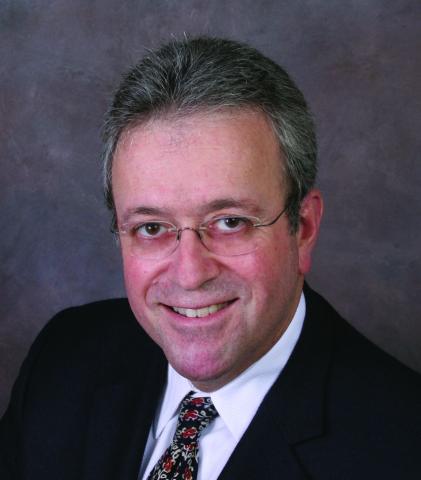
Novel stepwise method found to benefit patients with severe rhinophyma
DENVER –
Rhinophyma occurs primarily in the sixth and seventh decades of life and is marked by facial hypertrophy that leads to tumor-like growth, inflammation, fibrosis, and loss of the cosmetic nasal subunits. “When it becomes severe it leads to a degree of embarrassment as well,” one of the study authors, Patricia Richey, MD, said during an oral abstract session at the annual meeting of the American Society for Dermatologic Surgery. “We found that our method has been efficacious, but most often, and more importantly, leads to an improvement in the patient’s quality of life.”
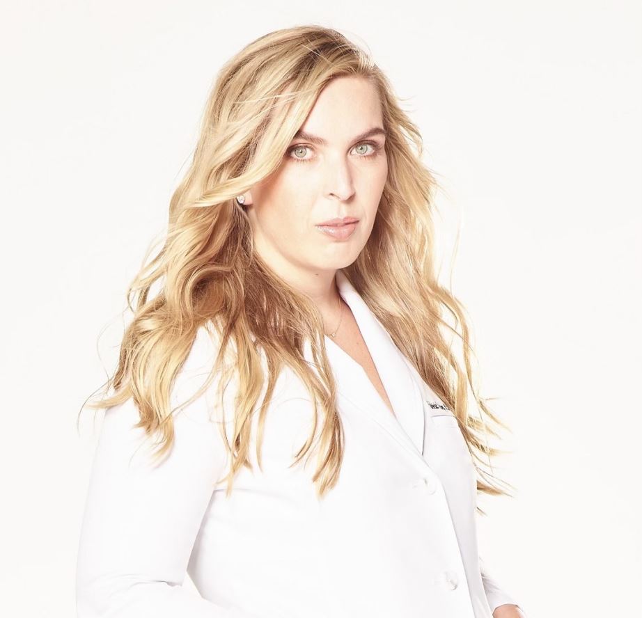
To date, clinicians have used fully ablative lasers to treat varying degrees of rhinophyma but at a cost of prolonged healing time and higher rates of scarring and pigment or textural changes. However, not all dermatologists use full-field ablative lasers in their practices.
“Fractionated ablative lasers have been used in the past for mild to moderate rhinophyma, but they cannot ablate to 100% density, which would be necessary to debulk the marked hypertrophy present in our patients,” said Dr. Richey, who practices Mohs surgery and cosmetic dermatology in Washington, D.C., and conducts research for the Wellman Center for Photomedicine and the Dermatology Laser and Cosmetic Center at Massachusetts General Hospital, Boston. “That’s why we added a surgical component.”
She and colleague Mathew M. Avram, MD, JD, developed a three-step method for treating severe rhinophyma that they performed on three elderly patients. Step 1 is the surgical debulk. Following infiltration of local anesthesia, a razor blade or 15-blade is used to excise the most prominent lobules of hypertrophied sebaceous tissue down to the fibrofatty layer of the nose as a partial thickness excision that does not reach the level of the perichondrium or cartilage. “Hemostasis is achieved with electrocoagulation and application of petrolatum ointment, followed by a pressure dressing,” Dr. Richey said. “The location of the debulk varies by patient.”
Step 2 involves fractionated ablative laser treatment 4 weeks later with either the CO2 or erbium:YAG (Er:YAG) 2,940-nm laser. According to Dr. Richey, the typical setting for the fractionated CO2 is a fluence of 70mJ/cm2 and a high density, performing six out of four passes with 60 seconds between each pass, “though these settings may vary based on the patient presentation,” she said.
The treatment level ranges from 5 (14% density) to 10 (70% density, for the most severe cases). Meanwhile, a representative setting for the ablative fractionated Er:YAG 2,940-nm laser is 250 mcm, no coagulation, 5.5% density, and one pass. “If a second surgical debulk is performed on the same day as ablative laser treatment, the sites of shave removal are typically avoided with the laser,” she said. If a certain portion of the nose has recently healed following surgical debulk 4 weeks prior, they may perform only two passes in this region.
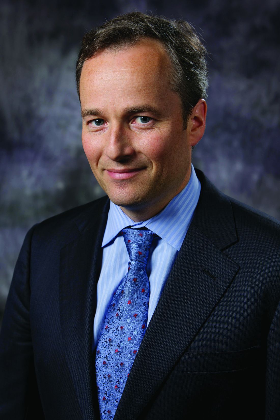
In an interview, Dr. Avram, who directs the MGH Dermatology Laser and Cosmetic Center, characterized the staged method as providing “transformative change to severe, cosmetically disfiguring rhinophyma. The ablative fractional laser provides more fine-tuned contouring.”
The three patients studied had an average of three to four monthly treatments. “There is typically a great deal of improvement by the second treatment,” Dr. Richey said. Add-on treatments may include low voltage electrodessication at 1.8 watts for patients with well-demarcated papules of sebaceous hyperplasia, and a vascular laser such as the pulsed dye laser if telangiectasias are present.
One limitation of the stepwise method, she said, is that the surgical debulk typically results in a scar, “but it’s rarely noticeable if carefully performed, likely due to fractionated ablative use during the scar remodeling period. It’s important to set expectations with your patient at the initial consult. We always discuss treatment goals and that while we aim achieve the most desirable outcome possible, we’re never going to get them back to having a completely normal nose. They’re always going to have some mild or moderate rhinophymatous changes present.”

Vincent Richer, MD, a Vancouver-based medical and cosmetic dermatologist who was asked to comment on these results, characterized the stepwise method as promising. “Though more treatments are required, the easier recovery, safe outcomes in the case presented and excellent cosmetic result made it an interesting alternative when fully ablative resurfacing is daunting, either for patients or physicians involved,” he said in an interview.
The researchers reported having no relevant disclosures. Dr. Richer disclosed that he performs clinical trials for AbbVie/Allergan, Galderma, Leo Pharma, Pfizer, and is a member of advisory board for Bausch, Celgene, Eli Lilly, Galderma, Janssen, Johnson & Johnson, Leo Pharma, L’Oréal, and Sanofi. He is also a consultant to AbbVie/Allergan, Bausch, Celgene, Eli Lilly, Galderma, Janssen, Johnson & Johnson, Leo Pharma, L’Oréal, Merz, and Sanofi.
DENVER –
Rhinophyma occurs primarily in the sixth and seventh decades of life and is marked by facial hypertrophy that leads to tumor-like growth, inflammation, fibrosis, and loss of the cosmetic nasal subunits. “When it becomes severe it leads to a degree of embarrassment as well,” one of the study authors, Patricia Richey, MD, said during an oral abstract session at the annual meeting of the American Society for Dermatologic Surgery. “We found that our method has been efficacious, but most often, and more importantly, leads to an improvement in the patient’s quality of life.”
To date, clinicians have used fully ablative lasers to treat varying degrees of rhinophyma but at a cost of prolonged healing time and higher rates of scarring and pigment or textural changes. However, not all dermatologists use full-field ablative lasers in their practices.
“Fractionated ablative lasers have been used in the past for mild to moderate rhinophyma, but they cannot ablate to 100% density, which would be necessary to debulk the marked hypertrophy present in our patients,” said Dr. Richey, who practices Mohs surgery and cosmetic dermatology in Washington, D.C., and conducts research for the Wellman Center for Photomedicine and the Dermatology Laser and Cosmetic Center at Massachusetts General Hospital, Boston. “That’s why we added a surgical component.”
She and colleague Mathew M. Avram, MD, JD, developed a three-step method for treating severe rhinophyma that they performed on three elderly patients. Step 1 is the surgical debulk. Following infiltration of local anesthesia, a razor blade or 15-blade is used to excise the most prominent lobules of hypertrophied sebaceous tissue down to the fibrofatty layer of the nose as a partial thickness excision that does not reach the level of the perichondrium or cartilage. “Hemostasis is achieved with electrocoagulation and application of petrolatum ointment, followed by a pressure dressing,” Dr. Richey said. “The location of the debulk varies by patient.”
Step 2 involves fractionated ablative laser treatment 4 weeks later with either the CO2 or erbium:YAG (Er:YAG) 2,940-nm laser. According to Dr. Richey, the typical setting for the fractionated CO2 is a fluence of 70mJ/cm2 and a high density, performing six out of four passes with 60 seconds between each pass, “though these settings may vary based on the patient presentation,” she said.
The treatment level ranges from 5 (14% density) to 10 (70% density, for the most severe cases). Meanwhile, a representative setting for the ablative fractionated Er:YAG 2,940-nm laser is 250 mcm, no coagulation, 5.5% density, and one pass. “If a second surgical debulk is performed on the same day as ablative laser treatment, the sites of shave removal are typically avoided with the laser,” she said. If a certain portion of the nose has recently healed following surgical debulk 4 weeks prior, they may perform only two passes in this region.
In an interview, Dr. Avram, who directs the MGH Dermatology Laser and Cosmetic Center, characterized the staged method as providing “transformative change to severe, cosmetically disfiguring rhinophyma. The ablative fractional laser provides more fine-tuned contouring.”
The three patients studied had an average of three to four monthly treatments. “There is typically a great deal of improvement by the second treatment,” Dr. Richey said. Add-on treatments may include low voltage electrodessication at 1.8 watts for patients with well-demarcated papules of sebaceous hyperplasia, and a vascular laser such as the pulsed dye laser if telangiectasias are present.
One limitation of the stepwise method, she said, is that the surgical debulk typically results in a scar, “but it’s rarely noticeable if carefully performed, likely due to fractionated ablative use during the scar remodeling period. It’s important to set expectations with your patient at the initial consult. We always discuss treatment goals and that while we aim achieve the most desirable outcome possible, we’re never going to get them back to having a completely normal nose. They’re always going to have some mild or moderate rhinophymatous changes present.”
Vincent Richer, MD, a Vancouver-based medical and cosmetic dermatologist who was asked to comment on these results, characterized the stepwise method as promising. “Though more treatments are required, the easier recovery, safe outcomes in the case presented and excellent cosmetic result made it an interesting alternative when fully ablative resurfacing is daunting, either for patients or physicians involved,” he said in an interview.
The researchers reported having no relevant disclosures. Dr. Richer disclosed that he performs clinical trials for AbbVie/Allergan, Galderma, Leo Pharma, Pfizer, and is a member of advisory board for Bausch, Celgene, Eli Lilly, Galderma, Janssen, Johnson & Johnson, Leo Pharma, L’Oréal, and Sanofi. He is also a consultant to AbbVie/Allergan, Bausch, Celgene, Eli Lilly, Galderma, Janssen, Johnson & Johnson, Leo Pharma, L’Oréal, Merz, and Sanofi.
DENVER –
Rhinophyma occurs primarily in the sixth and seventh decades of life and is marked by facial hypertrophy that leads to tumor-like growth, inflammation, fibrosis, and loss of the cosmetic nasal subunits. “When it becomes severe it leads to a degree of embarrassment as well,” one of the study authors, Patricia Richey, MD, said during an oral abstract session at the annual meeting of the American Society for Dermatologic Surgery. “We found that our method has been efficacious, but most often, and more importantly, leads to an improvement in the patient’s quality of life.”
To date, clinicians have used fully ablative lasers to treat varying degrees of rhinophyma but at a cost of prolonged healing time and higher rates of scarring and pigment or textural changes. However, not all dermatologists use full-field ablative lasers in their practices.
“Fractionated ablative lasers have been used in the past for mild to moderate rhinophyma, but they cannot ablate to 100% density, which would be necessary to debulk the marked hypertrophy present in our patients,” said Dr. Richey, who practices Mohs surgery and cosmetic dermatology in Washington, D.C., and conducts research for the Wellman Center for Photomedicine and the Dermatology Laser and Cosmetic Center at Massachusetts General Hospital, Boston. “That’s why we added a surgical component.”
She and colleague Mathew M. Avram, MD, JD, developed a three-step method for treating severe rhinophyma that they performed on three elderly patients. Step 1 is the surgical debulk. Following infiltration of local anesthesia, a razor blade or 15-blade is used to excise the most prominent lobules of hypertrophied sebaceous tissue down to the fibrofatty layer of the nose as a partial thickness excision that does not reach the level of the perichondrium or cartilage. “Hemostasis is achieved with electrocoagulation and application of petrolatum ointment, followed by a pressure dressing,” Dr. Richey said. “The location of the debulk varies by patient.”
Step 2 involves fractionated ablative laser treatment 4 weeks later with either the CO2 or erbium:YAG (Er:YAG) 2,940-nm laser. According to Dr. Richey, the typical setting for the fractionated CO2 is a fluence of 70mJ/cm2 and a high density, performing six out of four passes with 60 seconds between each pass, “though these settings may vary based on the patient presentation,” she said.
The treatment level ranges from 5 (14% density) to 10 (70% density, for the most severe cases). Meanwhile, a representative setting for the ablative fractionated Er:YAG 2,940-nm laser is 250 mcm, no coagulation, 5.5% density, and one pass. “If a second surgical debulk is performed on the same day as ablative laser treatment, the sites of shave removal are typically avoided with the laser,” she said. If a certain portion of the nose has recently healed following surgical debulk 4 weeks prior, they may perform only two passes in this region.
In an interview, Dr. Avram, who directs the MGH Dermatology Laser and Cosmetic Center, characterized the staged method as providing “transformative change to severe, cosmetically disfiguring rhinophyma. The ablative fractional laser provides more fine-tuned contouring.”
The three patients studied had an average of three to four monthly treatments. “There is typically a great deal of improvement by the second treatment,” Dr. Richey said. Add-on treatments may include low voltage electrodessication at 1.8 watts for patients with well-demarcated papules of sebaceous hyperplasia, and a vascular laser such as the pulsed dye laser if telangiectasias are present.
One limitation of the stepwise method, she said, is that the surgical debulk typically results in a scar, “but it’s rarely noticeable if carefully performed, likely due to fractionated ablative use during the scar remodeling period. It’s important to set expectations with your patient at the initial consult. We always discuss treatment goals and that while we aim achieve the most desirable outcome possible, we’re never going to get them back to having a completely normal nose. They’re always going to have some mild or moderate rhinophymatous changes present.”
Vincent Richer, MD, a Vancouver-based medical and cosmetic dermatologist who was asked to comment on these results, characterized the stepwise method as promising. “Though more treatments are required, the easier recovery, safe outcomes in the case presented and excellent cosmetic result made it an interesting alternative when fully ablative resurfacing is daunting, either for patients or physicians involved,” he said in an interview.
The researchers reported having no relevant disclosures. Dr. Richer disclosed that he performs clinical trials for AbbVie/Allergan, Galderma, Leo Pharma, Pfizer, and is a member of advisory board for Bausch, Celgene, Eli Lilly, Galderma, Janssen, Johnson & Johnson, Leo Pharma, L’Oréal, and Sanofi. He is also a consultant to AbbVie/Allergan, Bausch, Celgene, Eli Lilly, Galderma, Janssen, Johnson & Johnson, Leo Pharma, L’Oréal, Merz, and Sanofi.
AT ASDS 2022
Vedolizumab linked to increased treatment failure in older patients with Crohn’s
Findings indicate that vedolizumab is associated with an increased risk for treatment failure in older patients with inflammatory bowel disease (IBD), as compared with tumor necrosis factor (TNF) antagonists, according to a new study published in JAMA Network Open.

Although the incidence and prevalence of IBD among older adults are rapidly increasing, there is a lack of evidence-based treatment guidance for these patients, who represent less than 5% of participants in IBD-related clinical trials, wrote Siddharth Singh, MD, a gastroenterologist and assistant professor of medicine at the University of California, San Diego, and colleagues.
“Older patients are frequently undertreated and mismanaged with long-term corticosteroid use and limited use of steroid-sparing therapies owing to patients’ and clinicians’ concerns about the safety of immunosuppressive therapy,” the authors wrote. “There is considerable need for evidence-based treatment guidance for older patients with IBD.”
The researchers undertook an observational study of the comparative effectiveness of vedolizumab versus TNF antagonists (namely infliximab, adalimumab, and golimumab) among older patients with IBD in Denmark. Using the Danish National Patient Register, the authors included 754 patients aged 50 years and older who received treatment between 2005 and 2018.
The primary effectiveness outcome was treatment failure, defined as the composite 1-year risk of IBD-related hospitalization, IBD-related surgery, or a new corticosteroid prescription more than 6 weeks after initiation of treatment with a biologic. Secondary effectiveness outcomes included time to each component included in the composite score.
The primary safety outcome was the risk of serious infections, defined as those that required hospitalization. Secondary safety outcomes were risk of cancer and major adverse cardiovascular or venous thromboembolic events.
The researchers conducted a 1:1 propensity score-matched analysis, accounting for patient, disease, and treatment factors. The 754 patients included 377 incident users of vedolizumab, including 177 with Crohn’s disease; and 377 incident users of TNF antagonists, including 182 with Crohn’s disease. The average follow-up after treatment initiation occurred between 32 and 40 weeks.
Notably, patients treated with vedolizumab were more likely than those treated with TNF antagonists to have multimorbidity, at 16.2% versus 14.1%, and a higher burden of frailty, at 2.7% versus 1.9%. No significant differences were observed in the proportion of patients with recent immunomodulator and corticosteroid exposure.
Overall, vedolizumab was associated with a 31% increased risk of treatment failure (45.4%), compared with TNF antagonists (34.7%). This included an increased risk of IBD-related hospitalization (27.8% versus 16.3%) and IBD-related major abdominal surgery (21.3% versus 8%).
Among patients with Crohn’s disease, vedolizumab was associated with a 77% increased risk of treatment failure, as well as a greater need for corticosteroids. There was no significant difference in the risk of treatment failure or need for corticosteroids in patients with ulcerative colitis
No significant differences were seen in the risk of serious infections between patients treated with vedolizumab or TNF antagonists, at 8.2% versus 8.7%. This didn’t change by IBD phenotype, age at time of biologic therapy initiation, or treatment with biologic monotherapy versus combination therapy with immunomodulators.
The overall incidence of major adverse cardiovascular or venous thromboembolic events was similar among the groups. Rates of new malignant neoplasms were low, with fewer than five events.
In a subgroup analysis based on the Charlson Comorbidity Index, vedolizumab was associated with a 63% increased risk of treatment failure for patients without comorbidities but not for patients with comorbidities.
“This study adds to the body of literature comparing vedolizumab and anti-TNF in older adults. The findings have been mixed, in some part due to differences in study designs,” said Ashwin N. Ananthakrishnan, MBBS, MPH, associate professor of medicine at Harvard Medical School and a gastroenterologist at Massachusetts General Hospital, both in Boston.
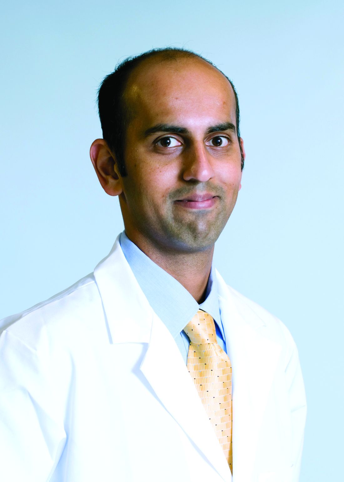
Dr. Ananthakrishnan, who wasn’t involved with this study, has previously researched the two treatments and found that they are comparably safe in older adults. In fact, among patients with significant comorbidity, vedolizumab may be safer. However, the Danish study wasn’t powered to describe that, he said. Moreover, patient characteristics and treatment approaches likely differ between the United States and Denmark.
“Overall, the findings are reassuring. Often when we treat older adults, the emphasis is on safety,” he said. “But by highlighting the difference in clinical response rates – their findings being consistent with a study we published a few years ago – it highlights the importance of also considering efficacy and onset of action for specific disease phenotypes in treatment selection.”
Dr. Ananthakrishnan and colleagues are currently developing clinical tools for risk stratification and prognostication in older adults with IBD, including functional and frailty assessments. “Biologically, older adults may be particularly vulnerable to specific treatment risks such as infections and cancer, but they are also vulnerable to the consequences of untreated disease, including loss of functional independence and frailty,” he explained. “Thus, arriving at the right risk to benefit balance is critically important when making treatment decisions for older adults.”
The study by Dr. Singh and colleagues was supported by grants from the National Institute of Diabetes and Digestive and Kidney Diseases and the Danish National Research Foundation. Dr. Singh reported receiving grants from pharmaceutical companies unrelated to the study, as well as support from the International Organization for the Study of Inflammatory Bowel Disease Operating Grant and Litwin Pioneers in IBD. No other disclosures were reported. Dr. Ananthakrishnan reported no relevant disclosures.
Findings indicate that vedolizumab is associated with an increased risk for treatment failure in older patients with inflammatory bowel disease (IBD), as compared with tumor necrosis factor (TNF) antagonists, according to a new study published in JAMA Network Open.
Although the incidence and prevalence of IBD among older adults are rapidly increasing, there is a lack of evidence-based treatment guidance for these patients, who represent less than 5% of participants in IBD-related clinical trials, wrote Siddharth Singh, MD, a gastroenterologist and assistant professor of medicine at the University of California, San Diego, and colleagues.
“Older patients are frequently undertreated and mismanaged with long-term corticosteroid use and limited use of steroid-sparing therapies owing to patients’ and clinicians’ concerns about the safety of immunosuppressive therapy,” the authors wrote. “There is considerable need for evidence-based treatment guidance for older patients with IBD.”
The researchers undertook an observational study of the comparative effectiveness of vedolizumab versus TNF antagonists (namely infliximab, adalimumab, and golimumab) among older patients with IBD in Denmark. Using the Danish National Patient Register, the authors included 754 patients aged 50 years and older who received treatment between 2005 and 2018.
The primary effectiveness outcome was treatment failure, defined as the composite 1-year risk of IBD-related hospitalization, IBD-related surgery, or a new corticosteroid prescription more than 6 weeks after initiation of treatment with a biologic. Secondary effectiveness outcomes included time to each component included in the composite score.
The primary safety outcome was the risk of serious infections, defined as those that required hospitalization. Secondary safety outcomes were risk of cancer and major adverse cardiovascular or venous thromboembolic events.
The researchers conducted a 1:1 propensity score-matched analysis, accounting for patient, disease, and treatment factors. The 754 patients included 377 incident users of vedolizumab, including 177 with Crohn’s disease; and 377 incident users of TNF antagonists, including 182 with Crohn’s disease. The average follow-up after treatment initiation occurred between 32 and 40 weeks.
Notably, patients treated with vedolizumab were more likely than those treated with TNF antagonists to have multimorbidity, at 16.2% versus 14.1%, and a higher burden of frailty, at 2.7% versus 1.9%. No significant differences were observed in the proportion of patients with recent immunomodulator and corticosteroid exposure.
Overall, vedolizumab was associated with a 31% increased risk of treatment failure (45.4%), compared with TNF antagonists (34.7%). This included an increased risk of IBD-related hospitalization (27.8% versus 16.3%) and IBD-related major abdominal surgery (21.3% versus 8%).
Among patients with Crohn’s disease, vedolizumab was associated with a 77% increased risk of treatment failure, as well as a greater need for corticosteroids. There was no significant difference in the risk of treatment failure or need for corticosteroids in patients with ulcerative colitis
No significant differences were seen in the risk of serious infections between patients treated with vedolizumab or TNF antagonists, at 8.2% versus 8.7%. This didn’t change by IBD phenotype, age at time of biologic therapy initiation, or treatment with biologic monotherapy versus combination therapy with immunomodulators.
The overall incidence of major adverse cardiovascular or venous thromboembolic events was similar among the groups. Rates of new malignant neoplasms were low, with fewer than five events.
In a subgroup analysis based on the Charlson Comorbidity Index, vedolizumab was associated with a 63% increased risk of treatment failure for patients without comorbidities but not for patients with comorbidities.
“This study adds to the body of literature comparing vedolizumab and anti-TNF in older adults. The findings have been mixed, in some part due to differences in study designs,” said Ashwin N. Ananthakrishnan, MBBS, MPH, associate professor of medicine at Harvard Medical School and a gastroenterologist at Massachusetts General Hospital, both in Boston.
Dr. Ananthakrishnan, who wasn’t involved with this study, has previously researched the two treatments and found that they are comparably safe in older adults. In fact, among patients with significant comorbidity, vedolizumab may be safer. However, the Danish study wasn’t powered to describe that, he said. Moreover, patient characteristics and treatment approaches likely differ between the United States and Denmark.
“Overall, the findings are reassuring. Often when we treat older adults, the emphasis is on safety,” he said. “But by highlighting the difference in clinical response rates – their findings being consistent with a study we published a few years ago – it highlights the importance of also considering efficacy and onset of action for specific disease phenotypes in treatment selection.”
Dr. Ananthakrishnan and colleagues are currently developing clinical tools for risk stratification and prognostication in older adults with IBD, including functional and frailty assessments. “Biologically, older adults may be particularly vulnerable to specific treatment risks such as infections and cancer, but they are also vulnerable to the consequences of untreated disease, including loss of functional independence and frailty,” he explained. “Thus, arriving at the right risk to benefit balance is critically important when making treatment decisions for older adults.”
The study by Dr. Singh and colleagues was supported by grants from the National Institute of Diabetes and Digestive and Kidney Diseases and the Danish National Research Foundation. Dr. Singh reported receiving grants from pharmaceutical companies unrelated to the study, as well as support from the International Organization for the Study of Inflammatory Bowel Disease Operating Grant and Litwin Pioneers in IBD. No other disclosures were reported. Dr. Ananthakrishnan reported no relevant disclosures.
Findings indicate that vedolizumab is associated with an increased risk for treatment failure in older patients with inflammatory bowel disease (IBD), as compared with tumor necrosis factor (TNF) antagonists, according to a new study published in JAMA Network Open.
Although the incidence and prevalence of IBD among older adults are rapidly increasing, there is a lack of evidence-based treatment guidance for these patients, who represent less than 5% of participants in IBD-related clinical trials, wrote Siddharth Singh, MD, a gastroenterologist and assistant professor of medicine at the University of California, San Diego, and colleagues.
“Older patients are frequently undertreated and mismanaged with long-term corticosteroid use and limited use of steroid-sparing therapies owing to patients’ and clinicians’ concerns about the safety of immunosuppressive therapy,” the authors wrote. “There is considerable need for evidence-based treatment guidance for older patients with IBD.”
The researchers undertook an observational study of the comparative effectiveness of vedolizumab versus TNF antagonists (namely infliximab, adalimumab, and golimumab) among older patients with IBD in Denmark. Using the Danish National Patient Register, the authors included 754 patients aged 50 years and older who received treatment between 2005 and 2018.
The primary effectiveness outcome was treatment failure, defined as the composite 1-year risk of IBD-related hospitalization, IBD-related surgery, or a new corticosteroid prescription more than 6 weeks after initiation of treatment with a biologic. Secondary effectiveness outcomes included time to each component included in the composite score.
The primary safety outcome was the risk of serious infections, defined as those that required hospitalization. Secondary safety outcomes were risk of cancer and major adverse cardiovascular or venous thromboembolic events.
The researchers conducted a 1:1 propensity score-matched analysis, accounting for patient, disease, and treatment factors. The 754 patients included 377 incident users of vedolizumab, including 177 with Crohn’s disease; and 377 incident users of TNF antagonists, including 182 with Crohn’s disease. The average follow-up after treatment initiation occurred between 32 and 40 weeks.
Notably, patients treated with vedolizumab were more likely than those treated with TNF antagonists to have multimorbidity, at 16.2% versus 14.1%, and a higher burden of frailty, at 2.7% versus 1.9%. No significant differences were observed in the proportion of patients with recent immunomodulator and corticosteroid exposure.
Overall, vedolizumab was associated with a 31% increased risk of treatment failure (45.4%), compared with TNF antagonists (34.7%). This included an increased risk of IBD-related hospitalization (27.8% versus 16.3%) and IBD-related major abdominal surgery (21.3% versus 8%).
Among patients with Crohn’s disease, vedolizumab was associated with a 77% increased risk of treatment failure, as well as a greater need for corticosteroids. There was no significant difference in the risk of treatment failure or need for corticosteroids in patients with ulcerative colitis
No significant differences were seen in the risk of serious infections between patients treated with vedolizumab or TNF antagonists, at 8.2% versus 8.7%. This didn’t change by IBD phenotype, age at time of biologic therapy initiation, or treatment with biologic monotherapy versus combination therapy with immunomodulators.
The overall incidence of major adverse cardiovascular or venous thromboembolic events was similar among the groups. Rates of new malignant neoplasms were low, with fewer than five events.
In a subgroup analysis based on the Charlson Comorbidity Index, vedolizumab was associated with a 63% increased risk of treatment failure for patients without comorbidities but not for patients with comorbidities.
“This study adds to the body of literature comparing vedolizumab and anti-TNF in older adults. The findings have been mixed, in some part due to differences in study designs,” said Ashwin N. Ananthakrishnan, MBBS, MPH, associate professor of medicine at Harvard Medical School and a gastroenterologist at Massachusetts General Hospital, both in Boston.
Dr. Ananthakrishnan, who wasn’t involved with this study, has previously researched the two treatments and found that they are comparably safe in older adults. In fact, among patients with significant comorbidity, vedolizumab may be safer. However, the Danish study wasn’t powered to describe that, he said. Moreover, patient characteristics and treatment approaches likely differ between the United States and Denmark.
“Overall, the findings are reassuring. Often when we treat older adults, the emphasis is on safety,” he said. “But by highlighting the difference in clinical response rates – their findings being consistent with a study we published a few years ago – it highlights the importance of also considering efficacy and onset of action for specific disease phenotypes in treatment selection.”
Dr. Ananthakrishnan and colleagues are currently developing clinical tools for risk stratification and prognostication in older adults with IBD, including functional and frailty assessments. “Biologically, older adults may be particularly vulnerable to specific treatment risks such as infections and cancer, but they are also vulnerable to the consequences of untreated disease, including loss of functional independence and frailty,” he explained. “Thus, arriving at the right risk to benefit balance is critically important when making treatment decisions for older adults.”
The study by Dr. Singh and colleagues was supported by grants from the National Institute of Diabetes and Digestive and Kidney Diseases and the Danish National Research Foundation. Dr. Singh reported receiving grants from pharmaceutical companies unrelated to the study, as well as support from the International Organization for the Study of Inflammatory Bowel Disease Operating Grant and Litwin Pioneers in IBD. No other disclosures were reported. Dr. Ananthakrishnan reported no relevant disclosures.
FROM JAMA NETWORK OPEN