User login
FDA approves dupilumab for children with eczema aged 6 months to 5 years
The whose disease is not adequately controlled with topical prescription therapies or when those therapies are not advisable.
The approval, announced on June 7, 2022, makes dupilumab (Dupixent), an interleukin-4 receptor alpha antagonist, the first biologic available in the United States to treat uncontrolled moderate to severe atopic dermatitis in this age group. In this age group, it is administered subcutaneously every 4 weeks. Dupilumab remains the only biologic treatment approved for patients aged 6 years and older for this indication.
Approval was based on data from a 16-week pivotal phase 3 trial that evaluated the efficacy and safety of dupilumab added to standard of care topical corticosteroids (TCS) in children aged 6 months to 5 years with uncontrolled moderate to severe atopic dermatitis. The trial’s principal investigator, Amy S. Paller, MD, professor and chair of dermatology at Northwestern University, Chicago, and colleagues, found that, at 16 weeks, 28% of patients who were treated with dupilumab, added to low-potency TCS, met the primary endpoint of clear or almost clear skin, compared with 4% of those who received low-potency TCS alone (P < .0001).
In addition, patients who received the combined treatment experienced a 70% average improvement in disease severity from baseline, compared with a 20% improvement among those in the TCS-only group (P < .0001). They also experienced a 49% improvement in itch, compared with a 2% improvement among their counterparts in the TCS-only group (P < .0001).
Outside of the United States, the study’s coprimary endpoint was achievement of 75% or greater improvement in overall disease severity. More than half of the patients who received combined treatment (53%) met this endpoint, compared with 11% in the TCS-only group (P < .0001), according to the company.
Safety results were generally consistent with the safety profile of dupilumab in atopic dermatitis for patients aged 6 years and older. The most common adverse events that were more commonly observed with dupilumab included conjunctivitis (5% vs 0% in the placebo group) and herpes viral infections (6% vs. 5% in the placebo group). Among those on dupilumab, ages 6 months to 5 years, hand,foot, and mouth disease was reported in 5% and skin papilloma were reported in 2%, but these cases did not lead to discontinuation of treatment, according to the company release.
A version of this article first appeared on Medscape.com.
The whose disease is not adequately controlled with topical prescription therapies or when those therapies are not advisable.
The approval, announced on June 7, 2022, makes dupilumab (Dupixent), an interleukin-4 receptor alpha antagonist, the first biologic available in the United States to treat uncontrolled moderate to severe atopic dermatitis in this age group. In this age group, it is administered subcutaneously every 4 weeks. Dupilumab remains the only biologic treatment approved for patients aged 6 years and older for this indication.
Approval was based on data from a 16-week pivotal phase 3 trial that evaluated the efficacy and safety of dupilumab added to standard of care topical corticosteroids (TCS) in children aged 6 months to 5 years with uncontrolled moderate to severe atopic dermatitis. The trial’s principal investigator, Amy S. Paller, MD, professor and chair of dermatology at Northwestern University, Chicago, and colleagues, found that, at 16 weeks, 28% of patients who were treated with dupilumab, added to low-potency TCS, met the primary endpoint of clear or almost clear skin, compared with 4% of those who received low-potency TCS alone (P < .0001).
In addition, patients who received the combined treatment experienced a 70% average improvement in disease severity from baseline, compared with a 20% improvement among those in the TCS-only group (P < .0001). They also experienced a 49% improvement in itch, compared with a 2% improvement among their counterparts in the TCS-only group (P < .0001).
Outside of the United States, the study’s coprimary endpoint was achievement of 75% or greater improvement in overall disease severity. More than half of the patients who received combined treatment (53%) met this endpoint, compared with 11% in the TCS-only group (P < .0001), according to the company.
Safety results were generally consistent with the safety profile of dupilumab in atopic dermatitis for patients aged 6 years and older. The most common adverse events that were more commonly observed with dupilumab included conjunctivitis (5% vs 0% in the placebo group) and herpes viral infections (6% vs. 5% in the placebo group). Among those on dupilumab, ages 6 months to 5 years, hand,foot, and mouth disease was reported in 5% and skin papilloma were reported in 2%, but these cases did not lead to discontinuation of treatment, according to the company release.
A version of this article first appeared on Medscape.com.
The whose disease is not adequately controlled with topical prescription therapies or when those therapies are not advisable.
The approval, announced on June 7, 2022, makes dupilumab (Dupixent), an interleukin-4 receptor alpha antagonist, the first biologic available in the United States to treat uncontrolled moderate to severe atopic dermatitis in this age group. In this age group, it is administered subcutaneously every 4 weeks. Dupilumab remains the only biologic treatment approved for patients aged 6 years and older for this indication.
Approval was based on data from a 16-week pivotal phase 3 trial that evaluated the efficacy and safety of dupilumab added to standard of care topical corticosteroids (TCS) in children aged 6 months to 5 years with uncontrolled moderate to severe atopic dermatitis. The trial’s principal investigator, Amy S. Paller, MD, professor and chair of dermatology at Northwestern University, Chicago, and colleagues, found that, at 16 weeks, 28% of patients who were treated with dupilumab, added to low-potency TCS, met the primary endpoint of clear or almost clear skin, compared with 4% of those who received low-potency TCS alone (P < .0001).
In addition, patients who received the combined treatment experienced a 70% average improvement in disease severity from baseline, compared with a 20% improvement among those in the TCS-only group (P < .0001). They also experienced a 49% improvement in itch, compared with a 2% improvement among their counterparts in the TCS-only group (P < .0001).
Outside of the United States, the study’s coprimary endpoint was achievement of 75% or greater improvement in overall disease severity. More than half of the patients who received combined treatment (53%) met this endpoint, compared with 11% in the TCS-only group (P < .0001), according to the company.
Safety results were generally consistent with the safety profile of dupilumab in atopic dermatitis for patients aged 6 years and older. The most common adverse events that were more commonly observed with dupilumab included conjunctivitis (5% vs 0% in the placebo group) and herpes viral infections (6% vs. 5% in the placebo group). Among those on dupilumab, ages 6 months to 5 years, hand,foot, and mouth disease was reported in 5% and skin papilloma were reported in 2%, but these cases did not lead to discontinuation of treatment, according to the company release.
A version of this article first appeared on Medscape.com.
Substance use the main cause of physician license actions
Despite a sharp uptick in 2011, substance use–specific license actions taken against physicians dropped in frequency between 2004 and 2020.
More than three fourths (76.3%) of license actions taken against physicians were related to substance use, according to a recent study published in JAMA. Psychological impairment was the reason associated with more than 1 in 10 (11.5%) actions taken against physicians’ licenses, while physical impairment was the reason behind approximately 12% of such actions, per the study.
Researchers analyzed 5032 actions taken against the licenses of U.S. physicians. The actions were reported to the National Practitioner Data Bank and were related to substance use, psychological impairment, and physical impairment. The National Practitioner Data Bank is a web-based repository of reports with information on medical malpractice payments and certain adverse actions related to healthcare practitioners, providers, and suppliers. It is provided by the Department of Health & Human Services.
“While there has been increased attention [on] the mental health of physicians, we wanted to understand the extent to which changes in attitudes and practices were reflected in actions taken by hospitals or licensing boards, which are reported in the National Practitioner Data Bank,” Lisa Rotenstein, MD, a primary care physician at Boston’s Brigham and Women’s Hospital and lead author of the study, told this news organization.
Dr. Rotenstein, who is an assistant professor at Harvard Medical School, Boston, studies issues of mental health among physicians and trainees. Dr. Rotenstein was the lead author of a 2016 study that found that more than a quarter (27.2%) of medical students have depressive symptoms. She was also lead author of a 2018 study published in JAMA on the prevalence of burnout among attending physicians.
Actions against physicians trending downward
2011 marked the peak in actions taken against physicians’ licenses for substance use, per the study, but actions related to substance use have otherwise maintained a steady decline over the past 17 years. Researchers found that physicians with license actions as a result of substance use or psychological impairment were more likely to receive indefinite penalties, while also having emergency action taken against their license to practice.
In addition, physicians who had actions taken against their licenses because of substance use or psychological impairment were more likely to accrue a greater number of actions over the course of their careers, according to the study.
About 47% of physicians reported experiencing burnout per Medscape’s Physician Burnout and Depression Report 2022: Stress, Anxiety, and Anger report. Burnout among emergency physicians spiked from 43% in 2020 to 60% in 2021, according to the report.
More than one quarter (26%) of physicians reported drinking alcohol to cope with burnout in 2020, according to Medscape’s 2021 Physician Burnout and Suicide Report. Per the 2021 report, 48% of physicians chose exercise to deal with burnout, while 35% indulged in eating junk food.
Peter Grinspoon, MD, a Boston-based primary care physician, wrote in The Los Angeles Times in 2016 that the rate of substance abuse among physicians starts at 10% and can go as high as 15%; by comparison, rates of substance use among the general population are 8%-10%. “What appears to account for the difference is physician distress, and in the case of drug abuse, plentiful access,” he added.
Dr. Grinspoon wrote a 2016 book called “Free Refills: A Doctor Confronts His Addiction,” which chronicles his experience in recovery and relapse as a physician who was dependent on opioid painkillers.
The findings from the recent study in JAMA “suggest we have made some progress in addressing issues related to substance use in ways that don’t result in license actions or even in meeting physicians’ need for support related to substance use,” said Dr. Rotenstein.
Still, she insists that there’s “substantial opportunity to improve mental health and support offerings for physicians and to reduce stigma related to seeking and receiving mental health support, ideally averting the need for license actions.”
According to Dr. Rotenstein, the cases listed in the National Practitioner Data Bank represent the most severe cases; these reports have risen to a high level of attention or concern and are the result of adverse action reports submitted by healthcare institutions and state licensing boards.
“There are many, many more physicians whose cases are not represented here but who struggle with depression, anxiety, substance use, and more,” said Dr. Rotenstein.
A version of this article first appeared on Medscape.com.
Despite a sharp uptick in 2011, substance use–specific license actions taken against physicians dropped in frequency between 2004 and 2020.
More than three fourths (76.3%) of license actions taken against physicians were related to substance use, according to a recent study published in JAMA. Psychological impairment was the reason associated with more than 1 in 10 (11.5%) actions taken against physicians’ licenses, while physical impairment was the reason behind approximately 12% of such actions, per the study.
Researchers analyzed 5032 actions taken against the licenses of U.S. physicians. The actions were reported to the National Practitioner Data Bank and were related to substance use, psychological impairment, and physical impairment. The National Practitioner Data Bank is a web-based repository of reports with information on medical malpractice payments and certain adverse actions related to healthcare practitioners, providers, and suppliers. It is provided by the Department of Health & Human Services.
“While there has been increased attention [on] the mental health of physicians, we wanted to understand the extent to which changes in attitudes and practices were reflected in actions taken by hospitals or licensing boards, which are reported in the National Practitioner Data Bank,” Lisa Rotenstein, MD, a primary care physician at Boston’s Brigham and Women’s Hospital and lead author of the study, told this news organization.
Dr. Rotenstein, who is an assistant professor at Harvard Medical School, Boston, studies issues of mental health among physicians and trainees. Dr. Rotenstein was the lead author of a 2016 study that found that more than a quarter (27.2%) of medical students have depressive symptoms. She was also lead author of a 2018 study published in JAMA on the prevalence of burnout among attending physicians.
Actions against physicians trending downward
2011 marked the peak in actions taken against physicians’ licenses for substance use, per the study, but actions related to substance use have otherwise maintained a steady decline over the past 17 years. Researchers found that physicians with license actions as a result of substance use or psychological impairment were more likely to receive indefinite penalties, while also having emergency action taken against their license to practice.
In addition, physicians who had actions taken against their licenses because of substance use or psychological impairment were more likely to accrue a greater number of actions over the course of their careers, according to the study.
About 47% of physicians reported experiencing burnout per Medscape’s Physician Burnout and Depression Report 2022: Stress, Anxiety, and Anger report. Burnout among emergency physicians spiked from 43% in 2020 to 60% in 2021, according to the report.
More than one quarter (26%) of physicians reported drinking alcohol to cope with burnout in 2020, according to Medscape’s 2021 Physician Burnout and Suicide Report. Per the 2021 report, 48% of physicians chose exercise to deal with burnout, while 35% indulged in eating junk food.
Peter Grinspoon, MD, a Boston-based primary care physician, wrote in The Los Angeles Times in 2016 that the rate of substance abuse among physicians starts at 10% and can go as high as 15%; by comparison, rates of substance use among the general population are 8%-10%. “What appears to account for the difference is physician distress, and in the case of drug abuse, plentiful access,” he added.
Dr. Grinspoon wrote a 2016 book called “Free Refills: A Doctor Confronts His Addiction,” which chronicles his experience in recovery and relapse as a physician who was dependent on opioid painkillers.
The findings from the recent study in JAMA “suggest we have made some progress in addressing issues related to substance use in ways that don’t result in license actions or even in meeting physicians’ need for support related to substance use,” said Dr. Rotenstein.
Still, she insists that there’s “substantial opportunity to improve mental health and support offerings for physicians and to reduce stigma related to seeking and receiving mental health support, ideally averting the need for license actions.”
According to Dr. Rotenstein, the cases listed in the National Practitioner Data Bank represent the most severe cases; these reports have risen to a high level of attention or concern and are the result of adverse action reports submitted by healthcare institutions and state licensing boards.
“There are many, many more physicians whose cases are not represented here but who struggle with depression, anxiety, substance use, and more,” said Dr. Rotenstein.
A version of this article first appeared on Medscape.com.
Despite a sharp uptick in 2011, substance use–specific license actions taken against physicians dropped in frequency between 2004 and 2020.
More than three fourths (76.3%) of license actions taken against physicians were related to substance use, according to a recent study published in JAMA. Psychological impairment was the reason associated with more than 1 in 10 (11.5%) actions taken against physicians’ licenses, while physical impairment was the reason behind approximately 12% of such actions, per the study.
Researchers analyzed 5032 actions taken against the licenses of U.S. physicians. The actions were reported to the National Practitioner Data Bank and were related to substance use, psychological impairment, and physical impairment. The National Practitioner Data Bank is a web-based repository of reports with information on medical malpractice payments and certain adverse actions related to healthcare practitioners, providers, and suppliers. It is provided by the Department of Health & Human Services.
“While there has been increased attention [on] the mental health of physicians, we wanted to understand the extent to which changes in attitudes and practices were reflected in actions taken by hospitals or licensing boards, which are reported in the National Practitioner Data Bank,” Lisa Rotenstein, MD, a primary care physician at Boston’s Brigham and Women’s Hospital and lead author of the study, told this news organization.
Dr. Rotenstein, who is an assistant professor at Harvard Medical School, Boston, studies issues of mental health among physicians and trainees. Dr. Rotenstein was the lead author of a 2016 study that found that more than a quarter (27.2%) of medical students have depressive symptoms. She was also lead author of a 2018 study published in JAMA on the prevalence of burnout among attending physicians.
Actions against physicians trending downward
2011 marked the peak in actions taken against physicians’ licenses for substance use, per the study, but actions related to substance use have otherwise maintained a steady decline over the past 17 years. Researchers found that physicians with license actions as a result of substance use or psychological impairment were more likely to receive indefinite penalties, while also having emergency action taken against their license to practice.
In addition, physicians who had actions taken against their licenses because of substance use or psychological impairment were more likely to accrue a greater number of actions over the course of their careers, according to the study.
About 47% of physicians reported experiencing burnout per Medscape’s Physician Burnout and Depression Report 2022: Stress, Anxiety, and Anger report. Burnout among emergency physicians spiked from 43% in 2020 to 60% in 2021, according to the report.
More than one quarter (26%) of physicians reported drinking alcohol to cope with burnout in 2020, according to Medscape’s 2021 Physician Burnout and Suicide Report. Per the 2021 report, 48% of physicians chose exercise to deal with burnout, while 35% indulged in eating junk food.
Peter Grinspoon, MD, a Boston-based primary care physician, wrote in The Los Angeles Times in 2016 that the rate of substance abuse among physicians starts at 10% and can go as high as 15%; by comparison, rates of substance use among the general population are 8%-10%. “What appears to account for the difference is physician distress, and in the case of drug abuse, plentiful access,” he added.
Dr. Grinspoon wrote a 2016 book called “Free Refills: A Doctor Confronts His Addiction,” which chronicles his experience in recovery and relapse as a physician who was dependent on opioid painkillers.
The findings from the recent study in JAMA “suggest we have made some progress in addressing issues related to substance use in ways that don’t result in license actions or even in meeting physicians’ need for support related to substance use,” said Dr. Rotenstein.
Still, she insists that there’s “substantial opportunity to improve mental health and support offerings for physicians and to reduce stigma related to seeking and receiving mental health support, ideally averting the need for license actions.”
According to Dr. Rotenstein, the cases listed in the National Practitioner Data Bank represent the most severe cases; these reports have risen to a high level of attention or concern and are the result of adverse action reports submitted by healthcare institutions and state licensing boards.
“There are many, many more physicians whose cases are not represented here but who struggle with depression, anxiety, substance use, and more,” said Dr. Rotenstein.
A version of this article first appeared on Medscape.com.
FROM JAMA
FDA panel strongly backs protein-based Novavax COVID-19 vaccine
than the cutting-edge technology used in mRNA-based shots.
The Vaccines and Related Biological Products Advisory Committee of the Food and Drug Administration voted almost unanimously June 7 in favor of Novavax’s two-dose COVID-19 vaccine for those 18 or older – despite some concerns over rare events of myocarditis and pericarditis.
The tally was 21 “yes” votes, without any “no” votes, but one abstention from a panelist who then offered a largely positive take on this vaccine.
Panelist Bruce Gellin, MD, explained at the end of the meeting that he would have cast a conditional vote in favor of the Novavax vaccine, called NVX-CoV2373, had that been an option. Dr. Gellin, chief of global public health strategy for the Rockefeller Foundation and a vaccine expert, said he didn’t want his abstention to be considered as signaling opposition to the Novavax shot.
Instead, he said, he expects FDA officials will gather more data and evidence about the Novavax vaccine, especially in relation to certain manufacturing issues, before making its decision on the company’s application.
Earlier in the day, a top FDA vaccine reviewer, Doran Fink, MD, PhD, noted that there were important manufacturing differences between the Novavax vaccine supply used in different projects, complicating efforts to assess the company’s application for emergency use authorization (EUA).
But Dr. Fink noted that the FDA staff already had made a convincing case in its briefing document, with enough evidence for an initial conditional clearance to be found in available data.
The FDA is not bound to follow the suggestions of its advisory committees but it often does.
Using the ‘bully pulpit’
At the beginning of the meeting, Peter Marks, MD, PhD, director of the FDA’s Center for Biologics Evaluation and Research, said he was seizing the “bully pulpit” in addressing the need to persuade more people in the United States to take shots against COVID-19.
About 67% of people in the United States aged 18 and older are fully vaccinated, but only about 50% of those in this group have had a first booster, according to the Centers for Disease Control and Prevention.
The two-dose mRNA vaccines from Pfizer and Moderna have been the subject of intense misinformation campaigns on social media, despite efforts by the FDA and other public health officials to convey the message about their strong benefit-risk profile. The FDA in May limited the authorized use of Johnson & Johnson’s single-dose COVID-19 shot, which is based on a different technology, because of concerns about rare and potentially life-threatening blood clots.
Novavax has been described as a more traditional vaccine – a protein subunit shot similar to one people have long received for protection against influenza, pertussis (whooping cough), diphtheria, and tetanus.
“Having a protein-based alternative may be more comfortable for some in terms of their acceptance of vaccines,” Dr. Marks said. “We do have a problem with vaccine uptake that is very serious in the United States. And anything we can do to get people more comfortable to be able to accept these potentially life-saving medical products is something that we feel we are compelled to do.”
Dr. Marks offered these remarks in answer to an FDA panelist’s question about the need to consider an EUA for yet another vaccine.
EUAs are special clearances the FDA can grant in connection with public health emergencies such as the pandemic. The FDA used EUAs for the initial December 2020 clearances of the Pfizer-BioNTech and Moderna vaccines. It has since granted normal approvals for both of these mRNA-based vaccines, based on larger bodies of evidence gathered and submitted by their developers.
During the meeting, the FDA panelists in general appeared comfortable with the idea of granting another EUA for a vaccine. There was agreement that the shot appeared to work in key tests, although these were done before the rise of the Omicron variant.
In a key test, known as study 301, the Novavax vaccine was judged to be 90.4% effective. In the study, 17 of the 17,272 people who got the Novavax vaccine developed COVID-19, compared with 79 of the 8,385 in the placebo group.
Panelists expressed disappointment with the lack of information about how the shot would work now.
“We’re looking at the efficacy against strains that don’t exist any longer,” said panelist Eric J. Rubin, MD, PhD, a Harvard professor and editor of the New England Journal of Medicine.
Still, Dr. Rubin added that he agreed with the argument the FDA’s Dr. Marks had made earlier for an EUA for the Novavax vaccine.
“If there really is a population of patients who are willing to take this and not willing to take the existing vaccines, I think it’s pretty compelling,” Dr. Rubin said.
Other FDA panelists were skeptical of this argument. Jay Portnoy, MD, who was listed on the FDA roster as the panel’s consumer representative, said he has close friends who are vaccine skeptics.
“Their hesitancy is more ideological than technological,” said Dr. Portnoy of Children’s Mercy Hospital, Kansas City, Mo. “So I really doubt that this vaccine is going to crack that nut, but perhaps some individuals would get this when they wouldn’t get the other ones.”
Myocarditis, pericarditis
The Novavax vaccine is already authorized in other countries, including Canada. Novavax in February announced that it had begun shipping its first doses of the vaccine to European Union member states. The vaccine can be moved through existing vaccine supply and cold chain channels instead of requiring complex new delivery procedures.
That could prove an advantage in time, said FDA panelist Michael Nelson, MD, PhD, of the University of Virginia, Charlottesville.
“Who knows even with supply chain challenges down the road, it will be nice to have options going forward,” Dr. Nelson said.
As with other COVID-19 vaccines, clinicians and researchers are still working to understand the potential risk for inflammation of heart muscle and nearby tissue with vaccination. Most patients with myocarditis or pericarditis who sought medical care for these conditions responded well to medicine and rest and felt better quickly, the CDC says on its website. They usually return to their normal daily activities after their symptoms improve.
At the June 7 meeting, Dr. Nelson said there may be cases of myocarditis that go undetected.
“Our signals are those who get admitted to the emergency room and the hospital,” he said. “I’m quite convinced that there are others who are experiencing cardiac events of lesser severity that are worthy of being studied, both from mechanistic and outcomes standpoints. So we have a lot of work to do.”
In looking at results for an initial pool of 40,000 people who received the Novavax vaccine, there were five reported cases of myocarditis or pericarditis developing within 20 days of people getting the shot, the FDA staff said in its presentation on safety.
In a briefing document released ahead of the advisory committee meeting, the FDA staff flagged this number of cases in a relatively small database as a concern, noting it “could be higher than reported during postauthorization use of mRNA COVID-19 vaccines (for which no cases were identified in preauthorization evaluation).”
Novavax officials took a somewhat unusual step of responding in public. The Gaithersburg, Md.–based company on June 3 issued a statement saying researchers had come to “expect to see natural background events of myocarditis in any sufficiently large database, and that young males are at higher risk.”
The data from the company’s placebo-controlled studies show that, overall, in its clinical development program, the rate of myocarditis was balanced between the vaccine and placebo arms (0.007% and 0.005%), Novavax said.
At the June 7 meeting, FDA panelists including Dr. Nelson, and Paul A. Offit, MD, of Children’s Hospital of Philadelphia, urged continued study to try to determine whether and how the vaccines could trigger myocarditis. Investments made now in pursuing these questions related to COVID-19 shots may pay off later, Dr. Offit said.
“We can use that knowledge to make safer vaccines for a disease that is going to be with us for decades, if not longer,” he said.
A version of this article first appeared on Medscape.com.
than the cutting-edge technology used in mRNA-based shots.
The Vaccines and Related Biological Products Advisory Committee of the Food and Drug Administration voted almost unanimously June 7 in favor of Novavax’s two-dose COVID-19 vaccine for those 18 or older – despite some concerns over rare events of myocarditis and pericarditis.
The tally was 21 “yes” votes, without any “no” votes, but one abstention from a panelist who then offered a largely positive take on this vaccine.
Panelist Bruce Gellin, MD, explained at the end of the meeting that he would have cast a conditional vote in favor of the Novavax vaccine, called NVX-CoV2373, had that been an option. Dr. Gellin, chief of global public health strategy for the Rockefeller Foundation and a vaccine expert, said he didn’t want his abstention to be considered as signaling opposition to the Novavax shot.
Instead, he said, he expects FDA officials will gather more data and evidence about the Novavax vaccine, especially in relation to certain manufacturing issues, before making its decision on the company’s application.
Earlier in the day, a top FDA vaccine reviewer, Doran Fink, MD, PhD, noted that there were important manufacturing differences between the Novavax vaccine supply used in different projects, complicating efforts to assess the company’s application for emergency use authorization (EUA).
But Dr. Fink noted that the FDA staff already had made a convincing case in its briefing document, with enough evidence for an initial conditional clearance to be found in available data.
The FDA is not bound to follow the suggestions of its advisory committees but it often does.
Using the ‘bully pulpit’
At the beginning of the meeting, Peter Marks, MD, PhD, director of the FDA’s Center for Biologics Evaluation and Research, said he was seizing the “bully pulpit” in addressing the need to persuade more people in the United States to take shots against COVID-19.
About 67% of people in the United States aged 18 and older are fully vaccinated, but only about 50% of those in this group have had a first booster, according to the Centers for Disease Control and Prevention.
The two-dose mRNA vaccines from Pfizer and Moderna have been the subject of intense misinformation campaigns on social media, despite efforts by the FDA and other public health officials to convey the message about their strong benefit-risk profile. The FDA in May limited the authorized use of Johnson & Johnson’s single-dose COVID-19 shot, which is based on a different technology, because of concerns about rare and potentially life-threatening blood clots.
Novavax has been described as a more traditional vaccine – a protein subunit shot similar to one people have long received for protection against influenza, pertussis (whooping cough), diphtheria, and tetanus.
“Having a protein-based alternative may be more comfortable for some in terms of their acceptance of vaccines,” Dr. Marks said. “We do have a problem with vaccine uptake that is very serious in the United States. And anything we can do to get people more comfortable to be able to accept these potentially life-saving medical products is something that we feel we are compelled to do.”
Dr. Marks offered these remarks in answer to an FDA panelist’s question about the need to consider an EUA for yet another vaccine.
EUAs are special clearances the FDA can grant in connection with public health emergencies such as the pandemic. The FDA used EUAs for the initial December 2020 clearances of the Pfizer-BioNTech and Moderna vaccines. It has since granted normal approvals for both of these mRNA-based vaccines, based on larger bodies of evidence gathered and submitted by their developers.
During the meeting, the FDA panelists in general appeared comfortable with the idea of granting another EUA for a vaccine. There was agreement that the shot appeared to work in key tests, although these were done before the rise of the Omicron variant.
In a key test, known as study 301, the Novavax vaccine was judged to be 90.4% effective. In the study, 17 of the 17,272 people who got the Novavax vaccine developed COVID-19, compared with 79 of the 8,385 in the placebo group.
Panelists expressed disappointment with the lack of information about how the shot would work now.
“We’re looking at the efficacy against strains that don’t exist any longer,” said panelist Eric J. Rubin, MD, PhD, a Harvard professor and editor of the New England Journal of Medicine.
Still, Dr. Rubin added that he agreed with the argument the FDA’s Dr. Marks had made earlier for an EUA for the Novavax vaccine.
“If there really is a population of patients who are willing to take this and not willing to take the existing vaccines, I think it’s pretty compelling,” Dr. Rubin said.
Other FDA panelists were skeptical of this argument. Jay Portnoy, MD, who was listed on the FDA roster as the panel’s consumer representative, said he has close friends who are vaccine skeptics.
“Their hesitancy is more ideological than technological,” said Dr. Portnoy of Children’s Mercy Hospital, Kansas City, Mo. “So I really doubt that this vaccine is going to crack that nut, but perhaps some individuals would get this when they wouldn’t get the other ones.”
Myocarditis, pericarditis
The Novavax vaccine is already authorized in other countries, including Canada. Novavax in February announced that it had begun shipping its first doses of the vaccine to European Union member states. The vaccine can be moved through existing vaccine supply and cold chain channels instead of requiring complex new delivery procedures.
That could prove an advantage in time, said FDA panelist Michael Nelson, MD, PhD, of the University of Virginia, Charlottesville.
“Who knows even with supply chain challenges down the road, it will be nice to have options going forward,” Dr. Nelson said.
As with other COVID-19 vaccines, clinicians and researchers are still working to understand the potential risk for inflammation of heart muscle and nearby tissue with vaccination. Most patients with myocarditis or pericarditis who sought medical care for these conditions responded well to medicine and rest and felt better quickly, the CDC says on its website. They usually return to their normal daily activities after their symptoms improve.
At the June 7 meeting, Dr. Nelson said there may be cases of myocarditis that go undetected.
“Our signals are those who get admitted to the emergency room and the hospital,” he said. “I’m quite convinced that there are others who are experiencing cardiac events of lesser severity that are worthy of being studied, both from mechanistic and outcomes standpoints. So we have a lot of work to do.”
In looking at results for an initial pool of 40,000 people who received the Novavax vaccine, there were five reported cases of myocarditis or pericarditis developing within 20 days of people getting the shot, the FDA staff said in its presentation on safety.
In a briefing document released ahead of the advisory committee meeting, the FDA staff flagged this number of cases in a relatively small database as a concern, noting it “could be higher than reported during postauthorization use of mRNA COVID-19 vaccines (for which no cases were identified in preauthorization evaluation).”
Novavax officials took a somewhat unusual step of responding in public. The Gaithersburg, Md.–based company on June 3 issued a statement saying researchers had come to “expect to see natural background events of myocarditis in any sufficiently large database, and that young males are at higher risk.”
The data from the company’s placebo-controlled studies show that, overall, in its clinical development program, the rate of myocarditis was balanced between the vaccine and placebo arms (0.007% and 0.005%), Novavax said.
At the June 7 meeting, FDA panelists including Dr. Nelson, and Paul A. Offit, MD, of Children’s Hospital of Philadelphia, urged continued study to try to determine whether and how the vaccines could trigger myocarditis. Investments made now in pursuing these questions related to COVID-19 shots may pay off later, Dr. Offit said.
“We can use that knowledge to make safer vaccines for a disease that is going to be with us for decades, if not longer,” he said.
A version of this article first appeared on Medscape.com.
than the cutting-edge technology used in mRNA-based shots.
The Vaccines and Related Biological Products Advisory Committee of the Food and Drug Administration voted almost unanimously June 7 in favor of Novavax’s two-dose COVID-19 vaccine for those 18 or older – despite some concerns over rare events of myocarditis and pericarditis.
The tally was 21 “yes” votes, without any “no” votes, but one abstention from a panelist who then offered a largely positive take on this vaccine.
Panelist Bruce Gellin, MD, explained at the end of the meeting that he would have cast a conditional vote in favor of the Novavax vaccine, called NVX-CoV2373, had that been an option. Dr. Gellin, chief of global public health strategy for the Rockefeller Foundation and a vaccine expert, said he didn’t want his abstention to be considered as signaling opposition to the Novavax shot.
Instead, he said, he expects FDA officials will gather more data and evidence about the Novavax vaccine, especially in relation to certain manufacturing issues, before making its decision on the company’s application.
Earlier in the day, a top FDA vaccine reviewer, Doran Fink, MD, PhD, noted that there were important manufacturing differences between the Novavax vaccine supply used in different projects, complicating efforts to assess the company’s application for emergency use authorization (EUA).
But Dr. Fink noted that the FDA staff already had made a convincing case in its briefing document, with enough evidence for an initial conditional clearance to be found in available data.
The FDA is not bound to follow the suggestions of its advisory committees but it often does.
Using the ‘bully pulpit’
At the beginning of the meeting, Peter Marks, MD, PhD, director of the FDA’s Center for Biologics Evaluation and Research, said he was seizing the “bully pulpit” in addressing the need to persuade more people in the United States to take shots against COVID-19.
About 67% of people in the United States aged 18 and older are fully vaccinated, but only about 50% of those in this group have had a first booster, according to the Centers for Disease Control and Prevention.
The two-dose mRNA vaccines from Pfizer and Moderna have been the subject of intense misinformation campaigns on social media, despite efforts by the FDA and other public health officials to convey the message about their strong benefit-risk profile. The FDA in May limited the authorized use of Johnson & Johnson’s single-dose COVID-19 shot, which is based on a different technology, because of concerns about rare and potentially life-threatening blood clots.
Novavax has been described as a more traditional vaccine – a protein subunit shot similar to one people have long received for protection against influenza, pertussis (whooping cough), diphtheria, and tetanus.
“Having a protein-based alternative may be more comfortable for some in terms of their acceptance of vaccines,” Dr. Marks said. “We do have a problem with vaccine uptake that is very serious in the United States. And anything we can do to get people more comfortable to be able to accept these potentially life-saving medical products is something that we feel we are compelled to do.”
Dr. Marks offered these remarks in answer to an FDA panelist’s question about the need to consider an EUA for yet another vaccine.
EUAs are special clearances the FDA can grant in connection with public health emergencies such as the pandemic. The FDA used EUAs for the initial December 2020 clearances of the Pfizer-BioNTech and Moderna vaccines. It has since granted normal approvals for both of these mRNA-based vaccines, based on larger bodies of evidence gathered and submitted by their developers.
During the meeting, the FDA panelists in general appeared comfortable with the idea of granting another EUA for a vaccine. There was agreement that the shot appeared to work in key tests, although these were done before the rise of the Omicron variant.
In a key test, known as study 301, the Novavax vaccine was judged to be 90.4% effective. In the study, 17 of the 17,272 people who got the Novavax vaccine developed COVID-19, compared with 79 of the 8,385 in the placebo group.
Panelists expressed disappointment with the lack of information about how the shot would work now.
“We’re looking at the efficacy against strains that don’t exist any longer,” said panelist Eric J. Rubin, MD, PhD, a Harvard professor and editor of the New England Journal of Medicine.
Still, Dr. Rubin added that he agreed with the argument the FDA’s Dr. Marks had made earlier for an EUA for the Novavax vaccine.
“If there really is a population of patients who are willing to take this and not willing to take the existing vaccines, I think it’s pretty compelling,” Dr. Rubin said.
Other FDA panelists were skeptical of this argument. Jay Portnoy, MD, who was listed on the FDA roster as the panel’s consumer representative, said he has close friends who are vaccine skeptics.
“Their hesitancy is more ideological than technological,” said Dr. Portnoy of Children’s Mercy Hospital, Kansas City, Mo. “So I really doubt that this vaccine is going to crack that nut, but perhaps some individuals would get this when they wouldn’t get the other ones.”
Myocarditis, pericarditis
The Novavax vaccine is already authorized in other countries, including Canada. Novavax in February announced that it had begun shipping its first doses of the vaccine to European Union member states. The vaccine can be moved through existing vaccine supply and cold chain channels instead of requiring complex new delivery procedures.
That could prove an advantage in time, said FDA panelist Michael Nelson, MD, PhD, of the University of Virginia, Charlottesville.
“Who knows even with supply chain challenges down the road, it will be nice to have options going forward,” Dr. Nelson said.
As with other COVID-19 vaccines, clinicians and researchers are still working to understand the potential risk for inflammation of heart muscle and nearby tissue with vaccination. Most patients with myocarditis or pericarditis who sought medical care for these conditions responded well to medicine and rest and felt better quickly, the CDC says on its website. They usually return to their normal daily activities after their symptoms improve.
At the June 7 meeting, Dr. Nelson said there may be cases of myocarditis that go undetected.
“Our signals are those who get admitted to the emergency room and the hospital,” he said. “I’m quite convinced that there are others who are experiencing cardiac events of lesser severity that are worthy of being studied, both from mechanistic and outcomes standpoints. So we have a lot of work to do.”
In looking at results for an initial pool of 40,000 people who received the Novavax vaccine, there were five reported cases of myocarditis or pericarditis developing within 20 days of people getting the shot, the FDA staff said in its presentation on safety.
In a briefing document released ahead of the advisory committee meeting, the FDA staff flagged this number of cases in a relatively small database as a concern, noting it “could be higher than reported during postauthorization use of mRNA COVID-19 vaccines (for which no cases were identified in preauthorization evaluation).”
Novavax officials took a somewhat unusual step of responding in public. The Gaithersburg, Md.–based company on June 3 issued a statement saying researchers had come to “expect to see natural background events of myocarditis in any sufficiently large database, and that young males are at higher risk.”
The data from the company’s placebo-controlled studies show that, overall, in its clinical development program, the rate of myocarditis was balanced between the vaccine and placebo arms (0.007% and 0.005%), Novavax said.
At the June 7 meeting, FDA panelists including Dr. Nelson, and Paul A. Offit, MD, of Children’s Hospital of Philadelphia, urged continued study to try to determine whether and how the vaccines could trigger myocarditis. Investments made now in pursuing these questions related to COVID-19 shots may pay off later, Dr. Offit said.
“We can use that knowledge to make safer vaccines for a disease that is going to be with us for decades, if not longer,” he said.
A version of this article first appeared on Medscape.com.

FDA adds RA indication for Riabni rituximab biosimilar
The Food and Drug Administration has approved adding adult patients with rheumatoid arthritis to the list of indications for the rituximab biosimilar Riabni (rituximab-arrx) on the basis of results of a randomized, double-blind, comparative clinical study with the CD20-directed cytolytic antibody reference product, Rituxan, the biosimilar’s manufacturer, Amgen, announced June 6.
The RA indication is specifically for adults with moderate to severely active disease who have had an inadequate response to one or more tumor necrosis factor inhibitors. Riabni was approved in December 2020 for the treatment of adult patients with non-Hodgkin lymphoma, chronic lymphocytic leukemia, granulomatosis with polyangiitis, and microscopic polyangiitis.
The clinical study testing Riabni against Rituxan involved 311 patients with moderate to severe RA who received Riabni, Rituxan manufactured in the United States, and Rituxan manufactured in the European Union. The patients who received the U.S.-manufactured Rituxan were transitioned to receive Riabni for their second dose of rituximab, whereas patients in other groups stayed with the same treatment. The trial’s primary efficacy endpoint of the change in Disease Activity Score in 28 joints using C-reactive protein from baseline to week 24 was within the predefined equivalence margin for clinical efficacy between Riabni and Rituxan. The two products also had similar safety, pharmacokinetics, and immunogenicity profiles, according to Amgen.
Currently, Riabni and Ruxience (rituximab-pvvr) are the only two approved rituximab biosimilars in the United States. Ruxience is approved for the same indications. Rituxan alone has protected orphan drug status for the indication of adult patients with moderate to severe pemphigus vulgaris.
A version of this article first appeared on Medscape.com.
The Food and Drug Administration has approved adding adult patients with rheumatoid arthritis to the list of indications for the rituximab biosimilar Riabni (rituximab-arrx) on the basis of results of a randomized, double-blind, comparative clinical study with the CD20-directed cytolytic antibody reference product, Rituxan, the biosimilar’s manufacturer, Amgen, announced June 6.
The RA indication is specifically for adults with moderate to severely active disease who have had an inadequate response to one or more tumor necrosis factor inhibitors. Riabni was approved in December 2020 for the treatment of adult patients with non-Hodgkin lymphoma, chronic lymphocytic leukemia, granulomatosis with polyangiitis, and microscopic polyangiitis.
The clinical study testing Riabni against Rituxan involved 311 patients with moderate to severe RA who received Riabni, Rituxan manufactured in the United States, and Rituxan manufactured in the European Union. The patients who received the U.S.-manufactured Rituxan were transitioned to receive Riabni for their second dose of rituximab, whereas patients in other groups stayed with the same treatment. The trial’s primary efficacy endpoint of the change in Disease Activity Score in 28 joints using C-reactive protein from baseline to week 24 was within the predefined equivalence margin for clinical efficacy between Riabni and Rituxan. The two products also had similar safety, pharmacokinetics, and immunogenicity profiles, according to Amgen.
Currently, Riabni and Ruxience (rituximab-pvvr) are the only two approved rituximab biosimilars in the United States. Ruxience is approved for the same indications. Rituxan alone has protected orphan drug status for the indication of adult patients with moderate to severe pemphigus vulgaris.
A version of this article first appeared on Medscape.com.
The Food and Drug Administration has approved adding adult patients with rheumatoid arthritis to the list of indications for the rituximab biosimilar Riabni (rituximab-arrx) on the basis of results of a randomized, double-blind, comparative clinical study with the CD20-directed cytolytic antibody reference product, Rituxan, the biosimilar’s manufacturer, Amgen, announced June 6.
The RA indication is specifically for adults with moderate to severely active disease who have had an inadequate response to one or more tumor necrosis factor inhibitors. Riabni was approved in December 2020 for the treatment of adult patients with non-Hodgkin lymphoma, chronic lymphocytic leukemia, granulomatosis with polyangiitis, and microscopic polyangiitis.
The clinical study testing Riabni against Rituxan involved 311 patients with moderate to severe RA who received Riabni, Rituxan manufactured in the United States, and Rituxan manufactured in the European Union. The patients who received the U.S.-manufactured Rituxan were transitioned to receive Riabni for their second dose of rituximab, whereas patients in other groups stayed with the same treatment. The trial’s primary efficacy endpoint of the change in Disease Activity Score in 28 joints using C-reactive protein from baseline to week 24 was within the predefined equivalence margin for clinical efficacy between Riabni and Rituxan. The two products also had similar safety, pharmacokinetics, and immunogenicity profiles, according to Amgen.
Currently, Riabni and Ruxience (rituximab-pvvr) are the only two approved rituximab biosimilars in the United States. Ruxience is approved for the same indications. Rituxan alone has protected orphan drug status for the indication of adult patients with moderate to severe pemphigus vulgaris.
A version of this article first appeared on Medscape.com.
Gastroenterologists’ income up 12%, most happy with career pick
Gastroenterologists, like many other physicians, fared better financially in 2021 than during the height of the pandemic in 2020, according to the 2022 Medscape Gastroenterology Compensation Report.
Gastroenterologists’ average annual income rose from $406,000 in 2020 to $453,000 in 2021 – an increase of 12% over the prior year, second only to otolaryngologists (+13%).
“Compensation for most physicians is trending back up as demand for physicians accelerates,” says James Taylor, group president and chief operating officer of AMN Healthcare’s Physician & Leadership Solutions. “The market for physicians has done a complete 180 over just 7 or 8 months.”
In terms of 2021 income gains, gastroenterologists finished toward the top of the 29+ specialties surveyed by Medscape. The average bonus gastroenterologists earned was also higher in 2021 than in 2020 ($74,000 vs. $60,000).
Competition, side gigs
This year, Medscape asked gastroenterologists how competition affects their income; 16% cited nonphysician practitioners as a source of competition (same as physicians overall).
Eight percent cited telemedicine as a source of competition; 5% cited “minute clinics” and other walk-in clinics in pharmacies. Roughly three-quarters said their income is not affected by competition from these sources.
About 30% of gastroenterologists added responsibilities to their medical workload. A few even have side jobs outside of medicine.
However, gastroenterologists are somewhat less likely to take on extra work than other specialties (36%).
“Physicians are fortunate to have a huge array of potential side gigs available to them,” notes Sylvie Stacy, MD, MPH, author of 50 Nonclinical Careers for Physicians. “Supplemental income that pays well is not difficult to find.” She says most who do take on side jobs are motivated to fund early retirement or desire greater financial independence. They also have high levels of student debt to pay off.
Getting paid well is one thing; feeling adequately paid can be another. Gastroenterologists landed toward the middle (53%) of all physicians in terms of feeling fairly compensated for their work. Neurologists were the least (42%), while public health and preventive medicine providers (72%) were most apt to feel fairly compensated.
Challenges and rewards
The challenges of working during the pandemic and the overall changing tone of medicine prompted some physicians to leave the profession, while disenchanting many others.
This year, a smaller percentage of gastroenterologists said they would enter medicine again, compared with last year (75% vs. 81%).
Yet most gastroenterologists surveyed this year said they would choose their specialty again (95%), which is similar to last year (93%). Family physicians and internists would be less willing than most other physicians to repeat their choice.
Gastroenterologists spend an average 14.3 hours each week handling paperwork and administration, placing them among the middle third of all physicians. This year, the average for physicians overall was about 15.5 hours per week.
Most gastroenterologists (73%) plan to continue taking Medicare and/or Medicaid patients. However, that rate is smaller than in last year’s report (80%).
Compared with last year, about the same number of gastroenterologists say they won’t take new Medicaid patients (about 4% vs. 3%), while a somewhat higher percentage are undecided (about 22% vs. 16%). Overall, 70% of physicians said they plan to continue taking Medicare and/or Medicaid patients.
Nearly one-quarter (23%) of gastroenterologists indicated that they would drop low-paying insurers, but most would not because of business, ethical, or other reasons.
What is most rewarding about being a gastroenterologist? Being good at what they do/finding answers, diagnoses tops the list (31%), followed by relationships with and gratitude from patients (29%), making the world a better place/helping others (15%), and making good money at a job they like (11%). A few cited teaching (6%) and pride in their profession (5%)
The most challenging part of their job is having to follow so many rules and regulations (21%). Other challenges include trouble getting fair reimbursement (18%), dealing with difficult patients (17%), having to work long hours (14%), and working with electronic health record systems (10%).
A version of this article first appeared on Medscape.com.
Gastroenterologists, like many other physicians, fared better financially in 2021 than during the height of the pandemic in 2020, according to the 2022 Medscape Gastroenterology Compensation Report.
Gastroenterologists’ average annual income rose from $406,000 in 2020 to $453,000 in 2021 – an increase of 12% over the prior year, second only to otolaryngologists (+13%).
“Compensation for most physicians is trending back up as demand for physicians accelerates,” says James Taylor, group president and chief operating officer of AMN Healthcare’s Physician & Leadership Solutions. “The market for physicians has done a complete 180 over just 7 or 8 months.”
In terms of 2021 income gains, gastroenterologists finished toward the top of the 29+ specialties surveyed by Medscape. The average bonus gastroenterologists earned was also higher in 2021 than in 2020 ($74,000 vs. $60,000).
Competition, side gigs
This year, Medscape asked gastroenterologists how competition affects their income; 16% cited nonphysician practitioners as a source of competition (same as physicians overall).
Eight percent cited telemedicine as a source of competition; 5% cited “minute clinics” and other walk-in clinics in pharmacies. Roughly three-quarters said their income is not affected by competition from these sources.
About 30% of gastroenterologists added responsibilities to their medical workload. A few even have side jobs outside of medicine.
However, gastroenterologists are somewhat less likely to take on extra work than other specialties (36%).
“Physicians are fortunate to have a huge array of potential side gigs available to them,” notes Sylvie Stacy, MD, MPH, author of 50 Nonclinical Careers for Physicians. “Supplemental income that pays well is not difficult to find.” She says most who do take on side jobs are motivated to fund early retirement or desire greater financial independence. They also have high levels of student debt to pay off.
Getting paid well is one thing; feeling adequately paid can be another. Gastroenterologists landed toward the middle (53%) of all physicians in terms of feeling fairly compensated for their work. Neurologists were the least (42%), while public health and preventive medicine providers (72%) were most apt to feel fairly compensated.
Challenges and rewards
The challenges of working during the pandemic and the overall changing tone of medicine prompted some physicians to leave the profession, while disenchanting many others.
This year, a smaller percentage of gastroenterologists said they would enter medicine again, compared with last year (75% vs. 81%).
Yet most gastroenterologists surveyed this year said they would choose their specialty again (95%), which is similar to last year (93%). Family physicians and internists would be less willing than most other physicians to repeat their choice.
Gastroenterologists spend an average 14.3 hours each week handling paperwork and administration, placing them among the middle third of all physicians. This year, the average for physicians overall was about 15.5 hours per week.
Most gastroenterologists (73%) plan to continue taking Medicare and/or Medicaid patients. However, that rate is smaller than in last year’s report (80%).
Compared with last year, about the same number of gastroenterologists say they won’t take new Medicaid patients (about 4% vs. 3%), while a somewhat higher percentage are undecided (about 22% vs. 16%). Overall, 70% of physicians said they plan to continue taking Medicare and/or Medicaid patients.
Nearly one-quarter (23%) of gastroenterologists indicated that they would drop low-paying insurers, but most would not because of business, ethical, or other reasons.
What is most rewarding about being a gastroenterologist? Being good at what they do/finding answers, diagnoses tops the list (31%), followed by relationships with and gratitude from patients (29%), making the world a better place/helping others (15%), and making good money at a job they like (11%). A few cited teaching (6%) and pride in their profession (5%)
The most challenging part of their job is having to follow so many rules and regulations (21%). Other challenges include trouble getting fair reimbursement (18%), dealing with difficult patients (17%), having to work long hours (14%), and working with electronic health record systems (10%).
A version of this article first appeared on Medscape.com.
Gastroenterologists, like many other physicians, fared better financially in 2021 than during the height of the pandemic in 2020, according to the 2022 Medscape Gastroenterology Compensation Report.
Gastroenterologists’ average annual income rose from $406,000 in 2020 to $453,000 in 2021 – an increase of 12% over the prior year, second only to otolaryngologists (+13%).
“Compensation for most physicians is trending back up as demand for physicians accelerates,” says James Taylor, group president and chief operating officer of AMN Healthcare’s Physician & Leadership Solutions. “The market for physicians has done a complete 180 over just 7 or 8 months.”
In terms of 2021 income gains, gastroenterologists finished toward the top of the 29+ specialties surveyed by Medscape. The average bonus gastroenterologists earned was also higher in 2021 than in 2020 ($74,000 vs. $60,000).
Competition, side gigs
This year, Medscape asked gastroenterologists how competition affects their income; 16% cited nonphysician practitioners as a source of competition (same as physicians overall).
Eight percent cited telemedicine as a source of competition; 5% cited “minute clinics” and other walk-in clinics in pharmacies. Roughly three-quarters said their income is not affected by competition from these sources.
About 30% of gastroenterologists added responsibilities to their medical workload. A few even have side jobs outside of medicine.
However, gastroenterologists are somewhat less likely to take on extra work than other specialties (36%).
“Physicians are fortunate to have a huge array of potential side gigs available to them,” notes Sylvie Stacy, MD, MPH, author of 50 Nonclinical Careers for Physicians. “Supplemental income that pays well is not difficult to find.” She says most who do take on side jobs are motivated to fund early retirement or desire greater financial independence. They also have high levels of student debt to pay off.
Getting paid well is one thing; feeling adequately paid can be another. Gastroenterologists landed toward the middle (53%) of all physicians in terms of feeling fairly compensated for their work. Neurologists were the least (42%), while public health and preventive medicine providers (72%) were most apt to feel fairly compensated.
Challenges and rewards
The challenges of working during the pandemic and the overall changing tone of medicine prompted some physicians to leave the profession, while disenchanting many others.
This year, a smaller percentage of gastroenterologists said they would enter medicine again, compared with last year (75% vs. 81%).
Yet most gastroenterologists surveyed this year said they would choose their specialty again (95%), which is similar to last year (93%). Family physicians and internists would be less willing than most other physicians to repeat their choice.
Gastroenterologists spend an average 14.3 hours each week handling paperwork and administration, placing them among the middle third of all physicians. This year, the average for physicians overall was about 15.5 hours per week.
Most gastroenterologists (73%) plan to continue taking Medicare and/or Medicaid patients. However, that rate is smaller than in last year’s report (80%).
Compared with last year, about the same number of gastroenterologists say they won’t take new Medicaid patients (about 4% vs. 3%), while a somewhat higher percentage are undecided (about 22% vs. 16%). Overall, 70% of physicians said they plan to continue taking Medicare and/or Medicaid patients.
Nearly one-quarter (23%) of gastroenterologists indicated that they would drop low-paying insurers, but most would not because of business, ethical, or other reasons.
What is most rewarding about being a gastroenterologist? Being good at what they do/finding answers, diagnoses tops the list (31%), followed by relationships with and gratitude from patients (29%), making the world a better place/helping others (15%), and making good money at a job they like (11%). A few cited teaching (6%) and pride in their profession (5%)
The most challenging part of their job is having to follow so many rules and regulations (21%). Other challenges include trouble getting fair reimbursement (18%), dealing with difficult patients (17%), having to work long hours (14%), and working with electronic health record systems (10%).
A version of this article first appeared on Medscape.com.
Endometriosis: Diagnosis, Surgical Management, and Overlapping Diagnosis.

As a gynecologist specializing in minimally invasive surgical techniques, what is your involvement in the process for diagnosing endometriosis?
Dr. Lager: At our multidisciplinary endometriosis center, we receive a range of referrals from excellent providers near San Francisco and beyond. As a result, patients will often have had extensive evaluations and multiple treatments. Nonetheless, it is important to take a thorough history, to gain an understanding of the progress of their disease, treatments they have taken and the results or side effects of those treatments, and the goals of the patient in order to guide next steps.
Reviewing previous operative reports, pathology, and surgical photos can also be helpful to guide next steps. Commonly, patients will present with dysmenorrhea, and depending on the severity of associated symptoms, such as dysuria, dyschezia, hematuria, or hematochezia, I may refer patients to our colleagues in urology or GI for further evaluation.
Patients will often have previous imaging such as an ultrasound or CT to evaluate anatomic etiology of the pain, but those studies are often negative. Depending on their history, I may order additional imaging, such as an MRI pelvis, and consideration of vaginal or rectal gel. We have worked closely with our radiologists who have developed a specific endometriosis protocol for deeply infiltrative endometriosis and have a multidisciplinary review committee to discuss complex cases.
Although the gold standard for diagnosis of endometriosis is surgical, this leads to a delay in treatment of 7 to 12 years.[1] So, if a patient presents with symptoms of endometriosis, I will discuss the likely diagnosis and start treatment.
Are there specific techniques that you prefer in your standard practice once a clear diagnosis is determined?
Dr. Lager: As I mentioned, although endometriosis is a surgical diagnosis, there may be findings on imaging which will lead to a diagnosis of endometriosis, including endometriomas, uterosacral thickening, a “kissing ovary” appearance, or hematosalpinx for example.
I discuss a broad range of treatment options based on the patient’s goals, from least invasive treatments to definitive surgery. I discuss dietary changes, integrative medicine (we are fortunate to have an integrative medicine gynecologist here at UCSF Osher Center), and pain psychology. Additionally, I review first-line hormonal management options such as: birth control pills, progestin-only pills, levenogestrol IUD, etonogestrel implant, and medroxyprogesterone acetate injection. In my practice, most patients have already tried initial treatment options, and are most interested in other options. I then review second-line options such as GnRH agonists, antagonists, danazol, and aromatase inhibitors. For patients that have had chronic pelvic pain, I also discuss peripheral and central sensitization, and overlapping diagnoses. Surgical management includes diagnostic laparoscopy and excision or ablation of endometriosis, hysterectomy, and oophorectomy.
Are there specific factors that you look for to help you decide whether surgical management is necessary?
Dr. Lager: There are several reasons why patients decide to proceed with surgical management. First, some patients are reticent to start treatment, particularly if they have had negative experiences with hormonal medications and desire a definitive diagnosis. Other patients choose to proceed with surgery for fertility reasons, and others have severe symptoms that are not managed by medications.
The goal of surgery is to remove all visible endometriotic lesions, restore normal anatomy and for pathologic diagnosis if there is atypical characteristics of an endometrial mass. The pelvic exam and imaging can often be helpful surgical planning. If there is a deeply infiltrative lesion in the bowel or bladder, I consult my urology and colorectal colleagues for surgical planning.
Endometriotic lesions are heterogenous, and can include superficial peritoneal lesions, clear vesicular lesions, “powder burn lesions”, endometriomas, and deep infiltrative lesions.
Additionally, I counsel patients on surgical options depending on the fertility desires. For patients with infertility and symptoms of endometriosis, primary surgery with excision or ablation increases pregnancy rates. One meta-analysis showed that operative laparoscopy improved live births and ongoing pregnancy rates.[2] This was found for the first laparoscopic surgery and not repeat surgery.
Can you talk a little bit more about some of the advancements and the controversies in surgical management, and how that impacts your practice or your treatment?
Dr. Lager: Controversy in surgical management includes excision versus ablation in surgical management of endometriosis. One randomized controlled trial showed an improvement with dyspareunia with excision versus ablation after 5 year follow up.[3] However, a recent meta-analysis from 2021 showed no difference in dysmenorrhea between excision and ablation.[4] I generally perform excision of endometriosis as it can provide a tissue for diagnosis and may allow for complete excision of a lesion that may have an underlying component not easily seen.
We also discussed some of the controversy related to fertility and endometriosis. Management of endometriomas in the face of desired fertility is unclear. Endometriomas that are >3 cm in diameter are associated with decreased anti-Mullerian hormone (AMH) levels, but ovarian cystectomy for endometriomas is also associated with decreased AMH levels. I will counsel patients regarding the risks and benefits of ovarian cystectomy and discuss with the reproductive endocrinologists if they recommend removal to improve oocyte retrieval.
Lastly, conservative versus definitive treatment is an important issue to discuss. Depending on a patient’s goals, conservative surgical management of endometriosis may be the most appropriate procedure. However, if a patient has multiple surgeries, does not desire to have children or has completed childbearing regardless of age and wants to decrease the risk of need for repeat surgery, I will discuss with patients that the risk of reoperation after hysterectomy versus conservative surgery is 8% vs 21% in 2 years and 59% vs 22% after 7 years, respectively.[5] Additionally, the patient may have an overlapping gynecological condition, such as adenomyosis or fibroids, and desire surgical management for those conditions as well. Management ultimately will depend on shared decision making,
You mentioned overlapping diagnosis. What are the impacts and barriers related to misdiagnosis or overlapping diagnosis, and what is your approach to recognizing those signs and symptoms?
Dr. Lager: The classic symptoms of endometriosis can overlap with several medical conditions. In addition to gynecologic issues such as adenomyosis and fibroids that I mentioned previously, symptoms such as pelvic pain, bloating, and dysuria can be associated with gastrointestinal conditions, painful bladder syndrome, neurologic, and musculoskeletal pain conditions. This is complex because the overlapping diagnoses can lead to misdiagnosis, and delay in diagnosis and missing an associated diagnosis can lead to inadequate treatment.
I approach the possibility of overlapping diagnoses in consultation with my colleagues who may recommend further testing, such as endoscopy and colonoscopy. Depending on the diagnoses, several treatments can be started concomitantly to address the multifactorial components of pain. For example, pelvic floor dysfunction related to pelvic pain can affect bowel habits, even without a diagnosis of IBS. Pelvic floor physical therapy can address one component of this. Similarly, even if we surgically or medically manage symptoms of endometriosis, the musculoskeletal pain can lead to persistent or worsening pain. The same goes for pain medicine and peripheral or central pain sensitization or neurological pain.
Was there anything else you’d like to share with your colleagues?
Dr. Lager: Endometriosis is a complex condition that requires a multifactorial approach that takes into consideration a patient’s goals. There is not a one-size fit for all patients with endometriosis due to all the issues we discussed. It will take time to address the varied components of pain and is an iterative process. Minimally invasive surgery has an important role in diagnosis and management of endometriosis but is one of several approaches to treat this complex condition. Thanks for taking the time to discuss this important condition that affects at least 10% of gynecological patients, and potentially more due to delayed and undiagnosed disease.
- Staal AH, van der Zanden M, Nap AW. Diagnostic delay of endometriosis in the Netherlands. Gynecol Obstet Invest. 2016;81(4):321-4. doi: 10.1159/000441911
- Duffy JM, Arambage K, Correa FJ, et al. Laparoscopic surgery for endometriosis. Cochrane Database Syst Rev. 2014;(4):CD011031. Update in: Cochrane Database Syst Rev. 2020;10:CD011031. doi:10.1002/14651858.CD011031.pub2
- Healey M, Cheng C, Kaur H. To excise or ablate endometriosis? A prospective randomized double-blinded trial after 5-year follow-up. J Minim Invasive Gynecol. 2014;21(6):999-1004. doi: 10.1016/j.jmig.2014.04.002
- Burks C, Lee M, DeSarno M, Findley J, Flyckt R. Excision versus ablation for management of minimal to mild endometriosis: a systematic review and meta-analysis. J Minim Invasive Gynecol. 2021;28(3):587-597. doi:10.1016/j.jmig.2020.11.028
- Shakiba K, Bena JF, McGill KM, Minger J, Falcone T. Surgical treatment of endometriosis: a 7-year follow-up on the requirement for further surgery. Obstet Gynecol. 2008;111(6):1285-92. Erratum in: Obstet Gynecol. 2008;112(3):710. doi:10.1097/AOG.0b013e3181758ec6
As a gynecologist specializing in minimally invasive surgical techniques, what is your involvement in the process for diagnosing endometriosis?
Dr. Lager: At our multidisciplinary endometriosis center, we receive a range of referrals from excellent providers near San Francisco and beyond. As a result, patients will often have had extensive evaluations and multiple treatments. Nonetheless, it is important to take a thorough history, to gain an understanding of the progress of their disease, treatments they have taken and the results or side effects of those treatments, and the goals of the patient in order to guide next steps.
Reviewing previous operative reports, pathology, and surgical photos can also be helpful to guide next steps. Commonly, patients will present with dysmenorrhea, and depending on the severity of associated symptoms, such as dysuria, dyschezia, hematuria, or hematochezia, I may refer patients to our colleagues in urology or GI for further evaluation.
Patients will often have previous imaging such as an ultrasound or CT to evaluate anatomic etiology of the pain, but those studies are often negative. Depending on their history, I may order additional imaging, such as an MRI pelvis, and consideration of vaginal or rectal gel. We have worked closely with our radiologists who have developed a specific endometriosis protocol for deeply infiltrative endometriosis and have a multidisciplinary review committee to discuss complex cases.
Although the gold standard for diagnosis of endometriosis is surgical, this leads to a delay in treatment of 7 to 12 years.[1] So, if a patient presents with symptoms of endometriosis, I will discuss the likely diagnosis and start treatment.
Are there specific techniques that you prefer in your standard practice once a clear diagnosis is determined?
Dr. Lager: As I mentioned, although endometriosis is a surgical diagnosis, there may be findings on imaging which will lead to a diagnosis of endometriosis, including endometriomas, uterosacral thickening, a “kissing ovary” appearance, or hematosalpinx for example.
I discuss a broad range of treatment options based on the patient’s goals, from least invasive treatments to definitive surgery. I discuss dietary changes, integrative medicine (we are fortunate to have an integrative medicine gynecologist here at UCSF Osher Center), and pain psychology. Additionally, I review first-line hormonal management options such as: birth control pills, progestin-only pills, levenogestrol IUD, etonogestrel implant, and medroxyprogesterone acetate injection. In my practice, most patients have already tried initial treatment options, and are most interested in other options. I then review second-line options such as GnRH agonists, antagonists, danazol, and aromatase inhibitors. For patients that have had chronic pelvic pain, I also discuss peripheral and central sensitization, and overlapping diagnoses. Surgical management includes diagnostic laparoscopy and excision or ablation of endometriosis, hysterectomy, and oophorectomy.
Are there specific factors that you look for to help you decide whether surgical management is necessary?
Dr. Lager: There are several reasons why patients decide to proceed with surgical management. First, some patients are reticent to start treatment, particularly if they have had negative experiences with hormonal medications and desire a definitive diagnosis. Other patients choose to proceed with surgery for fertility reasons, and others have severe symptoms that are not managed by medications.
The goal of surgery is to remove all visible endometriotic lesions, restore normal anatomy and for pathologic diagnosis if there is atypical characteristics of an endometrial mass. The pelvic exam and imaging can often be helpful surgical planning. If there is a deeply infiltrative lesion in the bowel or bladder, I consult my urology and colorectal colleagues for surgical planning.
Endometriotic lesions are heterogenous, and can include superficial peritoneal lesions, clear vesicular lesions, “powder burn lesions”, endometriomas, and deep infiltrative lesions.
Additionally, I counsel patients on surgical options depending on the fertility desires. For patients with infertility and symptoms of endometriosis, primary surgery with excision or ablation increases pregnancy rates. One meta-analysis showed that operative laparoscopy improved live births and ongoing pregnancy rates.[2] This was found for the first laparoscopic surgery and not repeat surgery.
Can you talk a little bit more about some of the advancements and the controversies in surgical management, and how that impacts your practice or your treatment?
Dr. Lager: Controversy in surgical management includes excision versus ablation in surgical management of endometriosis. One randomized controlled trial showed an improvement with dyspareunia with excision versus ablation after 5 year follow up.[3] However, a recent meta-analysis from 2021 showed no difference in dysmenorrhea between excision and ablation.[4] I generally perform excision of endometriosis as it can provide a tissue for diagnosis and may allow for complete excision of a lesion that may have an underlying component not easily seen.
We also discussed some of the controversy related to fertility and endometriosis. Management of endometriomas in the face of desired fertility is unclear. Endometriomas that are >3 cm in diameter are associated with decreased anti-Mullerian hormone (AMH) levels, but ovarian cystectomy for endometriomas is also associated with decreased AMH levels. I will counsel patients regarding the risks and benefits of ovarian cystectomy and discuss with the reproductive endocrinologists if they recommend removal to improve oocyte retrieval.
Lastly, conservative versus definitive treatment is an important issue to discuss. Depending on a patient’s goals, conservative surgical management of endometriosis may be the most appropriate procedure. However, if a patient has multiple surgeries, does not desire to have children or has completed childbearing regardless of age and wants to decrease the risk of need for repeat surgery, I will discuss with patients that the risk of reoperation after hysterectomy versus conservative surgery is 8% vs 21% in 2 years and 59% vs 22% after 7 years, respectively.[5] Additionally, the patient may have an overlapping gynecological condition, such as adenomyosis or fibroids, and desire surgical management for those conditions as well. Management ultimately will depend on shared decision making,
You mentioned overlapping diagnosis. What are the impacts and barriers related to misdiagnosis or overlapping diagnosis, and what is your approach to recognizing those signs and symptoms?
Dr. Lager: The classic symptoms of endometriosis can overlap with several medical conditions. In addition to gynecologic issues such as adenomyosis and fibroids that I mentioned previously, symptoms such as pelvic pain, bloating, and dysuria can be associated with gastrointestinal conditions, painful bladder syndrome, neurologic, and musculoskeletal pain conditions. This is complex because the overlapping diagnoses can lead to misdiagnosis, and delay in diagnosis and missing an associated diagnosis can lead to inadequate treatment.
I approach the possibility of overlapping diagnoses in consultation with my colleagues who may recommend further testing, such as endoscopy and colonoscopy. Depending on the diagnoses, several treatments can be started concomitantly to address the multifactorial components of pain. For example, pelvic floor dysfunction related to pelvic pain can affect bowel habits, even without a diagnosis of IBS. Pelvic floor physical therapy can address one component of this. Similarly, even if we surgically or medically manage symptoms of endometriosis, the musculoskeletal pain can lead to persistent or worsening pain. The same goes for pain medicine and peripheral or central pain sensitization or neurological pain.
Was there anything else you’d like to share with your colleagues?
Dr. Lager: Endometriosis is a complex condition that requires a multifactorial approach that takes into consideration a patient’s goals. There is not a one-size fit for all patients with endometriosis due to all the issues we discussed. It will take time to address the varied components of pain and is an iterative process. Minimally invasive surgery has an important role in diagnosis and management of endometriosis but is one of several approaches to treat this complex condition. Thanks for taking the time to discuss this important condition that affects at least 10% of gynecological patients, and potentially more due to delayed and undiagnosed disease.
As a gynecologist specializing in minimally invasive surgical techniques, what is your involvement in the process for diagnosing endometriosis?
Dr. Lager: At our multidisciplinary endometriosis center, we receive a range of referrals from excellent providers near San Francisco and beyond. As a result, patients will often have had extensive evaluations and multiple treatments. Nonetheless, it is important to take a thorough history, to gain an understanding of the progress of their disease, treatments they have taken and the results or side effects of those treatments, and the goals of the patient in order to guide next steps.
Reviewing previous operative reports, pathology, and surgical photos can also be helpful to guide next steps. Commonly, patients will present with dysmenorrhea, and depending on the severity of associated symptoms, such as dysuria, dyschezia, hematuria, or hematochezia, I may refer patients to our colleagues in urology or GI for further evaluation.
Patients will often have previous imaging such as an ultrasound or CT to evaluate anatomic etiology of the pain, but those studies are often negative. Depending on their history, I may order additional imaging, such as an MRI pelvis, and consideration of vaginal or rectal gel. We have worked closely with our radiologists who have developed a specific endometriosis protocol for deeply infiltrative endometriosis and have a multidisciplinary review committee to discuss complex cases.
Although the gold standard for diagnosis of endometriosis is surgical, this leads to a delay in treatment of 7 to 12 years.[1] So, if a patient presents with symptoms of endometriosis, I will discuss the likely diagnosis and start treatment.
Are there specific techniques that you prefer in your standard practice once a clear diagnosis is determined?
Dr. Lager: As I mentioned, although endometriosis is a surgical diagnosis, there may be findings on imaging which will lead to a diagnosis of endometriosis, including endometriomas, uterosacral thickening, a “kissing ovary” appearance, or hematosalpinx for example.
I discuss a broad range of treatment options based on the patient’s goals, from least invasive treatments to definitive surgery. I discuss dietary changes, integrative medicine (we are fortunate to have an integrative medicine gynecologist here at UCSF Osher Center), and pain psychology. Additionally, I review first-line hormonal management options such as: birth control pills, progestin-only pills, levenogestrol IUD, etonogestrel implant, and medroxyprogesterone acetate injection. In my practice, most patients have already tried initial treatment options, and are most interested in other options. I then review second-line options such as GnRH agonists, antagonists, danazol, and aromatase inhibitors. For patients that have had chronic pelvic pain, I also discuss peripheral and central sensitization, and overlapping diagnoses. Surgical management includes diagnostic laparoscopy and excision or ablation of endometriosis, hysterectomy, and oophorectomy.
Are there specific factors that you look for to help you decide whether surgical management is necessary?
Dr. Lager: There are several reasons why patients decide to proceed with surgical management. First, some patients are reticent to start treatment, particularly if they have had negative experiences with hormonal medications and desire a definitive diagnosis. Other patients choose to proceed with surgery for fertility reasons, and others have severe symptoms that are not managed by medications.
The goal of surgery is to remove all visible endometriotic lesions, restore normal anatomy and for pathologic diagnosis if there is atypical characteristics of an endometrial mass. The pelvic exam and imaging can often be helpful surgical planning. If there is a deeply infiltrative lesion in the bowel or bladder, I consult my urology and colorectal colleagues for surgical planning.
Endometriotic lesions are heterogenous, and can include superficial peritoneal lesions, clear vesicular lesions, “powder burn lesions”, endometriomas, and deep infiltrative lesions.
Additionally, I counsel patients on surgical options depending on the fertility desires. For patients with infertility and symptoms of endometriosis, primary surgery with excision or ablation increases pregnancy rates. One meta-analysis showed that operative laparoscopy improved live births and ongoing pregnancy rates.[2] This was found for the first laparoscopic surgery and not repeat surgery.
Can you talk a little bit more about some of the advancements and the controversies in surgical management, and how that impacts your practice or your treatment?
Dr. Lager: Controversy in surgical management includes excision versus ablation in surgical management of endometriosis. One randomized controlled trial showed an improvement with dyspareunia with excision versus ablation after 5 year follow up.[3] However, a recent meta-analysis from 2021 showed no difference in dysmenorrhea between excision and ablation.[4] I generally perform excision of endometriosis as it can provide a tissue for diagnosis and may allow for complete excision of a lesion that may have an underlying component not easily seen.
We also discussed some of the controversy related to fertility and endometriosis. Management of endometriomas in the face of desired fertility is unclear. Endometriomas that are >3 cm in diameter are associated with decreased anti-Mullerian hormone (AMH) levels, but ovarian cystectomy for endometriomas is also associated with decreased AMH levels. I will counsel patients regarding the risks and benefits of ovarian cystectomy and discuss with the reproductive endocrinologists if they recommend removal to improve oocyte retrieval.
Lastly, conservative versus definitive treatment is an important issue to discuss. Depending on a patient’s goals, conservative surgical management of endometriosis may be the most appropriate procedure. However, if a patient has multiple surgeries, does not desire to have children or has completed childbearing regardless of age and wants to decrease the risk of need for repeat surgery, I will discuss with patients that the risk of reoperation after hysterectomy versus conservative surgery is 8% vs 21% in 2 years and 59% vs 22% after 7 years, respectively.[5] Additionally, the patient may have an overlapping gynecological condition, such as adenomyosis or fibroids, and desire surgical management for those conditions as well. Management ultimately will depend on shared decision making,
You mentioned overlapping diagnosis. What are the impacts and barriers related to misdiagnosis or overlapping diagnosis, and what is your approach to recognizing those signs and symptoms?
Dr. Lager: The classic symptoms of endometriosis can overlap with several medical conditions. In addition to gynecologic issues such as adenomyosis and fibroids that I mentioned previously, symptoms such as pelvic pain, bloating, and dysuria can be associated with gastrointestinal conditions, painful bladder syndrome, neurologic, and musculoskeletal pain conditions. This is complex because the overlapping diagnoses can lead to misdiagnosis, and delay in diagnosis and missing an associated diagnosis can lead to inadequate treatment.
I approach the possibility of overlapping diagnoses in consultation with my colleagues who may recommend further testing, such as endoscopy and colonoscopy. Depending on the diagnoses, several treatments can be started concomitantly to address the multifactorial components of pain. For example, pelvic floor dysfunction related to pelvic pain can affect bowel habits, even without a diagnosis of IBS. Pelvic floor physical therapy can address one component of this. Similarly, even if we surgically or medically manage symptoms of endometriosis, the musculoskeletal pain can lead to persistent or worsening pain. The same goes for pain medicine and peripheral or central pain sensitization or neurological pain.
Was there anything else you’d like to share with your colleagues?
Dr. Lager: Endometriosis is a complex condition that requires a multifactorial approach that takes into consideration a patient’s goals. There is not a one-size fit for all patients with endometriosis due to all the issues we discussed. It will take time to address the varied components of pain and is an iterative process. Minimally invasive surgery has an important role in diagnosis and management of endometriosis but is one of several approaches to treat this complex condition. Thanks for taking the time to discuss this important condition that affects at least 10% of gynecological patients, and potentially more due to delayed and undiagnosed disease.
- Staal AH, van der Zanden M, Nap AW. Diagnostic delay of endometriosis in the Netherlands. Gynecol Obstet Invest. 2016;81(4):321-4. doi: 10.1159/000441911
- Duffy JM, Arambage K, Correa FJ, et al. Laparoscopic surgery for endometriosis. Cochrane Database Syst Rev. 2014;(4):CD011031. Update in: Cochrane Database Syst Rev. 2020;10:CD011031. doi:10.1002/14651858.CD011031.pub2
- Healey M, Cheng C, Kaur H. To excise or ablate endometriosis? A prospective randomized double-blinded trial after 5-year follow-up. J Minim Invasive Gynecol. 2014;21(6):999-1004. doi: 10.1016/j.jmig.2014.04.002
- Burks C, Lee M, DeSarno M, Findley J, Flyckt R. Excision versus ablation for management of minimal to mild endometriosis: a systematic review and meta-analysis. J Minim Invasive Gynecol. 2021;28(3):587-597. doi:10.1016/j.jmig.2020.11.028
- Shakiba K, Bena JF, McGill KM, Minger J, Falcone T. Surgical treatment of endometriosis: a 7-year follow-up on the requirement for further surgery. Obstet Gynecol. 2008;111(6):1285-92. Erratum in: Obstet Gynecol. 2008;112(3):710. doi:10.1097/AOG.0b013e3181758ec6
- Staal AH, van der Zanden M, Nap AW. Diagnostic delay of endometriosis in the Netherlands. Gynecol Obstet Invest. 2016;81(4):321-4. doi: 10.1159/000441911
- Duffy JM, Arambage K, Correa FJ, et al. Laparoscopic surgery for endometriosis. Cochrane Database Syst Rev. 2014;(4):CD011031. Update in: Cochrane Database Syst Rev. 2020;10:CD011031. doi:10.1002/14651858.CD011031.pub2
- Healey M, Cheng C, Kaur H. To excise or ablate endometriosis? A prospective randomized double-blinded trial after 5-year follow-up. J Minim Invasive Gynecol. 2014;21(6):999-1004. doi: 10.1016/j.jmig.2014.04.002
- Burks C, Lee M, DeSarno M, Findley J, Flyckt R. Excision versus ablation for management of minimal to mild endometriosis: a systematic review and meta-analysis. J Minim Invasive Gynecol. 2021;28(3):587-597. doi:10.1016/j.jmig.2020.11.028
- Shakiba K, Bena JF, McGill KM, Minger J, Falcone T. Surgical treatment of endometriosis: a 7-year follow-up on the requirement for further surgery. Obstet Gynecol. 2008;111(6):1285-92. Erratum in: Obstet Gynecol. 2008;112(3):710. doi:10.1097/AOG.0b013e3181758ec6
Children and COVID: Cases down, start of vaccinations near
The first decline in COVID-19 cases among children since early April may have been holiday related, but the shortened week also brought news about vaccination for the youngest children.
The Food and Drug Administration has accepted Pfizer’s application for a COVID-19 vaccine for children under age 5, so vaccination could begin as early as June 21, according to White House COVID-19 response coordinator Ashish Jha, MD.
“We know that many, many parents are eager to vaccinate their youngest kids and it’s important to do this right,” Dr. Jha said at a White House press briefing June 2. “We expect that vaccinations will begin in earnest as early as June 21 and really roll on throughout that week.”
Decline may just be underreporting
Over on the incidence side of the pandemic, “Testing and reporting may have been affected by the holiday weekend [since] states may change their reporting schedules, which may cause irregularities in trends,” the American Academy of Pediatrics and the Children’s Hospital association said in their latest COVID report.
The decline in new cases was not spread uniformly across the four major regions of the United States. The count actually went up in the West for the week of May 27 to June 2, while the South saw the largest decline. The Midwest and Northeast, meanwhile, saw new cases drop for the second straight week, the AAP and CHA said.
The cumulative number of COVID-19 cases in children was up to 13.45 million as of June 2, with children representing 18.9% of all cases since the start of the pandemic, according to the two organizations. The Centers for Disease Control and Prevention reported figures of 13.14 million and 17.5% on June 6.
The AAP/CHA estimates, however, are based on state data that have become increasingly hard to obtain and subject to inconsistency. “Shortages of COVID-19 tests during surges and the increasing use of COVID-19 home tests likely affect the undercounting of COVID-19 cases,” they noted, and “at times when COVID-19 transmission is low, states might reduce the frequency information is updated.”
Vaccinations held steady over the holiday
The ongoing vaccination effort in children aged 5 years and older did not show a Memorial Day drop-off, as initial vaccinations held at 43,000 in 5- to 11-year-olds and at 27,000 in 12- to 17-year-olds for a second consecutive week. That number has ranged from 34,000 to 70,000 for the younger children and from 25,000 to 47,000 for the older group since mid-March, the AAP said in a separate weekly report.
Despite weekly vaccine initiations that have been roughly double those of the older children for months, the 5- to 11-year-olds are still only at 36.0% coverage with at least one dose, compared with 69.5% for the 12- to-17-year-olds. Full vaccination for the two age groups comes in at 29.3% and 59.6%, respectively, as of June 6, according to the CDC’s COVID Data Tracker.
The first decline in COVID-19 cases among children since early April may have been holiday related, but the shortened week also brought news about vaccination for the youngest children.
The Food and Drug Administration has accepted Pfizer’s application for a COVID-19 vaccine for children under age 5, so vaccination could begin as early as June 21, according to White House COVID-19 response coordinator Ashish Jha, MD.
“We know that many, many parents are eager to vaccinate their youngest kids and it’s important to do this right,” Dr. Jha said at a White House press briefing June 2. “We expect that vaccinations will begin in earnest as early as June 21 and really roll on throughout that week.”
Decline may just be underreporting
Over on the incidence side of the pandemic, “Testing and reporting may have been affected by the holiday weekend [since] states may change their reporting schedules, which may cause irregularities in trends,” the American Academy of Pediatrics and the Children’s Hospital association said in their latest COVID report.
The decline in new cases was not spread uniformly across the four major regions of the United States. The count actually went up in the West for the week of May 27 to June 2, while the South saw the largest decline. The Midwest and Northeast, meanwhile, saw new cases drop for the second straight week, the AAP and CHA said.
The cumulative number of COVID-19 cases in children was up to 13.45 million as of June 2, with children representing 18.9% of all cases since the start of the pandemic, according to the two organizations. The Centers for Disease Control and Prevention reported figures of 13.14 million and 17.5% on June 6.
The AAP/CHA estimates, however, are based on state data that have become increasingly hard to obtain and subject to inconsistency. “Shortages of COVID-19 tests during surges and the increasing use of COVID-19 home tests likely affect the undercounting of COVID-19 cases,” they noted, and “at times when COVID-19 transmission is low, states might reduce the frequency information is updated.”
Vaccinations held steady over the holiday
The ongoing vaccination effort in children aged 5 years and older did not show a Memorial Day drop-off, as initial vaccinations held at 43,000 in 5- to 11-year-olds and at 27,000 in 12- to 17-year-olds for a second consecutive week. That number has ranged from 34,000 to 70,000 for the younger children and from 25,000 to 47,000 for the older group since mid-March, the AAP said in a separate weekly report.
Despite weekly vaccine initiations that have been roughly double those of the older children for months, the 5- to 11-year-olds are still only at 36.0% coverage with at least one dose, compared with 69.5% for the 12- to-17-year-olds. Full vaccination for the two age groups comes in at 29.3% and 59.6%, respectively, as of June 6, according to the CDC’s COVID Data Tracker.
The first decline in COVID-19 cases among children since early April may have been holiday related, but the shortened week also brought news about vaccination for the youngest children.
The Food and Drug Administration has accepted Pfizer’s application for a COVID-19 vaccine for children under age 5, so vaccination could begin as early as June 21, according to White House COVID-19 response coordinator Ashish Jha, MD.
“We know that many, many parents are eager to vaccinate their youngest kids and it’s important to do this right,” Dr. Jha said at a White House press briefing June 2. “We expect that vaccinations will begin in earnest as early as June 21 and really roll on throughout that week.”
Decline may just be underreporting
Over on the incidence side of the pandemic, “Testing and reporting may have been affected by the holiday weekend [since] states may change their reporting schedules, which may cause irregularities in trends,” the American Academy of Pediatrics and the Children’s Hospital association said in their latest COVID report.
The decline in new cases was not spread uniformly across the four major regions of the United States. The count actually went up in the West for the week of May 27 to June 2, while the South saw the largest decline. The Midwest and Northeast, meanwhile, saw new cases drop for the second straight week, the AAP and CHA said.
The cumulative number of COVID-19 cases in children was up to 13.45 million as of June 2, with children representing 18.9% of all cases since the start of the pandemic, according to the two organizations. The Centers for Disease Control and Prevention reported figures of 13.14 million and 17.5% on June 6.
The AAP/CHA estimates, however, are based on state data that have become increasingly hard to obtain and subject to inconsistency. “Shortages of COVID-19 tests during surges and the increasing use of COVID-19 home tests likely affect the undercounting of COVID-19 cases,” they noted, and “at times when COVID-19 transmission is low, states might reduce the frequency information is updated.”
Vaccinations held steady over the holiday
The ongoing vaccination effort in children aged 5 years and older did not show a Memorial Day drop-off, as initial vaccinations held at 43,000 in 5- to 11-year-olds and at 27,000 in 12- to 17-year-olds for a second consecutive week. That number has ranged from 34,000 to 70,000 for the younger children and from 25,000 to 47,000 for the older group since mid-March, the AAP said in a separate weekly report.
Despite weekly vaccine initiations that have been roughly double those of the older children for months, the 5- to 11-year-olds are still only at 36.0% coverage with at least one dose, compared with 69.5% for the 12- to-17-year-olds. Full vaccination for the two age groups comes in at 29.3% and 59.6%, respectively, as of June 6, according to the CDC’s COVID Data Tracker.
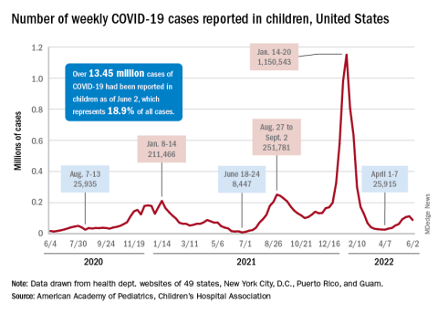
Cochlear implants benefit deaf children with developmental delays
Deaf babies and toddlers with developmental delays may benefit significantly from receiving cochlear implants over hearing aids.
A new study, published in the journal Pediatrics, pushes against the notion that children with low nonverbal cognition and adaptive functioning skills won’t improve if given cochlear implants. Some insurers cover hearing aids for children with developmental disorders but not the implants, which can cost between $60,000 and $100,000 per ear.
“We were surprised [by] the large magnitude of the improvements, not only in quality of life, but also in cognition, ability to function in daily living situations, and speech and language,” lead author John S. Oghalai, MD, of the University of Southern California, Los Angeles, told this news organization. “Remember, these are children with substantial developmental delays. Any improvements are incredibly important and meaningful.”
All children with severe hearing loss should be referred for cochlear implant evaluation, “regardless of the presence of other disabilities,” Dr. Oghalai said. “The younger this referral happens, the better the outcomes will be.”
Dr. Oghalai and his colleagues reviewed data from 204 children approximately 1-3 years old with hearing aids receiving treatment in Texas and California. Of these, 138 received a cochlear implant and had normal cognitive skills and social competence (referred to as adaptive behavior). Another 37 received a cochlear implant and also met criteria for early developmental impairment (EDI), defined by measures of nonverbal cognitive scores and adaptive functioning.
A third group of 29 children with EDI continued with hearing aids without a cochlear implant.
The children were evaluated annually for 1-5 years, with the average follow-up of 2 years. At baseline, no significant differences were noted between the children with EDI who received implants and those who did not on cognition, language, auditory skills, or measures of parental or child stress.
Overall, children who received implants scored higher on cognitive and social measures than those who continued using hearing aids.
Compared with children with EDI who received implants, children without EDI who received implants had significantly higher developmental scores by the study’s end (P ≤ .001), whereas children with EDI who did not receive implants had significantly lower scores (P ≤ .04).
Children who received implants, and their parents, also experienced less stress than those who did not receive the devices, according to the researchers.
Dr. Oghalai and colleagues also measured developmental trajectories for each cohort. Children without delays who received implants had the best outcomes, but those with EDI who received implants had better outcomes than those with EDI and hearing aids.
Findings ‘overdue’
“This study is overdue,” Howard Francis, MD, chair of the department of head and neck surgery & communication sciences at Duke University, Durham, N.C., told this news organization.
Dr. Francis called the new research “reasonably powered and designed,” and said it “documents benefits in the cognitive, language, and patient-child relationship domains” in children who received cochlear implants “compared to children with similar levels of developmental delay whose hearing loss was treated using hearing aids.”
However, “larger studies will be needed to account for potential effects of older age at intervention in the hearing aid group,” he said. Socioeconomic effects are a topic for future research as well, Dr. Francis added.
The researchers initially wanted to perform a controlled clinical trial. However, by the time they secured funding, health insurance policy had changed to cover cochlear implants for children without EDI because of demonstrated benefits shown in studies.
They also were unable to determine the reasons for families’ decisions to choose implants or hearing aids and were unable to assess the impact of insurance on the choice of implantation. But they did find that families with insurers who would cover implants often did choose the devices. Children were also followed for an average of 2 years, so long-term outcomes are unknown.
Despite these limitations, the results support the value of cochlear implantation in children with disabilities and developmental delays, and it should be discussed with parents, the researchers concluded.
“Cochlear implants are just a tool; they do not provide speech and language,” Dr. Oghalai said. “Any child with severe hearing loss requires significant therapy and education via sign language, auditory-verbal therapy, or both. Making the decision about what type of therapy to do is personal, and it depends upon the family and the options that are available to them in their community.”
The study was funded by the National Institutes of Health. The researchers and Dr. Francis have disclosed no relevant financial relationships.
A version of this article first appeared on Medscape.com.
Deaf babies and toddlers with developmental delays may benefit significantly from receiving cochlear implants over hearing aids.
A new study, published in the journal Pediatrics, pushes against the notion that children with low nonverbal cognition and adaptive functioning skills won’t improve if given cochlear implants. Some insurers cover hearing aids for children with developmental disorders but not the implants, which can cost between $60,000 and $100,000 per ear.
“We were surprised [by] the large magnitude of the improvements, not only in quality of life, but also in cognition, ability to function in daily living situations, and speech and language,” lead author John S. Oghalai, MD, of the University of Southern California, Los Angeles, told this news organization. “Remember, these are children with substantial developmental delays. Any improvements are incredibly important and meaningful.”
All children with severe hearing loss should be referred for cochlear implant evaluation, “regardless of the presence of other disabilities,” Dr. Oghalai said. “The younger this referral happens, the better the outcomes will be.”
Dr. Oghalai and his colleagues reviewed data from 204 children approximately 1-3 years old with hearing aids receiving treatment in Texas and California. Of these, 138 received a cochlear implant and had normal cognitive skills and social competence (referred to as adaptive behavior). Another 37 received a cochlear implant and also met criteria for early developmental impairment (EDI), defined by measures of nonverbal cognitive scores and adaptive functioning.
A third group of 29 children with EDI continued with hearing aids without a cochlear implant.
The children were evaluated annually for 1-5 years, with the average follow-up of 2 years. At baseline, no significant differences were noted between the children with EDI who received implants and those who did not on cognition, language, auditory skills, or measures of parental or child stress.
Overall, children who received implants scored higher on cognitive and social measures than those who continued using hearing aids.
Compared with children with EDI who received implants, children without EDI who received implants had significantly higher developmental scores by the study’s end (P ≤ .001), whereas children with EDI who did not receive implants had significantly lower scores (P ≤ .04).
Children who received implants, and their parents, also experienced less stress than those who did not receive the devices, according to the researchers.
Dr. Oghalai and colleagues also measured developmental trajectories for each cohort. Children without delays who received implants had the best outcomes, but those with EDI who received implants had better outcomes than those with EDI and hearing aids.
Findings ‘overdue’
“This study is overdue,” Howard Francis, MD, chair of the department of head and neck surgery & communication sciences at Duke University, Durham, N.C., told this news organization.
Dr. Francis called the new research “reasonably powered and designed,” and said it “documents benefits in the cognitive, language, and patient-child relationship domains” in children who received cochlear implants “compared to children with similar levels of developmental delay whose hearing loss was treated using hearing aids.”
However, “larger studies will be needed to account for potential effects of older age at intervention in the hearing aid group,” he said. Socioeconomic effects are a topic for future research as well, Dr. Francis added.
The researchers initially wanted to perform a controlled clinical trial. However, by the time they secured funding, health insurance policy had changed to cover cochlear implants for children without EDI because of demonstrated benefits shown in studies.
They also were unable to determine the reasons for families’ decisions to choose implants or hearing aids and were unable to assess the impact of insurance on the choice of implantation. But they did find that families with insurers who would cover implants often did choose the devices. Children were also followed for an average of 2 years, so long-term outcomes are unknown.
Despite these limitations, the results support the value of cochlear implantation in children with disabilities and developmental delays, and it should be discussed with parents, the researchers concluded.
“Cochlear implants are just a tool; they do not provide speech and language,” Dr. Oghalai said. “Any child with severe hearing loss requires significant therapy and education via sign language, auditory-verbal therapy, or both. Making the decision about what type of therapy to do is personal, and it depends upon the family and the options that are available to them in their community.”
The study was funded by the National Institutes of Health. The researchers and Dr. Francis have disclosed no relevant financial relationships.
A version of this article first appeared on Medscape.com.
Deaf babies and toddlers with developmental delays may benefit significantly from receiving cochlear implants over hearing aids.
A new study, published in the journal Pediatrics, pushes against the notion that children with low nonverbal cognition and adaptive functioning skills won’t improve if given cochlear implants. Some insurers cover hearing aids for children with developmental disorders but not the implants, which can cost between $60,000 and $100,000 per ear.
“We were surprised [by] the large magnitude of the improvements, not only in quality of life, but also in cognition, ability to function in daily living situations, and speech and language,” lead author John S. Oghalai, MD, of the University of Southern California, Los Angeles, told this news organization. “Remember, these are children with substantial developmental delays. Any improvements are incredibly important and meaningful.”
All children with severe hearing loss should be referred for cochlear implant evaluation, “regardless of the presence of other disabilities,” Dr. Oghalai said. “The younger this referral happens, the better the outcomes will be.”
Dr. Oghalai and his colleagues reviewed data from 204 children approximately 1-3 years old with hearing aids receiving treatment in Texas and California. Of these, 138 received a cochlear implant and had normal cognitive skills and social competence (referred to as adaptive behavior). Another 37 received a cochlear implant and also met criteria for early developmental impairment (EDI), defined by measures of nonverbal cognitive scores and adaptive functioning.
A third group of 29 children with EDI continued with hearing aids without a cochlear implant.
The children were evaluated annually for 1-5 years, with the average follow-up of 2 years. At baseline, no significant differences were noted between the children with EDI who received implants and those who did not on cognition, language, auditory skills, or measures of parental or child stress.
Overall, children who received implants scored higher on cognitive and social measures than those who continued using hearing aids.
Compared with children with EDI who received implants, children without EDI who received implants had significantly higher developmental scores by the study’s end (P ≤ .001), whereas children with EDI who did not receive implants had significantly lower scores (P ≤ .04).
Children who received implants, and their parents, also experienced less stress than those who did not receive the devices, according to the researchers.
Dr. Oghalai and colleagues also measured developmental trajectories for each cohort. Children without delays who received implants had the best outcomes, but those with EDI who received implants had better outcomes than those with EDI and hearing aids.
Findings ‘overdue’
“This study is overdue,” Howard Francis, MD, chair of the department of head and neck surgery & communication sciences at Duke University, Durham, N.C., told this news organization.
Dr. Francis called the new research “reasonably powered and designed,” and said it “documents benefits in the cognitive, language, and patient-child relationship domains” in children who received cochlear implants “compared to children with similar levels of developmental delay whose hearing loss was treated using hearing aids.”
However, “larger studies will be needed to account for potential effects of older age at intervention in the hearing aid group,” he said. Socioeconomic effects are a topic for future research as well, Dr. Francis added.
The researchers initially wanted to perform a controlled clinical trial. However, by the time they secured funding, health insurance policy had changed to cover cochlear implants for children without EDI because of demonstrated benefits shown in studies.
They also were unable to determine the reasons for families’ decisions to choose implants or hearing aids and were unable to assess the impact of insurance on the choice of implantation. But they did find that families with insurers who would cover implants often did choose the devices. Children were also followed for an average of 2 years, so long-term outcomes are unknown.
Despite these limitations, the results support the value of cochlear implantation in children with disabilities and developmental delays, and it should be discussed with parents, the researchers concluded.
“Cochlear implants are just a tool; they do not provide speech and language,” Dr. Oghalai said. “Any child with severe hearing loss requires significant therapy and education via sign language, auditory-verbal therapy, or both. Making the decision about what type of therapy to do is personal, and it depends upon the family and the options that are available to them in their community.”
The study was funded by the National Institutes of Health. The researchers and Dr. Francis have disclosed no relevant financial relationships.
A version of this article first appeared on Medscape.com.
FROM PEDIATRICS
Hope for quicker and more accurate endometriosis diagnosis
A new imaging study hopes to make diagnosing endometriosis quicker, more accurate and reduce the need for invasive surgery.
In October 2020 the All Party Parliamentary Group on Endometriosis published a report that included within its recommendations “a commitment to drive down diagnosis times” for women with the condition. On average, it takes around 8 years for a woman to get a diagnosis of endometriosis, a figure, said the authors of the report, that had “not improved in the last decade.”
Indeed, in its report the APPG said that it was seeking a commitment from Governments in all four nations to reduce average diagnosis times with “targets of 4 years or less by 2025, and a year or less by 2030.”
Surgery often needed for endometriosis diagnosis
Endometriosis affects 1 in 10 women between puberty and menopause – 1.5 million in the United Kingdom – often results in multiple general practitioner and accident and emergency department visits, multiple scans, and often laparoscopic surgery to confirm the diagnosis, as there is currently no simple diagnostic test for the condition. One of the main reasons for the delay in diagnosis is the lack of noninvasive tests capable of detecting all endometriosis subtypes – ovarian, superficial, and deep disease.
Now, experts at the Endometriosis CaRe Centre and Nuffield Department of Women’s and Reproductive Health, University of Oxford (England), in collaboration with British life sciences company Serac Healthcare, hope to establish a faster process for diagnosing endometriosis.
Christian Becker, codirector of the Endometriosis CaRe Centre in Oxford, and a study lead, said: “There is an urgent unmet clinical need for a noninvasive marker to identify or rule out endometriosis as it is such a very common disease affecting more than 190 million women worldwide.”
In the study, researchers will investigate whether a 20-minute imaging scan can detect the most common types of endometriosis, which currently require surgery to diagnose. In turn, they hope that earlier diagnosis of the condition will allow women to seek appropriate treatment sooner. They will use an experimental imaging marker – 99mTc-maraciclatide – that binds to areas of inflammation and that can be used in endometriosis to visualize the disease on a scan. The imaging marker has already been used for detecting inflammation in conditions such as rheumatoid arthritis.
Between 2 and 7 days before planned surgery for suspected endometriosis, participants will be invited for an imaging scan, and the team will compare the suspected locations of disease detected on the scan with those seen during surgery to confirm whether this imaging test could be an effective noninvasive method of detecting all endometriosis subtypes.
Doctor visits and repeated investigations reduced
The researchers commented that the potential strengths of the scan lie in the way the imaging marker binds to areas of inflammation, which may allow doctors to distinguish between new and old lesions and detect endometriosis in areas not easily seen during surgery, such as the lung.
They added that the development of a 20-minute imaging test would reduce the need for repeated visits to doctors, for repeated investigations, and for invasive surgery to obtain a diagnosis. This would ultimately “reduce the time taken to confirm or exclude endometriosis,” they pointed out.
Following the publication of the APPG report in October 2020 the group’s then chair, the late Sir David Amess, said: “Without investment in research, a reduction in diagnosis time, and appropriate NHS pathways, those with endometriosis will continue to face huge barriers in accessing the appropriate support at the right time.”
Krina Zondervan, head of department at the Nuffield Department of Women’s and Reproductive Health, University of Oxford, and a study lead, said: “This study highlights that close collaborations between academics, clinicians and industry are important to combine and accelerate discovery and innovation in addressing high-priority areas in women’s health such as endometriosis.”
David Hail, CEO of Serac Healthcare, said: “We are excited about the potential of 99mTc-maraciclatide to diagnose endometriosis noninvasively and delighted to be working with the internationally renowned team at Oxford on this important first study.”
A version of this article first appeared on Medscape UK.
A new imaging study hopes to make diagnosing endometriosis quicker, more accurate and reduce the need for invasive surgery.
In October 2020 the All Party Parliamentary Group on Endometriosis published a report that included within its recommendations “a commitment to drive down diagnosis times” for women with the condition. On average, it takes around 8 years for a woman to get a diagnosis of endometriosis, a figure, said the authors of the report, that had “not improved in the last decade.”
Indeed, in its report the APPG said that it was seeking a commitment from Governments in all four nations to reduce average diagnosis times with “targets of 4 years or less by 2025, and a year or less by 2030.”
Surgery often needed for endometriosis diagnosis
Endometriosis affects 1 in 10 women between puberty and menopause – 1.5 million in the United Kingdom – often results in multiple general practitioner and accident and emergency department visits, multiple scans, and often laparoscopic surgery to confirm the diagnosis, as there is currently no simple diagnostic test for the condition. One of the main reasons for the delay in diagnosis is the lack of noninvasive tests capable of detecting all endometriosis subtypes – ovarian, superficial, and deep disease.
Now, experts at the Endometriosis CaRe Centre and Nuffield Department of Women’s and Reproductive Health, University of Oxford (England), in collaboration with British life sciences company Serac Healthcare, hope to establish a faster process for diagnosing endometriosis.
Christian Becker, codirector of the Endometriosis CaRe Centre in Oxford, and a study lead, said: “There is an urgent unmet clinical need for a noninvasive marker to identify or rule out endometriosis as it is such a very common disease affecting more than 190 million women worldwide.”
In the study, researchers will investigate whether a 20-minute imaging scan can detect the most common types of endometriosis, which currently require surgery to diagnose. In turn, they hope that earlier diagnosis of the condition will allow women to seek appropriate treatment sooner. They will use an experimental imaging marker – 99mTc-maraciclatide – that binds to areas of inflammation and that can be used in endometriosis to visualize the disease on a scan. The imaging marker has already been used for detecting inflammation in conditions such as rheumatoid arthritis.
Between 2 and 7 days before planned surgery for suspected endometriosis, participants will be invited for an imaging scan, and the team will compare the suspected locations of disease detected on the scan with those seen during surgery to confirm whether this imaging test could be an effective noninvasive method of detecting all endometriosis subtypes.
Doctor visits and repeated investigations reduced
The researchers commented that the potential strengths of the scan lie in the way the imaging marker binds to areas of inflammation, which may allow doctors to distinguish between new and old lesions and detect endometriosis in areas not easily seen during surgery, such as the lung.
They added that the development of a 20-minute imaging test would reduce the need for repeated visits to doctors, for repeated investigations, and for invasive surgery to obtain a diagnosis. This would ultimately “reduce the time taken to confirm or exclude endometriosis,” they pointed out.
Following the publication of the APPG report in October 2020 the group’s then chair, the late Sir David Amess, said: “Without investment in research, a reduction in diagnosis time, and appropriate NHS pathways, those with endometriosis will continue to face huge barriers in accessing the appropriate support at the right time.”
Krina Zondervan, head of department at the Nuffield Department of Women’s and Reproductive Health, University of Oxford, and a study lead, said: “This study highlights that close collaborations between academics, clinicians and industry are important to combine and accelerate discovery and innovation in addressing high-priority areas in women’s health such as endometriosis.”
David Hail, CEO of Serac Healthcare, said: “We are excited about the potential of 99mTc-maraciclatide to diagnose endometriosis noninvasively and delighted to be working with the internationally renowned team at Oxford on this important first study.”
A version of this article first appeared on Medscape UK.
A new imaging study hopes to make diagnosing endometriosis quicker, more accurate and reduce the need for invasive surgery.
In October 2020 the All Party Parliamentary Group on Endometriosis published a report that included within its recommendations “a commitment to drive down diagnosis times” for women with the condition. On average, it takes around 8 years for a woman to get a diagnosis of endometriosis, a figure, said the authors of the report, that had “not improved in the last decade.”
Indeed, in its report the APPG said that it was seeking a commitment from Governments in all four nations to reduce average diagnosis times with “targets of 4 years or less by 2025, and a year or less by 2030.”
Surgery often needed for endometriosis diagnosis
Endometriosis affects 1 in 10 women between puberty and menopause – 1.5 million in the United Kingdom – often results in multiple general practitioner and accident and emergency department visits, multiple scans, and often laparoscopic surgery to confirm the diagnosis, as there is currently no simple diagnostic test for the condition. One of the main reasons for the delay in diagnosis is the lack of noninvasive tests capable of detecting all endometriosis subtypes – ovarian, superficial, and deep disease.
Now, experts at the Endometriosis CaRe Centre and Nuffield Department of Women’s and Reproductive Health, University of Oxford (England), in collaboration with British life sciences company Serac Healthcare, hope to establish a faster process for diagnosing endometriosis.
Christian Becker, codirector of the Endometriosis CaRe Centre in Oxford, and a study lead, said: “There is an urgent unmet clinical need for a noninvasive marker to identify or rule out endometriosis as it is such a very common disease affecting more than 190 million women worldwide.”
In the study, researchers will investigate whether a 20-minute imaging scan can detect the most common types of endometriosis, which currently require surgery to diagnose. In turn, they hope that earlier diagnosis of the condition will allow women to seek appropriate treatment sooner. They will use an experimental imaging marker – 99mTc-maraciclatide – that binds to areas of inflammation and that can be used in endometriosis to visualize the disease on a scan. The imaging marker has already been used for detecting inflammation in conditions such as rheumatoid arthritis.
Between 2 and 7 days before planned surgery for suspected endometriosis, participants will be invited for an imaging scan, and the team will compare the suspected locations of disease detected on the scan with those seen during surgery to confirm whether this imaging test could be an effective noninvasive method of detecting all endometriosis subtypes.
Doctor visits and repeated investigations reduced
The researchers commented that the potential strengths of the scan lie in the way the imaging marker binds to areas of inflammation, which may allow doctors to distinguish between new and old lesions and detect endometriosis in areas not easily seen during surgery, such as the lung.
They added that the development of a 20-minute imaging test would reduce the need for repeated visits to doctors, for repeated investigations, and for invasive surgery to obtain a diagnosis. This would ultimately “reduce the time taken to confirm or exclude endometriosis,” they pointed out.
Following the publication of the APPG report in October 2020 the group’s then chair, the late Sir David Amess, said: “Without investment in research, a reduction in diagnosis time, and appropriate NHS pathways, those with endometriosis will continue to face huge barriers in accessing the appropriate support at the right time.”
Krina Zondervan, head of department at the Nuffield Department of Women’s and Reproductive Health, University of Oxford, and a study lead, said: “This study highlights that close collaborations between academics, clinicians and industry are important to combine and accelerate discovery and innovation in addressing high-priority areas in women’s health such as endometriosis.”
David Hail, CEO of Serac Healthcare, said: “We are excited about the potential of 99mTc-maraciclatide to diagnose endometriosis noninvasively and delighted to be working with the internationally renowned team at Oxford on this important first study.”
A version of this article first appeared on Medscape UK.
Social activities may offset psychosis risk in poor communities
, new research suggests.
A study of more than 170 young participants showed reduced hippocampal volume in those living in poor neighborhoods who had low social engagement versus their peers with greater community engagement.
“These findings demonstrate the importance of considering broader environmental influences and indices of social engagement when conceptualizing adversity and potential interventions for individuals at clinical high risk for psychosis,” co-investigator Benson Ku, MD, a postdoctoral fellow and psychiatry resident at Emory University School of Medicine, Atlanta, told this news organization.

The results were presented at the virtual American Society of Clinical Psychopharmacology annual meeting.
A personal connection
It’s well known that growing up in low-income housing is associated with lower hippocampal volume and an increased risk for schizophrenia, said Dr. Ku.
“The inverse relationship between poverty and hippocampal gray matter volume has [also] been shown to be mediated by social stress, which can include things like lack of parental caregiving and stressful life events,” he added.
Dr. Ku himself grew up in a socioeconomically disadvantaged family in Queens, New York, and he said he had initially performed poorly in school. His early experiences have helped inform his clinical and research interests in the social determinants of mental health.
“I found community support in the Boys’ Club of New York and a local Magic Shop near where I lived, which helped me thrive and become the successful man I am today. I have also heard from my patients how their living conditions and neighborhood have significantly impacted their mental health,” Dr. Ku said.
“A more in-depth understanding of the social determinants of mental health has helped build rapport and empathy with my patients,” he added.
To explore the association between neighborhood poverty, social engagement, and hippocampal volume in youth at high risk for psychosis, the researchers analyzed data from the North American Prodrome Longitudinal Study Phase 2, a multisite consortium.
The researchers recruited and followed up with help-seeking adolescents and young adults from diverse neighborhoods. The analysis included 174 youth, ages 12-33 years, at high clinical risk for psychosis.
Hippocampal volume was assessed using structural MRI. Neighborhood poverty was defined as the percentage of residents with an annual income below the poverty level in the past year.
Social engagement was derived from the desirable events subscale items of the Life Events Scale. These activities included involvement in a church or synagogue; participation in a club, neighborhood, or other organization; taking a vacation; engaging in a hobby, sport, craft, or recreational activity; acquiring a pet; or making new friends.
Lower hippocampal volume
Results showed neighborhood poverty was associated with reduced hippocampal volume, even after controlling for several confounders, including race/ethnicity, family history of mental illnesses, household poverty, educational level, and stressful life events.
Among the 77 participants with lower social engagement, which was defined as three or fewer social activities, neighborhood poverty was associated with reduced hippocampal volume.
However, in the 97 participants who reported greater social engagement, which was defined as four or more social activities, neighborhood poverty was not significantly associated with hippocampal volume.
“It is possible that social engagement may mitigate the deleterious effects of neighborhood poverty on brain morphology, which may inform interventions offered to individuals from disadvantaged neighborhoods,” Dr. Ku said.
“If replication of the relationships between neighborhood poverty, hippocampal volume, and social engagement is established in other populations in longitudinal studies, then targeted interventions at the community level and increased social engagement may potentially play a major role in disease prevention among at-risk youth,” he said.
Dr. Ku noted social engagement might look different in urban versus rural settings.
“In urban areas, it might mean friends, clubs, neighborhood organizations, etc. In rural areas, it might mean family, pets, crafts, etc. The level of social engagement may also depend on neighborhood characteristics, and more research would be needed to better understand how geographic area characteristics – remote, rural, urban – affects social engagement,” he said.
Interesting, innovative
Nagy Youssef, MD, PhD, director of clinical research and professor of psychiatry, Ohio State University College of Medicine, Columbus, said the study suggests “social engagement may reduce the negative effect of poverty in this population, and if replicated in a larger study, could assist and be a part of the early intervention and prevention in psychosis.”

Overall, “this is an interesting and innovative study that has important medical and social implications and is a good step toward helping us understand these relationships and mitigate and prevent negative consequences, as best as possible, in this population,” said Dr. Youssef, who was not part of the research.
The analysis was supported by a grant from the National Institute of Mental Health to the North American Prodrome Longitudinal Study. Dr. Ku and Dr. Youssef report no relevant financial relationships.
A version of this article first appeared on Medscape.com.
, new research suggests.
A study of more than 170 young participants showed reduced hippocampal volume in those living in poor neighborhoods who had low social engagement versus their peers with greater community engagement.
“These findings demonstrate the importance of considering broader environmental influences and indices of social engagement when conceptualizing adversity and potential interventions for individuals at clinical high risk for psychosis,” co-investigator Benson Ku, MD, a postdoctoral fellow and psychiatry resident at Emory University School of Medicine, Atlanta, told this news organization.
The results were presented at the virtual American Society of Clinical Psychopharmacology annual meeting.
A personal connection
It’s well known that growing up in low-income housing is associated with lower hippocampal volume and an increased risk for schizophrenia, said Dr. Ku.
“The inverse relationship between poverty and hippocampal gray matter volume has [also] been shown to be mediated by social stress, which can include things like lack of parental caregiving and stressful life events,” he added.
Dr. Ku himself grew up in a socioeconomically disadvantaged family in Queens, New York, and he said he had initially performed poorly in school. His early experiences have helped inform his clinical and research interests in the social determinants of mental health.
“I found community support in the Boys’ Club of New York and a local Magic Shop near where I lived, which helped me thrive and become the successful man I am today. I have also heard from my patients how their living conditions and neighborhood have significantly impacted their mental health,” Dr. Ku said.
“A more in-depth understanding of the social determinants of mental health has helped build rapport and empathy with my patients,” he added.
To explore the association between neighborhood poverty, social engagement, and hippocampal volume in youth at high risk for psychosis, the researchers analyzed data from the North American Prodrome Longitudinal Study Phase 2, a multisite consortium.
The researchers recruited and followed up with help-seeking adolescents and young adults from diverse neighborhoods. The analysis included 174 youth, ages 12-33 years, at high clinical risk for psychosis.
Hippocampal volume was assessed using structural MRI. Neighborhood poverty was defined as the percentage of residents with an annual income below the poverty level in the past year.
Social engagement was derived from the desirable events subscale items of the Life Events Scale. These activities included involvement in a church or synagogue; participation in a club, neighborhood, or other organization; taking a vacation; engaging in a hobby, sport, craft, or recreational activity; acquiring a pet; or making new friends.
Lower hippocampal volume
Results showed neighborhood poverty was associated with reduced hippocampal volume, even after controlling for several confounders, including race/ethnicity, family history of mental illnesses, household poverty, educational level, and stressful life events.
Among the 77 participants with lower social engagement, which was defined as three or fewer social activities, neighborhood poverty was associated with reduced hippocampal volume.
However, in the 97 participants who reported greater social engagement, which was defined as four or more social activities, neighborhood poverty was not significantly associated with hippocampal volume.
“It is possible that social engagement may mitigate the deleterious effects of neighborhood poverty on brain morphology, which may inform interventions offered to individuals from disadvantaged neighborhoods,” Dr. Ku said.
“If replication of the relationships between neighborhood poverty, hippocampal volume, and social engagement is established in other populations in longitudinal studies, then targeted interventions at the community level and increased social engagement may potentially play a major role in disease prevention among at-risk youth,” he said.
Dr. Ku noted social engagement might look different in urban versus rural settings.
“In urban areas, it might mean friends, clubs, neighborhood organizations, etc. In rural areas, it might mean family, pets, crafts, etc. The level of social engagement may also depend on neighborhood characteristics, and more research would be needed to better understand how geographic area characteristics – remote, rural, urban – affects social engagement,” he said.
Interesting, innovative
Nagy Youssef, MD, PhD, director of clinical research and professor of psychiatry, Ohio State University College of Medicine, Columbus, said the study suggests “social engagement may reduce the negative effect of poverty in this population, and if replicated in a larger study, could assist and be a part of the early intervention and prevention in psychosis.”
Overall, “this is an interesting and innovative study that has important medical and social implications and is a good step toward helping us understand these relationships and mitigate and prevent negative consequences, as best as possible, in this population,” said Dr. Youssef, who was not part of the research.
The analysis was supported by a grant from the National Institute of Mental Health to the North American Prodrome Longitudinal Study. Dr. Ku and Dr. Youssef report no relevant financial relationships.
A version of this article first appeared on Medscape.com.
, new research suggests.
A study of more than 170 young participants showed reduced hippocampal volume in those living in poor neighborhoods who had low social engagement versus their peers with greater community engagement.
“These findings demonstrate the importance of considering broader environmental influences and indices of social engagement when conceptualizing adversity and potential interventions for individuals at clinical high risk for psychosis,” co-investigator Benson Ku, MD, a postdoctoral fellow and psychiatry resident at Emory University School of Medicine, Atlanta, told this news organization.
The results were presented at the virtual American Society of Clinical Psychopharmacology annual meeting.
A personal connection
It’s well known that growing up in low-income housing is associated with lower hippocampal volume and an increased risk for schizophrenia, said Dr. Ku.
“The inverse relationship between poverty and hippocampal gray matter volume has [also] been shown to be mediated by social stress, which can include things like lack of parental caregiving and stressful life events,” he added.
Dr. Ku himself grew up in a socioeconomically disadvantaged family in Queens, New York, and he said he had initially performed poorly in school. His early experiences have helped inform his clinical and research interests in the social determinants of mental health.
“I found community support in the Boys’ Club of New York and a local Magic Shop near where I lived, which helped me thrive and become the successful man I am today. I have also heard from my patients how their living conditions and neighborhood have significantly impacted their mental health,” Dr. Ku said.
“A more in-depth understanding of the social determinants of mental health has helped build rapport and empathy with my patients,” he added.
To explore the association between neighborhood poverty, social engagement, and hippocampal volume in youth at high risk for psychosis, the researchers analyzed data from the North American Prodrome Longitudinal Study Phase 2, a multisite consortium.
The researchers recruited and followed up with help-seeking adolescents and young adults from diverse neighborhoods. The analysis included 174 youth, ages 12-33 years, at high clinical risk for psychosis.
Hippocampal volume was assessed using structural MRI. Neighborhood poverty was defined as the percentage of residents with an annual income below the poverty level in the past year.
Social engagement was derived from the desirable events subscale items of the Life Events Scale. These activities included involvement in a church or synagogue; participation in a club, neighborhood, or other organization; taking a vacation; engaging in a hobby, sport, craft, or recreational activity; acquiring a pet; or making new friends.
Lower hippocampal volume
Results showed neighborhood poverty was associated with reduced hippocampal volume, even after controlling for several confounders, including race/ethnicity, family history of mental illnesses, household poverty, educational level, and stressful life events.
Among the 77 participants with lower social engagement, which was defined as three or fewer social activities, neighborhood poverty was associated with reduced hippocampal volume.
However, in the 97 participants who reported greater social engagement, which was defined as four or more social activities, neighborhood poverty was not significantly associated with hippocampal volume.
“It is possible that social engagement may mitigate the deleterious effects of neighborhood poverty on brain morphology, which may inform interventions offered to individuals from disadvantaged neighborhoods,” Dr. Ku said.
“If replication of the relationships between neighborhood poverty, hippocampal volume, and social engagement is established in other populations in longitudinal studies, then targeted interventions at the community level and increased social engagement may potentially play a major role in disease prevention among at-risk youth,” he said.
Dr. Ku noted social engagement might look different in urban versus rural settings.
“In urban areas, it might mean friends, clubs, neighborhood organizations, etc. In rural areas, it might mean family, pets, crafts, etc. The level of social engagement may also depend on neighborhood characteristics, and more research would be needed to better understand how geographic area characteristics – remote, rural, urban – affects social engagement,” he said.
Interesting, innovative
Nagy Youssef, MD, PhD, director of clinical research and professor of psychiatry, Ohio State University College of Medicine, Columbus, said the study suggests “social engagement may reduce the negative effect of poverty in this population, and if replicated in a larger study, could assist and be a part of the early intervention and prevention in psychosis.”
Overall, “this is an interesting and innovative study that has important medical and social implications and is a good step toward helping us understand these relationships and mitigate and prevent negative consequences, as best as possible, in this population,” said Dr. Youssef, who was not part of the research.
The analysis was supported by a grant from the National Institute of Mental Health to the North American Prodrome Longitudinal Study. Dr. Ku and Dr. Youssef report no relevant financial relationships.
A version of this article first appeared on Medscape.com.
FROM ASCP 2022