User login
Children and COVID: New cases up by 50%
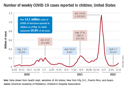
The latest increase in new child COVID-19 cases seems to be picking up steam, rising by 50% in the last week, according to the American Academy of Pediatrics and the Children’s Hospital Association.
That 50% week-to-week change follows increases of 17%, 44%, 12%, and 28% since the nationwide weekly total fell to its low point for the year (25,915) in the beginning of April, the AAP and CHA said in their weekly COVID report.
Regionally, the distribution of those 93,000 COVID cases was fairly even. The Northeast, which saw the biggest jump for the week, and the Midwest were both around 25,000 new cases, while the South had about 20,000 and the West was lowest with 18,000 or so. At the state/territory level, the largest percent increases over the last 2 weeks were found in Maine and Puerto Rico, with Massachusetts and Vermont just a step behind, the AAP/CHA data show.
In cumulative terms, there have been over 13.1 million cases of COVID-19 among children in the United States, with pediatric cases representing 19.0% of all cases since the pandemic began, the two organizations reported. They also noted a number of important limitations: New York state has never reported cases by age, several states have stopped updating their online dashboards, and states apply a variety of age ranges to define children (Alabama has the smallest range, 0-14 years; South Carolina, Tennessee, and West Virginia the largest, 0-20).
By comparison, the Centers for Disease Control and Prevention put the total number of cases in children aged 0-17 at 12.7 million, although that figure is based on a cumulative number of 73.4 million cases among all ages, which is well short of the reported total of almost 82.4 million as of May 16. COVID cases in children have led to 1,536 deaths so far, the CDC said.
The recent upward trend in new cases also can be seen in the CDC’s data, which show the weekly rate rising from 35 per 100,000 population on March 26 to 102 per 100,000 on May 7 in children aged 0-14 years, with commensurate increases seen among older children over the same period. In turn, the rate of new admissions for children aged 0-17 has gone from a low of 0.13 per 100,000 as late as April 10 up to 0.23 on May 13, the CDC said on its COVID Data Tracker.
One thing not going up these days is vaccinations among the youngest eligible children. The number of 5- to 11-year-olds receiving their initial dose was down to 40,000 for the week of May 5-11, the fewest since the vaccine was approved for that age group. For a change of pace, the number increased among children aged 12-17, as 37,000 got initial vaccinations that week, compared with 29,000 a week earlier, the AAP said in its weekly vaccination report.
The latest increase in new child COVID-19 cases seems to be picking up steam, rising by 50% in the last week, according to the American Academy of Pediatrics and the Children’s Hospital Association.
That 50% week-to-week change follows increases of 17%, 44%, 12%, and 28% since the nationwide weekly total fell to its low point for the year (25,915) in the beginning of April, the AAP and CHA said in their weekly COVID report.
Regionally, the distribution of those 93,000 COVID cases was fairly even. The Northeast, which saw the biggest jump for the week, and the Midwest were both around 25,000 new cases, while the South had about 20,000 and the West was lowest with 18,000 or so. At the state/territory level, the largest percent increases over the last 2 weeks were found in Maine and Puerto Rico, with Massachusetts and Vermont just a step behind, the AAP/CHA data show.
In cumulative terms, there have been over 13.1 million cases of COVID-19 among children in the United States, with pediatric cases representing 19.0% of all cases since the pandemic began, the two organizations reported. They also noted a number of important limitations: New York state has never reported cases by age, several states have stopped updating their online dashboards, and states apply a variety of age ranges to define children (Alabama has the smallest range, 0-14 years; South Carolina, Tennessee, and West Virginia the largest, 0-20).
By comparison, the Centers for Disease Control and Prevention put the total number of cases in children aged 0-17 at 12.7 million, although that figure is based on a cumulative number of 73.4 million cases among all ages, which is well short of the reported total of almost 82.4 million as of May 16. COVID cases in children have led to 1,536 deaths so far, the CDC said.
The recent upward trend in new cases also can be seen in the CDC’s data, which show the weekly rate rising from 35 per 100,000 population on March 26 to 102 per 100,000 on May 7 in children aged 0-14 years, with commensurate increases seen among older children over the same period. In turn, the rate of new admissions for children aged 0-17 has gone from a low of 0.13 per 100,000 as late as April 10 up to 0.23 on May 13, the CDC said on its COVID Data Tracker.
One thing not going up these days is vaccinations among the youngest eligible children. The number of 5- to 11-year-olds receiving their initial dose was down to 40,000 for the week of May 5-11, the fewest since the vaccine was approved for that age group. For a change of pace, the number increased among children aged 12-17, as 37,000 got initial vaccinations that week, compared with 29,000 a week earlier, the AAP said in its weekly vaccination report.
The latest increase in new child COVID-19 cases seems to be picking up steam, rising by 50% in the last week, according to the American Academy of Pediatrics and the Children’s Hospital Association.
That 50% week-to-week change follows increases of 17%, 44%, 12%, and 28% since the nationwide weekly total fell to its low point for the year (25,915) in the beginning of April, the AAP and CHA said in their weekly COVID report.
Regionally, the distribution of those 93,000 COVID cases was fairly even. The Northeast, which saw the biggest jump for the week, and the Midwest were both around 25,000 new cases, while the South had about 20,000 and the West was lowest with 18,000 or so. At the state/territory level, the largest percent increases over the last 2 weeks were found in Maine and Puerto Rico, with Massachusetts and Vermont just a step behind, the AAP/CHA data show.
In cumulative terms, there have been over 13.1 million cases of COVID-19 among children in the United States, with pediatric cases representing 19.0% of all cases since the pandemic began, the two organizations reported. They also noted a number of important limitations: New York state has never reported cases by age, several states have stopped updating their online dashboards, and states apply a variety of age ranges to define children (Alabama has the smallest range, 0-14 years; South Carolina, Tennessee, and West Virginia the largest, 0-20).
By comparison, the Centers for Disease Control and Prevention put the total number of cases in children aged 0-17 at 12.7 million, although that figure is based on a cumulative number of 73.4 million cases among all ages, which is well short of the reported total of almost 82.4 million as of May 16. COVID cases in children have led to 1,536 deaths so far, the CDC said.
The recent upward trend in new cases also can be seen in the CDC’s data, which show the weekly rate rising from 35 per 100,000 population on March 26 to 102 per 100,000 on May 7 in children aged 0-14 years, with commensurate increases seen among older children over the same period. In turn, the rate of new admissions for children aged 0-17 has gone from a low of 0.13 per 100,000 as late as April 10 up to 0.23 on May 13, the CDC said on its COVID Data Tracker.
One thing not going up these days is vaccinations among the youngest eligible children. The number of 5- to 11-year-olds receiving their initial dose was down to 40,000 for the week of May 5-11, the fewest since the vaccine was approved for that age group. For a change of pace, the number increased among children aged 12-17, as 37,000 got initial vaccinations that week, compared with 29,000 a week earlier, the AAP said in its weekly vaccination report.
Tuberculosis: The disease that changed world history
Almost forgotten today, tuberculosis is still one of the deadliest infectious diseases in the world. In an interview with Coliquio, Ronald D. Gerste, MD, PhD, an ophthalmologist and historian, looked back on this disease’s eventful history, which encompasses outstanding discoveries and catastrophic failures in diagnosis and treatment from the Middle Ages to the present day.
Under different names, TB has affected mankind for millennia. One of these names was the “aesthetic disease,” because it led to weight loss and pallor in the younger patients that it often affected. This was considered the ideal of beauty in the Victorian era. Many celebrities suffered from the disease, including poets and artists such as Friedrich Schiller, Lord Byron, and the Bronte family. As recently as the early 1990s, the disease almost changed world history, because Nelson Mandela became ill before the negotiations that led to the end of apartheid in South Africa.
Today, the global community is still not on track to meet its self-imposed targets for controlling the infectious disease, as reported by the World Health Organization on World TB Day in late March. Children and young people are the leading victims. In 2020 alone, 1.1 million children and adolescents under age 15 years were infected with TB, and 226,000 died of the disease, according to the WHO.
Q: Nelson Mandela was ill with tuberculosis during his imprisonment. How did the disease manifest itself in the future Nobel Peace Prize winner, and what is known about the treatment?
Ronald D. Gerste: Nelson Mandela contracted tuberculosis in 1988. At that time, he was 70 years old and had been in prison for 26 years. The disease presented in him with the almost classic symptom: He was coughing up blood and was also increasingly fatigued and losing weight. After doctors initially suspected a viral infection, but then TB was proven, he was treated with medication, and fluid was also drained from his lungs. [Mr.] Mandela was hospitalized for six weeks at Tygerberg Hospital in Cape Town, the second largest hospital in South Africa. The therapy worked well, but [Mr.] Mandela’s lungs remained damaged. He was subsequently prone to pneumonia and was repeatedly hospitalized for pneumonia in 2012 and 2013.
Q: Mandela was lucky that the treatment worked for him. A few years later, the first antibiotic-resistant pathogen strains developed. How did medical research respond to this development?
Gerste: The emergence of multidrug resistant (MDR) strains of the pathogen prompted the WHO to declare a “global health emergency” in 1993. Three years later, World TB Day was proclaimed to raise awareness of the threat posed by this disease, which has been known since ancient times. It always takes place on March 24, the day in 1882 when Robert Koch gave his famous lecture in Berlin in which he announced the discovery of the pathogen Mycobacterium tuberculosis.
Medical research has introduced new drugs into TB therapy, such as bedaquiline and delamanid. But MDR tuberculosis therapy remains a global challenge and has diminished hopes of eradicating tuberculosis, as we did with smallpox some 40 years ago. Today, only 56% of all MDR-TB patients worldwide are successfully treated.
Q: As already mentioned, the TB pathogen was discovered by Robert Koch. How did this come about?
Gerste: Along with cholera, TB was a great epidemic of the 19th century. For an ambitious researcher like Robert Koch, who had made a name for himself with the discovery of anthrax in 1876, there was no more rewarding goal than to find the cause of this infectious disease, which claimed the lives of many famous people such as Kafka, Dostoevsky, and Schiller, as well as many whose names are forgotten today.
[Dr.] Koch worked with his cultures for several years; the method of staining with methylene blue that was developed by the young Paul Ehrlich represented a breakthrough. To this method, [Dr.] Koch added a second, brownish dye. After countless experiments, this allowed slightly curved bacilli to be identified in tuberculous material under the microscope.
On the evening of March 24, 1882, [Dr.] Koch gave a lecture at the Institute of Physiology in Berlin with the title “Etiology of TB,” which sounded less than sensational on the invitations. One or two dozen participants had been expected, but more than one hundred came; numerous listeners had to make do with standing room behind the rows of chairs in the lecture hall. After a rather dry presentation ([Dr.] Koch was not a great orator nor a self-promoter), he presented his results to those present.
His assistants had set up a series of microscopes in the lecture hall through which everyone could get a glimpse of this enemy of humanity: the tubercle bacillus. When [Dr.] Koch had finished his remarks, there was silence in the hall. There was no burst of applause; the audience was too deeply aware that they had witnessed a historic moment. Paul Ehrlich later said that this evening had been the most significant scientific experience of his life. Over the next few weeks, the newspapers made a national hero out of Robert Koch, and the Emperor appointed him a Privy Councilor of the Government. The country doctor from Pomerania was now the figurehead of science in the young German Empire.
Q: Shortly after his discovery, [Dr.] Koch advertised a vaccination against TB with the active ingredient tuberculin. Was he able to convince with that too?
Gerste: No, this was the big flop, almost the disaster of a remarkable scientific career. The preparation of attenuated tubercle bacilli with water and glycerin not only did not prevent infection at all, it proved fatal for numerous users. However, tuberculin has survived in a modified form: as a tuberculin test, in which a characteristic skin rash indicates that a tested person has already had contact with the Mycobacterium.
Q: How have diagnostic options and treatment of the disease evolved since Robert Koch’s lifetime?
Gerste: A very decisive advance was made in diagnostics. With the rather accidental discovery of the rays soon named after him by Wilhelm Conrad Röntgen in the last days of 1895, it became possible to visualize the lung changes that tuberculosis caused in an unexpected way on living patients; the serial examinations for TB by X-rays were the logical consequence. Both scientists received Nobel Prizes, which were still new at the time, within a few years of each other: [Dr.] Röntgen in 1901 for physics, and [Dr]. Koch in 1905 for medicine and physiology.
Effective drugs were practically unavailable toward the end of the 19th century. For those who could afford it, however, a whole new world of (hoped-for or perceived) healing from “consumption” opened up: the sanatorium, located high in the mountains, surrounded by “fresh air.” The most famous of these climatic health resorts is probably Davos. It is no disrespect to the Swiss Confederation, which I hold in high esteem, to point out that Switzerland owes its high status as a tourist destination and thus its prosperity in part to TB.
Q: Things were quite different in earlier times. Until 250 years ago, the hopes of many patients rested on the medieval healing method of the “royal touch.” What’s that all about?
Gerste: In the Middle Ages, a “healing method” emerged from which not only lepers and other seriously ill people but also those suffering from consumption expected to be saved: the “royal touch,” which was first described by the Frankish king Clovis in 496. This ceremony was based on the idea that the king or queen, anointed by God, could improve or even cure the ailment of a sick person through a brief touch.
With the transition from the Middle Ages to the early modern period, this act, during which thousands often gathered in front of the ruler’s residence, was practiced on a large scale. The sufferers passed by the anointed ruler as if in a procession and were briefly touched by him or her. The extremely few “successes” were of course exploited by royal propaganda to proclaim the blessing that the reign of the king or queen meant for the country. But on those who nevertheless fell victim to TB or another ailment, the chroniclers remained silent.
Charles II of England, who ruled from 1660 to 1685 during the Restoration after the English Civil War, is said to have touched 92,102 sick people during this period, according to contemporary counts. The record for a single day’s performance is probably held by Louis XVI of France, who is said to have touched a total of 2,400 sufferers on June 14, 1775. Some of them may have stood and cheered in the Paris crowd 18 years later as the king climbed the steps to the guillotine.
Q: Another invention associated with TB diagnosis is the stethoscope. How did it come about?
Gerste: A young physician named René-Théophile-Hyacinthe Laënnec had already experienced the importance of diagnosing TB in his student years. His teacher in Paris was Xavier Bichat, considered the founder of histology, who died of TB in [Dr.] Laënnec’s second year at the age of only 30. [Dr.] Laënnec was a devotee of auscultation and made it work with a massively overweight patient by rolling up a sheet of paper, then placing this on the woman’s thorax to listen to her heart sounds. He developed the idea further and built a hollow wooden tube with a metal earpiece. In 1818, he presented the device at the meeting of the Academy of Sciences in Paris; he called it a stethoscope. He used his new instrument primarily to auscultate the lungs of patients with TB and distinguished the sounds of TB cavities from those of other lung diseases such as pneumonia and emphysema.
Q: Back to the present day: The WHO wants to eradicate TB once and for all. What are the hopes and fears in the fight against this disease?
Gerste: There is no doubt that we are currently taking a step backwards in these efforts, and this is not only due to multiresistant pathogens. Especially in poorer countries particularly affected by TB, treatment and screening programs have been disrupted by lockdown measures targeting COVID-19. The WHO suspects that in the first pandemic year, 2020, about half a million additional people may have died from TB because they never received a diagnosis.
Dr. Gerste, born in 1957, is a physician and historian. Dr. Gerste has lived for many years as a correspondent and book author in Washington, D.C., where he writes primarily for the New Journal of Zürich, the FAS, Back Then, the German Medical Journal, and other academic journals.
This article was translated from Coliquio.
Almost forgotten today, tuberculosis is still one of the deadliest infectious diseases in the world. In an interview with Coliquio, Ronald D. Gerste, MD, PhD, an ophthalmologist and historian, looked back on this disease’s eventful history, which encompasses outstanding discoveries and catastrophic failures in diagnosis and treatment from the Middle Ages to the present day.
Under different names, TB has affected mankind for millennia. One of these names was the “aesthetic disease,” because it led to weight loss and pallor in the younger patients that it often affected. This was considered the ideal of beauty in the Victorian era. Many celebrities suffered from the disease, including poets and artists such as Friedrich Schiller, Lord Byron, and the Bronte family. As recently as the early 1990s, the disease almost changed world history, because Nelson Mandela became ill before the negotiations that led to the end of apartheid in South Africa.
Today, the global community is still not on track to meet its self-imposed targets for controlling the infectious disease, as reported by the World Health Organization on World TB Day in late March. Children and young people are the leading victims. In 2020 alone, 1.1 million children and adolescents under age 15 years were infected with TB, and 226,000 died of the disease, according to the WHO.
Q: Nelson Mandela was ill with tuberculosis during his imprisonment. How did the disease manifest itself in the future Nobel Peace Prize winner, and what is known about the treatment?
Ronald D. Gerste: Nelson Mandela contracted tuberculosis in 1988. At that time, he was 70 years old and had been in prison for 26 years. The disease presented in him with the almost classic symptom: He was coughing up blood and was also increasingly fatigued and losing weight. After doctors initially suspected a viral infection, but then TB was proven, he was treated with medication, and fluid was also drained from his lungs. [Mr.] Mandela was hospitalized for six weeks at Tygerberg Hospital in Cape Town, the second largest hospital in South Africa. The therapy worked well, but [Mr.] Mandela’s lungs remained damaged. He was subsequently prone to pneumonia and was repeatedly hospitalized for pneumonia in 2012 and 2013.
Q: Mandela was lucky that the treatment worked for him. A few years later, the first antibiotic-resistant pathogen strains developed. How did medical research respond to this development?
Gerste: The emergence of multidrug resistant (MDR) strains of the pathogen prompted the WHO to declare a “global health emergency” in 1993. Three years later, World TB Day was proclaimed to raise awareness of the threat posed by this disease, which has been known since ancient times. It always takes place on March 24, the day in 1882 when Robert Koch gave his famous lecture in Berlin in which he announced the discovery of the pathogen Mycobacterium tuberculosis.
Medical research has introduced new drugs into TB therapy, such as bedaquiline and delamanid. But MDR tuberculosis therapy remains a global challenge and has diminished hopes of eradicating tuberculosis, as we did with smallpox some 40 years ago. Today, only 56% of all MDR-TB patients worldwide are successfully treated.
Q: As already mentioned, the TB pathogen was discovered by Robert Koch. How did this come about?
Gerste: Along with cholera, TB was a great epidemic of the 19th century. For an ambitious researcher like Robert Koch, who had made a name for himself with the discovery of anthrax in 1876, there was no more rewarding goal than to find the cause of this infectious disease, which claimed the lives of many famous people such as Kafka, Dostoevsky, and Schiller, as well as many whose names are forgotten today.
[Dr.] Koch worked with his cultures for several years; the method of staining with methylene blue that was developed by the young Paul Ehrlich represented a breakthrough. To this method, [Dr.] Koch added a second, brownish dye. After countless experiments, this allowed slightly curved bacilli to be identified in tuberculous material under the microscope.
On the evening of March 24, 1882, [Dr.] Koch gave a lecture at the Institute of Physiology in Berlin with the title “Etiology of TB,” which sounded less than sensational on the invitations. One or two dozen participants had been expected, but more than one hundred came; numerous listeners had to make do with standing room behind the rows of chairs in the lecture hall. After a rather dry presentation ([Dr.] Koch was not a great orator nor a self-promoter), he presented his results to those present.
His assistants had set up a series of microscopes in the lecture hall through which everyone could get a glimpse of this enemy of humanity: the tubercle bacillus. When [Dr.] Koch had finished his remarks, there was silence in the hall. There was no burst of applause; the audience was too deeply aware that they had witnessed a historic moment. Paul Ehrlich later said that this evening had been the most significant scientific experience of his life. Over the next few weeks, the newspapers made a national hero out of Robert Koch, and the Emperor appointed him a Privy Councilor of the Government. The country doctor from Pomerania was now the figurehead of science in the young German Empire.
Q: Shortly after his discovery, [Dr.] Koch advertised a vaccination against TB with the active ingredient tuberculin. Was he able to convince with that too?
Gerste: No, this was the big flop, almost the disaster of a remarkable scientific career. The preparation of attenuated tubercle bacilli with water and glycerin not only did not prevent infection at all, it proved fatal for numerous users. However, tuberculin has survived in a modified form: as a tuberculin test, in which a characteristic skin rash indicates that a tested person has already had contact with the Mycobacterium.
Q: How have diagnostic options and treatment of the disease evolved since Robert Koch’s lifetime?
Gerste: A very decisive advance was made in diagnostics. With the rather accidental discovery of the rays soon named after him by Wilhelm Conrad Röntgen in the last days of 1895, it became possible to visualize the lung changes that tuberculosis caused in an unexpected way on living patients; the serial examinations for TB by X-rays were the logical consequence. Both scientists received Nobel Prizes, which were still new at the time, within a few years of each other: [Dr.] Röntgen in 1901 for physics, and [Dr]. Koch in 1905 for medicine and physiology.
Effective drugs were practically unavailable toward the end of the 19th century. For those who could afford it, however, a whole new world of (hoped-for or perceived) healing from “consumption” opened up: the sanatorium, located high in the mountains, surrounded by “fresh air.” The most famous of these climatic health resorts is probably Davos. It is no disrespect to the Swiss Confederation, which I hold in high esteem, to point out that Switzerland owes its high status as a tourist destination and thus its prosperity in part to TB.
Q: Things were quite different in earlier times. Until 250 years ago, the hopes of many patients rested on the medieval healing method of the “royal touch.” What’s that all about?
Gerste: In the Middle Ages, a “healing method” emerged from which not only lepers and other seriously ill people but also those suffering from consumption expected to be saved: the “royal touch,” which was first described by the Frankish king Clovis in 496. This ceremony was based on the idea that the king or queen, anointed by God, could improve or even cure the ailment of a sick person through a brief touch.
With the transition from the Middle Ages to the early modern period, this act, during which thousands often gathered in front of the ruler’s residence, was practiced on a large scale. The sufferers passed by the anointed ruler as if in a procession and were briefly touched by him or her. The extremely few “successes” were of course exploited by royal propaganda to proclaim the blessing that the reign of the king or queen meant for the country. But on those who nevertheless fell victim to TB or another ailment, the chroniclers remained silent.
Charles II of England, who ruled from 1660 to 1685 during the Restoration after the English Civil War, is said to have touched 92,102 sick people during this period, according to contemporary counts. The record for a single day’s performance is probably held by Louis XVI of France, who is said to have touched a total of 2,400 sufferers on June 14, 1775. Some of them may have stood and cheered in the Paris crowd 18 years later as the king climbed the steps to the guillotine.
Q: Another invention associated with TB diagnosis is the stethoscope. How did it come about?
Gerste: A young physician named René-Théophile-Hyacinthe Laënnec had already experienced the importance of diagnosing TB in his student years. His teacher in Paris was Xavier Bichat, considered the founder of histology, who died of TB in [Dr.] Laënnec’s second year at the age of only 30. [Dr.] Laënnec was a devotee of auscultation and made it work with a massively overweight patient by rolling up a sheet of paper, then placing this on the woman’s thorax to listen to her heart sounds. He developed the idea further and built a hollow wooden tube with a metal earpiece. In 1818, he presented the device at the meeting of the Academy of Sciences in Paris; he called it a stethoscope. He used his new instrument primarily to auscultate the lungs of patients with TB and distinguished the sounds of TB cavities from those of other lung diseases such as pneumonia and emphysema.
Q: Back to the present day: The WHO wants to eradicate TB once and for all. What are the hopes and fears in the fight against this disease?
Gerste: There is no doubt that we are currently taking a step backwards in these efforts, and this is not only due to multiresistant pathogens. Especially in poorer countries particularly affected by TB, treatment and screening programs have been disrupted by lockdown measures targeting COVID-19. The WHO suspects that in the first pandemic year, 2020, about half a million additional people may have died from TB because they never received a diagnosis.
Dr. Gerste, born in 1957, is a physician and historian. Dr. Gerste has lived for many years as a correspondent and book author in Washington, D.C., where he writes primarily for the New Journal of Zürich, the FAS, Back Then, the German Medical Journal, and other academic journals.
This article was translated from Coliquio.
Almost forgotten today, tuberculosis is still one of the deadliest infectious diseases in the world. In an interview with Coliquio, Ronald D. Gerste, MD, PhD, an ophthalmologist and historian, looked back on this disease’s eventful history, which encompasses outstanding discoveries and catastrophic failures in diagnosis and treatment from the Middle Ages to the present day.
Under different names, TB has affected mankind for millennia. One of these names was the “aesthetic disease,” because it led to weight loss and pallor in the younger patients that it often affected. This was considered the ideal of beauty in the Victorian era. Many celebrities suffered from the disease, including poets and artists such as Friedrich Schiller, Lord Byron, and the Bronte family. As recently as the early 1990s, the disease almost changed world history, because Nelson Mandela became ill before the negotiations that led to the end of apartheid in South Africa.
Today, the global community is still not on track to meet its self-imposed targets for controlling the infectious disease, as reported by the World Health Organization on World TB Day in late March. Children and young people are the leading victims. In 2020 alone, 1.1 million children and adolescents under age 15 years were infected with TB, and 226,000 died of the disease, according to the WHO.
Q: Nelson Mandela was ill with tuberculosis during his imprisonment. How did the disease manifest itself in the future Nobel Peace Prize winner, and what is known about the treatment?
Ronald D. Gerste: Nelson Mandela contracted tuberculosis in 1988. At that time, he was 70 years old and had been in prison for 26 years. The disease presented in him with the almost classic symptom: He was coughing up blood and was also increasingly fatigued and losing weight. After doctors initially suspected a viral infection, but then TB was proven, he was treated with medication, and fluid was also drained from his lungs. [Mr.] Mandela was hospitalized for six weeks at Tygerberg Hospital in Cape Town, the second largest hospital in South Africa. The therapy worked well, but [Mr.] Mandela’s lungs remained damaged. He was subsequently prone to pneumonia and was repeatedly hospitalized for pneumonia in 2012 and 2013.
Q: Mandela was lucky that the treatment worked for him. A few years later, the first antibiotic-resistant pathogen strains developed. How did medical research respond to this development?
Gerste: The emergence of multidrug resistant (MDR) strains of the pathogen prompted the WHO to declare a “global health emergency” in 1993. Three years later, World TB Day was proclaimed to raise awareness of the threat posed by this disease, which has been known since ancient times. It always takes place on March 24, the day in 1882 when Robert Koch gave his famous lecture in Berlin in which he announced the discovery of the pathogen Mycobacterium tuberculosis.
Medical research has introduced new drugs into TB therapy, such as bedaquiline and delamanid. But MDR tuberculosis therapy remains a global challenge and has diminished hopes of eradicating tuberculosis, as we did with smallpox some 40 years ago. Today, only 56% of all MDR-TB patients worldwide are successfully treated.
Q: As already mentioned, the TB pathogen was discovered by Robert Koch. How did this come about?
Gerste: Along with cholera, TB was a great epidemic of the 19th century. For an ambitious researcher like Robert Koch, who had made a name for himself with the discovery of anthrax in 1876, there was no more rewarding goal than to find the cause of this infectious disease, which claimed the lives of many famous people such as Kafka, Dostoevsky, and Schiller, as well as many whose names are forgotten today.
[Dr.] Koch worked with his cultures for several years; the method of staining with methylene blue that was developed by the young Paul Ehrlich represented a breakthrough. To this method, [Dr.] Koch added a second, brownish dye. After countless experiments, this allowed slightly curved bacilli to be identified in tuberculous material under the microscope.
On the evening of March 24, 1882, [Dr.] Koch gave a lecture at the Institute of Physiology in Berlin with the title “Etiology of TB,” which sounded less than sensational on the invitations. One or two dozen participants had been expected, but more than one hundred came; numerous listeners had to make do with standing room behind the rows of chairs in the lecture hall. After a rather dry presentation ([Dr.] Koch was not a great orator nor a self-promoter), he presented his results to those present.
His assistants had set up a series of microscopes in the lecture hall through which everyone could get a glimpse of this enemy of humanity: the tubercle bacillus. When [Dr.] Koch had finished his remarks, there was silence in the hall. There was no burst of applause; the audience was too deeply aware that they had witnessed a historic moment. Paul Ehrlich later said that this evening had been the most significant scientific experience of his life. Over the next few weeks, the newspapers made a national hero out of Robert Koch, and the Emperor appointed him a Privy Councilor of the Government. The country doctor from Pomerania was now the figurehead of science in the young German Empire.
Q: Shortly after his discovery, [Dr.] Koch advertised a vaccination against TB with the active ingredient tuberculin. Was he able to convince with that too?
Gerste: No, this was the big flop, almost the disaster of a remarkable scientific career. The preparation of attenuated tubercle bacilli with water and glycerin not only did not prevent infection at all, it proved fatal for numerous users. However, tuberculin has survived in a modified form: as a tuberculin test, in which a characteristic skin rash indicates that a tested person has already had contact with the Mycobacterium.
Q: How have diagnostic options and treatment of the disease evolved since Robert Koch’s lifetime?
Gerste: A very decisive advance was made in diagnostics. With the rather accidental discovery of the rays soon named after him by Wilhelm Conrad Röntgen in the last days of 1895, it became possible to visualize the lung changes that tuberculosis caused in an unexpected way on living patients; the serial examinations for TB by X-rays were the logical consequence. Both scientists received Nobel Prizes, which were still new at the time, within a few years of each other: [Dr.] Röntgen in 1901 for physics, and [Dr]. Koch in 1905 for medicine and physiology.
Effective drugs were practically unavailable toward the end of the 19th century. For those who could afford it, however, a whole new world of (hoped-for or perceived) healing from “consumption” opened up: the sanatorium, located high in the mountains, surrounded by “fresh air.” The most famous of these climatic health resorts is probably Davos. It is no disrespect to the Swiss Confederation, which I hold in high esteem, to point out that Switzerland owes its high status as a tourist destination and thus its prosperity in part to TB.
Q: Things were quite different in earlier times. Until 250 years ago, the hopes of many patients rested on the medieval healing method of the “royal touch.” What’s that all about?
Gerste: In the Middle Ages, a “healing method” emerged from which not only lepers and other seriously ill people but also those suffering from consumption expected to be saved: the “royal touch,” which was first described by the Frankish king Clovis in 496. This ceremony was based on the idea that the king or queen, anointed by God, could improve or even cure the ailment of a sick person through a brief touch.
With the transition from the Middle Ages to the early modern period, this act, during which thousands often gathered in front of the ruler’s residence, was practiced on a large scale. The sufferers passed by the anointed ruler as if in a procession and were briefly touched by him or her. The extremely few “successes” were of course exploited by royal propaganda to proclaim the blessing that the reign of the king or queen meant for the country. But on those who nevertheless fell victim to TB or another ailment, the chroniclers remained silent.
Charles II of England, who ruled from 1660 to 1685 during the Restoration after the English Civil War, is said to have touched 92,102 sick people during this period, according to contemporary counts. The record for a single day’s performance is probably held by Louis XVI of France, who is said to have touched a total of 2,400 sufferers on June 14, 1775. Some of them may have stood and cheered in the Paris crowd 18 years later as the king climbed the steps to the guillotine.
Q: Another invention associated with TB diagnosis is the stethoscope. How did it come about?
Gerste: A young physician named René-Théophile-Hyacinthe Laënnec had already experienced the importance of diagnosing TB in his student years. His teacher in Paris was Xavier Bichat, considered the founder of histology, who died of TB in [Dr.] Laënnec’s second year at the age of only 30. [Dr.] Laënnec was a devotee of auscultation and made it work with a massively overweight patient by rolling up a sheet of paper, then placing this on the woman’s thorax to listen to her heart sounds. He developed the idea further and built a hollow wooden tube with a metal earpiece. In 1818, he presented the device at the meeting of the Academy of Sciences in Paris; he called it a stethoscope. He used his new instrument primarily to auscultate the lungs of patients with TB and distinguished the sounds of TB cavities from those of other lung diseases such as pneumonia and emphysema.
Q: Back to the present day: The WHO wants to eradicate TB once and for all. What are the hopes and fears in the fight against this disease?
Gerste: There is no doubt that we are currently taking a step backwards in these efforts, and this is not only due to multiresistant pathogens. Especially in poorer countries particularly affected by TB, treatment and screening programs have been disrupted by lockdown measures targeting COVID-19. The WHO suspects that in the first pandemic year, 2020, about half a million additional people may have died from TB because they never received a diagnosis.
Dr. Gerste, born in 1957, is a physician and historian. Dr. Gerste has lived for many years as a correspondent and book author in Washington, D.C., where he writes primarily for the New Journal of Zürich, the FAS, Back Then, the German Medical Journal, and other academic journals.
This article was translated from Coliquio.
Imiquimod cream offers alternative to surgery for vulvar lesions
Imiquimod cream is a safe, effective, first-line alternative to surgery for the treatment of vulvar high-grade squamous intraepithelial lesions (vHSILs), suggest the results from the first randomized trial to compare the two approaches directly.
The findings provide women with human papillomavirus (HPV)–related precancerous lesions with a new treatment option that can circumvent drawbacks of surgery, according to first author Gerda Trutnovsky, MD, deputy head of the Division of Gynecology at the Medical University of Graz, Austria.
“Surgical removal of [vulvar intraepithelial neoplasia] can cause wound healing disorders, scarring, and even sexual complaints later on,” she explained in a press statement. Further, recurrences are common, and repeat surgeries are often necessary, she said.
The results from the trial show that “imiquimod cream was effective and well tolerated, and the rate of success of this treatment equaled that of surgery,” Dr. Trutnovsky said.
The study was published online in The Lancet.
The findings are of note because HPV vaccination rates remain low, and the incidence of both cervical and vulvar intraepithelial neoplasia has increased in recent years, particularly among younger women, the authors comment.
First head-to-head trial
For the trial, Dr. Trutnovsky and her colleagues randomly assigned 110 women with vHSIL to receive either imiquimod treatment or surgery between June 2013 and January 2020. Of these patients, 78% had unifocal lesions, and 22% had multifocal lesions.
The participants (aged 18-90 years) were recruited from six hospitals in Austria. All had histologically confirmed vHSIL with visible unifocal or multifocal lesions. Those with suspected invasive disease, a history of vulvar cancer or severe inflammatory dermatosis of the vulva, or who had undergone active treatment for vHSIL in the prior 3 months were excluded.
Imiquimod treatment was self-administered. The dose was slowly escalated to no more than three times per week for 4-6 months. Surgery involved either excision or ablation.
The team reports that 98 patients (of the 110 who were randomly assigned) completed the study: 46 in the imiquinod arm and 52 in the surgery arm.
Complete clinical response rates at 6 months were 80% with imiquimod versus 79% with surgery. No significant difference was observed between the groups with respect to HPV clearance, adverse events, and treatment satisfaction, the authors report.
“Long-term follow-up ... is ongoing and will assess the effect of treatment modality on recurrence rates,” the team comments.
Dr. Trutnovsky and colleagues recommend that patients with vHSIL be counseled regarding the potential benefits and risks of treatment options. “On the basis of our results, the oncological safety of imiquimod treatment can be assumed as long as regular clinical check-ups are carried out,” they write.
They also note that good patient compliance is important for treatment with imiquimod to be successful and that surgery might remain the treatment of choice for patients who may not be adherent to treatment.
“In all other women with vHSIL, imiquimod can be considered a first-line treatment option,” the authors conclude.
The study was funded by the Austrian Science Fund and Austrian Gynaecological Oncology group. The authors have disclosed no relevant financial relationships.
A version of this article first appeared on Medscape.com.
Imiquimod cream is a safe, effective, first-line alternative to surgery for the treatment of vulvar high-grade squamous intraepithelial lesions (vHSILs), suggest the results from the first randomized trial to compare the two approaches directly.
The findings provide women with human papillomavirus (HPV)–related precancerous lesions with a new treatment option that can circumvent drawbacks of surgery, according to first author Gerda Trutnovsky, MD, deputy head of the Division of Gynecology at the Medical University of Graz, Austria.
“Surgical removal of [vulvar intraepithelial neoplasia] can cause wound healing disorders, scarring, and even sexual complaints later on,” she explained in a press statement. Further, recurrences are common, and repeat surgeries are often necessary, she said.
The results from the trial show that “imiquimod cream was effective and well tolerated, and the rate of success of this treatment equaled that of surgery,” Dr. Trutnovsky said.
The study was published online in The Lancet.
The findings are of note because HPV vaccination rates remain low, and the incidence of both cervical and vulvar intraepithelial neoplasia has increased in recent years, particularly among younger women, the authors comment.
First head-to-head trial
For the trial, Dr. Trutnovsky and her colleagues randomly assigned 110 women with vHSIL to receive either imiquimod treatment or surgery between June 2013 and January 2020. Of these patients, 78% had unifocal lesions, and 22% had multifocal lesions.
The participants (aged 18-90 years) were recruited from six hospitals in Austria. All had histologically confirmed vHSIL with visible unifocal or multifocal lesions. Those with suspected invasive disease, a history of vulvar cancer or severe inflammatory dermatosis of the vulva, or who had undergone active treatment for vHSIL in the prior 3 months were excluded.
Imiquimod treatment was self-administered. The dose was slowly escalated to no more than three times per week for 4-6 months. Surgery involved either excision or ablation.
The team reports that 98 patients (of the 110 who were randomly assigned) completed the study: 46 in the imiquinod arm and 52 in the surgery arm.
Complete clinical response rates at 6 months were 80% with imiquimod versus 79% with surgery. No significant difference was observed between the groups with respect to HPV clearance, adverse events, and treatment satisfaction, the authors report.
“Long-term follow-up ... is ongoing and will assess the effect of treatment modality on recurrence rates,” the team comments.
Dr. Trutnovsky and colleagues recommend that patients with vHSIL be counseled regarding the potential benefits and risks of treatment options. “On the basis of our results, the oncological safety of imiquimod treatment can be assumed as long as regular clinical check-ups are carried out,” they write.
They also note that good patient compliance is important for treatment with imiquimod to be successful and that surgery might remain the treatment of choice for patients who may not be adherent to treatment.
“In all other women with vHSIL, imiquimod can be considered a first-line treatment option,” the authors conclude.
The study was funded by the Austrian Science Fund and Austrian Gynaecological Oncology group. The authors have disclosed no relevant financial relationships.
A version of this article first appeared on Medscape.com.
Imiquimod cream is a safe, effective, first-line alternative to surgery for the treatment of vulvar high-grade squamous intraepithelial lesions (vHSILs), suggest the results from the first randomized trial to compare the two approaches directly.
The findings provide women with human papillomavirus (HPV)–related precancerous lesions with a new treatment option that can circumvent drawbacks of surgery, according to first author Gerda Trutnovsky, MD, deputy head of the Division of Gynecology at the Medical University of Graz, Austria.
“Surgical removal of [vulvar intraepithelial neoplasia] can cause wound healing disorders, scarring, and even sexual complaints later on,” she explained in a press statement. Further, recurrences are common, and repeat surgeries are often necessary, she said.
The results from the trial show that “imiquimod cream was effective and well tolerated, and the rate of success of this treatment equaled that of surgery,” Dr. Trutnovsky said.
The study was published online in The Lancet.
The findings are of note because HPV vaccination rates remain low, and the incidence of both cervical and vulvar intraepithelial neoplasia has increased in recent years, particularly among younger women, the authors comment.
First head-to-head trial
For the trial, Dr. Trutnovsky and her colleagues randomly assigned 110 women with vHSIL to receive either imiquimod treatment or surgery between June 2013 and January 2020. Of these patients, 78% had unifocal lesions, and 22% had multifocal lesions.
The participants (aged 18-90 years) were recruited from six hospitals in Austria. All had histologically confirmed vHSIL with visible unifocal or multifocal lesions. Those with suspected invasive disease, a history of vulvar cancer or severe inflammatory dermatosis of the vulva, or who had undergone active treatment for vHSIL in the prior 3 months were excluded.
Imiquimod treatment was self-administered. The dose was slowly escalated to no more than three times per week for 4-6 months. Surgery involved either excision or ablation.
The team reports that 98 patients (of the 110 who were randomly assigned) completed the study: 46 in the imiquinod arm and 52 in the surgery arm.
Complete clinical response rates at 6 months were 80% with imiquimod versus 79% with surgery. No significant difference was observed between the groups with respect to HPV clearance, adverse events, and treatment satisfaction, the authors report.
“Long-term follow-up ... is ongoing and will assess the effect of treatment modality on recurrence rates,” the team comments.
Dr. Trutnovsky and colleagues recommend that patients with vHSIL be counseled regarding the potential benefits and risks of treatment options. “On the basis of our results, the oncological safety of imiquimod treatment can be assumed as long as regular clinical check-ups are carried out,” they write.
They also note that good patient compliance is important for treatment with imiquimod to be successful and that surgery might remain the treatment of choice for patients who may not be adherent to treatment.
“In all other women with vHSIL, imiquimod can be considered a first-line treatment option,” the authors conclude.
The study was funded by the Austrian Science Fund and Austrian Gynaecological Oncology group. The authors have disclosed no relevant financial relationships.
A version of this article first appeared on Medscape.com.
COVID drove telehealth forward in high gear: Now what?
Before the pandemic hit in 2019, Pooja Aysola, MD, considered herself lucky because she could tap into telehealth for neurology consults in her work as an emergency department physician.
“We would wheel in a computer screen with a neurologist on board every time we had a suspected stroke patient. And I was able to talk directly to the neurologist about my patient’s symptoms. And it was great,” Dr. Aysola said.
The pandemic, however, prompted the need for telehealth in many situations beyond specialty care. As such, investment exploded over the past few years.
“We’re seeing telehealth across all specialties ... more than half of clinicians are now saying that they do believe that virtual visits will surpass in-person visits for primary care needs,” said Dr. Aysola, who also serves as senior director, clinical operations at Wheel, a Texas-based telehealth company.
Dr. Aysola spoke during an American Telemedicine Association conference panel addressing how COVID prompted an uptick in telehealth investment and utilization and how such virtual care is likely to evolve moving forward.
Nathaniel Lacktman, a partner at law firm Foley & Lardner, agreed with Dr. Aysola’s assessment of the market.
“The appetite for virtual care has become voracious,” said Mr. Lacktman, who chairs the firm’s telemedicine and digital health team. “It reminds me in some ways of taking my kids out to dinner and saying, ‘Try this new food.’ They’re like, ‘No, I won’t like it.’ They finally get a little taste and they’re like, ‘This is amazing.’”
While there is no doubt that stakeholders – from innovators to investors to providers to patients – will want more than just a taste of telehealth in the future, panelists addressed if this undeniable demand for virtual care was simply a short-term response to the pandemic or if there is a long-term desire to fundamentally change how care is delivered.
Expanding on the pandemic-driven ‘sandbox’
While the uptick in telehealth investment and utilization is not expected to continue at such jarring rates in the future, the panelists pointed out that innovation will proceed but perhaps at a different pace.
“The last 3 years have been a sandbox during which the industry was able to experiment,” said Mr. Lacktman. “What we’re going to see more of even post pandemic is building upon that experimental sandbox and creating models that aren’t just high growth and really quick but that are sustainable and meaningful.”
As such, patients and providers won’t be looking for telehealth to simply provide access to care but to provide a full scope of services while also improving quality.
Rachel Stillman, vice president of 7wireVentures, a Chicago-based venture capital firm, also expects interest in telehealth to continue but at a less frenetic pace. In 2021, the industry witnessed nearly $31 billion of venture financing directed towards digital health companies, she said.
“Now, Q1 2022 has had a little bit of a slower start. But with that said, we still have invested $6 billion in early stage companies. So ... we’re seeing some initial signs perhaps of – I don’t want to call it a slowdown – but increased discipline,” Ms. Stillman said.
Start-up companies will need to carefully position themselves for success in this post pandemic environment. “Ultimately, it really goes down to making sure your fundamentals are strong ... and having a really compelling [return on investment] case for your health plan, your self-insured employer, your health system, or your ultimate buyer,” Ms. Stillman said.
Two models are coming into play as innovation continues, she added. One is a traditional care delivery model whereby a start-up organization is building their own provider network specialized for the conditions or patient populations they are serving.
“Conversely, there are new entrants that are thinking about how they can leverage their insightful and strong technology foundations and platforms for existing provider networks that could benefit from a telemedicine partner,” Ms. Stillman pointed out.
Dr. Aysola added that companies are moving forward strategically to achieve post pandemic success. Some telehealth start-ups, for instance, are “capturing some of the low-hanging fruit, the simple UTIs, the really easy things to treat,” Dr. Aysola said.
Others are addressing the clinician’s experience. “Over 50% of clinicians have thought about leaving their jobs at some point during the pandemic. And so it’s becoming really clear that focusing on the clinician and the clinician’s needs are just imperative to [creating a] winning model post-pandemic,” Dr. Aysola said.
Adapting to the new normal
Health care provider organizations also need to adjust to post pandemic realities. “We work with a number of hospital systems, and it’s astounding how slow they are compared to the start-ups because there’s a lot more constituents; there’s bureaucracy,” Mr. Lacktman said. As a result, “the hospitals are in a more uncomfortable position post pandemic than the start-ups.”
To move forward successfully, these organizations, which are typically risk averse, need to create alignment among legal, compliance, and clinical leaders, Mr. Lacktman advised.
One of the first decisions that these teams need to make is whether they should proceed on their own or enter into a partnership with a start-up or pursue a merger and acquisition. In addition, some health systems, hospitals, and health plans are even opting to establish their own venture funds.
“Building your own venture fund or even investing ... in companies directly or in other venture funds [are strategies] that health systems might be able to leverage both to accelerate partnerships and also really be on top of key trends,” Ms. Stillman said.
No matter how health care systems invest in and implement telemedicine technologies, though, the need to move quickly is paramount.
Traditional health care systems “don’t always have the luxury of time. Things have to be done pretty quickly in order to remain competitive,” Dr. Aysola concluded. “We’ve found that companies can launch a virtual care offering in a matter of weeks. When in reality, if a traditional health care system were to try to launch it on their own, it could take upwards of 15 months.”
A version of this article first appeared on Medscape.com.
Before the pandemic hit in 2019, Pooja Aysola, MD, considered herself lucky because she could tap into telehealth for neurology consults in her work as an emergency department physician.
“We would wheel in a computer screen with a neurologist on board every time we had a suspected stroke patient. And I was able to talk directly to the neurologist about my patient’s symptoms. And it was great,” Dr. Aysola said.
The pandemic, however, prompted the need for telehealth in many situations beyond specialty care. As such, investment exploded over the past few years.
“We’re seeing telehealth across all specialties ... more than half of clinicians are now saying that they do believe that virtual visits will surpass in-person visits for primary care needs,” said Dr. Aysola, who also serves as senior director, clinical operations at Wheel, a Texas-based telehealth company.
Dr. Aysola spoke during an American Telemedicine Association conference panel addressing how COVID prompted an uptick in telehealth investment and utilization and how such virtual care is likely to evolve moving forward.
Nathaniel Lacktman, a partner at law firm Foley & Lardner, agreed with Dr. Aysola’s assessment of the market.
“The appetite for virtual care has become voracious,” said Mr. Lacktman, who chairs the firm’s telemedicine and digital health team. “It reminds me in some ways of taking my kids out to dinner and saying, ‘Try this new food.’ They’re like, ‘No, I won’t like it.’ They finally get a little taste and they’re like, ‘This is amazing.’”
While there is no doubt that stakeholders – from innovators to investors to providers to patients – will want more than just a taste of telehealth in the future, panelists addressed if this undeniable demand for virtual care was simply a short-term response to the pandemic or if there is a long-term desire to fundamentally change how care is delivered.
Expanding on the pandemic-driven ‘sandbox’
While the uptick in telehealth investment and utilization is not expected to continue at such jarring rates in the future, the panelists pointed out that innovation will proceed but perhaps at a different pace.
“The last 3 years have been a sandbox during which the industry was able to experiment,” said Mr. Lacktman. “What we’re going to see more of even post pandemic is building upon that experimental sandbox and creating models that aren’t just high growth and really quick but that are sustainable and meaningful.”
As such, patients and providers won’t be looking for telehealth to simply provide access to care but to provide a full scope of services while also improving quality.
Rachel Stillman, vice president of 7wireVentures, a Chicago-based venture capital firm, also expects interest in telehealth to continue but at a less frenetic pace. In 2021, the industry witnessed nearly $31 billion of venture financing directed towards digital health companies, she said.
“Now, Q1 2022 has had a little bit of a slower start. But with that said, we still have invested $6 billion in early stage companies. So ... we’re seeing some initial signs perhaps of – I don’t want to call it a slowdown – but increased discipline,” Ms. Stillman said.
Start-up companies will need to carefully position themselves for success in this post pandemic environment. “Ultimately, it really goes down to making sure your fundamentals are strong ... and having a really compelling [return on investment] case for your health plan, your self-insured employer, your health system, or your ultimate buyer,” Ms. Stillman said.
Two models are coming into play as innovation continues, she added. One is a traditional care delivery model whereby a start-up organization is building their own provider network specialized for the conditions or patient populations they are serving.
“Conversely, there are new entrants that are thinking about how they can leverage their insightful and strong technology foundations and platforms for existing provider networks that could benefit from a telemedicine partner,” Ms. Stillman pointed out.
Dr. Aysola added that companies are moving forward strategically to achieve post pandemic success. Some telehealth start-ups, for instance, are “capturing some of the low-hanging fruit, the simple UTIs, the really easy things to treat,” Dr. Aysola said.
Others are addressing the clinician’s experience. “Over 50% of clinicians have thought about leaving their jobs at some point during the pandemic. And so it’s becoming really clear that focusing on the clinician and the clinician’s needs are just imperative to [creating a] winning model post-pandemic,” Dr. Aysola said.
Adapting to the new normal
Health care provider organizations also need to adjust to post pandemic realities. “We work with a number of hospital systems, and it’s astounding how slow they are compared to the start-ups because there’s a lot more constituents; there’s bureaucracy,” Mr. Lacktman said. As a result, “the hospitals are in a more uncomfortable position post pandemic than the start-ups.”
To move forward successfully, these organizations, which are typically risk averse, need to create alignment among legal, compliance, and clinical leaders, Mr. Lacktman advised.
One of the first decisions that these teams need to make is whether they should proceed on their own or enter into a partnership with a start-up or pursue a merger and acquisition. In addition, some health systems, hospitals, and health plans are even opting to establish their own venture funds.
“Building your own venture fund or even investing ... in companies directly or in other venture funds [are strategies] that health systems might be able to leverage both to accelerate partnerships and also really be on top of key trends,” Ms. Stillman said.
No matter how health care systems invest in and implement telemedicine technologies, though, the need to move quickly is paramount.
Traditional health care systems “don’t always have the luxury of time. Things have to be done pretty quickly in order to remain competitive,” Dr. Aysola concluded. “We’ve found that companies can launch a virtual care offering in a matter of weeks. When in reality, if a traditional health care system were to try to launch it on their own, it could take upwards of 15 months.”
A version of this article first appeared on Medscape.com.
Before the pandemic hit in 2019, Pooja Aysola, MD, considered herself lucky because she could tap into telehealth for neurology consults in her work as an emergency department physician.
“We would wheel in a computer screen with a neurologist on board every time we had a suspected stroke patient. And I was able to talk directly to the neurologist about my patient’s symptoms. And it was great,” Dr. Aysola said.
The pandemic, however, prompted the need for telehealth in many situations beyond specialty care. As such, investment exploded over the past few years.
“We’re seeing telehealth across all specialties ... more than half of clinicians are now saying that they do believe that virtual visits will surpass in-person visits for primary care needs,” said Dr. Aysola, who also serves as senior director, clinical operations at Wheel, a Texas-based telehealth company.
Dr. Aysola spoke during an American Telemedicine Association conference panel addressing how COVID prompted an uptick in telehealth investment and utilization and how such virtual care is likely to evolve moving forward.
Nathaniel Lacktman, a partner at law firm Foley & Lardner, agreed with Dr. Aysola’s assessment of the market.
“The appetite for virtual care has become voracious,” said Mr. Lacktman, who chairs the firm’s telemedicine and digital health team. “It reminds me in some ways of taking my kids out to dinner and saying, ‘Try this new food.’ They’re like, ‘No, I won’t like it.’ They finally get a little taste and they’re like, ‘This is amazing.’”
While there is no doubt that stakeholders – from innovators to investors to providers to patients – will want more than just a taste of telehealth in the future, panelists addressed if this undeniable demand for virtual care was simply a short-term response to the pandemic or if there is a long-term desire to fundamentally change how care is delivered.
Expanding on the pandemic-driven ‘sandbox’
While the uptick in telehealth investment and utilization is not expected to continue at such jarring rates in the future, the panelists pointed out that innovation will proceed but perhaps at a different pace.
“The last 3 years have been a sandbox during which the industry was able to experiment,” said Mr. Lacktman. “What we’re going to see more of even post pandemic is building upon that experimental sandbox and creating models that aren’t just high growth and really quick but that are sustainable and meaningful.”
As such, patients and providers won’t be looking for telehealth to simply provide access to care but to provide a full scope of services while also improving quality.
Rachel Stillman, vice president of 7wireVentures, a Chicago-based venture capital firm, also expects interest in telehealth to continue but at a less frenetic pace. In 2021, the industry witnessed nearly $31 billion of venture financing directed towards digital health companies, she said.
“Now, Q1 2022 has had a little bit of a slower start. But with that said, we still have invested $6 billion in early stage companies. So ... we’re seeing some initial signs perhaps of – I don’t want to call it a slowdown – but increased discipline,” Ms. Stillman said.
Start-up companies will need to carefully position themselves for success in this post pandemic environment. “Ultimately, it really goes down to making sure your fundamentals are strong ... and having a really compelling [return on investment] case for your health plan, your self-insured employer, your health system, or your ultimate buyer,” Ms. Stillman said.
Two models are coming into play as innovation continues, she added. One is a traditional care delivery model whereby a start-up organization is building their own provider network specialized for the conditions or patient populations they are serving.
“Conversely, there are new entrants that are thinking about how they can leverage their insightful and strong technology foundations and platforms for existing provider networks that could benefit from a telemedicine partner,” Ms. Stillman pointed out.
Dr. Aysola added that companies are moving forward strategically to achieve post pandemic success. Some telehealth start-ups, for instance, are “capturing some of the low-hanging fruit, the simple UTIs, the really easy things to treat,” Dr. Aysola said.
Others are addressing the clinician’s experience. “Over 50% of clinicians have thought about leaving their jobs at some point during the pandemic. And so it’s becoming really clear that focusing on the clinician and the clinician’s needs are just imperative to [creating a] winning model post-pandemic,” Dr. Aysola said.
Adapting to the new normal
Health care provider organizations also need to adjust to post pandemic realities. “We work with a number of hospital systems, and it’s astounding how slow they are compared to the start-ups because there’s a lot more constituents; there’s bureaucracy,” Mr. Lacktman said. As a result, “the hospitals are in a more uncomfortable position post pandemic than the start-ups.”
To move forward successfully, these organizations, which are typically risk averse, need to create alignment among legal, compliance, and clinical leaders, Mr. Lacktman advised.
One of the first decisions that these teams need to make is whether they should proceed on their own or enter into a partnership with a start-up or pursue a merger and acquisition. In addition, some health systems, hospitals, and health plans are even opting to establish their own venture funds.
“Building your own venture fund or even investing ... in companies directly or in other venture funds [are strategies] that health systems might be able to leverage both to accelerate partnerships and also really be on top of key trends,” Ms. Stillman said.
No matter how health care systems invest in and implement telemedicine technologies, though, the need to move quickly is paramount.
Traditional health care systems “don’t always have the luxury of time. Things have to be done pretty quickly in order to remain competitive,” Dr. Aysola concluded. “We’ve found that companies can launch a virtual care offering in a matter of weeks. When in reality, if a traditional health care system were to try to launch it on their own, it could take upwards of 15 months.”
A version of this article first appeared on Medscape.com.
HPV strains covered by the vaccine have declined greatly in the U.S.
Twelve years after the human papillomavirus (HPV) vaccination program was introduced in the United States, the overall prevalence of cancer-causing HPV strains covered by the vaccine dropped by 85% among females – 90% among vaccinated females and 74% among unvaccinated females – a strong sign of herd immunity, a new analysis of a nationally representative database is showing.
“HPV vaccination is working well,” Hannah Rosenblum, MD, Centers for Disease Control and Prevention, Atlanta, told this news organization in an email.
“Twelve years after introduction of HPV vaccination in the United States, national data demonstrate increasing impact among females and strong herd effects among unvaccinated females,” she added. “[Although] vaccination coverage and completion of the recommended dose in the United States is lower than coverage with other adolescent vaccinations, HPV vaccination is the best way to prevent HPV infections that can lead to several cancers in both females and males.”
The study was published online in Annals of Internal Medicine.
NHANES survey
The authors used data from the National Health and Nutrition Examination Survey (NHANES) to examine the four HPV types in the quadrivalent vaccine before 2003 and 2006 (the pre-vaccine era) and then again between 2007-2010, 2011-2014, and 2015-2018 (the vaccine era). For females, they analyzed demographic and HPV prevalence data across each 4-year era.
“Analyses were limited to sexually experienced participants, to ensure that all those included had an opportunity for HPV exposure, and to participants aged 14-24 years with adequate self-collected cervicovaginal specimens,” the authors explain.
This resulted in a sample size of 3,197 females. Demographic and HPV prevalence data were also collected from males but only during the 2013-2016 era, because those are the only years for which male HPV typing data are available in NHANES. Again, analyses were limited to sexually experienced males aged 14-24 years with adequate self-collected penile specimens, which resulted in a sample size of 661 males.
Over the 12 years of follow-up for females, there was a steady increase in females reporting having received at least one dose of the HPV vaccine – from slightly over 25% during 2007-12 to 59% during 2015-2018. The percentage of males who reported having at least one HPV dose also increased, from 29.5% in 2016 to 34.5% in 2018.
During the earliest vaccine era (2007-2010), the prevalence of the four HPV strains covered by the vaccine was 7.3% among vaccinated females, compared with 20.4% among unvaccinated females. “By 2015 to 2018, the prevalence was 2.8% (prevalence ratio, 0.16; 95% confidence interval, 0.07-0.39). The prevalence of the four vaccine-covered types was only 1.9% in vaccinated females, compared with 4.8% in unvaccinated females (PR, 0.40; 95% CI, 0.11-1.41).
In contrast, the prevalence of HPV types that were not covered by the vaccine showed little change – from 51.1% in the pre-vaccine era to 47.6% during 2015-2018 (PR, 0.93; 95% CI, 0.80-1.08). The authors considered this a good sign because it indicates that vaccine-type HPV infections are not being replaced with other oncogenic HPV infections. Between 2013 and 2016, the difference in the prevalence of the four HPV vaccine types was smaller at 1.8% among vaccinated males and 3.5% among unvaccinated males (PR, 0.49; 95% CI, 0.11-2.20).
Again, the prevalence of non-HPV vaccine types was not significantly different between vaccinated and unvaccinated males: 30.7% versus 34.3%.
During the vaccine era, effectiveness for females ranged from 60% to 84%. For males, vaccine effectiveness could only be evaluated for the single 4-year period from 2013 to 2016, and it was estimated at 51%. Dr. Rosenblum noted that vaccine efficacy estimates were lower on this national survey than the almost 100% efficacy rates observed in clinical trials in both males and females.
“This might be due in part to many participants receiving the vaccine at an older age than is recommended when they could have been infected [with HPV] at the time of vaccination,” Dr. Rosenblum said. She also noted that because males were incorporated into the HPV vaccination program years after females, they likely also experienced strong herd effects from the vaccine, making it challenging to estimate vaccine effectiveness.
Dr. Rosenblum also noted that there have already been documented declines in cervical precancers and high-grade vulvar and vaginal precancers, as well as genital warts and juvenile-onset recurrent respiratory papillomatosis. At the same time, the incidence of cervical precancers has recently declined among U.S. females in their late teens and early 20s – “likely reflecting the impact of vaccination,” she said.
“This study is good news for the United States HPV vaccination program, and all efforts are needed to ensure that children and adolescents receive routinely recommended vaccinations [including vaccination against HPV],” Dr. Rosenblum added.
Editorial comment
Commenting on the findings, Rebecca Perkins, MD, Boston University School of Medicine, and colleagues point out that the COVID-19 pandemic has led to disruptions in HPV vaccination programs and has reversed much of the progress made in recent years. “During the pandemic, providers and health systems have deprioritized adolescent vaccination and particularly HPV vaccination, which in turn has led to more severe drops for HPV vaccination than for other adolescent vaccinations, and for adolescent vaccination, compared with early childhood and adult vaccinations,” Dr. Perkins and colleagues write in an accompanying editorial.
Thus, the need to compensate for the cumulative deficit of missed vaccinations over the past 2 years has created a “serious and urgent threat” to cancer prevention efforts – “a shortfall from which it may take a decade to recover,” the editorialists predict. To try and reverse this trend, several practices have been shown to improve HPV vaccination rates.
The first is a strong provider recommendation such as, “Your child is due for an HPV vaccine today.” The second is to give standing orders to allow nurses and medical assistants to administer vaccinations without requiring intervention by a physician. Lastly, programs to remind patients when vaccines are due and to recall them for appointments also work well.
“Using evidence-based methods and redoubling our efforts to prioritize HPV vaccination will be crucial to ensuring that we do not lose a generation to preventable HPV-associated cancer,” write Dr. Perkins and colleagues.
The study authors and editorialists have disclosed no relevant financial relationships.
A version of this article first appeared on Medscape.com.
Twelve years after the human papillomavirus (HPV) vaccination program was introduced in the United States, the overall prevalence of cancer-causing HPV strains covered by the vaccine dropped by 85% among females – 90% among vaccinated females and 74% among unvaccinated females – a strong sign of herd immunity, a new analysis of a nationally representative database is showing.
“HPV vaccination is working well,” Hannah Rosenblum, MD, Centers for Disease Control and Prevention, Atlanta, told this news organization in an email.
“Twelve years after introduction of HPV vaccination in the United States, national data demonstrate increasing impact among females and strong herd effects among unvaccinated females,” she added. “[Although] vaccination coverage and completion of the recommended dose in the United States is lower than coverage with other adolescent vaccinations, HPV vaccination is the best way to prevent HPV infections that can lead to several cancers in both females and males.”
The study was published online in Annals of Internal Medicine.
NHANES survey
The authors used data from the National Health and Nutrition Examination Survey (NHANES) to examine the four HPV types in the quadrivalent vaccine before 2003 and 2006 (the pre-vaccine era) and then again between 2007-2010, 2011-2014, and 2015-2018 (the vaccine era). For females, they analyzed demographic and HPV prevalence data across each 4-year era.
“Analyses were limited to sexually experienced participants, to ensure that all those included had an opportunity for HPV exposure, and to participants aged 14-24 years with adequate self-collected cervicovaginal specimens,” the authors explain.
This resulted in a sample size of 3,197 females. Demographic and HPV prevalence data were also collected from males but only during the 2013-2016 era, because those are the only years for which male HPV typing data are available in NHANES. Again, analyses were limited to sexually experienced males aged 14-24 years with adequate self-collected penile specimens, which resulted in a sample size of 661 males.
Over the 12 years of follow-up for females, there was a steady increase in females reporting having received at least one dose of the HPV vaccine – from slightly over 25% during 2007-12 to 59% during 2015-2018. The percentage of males who reported having at least one HPV dose also increased, from 29.5% in 2016 to 34.5% in 2018.
During the earliest vaccine era (2007-2010), the prevalence of the four HPV strains covered by the vaccine was 7.3% among vaccinated females, compared with 20.4% among unvaccinated females. “By 2015 to 2018, the prevalence was 2.8% (prevalence ratio, 0.16; 95% confidence interval, 0.07-0.39). The prevalence of the four vaccine-covered types was only 1.9% in vaccinated females, compared with 4.8% in unvaccinated females (PR, 0.40; 95% CI, 0.11-1.41).
In contrast, the prevalence of HPV types that were not covered by the vaccine showed little change – from 51.1% in the pre-vaccine era to 47.6% during 2015-2018 (PR, 0.93; 95% CI, 0.80-1.08). The authors considered this a good sign because it indicates that vaccine-type HPV infections are not being replaced with other oncogenic HPV infections. Between 2013 and 2016, the difference in the prevalence of the four HPV vaccine types was smaller at 1.8% among vaccinated males and 3.5% among unvaccinated males (PR, 0.49; 95% CI, 0.11-2.20).
Again, the prevalence of non-HPV vaccine types was not significantly different between vaccinated and unvaccinated males: 30.7% versus 34.3%.
During the vaccine era, effectiveness for females ranged from 60% to 84%. For males, vaccine effectiveness could only be evaluated for the single 4-year period from 2013 to 2016, and it was estimated at 51%. Dr. Rosenblum noted that vaccine efficacy estimates were lower on this national survey than the almost 100% efficacy rates observed in clinical trials in both males and females.
“This might be due in part to many participants receiving the vaccine at an older age than is recommended when they could have been infected [with HPV] at the time of vaccination,” Dr. Rosenblum said. She also noted that because males were incorporated into the HPV vaccination program years after females, they likely also experienced strong herd effects from the vaccine, making it challenging to estimate vaccine effectiveness.
Dr. Rosenblum also noted that there have already been documented declines in cervical precancers and high-grade vulvar and vaginal precancers, as well as genital warts and juvenile-onset recurrent respiratory papillomatosis. At the same time, the incidence of cervical precancers has recently declined among U.S. females in their late teens and early 20s – “likely reflecting the impact of vaccination,” she said.
“This study is good news for the United States HPV vaccination program, and all efforts are needed to ensure that children and adolescents receive routinely recommended vaccinations [including vaccination against HPV],” Dr. Rosenblum added.
Editorial comment
Commenting on the findings, Rebecca Perkins, MD, Boston University School of Medicine, and colleagues point out that the COVID-19 pandemic has led to disruptions in HPV vaccination programs and has reversed much of the progress made in recent years. “During the pandemic, providers and health systems have deprioritized adolescent vaccination and particularly HPV vaccination, which in turn has led to more severe drops for HPV vaccination than for other adolescent vaccinations, and for adolescent vaccination, compared with early childhood and adult vaccinations,” Dr. Perkins and colleagues write in an accompanying editorial.
Thus, the need to compensate for the cumulative deficit of missed vaccinations over the past 2 years has created a “serious and urgent threat” to cancer prevention efforts – “a shortfall from which it may take a decade to recover,” the editorialists predict. To try and reverse this trend, several practices have been shown to improve HPV vaccination rates.
The first is a strong provider recommendation such as, “Your child is due for an HPV vaccine today.” The second is to give standing orders to allow nurses and medical assistants to administer vaccinations without requiring intervention by a physician. Lastly, programs to remind patients when vaccines are due and to recall them for appointments also work well.
“Using evidence-based methods and redoubling our efforts to prioritize HPV vaccination will be crucial to ensuring that we do not lose a generation to preventable HPV-associated cancer,” write Dr. Perkins and colleagues.
The study authors and editorialists have disclosed no relevant financial relationships.
A version of this article first appeared on Medscape.com.
Twelve years after the human papillomavirus (HPV) vaccination program was introduced in the United States, the overall prevalence of cancer-causing HPV strains covered by the vaccine dropped by 85% among females – 90% among vaccinated females and 74% among unvaccinated females – a strong sign of herd immunity, a new analysis of a nationally representative database is showing.
“HPV vaccination is working well,” Hannah Rosenblum, MD, Centers for Disease Control and Prevention, Atlanta, told this news organization in an email.
“Twelve years after introduction of HPV vaccination in the United States, national data demonstrate increasing impact among females and strong herd effects among unvaccinated females,” she added. “[Although] vaccination coverage and completion of the recommended dose in the United States is lower than coverage with other adolescent vaccinations, HPV vaccination is the best way to prevent HPV infections that can lead to several cancers in both females and males.”
The study was published online in Annals of Internal Medicine.
NHANES survey
The authors used data from the National Health and Nutrition Examination Survey (NHANES) to examine the four HPV types in the quadrivalent vaccine before 2003 and 2006 (the pre-vaccine era) and then again between 2007-2010, 2011-2014, and 2015-2018 (the vaccine era). For females, they analyzed demographic and HPV prevalence data across each 4-year era.
“Analyses were limited to sexually experienced participants, to ensure that all those included had an opportunity for HPV exposure, and to participants aged 14-24 years with adequate self-collected cervicovaginal specimens,” the authors explain.
This resulted in a sample size of 3,197 females. Demographic and HPV prevalence data were also collected from males but only during the 2013-2016 era, because those are the only years for which male HPV typing data are available in NHANES. Again, analyses were limited to sexually experienced males aged 14-24 years with adequate self-collected penile specimens, which resulted in a sample size of 661 males.
Over the 12 years of follow-up for females, there was a steady increase in females reporting having received at least one dose of the HPV vaccine – from slightly over 25% during 2007-12 to 59% during 2015-2018. The percentage of males who reported having at least one HPV dose also increased, from 29.5% in 2016 to 34.5% in 2018.
During the earliest vaccine era (2007-2010), the prevalence of the four HPV strains covered by the vaccine was 7.3% among vaccinated females, compared with 20.4% among unvaccinated females. “By 2015 to 2018, the prevalence was 2.8% (prevalence ratio, 0.16; 95% confidence interval, 0.07-0.39). The prevalence of the four vaccine-covered types was only 1.9% in vaccinated females, compared with 4.8% in unvaccinated females (PR, 0.40; 95% CI, 0.11-1.41).
In contrast, the prevalence of HPV types that were not covered by the vaccine showed little change – from 51.1% in the pre-vaccine era to 47.6% during 2015-2018 (PR, 0.93; 95% CI, 0.80-1.08). The authors considered this a good sign because it indicates that vaccine-type HPV infections are not being replaced with other oncogenic HPV infections. Between 2013 and 2016, the difference in the prevalence of the four HPV vaccine types was smaller at 1.8% among vaccinated males and 3.5% among unvaccinated males (PR, 0.49; 95% CI, 0.11-2.20).
Again, the prevalence of non-HPV vaccine types was not significantly different between vaccinated and unvaccinated males: 30.7% versus 34.3%.
During the vaccine era, effectiveness for females ranged from 60% to 84%. For males, vaccine effectiveness could only be evaluated for the single 4-year period from 2013 to 2016, and it was estimated at 51%. Dr. Rosenblum noted that vaccine efficacy estimates were lower on this national survey than the almost 100% efficacy rates observed in clinical trials in both males and females.
“This might be due in part to many participants receiving the vaccine at an older age than is recommended when they could have been infected [with HPV] at the time of vaccination,” Dr. Rosenblum said. She also noted that because males were incorporated into the HPV vaccination program years after females, they likely also experienced strong herd effects from the vaccine, making it challenging to estimate vaccine effectiveness.
Dr. Rosenblum also noted that there have already been documented declines in cervical precancers and high-grade vulvar and vaginal precancers, as well as genital warts and juvenile-onset recurrent respiratory papillomatosis. At the same time, the incidence of cervical precancers has recently declined among U.S. females in their late teens and early 20s – “likely reflecting the impact of vaccination,” she said.
“This study is good news for the United States HPV vaccination program, and all efforts are needed to ensure that children and adolescents receive routinely recommended vaccinations [including vaccination against HPV],” Dr. Rosenblum added.
Editorial comment
Commenting on the findings, Rebecca Perkins, MD, Boston University School of Medicine, and colleagues point out that the COVID-19 pandemic has led to disruptions in HPV vaccination programs and has reversed much of the progress made in recent years. “During the pandemic, providers and health systems have deprioritized adolescent vaccination and particularly HPV vaccination, which in turn has led to more severe drops for HPV vaccination than for other adolescent vaccinations, and for adolescent vaccination, compared with early childhood and adult vaccinations,” Dr. Perkins and colleagues write in an accompanying editorial.
Thus, the need to compensate for the cumulative deficit of missed vaccinations over the past 2 years has created a “serious and urgent threat” to cancer prevention efforts – “a shortfall from which it may take a decade to recover,” the editorialists predict. To try and reverse this trend, several practices have been shown to improve HPV vaccination rates.
The first is a strong provider recommendation such as, “Your child is due for an HPV vaccine today.” The second is to give standing orders to allow nurses and medical assistants to administer vaccinations without requiring intervention by a physician. Lastly, programs to remind patients when vaccines are due and to recall them for appointments also work well.
“Using evidence-based methods and redoubling our efforts to prioritize HPV vaccination will be crucial to ensuring that we do not lose a generation to preventable HPV-associated cancer,” write Dr. Perkins and colleagues.
The study authors and editorialists have disclosed no relevant financial relationships.
A version of this article first appeared on Medscape.com.
FROM ANNALS OF INTERNAL MEDICINE
New law bans infant sleep products linked to 200 deaths
A new law will ban certain infant sleep products blamed for the deaths of more than 200 babies in the United States.
On May 16, President Joe Biden signed legislation that prohibits the manufacture and sale of crib bumpers or inclined sleepers for infants, due to the risk of suffocation, according to CBS News.
H.R. 3182, or the Safe Sleep for Babies Act of 2021, notes that sleepers and bumpers will be considered “banned hazardous products” under the Consumer Product Safety Act. It gives manufacturers and retailers 180 days to comply with the new rule.
“The dangers posed to babies have been apparent for years,” Teresa Murray, who directs the consumer watchdog office for the U.S. PIRG Education Fund, said in a statement.
“It’s unfortunate that this law could take months to take effect,” she said. “Parents and caregivers need to recognize the dangers of these products and get them out of their homes now.”
H.R. 3182 defines inclined sleepers as products that have a sleep surface slanted greater than 10 degrees and are intended for babies up to 1 year old. Crib bumpers include any material that is designed to cover the sides of a crib, which includes padding or vinyl bumper guards but not non-padded mesh crib liners.
The U.S. Consumer Product Safety Commission has received reports of more than 113 deaths involving crib bumpers between 1990 and 2019, as well as 113 nonfatal incidents between 2008 and 2019, according to a report from the commission.
More than 100 babies have died from infant-inclined sleep products, according to the commission, which has recalled numerous versions in recent years. But older models are still in circulation, CBS News reported.
Last year, the commission approved a federal safety rule that bans several types of sleep products for babies under 5 months old. Set to take effect next month, the rule requires products marketed for infants to meet the same federal safety standards as required for cribs and similar products.
Parents and advocates have called for a ban on these products for decades, according to CBS, since they can lead to suffocation when an infant’s nose and mouth are covered by a bumper or become stuck between a bumper and crib mattress.
Sudden unexpected infant death, or SUID – which includes sudden infant death syndrome, or SIDS – is the leading cause of injury death in infancy, according to the American Academy of Pediatrics. The group’s recommendations for safe sleep advise that infants should sleep on their back on a firm, flat surface without any extra padding, pillows, blankets, stuffed toys, bumpers, or other soft items in the sleep space.
A version of this article first appeared on WebMD.com.
A new law will ban certain infant sleep products blamed for the deaths of more than 200 babies in the United States.
On May 16, President Joe Biden signed legislation that prohibits the manufacture and sale of crib bumpers or inclined sleepers for infants, due to the risk of suffocation, according to CBS News.
H.R. 3182, or the Safe Sleep for Babies Act of 2021, notes that sleepers and bumpers will be considered “banned hazardous products” under the Consumer Product Safety Act. It gives manufacturers and retailers 180 days to comply with the new rule.
“The dangers posed to babies have been apparent for years,” Teresa Murray, who directs the consumer watchdog office for the U.S. PIRG Education Fund, said in a statement.
“It’s unfortunate that this law could take months to take effect,” she said. “Parents and caregivers need to recognize the dangers of these products and get them out of their homes now.”
H.R. 3182 defines inclined sleepers as products that have a sleep surface slanted greater than 10 degrees and are intended for babies up to 1 year old. Crib bumpers include any material that is designed to cover the sides of a crib, which includes padding or vinyl bumper guards but not non-padded mesh crib liners.
The U.S. Consumer Product Safety Commission has received reports of more than 113 deaths involving crib bumpers between 1990 and 2019, as well as 113 nonfatal incidents between 2008 and 2019, according to a report from the commission.
More than 100 babies have died from infant-inclined sleep products, according to the commission, which has recalled numerous versions in recent years. But older models are still in circulation, CBS News reported.
Last year, the commission approved a federal safety rule that bans several types of sleep products for babies under 5 months old. Set to take effect next month, the rule requires products marketed for infants to meet the same federal safety standards as required for cribs and similar products.
Parents and advocates have called for a ban on these products for decades, according to CBS, since they can lead to suffocation when an infant’s nose and mouth are covered by a bumper or become stuck between a bumper and crib mattress.
Sudden unexpected infant death, or SUID – which includes sudden infant death syndrome, or SIDS – is the leading cause of injury death in infancy, according to the American Academy of Pediatrics. The group’s recommendations for safe sleep advise that infants should sleep on their back on a firm, flat surface without any extra padding, pillows, blankets, stuffed toys, bumpers, or other soft items in the sleep space.
A version of this article first appeared on WebMD.com.
A new law will ban certain infant sleep products blamed for the deaths of more than 200 babies in the United States.
On May 16, President Joe Biden signed legislation that prohibits the manufacture and sale of crib bumpers or inclined sleepers for infants, due to the risk of suffocation, according to CBS News.
H.R. 3182, or the Safe Sleep for Babies Act of 2021, notes that sleepers and bumpers will be considered “banned hazardous products” under the Consumer Product Safety Act. It gives manufacturers and retailers 180 days to comply with the new rule.
“The dangers posed to babies have been apparent for years,” Teresa Murray, who directs the consumer watchdog office for the U.S. PIRG Education Fund, said in a statement.
“It’s unfortunate that this law could take months to take effect,” she said. “Parents and caregivers need to recognize the dangers of these products and get them out of their homes now.”
H.R. 3182 defines inclined sleepers as products that have a sleep surface slanted greater than 10 degrees and are intended for babies up to 1 year old. Crib bumpers include any material that is designed to cover the sides of a crib, which includes padding or vinyl bumper guards but not non-padded mesh crib liners.
The U.S. Consumer Product Safety Commission has received reports of more than 113 deaths involving crib bumpers between 1990 and 2019, as well as 113 nonfatal incidents between 2008 and 2019, according to a report from the commission.
More than 100 babies have died from infant-inclined sleep products, according to the commission, which has recalled numerous versions in recent years. But older models are still in circulation, CBS News reported.
Last year, the commission approved a federal safety rule that bans several types of sleep products for babies under 5 months old. Set to take effect next month, the rule requires products marketed for infants to meet the same federal safety standards as required for cribs and similar products.
Parents and advocates have called for a ban on these products for decades, according to CBS, since they can lead to suffocation when an infant’s nose and mouth are covered by a bumper or become stuck between a bumper and crib mattress.
Sudden unexpected infant death, or SUID – which includes sudden infant death syndrome, or SIDS – is the leading cause of injury death in infancy, according to the American Academy of Pediatrics. The group’s recommendations for safe sleep advise that infants should sleep on their back on a firm, flat surface without any extra padding, pillows, blankets, stuffed toys, bumpers, or other soft items in the sleep space.
A version of this article first appeared on WebMD.com.
Race difference seen in prenatal pot screens
Black patients and those with public insurance are more likely than their White, wealthier counterparts to be screened for marijuana use during pregnancy, researchers have found.
The data build on a growing body of evidence that disparities in age, insurance type, and race affect which women undergo drug testing during pregnancy and come under scrutiny from state social service agencies.
Many states require health care facilities to notify child protective services or law enforcement of a positive drug screening, but the consequences for women vary greatly from state to state. Twenty-four states and the District of Columbia consider prenatal drug use to be child abuse. But recent evidence suggests that urine drug screenings may not be reliable but can lead to separation of parents and babies.
“In many ways, the health system is better equipped to address these concerns than the criminal justice system,” Rebecca Stone, PhD, associate professor of sociology and criminal justice at Suffolk University, Boston, told this news organization. “They shouldn’t be criminal justice problems in many cases,” added Dr. Stone, who was not involved with the study.
The researchers analyzed data from the 2,045 patients who gave birth between January and July 2020. Of those, roughly one-fourth (24%) underwent a urine drug screening. The most common reason for a screen was that clinicians either suspected or patients self-reported use of marijuana during or shortly before pregnancy, according to the researchers, who presented their findings at the American College of Obstetricians and Gynecologists (ACOG) 2022 Annual Meeting.
Nearly 80% of the 209 patients who underwent drug testing because of suspected marijuana use were Black, and nearly 61% had public insurance. The median age of persons who underwent drug testing was 25 years; the overall median age of pregnant patients was 29 years.
Of the 1,561 patients who didn’t undergo drug screening, 43% were Black, and 37% had public insurance coverage.
Clinicians reported that nearly all patients (117/125; 94%) who tested positive for marijuana were reported to the Missouri child abuse/neglect hotline. Only four women who tested positive for marijuana use also tested positive for at least one other illegal drug.
“Marijuana did not predict other drug exposure; thus, we suggest that a history of marijuana use should not be used as a criteria for sending a urine drug screen on patients [who are admitted to the labor unit],” said Jeannie Kelly, MD, medical director of maternal-fetal transport and labor and delivery at the Washington University School of Medicine, St. Louis, who is the senior author of the study. “In our experience, this is a policy that increases inequitable screening without improving our ability to identify families who need extra support or monitoring.”
All patients in the study verbally agreed to a urine drug screening. Hospitals around the country have faced lawsuits for failing to gain consent from women undergoing such tests. A 2001 ruling from the U.S. Supreme Court made informed consent mandatory in the absence of a warrant.
Legal consequences of a positive test
Children exposed to marijuana in the womb are at heightened risk for impaired cognition and learning disabilities, according to a 2015 report from ACOG’s Committee on Obstetric Practice. However, a lack of care before birth can be harmful to infants and result in low birth weight and severe neurologic and other problems.
In a 2015 study, Dr. Stone found that women were less likely to seek prenatal care if they worried about the legal consequences of a positive test.
Dr. Kelly said the threat of interference from child protective services is often the top worry of pregnant women with substance use disorders. She argued that clinicians should treat marijuana the same way they do tobacco: discourage its use without reporting patients to law enforcement.
“Our suggestion is that this history you elicit of someone using marijuana probably shouldn’t be used [as a trigger for drug screening],” Dr. Kelly said.
She added that doctors can use discretion in choosing to screen for drugs, and she urged clinicians and health care institutions to reevaluate their drug screening practices to reduce harm and increase equitable care.
“We can only work the system in the places that we have control over,” she said. “I can’t control the downward cascade, but I can definitely control who I send a urine drug screen on.”
Dr. Kelly and Dr. Stone reported no relevant financial relationships.
A version of this article first appeared on Medscape.com.
Black patients and those with public insurance are more likely than their White, wealthier counterparts to be screened for marijuana use during pregnancy, researchers have found.
The data build on a growing body of evidence that disparities in age, insurance type, and race affect which women undergo drug testing during pregnancy and come under scrutiny from state social service agencies.
Many states require health care facilities to notify child protective services or law enforcement of a positive drug screening, but the consequences for women vary greatly from state to state. Twenty-four states and the District of Columbia consider prenatal drug use to be child abuse. But recent evidence suggests that urine drug screenings may not be reliable but can lead to separation of parents and babies.
“In many ways, the health system is better equipped to address these concerns than the criminal justice system,” Rebecca Stone, PhD, associate professor of sociology and criminal justice at Suffolk University, Boston, told this news organization. “They shouldn’t be criminal justice problems in many cases,” added Dr. Stone, who was not involved with the study.
The researchers analyzed data from the 2,045 patients who gave birth between January and July 2020. Of those, roughly one-fourth (24%) underwent a urine drug screening. The most common reason for a screen was that clinicians either suspected or patients self-reported use of marijuana during or shortly before pregnancy, according to the researchers, who presented their findings at the American College of Obstetricians and Gynecologists (ACOG) 2022 Annual Meeting.
Nearly 80% of the 209 patients who underwent drug testing because of suspected marijuana use were Black, and nearly 61% had public insurance. The median age of persons who underwent drug testing was 25 years; the overall median age of pregnant patients was 29 years.
Of the 1,561 patients who didn’t undergo drug screening, 43% were Black, and 37% had public insurance coverage.
Clinicians reported that nearly all patients (117/125; 94%) who tested positive for marijuana were reported to the Missouri child abuse/neglect hotline. Only four women who tested positive for marijuana use also tested positive for at least one other illegal drug.
“Marijuana did not predict other drug exposure; thus, we suggest that a history of marijuana use should not be used as a criteria for sending a urine drug screen on patients [who are admitted to the labor unit],” said Jeannie Kelly, MD, medical director of maternal-fetal transport and labor and delivery at the Washington University School of Medicine, St. Louis, who is the senior author of the study. “In our experience, this is a policy that increases inequitable screening without improving our ability to identify families who need extra support or monitoring.”
All patients in the study verbally agreed to a urine drug screening. Hospitals around the country have faced lawsuits for failing to gain consent from women undergoing such tests. A 2001 ruling from the U.S. Supreme Court made informed consent mandatory in the absence of a warrant.
Legal consequences of a positive test
Children exposed to marijuana in the womb are at heightened risk for impaired cognition and learning disabilities, according to a 2015 report from ACOG’s Committee on Obstetric Practice. However, a lack of care before birth can be harmful to infants and result in low birth weight and severe neurologic and other problems.
In a 2015 study, Dr. Stone found that women were less likely to seek prenatal care if they worried about the legal consequences of a positive test.
Dr. Kelly said the threat of interference from child protective services is often the top worry of pregnant women with substance use disorders. She argued that clinicians should treat marijuana the same way they do tobacco: discourage its use without reporting patients to law enforcement.
“Our suggestion is that this history you elicit of someone using marijuana probably shouldn’t be used [as a trigger for drug screening],” Dr. Kelly said.
She added that doctors can use discretion in choosing to screen for drugs, and she urged clinicians and health care institutions to reevaluate their drug screening practices to reduce harm and increase equitable care.
“We can only work the system in the places that we have control over,” she said. “I can’t control the downward cascade, but I can definitely control who I send a urine drug screen on.”
Dr. Kelly and Dr. Stone reported no relevant financial relationships.
A version of this article first appeared on Medscape.com.
Black patients and those with public insurance are more likely than their White, wealthier counterparts to be screened for marijuana use during pregnancy, researchers have found.
The data build on a growing body of evidence that disparities in age, insurance type, and race affect which women undergo drug testing during pregnancy and come under scrutiny from state social service agencies.
Many states require health care facilities to notify child protective services or law enforcement of a positive drug screening, but the consequences for women vary greatly from state to state. Twenty-four states and the District of Columbia consider prenatal drug use to be child abuse. But recent evidence suggests that urine drug screenings may not be reliable but can lead to separation of parents and babies.
“In many ways, the health system is better equipped to address these concerns than the criminal justice system,” Rebecca Stone, PhD, associate professor of sociology and criminal justice at Suffolk University, Boston, told this news organization. “They shouldn’t be criminal justice problems in many cases,” added Dr. Stone, who was not involved with the study.
The researchers analyzed data from the 2,045 patients who gave birth between January and July 2020. Of those, roughly one-fourth (24%) underwent a urine drug screening. The most common reason for a screen was that clinicians either suspected or patients self-reported use of marijuana during or shortly before pregnancy, according to the researchers, who presented their findings at the American College of Obstetricians and Gynecologists (ACOG) 2022 Annual Meeting.
Nearly 80% of the 209 patients who underwent drug testing because of suspected marijuana use were Black, and nearly 61% had public insurance. The median age of persons who underwent drug testing was 25 years; the overall median age of pregnant patients was 29 years.
Of the 1,561 patients who didn’t undergo drug screening, 43% were Black, and 37% had public insurance coverage.
Clinicians reported that nearly all patients (117/125; 94%) who tested positive for marijuana were reported to the Missouri child abuse/neglect hotline. Only four women who tested positive for marijuana use also tested positive for at least one other illegal drug.
“Marijuana did not predict other drug exposure; thus, we suggest that a history of marijuana use should not be used as a criteria for sending a urine drug screen on patients [who are admitted to the labor unit],” said Jeannie Kelly, MD, medical director of maternal-fetal transport and labor and delivery at the Washington University School of Medicine, St. Louis, who is the senior author of the study. “In our experience, this is a policy that increases inequitable screening without improving our ability to identify families who need extra support or monitoring.”
All patients in the study verbally agreed to a urine drug screening. Hospitals around the country have faced lawsuits for failing to gain consent from women undergoing such tests. A 2001 ruling from the U.S. Supreme Court made informed consent mandatory in the absence of a warrant.
Legal consequences of a positive test
Children exposed to marijuana in the womb are at heightened risk for impaired cognition and learning disabilities, according to a 2015 report from ACOG’s Committee on Obstetric Practice. However, a lack of care before birth can be harmful to infants and result in low birth weight and severe neurologic and other problems.
In a 2015 study, Dr. Stone found that women were less likely to seek prenatal care if they worried about the legal consequences of a positive test.
Dr. Kelly said the threat of interference from child protective services is often the top worry of pregnant women with substance use disorders. She argued that clinicians should treat marijuana the same way they do tobacco: discourage its use without reporting patients to law enforcement.
“Our suggestion is that this history you elicit of someone using marijuana probably shouldn’t be used [as a trigger for drug screening],” Dr. Kelly said.
She added that doctors can use discretion in choosing to screen for drugs, and she urged clinicians and health care institutions to reevaluate their drug screening practices to reduce harm and increase equitable care.
“We can only work the system in the places that we have control over,” she said. “I can’t control the downward cascade, but I can definitely control who I send a urine drug screen on.”
Dr. Kelly and Dr. Stone reported no relevant financial relationships.
A version of this article first appeared on Medscape.com.
FROM ACOG 2022
Most men with low-risk prostate tumors now forgoing treatment
, according to experts who say the dramatic increase reflects a growing understanding among both researchers and patients that low-grade prostate tumors can be safely watched for years without requiring treatment.
Roughly 60% of men eligible for AS chose that approach in 2021, up from 27% in 2014 and less than 10% in 2010, according to panel member Matthew Cooperberg, MD, MPH, of University of California, San Francisco. He presented the data for a panel of the American Urological Association (AUA) at the group’s annual meeting in New Orleans.
Dr. Cooperberg attributed the hike in AS rates in the United States to the growing scientific literature and guidelines supportive of the approach, which calls for periodic assessments of low-risk tumors but no surgery, radiation, or other therapies. In Canada and parts of Europe, approximately 80%-90% of men who are eligible for AS choose that approach, experts said.
Earlier this month, the AUA and the American Society for Radiation Oncology released the strongest guidelines to date supporting AS for low-risk patients, and, for the first time, for select patients with favorable intermediate-risk prostate cancer.
In 2012, the U.S. Preventative Services Task Force (USPSTF) recommended against screening for prostate-specific antigen (PSA), concluding that the benefits of the test did not outweigh the risks, such as overdiagnosis and overtreatment of low-risk prostate cancer.
Urologists blamed the USPSTF policy for a decline in PSA screening and an uptick in the diagnosis of advanced prostate cancer.
Dr. Cooperberg said the shift served as “a bit of a wake-up call for at least a segment of the urology community that if we didn’t fix the overtreatment problem, we would never retake the chunks of the conversation about screening and early detection.”
In 2018, following protests by urologists and patient advocates, the USPSTF revised its statements to include shared decisionmaking for PSA testing in men aged 55-69 years, reflecting emerging evidence of longer-term benefits and widespread adoption of active surveillance after detection of low-risk disease.
Laurence Klotz, MD, the University of Toronto researcher who named and helped develop AS 30 years ago, and who was not on the AUA panel, said other factors also help to explain the growing interest in AS. These include an increasing consensus among experts on the value of the strategy, mounting public awareness of its benefits, the efforts of support and advocacy groups, and the arrival of more sophisticated imaging and biomarkers that help further refine risk.
“We’re shrinking the gray zone,” Dr. Klotz said. “Remaining resistance to AS is due to legitimate concerns about missing significant cancer and losing a patient to metastatic disease, and perhaps financial drivers, particularly with less invasive technologies like radiation and focal therapy.”
The national rate for AS increased from 26.5% in 2014, when data were first reported through the AUA’s AQUA data registry. AQUA’s data comes from electronic health records and included 27,289 patients with newly diagnosed low-risk prostate cancer.
In 2014, radical prostatectomy was the leading treatment in the low-risk population, with 29.7% of these patients overall opting for surgery, edging out external beam radiotherapy (EBRT) and AS, at 28.2% and 26.5% respectively.
In 2015, AS and EBRT overtook surgery, and by 2021, 59.6% of low-risk patients had chosen AS, followed by 20.9% for EBRT and 15.8% for prostatectomy.
Aiming higher
William Catalona, MD, a panel member from Northwestern University Feinberg School of Medicine, Chicago, said the AUA’s Prostate Cancer Active Surveillance Project has set a goal of 80% uptake of AS in patients with low-risk prostate cancer. Dr. Catalona, an early critic of AS, called that figure “optimal and realistic,” something that should happen “as soon as possible.”
Dr. Catalona said the 80% benchmark matches acceptance of AS within the U.S. Department of Veterans Affairs hospitals.
However, Dr. Klotz said the American culture of treatment, which is driven at least in part by financial incentives on the part of physicians, may prevent the growth of AS above 80% in this country.
Dr. Cooperberg said financial incentives are real. “I think it’s a small minority of docs that are heavily driven by the financial incentive, but it certainly exists,” he told this news organization. When you look at the extreme variation of active surveillance rates, there is no question that factors like reimbursement are going to play a role.”
Dr. Catalona, who through the first decade of the 2000s regularly debated Dr. Klotz about the concept of AS, said he today recommends AS when appropriate.
“The variability of AS adoption among practices and physicians varies from 0% to 100%. Therefore, some are too ‘tight’ in recommending AS and some are ‘too loose.’ I do not attempt to steer [patients] into treatment unless I believe that would be their best option. Nevertheless, some opt for surveillance when I believe they are making a mistake, and some opt for treatment when I believe surveillance would have been a rational choice.”
Dr. Cooperberg agreed that a personalized approach is important and that both physicians and patients should be flexible in their decisionmaking. “There will always be some men with low-grade disease who should get immediate treatment. For example, a young man with very high-volume disease, even if it’s Gleason 3+3,” he said. “If it is clearly inevitable that he’s going to need treatment, he could reasonably make a decision to get immediate treatment.”
Dr. Cooperberg, Dr. Klotz, and Dr. Catalona have disclosed no relevant financial relationships.
A version of this article first appeared on Medscape.com.
, according to experts who say the dramatic increase reflects a growing understanding among both researchers and patients that low-grade prostate tumors can be safely watched for years without requiring treatment.
Roughly 60% of men eligible for AS chose that approach in 2021, up from 27% in 2014 and less than 10% in 2010, according to panel member Matthew Cooperberg, MD, MPH, of University of California, San Francisco. He presented the data for a panel of the American Urological Association (AUA) at the group’s annual meeting in New Orleans.
Dr. Cooperberg attributed the hike in AS rates in the United States to the growing scientific literature and guidelines supportive of the approach, which calls for periodic assessments of low-risk tumors but no surgery, radiation, or other therapies. In Canada and parts of Europe, approximately 80%-90% of men who are eligible for AS choose that approach, experts said.
Earlier this month, the AUA and the American Society for Radiation Oncology released the strongest guidelines to date supporting AS for low-risk patients, and, for the first time, for select patients with favorable intermediate-risk prostate cancer.
In 2012, the U.S. Preventative Services Task Force (USPSTF) recommended against screening for prostate-specific antigen (PSA), concluding that the benefits of the test did not outweigh the risks, such as overdiagnosis and overtreatment of low-risk prostate cancer.
Urologists blamed the USPSTF policy for a decline in PSA screening and an uptick in the diagnosis of advanced prostate cancer.
Dr. Cooperberg said the shift served as “a bit of a wake-up call for at least a segment of the urology community that if we didn’t fix the overtreatment problem, we would never retake the chunks of the conversation about screening and early detection.”
In 2018, following protests by urologists and patient advocates, the USPSTF revised its statements to include shared decisionmaking for PSA testing in men aged 55-69 years, reflecting emerging evidence of longer-term benefits and widespread adoption of active surveillance after detection of low-risk disease.
Laurence Klotz, MD, the University of Toronto researcher who named and helped develop AS 30 years ago, and who was not on the AUA panel, said other factors also help to explain the growing interest in AS. These include an increasing consensus among experts on the value of the strategy, mounting public awareness of its benefits, the efforts of support and advocacy groups, and the arrival of more sophisticated imaging and biomarkers that help further refine risk.
“We’re shrinking the gray zone,” Dr. Klotz said. “Remaining resistance to AS is due to legitimate concerns about missing significant cancer and losing a patient to metastatic disease, and perhaps financial drivers, particularly with less invasive technologies like radiation and focal therapy.”
The national rate for AS increased from 26.5% in 2014, when data were first reported through the AUA’s AQUA data registry. AQUA’s data comes from electronic health records and included 27,289 patients with newly diagnosed low-risk prostate cancer.
In 2014, radical prostatectomy was the leading treatment in the low-risk population, with 29.7% of these patients overall opting for surgery, edging out external beam radiotherapy (EBRT) and AS, at 28.2% and 26.5% respectively.
In 2015, AS and EBRT overtook surgery, and by 2021, 59.6% of low-risk patients had chosen AS, followed by 20.9% for EBRT and 15.8% for prostatectomy.
Aiming higher
William Catalona, MD, a panel member from Northwestern University Feinberg School of Medicine, Chicago, said the AUA’s Prostate Cancer Active Surveillance Project has set a goal of 80% uptake of AS in patients with low-risk prostate cancer. Dr. Catalona, an early critic of AS, called that figure “optimal and realistic,” something that should happen “as soon as possible.”
Dr. Catalona said the 80% benchmark matches acceptance of AS within the U.S. Department of Veterans Affairs hospitals.
However, Dr. Klotz said the American culture of treatment, which is driven at least in part by financial incentives on the part of physicians, may prevent the growth of AS above 80% in this country.
Dr. Cooperberg said financial incentives are real. “I think it’s a small minority of docs that are heavily driven by the financial incentive, but it certainly exists,” he told this news organization. When you look at the extreme variation of active surveillance rates, there is no question that factors like reimbursement are going to play a role.”
Dr. Catalona, who through the first decade of the 2000s regularly debated Dr. Klotz about the concept of AS, said he today recommends AS when appropriate.
“The variability of AS adoption among practices and physicians varies from 0% to 100%. Therefore, some are too ‘tight’ in recommending AS and some are ‘too loose.’ I do not attempt to steer [patients] into treatment unless I believe that would be their best option. Nevertheless, some opt for surveillance when I believe they are making a mistake, and some opt for treatment when I believe surveillance would have been a rational choice.”
Dr. Cooperberg agreed that a personalized approach is important and that both physicians and patients should be flexible in their decisionmaking. “There will always be some men with low-grade disease who should get immediate treatment. For example, a young man with very high-volume disease, even if it’s Gleason 3+3,” he said. “If it is clearly inevitable that he’s going to need treatment, he could reasonably make a decision to get immediate treatment.”
Dr. Cooperberg, Dr. Klotz, and Dr. Catalona have disclosed no relevant financial relationships.
A version of this article first appeared on Medscape.com.
, according to experts who say the dramatic increase reflects a growing understanding among both researchers and patients that low-grade prostate tumors can be safely watched for years without requiring treatment.
Roughly 60% of men eligible for AS chose that approach in 2021, up from 27% in 2014 and less than 10% in 2010, according to panel member Matthew Cooperberg, MD, MPH, of University of California, San Francisco. He presented the data for a panel of the American Urological Association (AUA) at the group’s annual meeting in New Orleans.
Dr. Cooperberg attributed the hike in AS rates in the United States to the growing scientific literature and guidelines supportive of the approach, which calls for periodic assessments of low-risk tumors but no surgery, radiation, or other therapies. In Canada and parts of Europe, approximately 80%-90% of men who are eligible for AS choose that approach, experts said.
Earlier this month, the AUA and the American Society for Radiation Oncology released the strongest guidelines to date supporting AS for low-risk patients, and, for the first time, for select patients with favorable intermediate-risk prostate cancer.
In 2012, the U.S. Preventative Services Task Force (USPSTF) recommended against screening for prostate-specific antigen (PSA), concluding that the benefits of the test did not outweigh the risks, such as overdiagnosis and overtreatment of low-risk prostate cancer.
Urologists blamed the USPSTF policy for a decline in PSA screening and an uptick in the diagnosis of advanced prostate cancer.
Dr. Cooperberg said the shift served as “a bit of a wake-up call for at least a segment of the urology community that if we didn’t fix the overtreatment problem, we would never retake the chunks of the conversation about screening and early detection.”
In 2018, following protests by urologists and patient advocates, the USPSTF revised its statements to include shared decisionmaking for PSA testing in men aged 55-69 years, reflecting emerging evidence of longer-term benefits and widespread adoption of active surveillance after detection of low-risk disease.
Laurence Klotz, MD, the University of Toronto researcher who named and helped develop AS 30 years ago, and who was not on the AUA panel, said other factors also help to explain the growing interest in AS. These include an increasing consensus among experts on the value of the strategy, mounting public awareness of its benefits, the efforts of support and advocacy groups, and the arrival of more sophisticated imaging and biomarkers that help further refine risk.
“We’re shrinking the gray zone,” Dr. Klotz said. “Remaining resistance to AS is due to legitimate concerns about missing significant cancer and losing a patient to metastatic disease, and perhaps financial drivers, particularly with less invasive technologies like radiation and focal therapy.”
The national rate for AS increased from 26.5% in 2014, when data were first reported through the AUA’s AQUA data registry. AQUA’s data comes from electronic health records and included 27,289 patients with newly diagnosed low-risk prostate cancer.
In 2014, radical prostatectomy was the leading treatment in the low-risk population, with 29.7% of these patients overall opting for surgery, edging out external beam radiotherapy (EBRT) and AS, at 28.2% and 26.5% respectively.
In 2015, AS and EBRT overtook surgery, and by 2021, 59.6% of low-risk patients had chosen AS, followed by 20.9% for EBRT and 15.8% for prostatectomy.
Aiming higher
William Catalona, MD, a panel member from Northwestern University Feinberg School of Medicine, Chicago, said the AUA’s Prostate Cancer Active Surveillance Project has set a goal of 80% uptake of AS in patients with low-risk prostate cancer. Dr. Catalona, an early critic of AS, called that figure “optimal and realistic,” something that should happen “as soon as possible.”
Dr. Catalona said the 80% benchmark matches acceptance of AS within the U.S. Department of Veterans Affairs hospitals.
However, Dr. Klotz said the American culture of treatment, which is driven at least in part by financial incentives on the part of physicians, may prevent the growth of AS above 80% in this country.
Dr. Cooperberg said financial incentives are real. “I think it’s a small minority of docs that are heavily driven by the financial incentive, but it certainly exists,” he told this news organization. When you look at the extreme variation of active surveillance rates, there is no question that factors like reimbursement are going to play a role.”
Dr. Catalona, who through the first decade of the 2000s regularly debated Dr. Klotz about the concept of AS, said he today recommends AS when appropriate.
“The variability of AS adoption among practices and physicians varies from 0% to 100%. Therefore, some are too ‘tight’ in recommending AS and some are ‘too loose.’ I do not attempt to steer [patients] into treatment unless I believe that would be their best option. Nevertheless, some opt for surveillance when I believe they are making a mistake, and some opt for treatment when I believe surveillance would have been a rational choice.”
Dr. Cooperberg agreed that a personalized approach is important and that both physicians and patients should be flexible in their decisionmaking. “There will always be some men with low-grade disease who should get immediate treatment. For example, a young man with very high-volume disease, even if it’s Gleason 3+3,” he said. “If it is clearly inevitable that he’s going to need treatment, he could reasonably make a decision to get immediate treatment.”
Dr. Cooperberg, Dr. Klotz, and Dr. Catalona have disclosed no relevant financial relationships.
A version of this article first appeared on Medscape.com.
FROM THE AUA ANNUAL MEETING
Pfizer COVID vaccine performs well in youth with rheumatic diseases
The Pfizer-BioNTech mRNA vaccine (Comirnaty) showed a good safety profile with minimal short-term side effects and no negative impact on disease activity in a cohort of adolescents and young adults with rheumatic diseases, according to research presented at the annual scientific meeting of the Childhood Arthritis and Rheumatology Research Alliance, held virtually this year.
Only 3% of patients experience a severe transient adverse event, according to Merav Heshin-Bekenstein, MD, of Dana-Dwek Children’s Hospital at the Tel Aviv Sourasky Medical Center in Israel. The findings were published in Rheumatology.

“We found that the mRNA Pfizer vaccine was immunogenic and induced an adequate humoral immune response in adolescent patients,” Dr. Heshin-Bekenstein told CARRA attendees. “It was definitely comparable to healthy controls and practically all patients were seropositive following the second vaccine, except for one patient with long-standing systemic sclerosis.”
The findings were not necessarily surprising but were encouraging to Melissa S. Oliver, MD, assistant professor of clinical pediatrics in the division of pediatric rheumatology at Indiana University, Indianapolis. Dr. Oliver wasn’t part of the study team.
“We know that the COVID vaccines in healthy adolescents have shown good efficacy with minimal side effects, and it’s good to see that this study showed that in those with rheumatic diseases on immunosuppressive therapy,” Dr. Oliver told this news organization.
Until now, the data on COVID-19 vaccines in teens with rheumatic illnesses has been limited, she said, so “many pediatric rheumatologists only have the data from adult studies to go on or personal experience with their own cohort of patients.”
But the high immunogenicity seen in the study was a pleasant surprise to Beth H. Rutstein, MD, assistant professor of clinical pediatrics in the division of rheumatology at Children’s Hospital of Philadelphia and the Perelman School of Medicine at the University of Pennsylvania.
“I was both surprised and thrilled with Dr. Heshin-Bekenstein’s findings suggesting near-universal seroconversion for patients with rheumatic disease regardless of underlying diagnosis or immunomodulatory therapy regimen, as much of the adult data has suggested a poorer seroconversion rate” and lower antibody titers in adults with similar illnesses, Dr. Rutstein said in an interview.
The study “provides essential reassurance that vaccination against COVID-19 does not increase the risk of disease flare or worsen disease severity scores,” said Dr. Rutstein, who was not associated with the research. “Rather than speaking purely anecdotally with our patients and their families, we can refer to the science – which is always more reassuring for both our patients and ourselves.”
Study included diverse conditions and therapies
Risk factors for poor outcomes with COVID-19 in children include obesity, cardiovascular disease, chronic lung disease, diabetes, and asthma, Dr. Heshin-Bekenstein told CARRA attendees. Multisystem inflammatory syndrome in children (MIS-C) and long COVID are also potential complications of COVID-19 with less understood risk factors.
Although COVID-19 is most often mild in children, certain severe, systemic rheumatic diseases increase hospitalization risk, including systemic lupus erythematosus (SLE) and vasculitis. Evidence has also shown that COVID-19 infection increases the risk of disease flare in teens with juvenile-onset rheumatic diseases, so it’s “crucial to prevent COVID-19 disease in this population,” Dr. Heshin-Bekenstein said.
Her study therefore aimed to assess the safety and immunogenicity of the Pfizer mRNA vaccine for teens with juvenile-onset rheumatic diseases and those taking immunomodulatory medications. The international prospective multicenter study ran from April to November 2021 at three pediatric rheumatology clinics in Israel and one in Slovenia. Endpoints included short-term side effects, vaccination impact on clinical disease activity, immunogenicity at 2-9 weeks after the second dose, and, secondarily, efficacy against COVID-19 infection.
The 91 participants included adolescents aged 12-18 and young adults aged 18-21. Nearly half of the participants (46%) had juvenile idiopathic arthritis (JIA), and 14% had SLE. Other participants’ conditions included systemic vasculitis, idiopathic uveitis, inflammatory bowel disease–related arthritis, systemic or localized scleroderma, juvenile dermatomyositis, or an autoinflammatory disease. Participants’ mean disease duration was 4.8 years.
The researchers compared the patients with a control group of 40 individuals with similar demographics but without rheumatic disease. The researchers used the LIAISON quantitative assay to assess serum IgG antibody levels against the SARS-CoV-2 spike protein in both groups.
Eight in 10 participants with rheumatic disease were taking an immunomodulatory medication, including a conventional synthetic disease-modifying antirheumatic drug (csDMARD) in 40%, a biologic DMARD in 37%, tumor necrosis factor (TNF) inhibitors in 32%, hydroxychloroquine (HCQ) in 19%, glucocorticoids in 14%, and mycophenolate in 11%. A smaller proportion were on other biologics: JAK inhibitors in 6.6%, anti-CD20 drugs in 4.4%, and an IL-6 inhibitor in 1%.
Side effects similar in both groups
None of the side effects reported by participants were statistically different between those with rheumatic disease and the control group. Localized pain was the most common side effect, reported by 73%-79% of participants after each dose. About twice as many participants with rheumatic disease experienced muscle aches and joint pains, compared with the control group, but the differences were not significant. Fever occurred more often in those with rheumatic disease (6%, five cases) than without (3%, one case). One-third of those with rheumatic disease felt tiredness, compared with 20% of the control group.
None of the healthy controls were hospitalized after vaccination, but three rheumatic patients were, including two after the first dose. Both were 17 years old, had systemic vasculitis with granulomatosis with polyangiitis (GPA), and were taking rituximab (Rituxan). One patient experienced acute onset of chronic renal failure, fever, dehydration, and high C-reactive protein within hours of vaccination. The other experienced new onset of pulmonary hemorrhage a week after vaccination.
In addition, a 14-year-old female with lupus, taking only HCQ, went to the emergency department with fever, headache, vomiting, and joint pain 1 day after the second vaccine dose. She had normal inflammatory markers and no change in disease activity score, and she was discharged with low-dose steroids tapered after 2 weeks.
Immune response high in patients with rheumatic disease
Immunogenicity was similar in both groups, with 97% seropositivity in the rheumatic disease group and 100% in the control group. Average IgG titers were 242 in the rheumatic group and 388 in the control group (P < .0001). Seropositivity was 88% in those taking mycophenolate with another drug (100% with mycophenolate monotherapy), 90% with HCQ, 94% with any csDMARDs and another drug (100% with csDMARD monotherapy), and 100% for all other drugs. During 3 months’ follow-up after vaccination, there were no COVID-19 cases among the participants.
Dr. Heshin-Bekenstein noted that their results showed better immunogenicity in teens, compared with adults, for two specific drugs. Seropositivity in teens taking methotrexate (Rheumatrex, Trexall) or rituximab was 100% in this study, compared with 84% in adults taking methotrexate and 39% in adults taking rituximab in a previous study. However, only three patients in this study were taking rituximab, and only seven were taking methotrexate.
The study’s heterogenous population was both a strength and a weakness of the study. “Due to the diversity of rheumatic diseases and medications included in this cohort, it was not possible to draw significant conclusions regarding the impact of the immunomodulatory medications and type of disease” on titers, Dr. Heshin-Bekenstein told attendees.
Still, “I think as pediatric rheumatologists, we can feel reassured in recommending the COVID-19 vaccine to our patients,” Dr. Oliver said. “I will add that every patient is different, and everyone should have a conversation with their physician about receiving the COVID-19 vaccine.” Dr. Oliver said she discusses vaccination, including COVID vaccination, with every patient, and it’s been challenging to address concerns in the midst of so much misinformation circulating about the vaccine.
These findings do raise questions about whether it’s still necessary to hold immunomodulatory medications to get the vaccine,” Dr. Rutstein said.
“Many families are nervous to pause their medications before and after the vaccine as is currently recommended for many therapies by the American College of Rheumatology, and I do share that concern for some of my patients with more clinically unstable disease, so I try to work with each family to decide on best timing and have delayed or deferred the series until some patients are on a steady dose of a new immunomodulatory medication if it has been recently started,” Dr. Rutstein said. “This is one of the reasons why Dr. Heshin-Bekenstein’s study is so important – we may be holding medications that can be safely continued and even further decrease the risk of disease flare.”
None of the physicians have disclosed any relevant financial relationships.
A version of this article first appeared on Medscape.com.
The Pfizer-BioNTech mRNA vaccine (Comirnaty) showed a good safety profile with minimal short-term side effects and no negative impact on disease activity in a cohort of adolescents and young adults with rheumatic diseases, according to research presented at the annual scientific meeting of the Childhood Arthritis and Rheumatology Research Alliance, held virtually this year.
Only 3% of patients experience a severe transient adverse event, according to Merav Heshin-Bekenstein, MD, of Dana-Dwek Children’s Hospital at the Tel Aviv Sourasky Medical Center in Israel. The findings were published in Rheumatology.
“We found that the mRNA Pfizer vaccine was immunogenic and induced an adequate humoral immune response in adolescent patients,” Dr. Heshin-Bekenstein told CARRA attendees. “It was definitely comparable to healthy controls and practically all patients were seropositive following the second vaccine, except for one patient with long-standing systemic sclerosis.”
The findings were not necessarily surprising but were encouraging to Melissa S. Oliver, MD, assistant professor of clinical pediatrics in the division of pediatric rheumatology at Indiana University, Indianapolis. Dr. Oliver wasn’t part of the study team.
“We know that the COVID vaccines in healthy adolescents have shown good efficacy with minimal side effects, and it’s good to see that this study showed that in those with rheumatic diseases on immunosuppressive therapy,” Dr. Oliver told this news organization.
Until now, the data on COVID-19 vaccines in teens with rheumatic illnesses has been limited, she said, so “many pediatric rheumatologists only have the data from adult studies to go on or personal experience with their own cohort of patients.”
But the high immunogenicity seen in the study was a pleasant surprise to Beth H. Rutstein, MD, assistant professor of clinical pediatrics in the division of rheumatology at Children’s Hospital of Philadelphia and the Perelman School of Medicine at the University of Pennsylvania.
“I was both surprised and thrilled with Dr. Heshin-Bekenstein’s findings suggesting near-universal seroconversion for patients with rheumatic disease regardless of underlying diagnosis or immunomodulatory therapy regimen, as much of the adult data has suggested a poorer seroconversion rate” and lower antibody titers in adults with similar illnesses, Dr. Rutstein said in an interview.
The study “provides essential reassurance that vaccination against COVID-19 does not increase the risk of disease flare or worsen disease severity scores,” said Dr. Rutstein, who was not associated with the research. “Rather than speaking purely anecdotally with our patients and their families, we can refer to the science – which is always more reassuring for both our patients and ourselves.”
Study included diverse conditions and therapies
Risk factors for poor outcomes with COVID-19 in children include obesity, cardiovascular disease, chronic lung disease, diabetes, and asthma, Dr. Heshin-Bekenstein told CARRA attendees. Multisystem inflammatory syndrome in children (MIS-C) and long COVID are also potential complications of COVID-19 with less understood risk factors.
Although COVID-19 is most often mild in children, certain severe, systemic rheumatic diseases increase hospitalization risk, including systemic lupus erythematosus (SLE) and vasculitis. Evidence has also shown that COVID-19 infection increases the risk of disease flare in teens with juvenile-onset rheumatic diseases, so it’s “crucial to prevent COVID-19 disease in this population,” Dr. Heshin-Bekenstein said.
Her study therefore aimed to assess the safety and immunogenicity of the Pfizer mRNA vaccine for teens with juvenile-onset rheumatic diseases and those taking immunomodulatory medications. The international prospective multicenter study ran from April to November 2021 at three pediatric rheumatology clinics in Israel and one in Slovenia. Endpoints included short-term side effects, vaccination impact on clinical disease activity, immunogenicity at 2-9 weeks after the second dose, and, secondarily, efficacy against COVID-19 infection.
The 91 participants included adolescents aged 12-18 and young adults aged 18-21. Nearly half of the participants (46%) had juvenile idiopathic arthritis (JIA), and 14% had SLE. Other participants’ conditions included systemic vasculitis, idiopathic uveitis, inflammatory bowel disease–related arthritis, systemic or localized scleroderma, juvenile dermatomyositis, or an autoinflammatory disease. Participants’ mean disease duration was 4.8 years.
The researchers compared the patients with a control group of 40 individuals with similar demographics but without rheumatic disease. The researchers used the LIAISON quantitative assay to assess serum IgG antibody levels against the SARS-CoV-2 spike protein in both groups.
Eight in 10 participants with rheumatic disease were taking an immunomodulatory medication, including a conventional synthetic disease-modifying antirheumatic drug (csDMARD) in 40%, a biologic DMARD in 37%, tumor necrosis factor (TNF) inhibitors in 32%, hydroxychloroquine (HCQ) in 19%, glucocorticoids in 14%, and mycophenolate in 11%. A smaller proportion were on other biologics: JAK inhibitors in 6.6%, anti-CD20 drugs in 4.4%, and an IL-6 inhibitor in 1%.
Side effects similar in both groups
None of the side effects reported by participants were statistically different between those with rheumatic disease and the control group. Localized pain was the most common side effect, reported by 73%-79% of participants after each dose. About twice as many participants with rheumatic disease experienced muscle aches and joint pains, compared with the control group, but the differences were not significant. Fever occurred more often in those with rheumatic disease (6%, five cases) than without (3%, one case). One-third of those with rheumatic disease felt tiredness, compared with 20% of the control group.
None of the healthy controls were hospitalized after vaccination, but three rheumatic patients were, including two after the first dose. Both were 17 years old, had systemic vasculitis with granulomatosis with polyangiitis (GPA), and were taking rituximab (Rituxan). One patient experienced acute onset of chronic renal failure, fever, dehydration, and high C-reactive protein within hours of vaccination. The other experienced new onset of pulmonary hemorrhage a week after vaccination.
In addition, a 14-year-old female with lupus, taking only HCQ, went to the emergency department with fever, headache, vomiting, and joint pain 1 day after the second vaccine dose. She had normal inflammatory markers and no change in disease activity score, and she was discharged with low-dose steroids tapered after 2 weeks.
Immune response high in patients with rheumatic disease
Immunogenicity was similar in both groups, with 97% seropositivity in the rheumatic disease group and 100% in the control group. Average IgG titers were 242 in the rheumatic group and 388 in the control group (P < .0001). Seropositivity was 88% in those taking mycophenolate with another drug (100% with mycophenolate monotherapy), 90% with HCQ, 94% with any csDMARDs and another drug (100% with csDMARD monotherapy), and 100% for all other drugs. During 3 months’ follow-up after vaccination, there were no COVID-19 cases among the participants.
Dr. Heshin-Bekenstein noted that their results showed better immunogenicity in teens, compared with adults, for two specific drugs. Seropositivity in teens taking methotrexate (Rheumatrex, Trexall) or rituximab was 100% in this study, compared with 84% in adults taking methotrexate and 39% in adults taking rituximab in a previous study. However, only three patients in this study were taking rituximab, and only seven were taking methotrexate.
The study’s heterogenous population was both a strength and a weakness of the study. “Due to the diversity of rheumatic diseases and medications included in this cohort, it was not possible to draw significant conclusions regarding the impact of the immunomodulatory medications and type of disease” on titers, Dr. Heshin-Bekenstein told attendees.
Still, “I think as pediatric rheumatologists, we can feel reassured in recommending the COVID-19 vaccine to our patients,” Dr. Oliver said. “I will add that every patient is different, and everyone should have a conversation with their physician about receiving the COVID-19 vaccine.” Dr. Oliver said she discusses vaccination, including COVID vaccination, with every patient, and it’s been challenging to address concerns in the midst of so much misinformation circulating about the vaccine.
These findings do raise questions about whether it’s still necessary to hold immunomodulatory medications to get the vaccine,” Dr. Rutstein said.
“Many families are nervous to pause their medications before and after the vaccine as is currently recommended for many therapies by the American College of Rheumatology, and I do share that concern for some of my patients with more clinically unstable disease, so I try to work with each family to decide on best timing and have delayed or deferred the series until some patients are on a steady dose of a new immunomodulatory medication if it has been recently started,” Dr. Rutstein said. “This is one of the reasons why Dr. Heshin-Bekenstein’s study is so important – we may be holding medications that can be safely continued and even further decrease the risk of disease flare.”
None of the physicians have disclosed any relevant financial relationships.
A version of this article first appeared on Medscape.com.
The Pfizer-BioNTech mRNA vaccine (Comirnaty) showed a good safety profile with minimal short-term side effects and no negative impact on disease activity in a cohort of adolescents and young adults with rheumatic diseases, according to research presented at the annual scientific meeting of the Childhood Arthritis and Rheumatology Research Alliance, held virtually this year.
Only 3% of patients experience a severe transient adverse event, according to Merav Heshin-Bekenstein, MD, of Dana-Dwek Children’s Hospital at the Tel Aviv Sourasky Medical Center in Israel. The findings were published in Rheumatology.
“We found that the mRNA Pfizer vaccine was immunogenic and induced an adequate humoral immune response in adolescent patients,” Dr. Heshin-Bekenstein told CARRA attendees. “It was definitely comparable to healthy controls and practically all patients were seropositive following the second vaccine, except for one patient with long-standing systemic sclerosis.”
The findings were not necessarily surprising but were encouraging to Melissa S. Oliver, MD, assistant professor of clinical pediatrics in the division of pediatric rheumatology at Indiana University, Indianapolis. Dr. Oliver wasn’t part of the study team.
“We know that the COVID vaccines in healthy adolescents have shown good efficacy with minimal side effects, and it’s good to see that this study showed that in those with rheumatic diseases on immunosuppressive therapy,” Dr. Oliver told this news organization.
Until now, the data on COVID-19 vaccines in teens with rheumatic illnesses has been limited, she said, so “many pediatric rheumatologists only have the data from adult studies to go on or personal experience with their own cohort of patients.”
But the high immunogenicity seen in the study was a pleasant surprise to Beth H. Rutstein, MD, assistant professor of clinical pediatrics in the division of rheumatology at Children’s Hospital of Philadelphia and the Perelman School of Medicine at the University of Pennsylvania.
“I was both surprised and thrilled with Dr. Heshin-Bekenstein’s findings suggesting near-universal seroconversion for patients with rheumatic disease regardless of underlying diagnosis or immunomodulatory therapy regimen, as much of the adult data has suggested a poorer seroconversion rate” and lower antibody titers in adults with similar illnesses, Dr. Rutstein said in an interview.
The study “provides essential reassurance that vaccination against COVID-19 does not increase the risk of disease flare or worsen disease severity scores,” said Dr. Rutstein, who was not associated with the research. “Rather than speaking purely anecdotally with our patients and their families, we can refer to the science – which is always more reassuring for both our patients and ourselves.”
Study included diverse conditions and therapies
Risk factors for poor outcomes with COVID-19 in children include obesity, cardiovascular disease, chronic lung disease, diabetes, and asthma, Dr. Heshin-Bekenstein told CARRA attendees. Multisystem inflammatory syndrome in children (MIS-C) and long COVID are also potential complications of COVID-19 with less understood risk factors.
Although COVID-19 is most often mild in children, certain severe, systemic rheumatic diseases increase hospitalization risk, including systemic lupus erythematosus (SLE) and vasculitis. Evidence has also shown that COVID-19 infection increases the risk of disease flare in teens with juvenile-onset rheumatic diseases, so it’s “crucial to prevent COVID-19 disease in this population,” Dr. Heshin-Bekenstein said.
Her study therefore aimed to assess the safety and immunogenicity of the Pfizer mRNA vaccine for teens with juvenile-onset rheumatic diseases and those taking immunomodulatory medications. The international prospective multicenter study ran from April to November 2021 at three pediatric rheumatology clinics in Israel and one in Slovenia. Endpoints included short-term side effects, vaccination impact on clinical disease activity, immunogenicity at 2-9 weeks after the second dose, and, secondarily, efficacy against COVID-19 infection.
The 91 participants included adolescents aged 12-18 and young adults aged 18-21. Nearly half of the participants (46%) had juvenile idiopathic arthritis (JIA), and 14% had SLE. Other participants’ conditions included systemic vasculitis, idiopathic uveitis, inflammatory bowel disease–related arthritis, systemic or localized scleroderma, juvenile dermatomyositis, or an autoinflammatory disease. Participants’ mean disease duration was 4.8 years.
The researchers compared the patients with a control group of 40 individuals with similar demographics but without rheumatic disease. The researchers used the LIAISON quantitative assay to assess serum IgG antibody levels against the SARS-CoV-2 spike protein in both groups.
Eight in 10 participants with rheumatic disease were taking an immunomodulatory medication, including a conventional synthetic disease-modifying antirheumatic drug (csDMARD) in 40%, a biologic DMARD in 37%, tumor necrosis factor (TNF) inhibitors in 32%, hydroxychloroquine (HCQ) in 19%, glucocorticoids in 14%, and mycophenolate in 11%. A smaller proportion were on other biologics: JAK inhibitors in 6.6%, anti-CD20 drugs in 4.4%, and an IL-6 inhibitor in 1%.
Side effects similar in both groups
None of the side effects reported by participants were statistically different between those with rheumatic disease and the control group. Localized pain was the most common side effect, reported by 73%-79% of participants after each dose. About twice as many participants with rheumatic disease experienced muscle aches and joint pains, compared with the control group, but the differences were not significant. Fever occurred more often in those with rheumatic disease (6%, five cases) than without (3%, one case). One-third of those with rheumatic disease felt tiredness, compared with 20% of the control group.
None of the healthy controls were hospitalized after vaccination, but three rheumatic patients were, including two after the first dose. Both were 17 years old, had systemic vasculitis with granulomatosis with polyangiitis (GPA), and were taking rituximab (Rituxan). One patient experienced acute onset of chronic renal failure, fever, dehydration, and high C-reactive protein within hours of vaccination. The other experienced new onset of pulmonary hemorrhage a week after vaccination.
In addition, a 14-year-old female with lupus, taking only HCQ, went to the emergency department with fever, headache, vomiting, and joint pain 1 day after the second vaccine dose. She had normal inflammatory markers and no change in disease activity score, and she was discharged with low-dose steroids tapered after 2 weeks.
Immune response high in patients with rheumatic disease
Immunogenicity was similar in both groups, with 97% seropositivity in the rheumatic disease group and 100% in the control group. Average IgG titers were 242 in the rheumatic group and 388 in the control group (P < .0001). Seropositivity was 88% in those taking mycophenolate with another drug (100% with mycophenolate monotherapy), 90% with HCQ, 94% with any csDMARDs and another drug (100% with csDMARD monotherapy), and 100% for all other drugs. During 3 months’ follow-up after vaccination, there were no COVID-19 cases among the participants.
Dr. Heshin-Bekenstein noted that their results showed better immunogenicity in teens, compared with adults, for two specific drugs. Seropositivity in teens taking methotrexate (Rheumatrex, Trexall) or rituximab was 100% in this study, compared with 84% in adults taking methotrexate and 39% in adults taking rituximab in a previous study. However, only three patients in this study were taking rituximab, and only seven were taking methotrexate.
The study’s heterogenous population was both a strength and a weakness of the study. “Due to the diversity of rheumatic diseases and medications included in this cohort, it was not possible to draw significant conclusions regarding the impact of the immunomodulatory medications and type of disease” on titers, Dr. Heshin-Bekenstein told attendees.
Still, “I think as pediatric rheumatologists, we can feel reassured in recommending the COVID-19 vaccine to our patients,” Dr. Oliver said. “I will add that every patient is different, and everyone should have a conversation with their physician about receiving the COVID-19 vaccine.” Dr. Oliver said she discusses vaccination, including COVID vaccination, with every patient, and it’s been challenging to address concerns in the midst of so much misinformation circulating about the vaccine.
These findings do raise questions about whether it’s still necessary to hold immunomodulatory medications to get the vaccine,” Dr. Rutstein said.
“Many families are nervous to pause their medications before and after the vaccine as is currently recommended for many therapies by the American College of Rheumatology, and I do share that concern for some of my patients with more clinically unstable disease, so I try to work with each family to decide on best timing and have delayed or deferred the series until some patients are on a steady dose of a new immunomodulatory medication if it has been recently started,” Dr. Rutstein said. “This is one of the reasons why Dr. Heshin-Bekenstein’s study is so important – we may be holding medications that can be safely continued and even further decrease the risk of disease flare.”
None of the physicians have disclosed any relevant financial relationships.
A version of this article first appeared on Medscape.com.
FROM CARRA 2022

Lenabasum improved skin symptoms in dermatomyositis, but future is uncertain
An – some of it statistically significant – in a phase 2, double-blind, randomized, controlled study.
Patients taking lenabasum experienced greater reductions in the Cutaneous Dermatomyositis Disease Area and Severity Index (CDASI) – a validated outcome designed to assess inflammatory skin involvement in the rare autoimmune disease – and improvements in patient-reported and biomarker outcomes, compared with those on placebo, dermatologist Victoria P. Werth, MD, and coinvestigators reported.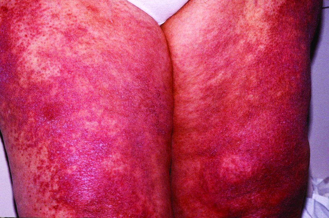
And in a recently completed phase 3 trial, reported by the manufacturer, a subpopulation of patients with active skin disease and no active muscle disease again showed greater reductions in CDASI activity scores – a secondary outcome in the trial.
However, the phase 3 DETERMINE trial produced negative findings overall. It enrolled a more heterogeneous group of patients – including those with both muscle weakness and skin involvement – and its primary outcome measure was a broader composite measure, the Total Improvement Score. The trial failed to meet this primary endpoint, Corbus Pharmaceuticals, the developer of lenabasum, announced in a press release in June 2021.
The phase 3 results are “frustrating” for patients with symptomatic and refractory skin manifestations of dermatomyositis (DM), given the promising findings from the phase 2 trial and from an open-label extension study, said Dr. Werth, professor of dermatology and medicine, University of Pennsylvania, Philadelphia, and principal investigator and coprincipal investigator of the phase 2 and phase 3 studies, respectively.
Dr. Werth is scheduled to present the results from the phase 3 trial at the annual European Alliance of Associations for Rheumatology meeting in June.
“With lenabasum, we have a therapy that doesn’t work for every patient, but does work for quite a number of them,” Dr. Werth said in an interview. “It’s oral, it’s not really that immunosuppressing, and there aren’t many side effects. Right now, patients are often being managed with steroids ... we really need treatments that are not as toxic.”
Robert Spiera, MD, a rheumatologist who led trials of lenabasum for treatment of diffuse cutaneous systemic sclerosis (dcSSc), agreed. “The CB2 agonist strategy is appealing because it’s nonimmunosuppressing and has both anti-inflammatory and antifibrotic properties,” he said in an interview. “I wouldn’t want to give up on it ... especially [for patients] with scleroderma and dermatomyositis who are treated with substantial drugs that are associated with morbidity.”
Lenabasum, he said, has proven to be “incredibly safe, and incredibly safe in the long term.”
While the phase 2 trial of the drug for dcSSc showed clear benefit over placebo, the phase 3 trial did not meet its primary endpoint using the American College of Rheumatology Combined Response Index in Diffuse Cutaneous Systemic Sclerosis.
It allowed background immunosuppressant therapy to reflect real-world clinical practice, and “there was such a high response rate to [that therapy, largely mycophenolate] that there was little room to show benefit beyond that,” said Dr. Spiera, director of the vasculitis and scleroderma program, Hospital for Special Surgery, New York.
The drug led to more improvement in the small subset of participants who were not receiving background immunotherapy during the trial, he noted.
Corbus is currently “seeking a partnership to further explore the drug” for treatment in different subpopulations, according to a company spokesperson. Results of a phase 2 trial of lenabasum for the treatment of systemic lupus erythematosus – with a pain rating as the primary outcome measure – are expected soon.
Phase 2 findings
The single-center phase 2 trial of lenabasum for DM enrolled 22 adults with minimal muscle involvement as evidenced by normal maximal resistance on muscle testing at entry and throughout the study. Most were taking immunosuppressant medication, and all had CDASI scores of at least 20, with mean scores in the severe range (> 26). Symptoms registered on patient-reported outcome measures were moderate to severe.
Patients received a half-dose of lenabasum (20 mg daily) for 1 month and a full dose (20 mg twice daily) for 2 months, or placebo, and were followed for an additional month without dosing.
Starting at day 43 – approximately 2 weeks after the dose was increased – there was “a trend for the change from baseline CDASI to be greater” in the lenabasum group, compared with those on placebo, Dr. Werth and colleagues reported. The differences reached statistical significance on day 113 (P = .038), a month after patients discontinued lenabasum, “suggesting that the modulation of the inflammatory response by lenabasum continued beyond its last dose.”
Five of the 11 patients treated with lenabasum (45%), and none of those on placebo, achieved at least a 40% reduction in the CDASI activity score by the end of the study.
Patients in the lenabasum group also had greater improvement in the Skindex-29 Symptoms scores – an objective measure of itch – and improvements in other secondary efficacy outcomes, including pain, though these did not reach statistical significance.
Skin biopsies before and after treatment showed significant reductions in inflammatory cytokines relevant to DM pathogenesis. Patients treated with the CB2 agonist had a downward trend in the CD4+ T cell population, which correlated with decreased CDASI activity scores, for instance, and a decrease in IL-31 protein expression, which correlated with decreased Skindex-29 Symptoms scores, the investigators reported.
There were no serious adverse events related to the CB2 agonist, and no treatment discontinuations.
The main part of the phase 2 trial, conducted from 2015 to 2017, was followed by a 3-year, open-label extension, in which 20 of the 22 patients took lenabasum 20 mg twice a day. The drug continued to be safe and well tolerated, and the CDASI activity score and other outcomes improved through year 1 and remained stable thereafter, according to a poster presented by Dr. Werth at the 2021 EULAR meeting.
After 1 year in the open-label extension, 60%-70% of patients had achieved mild skin disease, and 75% had achieved at least a 40% reduction in CDASI activity.
“A lot of patients, even if they weren’t completely cleared, were much happier in terms of their itch,” said Dr. Werth, also chief of dermatology, Corporal Michael J. Crescenz Veterans Affairs Medical Center, Philadelphia. “It’s been difficult for a lot of them now that they’re off the long-term extension ... a lot of them are flaring.”
The future
In the lab, with funding from the National Institutes of Health, Dr. Werth is continuing to investigate how lenabasum may be working in DM. A paper just published in the open access journal Arthritis Research & Therapy describes CB2 receptor distribution and up-regulation on key immune cells in the skin and blood, and how, in DM skin, its highest expression is on dendritic cells.
Through both mechanistic and more clinical research, “it’s important to understand the characteristics of the people [lenabasum] worked in or didn’t work in,” she said.
And in clinical trials, it’s important to capture meaningful improvement from the patient perspective. “It may be,” she noted, “that more global, systemic assessments are not the way to go for autoimmune skin disease.”
For dcSSc, Dr. Spiera said, it’s possible that a CB2 agonist may be helpful for patients who have been on immunosuppressants, particularly mycophenolate, for more than 6 months “and are still struggling.”
The phase 2 trial in DM was funded by the National Institutes of Health, the Department of Veterans Affairs, and Corbus Pharmaceuticals. The phase 3 trials in DM and in dcSSc were funded by Corbus. Dr. Werth disclosed grant support from Corbus and several other pharmaceutical companies. Dr. Spiera disclosed that he has received grant support or consulting fees from Roche-Genentech, GlaxoSmithKline, and several other pharmaceutical companies.
A version of this article first appeared on Medscape.com.
An – some of it statistically significant – in a phase 2, double-blind, randomized, controlled study.
Patients taking lenabasum experienced greater reductions in the Cutaneous Dermatomyositis Disease Area and Severity Index (CDASI) – a validated outcome designed to assess inflammatory skin involvement in the rare autoimmune disease – and improvements in patient-reported and biomarker outcomes, compared with those on placebo, dermatologist Victoria P. Werth, MD, and coinvestigators reported.
And in a recently completed phase 3 trial, reported by the manufacturer, a subpopulation of patients with active skin disease and no active muscle disease again showed greater reductions in CDASI activity scores – a secondary outcome in the trial.
However, the phase 3 DETERMINE trial produced negative findings overall. It enrolled a more heterogeneous group of patients – including those with both muscle weakness and skin involvement – and its primary outcome measure was a broader composite measure, the Total Improvement Score. The trial failed to meet this primary endpoint, Corbus Pharmaceuticals, the developer of lenabasum, announced in a press release in June 2021.
The phase 3 results are “frustrating” for patients with symptomatic and refractory skin manifestations of dermatomyositis (DM), given the promising findings from the phase 2 trial and from an open-label extension study, said Dr. Werth, professor of dermatology and medicine, University of Pennsylvania, Philadelphia, and principal investigator and coprincipal investigator of the phase 2 and phase 3 studies, respectively.
Dr. Werth is scheduled to present the results from the phase 3 trial at the annual European Alliance of Associations for Rheumatology meeting in June.
“With lenabasum, we have a therapy that doesn’t work for every patient, but does work for quite a number of them,” Dr. Werth said in an interview. “It’s oral, it’s not really that immunosuppressing, and there aren’t many side effects. Right now, patients are often being managed with steroids ... we really need treatments that are not as toxic.”
Robert Spiera, MD, a rheumatologist who led trials of lenabasum for treatment of diffuse cutaneous systemic sclerosis (dcSSc), agreed. “The CB2 agonist strategy is appealing because it’s nonimmunosuppressing and has both anti-inflammatory and antifibrotic properties,” he said in an interview. “I wouldn’t want to give up on it ... especially [for patients] with scleroderma and dermatomyositis who are treated with substantial drugs that are associated with morbidity.”
Lenabasum, he said, has proven to be “incredibly safe, and incredibly safe in the long term.”
While the phase 2 trial of the drug for dcSSc showed clear benefit over placebo, the phase 3 trial did not meet its primary endpoint using the American College of Rheumatology Combined Response Index in Diffuse Cutaneous Systemic Sclerosis.
It allowed background immunosuppressant therapy to reflect real-world clinical practice, and “there was such a high response rate to [that therapy, largely mycophenolate] that there was little room to show benefit beyond that,” said Dr. Spiera, director of the vasculitis and scleroderma program, Hospital for Special Surgery, New York.
The drug led to more improvement in the small subset of participants who were not receiving background immunotherapy during the trial, he noted.
Corbus is currently “seeking a partnership to further explore the drug” for treatment in different subpopulations, according to a company spokesperson. Results of a phase 2 trial of lenabasum for the treatment of systemic lupus erythematosus – with a pain rating as the primary outcome measure – are expected soon.
Phase 2 findings
The single-center phase 2 trial of lenabasum for DM enrolled 22 adults with minimal muscle involvement as evidenced by normal maximal resistance on muscle testing at entry and throughout the study. Most were taking immunosuppressant medication, and all had CDASI scores of at least 20, with mean scores in the severe range (> 26). Symptoms registered on patient-reported outcome measures were moderate to severe.
Patients received a half-dose of lenabasum (20 mg daily) for 1 month and a full dose (20 mg twice daily) for 2 months, or placebo, and were followed for an additional month without dosing.
Starting at day 43 – approximately 2 weeks after the dose was increased – there was “a trend for the change from baseline CDASI to be greater” in the lenabasum group, compared with those on placebo, Dr. Werth and colleagues reported. The differences reached statistical significance on day 113 (P = .038), a month after patients discontinued lenabasum, “suggesting that the modulation of the inflammatory response by lenabasum continued beyond its last dose.”
Five of the 11 patients treated with lenabasum (45%), and none of those on placebo, achieved at least a 40% reduction in the CDASI activity score by the end of the study.
Patients in the lenabasum group also had greater improvement in the Skindex-29 Symptoms scores – an objective measure of itch – and improvements in other secondary efficacy outcomes, including pain, though these did not reach statistical significance.
Skin biopsies before and after treatment showed significant reductions in inflammatory cytokines relevant to DM pathogenesis. Patients treated with the CB2 agonist had a downward trend in the CD4+ T cell population, which correlated with decreased CDASI activity scores, for instance, and a decrease in IL-31 protein expression, which correlated with decreased Skindex-29 Symptoms scores, the investigators reported.
There were no serious adverse events related to the CB2 agonist, and no treatment discontinuations.
The main part of the phase 2 trial, conducted from 2015 to 2017, was followed by a 3-year, open-label extension, in which 20 of the 22 patients took lenabasum 20 mg twice a day. The drug continued to be safe and well tolerated, and the CDASI activity score and other outcomes improved through year 1 and remained stable thereafter, according to a poster presented by Dr. Werth at the 2021 EULAR meeting.
After 1 year in the open-label extension, 60%-70% of patients had achieved mild skin disease, and 75% had achieved at least a 40% reduction in CDASI activity.
“A lot of patients, even if they weren’t completely cleared, were much happier in terms of their itch,” said Dr. Werth, also chief of dermatology, Corporal Michael J. Crescenz Veterans Affairs Medical Center, Philadelphia. “It’s been difficult for a lot of them now that they’re off the long-term extension ... a lot of them are flaring.”
The future
In the lab, with funding from the National Institutes of Health, Dr. Werth is continuing to investigate how lenabasum may be working in DM. A paper just published in the open access journal Arthritis Research & Therapy describes CB2 receptor distribution and up-regulation on key immune cells in the skin and blood, and how, in DM skin, its highest expression is on dendritic cells.
Through both mechanistic and more clinical research, “it’s important to understand the characteristics of the people [lenabasum] worked in or didn’t work in,” she said.
And in clinical trials, it’s important to capture meaningful improvement from the patient perspective. “It may be,” she noted, “that more global, systemic assessments are not the way to go for autoimmune skin disease.”
For dcSSc, Dr. Spiera said, it’s possible that a CB2 agonist may be helpful for patients who have been on immunosuppressants, particularly mycophenolate, for more than 6 months “and are still struggling.”
The phase 2 trial in DM was funded by the National Institutes of Health, the Department of Veterans Affairs, and Corbus Pharmaceuticals. The phase 3 trials in DM and in dcSSc were funded by Corbus. Dr. Werth disclosed grant support from Corbus and several other pharmaceutical companies. Dr. Spiera disclosed that he has received grant support or consulting fees from Roche-Genentech, GlaxoSmithKline, and several other pharmaceutical companies.
A version of this article first appeared on Medscape.com.
An – some of it statistically significant – in a phase 2, double-blind, randomized, controlled study.
Patients taking lenabasum experienced greater reductions in the Cutaneous Dermatomyositis Disease Area and Severity Index (CDASI) – a validated outcome designed to assess inflammatory skin involvement in the rare autoimmune disease – and improvements in patient-reported and biomarker outcomes, compared with those on placebo, dermatologist Victoria P. Werth, MD, and coinvestigators reported.
And in a recently completed phase 3 trial, reported by the manufacturer, a subpopulation of patients with active skin disease and no active muscle disease again showed greater reductions in CDASI activity scores – a secondary outcome in the trial.
However, the phase 3 DETERMINE trial produced negative findings overall. It enrolled a more heterogeneous group of patients – including those with both muscle weakness and skin involvement – and its primary outcome measure was a broader composite measure, the Total Improvement Score. The trial failed to meet this primary endpoint, Corbus Pharmaceuticals, the developer of lenabasum, announced in a press release in June 2021.
The phase 3 results are “frustrating” for patients with symptomatic and refractory skin manifestations of dermatomyositis (DM), given the promising findings from the phase 2 trial and from an open-label extension study, said Dr. Werth, professor of dermatology and medicine, University of Pennsylvania, Philadelphia, and principal investigator and coprincipal investigator of the phase 2 and phase 3 studies, respectively.
Dr. Werth is scheduled to present the results from the phase 3 trial at the annual European Alliance of Associations for Rheumatology meeting in June.
“With lenabasum, we have a therapy that doesn’t work for every patient, but does work for quite a number of them,” Dr. Werth said in an interview. “It’s oral, it’s not really that immunosuppressing, and there aren’t many side effects. Right now, patients are often being managed with steroids ... we really need treatments that are not as toxic.”
Robert Spiera, MD, a rheumatologist who led trials of lenabasum for treatment of diffuse cutaneous systemic sclerosis (dcSSc), agreed. “The CB2 agonist strategy is appealing because it’s nonimmunosuppressing and has both anti-inflammatory and antifibrotic properties,” he said in an interview. “I wouldn’t want to give up on it ... especially [for patients] with scleroderma and dermatomyositis who are treated with substantial drugs that are associated with morbidity.”
Lenabasum, he said, has proven to be “incredibly safe, and incredibly safe in the long term.”
While the phase 2 trial of the drug for dcSSc showed clear benefit over placebo, the phase 3 trial did not meet its primary endpoint using the American College of Rheumatology Combined Response Index in Diffuse Cutaneous Systemic Sclerosis.
It allowed background immunosuppressant therapy to reflect real-world clinical practice, and “there was such a high response rate to [that therapy, largely mycophenolate] that there was little room to show benefit beyond that,” said Dr. Spiera, director of the vasculitis and scleroderma program, Hospital for Special Surgery, New York.
The drug led to more improvement in the small subset of participants who were not receiving background immunotherapy during the trial, he noted.
Corbus is currently “seeking a partnership to further explore the drug” for treatment in different subpopulations, according to a company spokesperson. Results of a phase 2 trial of lenabasum for the treatment of systemic lupus erythematosus – with a pain rating as the primary outcome measure – are expected soon.
Phase 2 findings
The single-center phase 2 trial of lenabasum for DM enrolled 22 adults with minimal muscle involvement as evidenced by normal maximal resistance on muscle testing at entry and throughout the study. Most were taking immunosuppressant medication, and all had CDASI scores of at least 20, with mean scores in the severe range (> 26). Symptoms registered on patient-reported outcome measures were moderate to severe.
Patients received a half-dose of lenabasum (20 mg daily) for 1 month and a full dose (20 mg twice daily) for 2 months, or placebo, and were followed for an additional month without dosing.
Starting at day 43 – approximately 2 weeks after the dose was increased – there was “a trend for the change from baseline CDASI to be greater” in the lenabasum group, compared with those on placebo, Dr. Werth and colleagues reported. The differences reached statistical significance on day 113 (P = .038), a month after patients discontinued lenabasum, “suggesting that the modulation of the inflammatory response by lenabasum continued beyond its last dose.”
Five of the 11 patients treated with lenabasum (45%), and none of those on placebo, achieved at least a 40% reduction in the CDASI activity score by the end of the study.
Patients in the lenabasum group also had greater improvement in the Skindex-29 Symptoms scores – an objective measure of itch – and improvements in other secondary efficacy outcomes, including pain, though these did not reach statistical significance.
Skin biopsies before and after treatment showed significant reductions in inflammatory cytokines relevant to DM pathogenesis. Patients treated with the CB2 agonist had a downward trend in the CD4+ T cell population, which correlated with decreased CDASI activity scores, for instance, and a decrease in IL-31 protein expression, which correlated with decreased Skindex-29 Symptoms scores, the investigators reported.
There were no serious adverse events related to the CB2 agonist, and no treatment discontinuations.
The main part of the phase 2 trial, conducted from 2015 to 2017, was followed by a 3-year, open-label extension, in which 20 of the 22 patients took lenabasum 20 mg twice a day. The drug continued to be safe and well tolerated, and the CDASI activity score and other outcomes improved through year 1 and remained stable thereafter, according to a poster presented by Dr. Werth at the 2021 EULAR meeting.
After 1 year in the open-label extension, 60%-70% of patients had achieved mild skin disease, and 75% had achieved at least a 40% reduction in CDASI activity.
“A lot of patients, even if they weren’t completely cleared, were much happier in terms of their itch,” said Dr. Werth, also chief of dermatology, Corporal Michael J. Crescenz Veterans Affairs Medical Center, Philadelphia. “It’s been difficult for a lot of them now that they’re off the long-term extension ... a lot of them are flaring.”
The future
In the lab, with funding from the National Institutes of Health, Dr. Werth is continuing to investigate how lenabasum may be working in DM. A paper just published in the open access journal Arthritis Research & Therapy describes CB2 receptor distribution and up-regulation on key immune cells in the skin and blood, and how, in DM skin, its highest expression is on dendritic cells.
Through both mechanistic and more clinical research, “it’s important to understand the characteristics of the people [lenabasum] worked in or didn’t work in,” she said.
And in clinical trials, it’s important to capture meaningful improvement from the patient perspective. “It may be,” she noted, “that more global, systemic assessments are not the way to go for autoimmune skin disease.”
For dcSSc, Dr. Spiera said, it’s possible that a CB2 agonist may be helpful for patients who have been on immunosuppressants, particularly mycophenolate, for more than 6 months “and are still struggling.”
The phase 2 trial in DM was funded by the National Institutes of Health, the Department of Veterans Affairs, and Corbus Pharmaceuticals. The phase 3 trials in DM and in dcSSc were funded by Corbus. Dr. Werth disclosed grant support from Corbus and several other pharmaceutical companies. Dr. Spiera disclosed that he has received grant support or consulting fees from Roche-Genentech, GlaxoSmithKline, and several other pharmaceutical companies.
A version of this article first appeared on Medscape.com.
FROM THE JOURNAL OF INVESTIGATIVE DERMATOLOGY