User login
Migraine associated with more severe disability in patients with MS
DALLAS – researchers reported at the meeting held by the Americas Committee for Treatment and Research in Multiple Sclerosis.

“Traditional migraine risk factors such as obesity, anxiety, and depression were also overrepresented in our cohort” of patients with multiple sclerosis (MS) and migraine, said Anne M. Damian, MD, of Johns Hopkins University, Baltimore, and her research colleagues.
Migraine is common in patients with MS, but whether migraine plays a role in MS disease course or MS symptom severity is unknown. Dr. Damian and her colleagues conducted an observational study to examine the associations between migraine history, disability, and neurologic function in patients with MS and whether migraine tends to occur with other comorbid conditions in MS.
They analyzed data from 289 patients (79% female; mean age, 49.2 years) patients with MS who completed the Multiple Sclerosis Performance Test (MSPT), an iPad version of the MS Functional Composite. MS outcome measures included disability (such as the Patient Determined Disease Steps) and objective neurologic outcomes (such as walking speed, manual dexterity, and processing speed). Patients also completed a questionnaire about comorbidities, including history of physician-diagnosed migraine, diabetes, hypertension, hypercholesterolemia, heart disease, sleep apnea, depression, and anxiety.
The researchers used generalized linear models adjusted for age, sex, MS subtype, MS duration, years of education, and body mass index to evaluate the association between history of migraine and MS outcomes.
Compared with patients with MS without migraine, migraineurs (n = 65) tended to be younger (mean age, 44.3 years vs. 50.4 years) and were more likely to be overweight or obese (73.9% vs. 51.6%). In addition, patients with MS and migraine were more likely to have a history of depression (46.2% vs. 24.2%), anxiety (30.8% vs. 18.8%), and severe rather than mild disability (odds ratio, 3.08; 95% confidence, 1.04-9.20). Migraine also was associated with significantly slower walking speeds (9.08% slower; 95% CI, 0.82%-18.77%). Migraine was not associated with processing speed or manual dexterity, however.
If an association between migraine history and worse MS disability is confirmed, migraine history may be a factor that neurologists could consider when making MS treatment decisions, Dr. Damian said. The researchers noted that migraine was reported by patients and not detected using a validated questionnaire. Future studies should investigate whether MS lesions on MRI differ in migraineurs and whether migraine predicts future neurologic disability in patients with MS.
Collection of the MSPT outcomes was sponsored by Biogen.
SOURCE: Damian AM et al. ACTRIMS Forum 2019, Abstract 78.
DALLAS – researchers reported at the meeting held by the Americas Committee for Treatment and Research in Multiple Sclerosis.
“Traditional migraine risk factors such as obesity, anxiety, and depression were also overrepresented in our cohort” of patients with multiple sclerosis (MS) and migraine, said Anne M. Damian, MD, of Johns Hopkins University, Baltimore, and her research colleagues.
Migraine is common in patients with MS, but whether migraine plays a role in MS disease course or MS symptom severity is unknown. Dr. Damian and her colleagues conducted an observational study to examine the associations between migraine history, disability, and neurologic function in patients with MS and whether migraine tends to occur with other comorbid conditions in MS.
They analyzed data from 289 patients (79% female; mean age, 49.2 years) patients with MS who completed the Multiple Sclerosis Performance Test (MSPT), an iPad version of the MS Functional Composite. MS outcome measures included disability (such as the Patient Determined Disease Steps) and objective neurologic outcomes (such as walking speed, manual dexterity, and processing speed). Patients also completed a questionnaire about comorbidities, including history of physician-diagnosed migraine, diabetes, hypertension, hypercholesterolemia, heart disease, sleep apnea, depression, and anxiety.
The researchers used generalized linear models adjusted for age, sex, MS subtype, MS duration, years of education, and body mass index to evaluate the association between history of migraine and MS outcomes.
Compared with patients with MS without migraine, migraineurs (n = 65) tended to be younger (mean age, 44.3 years vs. 50.4 years) and were more likely to be overweight or obese (73.9% vs. 51.6%). In addition, patients with MS and migraine were more likely to have a history of depression (46.2% vs. 24.2%), anxiety (30.8% vs. 18.8%), and severe rather than mild disability (odds ratio, 3.08; 95% confidence, 1.04-9.20). Migraine also was associated with significantly slower walking speeds (9.08% slower; 95% CI, 0.82%-18.77%). Migraine was not associated with processing speed or manual dexterity, however.
If an association between migraine history and worse MS disability is confirmed, migraine history may be a factor that neurologists could consider when making MS treatment decisions, Dr. Damian said. The researchers noted that migraine was reported by patients and not detected using a validated questionnaire. Future studies should investigate whether MS lesions on MRI differ in migraineurs and whether migraine predicts future neurologic disability in patients with MS.
Collection of the MSPT outcomes was sponsored by Biogen.
SOURCE: Damian AM et al. ACTRIMS Forum 2019, Abstract 78.
DALLAS – researchers reported at the meeting held by the Americas Committee for Treatment and Research in Multiple Sclerosis.
“Traditional migraine risk factors such as obesity, anxiety, and depression were also overrepresented in our cohort” of patients with multiple sclerosis (MS) and migraine, said Anne M. Damian, MD, of Johns Hopkins University, Baltimore, and her research colleagues.
Migraine is common in patients with MS, but whether migraine plays a role in MS disease course or MS symptom severity is unknown. Dr. Damian and her colleagues conducted an observational study to examine the associations between migraine history, disability, and neurologic function in patients with MS and whether migraine tends to occur with other comorbid conditions in MS.
They analyzed data from 289 patients (79% female; mean age, 49.2 years) patients with MS who completed the Multiple Sclerosis Performance Test (MSPT), an iPad version of the MS Functional Composite. MS outcome measures included disability (such as the Patient Determined Disease Steps) and objective neurologic outcomes (such as walking speed, manual dexterity, and processing speed). Patients also completed a questionnaire about comorbidities, including history of physician-diagnosed migraine, diabetes, hypertension, hypercholesterolemia, heart disease, sleep apnea, depression, and anxiety.
The researchers used generalized linear models adjusted for age, sex, MS subtype, MS duration, years of education, and body mass index to evaluate the association between history of migraine and MS outcomes.
Compared with patients with MS without migraine, migraineurs (n = 65) tended to be younger (mean age, 44.3 years vs. 50.4 years) and were more likely to be overweight or obese (73.9% vs. 51.6%). In addition, patients with MS and migraine were more likely to have a history of depression (46.2% vs. 24.2%), anxiety (30.8% vs. 18.8%), and severe rather than mild disability (odds ratio, 3.08; 95% confidence, 1.04-9.20). Migraine also was associated with significantly slower walking speeds (9.08% slower; 95% CI, 0.82%-18.77%). Migraine was not associated with processing speed or manual dexterity, however.
If an association between migraine history and worse MS disability is confirmed, migraine history may be a factor that neurologists could consider when making MS treatment decisions, Dr. Damian said. The researchers noted that migraine was reported by patients and not detected using a validated questionnaire. Future studies should investigate whether MS lesions on MRI differ in migraineurs and whether migraine predicts future neurologic disability in patients with MS.
Collection of the MSPT outcomes was sponsored by Biogen.
SOURCE: Damian AM et al. ACTRIMS Forum 2019, Abstract 78.
REPORTING FROM ACTRIMS FORUM 2019
Post-HCT azithromycin doesn’t increase relapse risk, study suggests
HOUSTON — When given after transplant, azithromycin does not increase the risk of relapse in patients with moderate to severe chronic graft-versus-host disease (cGVHD) and bronchiolitis obliterans syndrome (BOS), according to a retrospective study.
A prior study, ALLOZITHRO (JAMA. 2017 Aug 8;318[6]:557-66), showed an increased risk of relapse and death in patients who received azithromycin as BOS prophylaxis prior to hematopoietic cell transplant (HCT).
That discovery prompted the Food and Drug Administration to release a safety communication warning prescribers about the risks associated with azithromycin as BOS prophylaxis. However, it wasn’t clear if the same risks exist when azithromycin is given for cGVHD management after HCT.
To gain some insight, Mark Shamoun, MD, of the University of Michigan, Ann Arbor, and his colleagues examined data on patients with moderate to severe cGVHD and BOS who received azithromycin after undergoing HCT to treat a hematologic malignancy.
Dr. Shamoun presented the group’s findings at the Transplantation & Cellular Therapy Meetings.
The researchers reviewed data on 239 patients enrolled in the University of Michigan’s HCT database from 2010 to 2017. The median age at baseline was 55 years (range, 4-72 years).
The patients received transplants to treat acute myeloid leukemia or myelodysplastic syndromes (n = 141), acute lymphoblastic leukemia (n = 40), lymphoma (n = 26), chronic leukemia (n = 24), multiple myeloma (n = 6), and myeloproliferative neoplasms (n = 2).
The patients had matched related donors (43%) or matched unrelated donors (57%). Most patients received peripheral blood transplants (84%), though some received bone marrow (14%) or cord blood (2%). All patients had moderate (38%) or severe (62%) cGVHD.
Patients were split into two cohorts. Patients in cohort A (n = 86) had BOS and received azithromycin for more than 14 days.
Patients in cohort B (n = 153) either did not receive azithromycin or received it for 14 days or less. Fewer than 5% of patients in cohort B had BOS.
Most other baseline characteristics were similar between the cohorts. However, severe cGVHD was more prevalent in cohort A than B — 78% and 51%, respectively.
In cohort A, the median time to the start of azithromycin was 15 months after HCT (range, 3-68 months). The median duration of azithromycin treatment was 26 months (range, 1-77 months).
Results
The 2-year relapse rate was significantly lower in patients who received azithromycin than in those who did not — 4% and 17%, respectively (P = .001).
There was a significant difference in relapse rate both from the time of HCT (P = .001) and from the start of azithromycin or cGVHD (P = .011).
There was no significant difference in overall survival between the cohorts, either from the time of HCT (P = .294) or from the start of azithromycin or cGVHD (P = .428).
Dr. Shamoun said these results suggest azithromycin does not increase the risk of relapse when it is used to manage cGVHD. However, this study is limited by its retrospective nature. In addition, most patients in cohort B did not have BOS, severe cGVHD was more common in cohort A, and the incidence of relapse was not calculated from the time of azithromycin initiation in both cohorts. Therefore, additional investigation is needed.
Dr. Shamoun presented these results at Transplantation & Cellular Therapy Meetings, which is held by the American Society for Blood and Marrow Transplantation and the Center for International Blood and Marrow Transplant Research. At the meeting, the American Society for Blood and Marrow Transplantation announced a new name for the society, the American Society for Transplantation and Cellular Therapy (ASTCT).
Dr. Shamoun reported no conflicts of interest.
SOURCE: Shamoun M et al. TCT 2019, Abstract 33.
HOUSTON — When given after transplant, azithromycin does not increase the risk of relapse in patients with moderate to severe chronic graft-versus-host disease (cGVHD) and bronchiolitis obliterans syndrome (BOS), according to a retrospective study.
A prior study, ALLOZITHRO (JAMA. 2017 Aug 8;318[6]:557-66), showed an increased risk of relapse and death in patients who received azithromycin as BOS prophylaxis prior to hematopoietic cell transplant (HCT).
That discovery prompted the Food and Drug Administration to release a safety communication warning prescribers about the risks associated with azithromycin as BOS prophylaxis. However, it wasn’t clear if the same risks exist when azithromycin is given for cGVHD management after HCT.
To gain some insight, Mark Shamoun, MD, of the University of Michigan, Ann Arbor, and his colleagues examined data on patients with moderate to severe cGVHD and BOS who received azithromycin after undergoing HCT to treat a hematologic malignancy.
Dr. Shamoun presented the group’s findings at the Transplantation & Cellular Therapy Meetings.
The researchers reviewed data on 239 patients enrolled in the University of Michigan’s HCT database from 2010 to 2017. The median age at baseline was 55 years (range, 4-72 years).
The patients received transplants to treat acute myeloid leukemia or myelodysplastic syndromes (n = 141), acute lymphoblastic leukemia (n = 40), lymphoma (n = 26), chronic leukemia (n = 24), multiple myeloma (n = 6), and myeloproliferative neoplasms (n = 2).
The patients had matched related donors (43%) or matched unrelated donors (57%). Most patients received peripheral blood transplants (84%), though some received bone marrow (14%) or cord blood (2%). All patients had moderate (38%) or severe (62%) cGVHD.
Patients were split into two cohorts. Patients in cohort A (n = 86) had BOS and received azithromycin for more than 14 days.
Patients in cohort B (n = 153) either did not receive azithromycin or received it for 14 days or less. Fewer than 5% of patients in cohort B had BOS.
Most other baseline characteristics were similar between the cohorts. However, severe cGVHD was more prevalent in cohort A than B — 78% and 51%, respectively.
In cohort A, the median time to the start of azithromycin was 15 months after HCT (range, 3-68 months). The median duration of azithromycin treatment was 26 months (range, 1-77 months).
Results
The 2-year relapse rate was significantly lower in patients who received azithromycin than in those who did not — 4% and 17%, respectively (P = .001).
There was a significant difference in relapse rate both from the time of HCT (P = .001) and from the start of azithromycin or cGVHD (P = .011).
There was no significant difference in overall survival between the cohorts, either from the time of HCT (P = .294) or from the start of azithromycin or cGVHD (P = .428).
Dr. Shamoun said these results suggest azithromycin does not increase the risk of relapse when it is used to manage cGVHD. However, this study is limited by its retrospective nature. In addition, most patients in cohort B did not have BOS, severe cGVHD was more common in cohort A, and the incidence of relapse was not calculated from the time of azithromycin initiation in both cohorts. Therefore, additional investigation is needed.
Dr. Shamoun presented these results at Transplantation & Cellular Therapy Meetings, which is held by the American Society for Blood and Marrow Transplantation and the Center for International Blood and Marrow Transplant Research. At the meeting, the American Society for Blood and Marrow Transplantation announced a new name for the society, the American Society for Transplantation and Cellular Therapy (ASTCT).
Dr. Shamoun reported no conflicts of interest.
SOURCE: Shamoun M et al. TCT 2019, Abstract 33.
HOUSTON — When given after transplant, azithromycin does not increase the risk of relapse in patients with moderate to severe chronic graft-versus-host disease (cGVHD) and bronchiolitis obliterans syndrome (BOS), according to a retrospective study.
A prior study, ALLOZITHRO (JAMA. 2017 Aug 8;318[6]:557-66), showed an increased risk of relapse and death in patients who received azithromycin as BOS prophylaxis prior to hematopoietic cell transplant (HCT).
That discovery prompted the Food and Drug Administration to release a safety communication warning prescribers about the risks associated with azithromycin as BOS prophylaxis. However, it wasn’t clear if the same risks exist when azithromycin is given for cGVHD management after HCT.
To gain some insight, Mark Shamoun, MD, of the University of Michigan, Ann Arbor, and his colleagues examined data on patients with moderate to severe cGVHD and BOS who received azithromycin after undergoing HCT to treat a hematologic malignancy.
Dr. Shamoun presented the group’s findings at the Transplantation & Cellular Therapy Meetings.
The researchers reviewed data on 239 patients enrolled in the University of Michigan’s HCT database from 2010 to 2017. The median age at baseline was 55 years (range, 4-72 years).
The patients received transplants to treat acute myeloid leukemia or myelodysplastic syndromes (n = 141), acute lymphoblastic leukemia (n = 40), lymphoma (n = 26), chronic leukemia (n = 24), multiple myeloma (n = 6), and myeloproliferative neoplasms (n = 2).
The patients had matched related donors (43%) or matched unrelated donors (57%). Most patients received peripheral blood transplants (84%), though some received bone marrow (14%) or cord blood (2%). All patients had moderate (38%) or severe (62%) cGVHD.
Patients were split into two cohorts. Patients in cohort A (n = 86) had BOS and received azithromycin for more than 14 days.
Patients in cohort B (n = 153) either did not receive azithromycin or received it for 14 days or less. Fewer than 5% of patients in cohort B had BOS.
Most other baseline characteristics were similar between the cohorts. However, severe cGVHD was more prevalent in cohort A than B — 78% and 51%, respectively.
In cohort A, the median time to the start of azithromycin was 15 months after HCT (range, 3-68 months). The median duration of azithromycin treatment was 26 months (range, 1-77 months).
Results
The 2-year relapse rate was significantly lower in patients who received azithromycin than in those who did not — 4% and 17%, respectively (P = .001).
There was a significant difference in relapse rate both from the time of HCT (P = .001) and from the start of azithromycin or cGVHD (P = .011).
There was no significant difference in overall survival between the cohorts, either from the time of HCT (P = .294) or from the start of azithromycin or cGVHD (P = .428).
Dr. Shamoun said these results suggest azithromycin does not increase the risk of relapse when it is used to manage cGVHD. However, this study is limited by its retrospective nature. In addition, most patients in cohort B did not have BOS, severe cGVHD was more common in cohort A, and the incidence of relapse was not calculated from the time of azithromycin initiation in both cohorts. Therefore, additional investigation is needed.
Dr. Shamoun presented these results at Transplantation & Cellular Therapy Meetings, which is held by the American Society for Blood and Marrow Transplantation and the Center for International Blood and Marrow Transplant Research. At the meeting, the American Society for Blood and Marrow Transplantation announced a new name for the society, the American Society for Transplantation and Cellular Therapy (ASTCT).
Dr. Shamoun reported no conflicts of interest.
SOURCE: Shamoun M et al. TCT 2019, Abstract 33.
REPORTING FROM TCT 2019
Medical tourism for MS stem cell therapy is common
DALLAS – “Stem cell therapy is something that has been a topic of interest for neurologists for a while,” said Wijdan Rai, MD, speaking at the meeting presented by the American Committee for Treatment and Research in Multiple Sclerosis.
However, “stem cell tourism is notoriously difficult to study, because it’s not regulated; there’s no database we can access to try to figure out what exactly is going on in these clinics,” said Dr. Rai.
Dr. Rai, a neurology resident at the Ohio State University, Columbus, said that she and her colleagues had noticed patients with multiple sclerosis (MS) were asking more frequently about stem cell therapies, and mesenchymal stem cell therapy in particular.
To translate these anecdotal observations into more concrete data, Dr. Rai and her colleagues surveyed academic neurologists in the outpatient setting to see if their patients were asking them about medical tourism for stem cell therapy. Additionally, they were asked about patients who actually had sought out the therapy and if there were adverse reactions from stem cell therapy.
The 25-item questionnaire was sent to academic neurologists via an online survey tool called Synapse through the American Academy of Neurology. Dr. Rai and her colleagues found that over 90% of respondents had been asked about stem cell therapies and that 25% of respondents said their patients had some kind of complication from the treatment.
“Most commonly, it was some variation of an infection, like meningitis, encephalitis, or hepatitis C,” said Dr. Rai. Other physicians reported that their patients had spinal cord tumors, deterioration of MS, or stroke.
with this evidence in hand, she hopes that a fact sheet can be developed and hosted on a website so physicians can point their patients to evidence-based information about stem cell therapies in MS.
DALLAS – “Stem cell therapy is something that has been a topic of interest for neurologists for a while,” said Wijdan Rai, MD, speaking at the meeting presented by the American Committee for Treatment and Research in Multiple Sclerosis.
However, “stem cell tourism is notoriously difficult to study, because it’s not regulated; there’s no database we can access to try to figure out what exactly is going on in these clinics,” said Dr. Rai.
Dr. Rai, a neurology resident at the Ohio State University, Columbus, said that she and her colleagues had noticed patients with multiple sclerosis (MS) were asking more frequently about stem cell therapies, and mesenchymal stem cell therapy in particular.
To translate these anecdotal observations into more concrete data, Dr. Rai and her colleagues surveyed academic neurologists in the outpatient setting to see if their patients were asking them about medical tourism for stem cell therapy. Additionally, they were asked about patients who actually had sought out the therapy and if there were adverse reactions from stem cell therapy.
The 25-item questionnaire was sent to academic neurologists via an online survey tool called Synapse through the American Academy of Neurology. Dr. Rai and her colleagues found that over 90% of respondents had been asked about stem cell therapies and that 25% of respondents said their patients had some kind of complication from the treatment.
“Most commonly, it was some variation of an infection, like meningitis, encephalitis, or hepatitis C,” said Dr. Rai. Other physicians reported that their patients had spinal cord tumors, deterioration of MS, or stroke.
with this evidence in hand, she hopes that a fact sheet can be developed and hosted on a website so physicians can point their patients to evidence-based information about stem cell therapies in MS.
DALLAS – “Stem cell therapy is something that has been a topic of interest for neurologists for a while,” said Wijdan Rai, MD, speaking at the meeting presented by the American Committee for Treatment and Research in Multiple Sclerosis.
However, “stem cell tourism is notoriously difficult to study, because it’s not regulated; there’s no database we can access to try to figure out what exactly is going on in these clinics,” said Dr. Rai.
Dr. Rai, a neurology resident at the Ohio State University, Columbus, said that she and her colleagues had noticed patients with multiple sclerosis (MS) were asking more frequently about stem cell therapies, and mesenchymal stem cell therapy in particular.
To translate these anecdotal observations into more concrete data, Dr. Rai and her colleagues surveyed academic neurologists in the outpatient setting to see if their patients were asking them about medical tourism for stem cell therapy. Additionally, they were asked about patients who actually had sought out the therapy and if there were adverse reactions from stem cell therapy.
The 25-item questionnaire was sent to academic neurologists via an online survey tool called Synapse through the American Academy of Neurology. Dr. Rai and her colleagues found that over 90% of respondents had been asked about stem cell therapies and that 25% of respondents said their patients had some kind of complication from the treatment.
“Most commonly, it was some variation of an infection, like meningitis, encephalitis, or hepatitis C,” said Dr. Rai. Other physicians reported that their patients had spinal cord tumors, deterioration of MS, or stroke.
with this evidence in hand, she hopes that a fact sheet can be developed and hosted on a website so physicians can point their patients to evidence-based information about stem cell therapies in MS.
REPORTING FROM ACTRIMS FORUM 2019
Smartphone assessment of motor, cognitive function in MS extends clinicians’ reach
DALLAS –
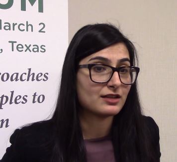
Alexandra Boukhvalova, a medical student at the University of Maryland School of Medicine, Baltimore, and her collaborators developed an interactive smartphone app to assess some aspects of cognitive and motor function for patients with multiple sclerosis (MS). Their findings were reported during a poster session at the meeting held by the American Society for Prevention and Treatment in Multiple Sclerosis.
“The clinician assessment is an hour-long assessment; it requires a trained neurologist,” Ms. Boukhvalova pointed out. In thinking about how app-based assessment could augment the clinical exam, she and her collaborators realized that “a lot of the neurologic exam is still quite subjective – so is there a way that we can make that exam more objective and quantifiable and also add a little bit of ease with accessibility and mobility?
“We created a suite of test apps ... to assess different neurological systems, ranging from motor function, cognitive and visual dysfunction, general fatigue, and strength,” she said. The apps included tapping tests and balloon-popping tasks, along with measures to give some indication of spasticity by assessing the smoothness of movements.
Participants completed the testing both in the clinic and from home. “We did not need an investigator present for patients to be able to complete the tests,” said Ms. Boukhvalova.
Ms. Boukhvalova and her colleagues compared performance on the gamified tasks between patients with MS and healthy controls. For all tasks, the participants with MS could clearly be differentiated from the healthy participants.
A further plus was that “The patients almost viewed these tests as games.” They reported that they enjoyed completing them, said Ms. Boukhvalova, adding that app-based assessments also offer an additional point of connection between MS patients and specialists, whom they may only see annually or semiannually.
Further app development may focus on utilizing sensor and accelerometer functions in smartphones to perform more natural and sophisticated motor analysis to look at gait and gross motor functioning, she said.
DALLAS –
Alexandra Boukhvalova, a medical student at the University of Maryland School of Medicine, Baltimore, and her collaborators developed an interactive smartphone app to assess some aspects of cognitive and motor function for patients with multiple sclerosis (MS). Their findings were reported during a poster session at the meeting held by the American Society for Prevention and Treatment in Multiple Sclerosis.
“The clinician assessment is an hour-long assessment; it requires a trained neurologist,” Ms. Boukhvalova pointed out. In thinking about how app-based assessment could augment the clinical exam, she and her collaborators realized that “a lot of the neurologic exam is still quite subjective – so is there a way that we can make that exam more objective and quantifiable and also add a little bit of ease with accessibility and mobility?
“We created a suite of test apps ... to assess different neurological systems, ranging from motor function, cognitive and visual dysfunction, general fatigue, and strength,” she said. The apps included tapping tests and balloon-popping tasks, along with measures to give some indication of spasticity by assessing the smoothness of movements.
Participants completed the testing both in the clinic and from home. “We did not need an investigator present for patients to be able to complete the tests,” said Ms. Boukhvalova.
Ms. Boukhvalova and her colleagues compared performance on the gamified tasks between patients with MS and healthy controls. For all tasks, the participants with MS could clearly be differentiated from the healthy participants.
A further plus was that “The patients almost viewed these tests as games.” They reported that they enjoyed completing them, said Ms. Boukhvalova, adding that app-based assessments also offer an additional point of connection between MS patients and specialists, whom they may only see annually or semiannually.
Further app development may focus on utilizing sensor and accelerometer functions in smartphones to perform more natural and sophisticated motor analysis to look at gait and gross motor functioning, she said.
DALLAS –
Alexandra Boukhvalova, a medical student at the University of Maryland School of Medicine, Baltimore, and her collaborators developed an interactive smartphone app to assess some aspects of cognitive and motor function for patients with multiple sclerosis (MS). Their findings were reported during a poster session at the meeting held by the American Society for Prevention and Treatment in Multiple Sclerosis.
“The clinician assessment is an hour-long assessment; it requires a trained neurologist,” Ms. Boukhvalova pointed out. In thinking about how app-based assessment could augment the clinical exam, she and her collaborators realized that “a lot of the neurologic exam is still quite subjective – so is there a way that we can make that exam more objective and quantifiable and also add a little bit of ease with accessibility and mobility?
“We created a suite of test apps ... to assess different neurological systems, ranging from motor function, cognitive and visual dysfunction, general fatigue, and strength,” she said. The apps included tapping tests and balloon-popping tasks, along with measures to give some indication of spasticity by assessing the smoothness of movements.
Participants completed the testing both in the clinic and from home. “We did not need an investigator present for patients to be able to complete the tests,” said Ms. Boukhvalova.
Ms. Boukhvalova and her colleagues compared performance on the gamified tasks between patients with MS and healthy controls. For all tasks, the participants with MS could clearly be differentiated from the healthy participants.
A further plus was that “The patients almost viewed these tests as games.” They reported that they enjoyed completing them, said Ms. Boukhvalova, adding that app-based assessments also offer an additional point of connection between MS patients and specialists, whom they may only see annually or semiannually.
Further app development may focus on utilizing sensor and accelerometer functions in smartphones to perform more natural and sophisticated motor analysis to look at gait and gross motor functioning, she said.
REPORTING FROM ACTRIMS FORUM 2019

Texting improves postpregnancy hypertension monitoring in black women
LAS VEGAS – Adi Hirshberg, MD, reported at the Pregnancy Meeting.

The text messaging system increased the compliance rate to 93%, compared with just 30% of those asked to return to the office after hospital discharge. Just as importantly, it completely erased racial disparity in compliance rates, compared with white women, with more than 90% of both groups complying, said Dr. Hirshberg of the University of Pennsylvania, Philadelphia.
“Our study shows that text-based monitoring eliminated the observed racial disparity in postpregnancy hypertension care,” she said at the meeting sponsored by the Society for Maternal-Fetal Medicine. “Using texts as a standard of care would have likely led to medication initiation or adjustment for an additional 20% or more women who missed an office visit. This is an innovative way to equally engage all women in the postpregnancy period.”
Dr. Hirshberg presented a preplanned subanalysis of the Remote Surveillance of Hypertension (TextBP) trial, published last year (BMJ Qual Saf. 2018;27:871-7). The publication gave overall data; this analysis broke results down by race.
TextBP equally randomized 206 postpartum women with pregnancy-induced hypertension to the usual practice of office-based BP monitoring, or to 2 weeks of text-based surveillance using a home BP cuff. It was open to all women with pregnancy-related hypertension who delivered in the Hospital of the University of Pennsylvania. Hypertension classes included gestational hypertension, preeclampsia, chronic hypertension with superimposed preeclampsia, and HELLP syndrome before or during the delivery admission. Women were randomized to usual care or to an office-based BP check within the first postpartum week.
The texting platform was developed through Way to Health, a web-based platform within the institution, with secure technological infrastructure. A starting introductory text message was sent by the Way to Health platform to the phone number provided on day of discharge.
Patients received reminders to text message their blood pressure twice daily for 2 weeks post partum, starting on the day after discharge. If the patient reported a systolic BP of more than 160 mm Hg or diastolic more than 110 mm Hg, the clinician received an alert.
The overall results showed that significantly more women in the texting group reported their blood pressure (92% vs. 44%), compared with the controls. Almost everyone in the texting group (84%) met the established criteria for BP measurement.
Dr. Hirshberg and her colleagues wanted to zero in on black women, to discover if the text reminders could help boost their compliance. This is important for a couple of reasons, she said. For one thing, black women face a higher baseline risk of hypertensive disorders and cardiovascular disease, both in general and during and after pregnancy. Second, “for every 100 black women who require postpartum hypertension surveillance, only about a third are likely to attended an office visit after discharge,” leaving them vulnerable to potentially preventable complications of hypertension.
Even in the original analysis, return rates for postpregnancy BP checks were low in both groups and, in fact, seemed to be declining over several years. In 2012, 56% of nonblack and 33% of black women returned for their checks. By 2014, that number had declined to 34% and 20%. The numbers parallel the increasing disparity in U.S. maternal death rate between black women and nonblack women. From 1989 to 2013, the death rate for black mothers doubled, jumping from 20 to 40 deaths per 100,000 live births. During the same period, the death rate for nonblack mothers increased from about 8 to about 11 per 10,000. Dr. Hirshberg said.
In the subanalysis, the primary outcomes were the percentage of patients in whom a blood pressure was obtained in the first 10 days following discharge. In the control office visit group, about 33% of black women had a measurement, compared with 70% of nonblack women – a significant difference. Text messaging, however, completely eliminated the disparity. In the texting group, 91% of nonblack women and 93% of black women had a BP measurement.
There were no hypertension readmissions in the texting arm; there were four in the office-visit arm, three of which occurred in black women.
“This suggests that the traditional office-based follow-up may have resulted in missed opportunities to start an antihypertensive in about 10 of the 55 women who missed their office visit,” Dr. Hirshberg said, adding that “these kinds of early interventions can reduce maternal morbidity and mortality and increase overall health in all.”
She reported no relevant financial disclosures.
SOURCE: Hirshberg A et al. Am J Obstet Gynecol. 2019 Jan;220(1):S6, Abstract 7.
LAS VEGAS – Adi Hirshberg, MD, reported at the Pregnancy Meeting.
The text messaging system increased the compliance rate to 93%, compared with just 30% of those asked to return to the office after hospital discharge. Just as importantly, it completely erased racial disparity in compliance rates, compared with white women, with more than 90% of both groups complying, said Dr. Hirshberg of the University of Pennsylvania, Philadelphia.
“Our study shows that text-based monitoring eliminated the observed racial disparity in postpregnancy hypertension care,” she said at the meeting sponsored by the Society for Maternal-Fetal Medicine. “Using texts as a standard of care would have likely led to medication initiation or adjustment for an additional 20% or more women who missed an office visit. This is an innovative way to equally engage all women in the postpregnancy period.”
Dr. Hirshberg presented a preplanned subanalysis of the Remote Surveillance of Hypertension (TextBP) trial, published last year (BMJ Qual Saf. 2018;27:871-7). The publication gave overall data; this analysis broke results down by race.
TextBP equally randomized 206 postpartum women with pregnancy-induced hypertension to the usual practice of office-based BP monitoring, or to 2 weeks of text-based surveillance using a home BP cuff. It was open to all women with pregnancy-related hypertension who delivered in the Hospital of the University of Pennsylvania. Hypertension classes included gestational hypertension, preeclampsia, chronic hypertension with superimposed preeclampsia, and HELLP syndrome before or during the delivery admission. Women were randomized to usual care or to an office-based BP check within the first postpartum week.
The texting platform was developed through Way to Health, a web-based platform within the institution, with secure technological infrastructure. A starting introductory text message was sent by the Way to Health platform to the phone number provided on day of discharge.
Patients received reminders to text message their blood pressure twice daily for 2 weeks post partum, starting on the day after discharge. If the patient reported a systolic BP of more than 160 mm Hg or diastolic more than 110 mm Hg, the clinician received an alert.
The overall results showed that significantly more women in the texting group reported their blood pressure (92% vs. 44%), compared with the controls. Almost everyone in the texting group (84%) met the established criteria for BP measurement.
Dr. Hirshberg and her colleagues wanted to zero in on black women, to discover if the text reminders could help boost their compliance. This is important for a couple of reasons, she said. For one thing, black women face a higher baseline risk of hypertensive disorders and cardiovascular disease, both in general and during and after pregnancy. Second, “for every 100 black women who require postpartum hypertension surveillance, only about a third are likely to attended an office visit after discharge,” leaving them vulnerable to potentially preventable complications of hypertension.
Even in the original analysis, return rates for postpregnancy BP checks were low in both groups and, in fact, seemed to be declining over several years. In 2012, 56% of nonblack and 33% of black women returned for their checks. By 2014, that number had declined to 34% and 20%. The numbers parallel the increasing disparity in U.S. maternal death rate between black women and nonblack women. From 1989 to 2013, the death rate for black mothers doubled, jumping from 20 to 40 deaths per 100,000 live births. During the same period, the death rate for nonblack mothers increased from about 8 to about 11 per 10,000. Dr. Hirshberg said.
In the subanalysis, the primary outcomes were the percentage of patients in whom a blood pressure was obtained in the first 10 days following discharge. In the control office visit group, about 33% of black women had a measurement, compared with 70% of nonblack women – a significant difference. Text messaging, however, completely eliminated the disparity. In the texting group, 91% of nonblack women and 93% of black women had a BP measurement.
There were no hypertension readmissions in the texting arm; there were four in the office-visit arm, three of which occurred in black women.
“This suggests that the traditional office-based follow-up may have resulted in missed opportunities to start an antihypertensive in about 10 of the 55 women who missed their office visit,” Dr. Hirshberg said, adding that “these kinds of early interventions can reduce maternal morbidity and mortality and increase overall health in all.”
She reported no relevant financial disclosures.
SOURCE: Hirshberg A et al. Am J Obstet Gynecol. 2019 Jan;220(1):S6, Abstract 7.
LAS VEGAS – Adi Hirshberg, MD, reported at the Pregnancy Meeting.
The text messaging system increased the compliance rate to 93%, compared with just 30% of those asked to return to the office after hospital discharge. Just as importantly, it completely erased racial disparity in compliance rates, compared with white women, with more than 90% of both groups complying, said Dr. Hirshberg of the University of Pennsylvania, Philadelphia.
“Our study shows that text-based monitoring eliminated the observed racial disparity in postpregnancy hypertension care,” she said at the meeting sponsored by the Society for Maternal-Fetal Medicine. “Using texts as a standard of care would have likely led to medication initiation or adjustment for an additional 20% or more women who missed an office visit. This is an innovative way to equally engage all women in the postpregnancy period.”
Dr. Hirshberg presented a preplanned subanalysis of the Remote Surveillance of Hypertension (TextBP) trial, published last year (BMJ Qual Saf. 2018;27:871-7). The publication gave overall data; this analysis broke results down by race.
TextBP equally randomized 206 postpartum women with pregnancy-induced hypertension to the usual practice of office-based BP monitoring, or to 2 weeks of text-based surveillance using a home BP cuff. It was open to all women with pregnancy-related hypertension who delivered in the Hospital of the University of Pennsylvania. Hypertension classes included gestational hypertension, preeclampsia, chronic hypertension with superimposed preeclampsia, and HELLP syndrome before or during the delivery admission. Women were randomized to usual care or to an office-based BP check within the first postpartum week.
The texting platform was developed through Way to Health, a web-based platform within the institution, with secure technological infrastructure. A starting introductory text message was sent by the Way to Health platform to the phone number provided on day of discharge.
Patients received reminders to text message their blood pressure twice daily for 2 weeks post partum, starting on the day after discharge. If the patient reported a systolic BP of more than 160 mm Hg or diastolic more than 110 mm Hg, the clinician received an alert.
The overall results showed that significantly more women in the texting group reported their blood pressure (92% vs. 44%), compared with the controls. Almost everyone in the texting group (84%) met the established criteria for BP measurement.
Dr. Hirshberg and her colleagues wanted to zero in on black women, to discover if the text reminders could help boost their compliance. This is important for a couple of reasons, she said. For one thing, black women face a higher baseline risk of hypertensive disorders and cardiovascular disease, both in general and during and after pregnancy. Second, “for every 100 black women who require postpartum hypertension surveillance, only about a third are likely to attended an office visit after discharge,” leaving them vulnerable to potentially preventable complications of hypertension.
Even in the original analysis, return rates for postpregnancy BP checks were low in both groups and, in fact, seemed to be declining over several years. In 2012, 56% of nonblack and 33% of black women returned for their checks. By 2014, that number had declined to 34% and 20%. The numbers parallel the increasing disparity in U.S. maternal death rate between black women and nonblack women. From 1989 to 2013, the death rate for black mothers doubled, jumping from 20 to 40 deaths per 100,000 live births. During the same period, the death rate for nonblack mothers increased from about 8 to about 11 per 10,000. Dr. Hirshberg said.
In the subanalysis, the primary outcomes were the percentage of patients in whom a blood pressure was obtained in the first 10 days following discharge. In the control office visit group, about 33% of black women had a measurement, compared with 70% of nonblack women – a significant difference. Text messaging, however, completely eliminated the disparity. In the texting group, 91% of nonblack women and 93% of black women had a BP measurement.
There were no hypertension readmissions in the texting arm; there were four in the office-visit arm, three of which occurred in black women.
“This suggests that the traditional office-based follow-up may have resulted in missed opportunities to start an antihypertensive in about 10 of the 55 women who missed their office visit,” Dr. Hirshberg said, adding that “these kinds of early interventions can reduce maternal morbidity and mortality and increase overall health in all.”
She reported no relevant financial disclosures.
SOURCE: Hirshberg A et al. Am J Obstet Gynecol. 2019 Jan;220(1):S6, Abstract 7.
REPORTING FROM THE PREGNANCY MEETING
Incidence of treated depression nearly 100% higher in patients with MS
DALLAS – according to an analysis of data from patients in the United Kingdom.

After a diagnosis of MS, the incidence of new treated depression is 229 per 10,000 person-years. In comparison, the incidence of new treated depression among matched patients without MS is 121 per 10,000 person-years, Neil Minton, MD, drug safety head at Celgene, reported at the meeting held by the Americas Committee for Treatment and Research in Multiple Sclerosis.
MS causes changes in the CNS that are associated with depression, but “data on rates of incident depression after MS diagnosis ... are limited,” Dr. Minton and his research colleagues said. To examine rates of treated incident depression in patients with MS after an MS diagnosis, compared with rates in a matched population of patients without MS, the researchers analyzed data from the U.K. Clinical Practice Research Datalink.
Their analysis included patients with MS who received a diagnosis of MS between 2001 and 2016, had at least 1 year of history available before the MS diagnosis, and had no history of treated depression. The researchers matched these patients with as many as 10 patients without MS by age, sex, general practice, record history length, and no history of treated depression. Treated depression was defined as a diagnosis code for depression and a prescription for an antidepressant treatment within 90 days. They used Byar’s method to estimate incidence rates and incidence rate ratios, the Kaplan-Meier method to generate cumulative incidence curves, and a log-rank test to compare the curves.
In all, 5,456 patients with MS and 45,712 matched patients without MS were included in the study. Patients’ median age was 42 years; 65% were female. Compared with patients without MS, patients with MS were more likely to have a history of untreated depression (9.6% vs. 7.5%) and to have received an antidepressant treatment for any indication before cohort entry (28.0% vs. 15.5%). Diagnoses for other psychiatric conditions were similar between the groups.
Incidence rates of treated depression were higher among women with MS, compared with men with MS – 241 versus 202 per 10,000 person-years. Compared with patients without MS, however, men with MS had a higher relative risk of treated depression (2.40 vs. 1.73).
The incidence rate ratios were similar in sensitivity analyses that excluded patients with a history of any psychiatric disorder at cohort entry and that did not require treatment to confirm a depression diagnosis.
The study was funded by a grant from Celgene.
SOURCE: Minton N et al. ACTRIMS Forum 2019, Abstract 82.
DALLAS – according to an analysis of data from patients in the United Kingdom.
After a diagnosis of MS, the incidence of new treated depression is 229 per 10,000 person-years. In comparison, the incidence of new treated depression among matched patients without MS is 121 per 10,000 person-years, Neil Minton, MD, drug safety head at Celgene, reported at the meeting held by the Americas Committee for Treatment and Research in Multiple Sclerosis.
MS causes changes in the CNS that are associated with depression, but “data on rates of incident depression after MS diagnosis ... are limited,” Dr. Minton and his research colleagues said. To examine rates of treated incident depression in patients with MS after an MS diagnosis, compared with rates in a matched population of patients without MS, the researchers analyzed data from the U.K. Clinical Practice Research Datalink.
Their analysis included patients with MS who received a diagnosis of MS between 2001 and 2016, had at least 1 year of history available before the MS diagnosis, and had no history of treated depression. The researchers matched these patients with as many as 10 patients without MS by age, sex, general practice, record history length, and no history of treated depression. Treated depression was defined as a diagnosis code for depression and a prescription for an antidepressant treatment within 90 days. They used Byar’s method to estimate incidence rates and incidence rate ratios, the Kaplan-Meier method to generate cumulative incidence curves, and a log-rank test to compare the curves.
In all, 5,456 patients with MS and 45,712 matched patients without MS were included in the study. Patients’ median age was 42 years; 65% were female. Compared with patients without MS, patients with MS were more likely to have a history of untreated depression (9.6% vs. 7.5%) and to have received an antidepressant treatment for any indication before cohort entry (28.0% vs. 15.5%). Diagnoses for other psychiatric conditions were similar between the groups.
Incidence rates of treated depression were higher among women with MS, compared with men with MS – 241 versus 202 per 10,000 person-years. Compared with patients without MS, however, men with MS had a higher relative risk of treated depression (2.40 vs. 1.73).
The incidence rate ratios were similar in sensitivity analyses that excluded patients with a history of any psychiatric disorder at cohort entry and that did not require treatment to confirm a depression diagnosis.
The study was funded by a grant from Celgene.
SOURCE: Minton N et al. ACTRIMS Forum 2019, Abstract 82.
DALLAS – according to an analysis of data from patients in the United Kingdom.
After a diagnosis of MS, the incidence of new treated depression is 229 per 10,000 person-years. In comparison, the incidence of new treated depression among matched patients without MS is 121 per 10,000 person-years, Neil Minton, MD, drug safety head at Celgene, reported at the meeting held by the Americas Committee for Treatment and Research in Multiple Sclerosis.
MS causes changes in the CNS that are associated with depression, but “data on rates of incident depression after MS diagnosis ... are limited,” Dr. Minton and his research colleagues said. To examine rates of treated incident depression in patients with MS after an MS diagnosis, compared with rates in a matched population of patients without MS, the researchers analyzed data from the U.K. Clinical Practice Research Datalink.
Their analysis included patients with MS who received a diagnosis of MS between 2001 and 2016, had at least 1 year of history available before the MS diagnosis, and had no history of treated depression. The researchers matched these patients with as many as 10 patients without MS by age, sex, general practice, record history length, and no history of treated depression. Treated depression was defined as a diagnosis code for depression and a prescription for an antidepressant treatment within 90 days. They used Byar’s method to estimate incidence rates and incidence rate ratios, the Kaplan-Meier method to generate cumulative incidence curves, and a log-rank test to compare the curves.
In all, 5,456 patients with MS and 45,712 matched patients without MS were included in the study. Patients’ median age was 42 years; 65% were female. Compared with patients without MS, patients with MS were more likely to have a history of untreated depression (9.6% vs. 7.5%) and to have received an antidepressant treatment for any indication before cohort entry (28.0% vs. 15.5%). Diagnoses for other psychiatric conditions were similar between the groups.
Incidence rates of treated depression were higher among women with MS, compared with men with MS – 241 versus 202 per 10,000 person-years. Compared with patients without MS, however, men with MS had a higher relative risk of treated depression (2.40 vs. 1.73).
The incidence rate ratios were similar in sensitivity analyses that excluded patients with a history of any psychiatric disorder at cohort entry and that did not require treatment to confirm a depression diagnosis.
The study was funded by a grant from Celgene.
SOURCE: Minton N et al. ACTRIMS Forum 2019, Abstract 82.
REPORTING FROM ACTRIMS FORUM 2019
Depigmentation therapy may be appropriate for patients with vitiligo
WASHINGTON – Seemal Desai, MD, said at the annual meeting of the American Academy of Dermatology.

Depigmentation therapy is “an underutilized resource for those patients who have recalcitrant disease or whose disease is so bad that you have not been able to improve their clinical, visible outcome,” said Dr. Desai, clinical assistant professor of dermatology at the University of Texas, Dallas. “Some of these patients simply desire to be one uniform color.”
Therapy is administered through medications that destroy residual melanocytes with the goal of achieving a uniform appearance of the skin. Monobenzyl ether of hydroquinone (MBEH) is the most common drug used in depigmentation therapy, according to Dr. Desai. It is available in concentrations of 20% and 40% and can be compounded in other concentrations. At first, patients should apply MBEH to a nickel-sized area of the skin (such as the forearm, the back of the hand, or the thigh) for 3 or 4 days. Treatment is administered in the morning and evening, but not at bedtime. A common side effect is irritant contact dermatitis.
When the patient is able to tolerate the medication in the first area, he or she can apply it to larger areas of the skin. Parts of the body are treated one at a time, and successful treatment takes time, he noted. Hair may become depigmented, but patients can be assured that the eyes will not.
Repigmentation has been reported after sun exposure. For all patients undergoing depigmentation therapy, dermatologists should provide extensive counseling about the need for lifelong photoprotection, said Dr. Desai. Protective measures can include wide-brimmed hats and broad-spectrum sunscreen. Paradoxical repigmentation can be treated with a stronger concentration of MBEH, liquid nitrogen therapy, microdermabrasion, and peels.
For patients with vitiligo, which results from the immune-mediated destruction of melanocytes, dermatologists should assess the patient’s psychological status and discuss “the impact that the disease has on not only the patient, but also the family,” Dr. Desai said. “The psychological trauma from this disease, especially for our patients who have rapidly progressing vitiligo or who have failed multiple therapies, is something that we cannot discount.”
Patients with vitiligo often have comorbid depression, and evening of the skin tone that depigmentation provides can benefit the patient’s mental state, he observed. Patients with severe depression related to vitiligo should be referred to a psychologist or psychiatrist. Finally, counseling and appropriate patient selection for depigmentation is of paramount importance.
During the presentation, he noted that hair dyes, resin products and adhesives, detergents, and leather preservatives have been associated with vitiligo. Dermatologic drugs such as imiquimod, chemotherapeutic agents, and interferon also may cause the condition.
He referred to the thousands of reported cases of vitiligo related to rhododenol, a phenolic compound that has been used in cosmetics and topical products. Most cases resolved when those affected stopped using the product, but some developed vitiligo vulgaris. Cosmetics containing rhododenol have been recalled in Japan since 2013, but over-the-counter products in Asia and Africa have been found to contain similar compounds, so dermatologists should ask patients about their travel history and about what products they are using for their skin, Dr. Desai advised.
He reported receiving grants and research funding from AbbVie, Dermira, Dr. Reddy’s Laboratories, and Menlo Therapeutics and serving as a consultant for several pharmaceutical companies.
WASHINGTON – Seemal Desai, MD, said at the annual meeting of the American Academy of Dermatology.
Depigmentation therapy is “an underutilized resource for those patients who have recalcitrant disease or whose disease is so bad that you have not been able to improve their clinical, visible outcome,” said Dr. Desai, clinical assistant professor of dermatology at the University of Texas, Dallas. “Some of these patients simply desire to be one uniform color.”
Therapy is administered through medications that destroy residual melanocytes with the goal of achieving a uniform appearance of the skin. Monobenzyl ether of hydroquinone (MBEH) is the most common drug used in depigmentation therapy, according to Dr. Desai. It is available in concentrations of 20% and 40% and can be compounded in other concentrations. At first, patients should apply MBEH to a nickel-sized area of the skin (such as the forearm, the back of the hand, or the thigh) for 3 or 4 days. Treatment is administered in the morning and evening, but not at bedtime. A common side effect is irritant contact dermatitis.
When the patient is able to tolerate the medication in the first area, he or she can apply it to larger areas of the skin. Parts of the body are treated one at a time, and successful treatment takes time, he noted. Hair may become depigmented, but patients can be assured that the eyes will not.
Repigmentation has been reported after sun exposure. For all patients undergoing depigmentation therapy, dermatologists should provide extensive counseling about the need for lifelong photoprotection, said Dr. Desai. Protective measures can include wide-brimmed hats and broad-spectrum sunscreen. Paradoxical repigmentation can be treated with a stronger concentration of MBEH, liquid nitrogen therapy, microdermabrasion, and peels.
For patients with vitiligo, which results from the immune-mediated destruction of melanocytes, dermatologists should assess the patient’s psychological status and discuss “the impact that the disease has on not only the patient, but also the family,” Dr. Desai said. “The psychological trauma from this disease, especially for our patients who have rapidly progressing vitiligo or who have failed multiple therapies, is something that we cannot discount.”
Patients with vitiligo often have comorbid depression, and evening of the skin tone that depigmentation provides can benefit the patient’s mental state, he observed. Patients with severe depression related to vitiligo should be referred to a psychologist or psychiatrist. Finally, counseling and appropriate patient selection for depigmentation is of paramount importance.
During the presentation, he noted that hair dyes, resin products and adhesives, detergents, and leather preservatives have been associated with vitiligo. Dermatologic drugs such as imiquimod, chemotherapeutic agents, and interferon also may cause the condition.
He referred to the thousands of reported cases of vitiligo related to rhododenol, a phenolic compound that has been used in cosmetics and topical products. Most cases resolved when those affected stopped using the product, but some developed vitiligo vulgaris. Cosmetics containing rhododenol have been recalled in Japan since 2013, but over-the-counter products in Asia and Africa have been found to contain similar compounds, so dermatologists should ask patients about their travel history and about what products they are using for their skin, Dr. Desai advised.
He reported receiving grants and research funding from AbbVie, Dermira, Dr. Reddy’s Laboratories, and Menlo Therapeutics and serving as a consultant for several pharmaceutical companies.
WASHINGTON – Seemal Desai, MD, said at the annual meeting of the American Academy of Dermatology.
Depigmentation therapy is “an underutilized resource for those patients who have recalcitrant disease or whose disease is so bad that you have not been able to improve their clinical, visible outcome,” said Dr. Desai, clinical assistant professor of dermatology at the University of Texas, Dallas. “Some of these patients simply desire to be one uniform color.”
Therapy is administered through medications that destroy residual melanocytes with the goal of achieving a uniform appearance of the skin. Monobenzyl ether of hydroquinone (MBEH) is the most common drug used in depigmentation therapy, according to Dr. Desai. It is available in concentrations of 20% and 40% and can be compounded in other concentrations. At first, patients should apply MBEH to a nickel-sized area of the skin (such as the forearm, the back of the hand, or the thigh) for 3 or 4 days. Treatment is administered in the morning and evening, but not at bedtime. A common side effect is irritant contact dermatitis.
When the patient is able to tolerate the medication in the first area, he or she can apply it to larger areas of the skin. Parts of the body are treated one at a time, and successful treatment takes time, he noted. Hair may become depigmented, but patients can be assured that the eyes will not.
Repigmentation has been reported after sun exposure. For all patients undergoing depigmentation therapy, dermatologists should provide extensive counseling about the need for lifelong photoprotection, said Dr. Desai. Protective measures can include wide-brimmed hats and broad-spectrum sunscreen. Paradoxical repigmentation can be treated with a stronger concentration of MBEH, liquid nitrogen therapy, microdermabrasion, and peels.
For patients with vitiligo, which results from the immune-mediated destruction of melanocytes, dermatologists should assess the patient’s psychological status and discuss “the impact that the disease has on not only the patient, but also the family,” Dr. Desai said. “The psychological trauma from this disease, especially for our patients who have rapidly progressing vitiligo or who have failed multiple therapies, is something that we cannot discount.”
Patients with vitiligo often have comorbid depression, and evening of the skin tone that depigmentation provides can benefit the patient’s mental state, he observed. Patients with severe depression related to vitiligo should be referred to a psychologist or psychiatrist. Finally, counseling and appropriate patient selection for depigmentation is of paramount importance.
During the presentation, he noted that hair dyes, resin products and adhesives, detergents, and leather preservatives have been associated with vitiligo. Dermatologic drugs such as imiquimod, chemotherapeutic agents, and interferon also may cause the condition.
He referred to the thousands of reported cases of vitiligo related to rhododenol, a phenolic compound that has been used in cosmetics and topical products. Most cases resolved when those affected stopped using the product, but some developed vitiligo vulgaris. Cosmetics containing rhododenol have been recalled in Japan since 2013, but over-the-counter products in Asia and Africa have been found to contain similar compounds, so dermatologists should ask patients about their travel history and about what products they are using for their skin, Dr. Desai advised.
He reported receiving grants and research funding from AbbVie, Dermira, Dr. Reddy’s Laboratories, and Menlo Therapeutics and serving as a consultant for several pharmaceutical companies.
EXPERT ANALYSIS FROM AAD 2019
What happens when RRMS patients discontinue their DMT?
DALLAS – results from a single-center study showed.

In addition, being over the age of 45 years was associated with a better disease course after treatment discontinuation.
“Being clinically and radiologically stable for more than 2 years can be a potential milestone to regard the discontinuation of DMT [disease-modifying therapy] as a reasonable option in a subset of patients, especially patients who are nondisabled,” lead study author Hajime Yano, MD, said in an interview at the Americas Committee for Treatment and Research in Multiple Sclerosis.
According to Dr. Yano, a research fellow at the Ann Romney Center for Neurologic Diseases and Partners Multiple Sclerosis Center in Boston, relapsing remitting multiple sclerosis (RRMS) patients without relapse for long periods on treatment may consider discontinuing DMT, but there is limited information regarding the impact of discontinuation, especially in terms of MRI activity.
In an effort to investigate the impact of DMT discontinuation on clinical and radiologic outcomes in RRMS patients, he and his colleagues identified 70 patients from the Comprehensive Longitudinal Investigation of Multiple Sclerosis at the Brigham and Women’s Hospital (CLIMB) study, which was initiated in 2000 and has enrolled more than 2,400 patients cared for at the Partners Multiple Sclerosis Center. Relapse date, symptoms, and Expanded Disability Status Scale (EDSS) were evaluated at 6-month intervals for each patient during the time of clinic visits by the treating neurologist. Additionally, brain MRIs were performed annually.
Next, the researchers matched the patients with 70 patients who remained on DMT identified by age, sex, treatment, treatment duration, disease duration, and EDSS. They used univariate and multivariable Cox proportional hazard models to test the differences between DMT discontinuation status with time to clinical relapse, MRI event, disability progression, and any inflammatory event (either clinical relapse or MRI event).
The mean age of patients was 45 years, 87% were female, their mean disease duration was about 13 years, and they had been receiving treatment for a mean of about 6 years. In adjusted analyses, the 70 pairs of patients who discontinued DMT and patients who continued DMT had similar outcomes in time to clinical relapse (hazard ratio, 0.93; P = .84), MRI event (HR, 1.01; P = .98), disability progression (HR, 1.33; P = .43), and any inflammatory event (HR, 0.93; P = .85). In a subgroup analysis, which compared the impact of DMT discontinuation between patients over the age of 45 years and those aged 45 years and younger, the researchers observed a statistically significant difference in effect of discontinuation on time to clinical relapse (P = .032), time to MRI event (P = .013), and time to any inflammatory event (P = .0005), all favoring patients over the age of 45 years.
“This finding makes sense since age has been reported as one of the factors that negatively impacts on the inflammatory activity in patients with RRMS,” Dr. Yano said. “However, our study is the first study [to find] that the impact of discontinuing DMT on RRMS patient prognosis may differ based on the age at the discontinuation. In short, stopping DMT at a younger age has a statistically significant higher risk on inflammatory activities, compared to [stopping DMT at an] older age.”
He acknowledged certain limitations of the study, including its small sample size and single-center design. However, Dr. Yano said that a key strength of the analysis was the inclusion of MRI activity prior to DMT as the definition of stable state, “which is an integral piece of information when physicians and patients consider DMT discontinuation in a ‘real world’ clinical setting. We also used MRI activity as an outcome measure, which is lacking in prior discontinuation studies.”
Dr. Yano reported that he has received a research grant from Yoshida Scholarship Foundation in Japan. His coauthors reported having numerous financial ties to industry.
SOURCE: Yano H et al. ACTRIMS Forum 2019, Poster 061.
DALLAS – results from a single-center study showed.
In addition, being over the age of 45 years was associated with a better disease course after treatment discontinuation.
“Being clinically and radiologically stable for more than 2 years can be a potential milestone to regard the discontinuation of DMT [disease-modifying therapy] as a reasonable option in a subset of patients, especially patients who are nondisabled,” lead study author Hajime Yano, MD, said in an interview at the Americas Committee for Treatment and Research in Multiple Sclerosis.
According to Dr. Yano, a research fellow at the Ann Romney Center for Neurologic Diseases and Partners Multiple Sclerosis Center in Boston, relapsing remitting multiple sclerosis (RRMS) patients without relapse for long periods on treatment may consider discontinuing DMT, but there is limited information regarding the impact of discontinuation, especially in terms of MRI activity.
In an effort to investigate the impact of DMT discontinuation on clinical and radiologic outcomes in RRMS patients, he and his colleagues identified 70 patients from the Comprehensive Longitudinal Investigation of Multiple Sclerosis at the Brigham and Women’s Hospital (CLIMB) study, which was initiated in 2000 and has enrolled more than 2,400 patients cared for at the Partners Multiple Sclerosis Center. Relapse date, symptoms, and Expanded Disability Status Scale (EDSS) were evaluated at 6-month intervals for each patient during the time of clinic visits by the treating neurologist. Additionally, brain MRIs were performed annually.
Next, the researchers matched the patients with 70 patients who remained on DMT identified by age, sex, treatment, treatment duration, disease duration, and EDSS. They used univariate and multivariable Cox proportional hazard models to test the differences between DMT discontinuation status with time to clinical relapse, MRI event, disability progression, and any inflammatory event (either clinical relapse or MRI event).
The mean age of patients was 45 years, 87% were female, their mean disease duration was about 13 years, and they had been receiving treatment for a mean of about 6 years. In adjusted analyses, the 70 pairs of patients who discontinued DMT and patients who continued DMT had similar outcomes in time to clinical relapse (hazard ratio, 0.93; P = .84), MRI event (HR, 1.01; P = .98), disability progression (HR, 1.33; P = .43), and any inflammatory event (HR, 0.93; P = .85). In a subgroup analysis, which compared the impact of DMT discontinuation between patients over the age of 45 years and those aged 45 years and younger, the researchers observed a statistically significant difference in effect of discontinuation on time to clinical relapse (P = .032), time to MRI event (P = .013), and time to any inflammatory event (P = .0005), all favoring patients over the age of 45 years.
“This finding makes sense since age has been reported as one of the factors that negatively impacts on the inflammatory activity in patients with RRMS,” Dr. Yano said. “However, our study is the first study [to find] that the impact of discontinuing DMT on RRMS patient prognosis may differ based on the age at the discontinuation. In short, stopping DMT at a younger age has a statistically significant higher risk on inflammatory activities, compared to [stopping DMT at an] older age.”
He acknowledged certain limitations of the study, including its small sample size and single-center design. However, Dr. Yano said that a key strength of the analysis was the inclusion of MRI activity prior to DMT as the definition of stable state, “which is an integral piece of information when physicians and patients consider DMT discontinuation in a ‘real world’ clinical setting. We also used MRI activity as an outcome measure, which is lacking in prior discontinuation studies.”
Dr. Yano reported that he has received a research grant from Yoshida Scholarship Foundation in Japan. His coauthors reported having numerous financial ties to industry.
SOURCE: Yano H et al. ACTRIMS Forum 2019, Poster 061.
DALLAS – results from a single-center study showed.
In addition, being over the age of 45 years was associated with a better disease course after treatment discontinuation.
“Being clinically and radiologically stable for more than 2 years can be a potential milestone to regard the discontinuation of DMT [disease-modifying therapy] as a reasonable option in a subset of patients, especially patients who are nondisabled,” lead study author Hajime Yano, MD, said in an interview at the Americas Committee for Treatment and Research in Multiple Sclerosis.
According to Dr. Yano, a research fellow at the Ann Romney Center for Neurologic Diseases and Partners Multiple Sclerosis Center in Boston, relapsing remitting multiple sclerosis (RRMS) patients without relapse for long periods on treatment may consider discontinuing DMT, but there is limited information regarding the impact of discontinuation, especially in terms of MRI activity.
In an effort to investigate the impact of DMT discontinuation on clinical and radiologic outcomes in RRMS patients, he and his colleagues identified 70 patients from the Comprehensive Longitudinal Investigation of Multiple Sclerosis at the Brigham and Women’s Hospital (CLIMB) study, which was initiated in 2000 and has enrolled more than 2,400 patients cared for at the Partners Multiple Sclerosis Center. Relapse date, symptoms, and Expanded Disability Status Scale (EDSS) were evaluated at 6-month intervals for each patient during the time of clinic visits by the treating neurologist. Additionally, brain MRIs were performed annually.
Next, the researchers matched the patients with 70 patients who remained on DMT identified by age, sex, treatment, treatment duration, disease duration, and EDSS. They used univariate and multivariable Cox proportional hazard models to test the differences between DMT discontinuation status with time to clinical relapse, MRI event, disability progression, and any inflammatory event (either clinical relapse or MRI event).
The mean age of patients was 45 years, 87% were female, their mean disease duration was about 13 years, and they had been receiving treatment for a mean of about 6 years. In adjusted analyses, the 70 pairs of patients who discontinued DMT and patients who continued DMT had similar outcomes in time to clinical relapse (hazard ratio, 0.93; P = .84), MRI event (HR, 1.01; P = .98), disability progression (HR, 1.33; P = .43), and any inflammatory event (HR, 0.93; P = .85). In a subgroup analysis, which compared the impact of DMT discontinuation between patients over the age of 45 years and those aged 45 years and younger, the researchers observed a statistically significant difference in effect of discontinuation on time to clinical relapse (P = .032), time to MRI event (P = .013), and time to any inflammatory event (P = .0005), all favoring patients over the age of 45 years.
“This finding makes sense since age has been reported as one of the factors that negatively impacts on the inflammatory activity in patients with RRMS,” Dr. Yano said. “However, our study is the first study [to find] that the impact of discontinuing DMT on RRMS patient prognosis may differ based on the age at the discontinuation. In short, stopping DMT at a younger age has a statistically significant higher risk on inflammatory activities, compared to [stopping DMT at an] older age.”
He acknowledged certain limitations of the study, including its small sample size and single-center design. However, Dr. Yano said that a key strength of the analysis was the inclusion of MRI activity prior to DMT as the definition of stable state, “which is an integral piece of information when physicians and patients consider DMT discontinuation in a ‘real world’ clinical setting. We also used MRI activity as an outcome measure, which is lacking in prior discontinuation studies.”
Dr. Yano reported that he has received a research grant from Yoshida Scholarship Foundation in Japan. His coauthors reported having numerous financial ties to industry.
SOURCE: Yano H et al. ACTRIMS Forum 2019, Poster 061.
REPORTING FROM ACTRIMS FORUM 2019
Key clinical point: Patients who discontinued disease-modifying therapy after a period of disease inactivity had a similar time to next event, compared with patients who remained on treatment.
Major finding: Compared with patients aged 45 years and younger, older patients who discontinued disease-modifying therapy had significantly favorable disease course in terms of time to clinical relapse (P = .032), time to MRI event (P = .013), and time to any inflammatory event (P = .0005).
Study details: A single-center study of 140 patients with relapsing remitting multiple sclerosis.
Disclosures: Dr. Yano reported that he has received a research grant from the Yoshida Scholarship Foundation in Japan. His coauthors reported having numerous financial ties to industry.Source: Yano H et al. ACTRIMS Forum 2019, Poster 061.
Gene expression signature reveals high-grade GCB DLBCL
New research suggests a gene expression signature can distinguish high-grade diffuse large B-cell lymphomas (DLBCLs) from other germinal center B-cell–like (GCB) DLBCLs.

Researchers identified GCB DLBCL patients with this 104-gene signature who had a “distinct mutational landscape” and inferior treatment outcomes. David W. Scott, MBChB, PhD, of the British Columbia Cancer Research Centre in Vancouver, and his colleagues described these patients in the Journal of Clinical Oncology.
The findings were published alongside a related editorial and a similar study from another group.
Dr. Scott and his colleagues began their study by analyzing data from 157 patients with de novo GCB DLBCL. Twenty-five of these patients had double- or triple-hit high-grade B-cell lymphoma with BCL2 translocations (HGBL-DH/TH-BCL2).
The researchers identified 104 genes that were the “most significantly differentially expressed between HGBL-DH/TH-BCL2 and other GCB DLBCLs” to create their double-hit gene signature (DHITsig).
The signature divided the patients into two groups — 42 patients (27%) whose tumors were positive for the DHITsig and 115 (73%) whose tumors were negative. Notably, 22 of the 25 HGBL-DH/TH-BCL2 tumors were DHITsig-positive and 3 were negative.
The DHITsig was not associated with clinical variables such as tumor volume, but it was associated with prognosis. Treatment outcomes were inferior in patients who were DHITsig-positive.
The 5-year time to progression rate was 81% in patients who were DHITsig-negative and 57% in those who were positive (P less than .001). The 5-year overall survival rate was 81% and 60%, respectively (P = .001).
The researchers observed similar results in a validation cohort of 262 patients with GCB-DLBCL who received rituximab-based therapy. The 5-year overall survival rate was 76% in patients who were DHITsig-negative and 49% in those who were positive (P less than .001).
Dr. Scott and his colleagues also evaluated the DHITsig in a second validation cohort of 162 patients with GCB DLBCL.
In analyzing data from all three cohorts, the researchers found that mutations in MYC, BCL2, CREBBP, EZH2Y646, DDX3X, TP53, and KMT2D were more frequent in DHITsig-positive patients and mutations in TNFAIP3, KLHL6, NFKBIE, TET2, CD58, and STAT3 were more common in DHITsig-negative patients.
Additional analyses suggested the cell of origin for DHITsig-positive tumors comes from the intermediate zone or dark zone of the germinal center.
Finally, the researchers found they could use a “clinically relevant assay” to detect the DHITsig. They added a 30-gene module to the Lymph3Cx assay to create a NanoString-based assay called DLBCL90.
The team tested DLBCL90 in 171 GCB DLBCL patients. In this group, 26% of patients were DHITsig-positive, 64% were negative, and 10% were indeterminate. The prognostic significance of the signature was maintained with the assay results, according to the researchers.
Dr. Scott and his colleagues also wanted to validate the association between the DHITsig and HGBL-DH/TH-BCL2, so they tested the DLBCL90 assay in two additional groups of patients.
First, the assay was used in 88 patients who had transformed follicular lymphoma with DLBCL morphology. Eleven of the 25 DHITsig-positive tumors and 4 of the 13 DHITsig-indeterminate tumors were HGBL-DH/TH-BCL2. However, none of the 50 DHITsig-negative tumors were HGBL-DH/TH-BCL2.
The researchers then used the DLBCL90 assay on 26 HGBL tumors. Twenty-three of these were DHITsig-positive and 3 were indeterminate.
This research was supported by the Canadian Cancer Society Research Institute and other organizations. The researchers reported relationships with Seattle Genetics, Roche, Janssen, Celgene, and various other companies.
SOURCE: Scott DW et al. J Clin Oncol. 2019 Jan 20;37(3):190-201.
New research suggests a gene expression signature can distinguish high-grade diffuse large B-cell lymphomas (DLBCLs) from other germinal center B-cell–like (GCB) DLBCLs.
Researchers identified GCB DLBCL patients with this 104-gene signature who had a “distinct mutational landscape” and inferior treatment outcomes. David W. Scott, MBChB, PhD, of the British Columbia Cancer Research Centre in Vancouver, and his colleagues described these patients in the Journal of Clinical Oncology.
The findings were published alongside a related editorial and a similar study from another group.
Dr. Scott and his colleagues began their study by analyzing data from 157 patients with de novo GCB DLBCL. Twenty-five of these patients had double- or triple-hit high-grade B-cell lymphoma with BCL2 translocations (HGBL-DH/TH-BCL2).
The researchers identified 104 genes that were the “most significantly differentially expressed between HGBL-DH/TH-BCL2 and other GCB DLBCLs” to create their double-hit gene signature (DHITsig).
The signature divided the patients into two groups — 42 patients (27%) whose tumors were positive for the DHITsig and 115 (73%) whose tumors were negative. Notably, 22 of the 25 HGBL-DH/TH-BCL2 tumors were DHITsig-positive and 3 were negative.
The DHITsig was not associated with clinical variables such as tumor volume, but it was associated with prognosis. Treatment outcomes were inferior in patients who were DHITsig-positive.
The 5-year time to progression rate was 81% in patients who were DHITsig-negative and 57% in those who were positive (P less than .001). The 5-year overall survival rate was 81% and 60%, respectively (P = .001).
The researchers observed similar results in a validation cohort of 262 patients with GCB-DLBCL who received rituximab-based therapy. The 5-year overall survival rate was 76% in patients who were DHITsig-negative and 49% in those who were positive (P less than .001).
Dr. Scott and his colleagues also evaluated the DHITsig in a second validation cohort of 162 patients with GCB DLBCL.
In analyzing data from all three cohorts, the researchers found that mutations in MYC, BCL2, CREBBP, EZH2Y646, DDX3X, TP53, and KMT2D were more frequent in DHITsig-positive patients and mutations in TNFAIP3, KLHL6, NFKBIE, TET2, CD58, and STAT3 were more common in DHITsig-negative patients.
Additional analyses suggested the cell of origin for DHITsig-positive tumors comes from the intermediate zone or dark zone of the germinal center.
Finally, the researchers found they could use a “clinically relevant assay” to detect the DHITsig. They added a 30-gene module to the Lymph3Cx assay to create a NanoString-based assay called DLBCL90.
The team tested DLBCL90 in 171 GCB DLBCL patients. In this group, 26% of patients were DHITsig-positive, 64% were negative, and 10% were indeterminate. The prognostic significance of the signature was maintained with the assay results, according to the researchers.
Dr. Scott and his colleagues also wanted to validate the association between the DHITsig and HGBL-DH/TH-BCL2, so they tested the DLBCL90 assay in two additional groups of patients.
First, the assay was used in 88 patients who had transformed follicular lymphoma with DLBCL morphology. Eleven of the 25 DHITsig-positive tumors and 4 of the 13 DHITsig-indeterminate tumors were HGBL-DH/TH-BCL2. However, none of the 50 DHITsig-negative tumors were HGBL-DH/TH-BCL2.
The researchers then used the DLBCL90 assay on 26 HGBL tumors. Twenty-three of these were DHITsig-positive and 3 were indeterminate.
This research was supported by the Canadian Cancer Society Research Institute and other organizations. The researchers reported relationships with Seattle Genetics, Roche, Janssen, Celgene, and various other companies.
SOURCE: Scott DW et al. J Clin Oncol. 2019 Jan 20;37(3):190-201.
New research suggests a gene expression signature can distinguish high-grade diffuse large B-cell lymphomas (DLBCLs) from other germinal center B-cell–like (GCB) DLBCLs.
Researchers identified GCB DLBCL patients with this 104-gene signature who had a “distinct mutational landscape” and inferior treatment outcomes. David W. Scott, MBChB, PhD, of the British Columbia Cancer Research Centre in Vancouver, and his colleagues described these patients in the Journal of Clinical Oncology.
The findings were published alongside a related editorial and a similar study from another group.
Dr. Scott and his colleagues began their study by analyzing data from 157 patients with de novo GCB DLBCL. Twenty-five of these patients had double- or triple-hit high-grade B-cell lymphoma with BCL2 translocations (HGBL-DH/TH-BCL2).
The researchers identified 104 genes that were the “most significantly differentially expressed between HGBL-DH/TH-BCL2 and other GCB DLBCLs” to create their double-hit gene signature (DHITsig).
The signature divided the patients into two groups — 42 patients (27%) whose tumors were positive for the DHITsig and 115 (73%) whose tumors were negative. Notably, 22 of the 25 HGBL-DH/TH-BCL2 tumors were DHITsig-positive and 3 were negative.
The DHITsig was not associated with clinical variables such as tumor volume, but it was associated with prognosis. Treatment outcomes were inferior in patients who were DHITsig-positive.
The 5-year time to progression rate was 81% in patients who were DHITsig-negative and 57% in those who were positive (P less than .001). The 5-year overall survival rate was 81% and 60%, respectively (P = .001).
The researchers observed similar results in a validation cohort of 262 patients with GCB-DLBCL who received rituximab-based therapy. The 5-year overall survival rate was 76% in patients who were DHITsig-negative and 49% in those who were positive (P less than .001).
Dr. Scott and his colleagues also evaluated the DHITsig in a second validation cohort of 162 patients with GCB DLBCL.
In analyzing data from all three cohorts, the researchers found that mutations in MYC, BCL2, CREBBP, EZH2Y646, DDX3X, TP53, and KMT2D were more frequent in DHITsig-positive patients and mutations in TNFAIP3, KLHL6, NFKBIE, TET2, CD58, and STAT3 were more common in DHITsig-negative patients.
Additional analyses suggested the cell of origin for DHITsig-positive tumors comes from the intermediate zone or dark zone of the germinal center.
Finally, the researchers found they could use a “clinically relevant assay” to detect the DHITsig. They added a 30-gene module to the Lymph3Cx assay to create a NanoString-based assay called DLBCL90.
The team tested DLBCL90 in 171 GCB DLBCL patients. In this group, 26% of patients were DHITsig-positive, 64% were negative, and 10% were indeterminate. The prognostic significance of the signature was maintained with the assay results, according to the researchers.
Dr. Scott and his colleagues also wanted to validate the association between the DHITsig and HGBL-DH/TH-BCL2, so they tested the DLBCL90 assay in two additional groups of patients.
First, the assay was used in 88 patients who had transformed follicular lymphoma with DLBCL morphology. Eleven of the 25 DHITsig-positive tumors and 4 of the 13 DHITsig-indeterminate tumors were HGBL-DH/TH-BCL2. However, none of the 50 DHITsig-negative tumors were HGBL-DH/TH-BCL2.
The researchers then used the DLBCL90 assay on 26 HGBL tumors. Twenty-three of these were DHITsig-positive and 3 were indeterminate.
This research was supported by the Canadian Cancer Society Research Institute and other organizations. The researchers reported relationships with Seattle Genetics, Roche, Janssen, Celgene, and various other companies.
SOURCE: Scott DW et al. J Clin Oncol. 2019 Jan 20;37(3):190-201.
FROM THE JOURNAL OF CLINICAL ONCOLOGY

Diagnostic devices may increasingly aid melanoma diagnosis
“If you incorporate the data from these devices into the biopsy decision, you can improve biopsy sensitivity and accuracy of selection. I think there’s no question with these technologies that’s a true statement,” said Dr. Rigel, of New York University Langone Health.
When considering diagnostic devices, evaluate whether they produce results that outperform dermatologists, are low cost, user-friendly, time-efficient and have a high sensitivity and specificity, Dr. Rigel advised. But since no device has a perfect sensitivity and specificity, they cannot be followed blindly. The data from these devices should be used to inform, but not replace, clinical decisions made by dermatologists.
“They’re basically additional information to integrate into the biopsy decision,” he said. “At the end of the day, if you see something with a low score but it really looks funky, the reality is you have to really consider it for biopsy.”
Dr. Rigel discussed five device types that were used to analyze a number of preselected, noninvasive melanoma lesions. The devices required little training to use and a dermatologist would be required to correctly identify the lesions in a deeper analysis.
- Multispectral digital skin lesion analysis (MDSLA) uses 10 spectral wavelengths to measure the light reflected from the tissue, generating a score from a proprietary algorithm that predicts the risk of melanoma. Use of MDSLA improved the biopsy sensitivity for melanoma from 65% to 93% among 179 dermatologists who reviewed images of 24 lesions, 5 of which were melanoma (Arch Dermatol. 2012;148(4):541-3).
- Spectrophotometric intracutaneous analysis scope uses a similar analytic method as the MDSLA device but is difficult to find in the United States; however, one recent study cited a sensitivity of 81.4% and a specificity of 86.4%, indicating it has value for diagnosing melanoma.
- Raman spectroscopy uses monochromatic laser light to analyze the vibratory patterns of cells and examines the shifts in the light to identify a “molecular fingerprint” of potentially cancerous cells, has a high sensitivity and a “relatively reasonable specificity,” Dr. Rigel said.
- Elastic scattering spectroscopy, a newer technology that uses a smartphone-sized device to measure the difference in light scattered from different cellular structures, holds promise to reduce the number of negative biopsies when differentiating between malignant and benign skin conditions. It is currently pending approval with the Food and Drug Administration.
- Electrical impedance spectroscopy (EIS), which uses an electrical alternating current to detect the electrical resistance of potentially cancerous tissue, generates a score with a high negative predictive value and a higher positive predictive value as the score increases. In a study of melanoma diagnoses made by dermatology trainees, use of EIS decreased the number of missed melanomas by 23.4% and resulted in fewer benign biopsies. (J Amer Acad Dermatol. 2019;80:285-7).
All these technologies have been proven effective, but have encountered various economic roadblocks, including delays in regulatory approval, which are partly responsible for why some are no longer on the market, Dr. Rigel said. “If you have to wait 5 years or 7 years to get approval of these devices, by the time they’re approved, the technology is already passed by.”
There are also issues with reimbursements, Dr. Rigel noted, which can further reduce the clinical implementation of these technologies.
Dr. Rigel reported relationships with Derm Tech International, Scibase (maker of EIS) and a number of dermatologic drug companies.
“If you incorporate the data from these devices into the biopsy decision, you can improve biopsy sensitivity and accuracy of selection. I think there’s no question with these technologies that’s a true statement,” said Dr. Rigel, of New York University Langone Health.
When considering diagnostic devices, evaluate whether they produce results that outperform dermatologists, are low cost, user-friendly, time-efficient and have a high sensitivity and specificity, Dr. Rigel advised. But since no device has a perfect sensitivity and specificity, they cannot be followed blindly. The data from these devices should be used to inform, but not replace, clinical decisions made by dermatologists.
“They’re basically additional information to integrate into the biopsy decision,” he said. “At the end of the day, if you see something with a low score but it really looks funky, the reality is you have to really consider it for biopsy.”
Dr. Rigel discussed five device types that were used to analyze a number of preselected, noninvasive melanoma lesions. The devices required little training to use and a dermatologist would be required to correctly identify the lesions in a deeper analysis.
- Multispectral digital skin lesion analysis (MDSLA) uses 10 spectral wavelengths to measure the light reflected from the tissue, generating a score from a proprietary algorithm that predicts the risk of melanoma. Use of MDSLA improved the biopsy sensitivity for melanoma from 65% to 93% among 179 dermatologists who reviewed images of 24 lesions, 5 of which were melanoma (Arch Dermatol. 2012;148(4):541-3).
- Spectrophotometric intracutaneous analysis scope uses a similar analytic method as the MDSLA device but is difficult to find in the United States; however, one recent study cited a sensitivity of 81.4% and a specificity of 86.4%, indicating it has value for diagnosing melanoma.
- Raman spectroscopy uses monochromatic laser light to analyze the vibratory patterns of cells and examines the shifts in the light to identify a “molecular fingerprint” of potentially cancerous cells, has a high sensitivity and a “relatively reasonable specificity,” Dr. Rigel said.
- Elastic scattering spectroscopy, a newer technology that uses a smartphone-sized device to measure the difference in light scattered from different cellular structures, holds promise to reduce the number of negative biopsies when differentiating between malignant and benign skin conditions. It is currently pending approval with the Food and Drug Administration.
- Electrical impedance spectroscopy (EIS), which uses an electrical alternating current to detect the electrical resistance of potentially cancerous tissue, generates a score with a high negative predictive value and a higher positive predictive value as the score increases. In a study of melanoma diagnoses made by dermatology trainees, use of EIS decreased the number of missed melanomas by 23.4% and resulted in fewer benign biopsies. (J Amer Acad Dermatol. 2019;80:285-7).
All these technologies have been proven effective, but have encountered various economic roadblocks, including delays in regulatory approval, which are partly responsible for why some are no longer on the market, Dr. Rigel said. “If you have to wait 5 years or 7 years to get approval of these devices, by the time they’re approved, the technology is already passed by.”
There are also issues with reimbursements, Dr. Rigel noted, which can further reduce the clinical implementation of these technologies.
Dr. Rigel reported relationships with Derm Tech International, Scibase (maker of EIS) and a number of dermatologic drug companies.
“If you incorporate the data from these devices into the biopsy decision, you can improve biopsy sensitivity and accuracy of selection. I think there’s no question with these technologies that’s a true statement,” said Dr. Rigel, of New York University Langone Health.
When considering diagnostic devices, evaluate whether they produce results that outperform dermatologists, are low cost, user-friendly, time-efficient and have a high sensitivity and specificity, Dr. Rigel advised. But since no device has a perfect sensitivity and specificity, they cannot be followed blindly. The data from these devices should be used to inform, but not replace, clinical decisions made by dermatologists.
“They’re basically additional information to integrate into the biopsy decision,” he said. “At the end of the day, if you see something with a low score but it really looks funky, the reality is you have to really consider it for biopsy.”
Dr. Rigel discussed five device types that were used to analyze a number of preselected, noninvasive melanoma lesions. The devices required little training to use and a dermatologist would be required to correctly identify the lesions in a deeper analysis.
- Multispectral digital skin lesion analysis (MDSLA) uses 10 spectral wavelengths to measure the light reflected from the tissue, generating a score from a proprietary algorithm that predicts the risk of melanoma. Use of MDSLA improved the biopsy sensitivity for melanoma from 65% to 93% among 179 dermatologists who reviewed images of 24 lesions, 5 of which were melanoma (Arch Dermatol. 2012;148(4):541-3).
- Spectrophotometric intracutaneous analysis scope uses a similar analytic method as the MDSLA device but is difficult to find in the United States; however, one recent study cited a sensitivity of 81.4% and a specificity of 86.4%, indicating it has value for diagnosing melanoma.
- Raman spectroscopy uses monochromatic laser light to analyze the vibratory patterns of cells and examines the shifts in the light to identify a “molecular fingerprint” of potentially cancerous cells, has a high sensitivity and a “relatively reasonable specificity,” Dr. Rigel said.
- Elastic scattering spectroscopy, a newer technology that uses a smartphone-sized device to measure the difference in light scattered from different cellular structures, holds promise to reduce the number of negative biopsies when differentiating between malignant and benign skin conditions. It is currently pending approval with the Food and Drug Administration.
- Electrical impedance spectroscopy (EIS), which uses an electrical alternating current to detect the electrical resistance of potentially cancerous tissue, generates a score with a high negative predictive value and a higher positive predictive value as the score increases. In a study of melanoma diagnoses made by dermatology trainees, use of EIS decreased the number of missed melanomas by 23.4% and resulted in fewer benign biopsies. (J Amer Acad Dermatol. 2019;80:285-7).
All these technologies have been proven effective, but have encountered various economic roadblocks, including delays in regulatory approval, which are partly responsible for why some are no longer on the market, Dr. Rigel said. “If you have to wait 5 years or 7 years to get approval of these devices, by the time they’re approved, the technology is already passed by.”
There are also issues with reimbursements, Dr. Rigel noted, which can further reduce the clinical implementation of these technologies.
Dr. Rigel reported relationships with Derm Tech International, Scibase (maker of EIS) and a number of dermatologic drug companies.
EXPERT ANALYSIS FROM AAD 2019