User login
Fitusiran is reversible during dosing suspension
PRAGUE – Fitusiran, a novel small interfering RNA therapeutic that decreases antithrombin (AT) synthesis and bleeding in patients with hemophilia A or B, with or without inhibitors, is reversible upon dosing cessation and becomes effective again with resumption of dosing. That finding is based on data obtained before, during, and after a phase 2 study dosing suspension.

In September 2017, the fitusiran open-label extension study was suspended to investigate a fatal thrombotic event. The suspension was lifted 3 months later, and the trial continued with reduced doses of replacement factors or bypassing agents for breakthrough bleeds.
The pause in the study was used to gather data about the effects of dosing cessation and resumption, which were reported by coauthor Craig Benson, MD, of Sanofi Genzyme in Cambridge, Mass. Dr. Benson presented findings at the annual congress of the European Association for Haemophilia and Allied Disorders on behalf of lead author John Pasi, MD, PhD, of the Haemophilia Centre at the Royal London Hospital and colleagues.
Prior to the suspension, 28 patients with hemophilia A or B, with or without inhibitors, were given 50-80 mg of fitusiran for up to 20 months with a median dose duration of 11 months. During dosing suspension, the investigators measured AT levels, thrombin generation, and annualized bleeding rates (ABR).
“We can see that the antithrombin knockdown effect of fitusiran is reversible with dosing hold,” Dr. Benson said, referring to an upward trend of median AT level.
Within 4 months of stopping fitusiran, median AT level was 60% of normal. This level continued to rise to about 90% by month 7, before returning to normal at month 11.
“This is an AT recovery we had previously seen by a few subjects that had discontinued [fitusiran] prior to the clinical hold,” Dr. Benson said, “but here, with a much larger sample size, we see a fairly consistent AT recovery.”
As expected, while AT levels rose, thrombin generation showed an inverse trend. “We see a similar temporal pattern,” Dr. Benson said. “The bulk of thrombin generation decrease is seen by the 4th month off fitusiran.”
When patients restarted fitusiran after the suspension was lifted, AT levels dropped to about 30% of normal by month 1 and about 20% of normal from month 2 onward. Again, in an inverse manner, thrombin generation increased.
Clinically, changes in AT and thrombin generation before, during, and after study suspension were reflected in median ABR, which rose from 1.43 events per year prior to cessation to 6.47 events per year during treatment hold before falling to 1.25 events per year after restarting fitusiran.
Overall, the results support the efficacy and reversibility of fitusiran. The agent is continuing to be studied in the phase 3 ATLAS trials.
The study was funded by Sanofi Genzyme and Alnylam. Dr. Benson is an employee of Sanofi Genzyme. Other investigators reported financial ties to Sanofi Genzyme, Alnylam, Baxalta, Octapharma, Pfizer, Shire, and others.
SOURCE: Pasi J et al. EAHAD 2019, Abstract OR16.
PRAGUE – Fitusiran, a novel small interfering RNA therapeutic that decreases antithrombin (AT) synthesis and bleeding in patients with hemophilia A or B, with or without inhibitors, is reversible upon dosing cessation and becomes effective again with resumption of dosing. That finding is based on data obtained before, during, and after a phase 2 study dosing suspension.
In September 2017, the fitusiran open-label extension study was suspended to investigate a fatal thrombotic event. The suspension was lifted 3 months later, and the trial continued with reduced doses of replacement factors or bypassing agents for breakthrough bleeds.
The pause in the study was used to gather data about the effects of dosing cessation and resumption, which were reported by coauthor Craig Benson, MD, of Sanofi Genzyme in Cambridge, Mass. Dr. Benson presented findings at the annual congress of the European Association for Haemophilia and Allied Disorders on behalf of lead author John Pasi, MD, PhD, of the Haemophilia Centre at the Royal London Hospital and colleagues.
Prior to the suspension, 28 patients with hemophilia A or B, with or without inhibitors, were given 50-80 mg of fitusiran for up to 20 months with a median dose duration of 11 months. During dosing suspension, the investigators measured AT levels, thrombin generation, and annualized bleeding rates (ABR).
“We can see that the antithrombin knockdown effect of fitusiran is reversible with dosing hold,” Dr. Benson said, referring to an upward trend of median AT level.
Within 4 months of stopping fitusiran, median AT level was 60% of normal. This level continued to rise to about 90% by month 7, before returning to normal at month 11.
“This is an AT recovery we had previously seen by a few subjects that had discontinued [fitusiran] prior to the clinical hold,” Dr. Benson said, “but here, with a much larger sample size, we see a fairly consistent AT recovery.”
As expected, while AT levels rose, thrombin generation showed an inverse trend. “We see a similar temporal pattern,” Dr. Benson said. “The bulk of thrombin generation decrease is seen by the 4th month off fitusiran.”
When patients restarted fitusiran after the suspension was lifted, AT levels dropped to about 30% of normal by month 1 and about 20% of normal from month 2 onward. Again, in an inverse manner, thrombin generation increased.
Clinically, changes in AT and thrombin generation before, during, and after study suspension were reflected in median ABR, which rose from 1.43 events per year prior to cessation to 6.47 events per year during treatment hold before falling to 1.25 events per year after restarting fitusiran.
Overall, the results support the efficacy and reversibility of fitusiran. The agent is continuing to be studied in the phase 3 ATLAS trials.
The study was funded by Sanofi Genzyme and Alnylam. Dr. Benson is an employee of Sanofi Genzyme. Other investigators reported financial ties to Sanofi Genzyme, Alnylam, Baxalta, Octapharma, Pfizer, Shire, and others.
SOURCE: Pasi J et al. EAHAD 2019, Abstract OR16.
PRAGUE – Fitusiran, a novel small interfering RNA therapeutic that decreases antithrombin (AT) synthesis and bleeding in patients with hemophilia A or B, with or without inhibitors, is reversible upon dosing cessation and becomes effective again with resumption of dosing. That finding is based on data obtained before, during, and after a phase 2 study dosing suspension.
In September 2017, the fitusiran open-label extension study was suspended to investigate a fatal thrombotic event. The suspension was lifted 3 months later, and the trial continued with reduced doses of replacement factors or bypassing agents for breakthrough bleeds.
The pause in the study was used to gather data about the effects of dosing cessation and resumption, which were reported by coauthor Craig Benson, MD, of Sanofi Genzyme in Cambridge, Mass. Dr. Benson presented findings at the annual congress of the European Association for Haemophilia and Allied Disorders on behalf of lead author John Pasi, MD, PhD, of the Haemophilia Centre at the Royal London Hospital and colleagues.
Prior to the suspension, 28 patients with hemophilia A or B, with or without inhibitors, were given 50-80 mg of fitusiran for up to 20 months with a median dose duration of 11 months. During dosing suspension, the investigators measured AT levels, thrombin generation, and annualized bleeding rates (ABR).
“We can see that the antithrombin knockdown effect of fitusiran is reversible with dosing hold,” Dr. Benson said, referring to an upward trend of median AT level.
Within 4 months of stopping fitusiran, median AT level was 60% of normal. This level continued to rise to about 90% by month 7, before returning to normal at month 11.
“This is an AT recovery we had previously seen by a few subjects that had discontinued [fitusiran] prior to the clinical hold,” Dr. Benson said, “but here, with a much larger sample size, we see a fairly consistent AT recovery.”
As expected, while AT levels rose, thrombin generation showed an inverse trend. “We see a similar temporal pattern,” Dr. Benson said. “The bulk of thrombin generation decrease is seen by the 4th month off fitusiran.”
When patients restarted fitusiran after the suspension was lifted, AT levels dropped to about 30% of normal by month 1 and about 20% of normal from month 2 onward. Again, in an inverse manner, thrombin generation increased.
Clinically, changes in AT and thrombin generation before, during, and after study suspension were reflected in median ABR, which rose from 1.43 events per year prior to cessation to 6.47 events per year during treatment hold before falling to 1.25 events per year after restarting fitusiran.
Overall, the results support the efficacy and reversibility of fitusiran. The agent is continuing to be studied in the phase 3 ATLAS trials.
The study was funded by Sanofi Genzyme and Alnylam. Dr. Benson is an employee of Sanofi Genzyme. Other investigators reported financial ties to Sanofi Genzyme, Alnylam, Baxalta, Octapharma, Pfizer, Shire, and others.
SOURCE: Pasi J et al. EAHAD 2019, Abstract OR16.
REPORTING FROM EAHAD 2019
Key clinical point: Major finding: Median antithrombin returned to levels greater than 60% of normal 4 months after stopping fitusiran.
Study details: A phase 2, open-label study involving 28 patients with hemophilia A or B.
Disclosures: The study was funded by Sanofi Genzyme and Alnylam. Dr. Benson is an employee of Sanofi Genzyme. Other investigators reported financial affiliations with Sanofi Genzyme, Alnylam, Baxalta, Octapharma, Pfizer, Shire, and others.
Source: Pasi J et al. EAHAD 2019, Abstract OR16.
A Pain He Can’t Walk Off
ANSWER
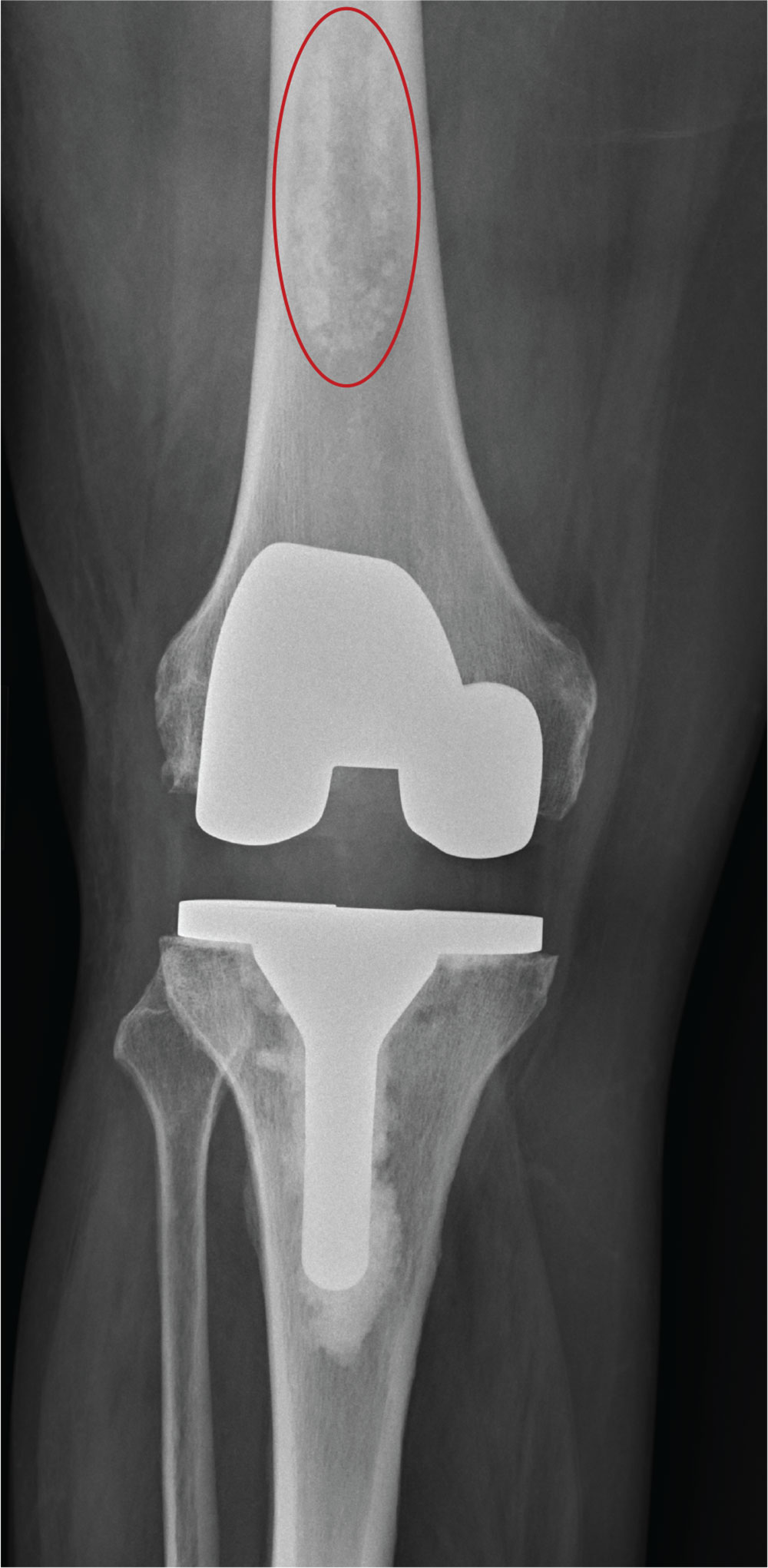
The radiograph shows a right knee prosthesis in place with no evidence of failure or displacement. Of note, there is a hyperdense, somewhat elongated lesion along the distal third of the femur. Radiographically, this is most likely consistent with an enchondroma. Enchondromas are typically benign bone lesions that originate from cartilage. They
The patient was referred to his orthopedist for follow-up.
ANSWER
The radiograph shows a right knee prosthesis in place with no evidence of failure or displacement. Of note, there is a hyperdense, somewhat elongated lesion along the distal third of the femur. Radiographically, this is most likely consistent with an enchondroma. Enchondromas are typically benign bone lesions that originate from cartilage. They
The patient was referred to his orthopedist for follow-up.
ANSWER
The radiograph shows a right knee prosthesis in place with no evidence of failure or displacement. Of note, there is a hyperdense, somewhat elongated lesion along the distal third of the femur. Radiographically, this is most likely consistent with an enchondroma. Enchondromas are typically benign bone lesions that originate from cartilage. They
The patient was referred to his orthopedist for follow-up.
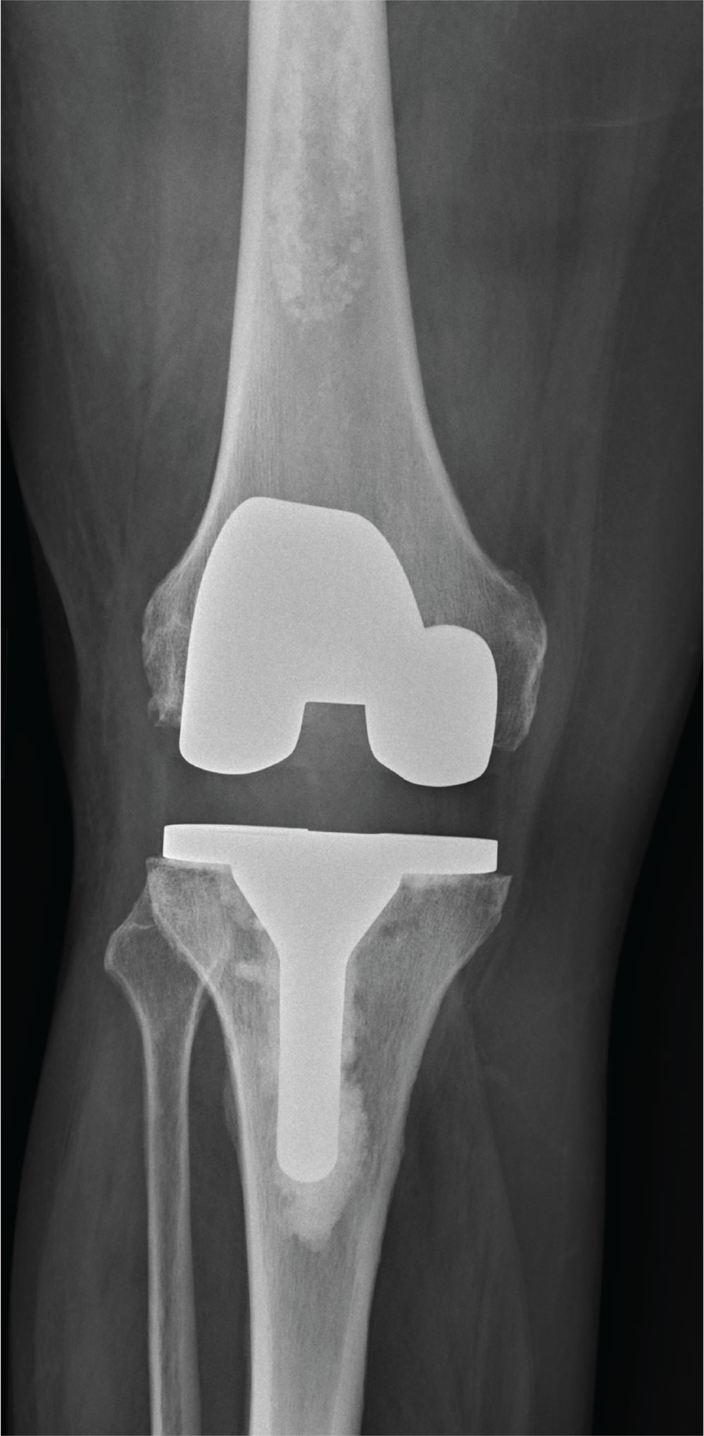
A 70-year-old man presents to the urgent care clinic for evaluation of right knee pain. He denies any specific injury or trauma. For the past several months, he says, he has had a “deep aching pain” that is exacerbated by walking and weight bearing.
His medical history is significant for mild hypertension and diabetes. His surgical history is significant for remote right total knee arthroplasty.
On examination, you note an elderly male in no obvious distress. His vital signs are normal. Inspection of the right knee shows a well-healed incision with no obvious effusion or erythema. He demonstrates a fairly good active range of motion. There is no evidence of ligament laxity.
You obtain a radiograph of the knee (shown). What is your impression?
Chronic Lymphocytic Leukemia and Infiltrates Seen During Excision of Nonmelanoma Skin Cancer
To the Editor:
Specific characteristics of a lymphocytic infiltrate noted on frozen section histologic examination during Mohs micrographic surgery (MMS) tumor excision should raise suspicion of an underlying chronic lymphocytic leukemia (CLL). This infiltrate may be the presenting sign of the underlying leukemia and has variable presentation that may mimic aggressive features. The following 3 cases highlight this phenomenon.
A 74-year-old man (patient 1) with a medical history of multiple nonmelanoma skin cancers (NMSCs) presented for definitive treatment of a biopsy-proven infiltrative basal cell carcinoma involving the right infra-auricular region. Mohs section histologic evaluation revealed patches of lymphocytic infiltrates so dense they obscured the tumor margins. The lymphocytic infiltrates persisted even after 3 MMS stages, though they were moderately less dense compared to the initial MMS stage. Clinical interpretation determined no relationship between the lymphocytic infiltrates and residual tumor. Due to concerns that this lymphocytic infiltrate may indicate an underlying leukemic process, preoperative laboratory tests were ordered prior to closure of the surgical wound, which demonstrated an elevated white blood cell count of 65,000/µL (reference range, 4500–11,000/µL) with 93% lymphocytes. A follow-up complete blood cell count (CBC) and blood smear confirmed the diagnosis of CLL. The patient was referred to a hematologist/oncologist.
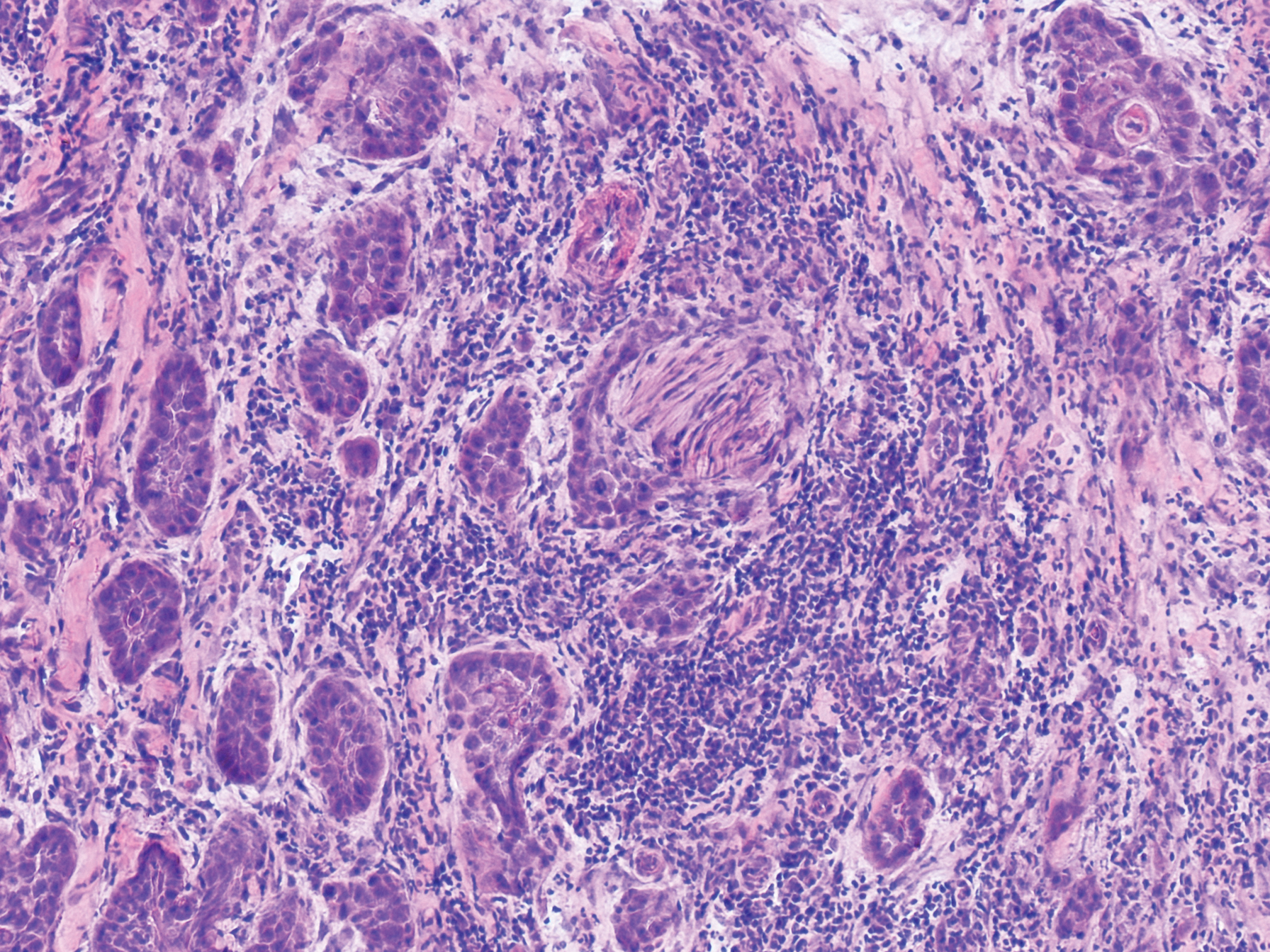
An 84-year old man (patient 2) with a medical history of numerous precancerous lesions and 1 squamous cell carcinoma (SCC) presented for a biopsy, which determined moderately differentiated SCC. Mohs micrographic surgery was performed. The initial stage of MMS histologic examination demonstrated basosquamous carcinoma in the specimen margins, including perineural growth, with an extensive lymphoid infiltrate surrounding the tumor (Figure 1). A second stage of MMS was performed, and although margins appeared to be clear of the basosquamous histology, complete assessment was difficult due to areas of dense inflammatory infiltrate (Figure 2), including perineural infiltration that remained and appeared to extend deeper into the tissues. Pathology was consulted and it was determined that the perineural infiltration was unlikely related to tumor spread but rather secondary to an unknown cause. Further investigation of the patient’s medical history revealed previously diagnosed CLL, which had been omitted by the patient, as he had forgotten this diagnosis and denied a history of cancer, lymphoma, and even leukemia. A query to the patient’s primary care physician found the most recent CBC demonstrated an elevated white blood cell count of 37,600/µL with 78% lymphocytes.
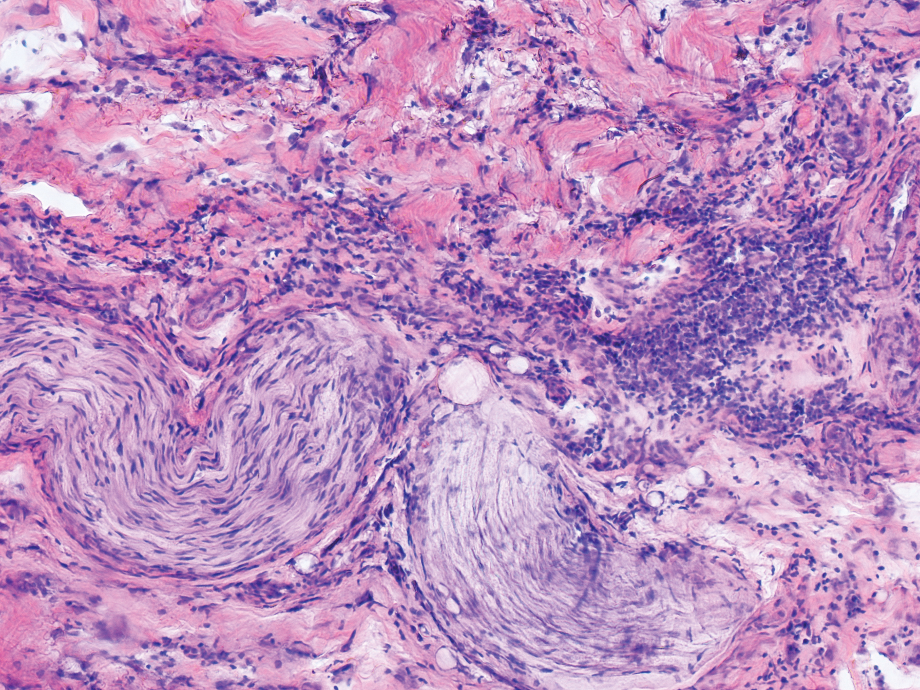
An 84-year-old man (patient 3) with a known history of CLL was referred for MMS excision of a 3.5×4.0-cm SCC on the right anterior temple extending onto the lateral upper and lower eyelids. Mohs frozen section histologic examination of excised tissue revealed patches of heavy lymphocytic infiltrates not found exclusively around the residual tumor but additionally around superficial and deep neurovascular bundles. The second stage of MMS appeared to be clear of tumor cells, but lymphocytic infiltrates remained. Because this patient had a clear history of CLL, the decision was made in conjunction with a dermatopathologist to conclude the surgery at this point. However, secondary to the aggressive, deeply invasive growth of this SCC, the patient was referred for adjunctive radiation therapy to the surgical site after wound reconstruction.
Chronic lymphocytic leukemia is the most common leukemia in the Western world1 and is estimated to account for 27% of all new cases of leukemia. An individual’s lifetime risk is 0.5%. Chronic lymphocytic leukemia is predominantly a disease of the elderly, with an average age at diagnosis of 71 years. It is more common among males, North American and European populations, and those with a positive family history. Although epidemiologic factors including farming, prolonged pesticide exposure, and contact with Agent Orange have tentative links to CLL, the relationships are poorly established.2
Symptoms associated with acute leukemia only rarely manifest in patients with CLL.3 If present, symptoms are vague and include weakness, fatigue, weight loss, fever, night sweats, and a feeling of abdominal fullness.2,3 On clinical examination, patients also may have lymphadenopathy, splenomegaly, or hepatomegaly. Increasing severity of symptoms at time of presentation directly correlates with the severity and staging at the time of diagnosis.4 Not only do patients with CLL have a greater incidence of NMSCs with more notable subclinical tumor extension than the average person, but these individuals also have a greatly increased risk for skin cancer recurrence posttreatment.5,6
Although tissue pathology is not routinely part of the diagnosis of patients with CLL, findings can add to clinical suspicion. Smudge cells, which are cell debris, are characteristic morphologic features found in CLL. Most CLL cells are characteristically small mature lymphocytes with a dense nucleus.3 The presence of aggregates of these cells may obscure tumor margins during resection of NMSCs.7 This infiltrate is present in more than one-third of patients with CLL, as described in one retrospective cohort. This study simultaneously demonstrated the relationship between CLL and a 2-fold increase in postoperative defect size, which was attributed to either subclinical tumor spread or extra tissue removal to ensure clearance due to the leukemic infiltrates themselves.8 The presence of perineural tumor growth, which can occur with aggressive SCC and basal cell carcinoma, may be mimicked by perineural involvement of CLL cells rather than the reactive inflammation associated with continued tumor margins.7
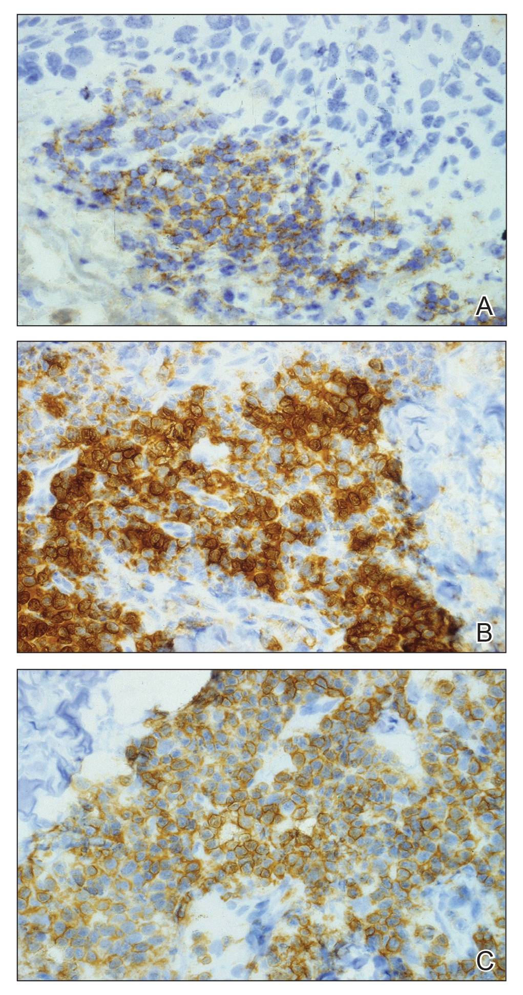
When evaluating a patient with suspected CLL, laboratory tests should include a CBC with differential and examination of the peripheral smear. If abnormal, immunophenotyping of lymphocytes by flow cytometry will rule out other lymphoproliferative diseases and verify CLL as the diagnosis.3 Diagnosis of CLL requires the presence of monoclonal B lymphocytes (≥5×10⁹/L) in the peripheral blood as confirmed by flow cytometry.3 Clonality of circulating B lymphocytes must be confirmed, and immunophenotyping will establish a diagnosis with leukemic cells having positive expression of CD20 (Figure 3A) and CD23 (Figure 3B)(characteristic of B-cell lineage) with coexpression of CD43 and CD5 (Figure 3C)(characteristic of T-cell lineage).7,9 This pattern of immunohistochemical markers can be differentiated from the normal immune response to cutaneous malignancies, which have the pattern of being CD3+, CD5+, and CD43+ with absence of B-cell markers (ie, CD20, CD23)(Table).7

The pathogenesis of this peritumoral infiltrate is unknown, though multiple theories exist. One theory is that the neoplastic lymphocytes are responding as a dysfunctional arm of the immune system to tumor-specific antigens. In patients with CLL, leukemic lymphocytes comprise a large portion of the circulating leukocyte population and this peritumoral infiltrate may simply be a reflection of the circulating leukocytic population. Another theory contends that neoplastic lymphocytes are simply nonspecific aggregations secondary to tumor neovascularization and increased vascular permeability.10
This neoplastic infiltrate seen incidentally during MMS excision of NMSCs not only provides a unique opportunity to diagnose and intervene in those with unknown CLL but also to be aware of complicating features that can spare the patient from unnecessary tissue removal, thereby maximizing the benefit of MMS. This infiltrate can obscure tumor margins; is unusually dense and patchy, with or without infiltrating perineural or perivascular components; and persists beyond what would seem to be an adequate margin to clear a tumor. These cases show these findings, which exemplify the peritumoral infiltrate of CLL and should prompt further workup.
- Rozman C, Monserrat E. Chronic lymphocytic leukemia. N Engl J Med. 1995;333:1052-1057.
- What are the risk factors for chronic lymphocytic leukemia? American Cancer Society website. https://www.cancer.org/cancer/chronic-lymphocytic-leukemia/causes-risks-prevention/risk-factors.html. Revised May 10, 2018. Accessed February 11, 2019.
- Hallek M, Cheson BD, Catovsky D, et al. Guidelines for the diagnosis and treatment of chronic lymphocytic leukemia: a report from the International Workshop on Chronic Lymphocytic Leukemia updating the National Cancer Institute-Working Group 1996 guidelines. Blood. 2008;111:5446-5456.
- Rai KR, Wasil T, Iqbal U, et al. Clinical staging and prognostic markers in chronic lymphocytic leukemia. Hematol Oncol Clin North Am. 2004;18:795-805, vii.
- Mehrany K, Weenig RH, Pittelkow MR, et al. High recurrence rates of squamous cell carcinoma after Mohs’ surgery in patients with chronic lymphocytic leukemia. Dermatol Surg. 2005;31:38-42.
- Brewer JD, Shanafelt TD, Khezri F, et al. Increased incidence and recurrence rates of nonmelanoma skin cancer in patients with non-Hodgkin lymphoma: a Rochester epidemiology project population-based study in Minnesota. J Am Acad Dermatol. 2015;72:302-309.
- Wilson ML, Elston DM, Tyler WB, et al. Dense lymphocytic infiltrates associated with non-melanoma skin cancer in patients with chronic lymphocytic leukemia. Dermatol Online J. 2010;16:4.
- Mehrany K, Byrd DR, Roenigk RK, et al. Lymphocytic infiltrates and subclinical epithelial tumor extension in patients with chronic leukemia and solid-organ transplantation. Dermatol Surg. 2003;29:129-134.
- Khandelwal A, Seilstad KH, Magro CM. Subclinical chronic lymphocytic leukaemia associated with a 13q deletion presenting initially in the skin: apropos of a case. J Cutan Pathol. 2006;33:256-259.
- Padgett JK, Parlette HL, English JC. A diagnosis of chronic lymphocytic leukemia prompted by cutaneous lymphocytic infiltrates present in mohs micrographic surgery frozen sections. Dermatol Surg. 2003;29:769-771.
To the Editor:
Specific characteristics of a lymphocytic infiltrate noted on frozen section histologic examination during Mohs micrographic surgery (MMS) tumor excision should raise suspicion of an underlying chronic lymphocytic leukemia (CLL). This infiltrate may be the presenting sign of the underlying leukemia and has variable presentation that may mimic aggressive features. The following 3 cases highlight this phenomenon.
A 74-year-old man (patient 1) with a medical history of multiple nonmelanoma skin cancers (NMSCs) presented for definitive treatment of a biopsy-proven infiltrative basal cell carcinoma involving the right infra-auricular region. Mohs section histologic evaluation revealed patches of lymphocytic infiltrates so dense they obscured the tumor margins. The lymphocytic infiltrates persisted even after 3 MMS stages, though they were moderately less dense compared to the initial MMS stage. Clinical interpretation determined no relationship between the lymphocytic infiltrates and residual tumor. Due to concerns that this lymphocytic infiltrate may indicate an underlying leukemic process, preoperative laboratory tests were ordered prior to closure of the surgical wound, which demonstrated an elevated white blood cell count of 65,000/µL (reference range, 4500–11,000/µL) with 93% lymphocytes. A follow-up complete blood cell count (CBC) and blood smear confirmed the diagnosis of CLL. The patient was referred to a hematologist/oncologist.
An 84-year old man (patient 2) with a medical history of numerous precancerous lesions and 1 squamous cell carcinoma (SCC) presented for a biopsy, which determined moderately differentiated SCC. Mohs micrographic surgery was performed. The initial stage of MMS histologic examination demonstrated basosquamous carcinoma in the specimen margins, including perineural growth, with an extensive lymphoid infiltrate surrounding the tumor (Figure 1). A second stage of MMS was performed, and although margins appeared to be clear of the basosquamous histology, complete assessment was difficult due to areas of dense inflammatory infiltrate (Figure 2), including perineural infiltration that remained and appeared to extend deeper into the tissues. Pathology was consulted and it was determined that the perineural infiltration was unlikely related to tumor spread but rather secondary to an unknown cause. Further investigation of the patient’s medical history revealed previously diagnosed CLL, which had been omitted by the patient, as he had forgotten this diagnosis and denied a history of cancer, lymphoma, and even leukemia. A query to the patient’s primary care physician found the most recent CBC demonstrated an elevated white blood cell count of 37,600/µL with 78% lymphocytes.
An 84-year-old man (patient 3) with a known history of CLL was referred for MMS excision of a 3.5×4.0-cm SCC on the right anterior temple extending onto the lateral upper and lower eyelids. Mohs frozen section histologic examination of excised tissue revealed patches of heavy lymphocytic infiltrates not found exclusively around the residual tumor but additionally around superficial and deep neurovascular bundles. The second stage of MMS appeared to be clear of tumor cells, but lymphocytic infiltrates remained. Because this patient had a clear history of CLL, the decision was made in conjunction with a dermatopathologist to conclude the surgery at this point. However, secondary to the aggressive, deeply invasive growth of this SCC, the patient was referred for adjunctive radiation therapy to the surgical site after wound reconstruction.
Chronic lymphocytic leukemia is the most common leukemia in the Western world1 and is estimated to account for 27% of all new cases of leukemia. An individual’s lifetime risk is 0.5%. Chronic lymphocytic leukemia is predominantly a disease of the elderly, with an average age at diagnosis of 71 years. It is more common among males, North American and European populations, and those with a positive family history. Although epidemiologic factors including farming, prolonged pesticide exposure, and contact with Agent Orange have tentative links to CLL, the relationships are poorly established.2
Symptoms associated with acute leukemia only rarely manifest in patients with CLL.3 If present, symptoms are vague and include weakness, fatigue, weight loss, fever, night sweats, and a feeling of abdominal fullness.2,3 On clinical examination, patients also may have lymphadenopathy, splenomegaly, or hepatomegaly. Increasing severity of symptoms at time of presentation directly correlates with the severity and staging at the time of diagnosis.4 Not only do patients with CLL have a greater incidence of NMSCs with more notable subclinical tumor extension than the average person, but these individuals also have a greatly increased risk for skin cancer recurrence posttreatment.5,6
Although tissue pathology is not routinely part of the diagnosis of patients with CLL, findings can add to clinical suspicion. Smudge cells, which are cell debris, are characteristic morphologic features found in CLL. Most CLL cells are characteristically small mature lymphocytes with a dense nucleus.3 The presence of aggregates of these cells may obscure tumor margins during resection of NMSCs.7 This infiltrate is present in more than one-third of patients with CLL, as described in one retrospective cohort. This study simultaneously demonstrated the relationship between CLL and a 2-fold increase in postoperative defect size, which was attributed to either subclinical tumor spread or extra tissue removal to ensure clearance due to the leukemic infiltrates themselves.8 The presence of perineural tumor growth, which can occur with aggressive SCC and basal cell carcinoma, may be mimicked by perineural involvement of CLL cells rather than the reactive inflammation associated with continued tumor margins.7
When evaluating a patient with suspected CLL, laboratory tests should include a CBC with differential and examination of the peripheral smear. If abnormal, immunophenotyping of lymphocytes by flow cytometry will rule out other lymphoproliferative diseases and verify CLL as the diagnosis.3 Diagnosis of CLL requires the presence of monoclonal B lymphocytes (≥5×10⁹/L) in the peripheral blood as confirmed by flow cytometry.3 Clonality of circulating B lymphocytes must be confirmed, and immunophenotyping will establish a diagnosis with leukemic cells having positive expression of CD20 (Figure 3A) and CD23 (Figure 3B)(characteristic of B-cell lineage) with coexpression of CD43 and CD5 (Figure 3C)(characteristic of T-cell lineage).7,9 This pattern of immunohistochemical markers can be differentiated from the normal immune response to cutaneous malignancies, which have the pattern of being CD3+, CD5+, and CD43+ with absence of B-cell markers (ie, CD20, CD23)(Table).7
The pathogenesis of this peritumoral infiltrate is unknown, though multiple theories exist. One theory is that the neoplastic lymphocytes are responding as a dysfunctional arm of the immune system to tumor-specific antigens. In patients with CLL, leukemic lymphocytes comprise a large portion of the circulating leukocyte population and this peritumoral infiltrate may simply be a reflection of the circulating leukocytic population. Another theory contends that neoplastic lymphocytes are simply nonspecific aggregations secondary to tumor neovascularization and increased vascular permeability.10
This neoplastic infiltrate seen incidentally during MMS excision of NMSCs not only provides a unique opportunity to diagnose and intervene in those with unknown CLL but also to be aware of complicating features that can spare the patient from unnecessary tissue removal, thereby maximizing the benefit of MMS. This infiltrate can obscure tumor margins; is unusually dense and patchy, with or without infiltrating perineural or perivascular components; and persists beyond what would seem to be an adequate margin to clear a tumor. These cases show these findings, which exemplify the peritumoral infiltrate of CLL and should prompt further workup.
To the Editor:
Specific characteristics of a lymphocytic infiltrate noted on frozen section histologic examination during Mohs micrographic surgery (MMS) tumor excision should raise suspicion of an underlying chronic lymphocytic leukemia (CLL). This infiltrate may be the presenting sign of the underlying leukemia and has variable presentation that may mimic aggressive features. The following 3 cases highlight this phenomenon.
A 74-year-old man (patient 1) with a medical history of multiple nonmelanoma skin cancers (NMSCs) presented for definitive treatment of a biopsy-proven infiltrative basal cell carcinoma involving the right infra-auricular region. Mohs section histologic evaluation revealed patches of lymphocytic infiltrates so dense they obscured the tumor margins. The lymphocytic infiltrates persisted even after 3 MMS stages, though they were moderately less dense compared to the initial MMS stage. Clinical interpretation determined no relationship between the lymphocytic infiltrates and residual tumor. Due to concerns that this lymphocytic infiltrate may indicate an underlying leukemic process, preoperative laboratory tests were ordered prior to closure of the surgical wound, which demonstrated an elevated white blood cell count of 65,000/µL (reference range, 4500–11,000/µL) with 93% lymphocytes. A follow-up complete blood cell count (CBC) and blood smear confirmed the diagnosis of CLL. The patient was referred to a hematologist/oncologist.
An 84-year old man (patient 2) with a medical history of numerous precancerous lesions and 1 squamous cell carcinoma (SCC) presented for a biopsy, which determined moderately differentiated SCC. Mohs micrographic surgery was performed. The initial stage of MMS histologic examination demonstrated basosquamous carcinoma in the specimen margins, including perineural growth, with an extensive lymphoid infiltrate surrounding the tumor (Figure 1). A second stage of MMS was performed, and although margins appeared to be clear of the basosquamous histology, complete assessment was difficult due to areas of dense inflammatory infiltrate (Figure 2), including perineural infiltration that remained and appeared to extend deeper into the tissues. Pathology was consulted and it was determined that the perineural infiltration was unlikely related to tumor spread but rather secondary to an unknown cause. Further investigation of the patient’s medical history revealed previously diagnosed CLL, which had been omitted by the patient, as he had forgotten this diagnosis and denied a history of cancer, lymphoma, and even leukemia. A query to the patient’s primary care physician found the most recent CBC demonstrated an elevated white blood cell count of 37,600/µL with 78% lymphocytes.
An 84-year-old man (patient 3) with a known history of CLL was referred for MMS excision of a 3.5×4.0-cm SCC on the right anterior temple extending onto the lateral upper and lower eyelids. Mohs frozen section histologic examination of excised tissue revealed patches of heavy lymphocytic infiltrates not found exclusively around the residual tumor but additionally around superficial and deep neurovascular bundles. The second stage of MMS appeared to be clear of tumor cells, but lymphocytic infiltrates remained. Because this patient had a clear history of CLL, the decision was made in conjunction with a dermatopathologist to conclude the surgery at this point. However, secondary to the aggressive, deeply invasive growth of this SCC, the patient was referred for adjunctive radiation therapy to the surgical site after wound reconstruction.
Chronic lymphocytic leukemia is the most common leukemia in the Western world1 and is estimated to account for 27% of all new cases of leukemia. An individual’s lifetime risk is 0.5%. Chronic lymphocytic leukemia is predominantly a disease of the elderly, with an average age at diagnosis of 71 years. It is more common among males, North American and European populations, and those with a positive family history. Although epidemiologic factors including farming, prolonged pesticide exposure, and contact with Agent Orange have tentative links to CLL, the relationships are poorly established.2
Symptoms associated with acute leukemia only rarely manifest in patients with CLL.3 If present, symptoms are vague and include weakness, fatigue, weight loss, fever, night sweats, and a feeling of abdominal fullness.2,3 On clinical examination, patients also may have lymphadenopathy, splenomegaly, or hepatomegaly. Increasing severity of symptoms at time of presentation directly correlates with the severity and staging at the time of diagnosis.4 Not only do patients with CLL have a greater incidence of NMSCs with more notable subclinical tumor extension than the average person, but these individuals also have a greatly increased risk for skin cancer recurrence posttreatment.5,6
Although tissue pathology is not routinely part of the diagnosis of patients with CLL, findings can add to clinical suspicion. Smudge cells, which are cell debris, are characteristic morphologic features found in CLL. Most CLL cells are characteristically small mature lymphocytes with a dense nucleus.3 The presence of aggregates of these cells may obscure tumor margins during resection of NMSCs.7 This infiltrate is present in more than one-third of patients with CLL, as described in one retrospective cohort. This study simultaneously demonstrated the relationship between CLL and a 2-fold increase in postoperative defect size, which was attributed to either subclinical tumor spread or extra tissue removal to ensure clearance due to the leukemic infiltrates themselves.8 The presence of perineural tumor growth, which can occur with aggressive SCC and basal cell carcinoma, may be mimicked by perineural involvement of CLL cells rather than the reactive inflammation associated with continued tumor margins.7
When evaluating a patient with suspected CLL, laboratory tests should include a CBC with differential and examination of the peripheral smear. If abnormal, immunophenotyping of lymphocytes by flow cytometry will rule out other lymphoproliferative diseases and verify CLL as the diagnosis.3 Diagnosis of CLL requires the presence of monoclonal B lymphocytes (≥5×10⁹/L) in the peripheral blood as confirmed by flow cytometry.3 Clonality of circulating B lymphocytes must be confirmed, and immunophenotyping will establish a diagnosis with leukemic cells having positive expression of CD20 (Figure 3A) and CD23 (Figure 3B)(characteristic of B-cell lineage) with coexpression of CD43 and CD5 (Figure 3C)(characteristic of T-cell lineage).7,9 This pattern of immunohistochemical markers can be differentiated from the normal immune response to cutaneous malignancies, which have the pattern of being CD3+, CD5+, and CD43+ with absence of B-cell markers (ie, CD20, CD23)(Table).7
The pathogenesis of this peritumoral infiltrate is unknown, though multiple theories exist. One theory is that the neoplastic lymphocytes are responding as a dysfunctional arm of the immune system to tumor-specific antigens. In patients with CLL, leukemic lymphocytes comprise a large portion of the circulating leukocyte population and this peritumoral infiltrate may simply be a reflection of the circulating leukocytic population. Another theory contends that neoplastic lymphocytes are simply nonspecific aggregations secondary to tumor neovascularization and increased vascular permeability.10
This neoplastic infiltrate seen incidentally during MMS excision of NMSCs not only provides a unique opportunity to diagnose and intervene in those with unknown CLL but also to be aware of complicating features that can spare the patient from unnecessary tissue removal, thereby maximizing the benefit of MMS. This infiltrate can obscure tumor margins; is unusually dense and patchy, with or without infiltrating perineural or perivascular components; and persists beyond what would seem to be an adequate margin to clear a tumor. These cases show these findings, which exemplify the peritumoral infiltrate of CLL and should prompt further workup.
- Rozman C, Monserrat E. Chronic lymphocytic leukemia. N Engl J Med. 1995;333:1052-1057.
- What are the risk factors for chronic lymphocytic leukemia? American Cancer Society website. https://www.cancer.org/cancer/chronic-lymphocytic-leukemia/causes-risks-prevention/risk-factors.html. Revised May 10, 2018. Accessed February 11, 2019.
- Hallek M, Cheson BD, Catovsky D, et al. Guidelines for the diagnosis and treatment of chronic lymphocytic leukemia: a report from the International Workshop on Chronic Lymphocytic Leukemia updating the National Cancer Institute-Working Group 1996 guidelines. Blood. 2008;111:5446-5456.
- Rai KR, Wasil T, Iqbal U, et al. Clinical staging and prognostic markers in chronic lymphocytic leukemia. Hematol Oncol Clin North Am. 2004;18:795-805, vii.
- Mehrany K, Weenig RH, Pittelkow MR, et al. High recurrence rates of squamous cell carcinoma after Mohs’ surgery in patients with chronic lymphocytic leukemia. Dermatol Surg. 2005;31:38-42.
- Brewer JD, Shanafelt TD, Khezri F, et al. Increased incidence and recurrence rates of nonmelanoma skin cancer in patients with non-Hodgkin lymphoma: a Rochester epidemiology project population-based study in Minnesota. J Am Acad Dermatol. 2015;72:302-309.
- Wilson ML, Elston DM, Tyler WB, et al. Dense lymphocytic infiltrates associated with non-melanoma skin cancer in patients with chronic lymphocytic leukemia. Dermatol Online J. 2010;16:4.
- Mehrany K, Byrd DR, Roenigk RK, et al. Lymphocytic infiltrates and subclinical epithelial tumor extension in patients with chronic leukemia and solid-organ transplantation. Dermatol Surg. 2003;29:129-134.
- Khandelwal A, Seilstad KH, Magro CM. Subclinical chronic lymphocytic leukaemia associated with a 13q deletion presenting initially in the skin: apropos of a case. J Cutan Pathol. 2006;33:256-259.
- Padgett JK, Parlette HL, English JC. A diagnosis of chronic lymphocytic leukemia prompted by cutaneous lymphocytic infiltrates present in mohs micrographic surgery frozen sections. Dermatol Surg. 2003;29:769-771.
- Rozman C, Monserrat E. Chronic lymphocytic leukemia. N Engl J Med. 1995;333:1052-1057.
- What are the risk factors for chronic lymphocytic leukemia? American Cancer Society website. https://www.cancer.org/cancer/chronic-lymphocytic-leukemia/causes-risks-prevention/risk-factors.html. Revised May 10, 2018. Accessed February 11, 2019.
- Hallek M, Cheson BD, Catovsky D, et al. Guidelines for the diagnosis and treatment of chronic lymphocytic leukemia: a report from the International Workshop on Chronic Lymphocytic Leukemia updating the National Cancer Institute-Working Group 1996 guidelines. Blood. 2008;111:5446-5456.
- Rai KR, Wasil T, Iqbal U, et al. Clinical staging and prognostic markers in chronic lymphocytic leukemia. Hematol Oncol Clin North Am. 2004;18:795-805, vii.
- Mehrany K, Weenig RH, Pittelkow MR, et al. High recurrence rates of squamous cell carcinoma after Mohs’ surgery in patients with chronic lymphocytic leukemia. Dermatol Surg. 2005;31:38-42.
- Brewer JD, Shanafelt TD, Khezri F, et al. Increased incidence and recurrence rates of nonmelanoma skin cancer in patients with non-Hodgkin lymphoma: a Rochester epidemiology project population-based study in Minnesota. J Am Acad Dermatol. 2015;72:302-309.
- Wilson ML, Elston DM, Tyler WB, et al. Dense lymphocytic infiltrates associated with non-melanoma skin cancer in patients with chronic lymphocytic leukemia. Dermatol Online J. 2010;16:4.
- Mehrany K, Byrd DR, Roenigk RK, et al. Lymphocytic infiltrates and subclinical epithelial tumor extension in patients with chronic leukemia and solid-organ transplantation. Dermatol Surg. 2003;29:129-134.
- Khandelwal A, Seilstad KH, Magro CM. Subclinical chronic lymphocytic leukaemia associated with a 13q deletion presenting initially in the skin: apropos of a case. J Cutan Pathol. 2006;33:256-259.
- Padgett JK, Parlette HL, English JC. A diagnosis of chronic lymphocytic leukemia prompted by cutaneous lymphocytic infiltrates present in mohs micrographic surgery frozen sections. Dermatol Surg. 2003;29:769-771.
Practice Points
- Chronic lymphocytic leukemia (CLL) may be seen during histologic examination of specimens during Mohs micrographic surgery as a monomorphic infiltrate of small mature lymphocytes with dense nuclei. Patients may be unaware of their diagnosis, which can be the presenting feature.
- An infiltrate of CLL may mimic aggressive behavior of nonmelanoma skin cancers including perineural invasion. A leukemic infiltrate may appear more dense and monomorphic. If needed, immunohistochemical staining of leukemic cells will show CD5 and CD23 positivity.
- Anecdotally, patients with CLL may not remember this pertinent medical history. Whether due to its asymptomatic nature or lack of treatment in early stages, direct questioning about CLL may be warranted if this characteristic infiltrate is encountered.
Few DLBCL patients benefit from nivolumab
Nivolumab may provide a benefit for a small group of patients with diffuse large B-cell lymphoma (DLBCL) who have failed a transplant or are ineligible for one, according to researchers.

Nivolumab produced a response in 10 of 121 DLBCL patients studied. Three patients achieved a complete response (CR) lasting 11 months or more.
Why this small group responded to nivolumab isn’t clear, according to Stephen M. Ansell, MD, PhD, of the Mayo Clinic in Rochester, Minn., and his colleagues.
Some responders did have 9p24.1 alterations, but others did not. None of the responders had tumor cells positive for programmed death–ligand 1 (PD-L1), and only one responder had detectable PD-L2 expression in malignant cells.
Dr. Ansell and his colleagues described these findings in the Journal of Clinical Oncology.
The researchers evaluated nivolumab in a phase 2 trial (NCT02038933) of patients with relapsed/refractory DLBCL. Of the 121 patients, 87 had failed autologous hematopoietic stem cell transplant (HSCT) and 34 were ineligible for autologous HSCT. The patients received nivolumab at 3 mg/kg every 2 weeks until disease progression, unacceptable toxicity, or study withdrawal.
HSCT failures
The patients who had failed HSCT had a median age of 62 years (range, 24-75 years). They had received a median of three prior systemic therapies (range, 1-11), and 28% were refractory to their most recent therapy.
About 40% of patients had germinal center B-cell–like (GCB) DLBCL, 32% had non-GCB disease, and data on disease subtype were missing for the rest of the group.
The patients received a median of four nivolumab doses (range, 1-44) and were followed for a median of 9 months (range, 0.1-25 months).
Nine patients (10%) achieved a response, and the median duration of response was 11 months. Responses occurred in patients with GCB and non-GCB DLBCL.
Three patients achieved a CR. Two of them were still on treatment and in CR at the data cutoff. Their responses had lasted 11 months and 14 months, respectively. The third complete responder did not progress on nivolumab but developed myelodysplastic syndrome, which was unrelated to the drug, and died.
Among all patients who had failed HSCT, the median progression-free survival was 1.9 months, and the median overall survival was 12.2 months.
Seven patients in this group were still receiving nivolumab at the data cutoff. Two of them were in CR, two had a partial response, and three had stable disease.
HSCT-ineligible group
The patients who were ineligible for HSCT had a median age of 68 years (range, 28-86 years). They had received a median of three prior systemic therapies (range, 1-7), and 59% were refractory to their most recent therapy.
About 56% of these patients had GCB DLBCL, 18% had non-GCB disease, and data on subtype were missing for the rest of the group.
The patients received a median of three nivolumab doses (range, 1-22) and were followed for a median of 6 months (range, 0.2-24 months). One patient (3%) achieved a partial response, which lasted 8.3 months.
The median progression-free survival in this group was 1.4 months, and the median overall survival was 5.8 months.
None of the patients in this group were still taking nivolumab at the data cutoff.
Biomarkers of response
Dr. Ansell and his colleagues were able to look for 9p24.1 alterations in archival tumor biopsy specimens from 74 patients. The team reported that 9p24.1 alterations were “infrequent,” but they were found in some responders.
Among the three complete responders, one patient had high-level 9p24.1 amplification, one had normal 9p24.1 copy number, and one did not have a biopsy available.
Among the seven patients who achieved a partial response, five had biopsy specimens. Three patients had low-level polysomy, one had copy gain, and one patient had normal 9p24.1.
None of the responders had PD-L1 expression in their tumor cells, but one complete responder had PD-L2–positive malignant cells.
Dr. Ansell and his colleagues wrote that the “biologic basis for response in the other two [complete responders] is unclear,” and the researchers were unable to assess associations between response and c-myc expression or double-hit lymphoma.
Safety
Of all 121 patients, 62% had a treatment-related adverse event (AE) and 24% had a grade 3/4–related AE.
The most common related AEs of any grade were nausea (17%), fatigue (17%), diarrhea (10%), neutropenia (7%), thrombocytopenia (6%), decreased appetite (6%), lipase increase (5%), rash (5%), and pyrexia (5%).
Four patients (3%) stopped taking nivolumab because of treatment-related AEs, including neutropenia, thrombocytopenia, diarrhea, pancreatitis, lipase increase, and psoriasiform dermatitis. There were no fatal treatment-related AEs.
This research was supported by Bristol-Myers Squibb and other groups. The study authors reported relationships with Bristol-Myers Squibb and other companies.
SOURCE: Ansell SM et al. J Clin Oncol. 2019 Jan 8. doi: 10.1200/JCO.18.00766.
Nivolumab may provide a benefit for a small group of patients with diffuse large B-cell lymphoma (DLBCL) who have failed a transplant or are ineligible for one, according to researchers.
Nivolumab produced a response in 10 of 121 DLBCL patients studied. Three patients achieved a complete response (CR) lasting 11 months or more.
Why this small group responded to nivolumab isn’t clear, according to Stephen M. Ansell, MD, PhD, of the Mayo Clinic in Rochester, Minn., and his colleagues.
Some responders did have 9p24.1 alterations, but others did not. None of the responders had tumor cells positive for programmed death–ligand 1 (PD-L1), and only one responder had detectable PD-L2 expression in malignant cells.
Dr. Ansell and his colleagues described these findings in the Journal of Clinical Oncology.
The researchers evaluated nivolumab in a phase 2 trial (NCT02038933) of patients with relapsed/refractory DLBCL. Of the 121 patients, 87 had failed autologous hematopoietic stem cell transplant (HSCT) and 34 were ineligible for autologous HSCT. The patients received nivolumab at 3 mg/kg every 2 weeks until disease progression, unacceptable toxicity, or study withdrawal.
HSCT failures
The patients who had failed HSCT had a median age of 62 years (range, 24-75 years). They had received a median of three prior systemic therapies (range, 1-11), and 28% were refractory to their most recent therapy.
About 40% of patients had germinal center B-cell–like (GCB) DLBCL, 32% had non-GCB disease, and data on disease subtype were missing for the rest of the group.
The patients received a median of four nivolumab doses (range, 1-44) and were followed for a median of 9 months (range, 0.1-25 months).
Nine patients (10%) achieved a response, and the median duration of response was 11 months. Responses occurred in patients with GCB and non-GCB DLBCL.
Three patients achieved a CR. Two of them were still on treatment and in CR at the data cutoff. Their responses had lasted 11 months and 14 months, respectively. The third complete responder did not progress on nivolumab but developed myelodysplastic syndrome, which was unrelated to the drug, and died.
Among all patients who had failed HSCT, the median progression-free survival was 1.9 months, and the median overall survival was 12.2 months.
Seven patients in this group were still receiving nivolumab at the data cutoff. Two of them were in CR, two had a partial response, and three had stable disease.
HSCT-ineligible group
The patients who were ineligible for HSCT had a median age of 68 years (range, 28-86 years). They had received a median of three prior systemic therapies (range, 1-7), and 59% were refractory to their most recent therapy.
About 56% of these patients had GCB DLBCL, 18% had non-GCB disease, and data on subtype were missing for the rest of the group.
The patients received a median of three nivolumab doses (range, 1-22) and were followed for a median of 6 months (range, 0.2-24 months). One patient (3%) achieved a partial response, which lasted 8.3 months.
The median progression-free survival in this group was 1.4 months, and the median overall survival was 5.8 months.
None of the patients in this group were still taking nivolumab at the data cutoff.
Biomarkers of response
Dr. Ansell and his colleagues were able to look for 9p24.1 alterations in archival tumor biopsy specimens from 74 patients. The team reported that 9p24.1 alterations were “infrequent,” but they were found in some responders.
Among the three complete responders, one patient had high-level 9p24.1 amplification, one had normal 9p24.1 copy number, and one did not have a biopsy available.
Among the seven patients who achieved a partial response, five had biopsy specimens. Three patients had low-level polysomy, one had copy gain, and one patient had normal 9p24.1.
None of the responders had PD-L1 expression in their tumor cells, but one complete responder had PD-L2–positive malignant cells.
Dr. Ansell and his colleagues wrote that the “biologic basis for response in the other two [complete responders] is unclear,” and the researchers were unable to assess associations between response and c-myc expression or double-hit lymphoma.
Safety
Of all 121 patients, 62% had a treatment-related adverse event (AE) and 24% had a grade 3/4–related AE.
The most common related AEs of any grade were nausea (17%), fatigue (17%), diarrhea (10%), neutropenia (7%), thrombocytopenia (6%), decreased appetite (6%), lipase increase (5%), rash (5%), and pyrexia (5%).
Four patients (3%) stopped taking nivolumab because of treatment-related AEs, including neutropenia, thrombocytopenia, diarrhea, pancreatitis, lipase increase, and psoriasiform dermatitis. There were no fatal treatment-related AEs.
This research was supported by Bristol-Myers Squibb and other groups. The study authors reported relationships with Bristol-Myers Squibb and other companies.
SOURCE: Ansell SM et al. J Clin Oncol. 2019 Jan 8. doi: 10.1200/JCO.18.00766.
Nivolumab may provide a benefit for a small group of patients with diffuse large B-cell lymphoma (DLBCL) who have failed a transplant or are ineligible for one, according to researchers.
Nivolumab produced a response in 10 of 121 DLBCL patients studied. Three patients achieved a complete response (CR) lasting 11 months or more.
Why this small group responded to nivolumab isn’t clear, according to Stephen M. Ansell, MD, PhD, of the Mayo Clinic in Rochester, Minn., and his colleagues.
Some responders did have 9p24.1 alterations, but others did not. None of the responders had tumor cells positive for programmed death–ligand 1 (PD-L1), and only one responder had detectable PD-L2 expression in malignant cells.
Dr. Ansell and his colleagues described these findings in the Journal of Clinical Oncology.
The researchers evaluated nivolumab in a phase 2 trial (NCT02038933) of patients with relapsed/refractory DLBCL. Of the 121 patients, 87 had failed autologous hematopoietic stem cell transplant (HSCT) and 34 were ineligible for autologous HSCT. The patients received nivolumab at 3 mg/kg every 2 weeks until disease progression, unacceptable toxicity, or study withdrawal.
HSCT failures
The patients who had failed HSCT had a median age of 62 years (range, 24-75 years). They had received a median of three prior systemic therapies (range, 1-11), and 28% were refractory to their most recent therapy.
About 40% of patients had germinal center B-cell–like (GCB) DLBCL, 32% had non-GCB disease, and data on disease subtype were missing for the rest of the group.
The patients received a median of four nivolumab doses (range, 1-44) and were followed for a median of 9 months (range, 0.1-25 months).
Nine patients (10%) achieved a response, and the median duration of response was 11 months. Responses occurred in patients with GCB and non-GCB DLBCL.
Three patients achieved a CR. Two of them were still on treatment and in CR at the data cutoff. Their responses had lasted 11 months and 14 months, respectively. The third complete responder did not progress on nivolumab but developed myelodysplastic syndrome, which was unrelated to the drug, and died.
Among all patients who had failed HSCT, the median progression-free survival was 1.9 months, and the median overall survival was 12.2 months.
Seven patients in this group were still receiving nivolumab at the data cutoff. Two of them were in CR, two had a partial response, and three had stable disease.
HSCT-ineligible group
The patients who were ineligible for HSCT had a median age of 68 years (range, 28-86 years). They had received a median of three prior systemic therapies (range, 1-7), and 59% were refractory to their most recent therapy.
About 56% of these patients had GCB DLBCL, 18% had non-GCB disease, and data on subtype were missing for the rest of the group.
The patients received a median of three nivolumab doses (range, 1-22) and were followed for a median of 6 months (range, 0.2-24 months). One patient (3%) achieved a partial response, which lasted 8.3 months.
The median progression-free survival in this group was 1.4 months, and the median overall survival was 5.8 months.
None of the patients in this group were still taking nivolumab at the data cutoff.
Biomarkers of response
Dr. Ansell and his colleagues were able to look for 9p24.1 alterations in archival tumor biopsy specimens from 74 patients. The team reported that 9p24.1 alterations were “infrequent,” but they were found in some responders.
Among the three complete responders, one patient had high-level 9p24.1 amplification, one had normal 9p24.1 copy number, and one did not have a biopsy available.
Among the seven patients who achieved a partial response, five had biopsy specimens. Three patients had low-level polysomy, one had copy gain, and one patient had normal 9p24.1.
None of the responders had PD-L1 expression in their tumor cells, but one complete responder had PD-L2–positive malignant cells.
Dr. Ansell and his colleagues wrote that the “biologic basis for response in the other two [complete responders] is unclear,” and the researchers were unable to assess associations between response and c-myc expression or double-hit lymphoma.
Safety
Of all 121 patients, 62% had a treatment-related adverse event (AE) and 24% had a grade 3/4–related AE.
The most common related AEs of any grade were nausea (17%), fatigue (17%), diarrhea (10%), neutropenia (7%), thrombocytopenia (6%), decreased appetite (6%), lipase increase (5%), rash (5%), and pyrexia (5%).
Four patients (3%) stopped taking nivolumab because of treatment-related AEs, including neutropenia, thrombocytopenia, diarrhea, pancreatitis, lipase increase, and psoriasiform dermatitis. There were no fatal treatment-related AEs.
This research was supported by Bristol-Myers Squibb and other groups. The study authors reported relationships with Bristol-Myers Squibb and other companies.
SOURCE: Ansell SM et al. J Clin Oncol. 2019 Jan 8. doi: 10.1200/JCO.18.00766.
FROM THE JOURNAL OF CLINICAL ONCOLOGY
Key clinical point:
Major finding: Nivolumab produced a response in 10 of 121 patients, including three complete responses.
Study details: A phase 2 study of 121 patients with relapsed/refractory diffuse large B-cell lymphoma.
Disclosures: This research was supported by Bristol-Myers Squibb and other organizations. The study authors reported relationships with Bristol-Myers Squibb and other companies.
Source: Ansell SM et al. J Clin Oncol. 2019 Jan 8. doi: 10.1200/JCO.18.00766.
TNFi use may not affect joint replacement rates for RA patients
Patients with rheumatoid arthritis using tumor necrosis factor inhibitors do not appear to have a lower rate of joint replacement when compared with patients taking conventional synthetic disease-modifying antirheumatic drugs, according to an analysis of data in the British Society for Rheumatology Biologics Register for RA.

Although there was not a general protective effect, patients with rheumatoid arthritis (RA) who were 60 years or older had a 40% reduction in total hip replacement (THR) when using tumor necrosis factor inhibitors (TNFi), according to first author Samuel Hawley from the Nuffield Department of Orthopaedics in the Rheumatology and Musculoskeletal Sciences at the University of Oxford (England) and his colleagues.
“While a reduction in THR amongst older TNFi users offers some support for biologics playing a role in reducing need for joint replacement, it must also be noted that the lack of an overall protective effect is suggestive that other factors apart from TNFi are likely to be involved in the ... downward population trends in joint replacement rates in RA,” Mr. Hawley and his colleagues wrote in their report published in the journal Rheumatology.
The researchers analyzed prospectively collected data on 11,202 RA patients from the British Society for Rheumatology Biologics Register for RA (BSRBR-RA) from 2001-2016 who were using TNFi (n = 9,558) or conventional synthetic disease-modifying antirheumatic drugs (csDMARDs; n = 1,644). Patients had a median disease duration of 11.0 years in the TNFi group and 10.8 years in the csDMARD group. TNFi and csDMARD users were matched based on their propensity to receive treatment, and researchers used a Cox regression analysis to compare the rates of total knee replacement (TKR), THR, and other joint replacement. The researchers utilized each csDMARD user a median of three times (interquartile range, one to six) in the comparisons.
The incidence rate for THR was 5.22/1,000 person-years for TNFi users and 6.30/1,000 person-years for csDMARD users, while the incidence rate for TKR was 8.89/1,000 person-years for TNFi users and 8.09/1,000 person-years for csDMARD users. Mr. Hawley and his colleagues found no association between TNFi use and THR when compared with csDMARD users (adjusted pooled hazard ratio, 0.86; 95% confidence interval, 0.60-1.22; P = .39) based on 589 THRs during follow-up. There was also no association between the incidence of TKR and TNFi use when compared with csDMARD users (adjusted pooled HR, 1.11; 95% CI, 0.84-1.47; P = .46) based on 846 TKRs during follow-up. When the researchers examined 336 other joint replacements performed during follow-up, there was also no significant difference in incidence between TNFi and csDMARD users (HR, 1.15; 95% CI, 0.75-1.77).
For patients 60 years or older, TNFi use was associated with a 40% reduction in THR incidence (HR, 0.60; 95% CI, 0.41-0.87; P = .008), but not in TKR incidence. However, younger patients using TNFi did not have a reduced incidence of THR, and there were no associations between TNFi use and incidence of TKR or other joint replacements.
“It could be that the relatively long disease duration at our baseline meant there was greater potential for prevention of joint destruction at the hip over knee, although details of differential natural history of RA disease at these two joints are not well established,” the researchers wrote. “It is also very difficult to disentangle the impact of TNFi on improved function and overall quality of life and how this may have mediated effects on longer-term progression of joint damage, potentially differentially at the knee and hip.”
The researchers said the study was limited by the potential for residual confounding by indication, and the long disease duration of patients means that the results would not be generalizable to patients with early RA. In addition, underreporting of joint replacement could create bias because the registry information is a combination of physician-reported and self-reported incidences, they added.
This study was funded by an award from the National Institute for Health Research (NIHR) and support from the Oxford NIHR Biomedical Research Unit. Four authors disclosed financial relationships with industry, including many companies marketing biologics for RA. Other authors reported no relevant conflicts of interest.
SOURCE: Hawley S et al. Rheumatology. 2019 Jan 10. doi: 10.1093/rheumatology/key424.
The arrival and widespread use of tumor necrosis factor inhibitors (TNFi) in the late 1990s has “created a perception of causality” that led many to believe that TNFi use was associated with decreased rates of joint replacement. However, the decline in total hip arthroplasties (THAs), total knee arthroplasties (TKAs), and other joint replacements is likely because of a confluence of factors, Susan M. Goodman, MD, and Anne R. Bass, MD, wrote in an editorial accompanying the report by Hawley et al. (Rheumatology. 2019 Jan 10. doi: 10.1093/rheumatology/kez022).

Although Hawley et al. attempted to mitigate confounding in their study by using a propensity score when comparing TNFi and conventional synthetic disease-modifying antirheumatic drug (csDMARD) users, there was a preference for physicians prescribing biologics at a rate of 87% versus 13%, and the biologic preference was associated with disease severity, which is “a strong driver of the need for surgery.” In addition, in patients 60 years or older for whom TNFi reduced indications for joint replacement, “[t]he differential effect of TNFi use on THA utilization in the elderly is especially curious because a previous study by the same authors demonstrated that TKA, but not THA, rates were impacted by introduction of NICE guidance in 2002.”

The authors also noted clinicians should exercise caution in extrapolating the results of Hawley et al. because the effects of biologic treatment in patients with a long disease duration, such as in this study, may not be generalizable to most RA patients.
Dr. Goodman and Dr. Bass are rheumatologists and professors of clinical medicine at Cornell University and the Hospital for Special Surgery, both in New York. Dr. Goodman disclosed financial relationships with Novartis and UCB outside the scope of this work.
The arrival and widespread use of tumor necrosis factor inhibitors (TNFi) in the late 1990s has “created a perception of causality” that led many to believe that TNFi use was associated with decreased rates of joint replacement. However, the decline in total hip arthroplasties (THAs), total knee arthroplasties (TKAs), and other joint replacements is likely because of a confluence of factors, Susan M. Goodman, MD, and Anne R. Bass, MD, wrote in an editorial accompanying the report by Hawley et al. (Rheumatology. 2019 Jan 10. doi: 10.1093/rheumatology/kez022).
Although Hawley et al. attempted to mitigate confounding in their study by using a propensity score when comparing TNFi and conventional synthetic disease-modifying antirheumatic drug (csDMARD) users, there was a preference for physicians prescribing biologics at a rate of 87% versus 13%, and the biologic preference was associated with disease severity, which is “a strong driver of the need for surgery.” In addition, in patients 60 years or older for whom TNFi reduced indications for joint replacement, “[t]he differential effect of TNFi use on THA utilization in the elderly is especially curious because a previous study by the same authors demonstrated that TKA, but not THA, rates were impacted by introduction of NICE guidance in 2002.”
The authors also noted clinicians should exercise caution in extrapolating the results of Hawley et al. because the effects of biologic treatment in patients with a long disease duration, such as in this study, may not be generalizable to most RA patients.
Dr. Goodman and Dr. Bass are rheumatologists and professors of clinical medicine at Cornell University and the Hospital for Special Surgery, both in New York. Dr. Goodman disclosed financial relationships with Novartis and UCB outside the scope of this work.
The arrival and widespread use of tumor necrosis factor inhibitors (TNFi) in the late 1990s has “created a perception of causality” that led many to believe that TNFi use was associated with decreased rates of joint replacement. However, the decline in total hip arthroplasties (THAs), total knee arthroplasties (TKAs), and other joint replacements is likely because of a confluence of factors, Susan M. Goodman, MD, and Anne R. Bass, MD, wrote in an editorial accompanying the report by Hawley et al. (Rheumatology. 2019 Jan 10. doi: 10.1093/rheumatology/kez022).
Although Hawley et al. attempted to mitigate confounding in their study by using a propensity score when comparing TNFi and conventional synthetic disease-modifying antirheumatic drug (csDMARD) users, there was a preference for physicians prescribing biologics at a rate of 87% versus 13%, and the biologic preference was associated with disease severity, which is “a strong driver of the need for surgery.” In addition, in patients 60 years or older for whom TNFi reduced indications for joint replacement, “[t]he differential effect of TNFi use on THA utilization in the elderly is especially curious because a previous study by the same authors demonstrated that TKA, but not THA, rates were impacted by introduction of NICE guidance in 2002.”
The authors also noted clinicians should exercise caution in extrapolating the results of Hawley et al. because the effects of biologic treatment in patients with a long disease duration, such as in this study, may not be generalizable to most RA patients.
Dr. Goodman and Dr. Bass are rheumatologists and professors of clinical medicine at Cornell University and the Hospital for Special Surgery, both in New York. Dr. Goodman disclosed financial relationships with Novartis and UCB outside the scope of this work.
Patients with rheumatoid arthritis using tumor necrosis factor inhibitors do not appear to have a lower rate of joint replacement when compared with patients taking conventional synthetic disease-modifying antirheumatic drugs, according to an analysis of data in the British Society for Rheumatology Biologics Register for RA.
Although there was not a general protective effect, patients with rheumatoid arthritis (RA) who were 60 years or older had a 40% reduction in total hip replacement (THR) when using tumor necrosis factor inhibitors (TNFi), according to first author Samuel Hawley from the Nuffield Department of Orthopaedics in the Rheumatology and Musculoskeletal Sciences at the University of Oxford (England) and his colleagues.
“While a reduction in THR amongst older TNFi users offers some support for biologics playing a role in reducing need for joint replacement, it must also be noted that the lack of an overall protective effect is suggestive that other factors apart from TNFi are likely to be involved in the ... downward population trends in joint replacement rates in RA,” Mr. Hawley and his colleagues wrote in their report published in the journal Rheumatology.
The researchers analyzed prospectively collected data on 11,202 RA patients from the British Society for Rheumatology Biologics Register for RA (BSRBR-RA) from 2001-2016 who were using TNFi (n = 9,558) or conventional synthetic disease-modifying antirheumatic drugs (csDMARDs; n = 1,644). Patients had a median disease duration of 11.0 years in the TNFi group and 10.8 years in the csDMARD group. TNFi and csDMARD users were matched based on their propensity to receive treatment, and researchers used a Cox regression analysis to compare the rates of total knee replacement (TKR), THR, and other joint replacement. The researchers utilized each csDMARD user a median of three times (interquartile range, one to six) in the comparisons.
The incidence rate for THR was 5.22/1,000 person-years for TNFi users and 6.30/1,000 person-years for csDMARD users, while the incidence rate for TKR was 8.89/1,000 person-years for TNFi users and 8.09/1,000 person-years for csDMARD users. Mr. Hawley and his colleagues found no association between TNFi use and THR when compared with csDMARD users (adjusted pooled hazard ratio, 0.86; 95% confidence interval, 0.60-1.22; P = .39) based on 589 THRs during follow-up. There was also no association between the incidence of TKR and TNFi use when compared with csDMARD users (adjusted pooled HR, 1.11; 95% CI, 0.84-1.47; P = .46) based on 846 TKRs during follow-up. When the researchers examined 336 other joint replacements performed during follow-up, there was also no significant difference in incidence between TNFi and csDMARD users (HR, 1.15; 95% CI, 0.75-1.77).
For patients 60 years or older, TNFi use was associated with a 40% reduction in THR incidence (HR, 0.60; 95% CI, 0.41-0.87; P = .008), but not in TKR incidence. However, younger patients using TNFi did not have a reduced incidence of THR, and there were no associations between TNFi use and incidence of TKR or other joint replacements.
“It could be that the relatively long disease duration at our baseline meant there was greater potential for prevention of joint destruction at the hip over knee, although details of differential natural history of RA disease at these two joints are not well established,” the researchers wrote. “It is also very difficult to disentangle the impact of TNFi on improved function and overall quality of life and how this may have mediated effects on longer-term progression of joint damage, potentially differentially at the knee and hip.”
The researchers said the study was limited by the potential for residual confounding by indication, and the long disease duration of patients means that the results would not be generalizable to patients with early RA. In addition, underreporting of joint replacement could create bias because the registry information is a combination of physician-reported and self-reported incidences, they added.
This study was funded by an award from the National Institute for Health Research (NIHR) and support from the Oxford NIHR Biomedical Research Unit. Four authors disclosed financial relationships with industry, including many companies marketing biologics for RA. Other authors reported no relevant conflicts of interest.
SOURCE: Hawley S et al. Rheumatology. 2019 Jan 10. doi: 10.1093/rheumatology/key424.
Patients with rheumatoid arthritis using tumor necrosis factor inhibitors do not appear to have a lower rate of joint replacement when compared with patients taking conventional synthetic disease-modifying antirheumatic drugs, according to an analysis of data in the British Society for Rheumatology Biologics Register for RA.
Although there was not a general protective effect, patients with rheumatoid arthritis (RA) who were 60 years or older had a 40% reduction in total hip replacement (THR) when using tumor necrosis factor inhibitors (TNFi), according to first author Samuel Hawley from the Nuffield Department of Orthopaedics in the Rheumatology and Musculoskeletal Sciences at the University of Oxford (England) and his colleagues.
“While a reduction in THR amongst older TNFi users offers some support for biologics playing a role in reducing need for joint replacement, it must also be noted that the lack of an overall protective effect is suggestive that other factors apart from TNFi are likely to be involved in the ... downward population trends in joint replacement rates in RA,” Mr. Hawley and his colleagues wrote in their report published in the journal Rheumatology.
The researchers analyzed prospectively collected data on 11,202 RA patients from the British Society for Rheumatology Biologics Register for RA (BSRBR-RA) from 2001-2016 who were using TNFi (n = 9,558) or conventional synthetic disease-modifying antirheumatic drugs (csDMARDs; n = 1,644). Patients had a median disease duration of 11.0 years in the TNFi group and 10.8 years in the csDMARD group. TNFi and csDMARD users were matched based on their propensity to receive treatment, and researchers used a Cox regression analysis to compare the rates of total knee replacement (TKR), THR, and other joint replacement. The researchers utilized each csDMARD user a median of three times (interquartile range, one to six) in the comparisons.
The incidence rate for THR was 5.22/1,000 person-years for TNFi users and 6.30/1,000 person-years for csDMARD users, while the incidence rate for TKR was 8.89/1,000 person-years for TNFi users and 8.09/1,000 person-years for csDMARD users. Mr. Hawley and his colleagues found no association between TNFi use and THR when compared with csDMARD users (adjusted pooled hazard ratio, 0.86; 95% confidence interval, 0.60-1.22; P = .39) based on 589 THRs during follow-up. There was also no association between the incidence of TKR and TNFi use when compared with csDMARD users (adjusted pooled HR, 1.11; 95% CI, 0.84-1.47; P = .46) based on 846 TKRs during follow-up. When the researchers examined 336 other joint replacements performed during follow-up, there was also no significant difference in incidence between TNFi and csDMARD users (HR, 1.15; 95% CI, 0.75-1.77).
For patients 60 years or older, TNFi use was associated with a 40% reduction in THR incidence (HR, 0.60; 95% CI, 0.41-0.87; P = .008), but not in TKR incidence. However, younger patients using TNFi did not have a reduced incidence of THR, and there were no associations between TNFi use and incidence of TKR or other joint replacements.
“It could be that the relatively long disease duration at our baseline meant there was greater potential for prevention of joint destruction at the hip over knee, although details of differential natural history of RA disease at these two joints are not well established,” the researchers wrote. “It is also very difficult to disentangle the impact of TNFi on improved function and overall quality of life and how this may have mediated effects on longer-term progression of joint damage, potentially differentially at the knee and hip.”
The researchers said the study was limited by the potential for residual confounding by indication, and the long disease duration of patients means that the results would not be generalizable to patients with early RA. In addition, underreporting of joint replacement could create bias because the registry information is a combination of physician-reported and self-reported incidences, they added.
This study was funded by an award from the National Institute for Health Research (NIHR) and support from the Oxford NIHR Biomedical Research Unit. Four authors disclosed financial relationships with industry, including many companies marketing biologics for RA. Other authors reported no relevant conflicts of interest.
SOURCE: Hawley S et al. Rheumatology. 2019 Jan 10. doi: 10.1093/rheumatology/key424.
FROM RHEUMATOLOGY
Key clinical point: The rate of joint replacement did not differ among patients with RA using conventional synthetic disease-modifying antirheumatic drugs (csDMARDs) or tumor necrosis factor inhibitors (TNFis).
Major finding: There was no association between TNFi use and total hip replacement when compared with csDMARD users based on an adjusted pooled hazard ratio of 0.86 (95% confidence interval, 0.60-1.22), but patients older than 60 years using TNFi had a significantly greater reduction in total hip replacement.
Study details: An observational study of 11,202 prospectively collected RA patients in the British Society for Rheumatology Biologics Register for RA.
Disclosures: This study was funded by an award from the National Institute for Health Research (NIHR) and support from the Oxford NIHR Biomedical Research Unit. Four authors disclosed financial relationships with industry, including many companies marketing biologics for RA. Other authors reported no relevant conflicts of interest.
Source: Hawley S et al. Rheumatology. 2019 Jan 10. doi: 10.1093/rheumatology/key424.
Small study identifies skin microbiome changes after UV exposure
, in a small pilot study published in Experimental Dermatology.
“Human skin and its microbial inhabitants are also subject to the effects of external and environmental stressors, such as ultraviolet radiation,” wrote Erin M. Burns, MS, PhD, of the department of dermatology at the University of Alabama at Birmingham, and her coauthors. “Environmental factors specific to the individual may modulate colonization of skin microbiota, and knowledge of how they do so can advance our understanding of the delicate balance between host and microorganisms,” they added.
In the study, the researchers conducted a prospective analysis of six males with Fitzpatrick types I or II skin exposed to various sources of UVR, including UVA1 and narrowband UVB. Participants received differing doses of irradiation, which was applied to the left upper or mid-back.
Both pre- and postexposure skin samples were obtained directly and 24 hours following UVR exposure. DNA from the cutaneous swabs was isolated and replicated via sequencing technology, which was used to analyze the microbial makeup of each sample.
After genomic analysis, Dr. Burns and her colleagues found extensive microbial differences among participants at each data collection point. Bacterial composition of the skin biome was altered 24 hours post UVR exposure both within and between study subjects, which included species- and phylum-level changes. While changes varied widely, trends that they identified included a “general increase” in the phylum Cyanobacteria and decreases in the family Lactobacillaceae and Pseudomonadaceae, they wrote.
Two key limitations of the study were the small sample size and inclusion of male participants only, which limits the generalizability of the results, the authors noted.
“These findings could add new insight into the treatment of UV-induced cutaneous inflammation and other skin diseases linked to microbiome shifts or UVR exposure,” they concluded. Future studies, they added, could address “the question of a protective nature of microbial inhabitants of the healthy skin microbiome,” which “may pave the way for the use of prophylactic probiotics for dermatologic health management.”
This study was supported by grant funding from the National Institutes of Health and University of Alabama at Birmingham. The authors – from the University of Alabama as well as Henry Ford Hospital, Detroit, and Florida State University, Tallahassee – reported no conflicts of interest.
SOURCE: Burns EM et al. Exp Dermatol. 2018 Dec 02. doi: 10.1111/exd.13854.
, in a small pilot study published in Experimental Dermatology.
“Human skin and its microbial inhabitants are also subject to the effects of external and environmental stressors, such as ultraviolet radiation,” wrote Erin M. Burns, MS, PhD, of the department of dermatology at the University of Alabama at Birmingham, and her coauthors. “Environmental factors specific to the individual may modulate colonization of skin microbiota, and knowledge of how they do so can advance our understanding of the delicate balance between host and microorganisms,” they added.
In the study, the researchers conducted a prospective analysis of six males with Fitzpatrick types I or II skin exposed to various sources of UVR, including UVA1 and narrowband UVB. Participants received differing doses of irradiation, which was applied to the left upper or mid-back.
Both pre- and postexposure skin samples were obtained directly and 24 hours following UVR exposure. DNA from the cutaneous swabs was isolated and replicated via sequencing technology, which was used to analyze the microbial makeup of each sample.
After genomic analysis, Dr. Burns and her colleagues found extensive microbial differences among participants at each data collection point. Bacterial composition of the skin biome was altered 24 hours post UVR exposure both within and between study subjects, which included species- and phylum-level changes. While changes varied widely, trends that they identified included a “general increase” in the phylum Cyanobacteria and decreases in the family Lactobacillaceae and Pseudomonadaceae, they wrote.
Two key limitations of the study were the small sample size and inclusion of male participants only, which limits the generalizability of the results, the authors noted.
“These findings could add new insight into the treatment of UV-induced cutaneous inflammation and other skin diseases linked to microbiome shifts or UVR exposure,” they concluded. Future studies, they added, could address “the question of a protective nature of microbial inhabitants of the healthy skin microbiome,” which “may pave the way for the use of prophylactic probiotics for dermatologic health management.”
This study was supported by grant funding from the National Institutes of Health and University of Alabama at Birmingham. The authors – from the University of Alabama as well as Henry Ford Hospital, Detroit, and Florida State University, Tallahassee – reported no conflicts of interest.
SOURCE: Burns EM et al. Exp Dermatol. 2018 Dec 02. doi: 10.1111/exd.13854.
, in a small pilot study published in Experimental Dermatology.
“Human skin and its microbial inhabitants are also subject to the effects of external and environmental stressors, such as ultraviolet radiation,” wrote Erin M. Burns, MS, PhD, of the department of dermatology at the University of Alabama at Birmingham, and her coauthors. “Environmental factors specific to the individual may modulate colonization of skin microbiota, and knowledge of how they do so can advance our understanding of the delicate balance between host and microorganisms,” they added.
In the study, the researchers conducted a prospective analysis of six males with Fitzpatrick types I or II skin exposed to various sources of UVR, including UVA1 and narrowband UVB. Participants received differing doses of irradiation, which was applied to the left upper or mid-back.
Both pre- and postexposure skin samples were obtained directly and 24 hours following UVR exposure. DNA from the cutaneous swabs was isolated and replicated via sequencing technology, which was used to analyze the microbial makeup of each sample.
After genomic analysis, Dr. Burns and her colleagues found extensive microbial differences among participants at each data collection point. Bacterial composition of the skin biome was altered 24 hours post UVR exposure both within and between study subjects, which included species- and phylum-level changes. While changes varied widely, trends that they identified included a “general increase” in the phylum Cyanobacteria and decreases in the family Lactobacillaceae and Pseudomonadaceae, they wrote.
Two key limitations of the study were the small sample size and inclusion of male participants only, which limits the generalizability of the results, the authors noted.
“These findings could add new insight into the treatment of UV-induced cutaneous inflammation and other skin diseases linked to microbiome shifts or UVR exposure,” they concluded. Future studies, they added, could address “the question of a protective nature of microbial inhabitants of the healthy skin microbiome,” which “may pave the way for the use of prophylactic probiotics for dermatologic health management.”
This study was supported by grant funding from the National Institutes of Health and University of Alabama at Birmingham. The authors – from the University of Alabama as well as Henry Ford Hospital, Detroit, and Florida State University, Tallahassee – reported no conflicts of interest.
SOURCE: Burns EM et al. Exp Dermatol. 2018 Dec 02. doi: 10.1111/exd.13854.
FROM EXPERIMENTAL DERMATOLOGY
Key clinical point: The makeup of the human skin microbiome was altered following ultraviolet radiation (UVR) exposure.
Major finding: Bacterial composition was changed both qualitatively and quantitatively 24 hours post UVR exposure.
Study details: A pilot study of six men with Fitzpatrick types I or II skin exposed to UVA and UVB radiation.
Disclosures: This study was supported by grant funding from the National Institutes of Health and University of Alabama at Birmingham. The authors reported no conflicts of interest.
Source: Burns EM et al. Exp Dermatol. 2018 Dec 2. doi: 10.1111/exd.13854.
Novel FVIII biomarker could help tailor hemophilia treatment
Patient-specific half maximal factor VIII effective concentrations measured using thromboelastography may be an accurate biomarker to predict efficacy of prophylaxis in severe hemophilia A.
Ihosvany Fernández-Bello, PhD, of La Paz University Hospital in Spain, along with his colleagues, used thromboelastography to investigate the pharmacodynamics of FVIII in young patients with severe hemophilia A who were under a long-term-prophylactic regimen. The findings of the observational, cross-sectional study were published in the European Journal of Pharmaceutical Sciences.
The researchers followed 19 patients under 18 years of age with severe hemophilia A receiving prophylactic treatment. They analyzed their joint condition, bleeding phenotype, and physical activity, compared with their individual pharmacodynamic/pharmacokinetic parameters of FVIII.
They analyzed whole blood withdrawn before FVIII administration and at five time points after treatment. Treated spontaneous bleeding events in the previous 2 years were retrospectively assessed.
Dr. Fernández-Bello and his colleagues measured half maximal factor VIII effective concentrations using thromboelastography, which was correlated with variables related to individual bleeding phenotype.
After analysis, only half maximal FVIII effective concentration of FVIII for thromboelastography parameters R-time, K-time, and alpha-angle were correlated with the bleeding phenotype. Other parameters, including joint condition, type of prophylaxis regimen, and FVIII trough concentrations were not predictive of bleeding phenotype.
“Our results are encouraging and offer the added value of having been obtained in a particularly fragile population considered particularly difficult to study,” they wrote.
The authors noted two key limitations were the small sample size and retrospective nature of the study. Despite the low number of participants, statistical significance of key bleeding outcomes were still achieved.
“This study reveals [half maximal effective concentration of FVIII] for the first time as a valuable biomarker to anticipate individual efficacy of prophylaxis in [severe hemophilia A],” they concluded.
The study was supported by grant funding from Novo Nordisk, the World Federation of Hemophilia, and the Spanish Society of Thrombosis and Hemostasis. The authors’ financial affiliations were not reported.
SOURCE: Fernández-Bello I et al. Eur J Pharm Sci. 2019 Feb 1;128:215-21.
Patient-specific half maximal factor VIII effective concentrations measured using thromboelastography may be an accurate biomarker to predict efficacy of prophylaxis in severe hemophilia A.
Ihosvany Fernández-Bello, PhD, of La Paz University Hospital in Spain, along with his colleagues, used thromboelastography to investigate the pharmacodynamics of FVIII in young patients with severe hemophilia A who were under a long-term-prophylactic regimen. The findings of the observational, cross-sectional study were published in the European Journal of Pharmaceutical Sciences.
The researchers followed 19 patients under 18 years of age with severe hemophilia A receiving prophylactic treatment. They analyzed their joint condition, bleeding phenotype, and physical activity, compared with their individual pharmacodynamic/pharmacokinetic parameters of FVIII.
They analyzed whole blood withdrawn before FVIII administration and at five time points after treatment. Treated spontaneous bleeding events in the previous 2 years were retrospectively assessed.
Dr. Fernández-Bello and his colleagues measured half maximal factor VIII effective concentrations using thromboelastography, which was correlated with variables related to individual bleeding phenotype.
After analysis, only half maximal FVIII effective concentration of FVIII for thromboelastography parameters R-time, K-time, and alpha-angle were correlated with the bleeding phenotype. Other parameters, including joint condition, type of prophylaxis regimen, and FVIII trough concentrations were not predictive of bleeding phenotype.
“Our results are encouraging and offer the added value of having been obtained in a particularly fragile population considered particularly difficult to study,” they wrote.
The authors noted two key limitations were the small sample size and retrospective nature of the study. Despite the low number of participants, statistical significance of key bleeding outcomes were still achieved.
“This study reveals [half maximal effective concentration of FVIII] for the first time as a valuable biomarker to anticipate individual efficacy of prophylaxis in [severe hemophilia A],” they concluded.
The study was supported by grant funding from Novo Nordisk, the World Federation of Hemophilia, and the Spanish Society of Thrombosis and Hemostasis. The authors’ financial affiliations were not reported.
SOURCE: Fernández-Bello I et al. Eur J Pharm Sci. 2019 Feb 1;128:215-21.
Patient-specific half maximal factor VIII effective concentrations measured using thromboelastography may be an accurate biomarker to predict efficacy of prophylaxis in severe hemophilia A.
Ihosvany Fernández-Bello, PhD, of La Paz University Hospital in Spain, along with his colleagues, used thromboelastography to investigate the pharmacodynamics of FVIII in young patients with severe hemophilia A who were under a long-term-prophylactic regimen. The findings of the observational, cross-sectional study were published in the European Journal of Pharmaceutical Sciences.
The researchers followed 19 patients under 18 years of age with severe hemophilia A receiving prophylactic treatment. They analyzed their joint condition, bleeding phenotype, and physical activity, compared with their individual pharmacodynamic/pharmacokinetic parameters of FVIII.
They analyzed whole blood withdrawn before FVIII administration and at five time points after treatment. Treated spontaneous bleeding events in the previous 2 years were retrospectively assessed.
Dr. Fernández-Bello and his colleagues measured half maximal factor VIII effective concentrations using thromboelastography, which was correlated with variables related to individual bleeding phenotype.
After analysis, only half maximal FVIII effective concentration of FVIII for thromboelastography parameters R-time, K-time, and alpha-angle were correlated with the bleeding phenotype. Other parameters, including joint condition, type of prophylaxis regimen, and FVIII trough concentrations were not predictive of bleeding phenotype.
“Our results are encouraging and offer the added value of having been obtained in a particularly fragile population considered particularly difficult to study,” they wrote.
The authors noted two key limitations were the small sample size and retrospective nature of the study. Despite the low number of participants, statistical significance of key bleeding outcomes were still achieved.
“This study reveals [half maximal effective concentration of FVIII] for the first time as a valuable biomarker to anticipate individual efficacy of prophylaxis in [severe hemophilia A],” they concluded.
The study was supported by grant funding from Novo Nordisk, the World Federation of Hemophilia, and the Spanish Society of Thrombosis and Hemostasis. The authors’ financial affiliations were not reported.
SOURCE: Fernández-Bello I et al. Eur J Pharm Sci. 2019 Feb 1;128:215-21.
FROM EUROPEAN JOURNAL OF PHARMACEUTICAL SCIENCES
Key clinical point:
Major finding: Only half maximal FVIII effective concentrations measured with thromboelastography predicted patient-specific bleeding phenotype in study participants.
Study details: An observational, cross-sectional study of 19 patients with severe hemophilia A.
Disclosures: The study was supported by grant funding from Novo Nordisk, the World Federation of Hemophilia, and the Spanish Society of Thrombosis and Hemostasis. The authors’ financial affiliations were not reported.
Source: Fernández-Bello I et al. Eur J Pharm Sci. 2019 Feb 1;128:215-21.
How are you at coping with transparency?
As reported in the Wall Street Journal, the current administration has proposed a suite of initiatives that could improve patients’ access to their health data, including doctors’ and hospitals’ electronic records as well as insurance claim information (“Rules to Ease Patient Access to Health Data Are Proposed,” by Anna Wilde Mathews, Feb. 11, 2019). One of the draft rules would mandate new technology standards that allow health information data to flow seamlessly between providers and hospitals using different electronic systems, a step that should have been taken well before the federal government began cajoling physicians into adopting not-ready-for-prime-time EMR systems and rewarding their “meaningful use.” Other rules are aimed at discouraging the patient-unfriendly practice of delaying and charging for the transfer of medical records.
Apple already has begun research and development on systems and tools that would allow patients to receive and store their health information on their smart phones and tablets. Arriving at the ED or a consulting physician, the patient would need only unlock his or her device to share his or her medical record.
These proposals are long overdue and in the long run should save providers and patients time and expense. As long as they also include rules mandating true transparency in hospital billing, these initiatives appear to be heading us in the right direction.
Do you create your office notes with the assumption that your patient will be reading them? Seventy-five years ago, physicians, many of whom were in solo practice, scrawled their notes as simple mnemonics. They could barely decipher their own scribbles. If they needed to share information with a consultant, it was with a phone call or dictated letter. You probably are more aware of creating a readable note because you rely on covering physicians ... and you know that the folks who pay you will be auditing your charts.
Depending on your patient mix, most of the notes you generate probably don’t contain many observations that you are hesitant to share with the patient. If you haven’t already discussed his body mass index with the patient you have described as “obese,” you aren’t doing your job. However, occasionally there are topics that have arisen in the family and social history that may not be pertinent to the patient’s current problem, but provide a more nuanced picture of her and serve as a mnemonic at a later visit. Will the patient mind if you include these tidbits in an electronic record that may be shared by a wide audience outside the confines of your exam room?

How do you deal with situations like this when the threat of transparency could interfere with our relationship with our patients? You could ask the patient, “ Do you mind if I include that event you just told me, in your EMR?” You could create a “shadow record” that includes information the patient prefers not to be shared and your own observations that you don’t feel comfortable sharing with the patient. Is this “shadow record” something electronic that could be redacted by simply toggling a clickable box? Or is it an old-fashioned paper note you keep in a separate file in a locked drawer in a file cabinet (if you even have a file cabinet)? I fear the lawyers would have something to say about both those options. The best solution may simply be to rely on your memory. If you have so many patients that you can’t remember those occasional sensitive issues that have been shared with you, then maybe you have too many patients.
Dr. Wilkoff practiced primary care pediatrics in Brunswick, Maine for nearly 40 years. He has authored several books on behavioral pediatrics, including “Coping with a Picky Eater.” Email him at pdnews@mdedge.com.
As reported in the Wall Street Journal, the current administration has proposed a suite of initiatives that could improve patients’ access to their health data, including doctors’ and hospitals’ electronic records as well as insurance claim information (“Rules to Ease Patient Access to Health Data Are Proposed,” by Anna Wilde Mathews, Feb. 11, 2019). One of the draft rules would mandate new technology standards that allow health information data to flow seamlessly between providers and hospitals using different electronic systems, a step that should have been taken well before the federal government began cajoling physicians into adopting not-ready-for-prime-time EMR systems and rewarding their “meaningful use.” Other rules are aimed at discouraging the patient-unfriendly practice of delaying and charging for the transfer of medical records.
Apple already has begun research and development on systems and tools that would allow patients to receive and store their health information on their smart phones and tablets. Arriving at the ED or a consulting physician, the patient would need only unlock his or her device to share his or her medical record.
These proposals are long overdue and in the long run should save providers and patients time and expense. As long as they also include rules mandating true transparency in hospital billing, these initiatives appear to be heading us in the right direction.
Do you create your office notes with the assumption that your patient will be reading them? Seventy-five years ago, physicians, many of whom were in solo practice, scrawled their notes as simple mnemonics. They could barely decipher their own scribbles. If they needed to share information with a consultant, it was with a phone call or dictated letter. You probably are more aware of creating a readable note because you rely on covering physicians ... and you know that the folks who pay you will be auditing your charts.
Depending on your patient mix, most of the notes you generate probably don’t contain many observations that you are hesitant to share with the patient. If you haven’t already discussed his body mass index with the patient you have described as “obese,” you aren’t doing your job. However, occasionally there are topics that have arisen in the family and social history that may not be pertinent to the patient’s current problem, but provide a more nuanced picture of her and serve as a mnemonic at a later visit. Will the patient mind if you include these tidbits in an electronic record that may be shared by a wide audience outside the confines of your exam room?
How do you deal with situations like this when the threat of transparency could interfere with our relationship with our patients? You could ask the patient, “ Do you mind if I include that event you just told me, in your EMR?” You could create a “shadow record” that includes information the patient prefers not to be shared and your own observations that you don’t feel comfortable sharing with the patient. Is this “shadow record” something electronic that could be redacted by simply toggling a clickable box? Or is it an old-fashioned paper note you keep in a separate file in a locked drawer in a file cabinet (if you even have a file cabinet)? I fear the lawyers would have something to say about both those options. The best solution may simply be to rely on your memory. If you have so many patients that you can’t remember those occasional sensitive issues that have been shared with you, then maybe you have too many patients.
Dr. Wilkoff practiced primary care pediatrics in Brunswick, Maine for nearly 40 years. He has authored several books on behavioral pediatrics, including “Coping with a Picky Eater.” Email him at pdnews@mdedge.com.
As reported in the Wall Street Journal, the current administration has proposed a suite of initiatives that could improve patients’ access to their health data, including doctors’ and hospitals’ electronic records as well as insurance claim information (“Rules to Ease Patient Access to Health Data Are Proposed,” by Anna Wilde Mathews, Feb. 11, 2019). One of the draft rules would mandate new technology standards that allow health information data to flow seamlessly between providers and hospitals using different electronic systems, a step that should have been taken well before the federal government began cajoling physicians into adopting not-ready-for-prime-time EMR systems and rewarding their “meaningful use.” Other rules are aimed at discouraging the patient-unfriendly practice of delaying and charging for the transfer of medical records.
Apple already has begun research and development on systems and tools that would allow patients to receive and store their health information on their smart phones and tablets. Arriving at the ED or a consulting physician, the patient would need only unlock his or her device to share his or her medical record.
These proposals are long overdue and in the long run should save providers and patients time and expense. As long as they also include rules mandating true transparency in hospital billing, these initiatives appear to be heading us in the right direction.
Do you create your office notes with the assumption that your patient will be reading them? Seventy-five years ago, physicians, many of whom were in solo practice, scrawled their notes as simple mnemonics. They could barely decipher their own scribbles. If they needed to share information with a consultant, it was with a phone call or dictated letter. You probably are more aware of creating a readable note because you rely on covering physicians ... and you know that the folks who pay you will be auditing your charts.
Depending on your patient mix, most of the notes you generate probably don’t contain many observations that you are hesitant to share with the patient. If you haven’t already discussed his body mass index with the patient you have described as “obese,” you aren’t doing your job. However, occasionally there are topics that have arisen in the family and social history that may not be pertinent to the patient’s current problem, but provide a more nuanced picture of her and serve as a mnemonic at a later visit. Will the patient mind if you include these tidbits in an electronic record that may be shared by a wide audience outside the confines of your exam room?
How do you deal with situations like this when the threat of transparency could interfere with our relationship with our patients? You could ask the patient, “ Do you mind if I include that event you just told me, in your EMR?” You could create a “shadow record” that includes information the patient prefers not to be shared and your own observations that you don’t feel comfortable sharing with the patient. Is this “shadow record” something electronic that could be redacted by simply toggling a clickable box? Or is it an old-fashioned paper note you keep in a separate file in a locked drawer in a file cabinet (if you even have a file cabinet)? I fear the lawyers would have something to say about both those options. The best solution may simply be to rely on your memory. If you have so many patients that you can’t remember those occasional sensitive issues that have been shared with you, then maybe you have too many patients.
Dr. Wilkoff practiced primary care pediatrics in Brunswick, Maine for nearly 40 years. He has authored several books on behavioral pediatrics, including “Coping with a Picky Eater.” Email him at pdnews@mdedge.com.
What comes first with SGLT2 inhibitors – diabetes or heart failure?
This week in MDedge Cardiocast: Most tPA-eligible stroke patients now get treated within an hour, atrial fib patients with prior hemorrhagic stroke who get their LAA closed can be safely treated, , and the MESA cardiovascular disease risk calculator can be an improvement on the ACC/AHA version.
This week in MDedge Cardiocast: Most tPA-eligible stroke patients now get treated within an hour, atrial fib patients with prior hemorrhagic stroke who get their LAA closed can be safely treated, , and the MESA cardiovascular disease risk calculator can be an improvement on the ACC/AHA version.
This week in MDedge Cardiocast: Most tPA-eligible stroke patients now get treated within an hour, atrial fib patients with prior hemorrhagic stroke who get their LAA closed can be safely treated, , and the MESA cardiovascular disease risk calculator can be an improvement on the ACC/AHA version.

Does adherence to a Mediterranean diet reduce the risk of Parkinson’s disease?
Among older adults, adherence to a Mediterranean diet is associated with lower probability of prodromal Parkinson’s disease, according to research published in Movement Disorders.

“Recommending the Mediterranean diet pattern, either to reduce the risk or lessen the effects ... of prodromal Parkinson’s disease, needs to be considered and further explored,” said lead author Maria I. Maraki, PhD, of the department of nutrition and dietetics at Harokopio University in Athens, Greece, and her research colleagues.
Evidence regarding the effect of a Mediterranean diet on Parkinson’s disease risk remains limited, however, and physicians should be cautious in interpreting the data, researchers noted in accompanying editorials.
“There is a puzzling constellation of information and data that cannot be reconciled with a simple model accounting for the role of diet, vascular risk factors, and the neurodegenerative process and mechanisms underlying Parkinson’s disease,” Connie Marras, MD, PhD, and Jose A. Obeso, MD, PhD, said in an editorial. Given Maraki et al.’s findings, “most of us would be glad to accept that such a causal inverse association exists and can therefore be strongly recommended to our patients,” but “further work is needed before definitive conclusions can be reached,” Dr. Marras and Dr. Obeso wrote. Dr. Marras is affiliated with the University Health Network and the University of Toronto. Dr. Obeso is affiliated with University Hospital HM Puerta del Sur, CEU San Pablo University, Móstoles, Spain.
The role of diet
Prior research has suggested that adherence to the Mediterranean diet – characterized by consumption of nonrefined cereals, fruits, vegetables, legumes, potatoes, fish, and olive oil – may be associated with reduced risk of Parkinson’s disease. In addition, studies have found that adherence to the Mediterranean diet may be protective in other diseases, including dementia and cardiovascular disease. Dr. Maraki and her colleagues sought to assess whether adherence to the Mediterranean diet is associated with the likelihood of prodromal Parkinson’s disease or its manifestations. To calculate the probability of prodromal Parkinson’s disease, the investigators used a tool created by the International Parkinson and Movement Disorder Society (MDS) that takes into account baseline risk factors as well as prodromal markers such as constipation and motor slowing.
They analyzed data from 1,731 participants in the population-based Hellenic Longitudinal Investigation of Aging and Diet (HELIAD) cohort in Greece. Participants, 41% of whom were male, were aged 65 years or older and did not have Parkinson’s disease. They completed a detailed food frequency questionnaire, and the researchers calculated how closely each participant’s diet adhered to the Mediterranean diet. Diet adherence scores ranged from 0 to 55, with higher scores indicating greater adherence.
The median probability of prodromal Parkinson’s disease was 1.9% (range, 0.2%-96.7%), and the probability was lower among those with greater adherence to the Mediterranean diet. This difference was “driven mostly by nonmotor markers of prodromal Parkinson’s disease,” including depression, constipation, urinary dysfunction, and daytime somnolence, the researchers said. “Each unit increase in Mediterranean diet score was associated with a 2% decreased probability for prodromal Parkinson’s disease.” Compared with participants in the lowest quartile of Mediterranean diet adherence, those in the highest quartile had an approximately 21% lower probability for prodromal Parkinson’s disease.
Potential confounding
“This study pushes the prodromal criteria into performing a job they were never designed to do,” which presents potential pitfalls, Ronald B. Postuma, MD, of the department of neurology at Montreal General Hospital in Quebec, said in an accompanying editorial.
While the MDS criteria were designed to assess the likelihood that any person over age 50 years is in a state of prodromal Parkinson’s disease, the present study aimed to evaluate whether a single putative risk factor for Parkinson’s disease is associated with the likelihood of its prodromal state.
In addition, the analysis did not include some of the prodromal markers that are part of the MDS criteria, including olfaction, polysomnographic-proven REM sleep behavior disorder, and dopaminergic functional neuroimaging.
“As pointed out by the researchers, many of the risk factors in the prodromal criteria are potentially confounded by factors other than Parkinson’s disease; for example, one could imagine that older people, men, or farmers (with their higher pesticide exposure) are less likely to follow the Mediterranean diet simply because of different cultural lifestyle patterns,” Dr. Postuma said.
It is also possible that the Mediterranean diet affects prodromal markers such as constipation, sleep, or depression without affecting underlying neurodegenerative disease. In any case, the effect sizes observed in the study were small, and there was no evidence that participants who adhered most closely to a Mediterranean diet had less parkinsonism, Dr. Postuma said.
These limitations do not preclude physicians from recommending the diet for other reasons. “Numerous studies, reviews, meta-analyses, and randomized controlled trials consistently rank the Mediterranean diet as among the healthiest diets available,” Dr. Postuma said. “So, one can clearly recommend diets such as these, even if not necessarily for Parkinson’s disease prevention.”
Adding insights
The researchers used a Mediterranean diet score that was developed in a population of adults from metropolitan Athens, “an area not unlike the one in which the score is being applied in the HELIAD study,” Christy C. Tangney, PhD, professor of clinical nutrition and preventive medicine and associate dean for research at Rush University Medical Center, Chicago, said in a separate editorial. As expected, the average Mediterranean diet adherence score in this study was higher than that in the Chicago Health and Aging Project (33.2 vs. 28.2).
“If we can identify differences in diet or lifestyle patterns and risk of this latent phase of Parkinson’s disease neurodegeneration, we may be one step closer to identifying preventive measures,” she said. Follow-up reports from HELIAD and other cohorts may allow researchers to assess how changes in dietary patterns relate to changes in Parkinson’s disease markers, the probability of prodromal Parkinson’s disease, and incident Parkinson’s disease, Dr. Tangney said.
The study authors had no conflicts of interest or financial disclosures. The study was supported by a grant from the Alzheimer’s Association, an ESPA‐EU grant cofunded by the European Social Fund and Greek National resources, and a grant from the Ministry for Health and Social Solidarity (Greece). Dr. Maraki and a coauthor have received financial support from the Greek State Scholarships Foundation. Dr. Tangney and Dr. Postuma had no conflicts of interest.
SOURCE: Maraki MI et al. Mov Disord. 2018 Oct 10. doi: 10.1002/mds.27489.
Among older adults, adherence to a Mediterranean diet is associated with lower probability of prodromal Parkinson’s disease, according to research published in Movement Disorders.
“Recommending the Mediterranean diet pattern, either to reduce the risk or lessen the effects ... of prodromal Parkinson’s disease, needs to be considered and further explored,” said lead author Maria I. Maraki, PhD, of the department of nutrition and dietetics at Harokopio University in Athens, Greece, and her research colleagues.
Evidence regarding the effect of a Mediterranean diet on Parkinson’s disease risk remains limited, however, and physicians should be cautious in interpreting the data, researchers noted in accompanying editorials.
“There is a puzzling constellation of information and data that cannot be reconciled with a simple model accounting for the role of diet, vascular risk factors, and the neurodegenerative process and mechanisms underlying Parkinson’s disease,” Connie Marras, MD, PhD, and Jose A. Obeso, MD, PhD, said in an editorial. Given Maraki et al.’s findings, “most of us would be glad to accept that such a causal inverse association exists and can therefore be strongly recommended to our patients,” but “further work is needed before definitive conclusions can be reached,” Dr. Marras and Dr. Obeso wrote. Dr. Marras is affiliated with the University Health Network and the University of Toronto. Dr. Obeso is affiliated with University Hospital HM Puerta del Sur, CEU San Pablo University, Móstoles, Spain.
The role of diet
Prior research has suggested that adherence to the Mediterranean diet – characterized by consumption of nonrefined cereals, fruits, vegetables, legumes, potatoes, fish, and olive oil – may be associated with reduced risk of Parkinson’s disease. In addition, studies have found that adherence to the Mediterranean diet may be protective in other diseases, including dementia and cardiovascular disease. Dr. Maraki and her colleagues sought to assess whether adherence to the Mediterranean diet is associated with the likelihood of prodromal Parkinson’s disease or its manifestations. To calculate the probability of prodromal Parkinson’s disease, the investigators used a tool created by the International Parkinson and Movement Disorder Society (MDS) that takes into account baseline risk factors as well as prodromal markers such as constipation and motor slowing.
They analyzed data from 1,731 participants in the population-based Hellenic Longitudinal Investigation of Aging and Diet (HELIAD) cohort in Greece. Participants, 41% of whom were male, were aged 65 years or older and did not have Parkinson’s disease. They completed a detailed food frequency questionnaire, and the researchers calculated how closely each participant’s diet adhered to the Mediterranean diet. Diet adherence scores ranged from 0 to 55, with higher scores indicating greater adherence.
The median probability of prodromal Parkinson’s disease was 1.9% (range, 0.2%-96.7%), and the probability was lower among those with greater adherence to the Mediterranean diet. This difference was “driven mostly by nonmotor markers of prodromal Parkinson’s disease,” including depression, constipation, urinary dysfunction, and daytime somnolence, the researchers said. “Each unit increase in Mediterranean diet score was associated with a 2% decreased probability for prodromal Parkinson’s disease.” Compared with participants in the lowest quartile of Mediterranean diet adherence, those in the highest quartile had an approximately 21% lower probability for prodromal Parkinson’s disease.
Potential confounding
“This study pushes the prodromal criteria into performing a job they were never designed to do,” which presents potential pitfalls, Ronald B. Postuma, MD, of the department of neurology at Montreal General Hospital in Quebec, said in an accompanying editorial.
While the MDS criteria were designed to assess the likelihood that any person over age 50 years is in a state of prodromal Parkinson’s disease, the present study aimed to evaluate whether a single putative risk factor for Parkinson’s disease is associated with the likelihood of its prodromal state.
In addition, the analysis did not include some of the prodromal markers that are part of the MDS criteria, including olfaction, polysomnographic-proven REM sleep behavior disorder, and dopaminergic functional neuroimaging.
“As pointed out by the researchers, many of the risk factors in the prodromal criteria are potentially confounded by factors other than Parkinson’s disease; for example, one could imagine that older people, men, or farmers (with their higher pesticide exposure) are less likely to follow the Mediterranean diet simply because of different cultural lifestyle patterns,” Dr. Postuma said.
It is also possible that the Mediterranean diet affects prodromal markers such as constipation, sleep, or depression without affecting underlying neurodegenerative disease. In any case, the effect sizes observed in the study were small, and there was no evidence that participants who adhered most closely to a Mediterranean diet had less parkinsonism, Dr. Postuma said.
These limitations do not preclude physicians from recommending the diet for other reasons. “Numerous studies, reviews, meta-analyses, and randomized controlled trials consistently rank the Mediterranean diet as among the healthiest diets available,” Dr. Postuma said. “So, one can clearly recommend diets such as these, even if not necessarily for Parkinson’s disease prevention.”
Adding insights
The researchers used a Mediterranean diet score that was developed in a population of adults from metropolitan Athens, “an area not unlike the one in which the score is being applied in the HELIAD study,” Christy C. Tangney, PhD, professor of clinical nutrition and preventive medicine and associate dean for research at Rush University Medical Center, Chicago, said in a separate editorial. As expected, the average Mediterranean diet adherence score in this study was higher than that in the Chicago Health and Aging Project (33.2 vs. 28.2).
“If we can identify differences in diet or lifestyle patterns and risk of this latent phase of Parkinson’s disease neurodegeneration, we may be one step closer to identifying preventive measures,” she said. Follow-up reports from HELIAD and other cohorts may allow researchers to assess how changes in dietary patterns relate to changes in Parkinson’s disease markers, the probability of prodromal Parkinson’s disease, and incident Parkinson’s disease, Dr. Tangney said.
The study authors had no conflicts of interest or financial disclosures. The study was supported by a grant from the Alzheimer’s Association, an ESPA‐EU grant cofunded by the European Social Fund and Greek National resources, and a grant from the Ministry for Health and Social Solidarity (Greece). Dr. Maraki and a coauthor have received financial support from the Greek State Scholarships Foundation. Dr. Tangney and Dr. Postuma had no conflicts of interest.
SOURCE: Maraki MI et al. Mov Disord. 2018 Oct 10. doi: 10.1002/mds.27489.
Among older adults, adherence to a Mediterranean diet is associated with lower probability of prodromal Parkinson’s disease, according to research published in Movement Disorders.
“Recommending the Mediterranean diet pattern, either to reduce the risk or lessen the effects ... of prodromal Parkinson’s disease, needs to be considered and further explored,” said lead author Maria I. Maraki, PhD, of the department of nutrition and dietetics at Harokopio University in Athens, Greece, and her research colleagues.
Evidence regarding the effect of a Mediterranean diet on Parkinson’s disease risk remains limited, however, and physicians should be cautious in interpreting the data, researchers noted in accompanying editorials.
“There is a puzzling constellation of information and data that cannot be reconciled with a simple model accounting for the role of diet, vascular risk factors, and the neurodegenerative process and mechanisms underlying Parkinson’s disease,” Connie Marras, MD, PhD, and Jose A. Obeso, MD, PhD, said in an editorial. Given Maraki et al.’s findings, “most of us would be glad to accept that such a causal inverse association exists and can therefore be strongly recommended to our patients,” but “further work is needed before definitive conclusions can be reached,” Dr. Marras and Dr. Obeso wrote. Dr. Marras is affiliated with the University Health Network and the University of Toronto. Dr. Obeso is affiliated with University Hospital HM Puerta del Sur, CEU San Pablo University, Móstoles, Spain.
The role of diet
Prior research has suggested that adherence to the Mediterranean diet – characterized by consumption of nonrefined cereals, fruits, vegetables, legumes, potatoes, fish, and olive oil – may be associated with reduced risk of Parkinson’s disease. In addition, studies have found that adherence to the Mediterranean diet may be protective in other diseases, including dementia and cardiovascular disease. Dr. Maraki and her colleagues sought to assess whether adherence to the Mediterranean diet is associated with the likelihood of prodromal Parkinson’s disease or its manifestations. To calculate the probability of prodromal Parkinson’s disease, the investigators used a tool created by the International Parkinson and Movement Disorder Society (MDS) that takes into account baseline risk factors as well as prodromal markers such as constipation and motor slowing.
They analyzed data from 1,731 participants in the population-based Hellenic Longitudinal Investigation of Aging and Diet (HELIAD) cohort in Greece. Participants, 41% of whom were male, were aged 65 years or older and did not have Parkinson’s disease. They completed a detailed food frequency questionnaire, and the researchers calculated how closely each participant’s diet adhered to the Mediterranean diet. Diet adherence scores ranged from 0 to 55, with higher scores indicating greater adherence.
The median probability of prodromal Parkinson’s disease was 1.9% (range, 0.2%-96.7%), and the probability was lower among those with greater adherence to the Mediterranean diet. This difference was “driven mostly by nonmotor markers of prodromal Parkinson’s disease,” including depression, constipation, urinary dysfunction, and daytime somnolence, the researchers said. “Each unit increase in Mediterranean diet score was associated with a 2% decreased probability for prodromal Parkinson’s disease.” Compared with participants in the lowest quartile of Mediterranean diet adherence, those in the highest quartile had an approximately 21% lower probability for prodromal Parkinson’s disease.
Potential confounding
“This study pushes the prodromal criteria into performing a job they were never designed to do,” which presents potential pitfalls, Ronald B. Postuma, MD, of the department of neurology at Montreal General Hospital in Quebec, said in an accompanying editorial.
While the MDS criteria were designed to assess the likelihood that any person over age 50 years is in a state of prodromal Parkinson’s disease, the present study aimed to evaluate whether a single putative risk factor for Parkinson’s disease is associated with the likelihood of its prodromal state.
In addition, the analysis did not include some of the prodromal markers that are part of the MDS criteria, including olfaction, polysomnographic-proven REM sleep behavior disorder, and dopaminergic functional neuroimaging.
“As pointed out by the researchers, many of the risk factors in the prodromal criteria are potentially confounded by factors other than Parkinson’s disease; for example, one could imagine that older people, men, or farmers (with their higher pesticide exposure) are less likely to follow the Mediterranean diet simply because of different cultural lifestyle patterns,” Dr. Postuma said.
It is also possible that the Mediterranean diet affects prodromal markers such as constipation, sleep, or depression without affecting underlying neurodegenerative disease. In any case, the effect sizes observed in the study were small, and there was no evidence that participants who adhered most closely to a Mediterranean diet had less parkinsonism, Dr. Postuma said.
These limitations do not preclude physicians from recommending the diet for other reasons. “Numerous studies, reviews, meta-analyses, and randomized controlled trials consistently rank the Mediterranean diet as among the healthiest diets available,” Dr. Postuma said. “So, one can clearly recommend diets such as these, even if not necessarily for Parkinson’s disease prevention.”
Adding insights
The researchers used a Mediterranean diet score that was developed in a population of adults from metropolitan Athens, “an area not unlike the one in which the score is being applied in the HELIAD study,” Christy C. Tangney, PhD, professor of clinical nutrition and preventive medicine and associate dean for research at Rush University Medical Center, Chicago, said in a separate editorial. As expected, the average Mediterranean diet adherence score in this study was higher than that in the Chicago Health and Aging Project (33.2 vs. 28.2).
“If we can identify differences in diet or lifestyle patterns and risk of this latent phase of Parkinson’s disease neurodegeneration, we may be one step closer to identifying preventive measures,” she said. Follow-up reports from HELIAD and other cohorts may allow researchers to assess how changes in dietary patterns relate to changes in Parkinson’s disease markers, the probability of prodromal Parkinson’s disease, and incident Parkinson’s disease, Dr. Tangney said.
The study authors had no conflicts of interest or financial disclosures. The study was supported by a grant from the Alzheimer’s Association, an ESPA‐EU grant cofunded by the European Social Fund and Greek National resources, and a grant from the Ministry for Health and Social Solidarity (Greece). Dr. Maraki and a coauthor have received financial support from the Greek State Scholarships Foundation. Dr. Tangney and Dr. Postuma had no conflicts of interest.
SOURCE: Maraki MI et al. Mov Disord. 2018 Oct 10. doi: 10.1002/mds.27489.
FROM MOVEMENT DISORDERS
Key clinical point: Adherence to a Mediterranean diet is associated with lower probability of prodromal Parkinson’s disease.
Major finding: Each 1-unit increase in Mediterranean diet score was associated with a 2% decreased probability for prodromal Parkinson’s disease.
Study details: A study of 1,731 older adults in the population-based Hellenic Longitudinal Investigation of Aging and Diet (HELIAD) cohort in Greece.
Disclosures: The study authors had no conflicts of interest or financial disclosures. The study was supported by a grant from the Alzheimer’s Association, an ESPA‐EU grant cofunded by the European Social Fund and Greek National resources, and a grant from the Ministry for Health and Social Solidarity (Greece). Dr. Maraki and a coauthor have received financial support from the Greek State Scholarships Foundation.
Source: Maraki MI et al. Mov Disord. 2018 Oct 10. doi:10.1002/mds.27489.