User login
Antimicrobial De‐Escalation Strategies
Two conflicting aims collide when choosing initial empiric therapy for patients with a potential life‐threatening infection. On the one hand, the clinical picture and seriousness of the suspected infectionsometimes with a multi‐drug resistant (MDR) pathogenpoint to the need for immediate empiric therapy with a broad‐spectrum regimen covering the most likely pathogens. This getting it right the first time approach1 is clearly a reasonable one given the significant negative impact of inappropriate or inadequate initial therapy on patient outcomes and costs,24 and the apparent inability to remedy the initial error by subsequent antimicrobial regimen adjustment.57 On the other hand, use of a broad‐spectrum regimen increases the risk of emergent antimicrobial‐resistant pathogens, with potential harm for the immediate patient and all subsequent patients who become exposed and infected with the resistant pathogen. Hence, the aim of optimizing initial empiric therapy comes into conflict with an important aim of antimicrobial stewardship, namely, to use antimicrobials in a manner that does not excessively promote development or selection of antimicrobial‐resistant pathogens.
The de‐escalation strategy is an approach that attempts to balance these conflicting aims by providing optimal initial patient management without inordinately promoting development of antimicrobial resistance. As discussed more fully in the corresponding supplement article by Dr Syndman, the first part of this strategy involves collecting cultures from suitable patients prior to initiating broad‐spectrum empiric antimicrobial therapy designed to cover the most likely pathogenic microorganisms, based on local patterns of prevalence and susceptibility, and the presence of risk factors for infection with drug‐resistant species.810 The second critical step involves modification of initial empiric therapy (when warranted) based on clinical status and when culture results are available.810 In this manner, the initial broad‐spectrum regimen can often be streamlined or de‐escalated to a more narrow‐spectrum regimen or, in some cases, terminated when negative cultures suggest no infection. Frequently, initial combination therapy can be replaced by monotherapy targeting the pathogenic organism identified in culture. Sometimes culture results indicate that initial empiric therapy was inappropriate/emnadequate and requires replacement or other modification. Thus, by modifying empiric antimicrobial therapy on the basis of culture results and clinical criteria, the de‐escalation strategy enables more effective targeting of the causative pathogen(s), elimination of redundant therapy, a decrease in antimicrobial pressure for emergence of resistance, and cost savings.10, 11 Decreasing the number of antimicrobial agents and/or the spectrum of coverage is also expected to decrease the risk of adverse events, drugdrug interactions, and Clostridium difficile‐associated disease.12, 13 A number of studies have demonstrated that de‐escalation of initially appropriate therapy can be successfully accomplished with either improved outcomes14, 15 or with comparable effectiveness as continued initial therapy,1618 but with reduced antimicrobial exposure and costs.19
The timing of streamlining or other modification of initial empiric therapy typically occurs when microbiological culture results become available. Assuming blood or other relevant tissue cultures were obtained prior to initiating empiric therapy, this means de‐escalation or other modifications of initial therapy generally occurs 24 days after hospitalization and/or the beginning of empiric therapy. If rapid diagnostic tests are used to identify or rule out particular pathogens, then de‐escalation may occur slightly sooner. In addition to culture results, observation of the patient in the hospital setting and improved clarity as to his or her clinical status also affect the decision about whether and how to modify the initial empiric antimicrobial regimen. The clinical scenario of the patient and his or her response to initial antimicrobial therapy is also typically clearer by day 3 of antibiotic therapy. If, for some reason, cultures were not obtained prior to beginning empiric therapy, then observations of clinical status and consideration of patient risk factors for resistant pathogens become predominant in the decision‐making process. With respect to the timing of culture attainment, this should occur prior to beginning antimicrobial therapy, because therapy may reduce culture yield and result in false negative or other misleading findings.20, 21
CASE 1: HEALTHCARE‐ASSOCIATED PNEUMONIA
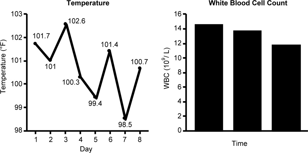
Case 1 is a 72‐year‐old woman admitted with findings consistent with healthcare‐associated pneumonia (HCAP). Empiric therapy was initiated with vancomycin and piperacillin/tazobactam. Figure 1 provides the laboratory (white blood cell [WBC] counts) and body temperature data for the patient since she entered the hospital and began empiric antibiotic therapy 3 days earlier. The WBC counts suggest the patient is responding to the antibiotic regimen, as demonstrated by a progressive reduction over the time period. However, her counts were still elevated above normal at last measurement, suggesting an incompletely resolved infection at this time. In addition, the patient is still coughing, but has less sputum production, and has some energy to get up and move around. Crackles are apparent at the right lung base. The patient's fever curve has trended down, but still shows notable fever spikes, with a temperature maximum of 101.4F for the past 24 hours. Her blood pressure (135/84 mmHg), pulse (74 bpm), and respiratory rate (14 breaths per minute) are normal, with slightly decreased oxygen saturation (94%) on room air, although improved from initial examination 3 days earlier (92%). The blood culture shows no growth; the sputum culture simply shows oropharyngeal flora. In other words, the culture results have not isolated a causative pathogen. In addition to vancomycin and piperacillin/tazobactam, the patient continues to receive her usual medications for a past history of myocardial infarction (low‐dose aspirin, metoprolol) and hypertension (enalapril, furosemide).
HCAP is a common infection often requiring initial empiric therapy with a broad‐spectrum regimen that covers possible involvement of resistant bacteria. As such, HCAP frequently provides excellent opportunities for de‐escalation. Figure 2 presents the general strategy from the 2005 American Thoracic Society and Infectious Diseases Society of America (ATS/IDSA) guidelines for the management of HCAP, hospital‐acquired pneumonia (HAP), or ventilator‐associated pneumonia (VAP).22 According to the guidelines, HCAP, HAP, and VAP should be similarly managed. Broad‐spectrum initial empiric antibiotic therapy is recommended for patients with late‐onset disease or those with risk factors for MDR pathogens (including high prevalence of resistance based on local antibiograms), while limited‐spectrum antibiotic therapy is recommended for all other patients. Note that consideration of de‐escalation or streamlining of initial therapy begins 2‐3 days after initiation of therapy. Data that should be reviewed prior to instituting de‐escalation include blood cultures and respiratory cultures, as well as the clinical status of the patient. The adequacy of respiratory samples used for culturing should factor into the decision‐making process. For example, in patients who are not intubated or mechanically ventilated, it can be challenging to obtain a quality respiratory specimen for culture. If clinicians are uncertain as to the quality of the respiratory specimen that was cultured, then de‐escalation decisions should be based more on the clinical status of the patient.
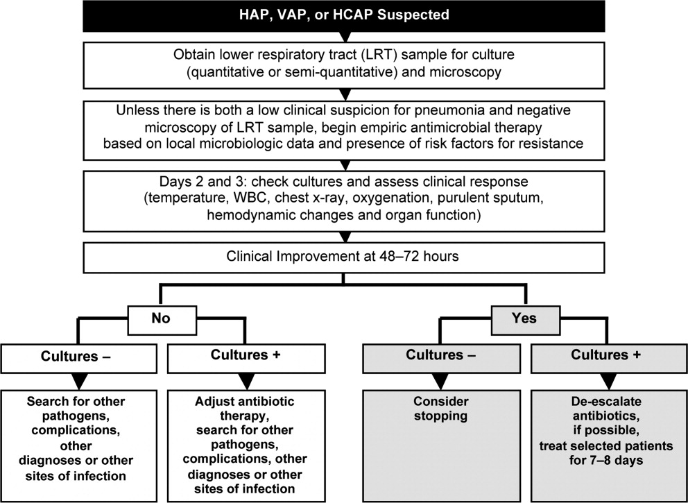
The clinical status of the patient, 2 days after beginning treatment, and culture results are critical in guiding the de‐escalation process.9, 22 The ATS/IDSA guidelines recommend serial assessments of clinical parameters to define the response to initial empiric therapy. If the therapy regimen is effective, an improvement in clinical response should be apparent within 2‐3 days of its initiation.22 Hence, no change in antimicrobial therapy should be undertaken before 3 days, unless there is evidence of rapid deterioration in clinical status or infectious diseases experts recommend a change. With respect to culture results, failure to isolate a group of MDR pathogens for which initial broad‐spectrum empiric therapy was selected affords an opportunity to now streamline therapy or treat with a more narrow‐spectrum regimen.9 Similarly, isolation of a particular pathogen can guide treatment modifications (when necessary), while a negative culture raises the possibility of terminating antimicrobial therapy, provided the culture was collected before initiating therapy. Confidence in this latter decision is bolstered when the patient exhibits rapid improvement in clinical status that is backed by radiographic resolution of lung abnormalities, or an alternative diagnosis has been established for which antimicrobial therapy is not indicated.9
At this stage in the process3 days after initiating empiric therapy, and with culture results in hand and evidence of clinical improvementthe first decision or question is whether antimicrobial therapy can be stopped altogether, ie, do the current data suggest a noninfectious diagnosis (eg, pulmonary embolism, atelectasis) or that bacterial pneumonia is unlikely or has resolved. A 2000 study by Singh et al. highlighted the feasibility of using operational criteria in the form of clinical pulmonary infection score (CPIS) to decide whether to terminate or shorten the duration of initial empiric antibiotic therapy for suspected VAP.23 More specifically, patients with pulmonary infiltrates but a low likelihood of pneumonia (CPIS 6) were randomized to receive either standard antibiotic therapy or ciprofloxacin monotherapy. The situation was re‐evaluated at 3 days, and ciprofloxacin therapy was discontinued if the CPIS remained 6. Results showed no difference in mortality between the ciprofloxacin and standard therapy groups, despite shorter duration of therapy for the former, together with lower antimicrobial exposure and costs for the ciprofloxacin group. (Use of the CPIS to shorten the duration of empiric therapy and limit antimicrobial exposure is discussed in greater detail in the corresponding article in this supplement by Dr File.) Having said that, the case study before us describes a patient with pneumonia by clinical criteria who has responded to broad‐spectrum therapy. Alternative noninfectious diagnoses are not apparent, and even though cultures have returned without significant growth, the patient should continue to receive antimicrobial treatment. The question now is whether to de‐escalate/streamline to a more narrow‐spectrum regimen, or continue the current one.
De‐escalation often targets antimicrobials that provide unnecessarily broad coverage, eg, those with antipseudomonal activity (particularly antipseudomonal carbapenems) and/or agents with activity against methicillin‐resistant Staphylococcus aureus (MRSA). In the absence of definitive culture results isolating a particular pathogen(s), decisions regarding which antibiotics to stop or change often depends, in large part, on patient characteristics (eg, history of prior infection with resistant pathogens, as well as drug allergies or renal insufficiency) and local antibiograms indicating the prevalence and antimicrobial susceptibility of different pneumonia pathogens in the hospital at large or particular wards within the hospital. However, negative culture results can also be useful in guiding subsequent therapy decisions or modifications. In the present case, MRSA was not grown from any cultures, and there was no evidence of Gram‐positive cocci clusters with Gram staining. This suggests that vancomycin should be stopped, and antimicrobial therapy continued with a single antibiotic or antibiotic product that does include MRSA coverage. The question then is whether to continue piperacillin/tazobactam or replace it with another antibiotic.
Because Pseudomonas aeruginosa was not isolated, the clinician might consider streamlining piperacillin/tazobactam to an antibiotic with less pseudomonal and anaerobic coverage, possibly a nonpseudomonal third‐generation cephalosporin or nonpseudomonal carbapenem, such as ertapenem. Given the activity of piperacillin/tazobactam against aerobic Gram‐positive and Gram‐negative pathogens, continuing piperacillin‐tazobactam as single‐agent therapy would also be a viable alternative. However, in the spirit of stewardship and lack of need for pseudomonal coverage, a decision was made to replace piperacillin/tazobactam with ceftriaxone. Ceftriaxone is a nonpseudomonal third‐generation cephalosporin with activity against most other Gram‐negative bacteria. Note that in this case, only oropharyngeal flora grew from the respiratory culture, and the blood culture was negative. However, if a pathogen had grown from either respiratory or blood cultures, then single‐agent therapy could have been used to target that specific pathogen. For example, if Klebsiella spp susceptible to ceftriaxone was isolated from the respiratory culture, then ceftriaxone would have been the obvious choice. If MRSA was isolated, then vancomycin (or another appropriate active agent, such as linezolid or clindamycin) could be administered as a single agent.
CASE 2: INTRA‐ABDOMINAL INFECTION (DIVERTICULITIS)
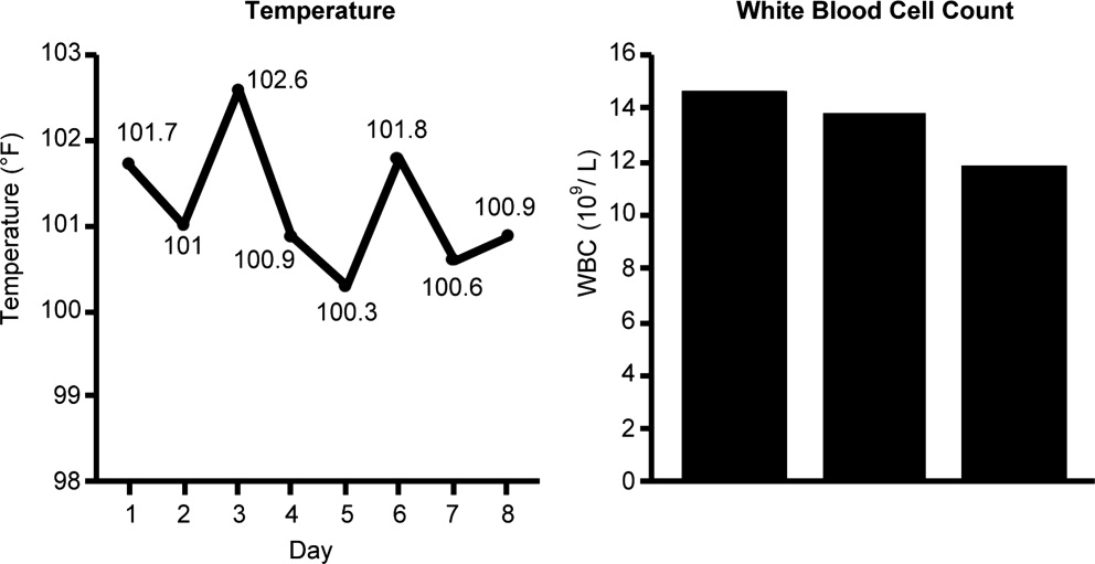
Case 2 is a 56‐year‐old woman who presents with a diverticular abscess and walled off perforation. Interventional radiology inserts a drain, and the patient is treated with ciprofloxacin plus metronidazole. This regimen is consistent with guidelines from the Surgical Infection Society and IDSA for initial empiric treatment of complicated intra‐abdominal infection of mild‐to‐moderate severity.24 On day 3 following hospital admission and initiation of empiric therapy, the patient seems to show treatment response, as evidenced by downward trends in body temperature and WBC count (Figure 3). However, although the body temperature measures are trending in the right direction, there is still concern about continuing fever spikes and fever at last measure (100.9F). In addition, the WBC count is still elevated, though improving. The patient's blood pressure has normalized (112/72 mmHg vs 84/58 mmHg at admission), and oxygen saturation (98%) measures are normal. The patient's lungs are clear, and her abdominal examination results are improving, though there is still some diffuse tenderness. Microbiological data show blood cultures with no growth, and isolation of Gram‐negative rods from cultures of the abdominal abscess.
We now have preliminary microbiological data for a patient who remains febrile and has continuing abdominal tenderness, but who is otherwise clinically stable. Can her antimicrobial regimen be de‐escalated at this point, based on what is currently known? When managing a patient after the first 3 or 4 days of empiric treatment, it is important to realize that the patient's condition with regards to infection might reflect issues unrelated to inadequate antimicrobial coverage. If the patient's clinical status has not improved, or if he or she remains febrile even 3 or 4 days into therapy, the clinician should not automatically assume the lack of improvement is due to antibiotic failure. At this point, it is important to consider possible nonantibiotic causes of persistent clinical abnormalities and fever, and for the case here, one possibility is inadequate abscess drainage. The patient should be evaluated with abdominal imaging to ascertain whether the abscess is being adequately drained. With respect to antimicrobial therapy, the patient's blood pressure has stabilized, and her fever is trending downward. In many cases, a lingering fever such as the one observed here, in the context of improving WBC counts and clinical stabilization, may reflect inadequate mechanical drainage of the abscess. Certainly the antimicrobial therapy should not be broadened at this time, and consideration should be given to de‐escalation based on the available microbiological data.
If a type of pathogenic organism is preliminarily identified from culture, but the exact identification of the organism is pending, adjustments of therapy can still be made. Adjustments can also be made based on what is not growing. In this case, the abscess culture has grown Gram‐negative rods, but no Gram‐positive organisms. Hence, continued coverage of Gram‐negative organisms is warranted. In addition, anaerobes often will not readily grow in clinical cultures, and because anaerobes are frequent co‐pathogens, it is appropriate to continue to provide anaerobic coverage. Based on this information, continuation of both ciprofloxacin (for aerobic Gram‐negative coverage) and metronidazole (to cover for anaerobic bacteria) is appropriate in the present case. In other words, the initial empiric therapy should be continued until subsequent culture identifies a particular pathogen, at which time the therapy can be streamlined.
Now, 1 day later (day 4 of hospital admission and empiric therapy), the patient's clinical status is essentially unchangedexcept for a spike in fever to 103.2F. The WBC count is unchanged. Moreover, additional abscess culture data are available, showing definitive identification of an extended‐spectrum ‐lactamase (ESBL)‐producing Escherichia coli organism. The blood culture is still negative. The first observation is that ESBL‐producing E coli is a relatively unusual pathogen in a community‐based infection. However, the patient here did have risk factors for antibiotic‐resistant pathogens, notably prior antimicrobial therapy as an outpatient. It is also important to recognize that community‐acquired infections with ESBL‐producing bacteria (mostly isolated from the urinary tract) have been reported in many parts of the world, and even in some parts of the United States.25
Based on these additional microbiological data, the patient was switched to treatment with ertapenem, a nonpseudomonal carbapenem with activity against ESBL‐producing Enterobacteriaceae.26 In addition, ertapenem, and other carbapenems, have excellent activity against anaerobes,26 and it is prudent to continue coverage for anaerobes even though anaerobes were not grown in the culture. As mentioned above, these organisms are difficult to grow in clinical culture, and they are common pathogens or co‐pathogens in intra‐abdominal infections. Carbapenems are widely regarded as the antimicrobials of choice for treatment of serious, invasive infections with ESBL‐producing bacteria.27 Furthermore, by choosing a nonpseudomonal carbapenem, compared with an antipseudomonal carbapenem, the new antibiotic regimen provides coverage of the isolated ESBL‐producing E coli organismas well as covering possible anaerobe involvementwithout exposing host bacteria to unnecessarily broad antipseudomonal activity. Cephalosporins, monobactams, and fluoroquinolones are generally not active against ESBL‐producing Enterobacteriaceae, and ‐lactam/‐lactamase inhibitor combinations (eg, ampicillin/sulbactam, piperacillin/tazobactam) do not have reliable activity in serious, high inoculum infections caused by ESBL‐producing Enterobacteriaceae.27
CASE 3: CENTRAL LINE‐ASSOCIATED BLOODSTREAM INFECTION
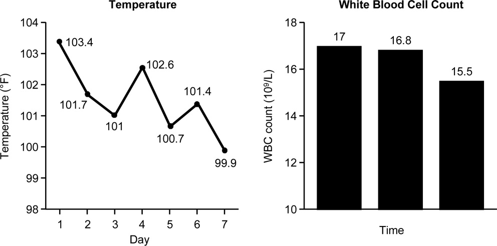
Case 3 is a 56‐year‐old man who presented to the hospital emergency department with status epilepticus. He was intubated, had a central line placed in the internal jugular vein, and was admitted to the intensive care unit (ICU). The seizure was successfully broken by aggressive treatment with repeated intravenous dosing of lorazepam and loading with fosphenytoin. Empiric antibiotic therapy was initiated with vancomycin and piperacillin/tazobactam on day 5, after spiking a fever of 103.4F. No clear source of the fever was identified. While in the ICU with a central line in place, 2 sets of blood cultures were drawn. Now on hospital day 6, the patient is still spiking fever, although the fever trend appears to be decreasing. The patient is hemodynamically stable, with no other abnormal findings (besides persistent fever) on physical examination. WBC count remains elevated, and both sets of blood cultures are notable for growth of Gram‐positive cocci.
Bloodstream infection is a serious condition in hospitalized patients that is associated with significant morbidity and mortality.28 Patients with suspected bloodstream infection typically receive empiric broad‐spectrum antimicrobial therapy, and are thus good candidates for de‐escalation based on subsequent clinical status and blood culture results. Because of the seriousness of bloodstream infection, healthcare workers are sometimes hesitant to de‐escalate initial empiric therapy, even when cultures isolate a pathogen susceptible to narrower‐spectrum agents, particularly if the patient appears to be improving on such therapy. This is true for various serious hospital or healthcare‐associated infections,16, 29 but particularly for bloodstream infections. Moreover, when central line‐associated bloodstream infection (CLABSI) is suspected, the most important initial intervention is to remove the infected central venous catheter. For a patient with a short‐term catheter and a CLABSI due to Gram‐negative bacilli, S aureus (which appears to be a likely pathogen for the case patient here), enterococci, fungi, or mycobacteria, the 2009 IDSA guidelines for management of intravascular catheter‐related infections recommend catheter removal.30 Catheter removal is even more important than antibiotic coverage; this point cannot be stressed enough. In some extreme cases, when the line cannot be removed for clinical reasons, antibiotic lock therapy can be used to supplement systemic antimicrobial therapy.30 This involves instilling a high antibiotic solution into the catheter lumen for a period of time in order to sterilize the lumen and prevent biofilm formation.31
The first step taken for the patient here was to remove the central venous catheter. Then, turning to the preliminary culture data, there is evidence for Gram‐positive cocci in the patient's blood. The blood culture did not grow any Gram‐negative organisms. Gram‐positive cocci (coagulase‐negative staphylococci, S aureus [methicillin‐susceptible or MRSA]) are the most common causes of CLABSI.32 Can the physician de‐escalate antibiotic therapy in this patient with CLABSI based on the preliminary information? Yes. The information is solid enough to suggest removal of the catheter which was providing coverage for Gram‐negative bacteria (piperacillin/tazobactam), while continuing vancomycin for coverage of possible MRSA, pending further review, ie, until the Gram‐positive cocci are speciated. Rapid diagnostic methods, including polymerase chain reaction (PCR) and nucleic acid probes, can be used to provide more information about certain pathogens (such as MRSA33, 34) before final culture and susceptibility results are available, but these are not routinely available in many clinical microbiology laboratories. Furthermore, these newer technologies remain fairly expensive.
Revisiting the patient 1 day later (hospital day 7), after narrowing the initial combination antibiotic regimen to vancomycin monotherapy, the physical examination indicates the patient is clinically stable, with continued improvement in fever and WBC count (Figure 4). Blood culture analysis now isolates methicillin‐susceptible S aureus (MSSA). Methicillin resistance mediates resistance to all ‐lactams, including carbapenems, greatly limiting treatment options. Vancomycin is the most commonly utilized antibiotic for the treatment of MRSA, and the recent clinical practice guidelines from the IDSA recommend either vancomycin or daptomycin for management of MRSA bacteremia in adult patients.35 However, antistaphylococcal penicillins and first‐generation cephalosporins are the antibiotics of choice for MSSA infections, and particularly for MSSA bloodstream infections.
The activity provided by vancomycin (or daptomycin) is overly broad if MSSA is involved, and importantly, it is not as effective as treatment with an antistaphylococcal penicillin or first‐generation cephalosporin. A recent study by Stryjewski et al., of hemodialysis patients with MSSA bacteremia, reported a higher proportion of treatment failure with vancomycin versus first‐generation cephalosporin therapy (31% vs 13%; P = 0.02).36 Furthermore, multivariate analysis identified vancomycin (vs first‐generation cephalosporin) use as a significant independent predictor of treatment failure (odds ratio [OR], 3.53; 95% confidence interval [CI], 1.1513.45; P = 0.04). Similarly, Chang et al. reported nafcillin, an antistaphylococcal penicillin, was superior to vancomycin in preventing bacteriologic failure (persistent failure and/or relapse) in patients with MSSA bacteremia (0% vs 19%; P = 0.058), and used multivariate analysis to identify vancomycin as a significant independent predictor of relapse (OR, 6.5; 95% CI, 1.052.8; P 0.05).37 Another recent study by Lodise et al. reported that initial empiric therapy with vancomycin for endocarditis caused by MSSA was associated with a higher infection‐related mortality rate than initial empiric therapy with a ‐lactam‐containing regimen (39% vs 11%; P = 0.005).38 The negative impact of initial treatment with vancomycin persisted even in patients switched to a ‐lactam therapy after culture results became available.
Hence, if a patient is being treated with vancomycin for a bloodstream (or other) infection due to MSSA, the therapy is suboptimal. In such a scenariowhich corresponds to that for the case patient herevancomycin should be discontinued and replaced with an antistaphylococcal penicillin or first‐generation cephalosporin. Many times, clinicians are resistant to terminating vancomycin and de‐escalating to antistaphylococcal penicillin/first‐generation cephalosporin therapy in a patient with bacteremia who is apparently responding to vancomycin. However, as the studies just reviewed make clear, not only is vancomycin treatment overly broad for the circumstance, it is also suboptimal and does not represent best clinical practice or patient care. Furthermore, continuing vancomycin in this situation unnecessarily exposes the patient to possible renal toxicity, particularly when aggressive dosing or prolonged vancomycin treatment is involved.39 Because of these issues and concerns, case 3 was de‐escalated from vancomycin to cefazolin, a first‐generation cephalosporin. One word of caution, however, is that there is some controversy over using cefazolin in patients with S aureus native valve endocarditis, given the possibility of a Type A ‐lactamase‐producing species causing cefazolin degradation.40 As a result, the clinician should first rule out endocarditis in the patient here before proceeding with cefazolin therapy. Another alternative would be to use an antistaphylococcal penicillin, such as nafcillin.
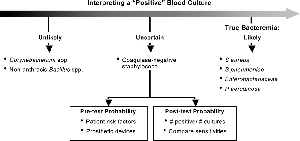
Finally, when dealing with bacteremia, and particularly when dealing with a possible CLABSI, the issue of potential culture contamination needs to be seriously considered and answered. Treating an actual infection, not what appears to be an infection because of culture contamination, is particularly important when dealing with possible CLABSI, because coagulase‐negative staphylococci (CoNS) are the most common cause of these types of infections,32 and CoNS are also frequent blood‐culture contaminants.41 Therefore, one needs to determine whether a blood culture growing a CoNS represents true bacteremia or simply contaminationwhich will obviously impact de‐escalation decisions.
In addition, when determining whether a blood culture is truly positive and clinically significant, it is important to consider whether the isolated pathogens are unlikely to be contaminants, likely to be contaminants, or the situation is unclear. A 2000 study by Kim et al.42 suggested that, among patients with 2 positive blood cultures for CoNS, routine identification of CoNS species and genotyping selected isolates using pulsed‐field gel electrophoresis may improve the process of discriminating contaminants from pathogens. Various additional factors need to be weighed when trying to interpret CoNS blood culture results, including patient risk factors, presence of prosthetic devices, number of blood cultures and number positive, and the antimicrobial sensitivity patterns of different isolates. For example, if the sensitivity patterns of 2 CoNS strains isolated from a patient are the same, the likelihood is increased that they represent true pathogens rather than contaminants. Figure 5 presents a schematic of this general approach.42
CONCLUSIONS
De‐escalation is a critical component of antimicrobial stewardship. As the prevalence of antimicrobial resistance grows in the hospital and community, de‐escalation will have an increasingly important role in limiting the further emergence of antimicrobial resistance. Pneumonia, intra‐abdominal infection, and bloodstream infection are commonly managed in the hospital setting. Each of these infection types presents excellent opportunities for de‐escalation, and each presents unique challenges and caveats. Concerted efforts must be made by clinicians and stewardship personnel to de‐escalate as soon as possible, based on culture results and clinical status. Although not discussed here, successful de‐escalation programs utilize structured process, guidelines, and algorithms to consistently implement de‐escalation efforts. These tools of implementation are more fully discussed in the corresponding article in this supplement by Dr Rosenberg.
- .Appropriate empirical antibacterial therapy for nosocomial infections: getting it right the first time.Drugs.2003;63:2157–2168.
- ,,, et al.Mortality and morbidity attributable to inadequate empirical antimicrobial therapy in patients admitted to the ICU with sepsis: a matched cohort study.J Antimicrob Chemother.2008;61:436–441.
- ,,,,,.Predictors of 30‐day mortality and hospital costs in patients with ventilator‐associated pneumonia attributed to potentially antibiotic‐resistant gram‐negative bacteria.Chest.2008;134:281–287.
- ,,,.Inadequate antimicrobial treatment of infections: a risk factor for hospital mortality among critically ill patients.Chest.1999;115:462–474.
- ,,, et al.Impact of BAL data on the therapy and outcome of ventilator‐associated pneumonia.Chest.1997;111:676–685.
- ,,,,,.Emergence of antibiotic‐resistant bacteria in cases of peritonitis after intraabdominal surgery affects the efficacy of empirical antimicrobial therapy.Clin Infect Dis.1996;23:486–494.
- ,,,,.Antimicrobial therapy escalation and hospital mortality among patients with health‐care‐associated pneumonia: a single‐center experience.Chest.2008;134:963–968.
- ,.De‐escalation in lower respiratory tract infections.Curr Opin Pulm Med.2006;12:364–368.
- .The importance of de‐escalating antimicrobial therapy in patients with ventilator‐associated pneumonia.Semin Respir Crit Care Med.2006;27:45–50.
- .Impact of antibiotic resistance in gram‐negative bacilli on empirical and definitive antibiotic therapy.Clin Infect Dis.2008;47(suppl 1):S14–S20.
- ,,, et al.Infectious Diseases Society of America and the Society for Healthcare Epidemiology of America guidelines for developing an institutional program to enhance antimicrobial stewardship.Clin Infect Dis.2007;44:159–177.
- Bad Bugs, No Drugs: As Antibiotic R46:155–164.
- ,,, et al.De‐escalation therapy rates are significantly higher by bronchoalveolar lavage than by tracheal aspirate.Intensive Care Med.2007;33:1533–1540.
- ,,, et al.Clinical characteristics and treatment patterns among patients with ventilator‐associated pneumonia.Chest.2006;129:1210–1218.
- ,,, et al.Empiric broad‐spectrum antibiotic therapy of nosocomial pneumonia in the intensive care unit: a prospective observational study.Crit Care.2006;10:R78.
- ,,,.Does de‐escalation of antibiotic therapy for ventilator‐associated pneumonia affect the likelihood of recurrent pneumonia or mortality in critically ill surgical patients?J Trauma.2009;66:1343–1348.
- ,,, et al.Ventilator‐associated pneumonia: breaking the vicious circle of antibiotic overuse.Crit Care Med.2007;35:379–385; quiz 386.
- ,,,,.Adjustment of antibiotic treatment according to the results of blood cultures leads to decreased antibiotic use and costs.J Antimicrob Chemother.2006;57:326–330.
- ,,,,.Diagnostic yield of blood cultures from antibiotic‐naive and antibiotically treated patients with haematological malignancies and high‐risk neutropenia.Scand J Infect Dis.2009;41:650–655.
- ,,, et al.Diagnostic accuracy of protected specimen brush and bronchoalveolar lavage in nosocomial pneumonia: impact of previous antimicrobial treatments.Crit Care Med.1998;26:236–244.
- Guidelines for the management of adults with hospital‐acquired, ventilator‐associated, and healthcare‐associated pneumonia.Am J Respir Crit Care Med.2005;171:388–416.
- ,,,,.Short‐course empiric antibiotic therapy for patients with pulmonary infiltrates in the intensive care unit. A proposed solution for indiscriminate antibiotic prescription.Am J Respir Crit Care Med.2000;162:505–511.
- ,,, et al.Diagnosis and management of complicated intra‐abdominal infection in adults and children: guidelines by the Surgical Infection Society and the Infectious Diseases Society of America.Clin Infect Dis.2010;50:133–164.
- ,,.Molecular characteristics of extended‐spectrum beta‐lactamase‐producing Escherichia coli from the Chicago area: high prevalence of ST131 producing CTX‐M‐15 in community hospitals.Int J Antimicrob Agents.2010;36:19–23.
- ,.Ertapenem: a review of its use in the treatment of bacterial infections.Drugs.2005;65:2151–2178.
- .Infections with extended‐spectrum beta‐lactamase‐producing Enterobacteriaceae: changing epidemiology and drug treatment choices.Drugs.2010;70:313–333.
- ,.Bloodstream infection in the ICU.Infect Dis Clin North Am.2009;23:557–569.
- ,,,,.Insights from the Society of Infectious Diseases Pharmacists on antimicrobial stewardship guidelines from the Infectious Diseases Society of America and the Society for Healthcare Epidemiology of America.Pharmacotherapy.2009;29:593–607.
- ,,, et al.Clinical practice guidelines for the diagnosis and management of intravascular catheter‐related infection: 2009 Update by the Infectious Diseases Society of America.Clin Infect Dis.2009;49:1–45.
- ,,,,,.Antimicrobial lock solutions for the prevention of infections associated with intravascular catheters in patients undergoing hemodialysis: systematic review and meta‐analysis of randomized, controlled trials.Clin Infect Dis.2008;47:83–93.
- ,.Catheter‐related bloodstream infections: catheter management according to pathogen.Int J Antimicrob Agents.2010;36(suppl 2):S26–S32.
- ,,,.Detection of methicillin‐resistant Staphylococcus aureus (MRSA) in specimens from various body sites: performance characteristics of the BD GeneOhm MRSA assay, the Xpert MRSA assay, and broth‐enriched culture in an area with a low prevalence of MRSA infections.J Clin Microbiol.2010;48:3882–3887.
- ,,,,.Clinical validation of the molecular BD GeneOhm StaphSR assay for direct detection of Staphylococcus aureus and methicillin‐resistant Staphylococcus aureus in positive blood cultures.J Clin Microbiol.2007;45:2191–2196.
- ,,, et al.Clinical practice guidelines by the Infectious Diseases Society of America for the treatment of methicillin‐resistant Staphylococcus aureus infections in adults and children.Clin Infect Dis.2011;52:e18–e55.
- ,,, et al.Use of vancomycin or first‐generation cephalosporins for the treatment of hemodialysis‐dependent patients with methicillin‐susceptible Staphylococcus aureus bacteremia.Clin Infect Dis.2007;44:190–196.
- ,,, et al.Staphylococcus aureus bacteremia: recurrence and the impact of antibiotic treatment in a prospective multicenter study.Medicine (Baltimore).2003;82:333–339.
- ,,,.Impact of empirical‐therapy selection on outcomes of intravenous drug users with infective endocarditis caused by methicillin‐susceptible Staphylococcus aureus.Antimicrob Agents Chemother.2007;51:3731–3733.
- ,,,,.A retrospective analysis of possible renal toxicity associated with vancomycin in patients with health care‐associated methicillin‐resistant Staphylococcus aureus pneumonia.Clin Ther.2007;29:1107–1115.
- ,,.Relapse of type A beta‐lactamase‐producing Staphylococcus aureus native valve endocarditis during cefazolin therapy: revisiting the issue.Clin Infect Dis.2003;37:1194–1198.
- ,.The origin of coagulase‐negative staphylococci isolated from blood cultures.J Hosp Infect.1995;30:217–223.
- ,,, et al.Determining the significance of coagulase‐negative staphylococci isolated from blood cultures at a community hospital: a role for species and strain identification.Infect Control Hosp Epidemiol.2000;21:213–217.
Two conflicting aims collide when choosing initial empiric therapy for patients with a potential life‐threatening infection. On the one hand, the clinical picture and seriousness of the suspected infectionsometimes with a multi‐drug resistant (MDR) pathogenpoint to the need for immediate empiric therapy with a broad‐spectrum regimen covering the most likely pathogens. This getting it right the first time approach1 is clearly a reasonable one given the significant negative impact of inappropriate or inadequate initial therapy on patient outcomes and costs,24 and the apparent inability to remedy the initial error by subsequent antimicrobial regimen adjustment.57 On the other hand, use of a broad‐spectrum regimen increases the risk of emergent antimicrobial‐resistant pathogens, with potential harm for the immediate patient and all subsequent patients who become exposed and infected with the resistant pathogen. Hence, the aim of optimizing initial empiric therapy comes into conflict with an important aim of antimicrobial stewardship, namely, to use antimicrobials in a manner that does not excessively promote development or selection of antimicrobial‐resistant pathogens.
The de‐escalation strategy is an approach that attempts to balance these conflicting aims by providing optimal initial patient management without inordinately promoting development of antimicrobial resistance. As discussed more fully in the corresponding supplement article by Dr Syndman, the first part of this strategy involves collecting cultures from suitable patients prior to initiating broad‐spectrum empiric antimicrobial therapy designed to cover the most likely pathogenic microorganisms, based on local patterns of prevalence and susceptibility, and the presence of risk factors for infection with drug‐resistant species.810 The second critical step involves modification of initial empiric therapy (when warranted) based on clinical status and when culture results are available.810 In this manner, the initial broad‐spectrum regimen can often be streamlined or de‐escalated to a more narrow‐spectrum regimen or, in some cases, terminated when negative cultures suggest no infection. Frequently, initial combination therapy can be replaced by monotherapy targeting the pathogenic organism identified in culture. Sometimes culture results indicate that initial empiric therapy was inappropriate/emnadequate and requires replacement or other modification. Thus, by modifying empiric antimicrobial therapy on the basis of culture results and clinical criteria, the de‐escalation strategy enables more effective targeting of the causative pathogen(s), elimination of redundant therapy, a decrease in antimicrobial pressure for emergence of resistance, and cost savings.10, 11 Decreasing the number of antimicrobial agents and/or the spectrum of coverage is also expected to decrease the risk of adverse events, drugdrug interactions, and Clostridium difficile‐associated disease.12, 13 A number of studies have demonstrated that de‐escalation of initially appropriate therapy can be successfully accomplished with either improved outcomes14, 15 or with comparable effectiveness as continued initial therapy,1618 but with reduced antimicrobial exposure and costs.19
The timing of streamlining or other modification of initial empiric therapy typically occurs when microbiological culture results become available. Assuming blood or other relevant tissue cultures were obtained prior to initiating empiric therapy, this means de‐escalation or other modifications of initial therapy generally occurs 24 days after hospitalization and/or the beginning of empiric therapy. If rapid diagnostic tests are used to identify or rule out particular pathogens, then de‐escalation may occur slightly sooner. In addition to culture results, observation of the patient in the hospital setting and improved clarity as to his or her clinical status also affect the decision about whether and how to modify the initial empiric antimicrobial regimen. The clinical scenario of the patient and his or her response to initial antimicrobial therapy is also typically clearer by day 3 of antibiotic therapy. If, for some reason, cultures were not obtained prior to beginning empiric therapy, then observations of clinical status and consideration of patient risk factors for resistant pathogens become predominant in the decision‐making process. With respect to the timing of culture attainment, this should occur prior to beginning antimicrobial therapy, because therapy may reduce culture yield and result in false negative or other misleading findings.20, 21
CASE 1: HEALTHCARE‐ASSOCIATED PNEUMONIA
Case 1 is a 72‐year‐old woman admitted with findings consistent with healthcare‐associated pneumonia (HCAP). Empiric therapy was initiated with vancomycin and piperacillin/tazobactam. Figure 1 provides the laboratory (white blood cell [WBC] counts) and body temperature data for the patient since she entered the hospital and began empiric antibiotic therapy 3 days earlier. The WBC counts suggest the patient is responding to the antibiotic regimen, as demonstrated by a progressive reduction over the time period. However, her counts were still elevated above normal at last measurement, suggesting an incompletely resolved infection at this time. In addition, the patient is still coughing, but has less sputum production, and has some energy to get up and move around. Crackles are apparent at the right lung base. The patient's fever curve has trended down, but still shows notable fever spikes, with a temperature maximum of 101.4F for the past 24 hours. Her blood pressure (135/84 mmHg), pulse (74 bpm), and respiratory rate (14 breaths per minute) are normal, with slightly decreased oxygen saturation (94%) on room air, although improved from initial examination 3 days earlier (92%). The blood culture shows no growth; the sputum culture simply shows oropharyngeal flora. In other words, the culture results have not isolated a causative pathogen. In addition to vancomycin and piperacillin/tazobactam, the patient continues to receive her usual medications for a past history of myocardial infarction (low‐dose aspirin, metoprolol) and hypertension (enalapril, furosemide).
HCAP is a common infection often requiring initial empiric therapy with a broad‐spectrum regimen that covers possible involvement of resistant bacteria. As such, HCAP frequently provides excellent opportunities for de‐escalation. Figure 2 presents the general strategy from the 2005 American Thoracic Society and Infectious Diseases Society of America (ATS/IDSA) guidelines for the management of HCAP, hospital‐acquired pneumonia (HAP), or ventilator‐associated pneumonia (VAP).22 According to the guidelines, HCAP, HAP, and VAP should be similarly managed. Broad‐spectrum initial empiric antibiotic therapy is recommended for patients with late‐onset disease or those with risk factors for MDR pathogens (including high prevalence of resistance based on local antibiograms), while limited‐spectrum antibiotic therapy is recommended for all other patients. Note that consideration of de‐escalation or streamlining of initial therapy begins 2‐3 days after initiation of therapy. Data that should be reviewed prior to instituting de‐escalation include blood cultures and respiratory cultures, as well as the clinical status of the patient. The adequacy of respiratory samples used for culturing should factor into the decision‐making process. For example, in patients who are not intubated or mechanically ventilated, it can be challenging to obtain a quality respiratory specimen for culture. If clinicians are uncertain as to the quality of the respiratory specimen that was cultured, then de‐escalation decisions should be based more on the clinical status of the patient.
The clinical status of the patient, 2 days after beginning treatment, and culture results are critical in guiding the de‐escalation process.9, 22 The ATS/IDSA guidelines recommend serial assessments of clinical parameters to define the response to initial empiric therapy. If the therapy regimen is effective, an improvement in clinical response should be apparent within 2‐3 days of its initiation.22 Hence, no change in antimicrobial therapy should be undertaken before 3 days, unless there is evidence of rapid deterioration in clinical status or infectious diseases experts recommend a change. With respect to culture results, failure to isolate a group of MDR pathogens for which initial broad‐spectrum empiric therapy was selected affords an opportunity to now streamline therapy or treat with a more narrow‐spectrum regimen.9 Similarly, isolation of a particular pathogen can guide treatment modifications (when necessary), while a negative culture raises the possibility of terminating antimicrobial therapy, provided the culture was collected before initiating therapy. Confidence in this latter decision is bolstered when the patient exhibits rapid improvement in clinical status that is backed by radiographic resolution of lung abnormalities, or an alternative diagnosis has been established for which antimicrobial therapy is not indicated.9
At this stage in the process3 days after initiating empiric therapy, and with culture results in hand and evidence of clinical improvementthe first decision or question is whether antimicrobial therapy can be stopped altogether, ie, do the current data suggest a noninfectious diagnosis (eg, pulmonary embolism, atelectasis) or that bacterial pneumonia is unlikely or has resolved. A 2000 study by Singh et al. highlighted the feasibility of using operational criteria in the form of clinical pulmonary infection score (CPIS) to decide whether to terminate or shorten the duration of initial empiric antibiotic therapy for suspected VAP.23 More specifically, patients with pulmonary infiltrates but a low likelihood of pneumonia (CPIS 6) were randomized to receive either standard antibiotic therapy or ciprofloxacin monotherapy. The situation was re‐evaluated at 3 days, and ciprofloxacin therapy was discontinued if the CPIS remained 6. Results showed no difference in mortality between the ciprofloxacin and standard therapy groups, despite shorter duration of therapy for the former, together with lower antimicrobial exposure and costs for the ciprofloxacin group. (Use of the CPIS to shorten the duration of empiric therapy and limit antimicrobial exposure is discussed in greater detail in the corresponding article in this supplement by Dr File.) Having said that, the case study before us describes a patient with pneumonia by clinical criteria who has responded to broad‐spectrum therapy. Alternative noninfectious diagnoses are not apparent, and even though cultures have returned without significant growth, the patient should continue to receive antimicrobial treatment. The question now is whether to de‐escalate/streamline to a more narrow‐spectrum regimen, or continue the current one.
De‐escalation often targets antimicrobials that provide unnecessarily broad coverage, eg, those with antipseudomonal activity (particularly antipseudomonal carbapenems) and/or agents with activity against methicillin‐resistant Staphylococcus aureus (MRSA). In the absence of definitive culture results isolating a particular pathogen(s), decisions regarding which antibiotics to stop or change often depends, in large part, on patient characteristics (eg, history of prior infection with resistant pathogens, as well as drug allergies or renal insufficiency) and local antibiograms indicating the prevalence and antimicrobial susceptibility of different pneumonia pathogens in the hospital at large or particular wards within the hospital. However, negative culture results can also be useful in guiding subsequent therapy decisions or modifications. In the present case, MRSA was not grown from any cultures, and there was no evidence of Gram‐positive cocci clusters with Gram staining. This suggests that vancomycin should be stopped, and antimicrobial therapy continued with a single antibiotic or antibiotic product that does include MRSA coverage. The question then is whether to continue piperacillin/tazobactam or replace it with another antibiotic.
Because Pseudomonas aeruginosa was not isolated, the clinician might consider streamlining piperacillin/tazobactam to an antibiotic with less pseudomonal and anaerobic coverage, possibly a nonpseudomonal third‐generation cephalosporin or nonpseudomonal carbapenem, such as ertapenem. Given the activity of piperacillin/tazobactam against aerobic Gram‐positive and Gram‐negative pathogens, continuing piperacillin‐tazobactam as single‐agent therapy would also be a viable alternative. However, in the spirit of stewardship and lack of need for pseudomonal coverage, a decision was made to replace piperacillin/tazobactam with ceftriaxone. Ceftriaxone is a nonpseudomonal third‐generation cephalosporin with activity against most other Gram‐negative bacteria. Note that in this case, only oropharyngeal flora grew from the respiratory culture, and the blood culture was negative. However, if a pathogen had grown from either respiratory or blood cultures, then single‐agent therapy could have been used to target that specific pathogen. For example, if Klebsiella spp susceptible to ceftriaxone was isolated from the respiratory culture, then ceftriaxone would have been the obvious choice. If MRSA was isolated, then vancomycin (or another appropriate active agent, such as linezolid or clindamycin) could be administered as a single agent.
CASE 2: INTRA‐ABDOMINAL INFECTION (DIVERTICULITIS)
Case 2 is a 56‐year‐old woman who presents with a diverticular abscess and walled off perforation. Interventional radiology inserts a drain, and the patient is treated with ciprofloxacin plus metronidazole. This regimen is consistent with guidelines from the Surgical Infection Society and IDSA for initial empiric treatment of complicated intra‐abdominal infection of mild‐to‐moderate severity.24 On day 3 following hospital admission and initiation of empiric therapy, the patient seems to show treatment response, as evidenced by downward trends in body temperature and WBC count (Figure 3). However, although the body temperature measures are trending in the right direction, there is still concern about continuing fever spikes and fever at last measure (100.9F). In addition, the WBC count is still elevated, though improving. The patient's blood pressure has normalized (112/72 mmHg vs 84/58 mmHg at admission), and oxygen saturation (98%) measures are normal. The patient's lungs are clear, and her abdominal examination results are improving, though there is still some diffuse tenderness. Microbiological data show blood cultures with no growth, and isolation of Gram‐negative rods from cultures of the abdominal abscess.
We now have preliminary microbiological data for a patient who remains febrile and has continuing abdominal tenderness, but who is otherwise clinically stable. Can her antimicrobial regimen be de‐escalated at this point, based on what is currently known? When managing a patient after the first 3 or 4 days of empiric treatment, it is important to realize that the patient's condition with regards to infection might reflect issues unrelated to inadequate antimicrobial coverage. If the patient's clinical status has not improved, or if he or she remains febrile even 3 or 4 days into therapy, the clinician should not automatically assume the lack of improvement is due to antibiotic failure. At this point, it is important to consider possible nonantibiotic causes of persistent clinical abnormalities and fever, and for the case here, one possibility is inadequate abscess drainage. The patient should be evaluated with abdominal imaging to ascertain whether the abscess is being adequately drained. With respect to antimicrobial therapy, the patient's blood pressure has stabilized, and her fever is trending downward. In many cases, a lingering fever such as the one observed here, in the context of improving WBC counts and clinical stabilization, may reflect inadequate mechanical drainage of the abscess. Certainly the antimicrobial therapy should not be broadened at this time, and consideration should be given to de‐escalation based on the available microbiological data.
If a type of pathogenic organism is preliminarily identified from culture, but the exact identification of the organism is pending, adjustments of therapy can still be made. Adjustments can also be made based on what is not growing. In this case, the abscess culture has grown Gram‐negative rods, but no Gram‐positive organisms. Hence, continued coverage of Gram‐negative organisms is warranted. In addition, anaerobes often will not readily grow in clinical cultures, and because anaerobes are frequent co‐pathogens, it is appropriate to continue to provide anaerobic coverage. Based on this information, continuation of both ciprofloxacin (for aerobic Gram‐negative coverage) and metronidazole (to cover for anaerobic bacteria) is appropriate in the present case. In other words, the initial empiric therapy should be continued until subsequent culture identifies a particular pathogen, at which time the therapy can be streamlined.
Now, 1 day later (day 4 of hospital admission and empiric therapy), the patient's clinical status is essentially unchangedexcept for a spike in fever to 103.2F. The WBC count is unchanged. Moreover, additional abscess culture data are available, showing definitive identification of an extended‐spectrum ‐lactamase (ESBL)‐producing Escherichia coli organism. The blood culture is still negative. The first observation is that ESBL‐producing E coli is a relatively unusual pathogen in a community‐based infection. However, the patient here did have risk factors for antibiotic‐resistant pathogens, notably prior antimicrobial therapy as an outpatient. It is also important to recognize that community‐acquired infections with ESBL‐producing bacteria (mostly isolated from the urinary tract) have been reported in many parts of the world, and even in some parts of the United States.25
Based on these additional microbiological data, the patient was switched to treatment with ertapenem, a nonpseudomonal carbapenem with activity against ESBL‐producing Enterobacteriaceae.26 In addition, ertapenem, and other carbapenems, have excellent activity against anaerobes,26 and it is prudent to continue coverage for anaerobes even though anaerobes were not grown in the culture. As mentioned above, these organisms are difficult to grow in clinical culture, and they are common pathogens or co‐pathogens in intra‐abdominal infections. Carbapenems are widely regarded as the antimicrobials of choice for treatment of serious, invasive infections with ESBL‐producing bacteria.27 Furthermore, by choosing a nonpseudomonal carbapenem, compared with an antipseudomonal carbapenem, the new antibiotic regimen provides coverage of the isolated ESBL‐producing E coli organismas well as covering possible anaerobe involvementwithout exposing host bacteria to unnecessarily broad antipseudomonal activity. Cephalosporins, monobactams, and fluoroquinolones are generally not active against ESBL‐producing Enterobacteriaceae, and ‐lactam/‐lactamase inhibitor combinations (eg, ampicillin/sulbactam, piperacillin/tazobactam) do not have reliable activity in serious, high inoculum infections caused by ESBL‐producing Enterobacteriaceae.27
CASE 3: CENTRAL LINE‐ASSOCIATED BLOODSTREAM INFECTION
Case 3 is a 56‐year‐old man who presented to the hospital emergency department with status epilepticus. He was intubated, had a central line placed in the internal jugular vein, and was admitted to the intensive care unit (ICU). The seizure was successfully broken by aggressive treatment with repeated intravenous dosing of lorazepam and loading with fosphenytoin. Empiric antibiotic therapy was initiated with vancomycin and piperacillin/tazobactam on day 5, after spiking a fever of 103.4F. No clear source of the fever was identified. While in the ICU with a central line in place, 2 sets of blood cultures were drawn. Now on hospital day 6, the patient is still spiking fever, although the fever trend appears to be decreasing. The patient is hemodynamically stable, with no other abnormal findings (besides persistent fever) on physical examination. WBC count remains elevated, and both sets of blood cultures are notable for growth of Gram‐positive cocci.
Bloodstream infection is a serious condition in hospitalized patients that is associated with significant morbidity and mortality.28 Patients with suspected bloodstream infection typically receive empiric broad‐spectrum antimicrobial therapy, and are thus good candidates for de‐escalation based on subsequent clinical status and blood culture results. Because of the seriousness of bloodstream infection, healthcare workers are sometimes hesitant to de‐escalate initial empiric therapy, even when cultures isolate a pathogen susceptible to narrower‐spectrum agents, particularly if the patient appears to be improving on such therapy. This is true for various serious hospital or healthcare‐associated infections,16, 29 but particularly for bloodstream infections. Moreover, when central line‐associated bloodstream infection (CLABSI) is suspected, the most important initial intervention is to remove the infected central venous catheter. For a patient with a short‐term catheter and a CLABSI due to Gram‐negative bacilli, S aureus (which appears to be a likely pathogen for the case patient here), enterococci, fungi, or mycobacteria, the 2009 IDSA guidelines for management of intravascular catheter‐related infections recommend catheter removal.30 Catheter removal is even more important than antibiotic coverage; this point cannot be stressed enough. In some extreme cases, when the line cannot be removed for clinical reasons, antibiotic lock therapy can be used to supplement systemic antimicrobial therapy.30 This involves instilling a high antibiotic solution into the catheter lumen for a period of time in order to sterilize the lumen and prevent biofilm formation.31
The first step taken for the patient here was to remove the central venous catheter. Then, turning to the preliminary culture data, there is evidence for Gram‐positive cocci in the patient's blood. The blood culture did not grow any Gram‐negative organisms. Gram‐positive cocci (coagulase‐negative staphylococci, S aureus [methicillin‐susceptible or MRSA]) are the most common causes of CLABSI.32 Can the physician de‐escalate antibiotic therapy in this patient with CLABSI based on the preliminary information? Yes. The information is solid enough to suggest removal of the catheter which was providing coverage for Gram‐negative bacteria (piperacillin/tazobactam), while continuing vancomycin for coverage of possible MRSA, pending further review, ie, until the Gram‐positive cocci are speciated. Rapid diagnostic methods, including polymerase chain reaction (PCR) and nucleic acid probes, can be used to provide more information about certain pathogens (such as MRSA33, 34) before final culture and susceptibility results are available, but these are not routinely available in many clinical microbiology laboratories. Furthermore, these newer technologies remain fairly expensive.
Revisiting the patient 1 day later (hospital day 7), after narrowing the initial combination antibiotic regimen to vancomycin monotherapy, the physical examination indicates the patient is clinically stable, with continued improvement in fever and WBC count (Figure 4). Blood culture analysis now isolates methicillin‐susceptible S aureus (MSSA). Methicillin resistance mediates resistance to all ‐lactams, including carbapenems, greatly limiting treatment options. Vancomycin is the most commonly utilized antibiotic for the treatment of MRSA, and the recent clinical practice guidelines from the IDSA recommend either vancomycin or daptomycin for management of MRSA bacteremia in adult patients.35 However, antistaphylococcal penicillins and first‐generation cephalosporins are the antibiotics of choice for MSSA infections, and particularly for MSSA bloodstream infections.
The activity provided by vancomycin (or daptomycin) is overly broad if MSSA is involved, and importantly, it is not as effective as treatment with an antistaphylococcal penicillin or first‐generation cephalosporin. A recent study by Stryjewski et al., of hemodialysis patients with MSSA bacteremia, reported a higher proportion of treatment failure with vancomycin versus first‐generation cephalosporin therapy (31% vs 13%; P = 0.02).36 Furthermore, multivariate analysis identified vancomycin (vs first‐generation cephalosporin) use as a significant independent predictor of treatment failure (odds ratio [OR], 3.53; 95% confidence interval [CI], 1.1513.45; P = 0.04). Similarly, Chang et al. reported nafcillin, an antistaphylococcal penicillin, was superior to vancomycin in preventing bacteriologic failure (persistent failure and/or relapse) in patients with MSSA bacteremia (0% vs 19%; P = 0.058), and used multivariate analysis to identify vancomycin as a significant independent predictor of relapse (OR, 6.5; 95% CI, 1.052.8; P 0.05).37 Another recent study by Lodise et al. reported that initial empiric therapy with vancomycin for endocarditis caused by MSSA was associated with a higher infection‐related mortality rate than initial empiric therapy with a ‐lactam‐containing regimen (39% vs 11%; P = 0.005).38 The negative impact of initial treatment with vancomycin persisted even in patients switched to a ‐lactam therapy after culture results became available.
Hence, if a patient is being treated with vancomycin for a bloodstream (or other) infection due to MSSA, the therapy is suboptimal. In such a scenariowhich corresponds to that for the case patient herevancomycin should be discontinued and replaced with an antistaphylococcal penicillin or first‐generation cephalosporin. Many times, clinicians are resistant to terminating vancomycin and de‐escalating to antistaphylococcal penicillin/first‐generation cephalosporin therapy in a patient with bacteremia who is apparently responding to vancomycin. However, as the studies just reviewed make clear, not only is vancomycin treatment overly broad for the circumstance, it is also suboptimal and does not represent best clinical practice or patient care. Furthermore, continuing vancomycin in this situation unnecessarily exposes the patient to possible renal toxicity, particularly when aggressive dosing or prolonged vancomycin treatment is involved.39 Because of these issues and concerns, case 3 was de‐escalated from vancomycin to cefazolin, a first‐generation cephalosporin. One word of caution, however, is that there is some controversy over using cefazolin in patients with S aureus native valve endocarditis, given the possibility of a Type A ‐lactamase‐producing species causing cefazolin degradation.40 As a result, the clinician should first rule out endocarditis in the patient here before proceeding with cefazolin therapy. Another alternative would be to use an antistaphylococcal penicillin, such as nafcillin.
Finally, when dealing with bacteremia, and particularly when dealing with a possible CLABSI, the issue of potential culture contamination needs to be seriously considered and answered. Treating an actual infection, not what appears to be an infection because of culture contamination, is particularly important when dealing with possible CLABSI, because coagulase‐negative staphylococci (CoNS) are the most common cause of these types of infections,32 and CoNS are also frequent blood‐culture contaminants.41 Therefore, one needs to determine whether a blood culture growing a CoNS represents true bacteremia or simply contaminationwhich will obviously impact de‐escalation decisions.
In addition, when determining whether a blood culture is truly positive and clinically significant, it is important to consider whether the isolated pathogens are unlikely to be contaminants, likely to be contaminants, or the situation is unclear. A 2000 study by Kim et al.42 suggested that, among patients with 2 positive blood cultures for CoNS, routine identification of CoNS species and genotyping selected isolates using pulsed‐field gel electrophoresis may improve the process of discriminating contaminants from pathogens. Various additional factors need to be weighed when trying to interpret CoNS blood culture results, including patient risk factors, presence of prosthetic devices, number of blood cultures and number positive, and the antimicrobial sensitivity patterns of different isolates. For example, if the sensitivity patterns of 2 CoNS strains isolated from a patient are the same, the likelihood is increased that they represent true pathogens rather than contaminants. Figure 5 presents a schematic of this general approach.42
CONCLUSIONS
De‐escalation is a critical component of antimicrobial stewardship. As the prevalence of antimicrobial resistance grows in the hospital and community, de‐escalation will have an increasingly important role in limiting the further emergence of antimicrobial resistance. Pneumonia, intra‐abdominal infection, and bloodstream infection are commonly managed in the hospital setting. Each of these infection types presents excellent opportunities for de‐escalation, and each presents unique challenges and caveats. Concerted efforts must be made by clinicians and stewardship personnel to de‐escalate as soon as possible, based on culture results and clinical status. Although not discussed here, successful de‐escalation programs utilize structured process, guidelines, and algorithms to consistently implement de‐escalation efforts. These tools of implementation are more fully discussed in the corresponding article in this supplement by Dr Rosenberg.
Two conflicting aims collide when choosing initial empiric therapy for patients with a potential life‐threatening infection. On the one hand, the clinical picture and seriousness of the suspected infectionsometimes with a multi‐drug resistant (MDR) pathogenpoint to the need for immediate empiric therapy with a broad‐spectrum regimen covering the most likely pathogens. This getting it right the first time approach1 is clearly a reasonable one given the significant negative impact of inappropriate or inadequate initial therapy on patient outcomes and costs,24 and the apparent inability to remedy the initial error by subsequent antimicrobial regimen adjustment.57 On the other hand, use of a broad‐spectrum regimen increases the risk of emergent antimicrobial‐resistant pathogens, with potential harm for the immediate patient and all subsequent patients who become exposed and infected with the resistant pathogen. Hence, the aim of optimizing initial empiric therapy comes into conflict with an important aim of antimicrobial stewardship, namely, to use antimicrobials in a manner that does not excessively promote development or selection of antimicrobial‐resistant pathogens.
The de‐escalation strategy is an approach that attempts to balance these conflicting aims by providing optimal initial patient management without inordinately promoting development of antimicrobial resistance. As discussed more fully in the corresponding supplement article by Dr Syndman, the first part of this strategy involves collecting cultures from suitable patients prior to initiating broad‐spectrum empiric antimicrobial therapy designed to cover the most likely pathogenic microorganisms, based on local patterns of prevalence and susceptibility, and the presence of risk factors for infection with drug‐resistant species.810 The second critical step involves modification of initial empiric therapy (when warranted) based on clinical status and when culture results are available.810 In this manner, the initial broad‐spectrum regimen can often be streamlined or de‐escalated to a more narrow‐spectrum regimen or, in some cases, terminated when negative cultures suggest no infection. Frequently, initial combination therapy can be replaced by monotherapy targeting the pathogenic organism identified in culture. Sometimes culture results indicate that initial empiric therapy was inappropriate/emnadequate and requires replacement or other modification. Thus, by modifying empiric antimicrobial therapy on the basis of culture results and clinical criteria, the de‐escalation strategy enables more effective targeting of the causative pathogen(s), elimination of redundant therapy, a decrease in antimicrobial pressure for emergence of resistance, and cost savings.10, 11 Decreasing the number of antimicrobial agents and/or the spectrum of coverage is also expected to decrease the risk of adverse events, drugdrug interactions, and Clostridium difficile‐associated disease.12, 13 A number of studies have demonstrated that de‐escalation of initially appropriate therapy can be successfully accomplished with either improved outcomes14, 15 or with comparable effectiveness as continued initial therapy,1618 but with reduced antimicrobial exposure and costs.19
The timing of streamlining or other modification of initial empiric therapy typically occurs when microbiological culture results become available. Assuming blood or other relevant tissue cultures were obtained prior to initiating empiric therapy, this means de‐escalation or other modifications of initial therapy generally occurs 24 days after hospitalization and/or the beginning of empiric therapy. If rapid diagnostic tests are used to identify or rule out particular pathogens, then de‐escalation may occur slightly sooner. In addition to culture results, observation of the patient in the hospital setting and improved clarity as to his or her clinical status also affect the decision about whether and how to modify the initial empiric antimicrobial regimen. The clinical scenario of the patient and his or her response to initial antimicrobial therapy is also typically clearer by day 3 of antibiotic therapy. If, for some reason, cultures were not obtained prior to beginning empiric therapy, then observations of clinical status and consideration of patient risk factors for resistant pathogens become predominant in the decision‐making process. With respect to the timing of culture attainment, this should occur prior to beginning antimicrobial therapy, because therapy may reduce culture yield and result in false negative or other misleading findings.20, 21
CASE 1: HEALTHCARE‐ASSOCIATED PNEUMONIA
Case 1 is a 72‐year‐old woman admitted with findings consistent with healthcare‐associated pneumonia (HCAP). Empiric therapy was initiated with vancomycin and piperacillin/tazobactam. Figure 1 provides the laboratory (white blood cell [WBC] counts) and body temperature data for the patient since she entered the hospital and began empiric antibiotic therapy 3 days earlier. The WBC counts suggest the patient is responding to the antibiotic regimen, as demonstrated by a progressive reduction over the time period. However, her counts were still elevated above normal at last measurement, suggesting an incompletely resolved infection at this time. In addition, the patient is still coughing, but has less sputum production, and has some energy to get up and move around. Crackles are apparent at the right lung base. The patient's fever curve has trended down, but still shows notable fever spikes, with a temperature maximum of 101.4F for the past 24 hours. Her blood pressure (135/84 mmHg), pulse (74 bpm), and respiratory rate (14 breaths per minute) are normal, with slightly decreased oxygen saturation (94%) on room air, although improved from initial examination 3 days earlier (92%). The blood culture shows no growth; the sputum culture simply shows oropharyngeal flora. In other words, the culture results have not isolated a causative pathogen. In addition to vancomycin and piperacillin/tazobactam, the patient continues to receive her usual medications for a past history of myocardial infarction (low‐dose aspirin, metoprolol) and hypertension (enalapril, furosemide).
HCAP is a common infection often requiring initial empiric therapy with a broad‐spectrum regimen that covers possible involvement of resistant bacteria. As such, HCAP frequently provides excellent opportunities for de‐escalation. Figure 2 presents the general strategy from the 2005 American Thoracic Society and Infectious Diseases Society of America (ATS/IDSA) guidelines for the management of HCAP, hospital‐acquired pneumonia (HAP), or ventilator‐associated pneumonia (VAP).22 According to the guidelines, HCAP, HAP, and VAP should be similarly managed. Broad‐spectrum initial empiric antibiotic therapy is recommended for patients with late‐onset disease or those with risk factors for MDR pathogens (including high prevalence of resistance based on local antibiograms), while limited‐spectrum antibiotic therapy is recommended for all other patients. Note that consideration of de‐escalation or streamlining of initial therapy begins 2‐3 days after initiation of therapy. Data that should be reviewed prior to instituting de‐escalation include blood cultures and respiratory cultures, as well as the clinical status of the patient. The adequacy of respiratory samples used for culturing should factor into the decision‐making process. For example, in patients who are not intubated or mechanically ventilated, it can be challenging to obtain a quality respiratory specimen for culture. If clinicians are uncertain as to the quality of the respiratory specimen that was cultured, then de‐escalation decisions should be based more on the clinical status of the patient.
The clinical status of the patient, 2 days after beginning treatment, and culture results are critical in guiding the de‐escalation process.9, 22 The ATS/IDSA guidelines recommend serial assessments of clinical parameters to define the response to initial empiric therapy. If the therapy regimen is effective, an improvement in clinical response should be apparent within 2‐3 days of its initiation.22 Hence, no change in antimicrobial therapy should be undertaken before 3 days, unless there is evidence of rapid deterioration in clinical status or infectious diseases experts recommend a change. With respect to culture results, failure to isolate a group of MDR pathogens for which initial broad‐spectrum empiric therapy was selected affords an opportunity to now streamline therapy or treat with a more narrow‐spectrum regimen.9 Similarly, isolation of a particular pathogen can guide treatment modifications (when necessary), while a negative culture raises the possibility of terminating antimicrobial therapy, provided the culture was collected before initiating therapy. Confidence in this latter decision is bolstered when the patient exhibits rapid improvement in clinical status that is backed by radiographic resolution of lung abnormalities, or an alternative diagnosis has been established for which antimicrobial therapy is not indicated.9
At this stage in the process3 days after initiating empiric therapy, and with culture results in hand and evidence of clinical improvementthe first decision or question is whether antimicrobial therapy can be stopped altogether, ie, do the current data suggest a noninfectious diagnosis (eg, pulmonary embolism, atelectasis) or that bacterial pneumonia is unlikely or has resolved. A 2000 study by Singh et al. highlighted the feasibility of using operational criteria in the form of clinical pulmonary infection score (CPIS) to decide whether to terminate or shorten the duration of initial empiric antibiotic therapy for suspected VAP.23 More specifically, patients with pulmonary infiltrates but a low likelihood of pneumonia (CPIS 6) were randomized to receive either standard antibiotic therapy or ciprofloxacin monotherapy. The situation was re‐evaluated at 3 days, and ciprofloxacin therapy was discontinued if the CPIS remained 6. Results showed no difference in mortality between the ciprofloxacin and standard therapy groups, despite shorter duration of therapy for the former, together with lower antimicrobial exposure and costs for the ciprofloxacin group. (Use of the CPIS to shorten the duration of empiric therapy and limit antimicrobial exposure is discussed in greater detail in the corresponding article in this supplement by Dr File.) Having said that, the case study before us describes a patient with pneumonia by clinical criteria who has responded to broad‐spectrum therapy. Alternative noninfectious diagnoses are not apparent, and even though cultures have returned without significant growth, the patient should continue to receive antimicrobial treatment. The question now is whether to de‐escalate/streamline to a more narrow‐spectrum regimen, or continue the current one.
De‐escalation often targets antimicrobials that provide unnecessarily broad coverage, eg, those with antipseudomonal activity (particularly antipseudomonal carbapenems) and/or agents with activity against methicillin‐resistant Staphylococcus aureus (MRSA). In the absence of definitive culture results isolating a particular pathogen(s), decisions regarding which antibiotics to stop or change often depends, in large part, on patient characteristics (eg, history of prior infection with resistant pathogens, as well as drug allergies or renal insufficiency) and local antibiograms indicating the prevalence and antimicrobial susceptibility of different pneumonia pathogens in the hospital at large or particular wards within the hospital. However, negative culture results can also be useful in guiding subsequent therapy decisions or modifications. In the present case, MRSA was not grown from any cultures, and there was no evidence of Gram‐positive cocci clusters with Gram staining. This suggests that vancomycin should be stopped, and antimicrobial therapy continued with a single antibiotic or antibiotic product that does include MRSA coverage. The question then is whether to continue piperacillin/tazobactam or replace it with another antibiotic.
Because Pseudomonas aeruginosa was not isolated, the clinician might consider streamlining piperacillin/tazobactam to an antibiotic with less pseudomonal and anaerobic coverage, possibly a nonpseudomonal third‐generation cephalosporin or nonpseudomonal carbapenem, such as ertapenem. Given the activity of piperacillin/tazobactam against aerobic Gram‐positive and Gram‐negative pathogens, continuing piperacillin‐tazobactam as single‐agent therapy would also be a viable alternative. However, in the spirit of stewardship and lack of need for pseudomonal coverage, a decision was made to replace piperacillin/tazobactam with ceftriaxone. Ceftriaxone is a nonpseudomonal third‐generation cephalosporin with activity against most other Gram‐negative bacteria. Note that in this case, only oropharyngeal flora grew from the respiratory culture, and the blood culture was negative. However, if a pathogen had grown from either respiratory or blood cultures, then single‐agent therapy could have been used to target that specific pathogen. For example, if Klebsiella spp susceptible to ceftriaxone was isolated from the respiratory culture, then ceftriaxone would have been the obvious choice. If MRSA was isolated, then vancomycin (or another appropriate active agent, such as linezolid or clindamycin) could be administered as a single agent.
CASE 2: INTRA‐ABDOMINAL INFECTION (DIVERTICULITIS)
Case 2 is a 56‐year‐old woman who presents with a diverticular abscess and walled off perforation. Interventional radiology inserts a drain, and the patient is treated with ciprofloxacin plus metronidazole. This regimen is consistent with guidelines from the Surgical Infection Society and IDSA for initial empiric treatment of complicated intra‐abdominal infection of mild‐to‐moderate severity.24 On day 3 following hospital admission and initiation of empiric therapy, the patient seems to show treatment response, as evidenced by downward trends in body temperature and WBC count (Figure 3). However, although the body temperature measures are trending in the right direction, there is still concern about continuing fever spikes and fever at last measure (100.9F). In addition, the WBC count is still elevated, though improving. The patient's blood pressure has normalized (112/72 mmHg vs 84/58 mmHg at admission), and oxygen saturation (98%) measures are normal. The patient's lungs are clear, and her abdominal examination results are improving, though there is still some diffuse tenderness. Microbiological data show blood cultures with no growth, and isolation of Gram‐negative rods from cultures of the abdominal abscess.
We now have preliminary microbiological data for a patient who remains febrile and has continuing abdominal tenderness, but who is otherwise clinically stable. Can her antimicrobial regimen be de‐escalated at this point, based on what is currently known? When managing a patient after the first 3 or 4 days of empiric treatment, it is important to realize that the patient's condition with regards to infection might reflect issues unrelated to inadequate antimicrobial coverage. If the patient's clinical status has not improved, or if he or she remains febrile even 3 or 4 days into therapy, the clinician should not automatically assume the lack of improvement is due to antibiotic failure. At this point, it is important to consider possible nonantibiotic causes of persistent clinical abnormalities and fever, and for the case here, one possibility is inadequate abscess drainage. The patient should be evaluated with abdominal imaging to ascertain whether the abscess is being adequately drained. With respect to antimicrobial therapy, the patient's blood pressure has stabilized, and her fever is trending downward. In many cases, a lingering fever such as the one observed here, in the context of improving WBC counts and clinical stabilization, may reflect inadequate mechanical drainage of the abscess. Certainly the antimicrobial therapy should not be broadened at this time, and consideration should be given to de‐escalation based on the available microbiological data.
If a type of pathogenic organism is preliminarily identified from culture, but the exact identification of the organism is pending, adjustments of therapy can still be made. Adjustments can also be made based on what is not growing. In this case, the abscess culture has grown Gram‐negative rods, but no Gram‐positive organisms. Hence, continued coverage of Gram‐negative organisms is warranted. In addition, anaerobes often will not readily grow in clinical cultures, and because anaerobes are frequent co‐pathogens, it is appropriate to continue to provide anaerobic coverage. Based on this information, continuation of both ciprofloxacin (for aerobic Gram‐negative coverage) and metronidazole (to cover for anaerobic bacteria) is appropriate in the present case. In other words, the initial empiric therapy should be continued until subsequent culture identifies a particular pathogen, at which time the therapy can be streamlined.
Now, 1 day later (day 4 of hospital admission and empiric therapy), the patient's clinical status is essentially unchangedexcept for a spike in fever to 103.2F. The WBC count is unchanged. Moreover, additional abscess culture data are available, showing definitive identification of an extended‐spectrum ‐lactamase (ESBL)‐producing Escherichia coli organism. The blood culture is still negative. The first observation is that ESBL‐producing E coli is a relatively unusual pathogen in a community‐based infection. However, the patient here did have risk factors for antibiotic‐resistant pathogens, notably prior antimicrobial therapy as an outpatient. It is also important to recognize that community‐acquired infections with ESBL‐producing bacteria (mostly isolated from the urinary tract) have been reported in many parts of the world, and even in some parts of the United States.25
Based on these additional microbiological data, the patient was switched to treatment with ertapenem, a nonpseudomonal carbapenem with activity against ESBL‐producing Enterobacteriaceae.26 In addition, ertapenem, and other carbapenems, have excellent activity against anaerobes,26 and it is prudent to continue coverage for anaerobes even though anaerobes were not grown in the culture. As mentioned above, these organisms are difficult to grow in clinical culture, and they are common pathogens or co‐pathogens in intra‐abdominal infections. Carbapenems are widely regarded as the antimicrobials of choice for treatment of serious, invasive infections with ESBL‐producing bacteria.27 Furthermore, by choosing a nonpseudomonal carbapenem, compared with an antipseudomonal carbapenem, the new antibiotic regimen provides coverage of the isolated ESBL‐producing E coli organismas well as covering possible anaerobe involvementwithout exposing host bacteria to unnecessarily broad antipseudomonal activity. Cephalosporins, monobactams, and fluoroquinolones are generally not active against ESBL‐producing Enterobacteriaceae, and ‐lactam/‐lactamase inhibitor combinations (eg, ampicillin/sulbactam, piperacillin/tazobactam) do not have reliable activity in serious, high inoculum infections caused by ESBL‐producing Enterobacteriaceae.27
CASE 3: CENTRAL LINE‐ASSOCIATED BLOODSTREAM INFECTION
Case 3 is a 56‐year‐old man who presented to the hospital emergency department with status epilepticus. He was intubated, had a central line placed in the internal jugular vein, and was admitted to the intensive care unit (ICU). The seizure was successfully broken by aggressive treatment with repeated intravenous dosing of lorazepam and loading with fosphenytoin. Empiric antibiotic therapy was initiated with vancomycin and piperacillin/tazobactam on day 5, after spiking a fever of 103.4F. No clear source of the fever was identified. While in the ICU with a central line in place, 2 sets of blood cultures were drawn. Now on hospital day 6, the patient is still spiking fever, although the fever trend appears to be decreasing. The patient is hemodynamically stable, with no other abnormal findings (besides persistent fever) on physical examination. WBC count remains elevated, and both sets of blood cultures are notable for growth of Gram‐positive cocci.
Bloodstream infection is a serious condition in hospitalized patients that is associated with significant morbidity and mortality.28 Patients with suspected bloodstream infection typically receive empiric broad‐spectrum antimicrobial therapy, and are thus good candidates for de‐escalation based on subsequent clinical status and blood culture results. Because of the seriousness of bloodstream infection, healthcare workers are sometimes hesitant to de‐escalate initial empiric therapy, even when cultures isolate a pathogen susceptible to narrower‐spectrum agents, particularly if the patient appears to be improving on such therapy. This is true for various serious hospital or healthcare‐associated infections,16, 29 but particularly for bloodstream infections. Moreover, when central line‐associated bloodstream infection (CLABSI) is suspected, the most important initial intervention is to remove the infected central venous catheter. For a patient with a short‐term catheter and a CLABSI due to Gram‐negative bacilli, S aureus (which appears to be a likely pathogen for the case patient here), enterococci, fungi, or mycobacteria, the 2009 IDSA guidelines for management of intravascular catheter‐related infections recommend catheter removal.30 Catheter removal is even more important than antibiotic coverage; this point cannot be stressed enough. In some extreme cases, when the line cannot be removed for clinical reasons, antibiotic lock therapy can be used to supplement systemic antimicrobial therapy.30 This involves instilling a high antibiotic solution into the catheter lumen for a period of time in order to sterilize the lumen and prevent biofilm formation.31
The first step taken for the patient here was to remove the central venous catheter. Then, turning to the preliminary culture data, there is evidence for Gram‐positive cocci in the patient's blood. The blood culture did not grow any Gram‐negative organisms. Gram‐positive cocci (coagulase‐negative staphylococci, S aureus [methicillin‐susceptible or MRSA]) are the most common causes of CLABSI.32 Can the physician de‐escalate antibiotic therapy in this patient with CLABSI based on the preliminary information? Yes. The information is solid enough to suggest removal of the catheter which was providing coverage for Gram‐negative bacteria (piperacillin/tazobactam), while continuing vancomycin for coverage of possible MRSA, pending further review, ie, until the Gram‐positive cocci are speciated. Rapid diagnostic methods, including polymerase chain reaction (PCR) and nucleic acid probes, can be used to provide more information about certain pathogens (such as MRSA33, 34) before final culture and susceptibility results are available, but these are not routinely available in many clinical microbiology laboratories. Furthermore, these newer technologies remain fairly expensive.
Revisiting the patient 1 day later (hospital day 7), after narrowing the initial combination antibiotic regimen to vancomycin monotherapy, the physical examination indicates the patient is clinically stable, with continued improvement in fever and WBC count (Figure 4). Blood culture analysis now isolates methicillin‐susceptible S aureus (MSSA). Methicillin resistance mediates resistance to all ‐lactams, including carbapenems, greatly limiting treatment options. Vancomycin is the most commonly utilized antibiotic for the treatment of MRSA, and the recent clinical practice guidelines from the IDSA recommend either vancomycin or daptomycin for management of MRSA bacteremia in adult patients.35 However, antistaphylococcal penicillins and first‐generation cephalosporins are the antibiotics of choice for MSSA infections, and particularly for MSSA bloodstream infections.
The activity provided by vancomycin (or daptomycin) is overly broad if MSSA is involved, and importantly, it is not as effective as treatment with an antistaphylococcal penicillin or first‐generation cephalosporin. A recent study by Stryjewski et al., of hemodialysis patients with MSSA bacteremia, reported a higher proportion of treatment failure with vancomycin versus first‐generation cephalosporin therapy (31% vs 13%; P = 0.02).36 Furthermore, multivariate analysis identified vancomycin (vs first‐generation cephalosporin) use as a significant independent predictor of treatment failure (odds ratio [OR], 3.53; 95% confidence interval [CI], 1.1513.45; P = 0.04). Similarly, Chang et al. reported nafcillin, an antistaphylococcal penicillin, was superior to vancomycin in preventing bacteriologic failure (persistent failure and/or relapse) in patients with MSSA bacteremia (0% vs 19%; P = 0.058), and used multivariate analysis to identify vancomycin as a significant independent predictor of relapse (OR, 6.5; 95% CI, 1.052.8; P 0.05).37 Another recent study by Lodise et al. reported that initial empiric therapy with vancomycin for endocarditis caused by MSSA was associated with a higher infection‐related mortality rate than initial empiric therapy with a ‐lactam‐containing regimen (39% vs 11%; P = 0.005).38 The negative impact of initial treatment with vancomycin persisted even in patients switched to a ‐lactam therapy after culture results became available.
Hence, if a patient is being treated with vancomycin for a bloodstream (or other) infection due to MSSA, the therapy is suboptimal. In such a scenariowhich corresponds to that for the case patient herevancomycin should be discontinued and replaced with an antistaphylococcal penicillin or first‐generation cephalosporin. Many times, clinicians are resistant to terminating vancomycin and de‐escalating to antistaphylococcal penicillin/first‐generation cephalosporin therapy in a patient with bacteremia who is apparently responding to vancomycin. However, as the studies just reviewed make clear, not only is vancomycin treatment overly broad for the circumstance, it is also suboptimal and does not represent best clinical practice or patient care. Furthermore, continuing vancomycin in this situation unnecessarily exposes the patient to possible renal toxicity, particularly when aggressive dosing or prolonged vancomycin treatment is involved.39 Because of these issues and concerns, case 3 was de‐escalated from vancomycin to cefazolin, a first‐generation cephalosporin. One word of caution, however, is that there is some controversy over using cefazolin in patients with S aureus native valve endocarditis, given the possibility of a Type A ‐lactamase‐producing species causing cefazolin degradation.40 As a result, the clinician should first rule out endocarditis in the patient here before proceeding with cefazolin therapy. Another alternative would be to use an antistaphylococcal penicillin, such as nafcillin.
Finally, when dealing with bacteremia, and particularly when dealing with a possible CLABSI, the issue of potential culture contamination needs to be seriously considered and answered. Treating an actual infection, not what appears to be an infection because of culture contamination, is particularly important when dealing with possible CLABSI, because coagulase‐negative staphylococci (CoNS) are the most common cause of these types of infections,32 and CoNS are also frequent blood‐culture contaminants.41 Therefore, one needs to determine whether a blood culture growing a CoNS represents true bacteremia or simply contaminationwhich will obviously impact de‐escalation decisions.
In addition, when determining whether a blood culture is truly positive and clinically significant, it is important to consider whether the isolated pathogens are unlikely to be contaminants, likely to be contaminants, or the situation is unclear. A 2000 study by Kim et al.42 suggested that, among patients with 2 positive blood cultures for CoNS, routine identification of CoNS species and genotyping selected isolates using pulsed‐field gel electrophoresis may improve the process of discriminating contaminants from pathogens. Various additional factors need to be weighed when trying to interpret CoNS blood culture results, including patient risk factors, presence of prosthetic devices, number of blood cultures and number positive, and the antimicrobial sensitivity patterns of different isolates. For example, if the sensitivity patterns of 2 CoNS strains isolated from a patient are the same, the likelihood is increased that they represent true pathogens rather than contaminants. Figure 5 presents a schematic of this general approach.42
CONCLUSIONS
De‐escalation is a critical component of antimicrobial stewardship. As the prevalence of antimicrobial resistance grows in the hospital and community, de‐escalation will have an increasingly important role in limiting the further emergence of antimicrobial resistance. Pneumonia, intra‐abdominal infection, and bloodstream infection are commonly managed in the hospital setting. Each of these infection types presents excellent opportunities for de‐escalation, and each presents unique challenges and caveats. Concerted efforts must be made by clinicians and stewardship personnel to de‐escalate as soon as possible, based on culture results and clinical status. Although not discussed here, successful de‐escalation programs utilize structured process, guidelines, and algorithms to consistently implement de‐escalation efforts. These tools of implementation are more fully discussed in the corresponding article in this supplement by Dr Rosenberg.
- .Appropriate empirical antibacterial therapy for nosocomial infections: getting it right the first time.Drugs.2003;63:2157–2168.
- ,,, et al.Mortality and morbidity attributable to inadequate empirical antimicrobial therapy in patients admitted to the ICU with sepsis: a matched cohort study.J Antimicrob Chemother.2008;61:436–441.
- ,,,,,.Predictors of 30‐day mortality and hospital costs in patients with ventilator‐associated pneumonia attributed to potentially antibiotic‐resistant gram‐negative bacteria.Chest.2008;134:281–287.
- ,,,.Inadequate antimicrobial treatment of infections: a risk factor for hospital mortality among critically ill patients.Chest.1999;115:462–474.
- ,,, et al.Impact of BAL data on the therapy and outcome of ventilator‐associated pneumonia.Chest.1997;111:676–685.
- ,,,,,.Emergence of antibiotic‐resistant bacteria in cases of peritonitis after intraabdominal surgery affects the efficacy of empirical antimicrobial therapy.Clin Infect Dis.1996;23:486–494.
- ,,,,.Antimicrobial therapy escalation and hospital mortality among patients with health‐care‐associated pneumonia: a single‐center experience.Chest.2008;134:963–968.
- ,.De‐escalation in lower respiratory tract infections.Curr Opin Pulm Med.2006;12:364–368.
- .The importance of de‐escalating antimicrobial therapy in patients with ventilator‐associated pneumonia.Semin Respir Crit Care Med.2006;27:45–50.
- .Impact of antibiotic resistance in gram‐negative bacilli on empirical and definitive antibiotic therapy.Clin Infect Dis.2008;47(suppl 1):S14–S20.
- ,,, et al.Infectious Diseases Society of America and the Society for Healthcare Epidemiology of America guidelines for developing an institutional program to enhance antimicrobial stewardship.Clin Infect Dis.2007;44:159–177.
- Bad Bugs, No Drugs: As Antibiotic R46:155–164.
- ,,, et al.De‐escalation therapy rates are significantly higher by bronchoalveolar lavage than by tracheal aspirate.Intensive Care Med.2007;33:1533–1540.
- ,,, et al.Clinical characteristics and treatment patterns among patients with ventilator‐associated pneumonia.Chest.2006;129:1210–1218.
- ,,, et al.Empiric broad‐spectrum antibiotic therapy of nosocomial pneumonia in the intensive care unit: a prospective observational study.Crit Care.2006;10:R78.
- ,,,.Does de‐escalation of antibiotic therapy for ventilator‐associated pneumonia affect the likelihood of recurrent pneumonia or mortality in critically ill surgical patients?J Trauma.2009;66:1343–1348.
- ,,, et al.Ventilator‐associated pneumonia: breaking the vicious circle of antibiotic overuse.Crit Care Med.2007;35:379–385; quiz 386.
- ,,,,.Adjustment of antibiotic treatment according to the results of blood cultures leads to decreased antibiotic use and costs.J Antimicrob Chemother.2006;57:326–330.
- ,,,,.Diagnostic yield of blood cultures from antibiotic‐naive and antibiotically treated patients with haematological malignancies and high‐risk neutropenia.Scand J Infect Dis.2009;41:650–655.
- ,,, et al.Diagnostic accuracy of protected specimen brush and bronchoalveolar lavage in nosocomial pneumonia: impact of previous antimicrobial treatments.Crit Care Med.1998;26:236–244.
- Guidelines for the management of adults with hospital‐acquired, ventilator‐associated, and healthcare‐associated pneumonia.Am J Respir Crit Care Med.2005;171:388–416.
- ,,,,.Short‐course empiric antibiotic therapy for patients with pulmonary infiltrates in the intensive care unit. A proposed solution for indiscriminate antibiotic prescription.Am J Respir Crit Care Med.2000;162:505–511.
- ,,, et al.Diagnosis and management of complicated intra‐abdominal infection in adults and children: guidelines by the Surgical Infection Society and the Infectious Diseases Society of America.Clin Infect Dis.2010;50:133–164.
- ,,.Molecular characteristics of extended‐spectrum beta‐lactamase‐producing Escherichia coli from the Chicago area: high prevalence of ST131 producing CTX‐M‐15 in community hospitals.Int J Antimicrob Agents.2010;36:19–23.
- ,.Ertapenem: a review of its use in the treatment of bacterial infections.Drugs.2005;65:2151–2178.
- .Infections with extended‐spectrum beta‐lactamase‐producing Enterobacteriaceae: changing epidemiology and drug treatment choices.Drugs.2010;70:313–333.
- ,.Bloodstream infection in the ICU.Infect Dis Clin North Am.2009;23:557–569.
- ,,,,.Insights from the Society of Infectious Diseases Pharmacists on antimicrobial stewardship guidelines from the Infectious Diseases Society of America and the Society for Healthcare Epidemiology of America.Pharmacotherapy.2009;29:593–607.
- ,,, et al.Clinical practice guidelines for the diagnosis and management of intravascular catheter‐related infection: 2009 Update by the Infectious Diseases Society of America.Clin Infect Dis.2009;49:1–45.
- ,,,,,.Antimicrobial lock solutions for the prevention of infections associated with intravascular catheters in patients undergoing hemodialysis: systematic review and meta‐analysis of randomized, controlled trials.Clin Infect Dis.2008;47:83–93.
- ,.Catheter‐related bloodstream infections: catheter management according to pathogen.Int J Antimicrob Agents.2010;36(suppl 2):S26–S32.
- ,,,.Detection of methicillin‐resistant Staphylococcus aureus (MRSA) in specimens from various body sites: performance characteristics of the BD GeneOhm MRSA assay, the Xpert MRSA assay, and broth‐enriched culture in an area with a low prevalence of MRSA infections.J Clin Microbiol.2010;48:3882–3887.
- ,,,,.Clinical validation of the molecular BD GeneOhm StaphSR assay for direct detection of Staphylococcus aureus and methicillin‐resistant Staphylococcus aureus in positive blood cultures.J Clin Microbiol.2007;45:2191–2196.
- ,,, et al.Clinical practice guidelines by the Infectious Diseases Society of America for the treatment of methicillin‐resistant Staphylococcus aureus infections in adults and children.Clin Infect Dis.2011;52:e18–e55.
- ,,, et al.Use of vancomycin or first‐generation cephalosporins for the treatment of hemodialysis‐dependent patients with methicillin‐susceptible Staphylococcus aureus bacteremia.Clin Infect Dis.2007;44:190–196.
- ,,, et al.Staphylococcus aureus bacteremia: recurrence and the impact of antibiotic treatment in a prospective multicenter study.Medicine (Baltimore).2003;82:333–339.
- ,,,.Impact of empirical‐therapy selection on outcomes of intravenous drug users with infective endocarditis caused by methicillin‐susceptible Staphylococcus aureus.Antimicrob Agents Chemother.2007;51:3731–3733.
- ,,,,.A retrospective analysis of possible renal toxicity associated with vancomycin in patients with health care‐associated methicillin‐resistant Staphylococcus aureus pneumonia.Clin Ther.2007;29:1107–1115.
- ,,.Relapse of type A beta‐lactamase‐producing Staphylococcus aureus native valve endocarditis during cefazolin therapy: revisiting the issue.Clin Infect Dis.2003;37:1194–1198.
- ,.The origin of coagulase‐negative staphylococci isolated from blood cultures.J Hosp Infect.1995;30:217–223.
- ,,, et al.Determining the significance of coagulase‐negative staphylococci isolated from blood cultures at a community hospital: a role for species and strain identification.Infect Control Hosp Epidemiol.2000;21:213–217.
- .Appropriate empirical antibacterial therapy for nosocomial infections: getting it right the first time.Drugs.2003;63:2157–2168.
- ,,, et al.Mortality and morbidity attributable to inadequate empirical antimicrobial therapy in patients admitted to the ICU with sepsis: a matched cohort study.J Antimicrob Chemother.2008;61:436–441.
- ,,,,,.Predictors of 30‐day mortality and hospital costs in patients with ventilator‐associated pneumonia attributed to potentially antibiotic‐resistant gram‐negative bacteria.Chest.2008;134:281–287.
- ,,,.Inadequate antimicrobial treatment of infections: a risk factor for hospital mortality among critically ill patients.Chest.1999;115:462–474.
- ,,, et al.Impact of BAL data on the therapy and outcome of ventilator‐associated pneumonia.Chest.1997;111:676–685.
- ,,,,,.Emergence of antibiotic‐resistant bacteria in cases of peritonitis after intraabdominal surgery affects the efficacy of empirical antimicrobial therapy.Clin Infect Dis.1996;23:486–494.
- ,,,,.Antimicrobial therapy escalation and hospital mortality among patients with health‐care‐associated pneumonia: a single‐center experience.Chest.2008;134:963–968.
- ,.De‐escalation in lower respiratory tract infections.Curr Opin Pulm Med.2006;12:364–368.
- .The importance of de‐escalating antimicrobial therapy in patients with ventilator‐associated pneumonia.Semin Respir Crit Care Med.2006;27:45–50.
- .Impact of antibiotic resistance in gram‐negative bacilli on empirical and definitive antibiotic therapy.Clin Infect Dis.2008;47(suppl 1):S14–S20.
- ,,, et al.Infectious Diseases Society of America and the Society for Healthcare Epidemiology of America guidelines for developing an institutional program to enhance antimicrobial stewardship.Clin Infect Dis.2007;44:159–177.
- Bad Bugs, No Drugs: As Antibiotic R46:155–164.
- ,,, et al.De‐escalation therapy rates are significantly higher by bronchoalveolar lavage than by tracheal aspirate.Intensive Care Med.2007;33:1533–1540.
- ,,, et al.Clinical characteristics and treatment patterns among patients with ventilator‐associated pneumonia.Chest.2006;129:1210–1218.
- ,,, et al.Empiric broad‐spectrum antibiotic therapy of nosocomial pneumonia in the intensive care unit: a prospective observational study.Crit Care.2006;10:R78.
- ,,,.Does de‐escalation of antibiotic therapy for ventilator‐associated pneumonia affect the likelihood of recurrent pneumonia or mortality in critically ill surgical patients?J Trauma.2009;66:1343–1348.
- ,,, et al.Ventilator‐associated pneumonia: breaking the vicious circle of antibiotic overuse.Crit Care Med.2007;35:379–385; quiz 386.
- ,,,,.Adjustment of antibiotic treatment according to the results of blood cultures leads to decreased antibiotic use and costs.J Antimicrob Chemother.2006;57:326–330.
- ,,,,.Diagnostic yield of blood cultures from antibiotic‐naive and antibiotically treated patients with haematological malignancies and high‐risk neutropenia.Scand J Infect Dis.2009;41:650–655.
- ,,, et al.Diagnostic accuracy of protected specimen brush and bronchoalveolar lavage in nosocomial pneumonia: impact of previous antimicrobial treatments.Crit Care Med.1998;26:236–244.
- Guidelines for the management of adults with hospital‐acquired, ventilator‐associated, and healthcare‐associated pneumonia.Am J Respir Crit Care Med.2005;171:388–416.
- ,,,,.Short‐course empiric antibiotic therapy for patients with pulmonary infiltrates in the intensive care unit. A proposed solution for indiscriminate antibiotic prescription.Am J Respir Crit Care Med.2000;162:505–511.
- ,,, et al.Diagnosis and management of complicated intra‐abdominal infection in adults and children: guidelines by the Surgical Infection Society and the Infectious Diseases Society of America.Clin Infect Dis.2010;50:133–164.
- ,,.Molecular characteristics of extended‐spectrum beta‐lactamase‐producing Escherichia coli from the Chicago area: high prevalence of ST131 producing CTX‐M‐15 in community hospitals.Int J Antimicrob Agents.2010;36:19–23.
- ,.Ertapenem: a review of its use in the treatment of bacterial infections.Drugs.2005;65:2151–2178.
- .Infections with extended‐spectrum beta‐lactamase‐producing Enterobacteriaceae: changing epidemiology and drug treatment choices.Drugs.2010;70:313–333.
- ,.Bloodstream infection in the ICU.Infect Dis Clin North Am.2009;23:557–569.
- ,,,,.Insights from the Society of Infectious Diseases Pharmacists on antimicrobial stewardship guidelines from the Infectious Diseases Society of America and the Society for Healthcare Epidemiology of America.Pharmacotherapy.2009;29:593–607.
- ,,, et al.Clinical practice guidelines for the diagnosis and management of intravascular catheter‐related infection: 2009 Update by the Infectious Diseases Society of America.Clin Infect Dis.2009;49:1–45.
- ,,,,,.Antimicrobial lock solutions for the prevention of infections associated with intravascular catheters in patients undergoing hemodialysis: systematic review and meta‐analysis of randomized, controlled trials.Clin Infect Dis.2008;47:83–93.
- ,.Catheter‐related bloodstream infections: catheter management according to pathogen.Int J Antimicrob Agents.2010;36(suppl 2):S26–S32.
- ,,,.Detection of methicillin‐resistant Staphylococcus aureus (MRSA) in specimens from various body sites: performance characteristics of the BD GeneOhm MRSA assay, the Xpert MRSA assay, and broth‐enriched culture in an area with a low prevalence of MRSA infections.J Clin Microbiol.2010;48:3882–3887.
- ,,,,.Clinical validation of the molecular BD GeneOhm StaphSR assay for direct detection of Staphylococcus aureus and methicillin‐resistant Staphylococcus aureus in positive blood cultures.J Clin Microbiol.2007;45:2191–2196.
- ,,, et al.Clinical practice guidelines by the Infectious Diseases Society of America for the treatment of methicillin‐resistant Staphylococcus aureus infections in adults and children.Clin Infect Dis.2011;52:e18–e55.
- ,,, et al.Use of vancomycin or first‐generation cephalosporins for the treatment of hemodialysis‐dependent patients with methicillin‐susceptible Staphylococcus aureus bacteremia.Clin Infect Dis.2007;44:190–196.
- ,,, et al.Staphylococcus aureus bacteremia: recurrence and the impact of antibiotic treatment in a prospective multicenter study.Medicine (Baltimore).2003;82:333–339.
- ,,,.Impact of empirical‐therapy selection on outcomes of intravenous drug users with infective endocarditis caused by methicillin‐susceptible Staphylococcus aureus.Antimicrob Agents Chemother.2007;51:3731–3733.
- ,,,,.A retrospective analysis of possible renal toxicity associated with vancomycin in patients with health care‐associated methicillin‐resistant Staphylococcus aureus pneumonia.Clin Ther.2007;29:1107–1115.
- ,,.Relapse of type A beta‐lactamase‐producing Staphylococcus aureus native valve endocarditis during cefazolin therapy: revisiting the issue.Clin Infect Dis.2003;37:1194–1198.
- ,.The origin of coagulase‐negative staphylococci isolated from blood cultures.J Hosp Infect.1995;30:217–223.
- ,,, et al.Determining the significance of coagulase‐negative staphylococci isolated from blood cultures at a community hospital: a role for species and strain identification.Infect Control Hosp Epidemiol.2000;21:213–217.
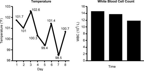
Duration and Cessation of Treatment
The appropriate duration of antimicrobial therapy for serious infections such as hospital‐ or healthcare‐associated pneumonia, complicated intra‐abdominal infection, and bacteremia has not been well studied. To the extent that guidelines for treatment duration exist, they are largely based on observational studies, clinical experience, and consensus, rather than data from well‐designed clinical studiesalthough such studies and data are beginning to emerge, more so in some areas (pneumonia) than others (intra‐abdominal infections and catheter‐related bacteremia). Additional studies supporting treatment durations for these and other important infections are encouraged, given the widely recognized relationships between antimicrobial use and development of antimicrobial resistance, and between antimicrobial resistance and increased morbidity, mortality, and healthcare costs.13 Duration is a component of antimicrobial exposure, and together with optimal dosing, has been linked with antimicrobial resistance and other adverse or unintended consequences of antimicrobial therapy. The general idea is to eradicate (kill) the pathogen as soon as possible, and then stop therapy, since dead bugs don't mutate.
An overwhelming body of work has established a link between antimicrobial use and emergence of antimicrobial‐resistant bacteria. This relationship holds for most, if not all, antimicrobial,47 but appears to be particularly strong for broader‐spectrum agents like fluoroquinolones,814 extended‐spectrum cephalosporins,1518 and carbapenems.4, 1822 Using an antimicrobial from a particular drug class typically promotes development of resistance to all members of the class, but can also lead to more broad‐based resistance including other drug classes, depending on the mechanisms of resistance. Emergence of resistance is expected to be especially high when a suboptimal antimicrobial regimen is administered for a prolonged time or duration,7, 23 as these conditions optimize pressure for selection of preexistent resistant strains or development of new ones.
Optimal efficacy and safety of antimicrobial therapy depends, first, on avoiding antimicrobials when they are not indicated, and second, when they are used, focusing on the 4 Ds of optimal antimicrobial therapy: right Drug, right Dose, De‐escalation to pathogen‐directed therapy, and right Duration of therapy.24 Corresponding articles in this supplement have focused on the first 3 Ds: Dr Syndman on selection of the right drug and dose, and Dr Kaye on de‐escalation of initial empiric therapy, when circumstances warrant it. The current article examines the rationale for reducing the duration of antimicrobial therapy (when possible), and current evidence or guidelines supporting the use of shorter courses of antimicrobial therapy for such infections as pneumonia (community‐, hospital‐, or healthcare‐acquired/associated), complicated intra‐abdominal infection, and bacteremia or sepsis. Key points will be illustrated through 3 case studies dealing with each of these general infection categories.
ADJUSTING DURATION TO OPTIMIZE ANTIMICROBIAL THERAPY
The ultimate goals of short‐course antimicrobial therapy are to rapidly eradicate pathogenic microorganisms and reduce selective pressure for emergence of resistance. The primary potential advantages of shorter duration antimicrobial therapy include lower cost, less toxicity, better adherence, reduced antimicrobial resistance, and reduced disruption of endogenous flora and risk of superinfections, such as Clostridium difficile‐associated disease.23 Other potential benefits of shorter antimicrobial durations include a shorter length of hospital stay and (perhaps) earlier removal of an intravenous catheter, which would be expected to reduce risk of iatrogenic complications and facilitate early mobility and earlier return to full health. Effective short‐course antimicrobial therapy also appears to better meet patient expectations of therapy than longer courses.25
Rapid or early eradication of pathogens depends not only on selecting an agent or combination of agents with activity against the causative pathogen, but also administering the agent in a manner that enables it to achieve its pharmacodynamic (PD) target for pathogen eradication in a rapid fashion.23, 26 The PD parameter that best predicts efficacy will vary for different antimicrobial classes, but the general idea is to use a dose, dosing schedule, and route of administration that rapidly achieves adequate tissue penetration and drug concentration at the infection site for a sufficient length of time for maximum efficacy. In brief, the general concept for short‐course antimicrobial therapy is to hit hard and fast then leave as soon as possible.23
The World Health Organization (WHO) 2000 report on overcoming antimicrobial resistance also recognizes that ideal antimicrobial usage includes using the correct drug, administered by the best route, in the right amount, at optimal intervals, for the appropriate period, after an accurate diagnosis.27 Administering antimicrobials for the wrong period of time (ie, duration) increases risk of resistance. In essence, the WHO report is another call to treat aggressively with shorter courses to help reduce antimicrobial resistance, and to avoid antimicrobial therapy when it is not warranted.
However, while there is general agreement about the utility of using as short an antimicrobial course as is consistent with efficacy, there has been a general dearth of information about exactly what the optimal duration is for particular agents (or drug classes) used to treat particular infections. This is especially the case for most infections occurring in critically ill patients in the hospital setting. Appropriate duration of therapy has been established for some infections, notably group A streptococcus pharyngitis, urinary tract infections, and some sexually transmitted diseases,2831 but treatment duration has not been firmly established for most serious infections. Furthermore, clinicians are often reluctant to shorten the duration of antimicrobial therapy in patients with serious infections for fear of incompletely eradicating the pathogen, thereby leading to relapses and significant morbidity or mortality.
Nevertheless, several studies have now been published that point to the effectiveness of shorter‐course antimicrobial therapy for community‐acquired pneumonia (CAP)3235 and hospital‐acquired pneumonia (HAP) or ventilator‐associated pneumonia (VAP),3645 and a more limited number pointing to the effectiveness of shorter‐course therapy for intra‐abdominal infections38, 46, 47 or bacteremia.4851 In addition, clinical practice guidelines recommend shorter‐course antimicrobial therapy for most patients with CAP,52 uncomplicated healthcare‐associated pneumonia (HCAP) or HAP/VAP,53 and complicated intra‐abdominal infections54and clinical practice guidelines for the management of intravascular catheter‐related infection, including bacteremia, specify a standard duration of therapy and conditions under which a shorter (or longer) course may be considered.55 Shorter‐course therapy can be best implemented based on clinical parameters (eg, resolution of fever, reduction of leukocytosis) along with clinical judgment of the well‐informed clinician with guidance from evidenced‐based guidelines.
The remainder of this section will examine some of the preclinical and clinical evidence supporting shorter‐course therapy for CAP. Subsequent sections of the article utilize 3 case studies to discuss current guidelines and supportive evidence for use of shorter‐course antimicrobial therapy in patients with HCAP or HAP/VAP, complicated intra‐abdominal infections, and bacteremia. The discussion of CAP is intended as an introduction that lays down some general concepts concerning shorter‐duration therapy before delving into the serious hospital‐ or healthcare‐related infections outlined above. Because there is more clinical research on duration of treatment for patients with HAP/VAP than for complicated intra‐abdominal infections or bacteremia, the section on HCAP/HAP/VAP is much longer and detailed than the ones for complicated intra‐abdominal infections or bacteremia.
CAP is defined as pneumonia developing in individuals who are not residents in a nursing home or extended‐care facility, and who have not recently been hospitalized or had significant exposure to the healthcare setting. Pneumonia developing after 48 hours of hospital admission, and that was not incubating at the time of admission, is known as HAP,53, 56 and VAP is a subset of HAP, more precisely defined as HAP that arises after endotracheal intubation.53 HCAP includes patients characterized by residence in a nursing home or extended‐care facility or hospitalization for 2 days in the preceding 90 days or other significant exposure to the healthcare setting.53, 57, 58
DURATION OF THERAPY FOR CAP
A number of studies have reported similar efficacy with shortened versus longer durations of antimicrobial therapy for CAP.33, 5964 Consistent with this, 2 recent meta‐analyses of studies comparing shorter‐ versus longer‐course therapy for mild‐to‐moderate CAP (22 randomized controlled trials and >8000 patients between them) reported similar efficacy and safety with shorter‐course therapy.65, 66 In addition, other studies have reported an association between longer durations of antimicrobial therapy and development of resistance by community respiratory pathogens, especially when lower doses have been used.67, 68 These findings are consistent with the belief that prolonged treatment with a suboptimal antimicrobial regimen creates particularly fertile conditions for selection or development of antimicrobial‐resistant strains.65, 66
Data from preclinical studies provide a basis for understanding the effectiveness of shorter‐dosing regimens of adequate antimicrobial therapy for CAP or other forms of pneumonia. In particular, in vitro time‐kill studies6974 and animal models of infection7577 have demonstrated that Streptococcus pneumoniae can be rapidly eradicated without use of long‐term therapy when appropriate antimicrobials are used. Consistent with these preclinical data, various clinical studies have also shown that S pneumoniae and other respiratory pathogens are rapidly eradicated from lower respiratory tract secretions after initiation of appropriate antimicrobial treatment. For example, Montravers et al. reported that 94% of respiratory pathogens were eradicated from the lungs of 76 patients with VAP after just 3 days of antimicrobial therapy.78
Based on the available data, the 2007 Infectious Diseases Society of America (IDSA)/America Thoracic Society (ATS) guidelines for CAP management recommend a minimum of 5 days of antimicrobial treatment, while noting that most patients become clinically stable within 3‐7 days of treatment onset and rarely require longer durations.52 The guidelines further recommend that CAP patients should be afebrile for 4872 hours and should have no more than 1 CAP‐associated sign of clinical instability before discontinuation of therapy. Although the general movement is toward use of shorter‐duration treatment courses than the traditional 710 days or longer, the IDSA/ATS guidelines acknowledge that longer durations may be needed in certain situations.79
CASE 1: HEALTHCARE‐ASSOCIATED PNEUMONIA
Case 1 is a 72‐year‐old woman admitted with findings consistent with HCAP who was initiated on an empiric therapy regimen of vancomycin and piperacillin‐tazobactam. Results from blood and sputum cultures obtained prior to treatment initiation came back on day 3, and were negative for pathogenic bacteria. White blood cell (WBC) counts were trending downward, and the patient appeared to be stabilizing. She still had an elevated WBC count, slight fever (temperature maximum of 101.4F for the past 24 hours), and lung crackles at the right lung base. Because Gram stain failed to identify Gram‐positive cocci clusters, and there was no culture evidence of methicillin‐resistant Staphylococcus aureus (MRSA) or Pseudomonas aeruginosa, vancomycin treatment was terminated and the patient was switched to single‐agent therapy with intravenous ceftriaxone, a nonpseudomonal third‐generation cephalosporin. On hospital day 5, there was continuing evidence of response to antimicrobial therapy. The patient reported feeling better and she was breathing comfortably. Her cough was much improved, sputum production was markedly decreased, and her fever had resolved. Now, on day 7, the patient is still afebrile, her WBC count is normal, and she has 96% oxygen saturation on room air.
The question before the clinician is whether to terminate or continue antimicrobial therapy, and if continued, with what regimen and for how long? In addition, if a decision is made to continue antimicrobial therapy, there is a possibility of switching from an intravenous to oral treatment regimen. An examination of the literature and current treatment guidelines for HCAP/HAP/VAP should enable a more informed decision, one that optimally benefits not only this patient, but all subsequent ones who might be exposed and infected with a resistant pathogen that develops when treatment is continued longer than necessary.
Using Clinical Parameters to Shorten Antimicrobial Therapy
A prospective study by Dennesen et al., published 10 years ago, was one of the first suggesting the possibility of shortened duration of antimicrobial therapy for VAP.80 At the time, duration of antimicrobial therapy for VAP typically ranged from 7 to 21 days, and was most commonly 14 to 21 days. In this study, Dennesen and coworkers examined symptom resolution in 27 patients diagnosed with VAP based on clinical, radiologic, and microbiological criteria, each of whom received appropriate antimicrobial therapy based on culture susceptibility data.80 Significant improvements were observed for all clinical parameters examined (highest temperature, leukocyte count, pressure of arterial oxygen to fractional inspired oxygen [PaO2/FIO2] ratio, semiquantitative culture result of endotracheal aspirate), usually first appearing within the first 6 days of antimicrobial therapy. Furthermore, analyses of specific pathogens showed that appropriate antimicrobial therapy rapidly eradicated endotracheal colonization with S pneumoniae, Haemophilus influenzae, and S aureus, but not of P aeruginosa or Enterobacteriaceae. Moreover, endotracheal colonization with resistant pathogens tended to occur when antimicrobial therapy was continued beyond the first week. Taken together, these results suggested that prolonged antimicrobials beyond 7 days usually did not benefit VAP patients, and in fact increased risk of superinfection with a resistant strain. However, it is important to make a distinction between VAP and, for example, skin or bloodstream infections involving S aureus. While improved signs and symptoms generally indicate clinical cure for VAP, this reasoning should not be applied to S aureus bacteremia.
The findings from Dennesen et al. are generally consistent with those from Montravers et al., which showed that 94% of respiratory pathogens were eradicated from the lungs of VAP patients 3 days after initiation of antimicrobial therapy.78 They are also consistent with the findings from a 2005 study by Vidaur et al., which demonstrated resolution of fever (38C), PaO2/FIO2 (>250 mmHg), and WBC/leukocyte count (10,000) in 73%, 75%, and 53% of VAP patients, respectively, without acute respiratory distress syndrome (ARDS; n = 75) after 3 days of appropriate antimicrobial therapy.81 However, Vidaur et al. reported that fever took roughly twice as long to resolve in VAP patients with ARDS (n = 20) versus without ARDS, and that hypoxia resolution was less useful when evaluating treatment response in ARDS patients. As with the Dennesen et al. study,80 the results from Vidaur et al. suggest that measures of core body temperature and oxygenation can be useful guides for clinicians in determining whether to shorten the duration of antimicrobial therapy for patients with VAP, HAP, or HCAP.81
Along the same lines, the clinical pulmonary infection score (CPIS) has established itself as a means for the early termination (shortening) of initial empiric antimicrobial therapy in particular VAP patients. The CPIS is derived by scoring 57 clinical indices relevant for the diagnosis of VAP, as illustrated in Table 1.82 A score of >6 is considered suggestive of pneumonia, while one 6 implies low likelihood of pneumonia. A 2000 study by Singh et al. randomized 81 consecutive patients with pulmonary infiltrates and a CPIS 6 to receive either standard antimicrobial therapy (at discretion of the clinician) or ciprofloxacin monotherapy, with the intention of reevaluating patients at day 3.45 For patients in the ciprofloxacin (experimental) group, antimicrobial therapy was terminated at day 3 if the CPIS remained 6. As a result, only 28% of patients in the experimental group had antimicrobial therapy continued beyond day 3, compared with 90% of patients in the standard therapy group (P = 0.0001). More importantly, there were no significant differences in mortality between patients in the 2 treatment groups, despite a significantly shorter treatment duration for those in the experimental group (3.0 vs 9.8 days, P = 0.0001). In addition, mean length of intensive care unit (ICU) stay was significantly shorter (9.4 vs 14.7 days, P= 0.04) and mean antimicrobial cost was significantly lower ($259 vs $640, P = 0.0001) for patients in the experimental versus standard therapy group.
| Points | |
|---|---|
| |
| Temperature C | |
| 36.5 and 38.4 | 0 |
| 38.5 and 38.9 | 1 |
| 39 or 36.0 | 2 |
| Tracheal secretions | |
| Absence of secretions | 0 |
| Presence of non‐purulent secretions | 1 |
| Presence of purulent secretions | 2 |
| Pulmonary radiography (chest X‐ray) | |
| No infiltrate | 0 |
| Diffused (or patchy) infiltrate | 1 |
| Localized infiltrate | 2 |
| WBCs, leukocytes/mm3 | |
| 4000 and 11,000 | 0 |
| 4000 or >11,000 | 1 |
| +Band forms 500 | 2 |
| Oxygenation: PaO2/FIO2 mmHg | |
| >240 or ARDS | 0 |
| 240 and no evidence of ARDS | 2 |
| Culture of tracheal aspirate (semiquantitative: 012 or 3+) | |
| Pathogenic bacteria cultured 1+ or no growth | 0 |
| Pathogenic bacteria cultured >1+ + same pathogenic bacteria seen on the gram stain >1+ | 1 2 |
| Progression of pulmonary infiltrate | |
| No radiographic progression | 0 |
| Radiographic progression (ARDS excluded) | 2 |
Furthermore, a significantly greater proportion of patients in the standard versus experimental therapy group exhibited evidence of antimicrobial resistance or superinfections (38% vs 14%, P = 0.017). The 2005 clinical practice guidelines for HAP, VAP, or HCAP state, A modified CPIS of 6 or less for 3 days, proposed by Singh and coworkers, is an objective criterion to select patients at low risk for early discontinuation of empiric treatment of HAP.53 While the Singh et al. study provides the rationale for shorter‐course therapy in ICU patients with pulmonary infiltrates who have low likelihood of pneumonia (CPIS 6), this criterion may or may not pertain to HAP/VAP more strictly, and still requires validation in patients with more severe forms of VAP. Incidentally, although the CPIS was designed to define VAP, and there are no data validating its use for other types of pneumonia, the clinical experience by this author indicates that it can be helpful in evaluating HCAP and non‐VAP HAP as well.
Clinical Trial to Support Shortened Duration of HCAP/HAP/VAP Therapy
A French study published in JAMA in 2003 provides more direct support that approximately 1 week of antimicrobial therapy produces effectiveness comparable to more traditional 23‐week therapy for most patients with VAP.37 In this prospective, multicenter, randomized, double‐blind (until day 8) clinical trial, 401 patients with microbiologically proven VAP were randomly assigned to receive either 8 days (n = 197) or 15 days (n = 204) of initial empiric antimicrobial therapy selected by the treating physician. No significant differences were observed between the 8‐day and 15‐day treatment groups for the 2 primary efficacy endpoints of death from any cause (18.8% vs 17.2%) and microbiologically documented pulmonary infection recurrence (28.9% vs 26.0%). There were also no differences between the groups for number of mechanical ventilation‐free days (8.7 vs 9.1 days), number of organ‐failure‐free days (8.7 vs 8.9 days), length of ICU stay (30.0 vs 27.5 days), unfavorable outcome (death, pulmonary infection recurrence, or prescription of a new antimicrobial) (46.2% vs 43.6%), mortality rate on day 60 (25.4% vs 27.9%), or in‐hospital mortality (32% vs 29.9%).
Conversely, patients in the 8‐day treatment group had significantly more antimicrobial‐free days (13.1 vs 8.7 days, P 0.001), and among patients who developed recurrent infections, multidrug‐resistant pathogens emerged more frequently in patients in the 15‐day versus 8‐day treatment group (62.0% vs 42.1%, P = 0.04). However, there was an apparent exception to the general comparable efficacy of the 8‐ and 15‐day treatment regimens for infections caused by nonfermenting Gram‐negative bacilli, including P aeruginosa. For primary infections caused by nonfermenting Gram‐negative bacilli, the 8‐day versus 15‐day regimen was associated with higher rates of pulmonary recurrence (40.6% vs 25.4%). Interestingly, the 8‐day regimen was not associated with more adverse outcomes here, just a higher recurrence rate. With respect to primary infections caused by MRSA, no differences were observed between the 2 treatment regimens for death for all causes (23.4% vs 30.2%) or pulmonary infection recurrence (33.3% vs 42.9%). Figure 1 presents the probability of survival data for the 8‐day and 15‐day treatment groups.
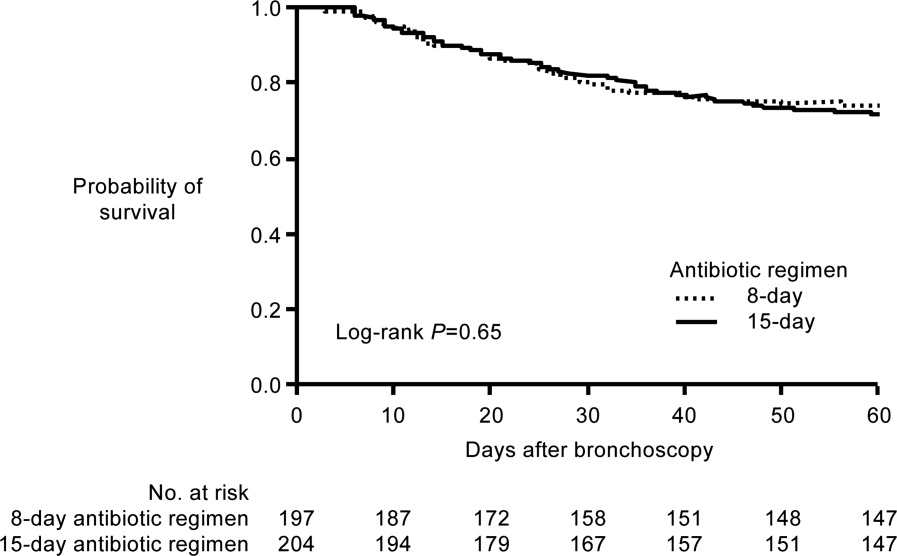
Hence, the data from the Chastre et al. study37 support use of an 8‐day (or shortened) regimen as standard antimicrobial therapy for most patients with VAP, with some possible exceptions. Additional studies provide further support for this general conclusion. For example, a prospective, randomized, controlled trial by Micek et al. evaluated the impact of using an antimicrobial discontinuation policy based on clinical criteria (discontinuation group; n = 150)versus the decision of treating physicians (conventional group, n = 140)to determine the duration of antimicrobial therapy for VAP, and observed a statistically shorter treatment duration in the discontinuation versus conventional management group (6.0 vs 8.0 days, P = 0.001), but no difference between the groups for hospital mortality (32.0% vs 37.1%), ICU length of stay (6.8 vs 7.0 days), or VAP recurrence (17.3% vs 19.3%).42 A prior study by the same group reported a shorter duration of antimicrobial therapy for VAP following implementation of an antimicrobial guideline (vs prior to implementation) (8.6 vs 14.8 days, P 0.001), and a lower rate of VAP recurrence among patients in the after period (7.7% vs 24.0%, P = 0.03). However, interpretation of the results was complicated by the fact that initial empiric therapy was more often appropriate during the after versus before guideline implementation period (94.2% vs 48.0%, P 0.001).40
A limited number of studies have focused further on shortened duration of therapy for patients with VAP caused by Gram‐negative bacteria, and particularly by nonfermenting Gram‐negative bacilli. A retrospective study by Hedrick et al. analyzed the relationship between antimicrobial duration and outcomes of 452 episodes of VAP in the ICU, 154 caused by nonfermenting Gram‐negative bacilli.39 In the study, 127 patients infected with a nonfermenting Gram‐negative bacillus received 9 days (mean 17.1 0.7 days) of antimicrobial therapy, while 27 received 3‐8 days (mean 6.4 0.3 days) of therapy. No significant differences were observed between the shorter‐ and longer‐duration groups for mortality (22% vs 14%, P = 0.38) or VAP recurrence (22% vs 34%, P = 0.27) for these patient populations. Table 2 provides the results for all 452 VAP episodes based on 8 days or 9 days of antimicrobial therapy.
| Patient Characteristic | 8 Days (n = 98) | 9 Days (n = 354) | P Value |
|---|---|---|---|
| |||
| Mean antimicrobial days | 6.2 | 16.8 | 0.0001 |
| Mean APACHE II | 18 | 20 | 0.0009 |
| % Trauma | 71 | 68 | 0.63 |
| Mean time to onset, days | 17.7 | 17.8 | 0.97 |
| Recurrence | 11% | 25% | 0.004 |
| Death | 13% | 11% | 0.59 |
| Nonfermenting Gram‐negative bacilli recurrence | 22% (n = 27) | 34% (n = 127) | 0.27 |
| Staphylococcus aureus recurrence | 20% (n = 10) | 38% (n = 47) | 0.47 |
The retrospective nature of the study limits the ability to more confidently interpret the results, but the data appear to be consistent with the conclusion that short‐duration therapy does not necessarily increase recurrence or worsen other outcomes in patients with VAP caused by nonfermenting Gram‐negative bacilli. The most common Gram‐negative bacilli associated with VAP in the study were P aeruginosa (18% of all infections), Enterobacter cloacae (11%), Acinetobacter spp (11%), Klebsiella pneumoniae (7%), Stenotrophomonas maltophilia (7%), Serratia spp (7%), H influenzae (6%), and Escherichia coli (4%). In addition, the study results suggest that short‐duration therapy is at least as effective as longer‐duration therapy for the overall VAP population, with potential benefits in terms of reduced antimicrobial use and lower rate of recurrence.
Another recent retrospective analysis examining an even shorter course of antimicrobial therapy (5 days) for patients with HAP associated with Gram‐negative bacteria reported a low overall recurrence rate (14%) and a critical care mortality rate (34.2%) in line with prior studies of short‐term therapy for VAP/HAP.44 However, the HAP relapse rate was significantly higher in patients with HAP caused by nonfermenting Gram‐negative bacilli versus other Gram‐negative species (17% vs 2%, P = 0.03).
A recent US pilot study explored the use of repeat bronchoalveolar lavage (BAL) to guide antimicrobial duration in 52 patients with VAP, and compared the results with a matched control group of 52 VAP patients treated before institution of the BAL pathway.43 Antimicrobial therapy in the pathway patients was discontinued if pathogen growth was 10,000 colony forming units/mL on the repeat BAL performed on day 4 of therapy. One objective was to determine whether a repeat BAL strategy, such as the one here, might be able to identify patients with VAP due to nonfermenting Gram‐negative bacilli or other microorganisms who could be safely and effectively treated with shorter‐duration therapy.
Results showed that the antimicrobial duration was significantly shorter for patients in the pathway group than the matched control group (9.8 vs 3.8 days, P 0.001), including the subset of patients with VAP associated with nonfermenting Gram‐negative bacilli (10.7 vs 14.4 days, P 0.001). No significant differences were observed between the overall treatment populations for VAP recurrence, mechanical ventilator‐free ICU days, ICU‐free hospital days, or mortality. Repeat BAL showed most VAP isolates in the study group (83%) responded to initial therapy with a mean duration of 8.8 days. Nonresponders without concomitant infections received significantly longer treatment than pure responding isolates (14.4 vs 7.3 days, P 0.001), and the most common nonresponding microorganisms were P aeruginosa (41% response rate) and S maltophilia (50% response rate), 2 nonfermenting Gram‐negative bacilli.
Most nonfermenting Gram‐negative bacilli‐associated VAP isolates in the study group did respond on repeat BAL (59%). These responders were treated for a mean duration of 8.2 days, and exhibited a similar recurrence rate versus that observed for the matched control group (12.0% vs 17.9%, P = 0.71). These pilot study results suggest that repeat BAL might be used to identify patients likely to benefit from short‐duration therapy, including patients infected with nonfermenting Gram‐negative bacilli. Further study on this is needed.
ATS/IDSA Guidelines for Duration of HCAP/HAP/VAP Therapy
Based largely on the studies by Dennesen et al.80 and Luna et al.83 indicating most VAP patients who respond to appropriate antimicrobial therapy do so within the first 6 days, and those by Chastre et al.37 and Singh et al.45 pointing to the efficacy and safety of shorter‐duration VAP therapy, the 2005 ATS/IDSA guidelines recommend the use of shorter‐duration antimicrobial therapy for most patients with HCAP or HAP/VAP.53 More specifically, the guidelines state, If patients receive an initially appropriate antimicrobial regimen, efforts should be made to shorten the duration of therapy from the traditional 14 to 21 days to periods as short as 7 days, provided that the etiologic pathogen is not P. aeruginosa, and that the patient has a good clinical response with resolution of clinical features of infection.53 Figure 2 presents an overview of the ATS/IDSA guidelines for HCAP/HAP/VAP management 48 to 72 hours after initiation of empiric antimicrobial therapy.53
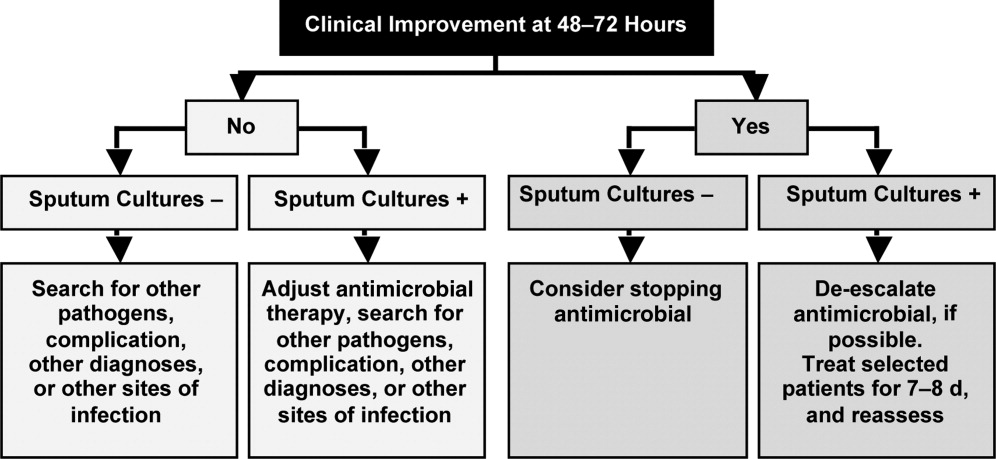
Note that the clinician should consider terminating antimicrobial therapy in patients with clinical improvement and negative cultures or other evidence suggestive of a noninfectious cause. CPIS can also be helpful when deciding whether to terminate initial empiric therapy in a patient with clinical improvement after 23 days of therapy and negative cultures. If cultures are positive, the clinician should consider whether antimicrobial de‐escalation is possible (as discussed by Dr Kaye in the corresponding supplement article), and aim to treat selected patients with an antimicrobial course lasting 78 days. After 78 days, patients should be reassessed for treatment termination or other appropriate actions.
The ATS/IDSA guidelines also provide recommendations for route of drug administration, and if and when to switch from an intravenous to oral agent. In particular, the guidelines state that all patients with HCAP, HAP, or VAP should initially receive therapy intravenously, but conversion to oral/enteral therapy may be possible in certain responding patients, ie, those with a good clinical response and a functioning intestinal tract.53 Fluoroquinolones and linezolid have oral formulations with bioavailability equivalent to the intravenous form, meaning the oral formulations are capable of achieving high levels at the site of infection. This may facilitate conversion to oral therapy in select patients. Early step‐down is safe and effective with fluoroquinolones.84, 85
Based on the information just reviewed, the antimicrobial can be terminated on day 7 for case 1. She is afebrile, and her WBC and oxygenation are normal. In fact, since her records show she was responding at day 5, consideration could have been given to switching from intravenous to oral therapy at that time, and perhaps even discharging her to the rehabilitation center.
CASE 2: INTRA‐ABDOMINAL INFECTION (DIVERTICULITIS)
Case 2 is a 56‐year‐old woman who presents with sepsis and diverticular abscess with walled‐off perforation. Upon hospital arrival, Interventional Radiology inserted a drain, and the patient was initiated on ciprofloxacin and metronidazole therapy. Day 3 examination showed improvement in WBC count and normal vital signs, but the patient still had a low‐grade fever (100.9F). Abdominal examination results were improved, but with some diffuse tenderness. Initial cultures of the abdominal abscess isolated Gram‐negative rods, and the patient was continued on ciprofloxacin/metronidazole. Further cultures on day 4 identified an extended‐spectrum ‐lactamase (ESBL)‐producing E coli organism as the causative pathogen. The patient was switched from ciprofloxacin/metronidazole to ertapenem. It is now hospital day 8, and the patient continues to show good response to treatment. She is afebrile and WBC count is normal. The abscess catheter is no longer draining. Her abdominal pain is improved, and she is complaining that she is hungry. A repeat computed tomography scan shows resolution of the abscess and no evidence of bowel perforation. Should antimicrobial therapy be continued in this patient, and if so, with what agent and for how long?
Guidelines from the Surgical Infection Society and IDSA state that antimicrobial therapy of established or complicated intra‐abdominal infection in adults should be limited to 47 days, unless it is difficult to achieve adequate source control.54 This is because extended antimicrobial exposure increases antimicrobial cost and risk of resistance, superinfection, C difficile‐associated colitis, or other untoward and unintended consequences of antimicrobial therapy, and there is no evidence that longer treatment durations improve outcomes.46, 47, 54, 86 Runyon et al. randomized 90 patients with spontaneous bacterial peritonitis or culture‐negative neutrocytic ascites to receive 5 days or 10 days of cefotaxime monotherapy, and reported similar rates of infection‐related mortality (0% vs 4.3%), hospitalization mortality (33% vs 43%), bacteriologic cure (93% vs 92%), and recurrence of ascitic fluid infection (12% vs 13%).46 Furthermore, shorter‐course therapy was associated with significantly lower antimicrobial administration and costs. Similarly, a recent prospective, randomized, double‐blind trial comparing 3 versus 5 days of ertapenem therapy in 111 patients with community‐acquired intra‐abdominal infection reported similar cure (93% vs 90%) and eradication rates (95% vs 94%).86 However, it should be noted that the mean duration of antimicrobial therapy in the longer‐duration group was still relatively short (5.7 days, range of 5‐10 days).
Studies also indicate there is a very low risk of infection recurrence or treatment failure when antimicrobial therapy is terminated in a patient diagnosed with a complicated intra‐abdominal infection who no longer shows signs of continuing infection.38, 87 Lennard et al. compared postoperative outcomes in 65 patients with or without leukocytosis and fever at the conclusion of antimicrobial therapy for intra‐abdominal sepsis, and reported development of intra‐abdominal infection in 7 of 21 (33%) with persistent leukocytosis.87 None of the 30 patients with normal WBC counts at the end of therapy developed an intra‐abdominal infection postoperatively. Furthermore, intra‐abdominal infection occurred postoperatively in 11 of 14 patients (79%) who responded to treatment but were still febrile at the time of antimicrobial discontinuation.
Similar results were obtained in a much larger, more recent study that retrospectively analyzed the relationship between duration of antimicrobial therapy and infectious complications for patients with intra‐abdominal infections.38 In the study, 929 patients with intra‐abdominal infections associated with either fever or leukocytosis were organized into 4 quartiles based on total duration of antimicrobial therapy (quartile 1: 07 days, n = 218; quartile 2: 812 days, n = 217; quartile 3: 1317 days, n = 246; and quartile 4: >17 days, n = 248) or antimicrobial duration after resolution of leukocytosis (quartile 1: 05 days, n = 130; quartile 2: 610 days, n = 127; quartile 3: 1115 days, n = 124; and quartile 4: >15 days, n = 118). Based on either total duration of antimicrobial therapy or duration after leukocytosis resolution, risk of recurrence was significantly higher for patients in quartiles 3 or 4 versus those in quartile 1, and there was no difference between quartiles 1 and 2.
Taken together, these results suggest that antimicrobial therapy for intra‐abdominal sepsis can be shortened in patients exhibiting a clinical response to treatment, if there are no signs of persistent leukocytosis or fever. Hence, clinicians should use the resolution of clinical signs of infection as a guide to determine when during the 47‐day window antimicrobial therapy should be terminated.54 In practical terms, this usually means treatment can be terminated when the patient is afebrile, has normal WBC counts, and is able to tolerate an oral diet.
Based on the clinical status of case 2 after 8 days of antimicrobial therapy (afebrile with normal WBC counts and requesting oral diet), the ertapenem regimen should be stopped. There is no reason to consider further outpatient antimicrobial therapy for this particular patient, but the Surgical Infection Society and IDSA guidelines discuss the type of patient who should be considered for oral or outpatient antimicrobial therapy. According to the guidelines, the patient convalescing from a complicated intra‐abdominal infection may receive oral antimicrobial therapy, but that therapy should only be included as a component within the brief treatment duration already mentioned, ie, in total, it should rarely exceed 7 days.54 Such therapy is rarely indicated for patients who are afebrile, with normal peripheral WBC/leukocyte counts, and with return of bowel function. These recommendations make it clear that no further antimicrobial therapy is warranted for case 2.
However, for appropriate patients who are recovering from a complicated intra‐abdominal infection and are able to tolerate an oral diet, an oral antimicrobial regimen selected on the basis of identified primary isolates may be used for completion of therapy.54 In the absence of cultures, an oral regimen that covers commonly isolated pathogens (eg, E coli, streptococci, and Bacteroides fragilis) should be considered. Common regimens include an oral cephalosporin or fluoroquinolone with metronidazole, or amoxicillin‐clavulanic acid, assuming susceptibility studies do not demonstrate resistance. Given the identification of an ESBL‐producing E coli for case 2a pathogen relatively resistant to oral antimicrobialan oral regimen probably would not have been viable for this patient even earlier in the treatment course. Lastly, a repeat computed tomography scan was used for the case here. It should be noted that there are currently no well‐established criteria for determining when repeat imaging is needed to confirm resolution of fluid collections. This should be a clinical decision. A general practice is that the catheter is left in place until there is minimal drainage (eg, 10 mL/day); catheter sinograms can also be helpful in determining the status of the abscess.
CASE 3: CENTRAL LINE‐ASSOCIATED BACTEREMIA
Case 3 is a 56‐year‐old man with status epilepticus, intubation, and ICU stay. He was initially treated with vancomycin and piperacillin‐tazobactam for a fever of 103.4F on day 5 of hospitalization. Blood cultures grew Gram‐positive cocci. The central venous catheter was removed, and the initial antimicrobial regimen was de‐escalated to vancomycin monotherapy, which was associated with continued improvement in fever and WBC count, and clinical stability on hospital day 7. At that time, further blood culture analyses isolated methicillin‐susceptible S aureus (MSSA), and the antimicrobial regimen was switched/de‐escalated from vancomycin to cefazolin. It is now hospital day 9 (day 3 of cefazolin) and the patient continues to respond and is afebrile. Repeat blood cultures show no bacterial growth, and a transesophageal echocardiograph (TEE) was performed and revealed normal heart valves. Should the antimicrobial therapy be continued for this patient, and if so, with what agent and for how long?
The IDSA guidelines for management of intravascular catheter‐related infections recommend catheter removal and 46 weeks of antimicrobial therapy for patients with S aureus catheter‐related bloodstream infection (CRBSI), unless the patient has exceptions allowing consideration of shorter‐duration therapy (minimum of 14 days, with day 1 being the first day of negative blood culture results).55 These exceptions include absence of diabetes; immunocompetence (no immunosuppression); removal of the infected catheter; no prosthetic intravascular device (eg, pacemaker or recently placed vascular graft); no evidence of endocarditis or suppurative thrombophlebitis on TEE and ultrasound, respectively; fever and bacteremia resolved within 72 hours after initiation of appropriate antimicrobial therapy; and no evidence of metastatic infection on physical examination and sign‐ or symptom‐directed diagnostic tests.
Short‐duration (10‐16 day) antimicrobial therapy has been reported to yield similarly low recurrence or relapse rates as longer courses of therapy in patients with uncomplicated catheter‐associated S aureus bacteremia.50, 51, 88, 89 A small 1989 study by Ehni and Reller prospectively followed 13 patients with S aureus CRBSI who had received short‐course therapy (17 days), and reported only 1 case of relapse with endocarditis (8% relapse rate).50 A subsequent study by Malanoski et al. retrospectively analyzed the data from 55 patients with S aureus CRBSI.51 Excluding the 8 patients with early complications, the authors observed similar rates of relapse in patients treated for 1015 days and those receiving longer courses of antimicrobial therapy (0% vs 4.7%). The clinical characteristics of the 2 treatment duration groups were similar, and delayed catheter removal was linked with persistence of bacteremia (P = 0.01).
A more recent multicenter, prospective observational study by Chang et al. examined recurrence and the impact of antimicrobial treatment in 505 consecutive patients with S aureus bacteremia, and determined that duration of antimicrobial therapy was not a factor associated with relapse.88 This was true both for patients with bacteremia resulting from endocarditis, bacteremia with no apparent source, or bacteremia due to a focus that could not be cured or removed (28 days therapy after defervescence, 28 days therapy, or 28 days therapy), or those with bacteremia resulting from a source amenable to definitive cure, such as an intravascular device that could be removed, an abscess that could be incised and drained, or an infected bone that could be resected (>14 days, 1014 days, or 10 days therapy). Similarly, a 2005 prospective study by Thomas and Morris determined there was no relationship between treatment duration (7 vs 8 days, 10 vs 10 days, or 14 vs 15 days; P = 0.62, 0.87, and 0.16, respectively) and rate of relapse for 276 patients with cannula‐associated S aureus bacteremia.89 Longer‐duration antimicrobial therapy is warranted in patients with CRBSI and an early complicated course, eg, fever and/or bacteremia persisting for >3 days after catheter removal.90
According to the IDSA guidelines, a TEE should be obtained for all patients with CRBSI involving S aureus who are being considered for a shorter duration of therapy, and the TEE should be performed at least 57 days after onset of bacteremia to minimize risk of false‐negative results.55 High rates of infective endocarditis are observed in patients with S aureus bacteremia,89, 9193 with higher rates in patients with MSSA versus MRSA bacteremia (43.4% vs 19.6%, P 0.009).91 TEE is essential to diagnose endocarditis and detect other complications of bacteremia.92, 93 This recommendation for use of TEE does not necessarily apply to all patients with CRBSI when S aureus is not involved.
Figure 3 summarizes the general recommendations from the IDSA guidelines for the management of CRBSI in patients with a short‐term catheter.55 The figure illustrates the varied recommendations for treatment duration depending on whether the infection is complicated or uncomplicated, and based on the pathogenic microorganism. Returning to case 3, the patient meets the general criteria for shorter duration of antimicrobial therapy: he is not diabetic or immunosuppressed, his catheter has been removed, he does not have any prosthetic intravascular devices, his fever and bacteremia (based on blood cultures) resolved within 3 days of initiating cefazolin therapy, and there is no evidence of endocarditis or other complications of bacteremia. Hence, he is an excellent example of a patient with uncomplicated MSSA CRBSI who meets the criteria for consideration of shortened antimicrobial therapy. Based on the clinical practice guidelines, the patient should continue on intravenous cefazolin for a 14‐day course of therapy, at which time he can be re‐evaluated. A recent review of bloodstream infections caused by various pathogens similarly concluded that the minimum treatment duration for low‐risk patients with S aureus CRBSI is 14 days.48 As a final point, it is also important to note that there is no role for oral therapy in patients with CRBSI, so whether shortened or not, the chosen regimen should be administered intravenously.
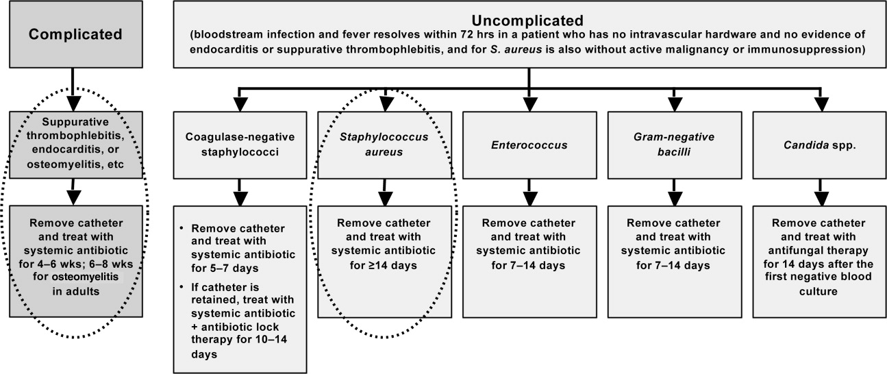
CONCLUSIONS
Shortening the duration of appropriate and adequate antimicrobial therapy represents one strategy for reducing pressure for selection or development of resistant pathogenic microorganisms. Other potential benefits of shorter courses of antimicrobial therapy include reduced risk of antimicrobial‐associated infections (superinfection, C difficile‐associated diarrhea) and other antimicrobial‐related adverse events, improved compliance, and reduced antimicrobial costs. Clinicians are sometimes concerned that reducing antimicrobial courses for patients with serious infections, such as HCAP/HAP/VAP, complicated intra‐abdominal infection, and CRBSI, will lead to incomplete eradication of pathogenic microorganisms, leading to disease recurrence and increased morbidity and mortality. When managing patients with these serious infections, clinicians often turn to the literature and recommendations from professional organizations for guidance. Available data from randomized controlled and nonrandomized clinical trials indicate that shorter‐course therapy is effective and safe for patients with CAP, HCAP/HAP/VAP, complicated intra‐abdominal infections, and CRBSI. Based on these data, and consensus/expert opinion, clinical practice guidelines have been developed that recommend specific durations of antimicrobial therapy for each of these infections.
Although greater study of antimicrobial therapy duration is needed, the current and developing literature and current treatment guidelines should enable clinicians to recognize patients who would benefit from shortened courses of antimicrobial therapy. In doing so, they would help to lower antimicrobial costs and reduce the growing problem of antimicrobial resistance, with its wide‐ranging, negative consequences for current and future patients, and the clinicians who treat them.
- .The relationship between antimicrobial resistance and patient outcomes: mortality, length of hospital stay, and health care costs.Clin Infect Dis.2006;42(suppl 2):S82–S89.
- ,,.Clinical and economic burden of antimicrobial resistance.Expert Rev Anti Infect Ther.2008;6:751–763.
- .Economics of antibiotic resistance.Expert Rev Anti Infect Ther.2008;6:523–539.
- ,,,.Emergence of antibiotic‐resistant Pseudomonas aeruginosa: comparison of risks associated with different antipseudomonal agents.Antimicrob Agents Chemother.1999;43:1379–1382.
- ,,,,.Effect of antibiotic prescribing in primary care on antimicrobial resistance in individual patients: systematic review and meta‐analysis.BMJ.2010;340:c2096.
- ,,,,,.Risk factors for invasive pneumococcal disease in children: a population‐based case‐control study in North America.Pediatrics.1999;103:E28.
- ,,.Antimicrobial resistance: consideration as an adverse drug event.Crit Care Med.2010;38:S155–S161.
- ,,,,,.Fluoroquinolone‐resistant Pseudomonas aeruginosa: assessment of risk factors and clinical impact.Am J Med.2006;119:526.e519–526.525.
- ,,, et al.Emergence of fluoroquinolone resistance in outpatient urinary Escherichia coli isolates.Am J Med.2008;121:876–884.
- ,,,,,.Imipenem resistance among Pseudomonas aeruginosa isolates: risk factors for infection and impact of resistance on clinical and economic outcomes.Infect Control Hosp Epidemiol.2006;27:893–900.
- ,,,,.Hospital and community fluoroquinolone use and resistance in Staphylococcus aureus and Escherichia coli in 17 US hospitals.Clin Infect Dis.2005;41:435–440.
- ,,,.Evaluation of an intervention designed to decrease the rate of nosocomial methicillin‐resistant Staphylococcus aureus infection by encouraging decreased fluoroquinolone use.Infect Control Hosp Epidemiol.2006;27:155–169.
- ,,,,,.Antibiotic resistance among gram‐negative bacilli in US intensive care units: implications for fluoroquinolone use.JAMA.2003;289:885–888.
- ,,, et al.Relationship between rates of antimicrobial consumption and the incidence of antimicrobial resistance in Staphylococcus aureus and Pseudomonas aeruginosa isolates from 47 French hospitals.Infect Control Hosp Epidemiol.2007;28:1389–1395.
- ,,, et al.Citywide clonal outbreak of multiresistant Acinetobacter baumannii and Pseudomonas aeruginosa in Brooklyn, NY: the preantibiotic era has returned.Arch Intern Med.2002;162:1515–1520.
- ,,,,.Nosocomial outbreak of Klebsiella infection resistant to late‐generation cephalosporins.Ann Intern Med.1993;119:353–358.
- ,,.The effect of an antimicrobial restriction program on Pseudomonas aeruginosa resistance to beta‐lactams in a large teaching hospital.Pharmacotherapy.2003;23:618–624.
- ,,,.Association between antibiotic usage and subsequent colonization or infection of extensive drug‐resistant Acinetobacter baumannii: a matched case‐control study in intensive care units.Diagn Microbiol Infect Dis.2008;62:298–305.
- ,,.Relationships between antimicrobial use and antimicrobial resistance in Gram‐negative bacteria causing nosocomial infections from 1991–2003 at a university hospital in Taiwan.Int J Antimicrob Agents.2005;26:463–472.
- ,,, et al.Risk factors for acquisition of imipenem‐resistant Acinetobacter baumannii: a case‐control study.Antimicrob Agents Chemother.2004;48:224–228.
- ,,,,.Consumption of imipenem correlates with beta‐lactam resistance in Pseudomonas aeruginosa.Antimicrob Agents Chemother.2002;46:2920–2925.
- ,,.Relationship of carbapenem restriction in 22 university teaching hospitals to carbapenem use and carbapenem‐resistant Pseudomonas aeruginosa.Antimicrob Agents Chemother.2009;53:1983–1986.
- .Clinical efficacy of newer agents in short‐duration therapy for community‐acquired pneumonia.Clin Infect Dis.2004;39(suppl 3):S159–S164.
- ,.The role of carbapenems in the treatment of severe nosocomial respiratory tract infections.Expert Opin Pharmacother.2008;9:561–575.
- ,.Does short‐course antibiotic therapy better meet patient expectations?Int J Antimicrob Agents.2003;21:222–228.
- ,,,.Tackling empirical antibiotic therapy for ventilator‐associated pneumonia in your ICU: guidance for implementing the guidelines.Semin Respir Crit Care Med.2009;30:102–115.
- World Health Organization (WHO) report on infectious diseases 2000. Overcoming antimicrobial resistance. Chapter 5. Call to action: A massive effort to provide proper treatment. Available at: http://www.who.int/infectious‐disease‐report/2000/index.html. Accessed January 14,2011.
- ,,,,.Diagnosis and management of group A streptococcal pharyngitis: a practice guideline. Infectious Diseases Society of America.Clin Infect Dis.1997;25:574–583.
- ,,,,,.Shorter‐course antibiotic therapy (SCAT): principles, current data, and caveats. In: Owens RCJ, Lautenbach E, editors.Antimicrobial Resistance: Problem Pathogens and Clinical Countermeasures.New York, NY:Informa Healthcare;2008:337–370.
- ,,, et al.International clinical practice guidelines for the treatment of acute uncomplicated cystitis and pyelonephritis in women: A 2010 update by the Infectious Diseases Society of America and the European Society for Microbiology and Infectious Diseases.Clin Infect Dis.2011;52:e103–e120.
- ,.Sexually transmitted diseases treatment guidelines, 2006.MMWR Recomm Rep.2006;55:1–94.
- ,,,,.Novel, single‐dose microsphere formulation of azithromycin versus 7‐day levofloxacin therapy for treatment of mild to moderate community‐acquired pneumonia in adults.Antimicrob Agents Chemother.2005;49:4035–4041.
- ,,, et al.High‐dose, short‐course levofloxacin for community‐acquired pneumonia: a new treatment paradigm.Clin Infect Dis.2003;37:752–760.
- ,.Randomized, multicentre study of the efficacy and tolerance of azithromycin versus clarithromycin in the treatment of adults with mild to moderate community‐acquired pneumonia. Azithromycin Study Group.Eur J Clin Microbiol Infect Dis.1998;17:828–833.
- ,,,,,.A prospective randomized study of inpatient IV antibiotics for community‐acquired pneumonia. The optimal duration of therapy.Chest.1996;110:965–971.
- ,,,.Use of quantitative cultures and reduced duration of antibiotic regimens for patients with ventilator‐associated pneumonia to decrease resistance in the intensive care unit.Clin Infect Dis.2006;43(suppl 2):S75–S81.
- ,,, et al.Comparison of 8 vs 15 days of antibiotic therapy for ventilator‐associated pneumonia in adults: a randomized trial.JAMA.2003;290:2588–2598.
- ,,, et al.Can we define the ideal duration of antibiotic therapy?Surg Infect (Larchmt).2006;7:419–432.
- ,,,,,.Duration of antibiotic therapy for ventilator‐associated pneumonia caused by non‐fermentative gram‐negative bacilli.Surg Infect (Larchmt).2007;8:589–597.
- ,,,,,.Experience with a clinical guideline for the treatment of ventilator‐associated pneumonia.Crit Care Med.2001;29:1109–1115.
- ,.Antibiotic utilization and outcomes for patients with clinically suspected ventilator‐associated pneumonia and negative quantitative BAL culture results.Chest.2005;128:2706–2713.
- ,,,.A randomized controlled trial of an antibiotic discontinuation policy for clinically suspected ventilator‐associated pneumonia.Chest.2004;125:1791–1799.
- ,,, et al.Repeat bronchoalveolar lavage to guide antibiotic duration for ventilator‐associated pneumonia.J Trauma.2007;63:1329–1337.
- ,,.Short course antibiotic therapy for Gram‐negative hospital‐acquired pneumonia in the critically ill.J Hosp Infect.2010;74:337–343.
- ,,,,.Short‐course empiric antibiotic therapy for patients with pulmonary infiltrates in the intensive care unit. A proposed solution for indiscriminate antibiotic prescription.Am J Respir Crit Care Med.2000;162:505–511.
- ,,,,.Short‐course versus long‐course antibiotic treatment of spontaneous bacterial peritonitis. A randomized controlled study of 100 patients.Gastroenterology.1991;100:1737–1742.
- ,,,,.Complicated appendicitis: is there a minimum intravenous antibiotic requirement? A prospective randomized trial.Am Surg.2000;66:887–890.
- ,,.Short‐course therapy for bloodstream infections in immunocompetent adults.Int J Antimicrob Agents.2009;34(suppl 4):S47–S51.
- ,,,.Short‐course monotherapy strategy for treating bacteremia in the critically ill.Minerva Anestesiol.2006;72:841–857.
- ,.Short‐course therapy for catheter‐associated Staphylococcus aureus bacteremia.Arch Intern Med.1989;149:533–536.
- ,,,.Staphylococcus aureus catheter‐associated bacteremia. Minimal effective therapy and unusual infectious complications associated with arterial sheath catheters.Arch Intern Med.1995;155:1161–1166.
- ,,, et al.Infectious Diseases Society of America/American Thoracic Society consensus guidelines on the management of community‐acquired pneumonia in adults.Clin Infect Dis.2007;44(suppl 2):S27–S72.
- Guidelines for the management of adults with hospital‐acquired, ventilator‐associated, and healthcare‐associated pneumonia.Am J Respir Crit Care Med.2005;171:388–416.
- ,,, et al.Diagnosis and management of complicated intra‐abdominal infection in adults and children: guidelines by the Surgical Infection Society and the Infectious Diseases Society of America.Clin Infect Dis.2010;50:133–164.
- ,,, et al.Clinical practice guidelines for the diagnosis and management of intravascular catheter‐related infection: 2009 update by the Infectious Diseases Society of America.Clin Infect Dis.2009;49:1–45.
- ,.Hospital‐acquired pneumonia: pathophysiology, diagnosis, and treatment.Surg Clin North Am.2009;89:439–461, ix.
- ,,.Healthcare‐associated pneumonia in adults: management principles to improve outcomes.Infect Dis Clin North Am.2004;18:939–962.
- ,,.Healthcare‐associated infections. A useful concept?Curr Opin Crit Care.2009;15:419–424.
- ,,,,,.Efficacy of 750‐mg, 5‐day levofloxacin in the treatment of community‐acquired pneumonia caused by atypical pathogens.Curr Med Res Opin.2004;20:555–563.
- ,,, et al.Effectiveness of discontinuing antibiotic treatment after three days versus eight days in mild to moderate‐severe community acquired pneumonia: randomised, double blind study.BMJ.2006;332:1355.
- ,,,,.Gemifloxacin once daily for 5 days versus 7 days for the treatment of community‐acquired pneumonia: a randomized, multicentre, double‐blind study.J Antimicrob Chemother.2007;60:112–120.
- ,,.Gemifloxacin once daily for 7 days compared to amoxicillin/clavulanic acid thrice daily for 10 days for the treatment of community‐acquired pneumonia of suspected pneumococcal origin.Respir Med.2004;98:708–720.
- ,,,.Comparison of 7 versus 10 days of antibiotic therapy for hospitalized patients with uncomplicated community‐acquired pneumonia: a prospective, randomized, double‐blind study.Am J Ther.1999;6:217–222.
- ,,,,.Clinical and bacteriological efficacy and safety of 5 and 7 day regimens of telithromycin once daily compared with a 10 day regimen of clarithromycin twice daily in patients with mild to moderate community‐acquired pneumonia.J Antimicrob Chemother.2004;54:515–523.
- ,,,,,.Short‐ versus long‐course antibacterial therapy for community‐acquired pneumonia: a meta‐analysis.Drugs.2008;68:1841–1854.
- ,,,.Efficacy of short‐course antibiotic regimens for community‐acquired pneumonia: a meta‐analysis.Am J Med.2007;120:783–790.
- ,,, et al.Low dosage and long treatment duration of beta‐lactam: risk factors for carriage of penicillin‐resistant Streptococcus pneumoniae.JAMA.1998;279:365–370.
- ,,, et al.Effect of short‐course, high‐dose amoxicillin therapy on resistant pneumococcal carriage: a randomized trial.JAMA.2001;286:49–56.
- ,,,.Antipneumococcal activity of ertapenem (MK‐0826) compared to those of other agents.Antimicrob Agents Chemother.2002;46:42–46.
- ,,.MIC and time‐kill study of antipneumococcal activities of RPR 106972 (a new oral streptogramin), RP 59500 (quinupristin‐dalfopristin), pyostacine (RP 7293), penicillin G, cefotaxime, erythromycin, and clarithromycin against 10 penicillin‐susceptible and ‐resistant pneumococci.Antimicrob Agents Chemother.1996;40:2071–2074.
- ,,.Bactericidal activity of daptomycin against Streptococcus pneumoniae compared with eight other antimicrobials.J Antimicrob Chemother.2003;51:443–446.
- ,,.Antipneumococcal activity of ertapenem compared to nine other compounds by time‐kill [abstract E‐800]. In:Program and Abstracts of the 41st Interscience Conference on Antimicrobial Agents and Chemotherapy (Chicago).Washington, DC:American Society for Microbiology,2001:184.
- ,,.Time‐kill analysis of the antipneumococcal activity of daptomycin compared with 8 other agents. In:Program and Abstracts of the 42nd Interscience Conference on Antimicrobial Agents and Chemotherapy (San Diego).Washington, DC:American Society for Microbiology,2002:161.
- ,,.Post‐antibiotic effect of garenoxacin against gram‐positive and gram‐negative organisms [abstract A‐496]. In:Program and Abstracts of the 42nd Interscience Conference on Antimicrobial Agents and Chemotherapy (San Diego).Washington, DC:American Society for Microbiology,2002:42.
- ,,, et al.Short‐course therapy of gemifloxacin effective against pneumococcal pneumonia in mice.J Chemother.2006;18:634–640.
- ,,.Animal models of Streptococcus pneumoniae disease.Clin Microbiol Rev.2008;21:666–685.
- ,,,,.Protective effect of trovafloxacin, ciprofloxacin and ampicillin against Streptococcus pneumoniae in a murine sepsis model.J Antimicrob Chemother.1999;44:477–481.
- ,,, et al.Follow‐up protected specimen brushes to assess treatment in nosocomial pneumonia.Am Rev Respir Dis.1993;147:38–44.
- ,.Antimicrobial therapy of community‐acquired pneumonia.Infect Dis Clin North Am.2004;18:993–1016, xi.
- ,,,,.Resolution of infectious parameters after antimicrobial therapy in patients with ventilator‐associated pneumonia.Am J Respir Crit Care Med.2001;163:1371–1375.
- ,,, et al.Clinical resolution in patients with suspicion of ventilator‐associated pneumonia: a cohort study comparing patients with and without acute respiratory distress syndrome.Crit Care Med.2005;33:1248–1253.
- ,,,,,.Diagnosis of ventilator‐associated pneumonia by bacteriologic analysis of bronchoscopic and nonbronchoscopic “blind” bronchoalveolar lavage fluid.Am Rev Respir Dis.1991;143:1121–1129.
- ,,, et al.Resolution of ventilator‐associated pneumonia: prospective evaluation of the clinical pulmonary infection score as an early clinical predictor of outcome.Crit Care Med.2003;31:676–682.
- .Pharmacoeconomic comparison of sequential IV/oral ciprofloxacin versus ceftazidime in the treatment of nosocomial pneumonia.Can J Hosp Pharm.1995;48:276–283.
- ,,, et al.Clinical and economic evaluation of oral ciprofloxacin after an abbreviated course of intravenous antibiotics.Am J Med.1991;91:462–470.
- ,,, et al.A prospective, double‐blind, multicenter, randomized trial comparing ertapenem 3 vs > or = 5 days in community‐acquired intraabdominal infection.J Gastrointest Surg.2008;12:592–600.
- ,,,.Implications of leukocytosis and fever at conclusion of antibiotic therapy for intra‐abdominal sepsis.Ann Surg.1982;195:19–24.
- ,,, et al.Staphylococcus aureus bacteremia: recurrence and the impact of antibiotic treatment in a prospective multicenter study.Medicine (Baltimore).2003;82:333–339.
- ,.Cannula‐associated Staphylococcus aureus bacteraemia: outcome in relation to treatment.Intern Med J.2005;35:319–330.
- ,.Optimal duration of therapy for catheter‐related Staphylococcus aureus bacteremia: a study of 55 cases and review.Clin Infect Dis.1992;14:75–82.
- ,,,,.Staphylococcus aureus bacteremia and endocarditis: the Grady Memorial Hospital experience with methicillin‐sensitive S aureus and methicillin‐resistant S aureus bacteremia.Am Heart J.2004;147:536–539.
- ,,, et al.Role of echocardiography in evaluation of patients with Staphylococcus aureus bacteremia: experience in 103 patients.J Am Coll Cardiol.1997;30:1072–1078.
- ,,, et al.Infective endocarditis due to Staphylococcus aureus: 59 prospectively identified cases with follow‐up.Clin Infect Dis.1999;28:106–114.
The appropriate duration of antimicrobial therapy for serious infections such as hospital‐ or healthcare‐associated pneumonia, complicated intra‐abdominal infection, and bacteremia has not been well studied. To the extent that guidelines for treatment duration exist, they are largely based on observational studies, clinical experience, and consensus, rather than data from well‐designed clinical studiesalthough such studies and data are beginning to emerge, more so in some areas (pneumonia) than others (intra‐abdominal infections and catheter‐related bacteremia). Additional studies supporting treatment durations for these and other important infections are encouraged, given the widely recognized relationships between antimicrobial use and development of antimicrobial resistance, and between antimicrobial resistance and increased morbidity, mortality, and healthcare costs.13 Duration is a component of antimicrobial exposure, and together with optimal dosing, has been linked with antimicrobial resistance and other adverse or unintended consequences of antimicrobial therapy. The general idea is to eradicate (kill) the pathogen as soon as possible, and then stop therapy, since dead bugs don't mutate.
An overwhelming body of work has established a link between antimicrobial use and emergence of antimicrobial‐resistant bacteria. This relationship holds for most, if not all, antimicrobial,47 but appears to be particularly strong for broader‐spectrum agents like fluoroquinolones,814 extended‐spectrum cephalosporins,1518 and carbapenems.4, 1822 Using an antimicrobial from a particular drug class typically promotes development of resistance to all members of the class, but can also lead to more broad‐based resistance including other drug classes, depending on the mechanisms of resistance. Emergence of resistance is expected to be especially high when a suboptimal antimicrobial regimen is administered for a prolonged time or duration,7, 23 as these conditions optimize pressure for selection of preexistent resistant strains or development of new ones.
Optimal efficacy and safety of antimicrobial therapy depends, first, on avoiding antimicrobials when they are not indicated, and second, when they are used, focusing on the 4 Ds of optimal antimicrobial therapy: right Drug, right Dose, De‐escalation to pathogen‐directed therapy, and right Duration of therapy.24 Corresponding articles in this supplement have focused on the first 3 Ds: Dr Syndman on selection of the right drug and dose, and Dr Kaye on de‐escalation of initial empiric therapy, when circumstances warrant it. The current article examines the rationale for reducing the duration of antimicrobial therapy (when possible), and current evidence or guidelines supporting the use of shorter courses of antimicrobial therapy for such infections as pneumonia (community‐, hospital‐, or healthcare‐acquired/associated), complicated intra‐abdominal infection, and bacteremia or sepsis. Key points will be illustrated through 3 case studies dealing with each of these general infection categories.
ADJUSTING DURATION TO OPTIMIZE ANTIMICROBIAL THERAPY
The ultimate goals of short‐course antimicrobial therapy are to rapidly eradicate pathogenic microorganisms and reduce selective pressure for emergence of resistance. The primary potential advantages of shorter duration antimicrobial therapy include lower cost, less toxicity, better adherence, reduced antimicrobial resistance, and reduced disruption of endogenous flora and risk of superinfections, such as Clostridium difficile‐associated disease.23 Other potential benefits of shorter antimicrobial durations include a shorter length of hospital stay and (perhaps) earlier removal of an intravenous catheter, which would be expected to reduce risk of iatrogenic complications and facilitate early mobility and earlier return to full health. Effective short‐course antimicrobial therapy also appears to better meet patient expectations of therapy than longer courses.25
Rapid or early eradication of pathogens depends not only on selecting an agent or combination of agents with activity against the causative pathogen, but also administering the agent in a manner that enables it to achieve its pharmacodynamic (PD) target for pathogen eradication in a rapid fashion.23, 26 The PD parameter that best predicts efficacy will vary for different antimicrobial classes, but the general idea is to use a dose, dosing schedule, and route of administration that rapidly achieves adequate tissue penetration and drug concentration at the infection site for a sufficient length of time for maximum efficacy. In brief, the general concept for short‐course antimicrobial therapy is to hit hard and fast then leave as soon as possible.23
The World Health Organization (WHO) 2000 report on overcoming antimicrobial resistance also recognizes that ideal antimicrobial usage includes using the correct drug, administered by the best route, in the right amount, at optimal intervals, for the appropriate period, after an accurate diagnosis.27 Administering antimicrobials for the wrong period of time (ie, duration) increases risk of resistance. In essence, the WHO report is another call to treat aggressively with shorter courses to help reduce antimicrobial resistance, and to avoid antimicrobial therapy when it is not warranted.
However, while there is general agreement about the utility of using as short an antimicrobial course as is consistent with efficacy, there has been a general dearth of information about exactly what the optimal duration is for particular agents (or drug classes) used to treat particular infections. This is especially the case for most infections occurring in critically ill patients in the hospital setting. Appropriate duration of therapy has been established for some infections, notably group A streptococcus pharyngitis, urinary tract infections, and some sexually transmitted diseases,2831 but treatment duration has not been firmly established for most serious infections. Furthermore, clinicians are often reluctant to shorten the duration of antimicrobial therapy in patients with serious infections for fear of incompletely eradicating the pathogen, thereby leading to relapses and significant morbidity or mortality.
Nevertheless, several studies have now been published that point to the effectiveness of shorter‐course antimicrobial therapy for community‐acquired pneumonia (CAP)3235 and hospital‐acquired pneumonia (HAP) or ventilator‐associated pneumonia (VAP),3645 and a more limited number pointing to the effectiveness of shorter‐course therapy for intra‐abdominal infections38, 46, 47 or bacteremia.4851 In addition, clinical practice guidelines recommend shorter‐course antimicrobial therapy for most patients with CAP,52 uncomplicated healthcare‐associated pneumonia (HCAP) or HAP/VAP,53 and complicated intra‐abdominal infections54and clinical practice guidelines for the management of intravascular catheter‐related infection, including bacteremia, specify a standard duration of therapy and conditions under which a shorter (or longer) course may be considered.55 Shorter‐course therapy can be best implemented based on clinical parameters (eg, resolution of fever, reduction of leukocytosis) along with clinical judgment of the well‐informed clinician with guidance from evidenced‐based guidelines.
The remainder of this section will examine some of the preclinical and clinical evidence supporting shorter‐course therapy for CAP. Subsequent sections of the article utilize 3 case studies to discuss current guidelines and supportive evidence for use of shorter‐course antimicrobial therapy in patients with HCAP or HAP/VAP, complicated intra‐abdominal infections, and bacteremia. The discussion of CAP is intended as an introduction that lays down some general concepts concerning shorter‐duration therapy before delving into the serious hospital‐ or healthcare‐related infections outlined above. Because there is more clinical research on duration of treatment for patients with HAP/VAP than for complicated intra‐abdominal infections or bacteremia, the section on HCAP/HAP/VAP is much longer and detailed than the ones for complicated intra‐abdominal infections or bacteremia.
CAP is defined as pneumonia developing in individuals who are not residents in a nursing home or extended‐care facility, and who have not recently been hospitalized or had significant exposure to the healthcare setting. Pneumonia developing after 48 hours of hospital admission, and that was not incubating at the time of admission, is known as HAP,53, 56 and VAP is a subset of HAP, more precisely defined as HAP that arises after endotracheal intubation.53 HCAP includes patients characterized by residence in a nursing home or extended‐care facility or hospitalization for 2 days in the preceding 90 days or other significant exposure to the healthcare setting.53, 57, 58
DURATION OF THERAPY FOR CAP
A number of studies have reported similar efficacy with shortened versus longer durations of antimicrobial therapy for CAP.33, 5964 Consistent with this, 2 recent meta‐analyses of studies comparing shorter‐ versus longer‐course therapy for mild‐to‐moderate CAP (22 randomized controlled trials and >8000 patients between them) reported similar efficacy and safety with shorter‐course therapy.65, 66 In addition, other studies have reported an association between longer durations of antimicrobial therapy and development of resistance by community respiratory pathogens, especially when lower doses have been used.67, 68 These findings are consistent with the belief that prolonged treatment with a suboptimal antimicrobial regimen creates particularly fertile conditions for selection or development of antimicrobial‐resistant strains.65, 66
Data from preclinical studies provide a basis for understanding the effectiveness of shorter‐dosing regimens of adequate antimicrobial therapy for CAP or other forms of pneumonia. In particular, in vitro time‐kill studies6974 and animal models of infection7577 have demonstrated that Streptococcus pneumoniae can be rapidly eradicated without use of long‐term therapy when appropriate antimicrobials are used. Consistent with these preclinical data, various clinical studies have also shown that S pneumoniae and other respiratory pathogens are rapidly eradicated from lower respiratory tract secretions after initiation of appropriate antimicrobial treatment. For example, Montravers et al. reported that 94% of respiratory pathogens were eradicated from the lungs of 76 patients with VAP after just 3 days of antimicrobial therapy.78
Based on the available data, the 2007 Infectious Diseases Society of America (IDSA)/America Thoracic Society (ATS) guidelines for CAP management recommend a minimum of 5 days of antimicrobial treatment, while noting that most patients become clinically stable within 3‐7 days of treatment onset and rarely require longer durations.52 The guidelines further recommend that CAP patients should be afebrile for 4872 hours and should have no more than 1 CAP‐associated sign of clinical instability before discontinuation of therapy. Although the general movement is toward use of shorter‐duration treatment courses than the traditional 710 days or longer, the IDSA/ATS guidelines acknowledge that longer durations may be needed in certain situations.79
CASE 1: HEALTHCARE‐ASSOCIATED PNEUMONIA
Case 1 is a 72‐year‐old woman admitted with findings consistent with HCAP who was initiated on an empiric therapy regimen of vancomycin and piperacillin‐tazobactam. Results from blood and sputum cultures obtained prior to treatment initiation came back on day 3, and were negative for pathogenic bacteria. White blood cell (WBC) counts were trending downward, and the patient appeared to be stabilizing. She still had an elevated WBC count, slight fever (temperature maximum of 101.4F for the past 24 hours), and lung crackles at the right lung base. Because Gram stain failed to identify Gram‐positive cocci clusters, and there was no culture evidence of methicillin‐resistant Staphylococcus aureus (MRSA) or Pseudomonas aeruginosa, vancomycin treatment was terminated and the patient was switched to single‐agent therapy with intravenous ceftriaxone, a nonpseudomonal third‐generation cephalosporin. On hospital day 5, there was continuing evidence of response to antimicrobial therapy. The patient reported feeling better and she was breathing comfortably. Her cough was much improved, sputum production was markedly decreased, and her fever had resolved. Now, on day 7, the patient is still afebrile, her WBC count is normal, and she has 96% oxygen saturation on room air.
The question before the clinician is whether to terminate or continue antimicrobial therapy, and if continued, with what regimen and for how long? In addition, if a decision is made to continue antimicrobial therapy, there is a possibility of switching from an intravenous to oral treatment regimen. An examination of the literature and current treatment guidelines for HCAP/HAP/VAP should enable a more informed decision, one that optimally benefits not only this patient, but all subsequent ones who might be exposed and infected with a resistant pathogen that develops when treatment is continued longer than necessary.
Using Clinical Parameters to Shorten Antimicrobial Therapy
A prospective study by Dennesen et al., published 10 years ago, was one of the first suggesting the possibility of shortened duration of antimicrobial therapy for VAP.80 At the time, duration of antimicrobial therapy for VAP typically ranged from 7 to 21 days, and was most commonly 14 to 21 days. In this study, Dennesen and coworkers examined symptom resolution in 27 patients diagnosed with VAP based on clinical, radiologic, and microbiological criteria, each of whom received appropriate antimicrobial therapy based on culture susceptibility data.80 Significant improvements were observed for all clinical parameters examined (highest temperature, leukocyte count, pressure of arterial oxygen to fractional inspired oxygen [PaO2/FIO2] ratio, semiquantitative culture result of endotracheal aspirate), usually first appearing within the first 6 days of antimicrobial therapy. Furthermore, analyses of specific pathogens showed that appropriate antimicrobial therapy rapidly eradicated endotracheal colonization with S pneumoniae, Haemophilus influenzae, and S aureus, but not of P aeruginosa or Enterobacteriaceae. Moreover, endotracheal colonization with resistant pathogens tended to occur when antimicrobial therapy was continued beyond the first week. Taken together, these results suggested that prolonged antimicrobials beyond 7 days usually did not benefit VAP patients, and in fact increased risk of superinfection with a resistant strain. However, it is important to make a distinction between VAP and, for example, skin or bloodstream infections involving S aureus. While improved signs and symptoms generally indicate clinical cure for VAP, this reasoning should not be applied to S aureus bacteremia.
The findings from Dennesen et al. are generally consistent with those from Montravers et al., which showed that 94% of respiratory pathogens were eradicated from the lungs of VAP patients 3 days after initiation of antimicrobial therapy.78 They are also consistent with the findings from a 2005 study by Vidaur et al., which demonstrated resolution of fever (38C), PaO2/FIO2 (>250 mmHg), and WBC/leukocyte count (10,000) in 73%, 75%, and 53% of VAP patients, respectively, without acute respiratory distress syndrome (ARDS; n = 75) after 3 days of appropriate antimicrobial therapy.81 However, Vidaur et al. reported that fever took roughly twice as long to resolve in VAP patients with ARDS (n = 20) versus without ARDS, and that hypoxia resolution was less useful when evaluating treatment response in ARDS patients. As with the Dennesen et al. study,80 the results from Vidaur et al. suggest that measures of core body temperature and oxygenation can be useful guides for clinicians in determining whether to shorten the duration of antimicrobial therapy for patients with VAP, HAP, or HCAP.81
Along the same lines, the clinical pulmonary infection score (CPIS) has established itself as a means for the early termination (shortening) of initial empiric antimicrobial therapy in particular VAP patients. The CPIS is derived by scoring 57 clinical indices relevant for the diagnosis of VAP, as illustrated in Table 1.82 A score of >6 is considered suggestive of pneumonia, while one 6 implies low likelihood of pneumonia. A 2000 study by Singh et al. randomized 81 consecutive patients with pulmonary infiltrates and a CPIS 6 to receive either standard antimicrobial therapy (at discretion of the clinician) or ciprofloxacin monotherapy, with the intention of reevaluating patients at day 3.45 For patients in the ciprofloxacin (experimental) group, antimicrobial therapy was terminated at day 3 if the CPIS remained 6. As a result, only 28% of patients in the experimental group had antimicrobial therapy continued beyond day 3, compared with 90% of patients in the standard therapy group (P = 0.0001). More importantly, there were no significant differences in mortality between patients in the 2 treatment groups, despite a significantly shorter treatment duration for those in the experimental group (3.0 vs 9.8 days, P = 0.0001). In addition, mean length of intensive care unit (ICU) stay was significantly shorter (9.4 vs 14.7 days, P= 0.04) and mean antimicrobial cost was significantly lower ($259 vs $640, P = 0.0001) for patients in the experimental versus standard therapy group.
| Points | |
|---|---|
| |
| Temperature C | |
| 36.5 and 38.4 | 0 |
| 38.5 and 38.9 | 1 |
| 39 or 36.0 | 2 |
| Tracheal secretions | |
| Absence of secretions | 0 |
| Presence of non‐purulent secretions | 1 |
| Presence of purulent secretions | 2 |
| Pulmonary radiography (chest X‐ray) | |
| No infiltrate | 0 |
| Diffused (or patchy) infiltrate | 1 |
| Localized infiltrate | 2 |
| WBCs, leukocytes/mm3 | |
| 4000 and 11,000 | 0 |
| 4000 or >11,000 | 1 |
| +Band forms 500 | 2 |
| Oxygenation: PaO2/FIO2 mmHg | |
| >240 or ARDS | 0 |
| 240 and no evidence of ARDS | 2 |
| Culture of tracheal aspirate (semiquantitative: 012 or 3+) | |
| Pathogenic bacteria cultured 1+ or no growth | 0 |
| Pathogenic bacteria cultured >1+ + same pathogenic bacteria seen on the gram stain >1+ | 1 2 |
| Progression of pulmonary infiltrate | |
| No radiographic progression | 0 |
| Radiographic progression (ARDS excluded) | 2 |
Furthermore, a significantly greater proportion of patients in the standard versus experimental therapy group exhibited evidence of antimicrobial resistance or superinfections (38% vs 14%, P = 0.017). The 2005 clinical practice guidelines for HAP, VAP, or HCAP state, A modified CPIS of 6 or less for 3 days, proposed by Singh and coworkers, is an objective criterion to select patients at low risk for early discontinuation of empiric treatment of HAP.53 While the Singh et al. study provides the rationale for shorter‐course therapy in ICU patients with pulmonary infiltrates who have low likelihood of pneumonia (CPIS 6), this criterion may or may not pertain to HAP/VAP more strictly, and still requires validation in patients with more severe forms of VAP. Incidentally, although the CPIS was designed to define VAP, and there are no data validating its use for other types of pneumonia, the clinical experience by this author indicates that it can be helpful in evaluating HCAP and non‐VAP HAP as well.
Clinical Trial to Support Shortened Duration of HCAP/HAP/VAP Therapy
A French study published in JAMA in 2003 provides more direct support that approximately 1 week of antimicrobial therapy produces effectiveness comparable to more traditional 23‐week therapy for most patients with VAP.37 In this prospective, multicenter, randomized, double‐blind (until day 8) clinical trial, 401 patients with microbiologically proven VAP were randomly assigned to receive either 8 days (n = 197) or 15 days (n = 204) of initial empiric antimicrobial therapy selected by the treating physician. No significant differences were observed between the 8‐day and 15‐day treatment groups for the 2 primary efficacy endpoints of death from any cause (18.8% vs 17.2%) and microbiologically documented pulmonary infection recurrence (28.9% vs 26.0%). There were also no differences between the groups for number of mechanical ventilation‐free days (8.7 vs 9.1 days), number of organ‐failure‐free days (8.7 vs 8.9 days), length of ICU stay (30.0 vs 27.5 days), unfavorable outcome (death, pulmonary infection recurrence, or prescription of a new antimicrobial) (46.2% vs 43.6%), mortality rate on day 60 (25.4% vs 27.9%), or in‐hospital mortality (32% vs 29.9%).
Conversely, patients in the 8‐day treatment group had significantly more antimicrobial‐free days (13.1 vs 8.7 days, P 0.001), and among patients who developed recurrent infections, multidrug‐resistant pathogens emerged more frequently in patients in the 15‐day versus 8‐day treatment group (62.0% vs 42.1%, P = 0.04). However, there was an apparent exception to the general comparable efficacy of the 8‐ and 15‐day treatment regimens for infections caused by nonfermenting Gram‐negative bacilli, including P aeruginosa. For primary infections caused by nonfermenting Gram‐negative bacilli, the 8‐day versus 15‐day regimen was associated with higher rates of pulmonary recurrence (40.6% vs 25.4%). Interestingly, the 8‐day regimen was not associated with more adverse outcomes here, just a higher recurrence rate. With respect to primary infections caused by MRSA, no differences were observed between the 2 treatment regimens for death for all causes (23.4% vs 30.2%) or pulmonary infection recurrence (33.3% vs 42.9%). Figure 1 presents the probability of survival data for the 8‐day and 15‐day treatment groups.
Hence, the data from the Chastre et al. study37 support use of an 8‐day (or shortened) regimen as standard antimicrobial therapy for most patients with VAP, with some possible exceptions. Additional studies provide further support for this general conclusion. For example, a prospective, randomized, controlled trial by Micek et al. evaluated the impact of using an antimicrobial discontinuation policy based on clinical criteria (discontinuation group; n = 150)versus the decision of treating physicians (conventional group, n = 140)to determine the duration of antimicrobial therapy for VAP, and observed a statistically shorter treatment duration in the discontinuation versus conventional management group (6.0 vs 8.0 days, P = 0.001), but no difference between the groups for hospital mortality (32.0% vs 37.1%), ICU length of stay (6.8 vs 7.0 days), or VAP recurrence (17.3% vs 19.3%).42 A prior study by the same group reported a shorter duration of antimicrobial therapy for VAP following implementation of an antimicrobial guideline (vs prior to implementation) (8.6 vs 14.8 days, P 0.001), and a lower rate of VAP recurrence among patients in the after period (7.7% vs 24.0%, P = 0.03). However, interpretation of the results was complicated by the fact that initial empiric therapy was more often appropriate during the after versus before guideline implementation period (94.2% vs 48.0%, P 0.001).40
A limited number of studies have focused further on shortened duration of therapy for patients with VAP caused by Gram‐negative bacteria, and particularly by nonfermenting Gram‐negative bacilli. A retrospective study by Hedrick et al. analyzed the relationship between antimicrobial duration and outcomes of 452 episodes of VAP in the ICU, 154 caused by nonfermenting Gram‐negative bacilli.39 In the study, 127 patients infected with a nonfermenting Gram‐negative bacillus received 9 days (mean 17.1 0.7 days) of antimicrobial therapy, while 27 received 3‐8 days (mean 6.4 0.3 days) of therapy. No significant differences were observed between the shorter‐ and longer‐duration groups for mortality (22% vs 14%, P = 0.38) or VAP recurrence (22% vs 34%, P = 0.27) for these patient populations. Table 2 provides the results for all 452 VAP episodes based on 8 days or 9 days of antimicrobial therapy.
| Patient Characteristic | 8 Days (n = 98) | 9 Days (n = 354) | P Value |
|---|---|---|---|
| |||
| Mean antimicrobial days | 6.2 | 16.8 | 0.0001 |
| Mean APACHE II | 18 | 20 | 0.0009 |
| % Trauma | 71 | 68 | 0.63 |
| Mean time to onset, days | 17.7 | 17.8 | 0.97 |
| Recurrence | 11% | 25% | 0.004 |
| Death | 13% | 11% | 0.59 |
| Nonfermenting Gram‐negative bacilli recurrence | 22% (n = 27) | 34% (n = 127) | 0.27 |
| Staphylococcus aureus recurrence | 20% (n = 10) | 38% (n = 47) | 0.47 |
The retrospective nature of the study limits the ability to more confidently interpret the results, but the data appear to be consistent with the conclusion that short‐duration therapy does not necessarily increase recurrence or worsen other outcomes in patients with VAP caused by nonfermenting Gram‐negative bacilli. The most common Gram‐negative bacilli associated with VAP in the study were P aeruginosa (18% of all infections), Enterobacter cloacae (11%), Acinetobacter spp (11%), Klebsiella pneumoniae (7%), Stenotrophomonas maltophilia (7%), Serratia spp (7%), H influenzae (6%), and Escherichia coli (4%). In addition, the study results suggest that short‐duration therapy is at least as effective as longer‐duration therapy for the overall VAP population, with potential benefits in terms of reduced antimicrobial use and lower rate of recurrence.
Another recent retrospective analysis examining an even shorter course of antimicrobial therapy (5 days) for patients with HAP associated with Gram‐negative bacteria reported a low overall recurrence rate (14%) and a critical care mortality rate (34.2%) in line with prior studies of short‐term therapy for VAP/HAP.44 However, the HAP relapse rate was significantly higher in patients with HAP caused by nonfermenting Gram‐negative bacilli versus other Gram‐negative species (17% vs 2%, P = 0.03).
A recent US pilot study explored the use of repeat bronchoalveolar lavage (BAL) to guide antimicrobial duration in 52 patients with VAP, and compared the results with a matched control group of 52 VAP patients treated before institution of the BAL pathway.43 Antimicrobial therapy in the pathway patients was discontinued if pathogen growth was 10,000 colony forming units/mL on the repeat BAL performed on day 4 of therapy. One objective was to determine whether a repeat BAL strategy, such as the one here, might be able to identify patients with VAP due to nonfermenting Gram‐negative bacilli or other microorganisms who could be safely and effectively treated with shorter‐duration therapy.
Results showed that the antimicrobial duration was significantly shorter for patients in the pathway group than the matched control group (9.8 vs 3.8 days, P 0.001), including the subset of patients with VAP associated with nonfermenting Gram‐negative bacilli (10.7 vs 14.4 days, P 0.001). No significant differences were observed between the overall treatment populations for VAP recurrence, mechanical ventilator‐free ICU days, ICU‐free hospital days, or mortality. Repeat BAL showed most VAP isolates in the study group (83%) responded to initial therapy with a mean duration of 8.8 days. Nonresponders without concomitant infections received significantly longer treatment than pure responding isolates (14.4 vs 7.3 days, P 0.001), and the most common nonresponding microorganisms were P aeruginosa (41% response rate) and S maltophilia (50% response rate), 2 nonfermenting Gram‐negative bacilli.
Most nonfermenting Gram‐negative bacilli‐associated VAP isolates in the study group did respond on repeat BAL (59%). These responders were treated for a mean duration of 8.2 days, and exhibited a similar recurrence rate versus that observed for the matched control group (12.0% vs 17.9%, P = 0.71). These pilot study results suggest that repeat BAL might be used to identify patients likely to benefit from short‐duration therapy, including patients infected with nonfermenting Gram‐negative bacilli. Further study on this is needed.
ATS/IDSA Guidelines for Duration of HCAP/HAP/VAP Therapy
Based largely on the studies by Dennesen et al.80 and Luna et al.83 indicating most VAP patients who respond to appropriate antimicrobial therapy do so within the first 6 days, and those by Chastre et al.37 and Singh et al.45 pointing to the efficacy and safety of shorter‐duration VAP therapy, the 2005 ATS/IDSA guidelines recommend the use of shorter‐duration antimicrobial therapy for most patients with HCAP or HAP/VAP.53 More specifically, the guidelines state, If patients receive an initially appropriate antimicrobial regimen, efforts should be made to shorten the duration of therapy from the traditional 14 to 21 days to periods as short as 7 days, provided that the etiologic pathogen is not P. aeruginosa, and that the patient has a good clinical response with resolution of clinical features of infection.53 Figure 2 presents an overview of the ATS/IDSA guidelines for HCAP/HAP/VAP management 48 to 72 hours after initiation of empiric antimicrobial therapy.53
Note that the clinician should consider terminating antimicrobial therapy in patients with clinical improvement and negative cultures or other evidence suggestive of a noninfectious cause. CPIS can also be helpful when deciding whether to terminate initial empiric therapy in a patient with clinical improvement after 23 days of therapy and negative cultures. If cultures are positive, the clinician should consider whether antimicrobial de‐escalation is possible (as discussed by Dr Kaye in the corresponding supplement article), and aim to treat selected patients with an antimicrobial course lasting 78 days. After 78 days, patients should be reassessed for treatment termination or other appropriate actions.
The ATS/IDSA guidelines also provide recommendations for route of drug administration, and if and when to switch from an intravenous to oral agent. In particular, the guidelines state that all patients with HCAP, HAP, or VAP should initially receive therapy intravenously, but conversion to oral/enteral therapy may be possible in certain responding patients, ie, those with a good clinical response and a functioning intestinal tract.53 Fluoroquinolones and linezolid have oral formulations with bioavailability equivalent to the intravenous form, meaning the oral formulations are capable of achieving high levels at the site of infection. This may facilitate conversion to oral therapy in select patients. Early step‐down is safe and effective with fluoroquinolones.84, 85
Based on the information just reviewed, the antimicrobial can be terminated on day 7 for case 1. She is afebrile, and her WBC and oxygenation are normal. In fact, since her records show she was responding at day 5, consideration could have been given to switching from intravenous to oral therapy at that time, and perhaps even discharging her to the rehabilitation center.
CASE 2: INTRA‐ABDOMINAL INFECTION (DIVERTICULITIS)
Case 2 is a 56‐year‐old woman who presents with sepsis and diverticular abscess with walled‐off perforation. Upon hospital arrival, Interventional Radiology inserted a drain, and the patient was initiated on ciprofloxacin and metronidazole therapy. Day 3 examination showed improvement in WBC count and normal vital signs, but the patient still had a low‐grade fever (100.9F). Abdominal examination results were improved, but with some diffuse tenderness. Initial cultures of the abdominal abscess isolated Gram‐negative rods, and the patient was continued on ciprofloxacin/metronidazole. Further cultures on day 4 identified an extended‐spectrum ‐lactamase (ESBL)‐producing E coli organism as the causative pathogen. The patient was switched from ciprofloxacin/metronidazole to ertapenem. It is now hospital day 8, and the patient continues to show good response to treatment. She is afebrile and WBC count is normal. The abscess catheter is no longer draining. Her abdominal pain is improved, and she is complaining that she is hungry. A repeat computed tomography scan shows resolution of the abscess and no evidence of bowel perforation. Should antimicrobial therapy be continued in this patient, and if so, with what agent and for how long?
Guidelines from the Surgical Infection Society and IDSA state that antimicrobial therapy of established or complicated intra‐abdominal infection in adults should be limited to 47 days, unless it is difficult to achieve adequate source control.54 This is because extended antimicrobial exposure increases antimicrobial cost and risk of resistance, superinfection, C difficile‐associated colitis, or other untoward and unintended consequences of antimicrobial therapy, and there is no evidence that longer treatment durations improve outcomes.46, 47, 54, 86 Runyon et al. randomized 90 patients with spontaneous bacterial peritonitis or culture‐negative neutrocytic ascites to receive 5 days or 10 days of cefotaxime monotherapy, and reported similar rates of infection‐related mortality (0% vs 4.3%), hospitalization mortality (33% vs 43%), bacteriologic cure (93% vs 92%), and recurrence of ascitic fluid infection (12% vs 13%).46 Furthermore, shorter‐course therapy was associated with significantly lower antimicrobial administration and costs. Similarly, a recent prospective, randomized, double‐blind trial comparing 3 versus 5 days of ertapenem therapy in 111 patients with community‐acquired intra‐abdominal infection reported similar cure (93% vs 90%) and eradication rates (95% vs 94%).86 However, it should be noted that the mean duration of antimicrobial therapy in the longer‐duration group was still relatively short (5.7 days, range of 5‐10 days).
Studies also indicate there is a very low risk of infection recurrence or treatment failure when antimicrobial therapy is terminated in a patient diagnosed with a complicated intra‐abdominal infection who no longer shows signs of continuing infection.38, 87 Lennard et al. compared postoperative outcomes in 65 patients with or without leukocytosis and fever at the conclusion of antimicrobial therapy for intra‐abdominal sepsis, and reported development of intra‐abdominal infection in 7 of 21 (33%) with persistent leukocytosis.87 None of the 30 patients with normal WBC counts at the end of therapy developed an intra‐abdominal infection postoperatively. Furthermore, intra‐abdominal infection occurred postoperatively in 11 of 14 patients (79%) who responded to treatment but were still febrile at the time of antimicrobial discontinuation.
Similar results were obtained in a much larger, more recent study that retrospectively analyzed the relationship between duration of antimicrobial therapy and infectious complications for patients with intra‐abdominal infections.38 In the study, 929 patients with intra‐abdominal infections associated with either fever or leukocytosis were organized into 4 quartiles based on total duration of antimicrobial therapy (quartile 1: 07 days, n = 218; quartile 2: 812 days, n = 217; quartile 3: 1317 days, n = 246; and quartile 4: >17 days, n = 248) or antimicrobial duration after resolution of leukocytosis (quartile 1: 05 days, n = 130; quartile 2: 610 days, n = 127; quartile 3: 1115 days, n = 124; and quartile 4: >15 days, n = 118). Based on either total duration of antimicrobial therapy or duration after leukocytosis resolution, risk of recurrence was significantly higher for patients in quartiles 3 or 4 versus those in quartile 1, and there was no difference between quartiles 1 and 2.
Taken together, these results suggest that antimicrobial therapy for intra‐abdominal sepsis can be shortened in patients exhibiting a clinical response to treatment, if there are no signs of persistent leukocytosis or fever. Hence, clinicians should use the resolution of clinical signs of infection as a guide to determine when during the 47‐day window antimicrobial therapy should be terminated.54 In practical terms, this usually means treatment can be terminated when the patient is afebrile, has normal WBC counts, and is able to tolerate an oral diet.
Based on the clinical status of case 2 after 8 days of antimicrobial therapy (afebrile with normal WBC counts and requesting oral diet), the ertapenem regimen should be stopped. There is no reason to consider further outpatient antimicrobial therapy for this particular patient, but the Surgical Infection Society and IDSA guidelines discuss the type of patient who should be considered for oral or outpatient antimicrobial therapy. According to the guidelines, the patient convalescing from a complicated intra‐abdominal infection may receive oral antimicrobial therapy, but that therapy should only be included as a component within the brief treatment duration already mentioned, ie, in total, it should rarely exceed 7 days.54 Such therapy is rarely indicated for patients who are afebrile, with normal peripheral WBC/leukocyte counts, and with return of bowel function. These recommendations make it clear that no further antimicrobial therapy is warranted for case 2.
However, for appropriate patients who are recovering from a complicated intra‐abdominal infection and are able to tolerate an oral diet, an oral antimicrobial regimen selected on the basis of identified primary isolates may be used for completion of therapy.54 In the absence of cultures, an oral regimen that covers commonly isolated pathogens (eg, E coli, streptococci, and Bacteroides fragilis) should be considered. Common regimens include an oral cephalosporin or fluoroquinolone with metronidazole, or amoxicillin‐clavulanic acid, assuming susceptibility studies do not demonstrate resistance. Given the identification of an ESBL‐producing E coli for case 2a pathogen relatively resistant to oral antimicrobialan oral regimen probably would not have been viable for this patient even earlier in the treatment course. Lastly, a repeat computed tomography scan was used for the case here. It should be noted that there are currently no well‐established criteria for determining when repeat imaging is needed to confirm resolution of fluid collections. This should be a clinical decision. A general practice is that the catheter is left in place until there is minimal drainage (eg, 10 mL/day); catheter sinograms can also be helpful in determining the status of the abscess.
CASE 3: CENTRAL LINE‐ASSOCIATED BACTEREMIA
Case 3 is a 56‐year‐old man with status epilepticus, intubation, and ICU stay. He was initially treated with vancomycin and piperacillin‐tazobactam for a fever of 103.4F on day 5 of hospitalization. Blood cultures grew Gram‐positive cocci. The central venous catheter was removed, and the initial antimicrobial regimen was de‐escalated to vancomycin monotherapy, which was associated with continued improvement in fever and WBC count, and clinical stability on hospital day 7. At that time, further blood culture analyses isolated methicillin‐susceptible S aureus (MSSA), and the antimicrobial regimen was switched/de‐escalated from vancomycin to cefazolin. It is now hospital day 9 (day 3 of cefazolin) and the patient continues to respond and is afebrile. Repeat blood cultures show no bacterial growth, and a transesophageal echocardiograph (TEE) was performed and revealed normal heart valves. Should the antimicrobial therapy be continued for this patient, and if so, with what agent and for how long?
The IDSA guidelines for management of intravascular catheter‐related infections recommend catheter removal and 46 weeks of antimicrobial therapy for patients with S aureus catheter‐related bloodstream infection (CRBSI), unless the patient has exceptions allowing consideration of shorter‐duration therapy (minimum of 14 days, with day 1 being the first day of negative blood culture results).55 These exceptions include absence of diabetes; immunocompetence (no immunosuppression); removal of the infected catheter; no prosthetic intravascular device (eg, pacemaker or recently placed vascular graft); no evidence of endocarditis or suppurative thrombophlebitis on TEE and ultrasound, respectively; fever and bacteremia resolved within 72 hours after initiation of appropriate antimicrobial therapy; and no evidence of metastatic infection on physical examination and sign‐ or symptom‐directed diagnostic tests.
Short‐duration (10‐16 day) antimicrobial therapy has been reported to yield similarly low recurrence or relapse rates as longer courses of therapy in patients with uncomplicated catheter‐associated S aureus bacteremia.50, 51, 88, 89 A small 1989 study by Ehni and Reller prospectively followed 13 patients with S aureus CRBSI who had received short‐course therapy (17 days), and reported only 1 case of relapse with endocarditis (8% relapse rate).50 A subsequent study by Malanoski et al. retrospectively analyzed the data from 55 patients with S aureus CRBSI.51 Excluding the 8 patients with early complications, the authors observed similar rates of relapse in patients treated for 1015 days and those receiving longer courses of antimicrobial therapy (0% vs 4.7%). The clinical characteristics of the 2 treatment duration groups were similar, and delayed catheter removal was linked with persistence of bacteremia (P = 0.01).
A more recent multicenter, prospective observational study by Chang et al. examined recurrence and the impact of antimicrobial treatment in 505 consecutive patients with S aureus bacteremia, and determined that duration of antimicrobial therapy was not a factor associated with relapse.88 This was true both for patients with bacteremia resulting from endocarditis, bacteremia with no apparent source, or bacteremia due to a focus that could not be cured or removed (28 days therapy after defervescence, 28 days therapy, or 28 days therapy), or those with bacteremia resulting from a source amenable to definitive cure, such as an intravascular device that could be removed, an abscess that could be incised and drained, or an infected bone that could be resected (>14 days, 1014 days, or 10 days therapy). Similarly, a 2005 prospective study by Thomas and Morris determined there was no relationship between treatment duration (7 vs 8 days, 10 vs 10 days, or 14 vs 15 days; P = 0.62, 0.87, and 0.16, respectively) and rate of relapse for 276 patients with cannula‐associated S aureus bacteremia.89 Longer‐duration antimicrobial therapy is warranted in patients with CRBSI and an early complicated course, eg, fever and/or bacteremia persisting for >3 days after catheter removal.90
According to the IDSA guidelines, a TEE should be obtained for all patients with CRBSI involving S aureus who are being considered for a shorter duration of therapy, and the TEE should be performed at least 57 days after onset of bacteremia to minimize risk of false‐negative results.55 High rates of infective endocarditis are observed in patients with S aureus bacteremia,89, 9193 with higher rates in patients with MSSA versus MRSA bacteremia (43.4% vs 19.6%, P 0.009).91 TEE is essential to diagnose endocarditis and detect other complications of bacteremia.92, 93 This recommendation for use of TEE does not necessarily apply to all patients with CRBSI when S aureus is not involved.
Figure 3 summarizes the general recommendations from the IDSA guidelines for the management of CRBSI in patients with a short‐term catheter.55 The figure illustrates the varied recommendations for treatment duration depending on whether the infection is complicated or uncomplicated, and based on the pathogenic microorganism. Returning to case 3, the patient meets the general criteria for shorter duration of antimicrobial therapy: he is not diabetic or immunosuppressed, his catheter has been removed, he does not have any prosthetic intravascular devices, his fever and bacteremia (based on blood cultures) resolved within 3 days of initiating cefazolin therapy, and there is no evidence of endocarditis or other complications of bacteremia. Hence, he is an excellent example of a patient with uncomplicated MSSA CRBSI who meets the criteria for consideration of shortened antimicrobial therapy. Based on the clinical practice guidelines, the patient should continue on intravenous cefazolin for a 14‐day course of therapy, at which time he can be re‐evaluated. A recent review of bloodstream infections caused by various pathogens similarly concluded that the minimum treatment duration for low‐risk patients with S aureus CRBSI is 14 days.48 As a final point, it is also important to note that there is no role for oral therapy in patients with CRBSI, so whether shortened or not, the chosen regimen should be administered intravenously.
CONCLUSIONS
Shortening the duration of appropriate and adequate antimicrobial therapy represents one strategy for reducing pressure for selection or development of resistant pathogenic microorganisms. Other potential benefits of shorter courses of antimicrobial therapy include reduced risk of antimicrobial‐associated infections (superinfection, C difficile‐associated diarrhea) and other antimicrobial‐related adverse events, improved compliance, and reduced antimicrobial costs. Clinicians are sometimes concerned that reducing antimicrobial courses for patients with serious infections, such as HCAP/HAP/VAP, complicated intra‐abdominal infection, and CRBSI, will lead to incomplete eradication of pathogenic microorganisms, leading to disease recurrence and increased morbidity and mortality. When managing patients with these serious infections, clinicians often turn to the literature and recommendations from professional organizations for guidance. Available data from randomized controlled and nonrandomized clinical trials indicate that shorter‐course therapy is effective and safe for patients with CAP, HCAP/HAP/VAP, complicated intra‐abdominal infections, and CRBSI. Based on these data, and consensus/expert opinion, clinical practice guidelines have been developed that recommend specific durations of antimicrobial therapy for each of these infections.
Although greater study of antimicrobial therapy duration is needed, the current and developing literature and current treatment guidelines should enable clinicians to recognize patients who would benefit from shortened courses of antimicrobial therapy. In doing so, they would help to lower antimicrobial costs and reduce the growing problem of antimicrobial resistance, with its wide‐ranging, negative consequences for current and future patients, and the clinicians who treat them.
The appropriate duration of antimicrobial therapy for serious infections such as hospital‐ or healthcare‐associated pneumonia, complicated intra‐abdominal infection, and bacteremia has not been well studied. To the extent that guidelines for treatment duration exist, they are largely based on observational studies, clinical experience, and consensus, rather than data from well‐designed clinical studiesalthough such studies and data are beginning to emerge, more so in some areas (pneumonia) than others (intra‐abdominal infections and catheter‐related bacteremia). Additional studies supporting treatment durations for these and other important infections are encouraged, given the widely recognized relationships between antimicrobial use and development of antimicrobial resistance, and between antimicrobial resistance and increased morbidity, mortality, and healthcare costs.13 Duration is a component of antimicrobial exposure, and together with optimal dosing, has been linked with antimicrobial resistance and other adverse or unintended consequences of antimicrobial therapy. The general idea is to eradicate (kill) the pathogen as soon as possible, and then stop therapy, since dead bugs don't mutate.
An overwhelming body of work has established a link between antimicrobial use and emergence of antimicrobial‐resistant bacteria. This relationship holds for most, if not all, antimicrobial,47 but appears to be particularly strong for broader‐spectrum agents like fluoroquinolones,814 extended‐spectrum cephalosporins,1518 and carbapenems.4, 1822 Using an antimicrobial from a particular drug class typically promotes development of resistance to all members of the class, but can also lead to more broad‐based resistance including other drug classes, depending on the mechanisms of resistance. Emergence of resistance is expected to be especially high when a suboptimal antimicrobial regimen is administered for a prolonged time or duration,7, 23 as these conditions optimize pressure for selection of preexistent resistant strains or development of new ones.
Optimal efficacy and safety of antimicrobial therapy depends, first, on avoiding antimicrobials when they are not indicated, and second, when they are used, focusing on the 4 Ds of optimal antimicrobial therapy: right Drug, right Dose, De‐escalation to pathogen‐directed therapy, and right Duration of therapy.24 Corresponding articles in this supplement have focused on the first 3 Ds: Dr Syndman on selection of the right drug and dose, and Dr Kaye on de‐escalation of initial empiric therapy, when circumstances warrant it. The current article examines the rationale for reducing the duration of antimicrobial therapy (when possible), and current evidence or guidelines supporting the use of shorter courses of antimicrobial therapy for such infections as pneumonia (community‐, hospital‐, or healthcare‐acquired/associated), complicated intra‐abdominal infection, and bacteremia or sepsis. Key points will be illustrated through 3 case studies dealing with each of these general infection categories.
ADJUSTING DURATION TO OPTIMIZE ANTIMICROBIAL THERAPY
The ultimate goals of short‐course antimicrobial therapy are to rapidly eradicate pathogenic microorganisms and reduce selective pressure for emergence of resistance. The primary potential advantages of shorter duration antimicrobial therapy include lower cost, less toxicity, better adherence, reduced antimicrobial resistance, and reduced disruption of endogenous flora and risk of superinfections, such as Clostridium difficile‐associated disease.23 Other potential benefits of shorter antimicrobial durations include a shorter length of hospital stay and (perhaps) earlier removal of an intravenous catheter, which would be expected to reduce risk of iatrogenic complications and facilitate early mobility and earlier return to full health. Effective short‐course antimicrobial therapy also appears to better meet patient expectations of therapy than longer courses.25
Rapid or early eradication of pathogens depends not only on selecting an agent or combination of agents with activity against the causative pathogen, but also administering the agent in a manner that enables it to achieve its pharmacodynamic (PD) target for pathogen eradication in a rapid fashion.23, 26 The PD parameter that best predicts efficacy will vary for different antimicrobial classes, but the general idea is to use a dose, dosing schedule, and route of administration that rapidly achieves adequate tissue penetration and drug concentration at the infection site for a sufficient length of time for maximum efficacy. In brief, the general concept for short‐course antimicrobial therapy is to hit hard and fast then leave as soon as possible.23
The World Health Organization (WHO) 2000 report on overcoming antimicrobial resistance also recognizes that ideal antimicrobial usage includes using the correct drug, administered by the best route, in the right amount, at optimal intervals, for the appropriate period, after an accurate diagnosis.27 Administering antimicrobials for the wrong period of time (ie, duration) increases risk of resistance. In essence, the WHO report is another call to treat aggressively with shorter courses to help reduce antimicrobial resistance, and to avoid antimicrobial therapy when it is not warranted.
However, while there is general agreement about the utility of using as short an antimicrobial course as is consistent with efficacy, there has been a general dearth of information about exactly what the optimal duration is for particular agents (or drug classes) used to treat particular infections. This is especially the case for most infections occurring in critically ill patients in the hospital setting. Appropriate duration of therapy has been established for some infections, notably group A streptococcus pharyngitis, urinary tract infections, and some sexually transmitted diseases,2831 but treatment duration has not been firmly established for most serious infections. Furthermore, clinicians are often reluctant to shorten the duration of antimicrobial therapy in patients with serious infections for fear of incompletely eradicating the pathogen, thereby leading to relapses and significant morbidity or mortality.
Nevertheless, several studies have now been published that point to the effectiveness of shorter‐course antimicrobial therapy for community‐acquired pneumonia (CAP)3235 and hospital‐acquired pneumonia (HAP) or ventilator‐associated pneumonia (VAP),3645 and a more limited number pointing to the effectiveness of shorter‐course therapy for intra‐abdominal infections38, 46, 47 or bacteremia.4851 In addition, clinical practice guidelines recommend shorter‐course antimicrobial therapy for most patients with CAP,52 uncomplicated healthcare‐associated pneumonia (HCAP) or HAP/VAP,53 and complicated intra‐abdominal infections54and clinical practice guidelines for the management of intravascular catheter‐related infection, including bacteremia, specify a standard duration of therapy and conditions under which a shorter (or longer) course may be considered.55 Shorter‐course therapy can be best implemented based on clinical parameters (eg, resolution of fever, reduction of leukocytosis) along with clinical judgment of the well‐informed clinician with guidance from evidenced‐based guidelines.
The remainder of this section will examine some of the preclinical and clinical evidence supporting shorter‐course therapy for CAP. Subsequent sections of the article utilize 3 case studies to discuss current guidelines and supportive evidence for use of shorter‐course antimicrobial therapy in patients with HCAP or HAP/VAP, complicated intra‐abdominal infections, and bacteremia. The discussion of CAP is intended as an introduction that lays down some general concepts concerning shorter‐duration therapy before delving into the serious hospital‐ or healthcare‐related infections outlined above. Because there is more clinical research on duration of treatment for patients with HAP/VAP than for complicated intra‐abdominal infections or bacteremia, the section on HCAP/HAP/VAP is much longer and detailed than the ones for complicated intra‐abdominal infections or bacteremia.
CAP is defined as pneumonia developing in individuals who are not residents in a nursing home or extended‐care facility, and who have not recently been hospitalized or had significant exposure to the healthcare setting. Pneumonia developing after 48 hours of hospital admission, and that was not incubating at the time of admission, is known as HAP,53, 56 and VAP is a subset of HAP, more precisely defined as HAP that arises after endotracheal intubation.53 HCAP includes patients characterized by residence in a nursing home or extended‐care facility or hospitalization for 2 days in the preceding 90 days or other significant exposure to the healthcare setting.53, 57, 58
DURATION OF THERAPY FOR CAP
A number of studies have reported similar efficacy with shortened versus longer durations of antimicrobial therapy for CAP.33, 5964 Consistent with this, 2 recent meta‐analyses of studies comparing shorter‐ versus longer‐course therapy for mild‐to‐moderate CAP (22 randomized controlled trials and >8000 patients between them) reported similar efficacy and safety with shorter‐course therapy.65, 66 In addition, other studies have reported an association between longer durations of antimicrobial therapy and development of resistance by community respiratory pathogens, especially when lower doses have been used.67, 68 These findings are consistent with the belief that prolonged treatment with a suboptimal antimicrobial regimen creates particularly fertile conditions for selection or development of antimicrobial‐resistant strains.65, 66
Data from preclinical studies provide a basis for understanding the effectiveness of shorter‐dosing regimens of adequate antimicrobial therapy for CAP or other forms of pneumonia. In particular, in vitro time‐kill studies6974 and animal models of infection7577 have demonstrated that Streptococcus pneumoniae can be rapidly eradicated without use of long‐term therapy when appropriate antimicrobials are used. Consistent with these preclinical data, various clinical studies have also shown that S pneumoniae and other respiratory pathogens are rapidly eradicated from lower respiratory tract secretions after initiation of appropriate antimicrobial treatment. For example, Montravers et al. reported that 94% of respiratory pathogens were eradicated from the lungs of 76 patients with VAP after just 3 days of antimicrobial therapy.78
Based on the available data, the 2007 Infectious Diseases Society of America (IDSA)/America Thoracic Society (ATS) guidelines for CAP management recommend a minimum of 5 days of antimicrobial treatment, while noting that most patients become clinically stable within 3‐7 days of treatment onset and rarely require longer durations.52 The guidelines further recommend that CAP patients should be afebrile for 4872 hours and should have no more than 1 CAP‐associated sign of clinical instability before discontinuation of therapy. Although the general movement is toward use of shorter‐duration treatment courses than the traditional 710 days or longer, the IDSA/ATS guidelines acknowledge that longer durations may be needed in certain situations.79
CASE 1: HEALTHCARE‐ASSOCIATED PNEUMONIA
Case 1 is a 72‐year‐old woman admitted with findings consistent with HCAP who was initiated on an empiric therapy regimen of vancomycin and piperacillin‐tazobactam. Results from blood and sputum cultures obtained prior to treatment initiation came back on day 3, and were negative for pathogenic bacteria. White blood cell (WBC) counts were trending downward, and the patient appeared to be stabilizing. She still had an elevated WBC count, slight fever (temperature maximum of 101.4F for the past 24 hours), and lung crackles at the right lung base. Because Gram stain failed to identify Gram‐positive cocci clusters, and there was no culture evidence of methicillin‐resistant Staphylococcus aureus (MRSA) or Pseudomonas aeruginosa, vancomycin treatment was terminated and the patient was switched to single‐agent therapy with intravenous ceftriaxone, a nonpseudomonal third‐generation cephalosporin. On hospital day 5, there was continuing evidence of response to antimicrobial therapy. The patient reported feeling better and she was breathing comfortably. Her cough was much improved, sputum production was markedly decreased, and her fever had resolved. Now, on day 7, the patient is still afebrile, her WBC count is normal, and she has 96% oxygen saturation on room air.
The question before the clinician is whether to terminate or continue antimicrobial therapy, and if continued, with what regimen and for how long? In addition, if a decision is made to continue antimicrobial therapy, there is a possibility of switching from an intravenous to oral treatment regimen. An examination of the literature and current treatment guidelines for HCAP/HAP/VAP should enable a more informed decision, one that optimally benefits not only this patient, but all subsequent ones who might be exposed and infected with a resistant pathogen that develops when treatment is continued longer than necessary.
Using Clinical Parameters to Shorten Antimicrobial Therapy
A prospective study by Dennesen et al., published 10 years ago, was one of the first suggesting the possibility of shortened duration of antimicrobial therapy for VAP.80 At the time, duration of antimicrobial therapy for VAP typically ranged from 7 to 21 days, and was most commonly 14 to 21 days. In this study, Dennesen and coworkers examined symptom resolution in 27 patients diagnosed with VAP based on clinical, radiologic, and microbiological criteria, each of whom received appropriate antimicrobial therapy based on culture susceptibility data.80 Significant improvements were observed for all clinical parameters examined (highest temperature, leukocyte count, pressure of arterial oxygen to fractional inspired oxygen [PaO2/FIO2] ratio, semiquantitative culture result of endotracheal aspirate), usually first appearing within the first 6 days of antimicrobial therapy. Furthermore, analyses of specific pathogens showed that appropriate antimicrobial therapy rapidly eradicated endotracheal colonization with S pneumoniae, Haemophilus influenzae, and S aureus, but not of P aeruginosa or Enterobacteriaceae. Moreover, endotracheal colonization with resistant pathogens tended to occur when antimicrobial therapy was continued beyond the first week. Taken together, these results suggested that prolonged antimicrobials beyond 7 days usually did not benefit VAP patients, and in fact increased risk of superinfection with a resistant strain. However, it is important to make a distinction between VAP and, for example, skin or bloodstream infections involving S aureus. While improved signs and symptoms generally indicate clinical cure for VAP, this reasoning should not be applied to S aureus bacteremia.
The findings from Dennesen et al. are generally consistent with those from Montravers et al., which showed that 94% of respiratory pathogens were eradicated from the lungs of VAP patients 3 days after initiation of antimicrobial therapy.78 They are also consistent with the findings from a 2005 study by Vidaur et al., which demonstrated resolution of fever (38C), PaO2/FIO2 (>250 mmHg), and WBC/leukocyte count (10,000) in 73%, 75%, and 53% of VAP patients, respectively, without acute respiratory distress syndrome (ARDS; n = 75) after 3 days of appropriate antimicrobial therapy.81 However, Vidaur et al. reported that fever took roughly twice as long to resolve in VAP patients with ARDS (n = 20) versus without ARDS, and that hypoxia resolution was less useful when evaluating treatment response in ARDS patients. As with the Dennesen et al. study,80 the results from Vidaur et al. suggest that measures of core body temperature and oxygenation can be useful guides for clinicians in determining whether to shorten the duration of antimicrobial therapy for patients with VAP, HAP, or HCAP.81
Along the same lines, the clinical pulmonary infection score (CPIS) has established itself as a means for the early termination (shortening) of initial empiric antimicrobial therapy in particular VAP patients. The CPIS is derived by scoring 57 clinical indices relevant for the diagnosis of VAP, as illustrated in Table 1.82 A score of >6 is considered suggestive of pneumonia, while one 6 implies low likelihood of pneumonia. A 2000 study by Singh et al. randomized 81 consecutive patients with pulmonary infiltrates and a CPIS 6 to receive either standard antimicrobial therapy (at discretion of the clinician) or ciprofloxacin monotherapy, with the intention of reevaluating patients at day 3.45 For patients in the ciprofloxacin (experimental) group, antimicrobial therapy was terminated at day 3 if the CPIS remained 6. As a result, only 28% of patients in the experimental group had antimicrobial therapy continued beyond day 3, compared with 90% of patients in the standard therapy group (P = 0.0001). More importantly, there were no significant differences in mortality between patients in the 2 treatment groups, despite a significantly shorter treatment duration for those in the experimental group (3.0 vs 9.8 days, P = 0.0001). In addition, mean length of intensive care unit (ICU) stay was significantly shorter (9.4 vs 14.7 days, P= 0.04) and mean antimicrobial cost was significantly lower ($259 vs $640, P = 0.0001) for patients in the experimental versus standard therapy group.
| Points | |
|---|---|
| |
| Temperature C | |
| 36.5 and 38.4 | 0 |
| 38.5 and 38.9 | 1 |
| 39 or 36.0 | 2 |
| Tracheal secretions | |
| Absence of secretions | 0 |
| Presence of non‐purulent secretions | 1 |
| Presence of purulent secretions | 2 |
| Pulmonary radiography (chest X‐ray) | |
| No infiltrate | 0 |
| Diffused (or patchy) infiltrate | 1 |
| Localized infiltrate | 2 |
| WBCs, leukocytes/mm3 | |
| 4000 and 11,000 | 0 |
| 4000 or >11,000 | 1 |
| +Band forms 500 | 2 |
| Oxygenation: PaO2/FIO2 mmHg | |
| >240 or ARDS | 0 |
| 240 and no evidence of ARDS | 2 |
| Culture of tracheal aspirate (semiquantitative: 012 or 3+) | |
| Pathogenic bacteria cultured 1+ or no growth | 0 |
| Pathogenic bacteria cultured >1+ + same pathogenic bacteria seen on the gram stain >1+ | 1 2 |
| Progression of pulmonary infiltrate | |
| No radiographic progression | 0 |
| Radiographic progression (ARDS excluded) | 2 |
Furthermore, a significantly greater proportion of patients in the standard versus experimental therapy group exhibited evidence of antimicrobial resistance or superinfections (38% vs 14%, P = 0.017). The 2005 clinical practice guidelines for HAP, VAP, or HCAP state, A modified CPIS of 6 or less for 3 days, proposed by Singh and coworkers, is an objective criterion to select patients at low risk for early discontinuation of empiric treatment of HAP.53 While the Singh et al. study provides the rationale for shorter‐course therapy in ICU patients with pulmonary infiltrates who have low likelihood of pneumonia (CPIS 6), this criterion may or may not pertain to HAP/VAP more strictly, and still requires validation in patients with more severe forms of VAP. Incidentally, although the CPIS was designed to define VAP, and there are no data validating its use for other types of pneumonia, the clinical experience by this author indicates that it can be helpful in evaluating HCAP and non‐VAP HAP as well.
Clinical Trial to Support Shortened Duration of HCAP/HAP/VAP Therapy
A French study published in JAMA in 2003 provides more direct support that approximately 1 week of antimicrobial therapy produces effectiveness comparable to more traditional 23‐week therapy for most patients with VAP.37 In this prospective, multicenter, randomized, double‐blind (until day 8) clinical trial, 401 patients with microbiologically proven VAP were randomly assigned to receive either 8 days (n = 197) or 15 days (n = 204) of initial empiric antimicrobial therapy selected by the treating physician. No significant differences were observed between the 8‐day and 15‐day treatment groups for the 2 primary efficacy endpoints of death from any cause (18.8% vs 17.2%) and microbiologically documented pulmonary infection recurrence (28.9% vs 26.0%). There were also no differences between the groups for number of mechanical ventilation‐free days (8.7 vs 9.1 days), number of organ‐failure‐free days (8.7 vs 8.9 days), length of ICU stay (30.0 vs 27.5 days), unfavorable outcome (death, pulmonary infection recurrence, or prescription of a new antimicrobial) (46.2% vs 43.6%), mortality rate on day 60 (25.4% vs 27.9%), or in‐hospital mortality (32% vs 29.9%).
Conversely, patients in the 8‐day treatment group had significantly more antimicrobial‐free days (13.1 vs 8.7 days, P 0.001), and among patients who developed recurrent infections, multidrug‐resistant pathogens emerged more frequently in patients in the 15‐day versus 8‐day treatment group (62.0% vs 42.1%, P = 0.04). However, there was an apparent exception to the general comparable efficacy of the 8‐ and 15‐day treatment regimens for infections caused by nonfermenting Gram‐negative bacilli, including P aeruginosa. For primary infections caused by nonfermenting Gram‐negative bacilli, the 8‐day versus 15‐day regimen was associated with higher rates of pulmonary recurrence (40.6% vs 25.4%). Interestingly, the 8‐day regimen was not associated with more adverse outcomes here, just a higher recurrence rate. With respect to primary infections caused by MRSA, no differences were observed between the 2 treatment regimens for death for all causes (23.4% vs 30.2%) or pulmonary infection recurrence (33.3% vs 42.9%). Figure 1 presents the probability of survival data for the 8‐day and 15‐day treatment groups.
Hence, the data from the Chastre et al. study37 support use of an 8‐day (or shortened) regimen as standard antimicrobial therapy for most patients with VAP, with some possible exceptions. Additional studies provide further support for this general conclusion. For example, a prospective, randomized, controlled trial by Micek et al. evaluated the impact of using an antimicrobial discontinuation policy based on clinical criteria (discontinuation group; n = 150)versus the decision of treating physicians (conventional group, n = 140)to determine the duration of antimicrobial therapy for VAP, and observed a statistically shorter treatment duration in the discontinuation versus conventional management group (6.0 vs 8.0 days, P = 0.001), but no difference between the groups for hospital mortality (32.0% vs 37.1%), ICU length of stay (6.8 vs 7.0 days), or VAP recurrence (17.3% vs 19.3%).42 A prior study by the same group reported a shorter duration of antimicrobial therapy for VAP following implementation of an antimicrobial guideline (vs prior to implementation) (8.6 vs 14.8 days, P 0.001), and a lower rate of VAP recurrence among patients in the after period (7.7% vs 24.0%, P = 0.03). However, interpretation of the results was complicated by the fact that initial empiric therapy was more often appropriate during the after versus before guideline implementation period (94.2% vs 48.0%, P 0.001).40
A limited number of studies have focused further on shortened duration of therapy for patients with VAP caused by Gram‐negative bacteria, and particularly by nonfermenting Gram‐negative bacilli. A retrospective study by Hedrick et al. analyzed the relationship between antimicrobial duration and outcomes of 452 episodes of VAP in the ICU, 154 caused by nonfermenting Gram‐negative bacilli.39 In the study, 127 patients infected with a nonfermenting Gram‐negative bacillus received 9 days (mean 17.1 0.7 days) of antimicrobial therapy, while 27 received 3‐8 days (mean 6.4 0.3 days) of therapy. No significant differences were observed between the shorter‐ and longer‐duration groups for mortality (22% vs 14%, P = 0.38) or VAP recurrence (22% vs 34%, P = 0.27) for these patient populations. Table 2 provides the results for all 452 VAP episodes based on 8 days or 9 days of antimicrobial therapy.
| Patient Characteristic | 8 Days (n = 98) | 9 Days (n = 354) | P Value |
|---|---|---|---|
| |||
| Mean antimicrobial days | 6.2 | 16.8 | 0.0001 |
| Mean APACHE II | 18 | 20 | 0.0009 |
| % Trauma | 71 | 68 | 0.63 |
| Mean time to onset, days | 17.7 | 17.8 | 0.97 |
| Recurrence | 11% | 25% | 0.004 |
| Death | 13% | 11% | 0.59 |
| Nonfermenting Gram‐negative bacilli recurrence | 22% (n = 27) | 34% (n = 127) | 0.27 |
| Staphylococcus aureus recurrence | 20% (n = 10) | 38% (n = 47) | 0.47 |
The retrospective nature of the study limits the ability to more confidently interpret the results, but the data appear to be consistent with the conclusion that short‐duration therapy does not necessarily increase recurrence or worsen other outcomes in patients with VAP caused by nonfermenting Gram‐negative bacilli. The most common Gram‐negative bacilli associated with VAP in the study were P aeruginosa (18% of all infections), Enterobacter cloacae (11%), Acinetobacter spp (11%), Klebsiella pneumoniae (7%), Stenotrophomonas maltophilia (7%), Serratia spp (7%), H influenzae (6%), and Escherichia coli (4%). In addition, the study results suggest that short‐duration therapy is at least as effective as longer‐duration therapy for the overall VAP population, with potential benefits in terms of reduced antimicrobial use and lower rate of recurrence.
Another recent retrospective analysis examining an even shorter course of antimicrobial therapy (5 days) for patients with HAP associated with Gram‐negative bacteria reported a low overall recurrence rate (14%) and a critical care mortality rate (34.2%) in line with prior studies of short‐term therapy for VAP/HAP.44 However, the HAP relapse rate was significantly higher in patients with HAP caused by nonfermenting Gram‐negative bacilli versus other Gram‐negative species (17% vs 2%, P = 0.03).
A recent US pilot study explored the use of repeat bronchoalveolar lavage (BAL) to guide antimicrobial duration in 52 patients with VAP, and compared the results with a matched control group of 52 VAP patients treated before institution of the BAL pathway.43 Antimicrobial therapy in the pathway patients was discontinued if pathogen growth was 10,000 colony forming units/mL on the repeat BAL performed on day 4 of therapy. One objective was to determine whether a repeat BAL strategy, such as the one here, might be able to identify patients with VAP due to nonfermenting Gram‐negative bacilli or other microorganisms who could be safely and effectively treated with shorter‐duration therapy.
Results showed that the antimicrobial duration was significantly shorter for patients in the pathway group than the matched control group (9.8 vs 3.8 days, P 0.001), including the subset of patients with VAP associated with nonfermenting Gram‐negative bacilli (10.7 vs 14.4 days, P 0.001). No significant differences were observed between the overall treatment populations for VAP recurrence, mechanical ventilator‐free ICU days, ICU‐free hospital days, or mortality. Repeat BAL showed most VAP isolates in the study group (83%) responded to initial therapy with a mean duration of 8.8 days. Nonresponders without concomitant infections received significantly longer treatment than pure responding isolates (14.4 vs 7.3 days, P 0.001), and the most common nonresponding microorganisms were P aeruginosa (41% response rate) and S maltophilia (50% response rate), 2 nonfermenting Gram‐negative bacilli.
Most nonfermenting Gram‐negative bacilli‐associated VAP isolates in the study group did respond on repeat BAL (59%). These responders were treated for a mean duration of 8.2 days, and exhibited a similar recurrence rate versus that observed for the matched control group (12.0% vs 17.9%, P = 0.71). These pilot study results suggest that repeat BAL might be used to identify patients likely to benefit from short‐duration therapy, including patients infected with nonfermenting Gram‐negative bacilli. Further study on this is needed.
ATS/IDSA Guidelines for Duration of HCAP/HAP/VAP Therapy
Based largely on the studies by Dennesen et al.80 and Luna et al.83 indicating most VAP patients who respond to appropriate antimicrobial therapy do so within the first 6 days, and those by Chastre et al.37 and Singh et al.45 pointing to the efficacy and safety of shorter‐duration VAP therapy, the 2005 ATS/IDSA guidelines recommend the use of shorter‐duration antimicrobial therapy for most patients with HCAP or HAP/VAP.53 More specifically, the guidelines state, If patients receive an initially appropriate antimicrobial regimen, efforts should be made to shorten the duration of therapy from the traditional 14 to 21 days to periods as short as 7 days, provided that the etiologic pathogen is not P. aeruginosa, and that the patient has a good clinical response with resolution of clinical features of infection.53 Figure 2 presents an overview of the ATS/IDSA guidelines for HCAP/HAP/VAP management 48 to 72 hours after initiation of empiric antimicrobial therapy.53
Note that the clinician should consider terminating antimicrobial therapy in patients with clinical improvement and negative cultures or other evidence suggestive of a noninfectious cause. CPIS can also be helpful when deciding whether to terminate initial empiric therapy in a patient with clinical improvement after 23 days of therapy and negative cultures. If cultures are positive, the clinician should consider whether antimicrobial de‐escalation is possible (as discussed by Dr Kaye in the corresponding supplement article), and aim to treat selected patients with an antimicrobial course lasting 78 days. After 78 days, patients should be reassessed for treatment termination or other appropriate actions.
The ATS/IDSA guidelines also provide recommendations for route of drug administration, and if and when to switch from an intravenous to oral agent. In particular, the guidelines state that all patients with HCAP, HAP, or VAP should initially receive therapy intravenously, but conversion to oral/enteral therapy may be possible in certain responding patients, ie, those with a good clinical response and a functioning intestinal tract.53 Fluoroquinolones and linezolid have oral formulations with bioavailability equivalent to the intravenous form, meaning the oral formulations are capable of achieving high levels at the site of infection. This may facilitate conversion to oral therapy in select patients. Early step‐down is safe and effective with fluoroquinolones.84, 85
Based on the information just reviewed, the antimicrobial can be terminated on day 7 for case 1. She is afebrile, and her WBC and oxygenation are normal. In fact, since her records show she was responding at day 5, consideration could have been given to switching from intravenous to oral therapy at that time, and perhaps even discharging her to the rehabilitation center.
CASE 2: INTRA‐ABDOMINAL INFECTION (DIVERTICULITIS)
Case 2 is a 56‐year‐old woman who presents with sepsis and diverticular abscess with walled‐off perforation. Upon hospital arrival, Interventional Radiology inserted a drain, and the patient was initiated on ciprofloxacin and metronidazole therapy. Day 3 examination showed improvement in WBC count and normal vital signs, but the patient still had a low‐grade fever (100.9F). Abdominal examination results were improved, but with some diffuse tenderness. Initial cultures of the abdominal abscess isolated Gram‐negative rods, and the patient was continued on ciprofloxacin/metronidazole. Further cultures on day 4 identified an extended‐spectrum ‐lactamase (ESBL)‐producing E coli organism as the causative pathogen. The patient was switched from ciprofloxacin/metronidazole to ertapenem. It is now hospital day 8, and the patient continues to show good response to treatment. She is afebrile and WBC count is normal. The abscess catheter is no longer draining. Her abdominal pain is improved, and she is complaining that she is hungry. A repeat computed tomography scan shows resolution of the abscess and no evidence of bowel perforation. Should antimicrobial therapy be continued in this patient, and if so, with what agent and for how long?
Guidelines from the Surgical Infection Society and IDSA state that antimicrobial therapy of established or complicated intra‐abdominal infection in adults should be limited to 47 days, unless it is difficult to achieve adequate source control.54 This is because extended antimicrobial exposure increases antimicrobial cost and risk of resistance, superinfection, C difficile‐associated colitis, or other untoward and unintended consequences of antimicrobial therapy, and there is no evidence that longer treatment durations improve outcomes.46, 47, 54, 86 Runyon et al. randomized 90 patients with spontaneous bacterial peritonitis or culture‐negative neutrocytic ascites to receive 5 days or 10 days of cefotaxime monotherapy, and reported similar rates of infection‐related mortality (0% vs 4.3%), hospitalization mortality (33% vs 43%), bacteriologic cure (93% vs 92%), and recurrence of ascitic fluid infection (12% vs 13%).46 Furthermore, shorter‐course therapy was associated with significantly lower antimicrobial administration and costs. Similarly, a recent prospective, randomized, double‐blind trial comparing 3 versus 5 days of ertapenem therapy in 111 patients with community‐acquired intra‐abdominal infection reported similar cure (93% vs 90%) and eradication rates (95% vs 94%).86 However, it should be noted that the mean duration of antimicrobial therapy in the longer‐duration group was still relatively short (5.7 days, range of 5‐10 days).
Studies also indicate there is a very low risk of infection recurrence or treatment failure when antimicrobial therapy is terminated in a patient diagnosed with a complicated intra‐abdominal infection who no longer shows signs of continuing infection.38, 87 Lennard et al. compared postoperative outcomes in 65 patients with or without leukocytosis and fever at the conclusion of antimicrobial therapy for intra‐abdominal sepsis, and reported development of intra‐abdominal infection in 7 of 21 (33%) with persistent leukocytosis.87 None of the 30 patients with normal WBC counts at the end of therapy developed an intra‐abdominal infection postoperatively. Furthermore, intra‐abdominal infection occurred postoperatively in 11 of 14 patients (79%) who responded to treatment but were still febrile at the time of antimicrobial discontinuation.
Similar results were obtained in a much larger, more recent study that retrospectively analyzed the relationship between duration of antimicrobial therapy and infectious complications for patients with intra‐abdominal infections.38 In the study, 929 patients with intra‐abdominal infections associated with either fever or leukocytosis were organized into 4 quartiles based on total duration of antimicrobial therapy (quartile 1: 07 days, n = 218; quartile 2: 812 days, n = 217; quartile 3: 1317 days, n = 246; and quartile 4: >17 days, n = 248) or antimicrobial duration after resolution of leukocytosis (quartile 1: 05 days, n = 130; quartile 2: 610 days, n = 127; quartile 3: 1115 days, n = 124; and quartile 4: >15 days, n = 118). Based on either total duration of antimicrobial therapy or duration after leukocytosis resolution, risk of recurrence was significantly higher for patients in quartiles 3 or 4 versus those in quartile 1, and there was no difference between quartiles 1 and 2.
Taken together, these results suggest that antimicrobial therapy for intra‐abdominal sepsis can be shortened in patients exhibiting a clinical response to treatment, if there are no signs of persistent leukocytosis or fever. Hence, clinicians should use the resolution of clinical signs of infection as a guide to determine when during the 47‐day window antimicrobial therapy should be terminated.54 In practical terms, this usually means treatment can be terminated when the patient is afebrile, has normal WBC counts, and is able to tolerate an oral diet.
Based on the clinical status of case 2 after 8 days of antimicrobial therapy (afebrile with normal WBC counts and requesting oral diet), the ertapenem regimen should be stopped. There is no reason to consider further outpatient antimicrobial therapy for this particular patient, but the Surgical Infection Society and IDSA guidelines discuss the type of patient who should be considered for oral or outpatient antimicrobial therapy. According to the guidelines, the patient convalescing from a complicated intra‐abdominal infection may receive oral antimicrobial therapy, but that therapy should only be included as a component within the brief treatment duration already mentioned, ie, in total, it should rarely exceed 7 days.54 Such therapy is rarely indicated for patients who are afebrile, with normal peripheral WBC/leukocyte counts, and with return of bowel function. These recommendations make it clear that no further antimicrobial therapy is warranted for case 2.
However, for appropriate patients who are recovering from a complicated intra‐abdominal infection and are able to tolerate an oral diet, an oral antimicrobial regimen selected on the basis of identified primary isolates may be used for completion of therapy.54 In the absence of cultures, an oral regimen that covers commonly isolated pathogens (eg, E coli, streptococci, and Bacteroides fragilis) should be considered. Common regimens include an oral cephalosporin or fluoroquinolone with metronidazole, or amoxicillin‐clavulanic acid, assuming susceptibility studies do not demonstrate resistance. Given the identification of an ESBL‐producing E coli for case 2a pathogen relatively resistant to oral antimicrobialan oral regimen probably would not have been viable for this patient even earlier in the treatment course. Lastly, a repeat computed tomography scan was used for the case here. It should be noted that there are currently no well‐established criteria for determining when repeat imaging is needed to confirm resolution of fluid collections. This should be a clinical decision. A general practice is that the catheter is left in place until there is minimal drainage (eg, 10 mL/day); catheter sinograms can also be helpful in determining the status of the abscess.
CASE 3: CENTRAL LINE‐ASSOCIATED BACTEREMIA
Case 3 is a 56‐year‐old man with status epilepticus, intubation, and ICU stay. He was initially treated with vancomycin and piperacillin‐tazobactam for a fever of 103.4F on day 5 of hospitalization. Blood cultures grew Gram‐positive cocci. The central venous catheter was removed, and the initial antimicrobial regimen was de‐escalated to vancomycin monotherapy, which was associated with continued improvement in fever and WBC count, and clinical stability on hospital day 7. At that time, further blood culture analyses isolated methicillin‐susceptible S aureus (MSSA), and the antimicrobial regimen was switched/de‐escalated from vancomycin to cefazolin. It is now hospital day 9 (day 3 of cefazolin) and the patient continues to respond and is afebrile. Repeat blood cultures show no bacterial growth, and a transesophageal echocardiograph (TEE) was performed and revealed normal heart valves. Should the antimicrobial therapy be continued for this patient, and if so, with what agent and for how long?
The IDSA guidelines for management of intravascular catheter‐related infections recommend catheter removal and 46 weeks of antimicrobial therapy for patients with S aureus catheter‐related bloodstream infection (CRBSI), unless the patient has exceptions allowing consideration of shorter‐duration therapy (minimum of 14 days, with day 1 being the first day of negative blood culture results).55 These exceptions include absence of diabetes; immunocompetence (no immunosuppression); removal of the infected catheter; no prosthetic intravascular device (eg, pacemaker or recently placed vascular graft); no evidence of endocarditis or suppurative thrombophlebitis on TEE and ultrasound, respectively; fever and bacteremia resolved within 72 hours after initiation of appropriate antimicrobial therapy; and no evidence of metastatic infection on physical examination and sign‐ or symptom‐directed diagnostic tests.
Short‐duration (10‐16 day) antimicrobial therapy has been reported to yield similarly low recurrence or relapse rates as longer courses of therapy in patients with uncomplicated catheter‐associated S aureus bacteremia.50, 51, 88, 89 A small 1989 study by Ehni and Reller prospectively followed 13 patients with S aureus CRBSI who had received short‐course therapy (17 days), and reported only 1 case of relapse with endocarditis (8% relapse rate).50 A subsequent study by Malanoski et al. retrospectively analyzed the data from 55 patients with S aureus CRBSI.51 Excluding the 8 patients with early complications, the authors observed similar rates of relapse in patients treated for 1015 days and those receiving longer courses of antimicrobial therapy (0% vs 4.7%). The clinical characteristics of the 2 treatment duration groups were similar, and delayed catheter removal was linked with persistence of bacteremia (P = 0.01).
A more recent multicenter, prospective observational study by Chang et al. examined recurrence and the impact of antimicrobial treatment in 505 consecutive patients with S aureus bacteremia, and determined that duration of antimicrobial therapy was not a factor associated with relapse.88 This was true both for patients with bacteremia resulting from endocarditis, bacteremia with no apparent source, or bacteremia due to a focus that could not be cured or removed (28 days therapy after defervescence, 28 days therapy, or 28 days therapy), or those with bacteremia resulting from a source amenable to definitive cure, such as an intravascular device that could be removed, an abscess that could be incised and drained, or an infected bone that could be resected (>14 days, 1014 days, or 10 days therapy). Similarly, a 2005 prospective study by Thomas and Morris determined there was no relationship between treatment duration (7 vs 8 days, 10 vs 10 days, or 14 vs 15 days; P = 0.62, 0.87, and 0.16, respectively) and rate of relapse for 276 patients with cannula‐associated S aureus bacteremia.89 Longer‐duration antimicrobial therapy is warranted in patients with CRBSI and an early complicated course, eg, fever and/or bacteremia persisting for >3 days after catheter removal.90
According to the IDSA guidelines, a TEE should be obtained for all patients with CRBSI involving S aureus who are being considered for a shorter duration of therapy, and the TEE should be performed at least 57 days after onset of bacteremia to minimize risk of false‐negative results.55 High rates of infective endocarditis are observed in patients with S aureus bacteremia,89, 9193 with higher rates in patients with MSSA versus MRSA bacteremia (43.4% vs 19.6%, P 0.009).91 TEE is essential to diagnose endocarditis and detect other complications of bacteremia.92, 93 This recommendation for use of TEE does not necessarily apply to all patients with CRBSI when S aureus is not involved.
Figure 3 summarizes the general recommendations from the IDSA guidelines for the management of CRBSI in patients with a short‐term catheter.55 The figure illustrates the varied recommendations for treatment duration depending on whether the infection is complicated or uncomplicated, and based on the pathogenic microorganism. Returning to case 3, the patient meets the general criteria for shorter duration of antimicrobial therapy: he is not diabetic or immunosuppressed, his catheter has been removed, he does not have any prosthetic intravascular devices, his fever and bacteremia (based on blood cultures) resolved within 3 days of initiating cefazolin therapy, and there is no evidence of endocarditis or other complications of bacteremia. Hence, he is an excellent example of a patient with uncomplicated MSSA CRBSI who meets the criteria for consideration of shortened antimicrobial therapy. Based on the clinical practice guidelines, the patient should continue on intravenous cefazolin for a 14‐day course of therapy, at which time he can be re‐evaluated. A recent review of bloodstream infections caused by various pathogens similarly concluded that the minimum treatment duration for low‐risk patients with S aureus CRBSI is 14 days.48 As a final point, it is also important to note that there is no role for oral therapy in patients with CRBSI, so whether shortened or not, the chosen regimen should be administered intravenously.
CONCLUSIONS
Shortening the duration of appropriate and adequate antimicrobial therapy represents one strategy for reducing pressure for selection or development of resistant pathogenic microorganisms. Other potential benefits of shorter courses of antimicrobial therapy include reduced risk of antimicrobial‐associated infections (superinfection, C difficile‐associated diarrhea) and other antimicrobial‐related adverse events, improved compliance, and reduced antimicrobial costs. Clinicians are sometimes concerned that reducing antimicrobial courses for patients with serious infections, such as HCAP/HAP/VAP, complicated intra‐abdominal infection, and CRBSI, will lead to incomplete eradication of pathogenic microorganisms, leading to disease recurrence and increased morbidity and mortality. When managing patients with these serious infections, clinicians often turn to the literature and recommendations from professional organizations for guidance. Available data from randomized controlled and nonrandomized clinical trials indicate that shorter‐course therapy is effective and safe for patients with CAP, HCAP/HAP/VAP, complicated intra‐abdominal infections, and CRBSI. Based on these data, and consensus/expert opinion, clinical practice guidelines have been developed that recommend specific durations of antimicrobial therapy for each of these infections.
Although greater study of antimicrobial therapy duration is needed, the current and developing literature and current treatment guidelines should enable clinicians to recognize patients who would benefit from shortened courses of antimicrobial therapy. In doing so, they would help to lower antimicrobial costs and reduce the growing problem of antimicrobial resistance, with its wide‐ranging, negative consequences for current and future patients, and the clinicians who treat them.
- .The relationship between antimicrobial resistance and patient outcomes: mortality, length of hospital stay, and health care costs.Clin Infect Dis.2006;42(suppl 2):S82–S89.
- ,,.Clinical and economic burden of antimicrobial resistance.Expert Rev Anti Infect Ther.2008;6:751–763.
- .Economics of antibiotic resistance.Expert Rev Anti Infect Ther.2008;6:523–539.
- ,,,.Emergence of antibiotic‐resistant Pseudomonas aeruginosa: comparison of risks associated with different antipseudomonal agents.Antimicrob Agents Chemother.1999;43:1379–1382.
- ,,,,.Effect of antibiotic prescribing in primary care on antimicrobial resistance in individual patients: systematic review and meta‐analysis.BMJ.2010;340:c2096.
- ,,,,,.Risk factors for invasive pneumococcal disease in children: a population‐based case‐control study in North America.Pediatrics.1999;103:E28.
- ,,.Antimicrobial resistance: consideration as an adverse drug event.Crit Care Med.2010;38:S155–S161.
- ,,,,,.Fluoroquinolone‐resistant Pseudomonas aeruginosa: assessment of risk factors and clinical impact.Am J Med.2006;119:526.e519–526.525.
- ,,, et al.Emergence of fluoroquinolone resistance in outpatient urinary Escherichia coli isolates.Am J Med.2008;121:876–884.
- ,,,,,.Imipenem resistance among Pseudomonas aeruginosa isolates: risk factors for infection and impact of resistance on clinical and economic outcomes.Infect Control Hosp Epidemiol.2006;27:893–900.
- ,,,,.Hospital and community fluoroquinolone use and resistance in Staphylococcus aureus and Escherichia coli in 17 US hospitals.Clin Infect Dis.2005;41:435–440.
- ,,,.Evaluation of an intervention designed to decrease the rate of nosocomial methicillin‐resistant Staphylococcus aureus infection by encouraging decreased fluoroquinolone use.Infect Control Hosp Epidemiol.2006;27:155–169.
- ,,,,,.Antibiotic resistance among gram‐negative bacilli in US intensive care units: implications for fluoroquinolone use.JAMA.2003;289:885–888.
- ,,, et al.Relationship between rates of antimicrobial consumption and the incidence of antimicrobial resistance in Staphylococcus aureus and Pseudomonas aeruginosa isolates from 47 French hospitals.Infect Control Hosp Epidemiol.2007;28:1389–1395.
- ,,, et al.Citywide clonal outbreak of multiresistant Acinetobacter baumannii and Pseudomonas aeruginosa in Brooklyn, NY: the preantibiotic era has returned.Arch Intern Med.2002;162:1515–1520.
- ,,,,.Nosocomial outbreak of Klebsiella infection resistant to late‐generation cephalosporins.Ann Intern Med.1993;119:353–358.
- ,,.The effect of an antimicrobial restriction program on Pseudomonas aeruginosa resistance to beta‐lactams in a large teaching hospital.Pharmacotherapy.2003;23:618–624.
- ,,,.Association between antibiotic usage and subsequent colonization or infection of extensive drug‐resistant Acinetobacter baumannii: a matched case‐control study in intensive care units.Diagn Microbiol Infect Dis.2008;62:298–305.
- ,,.Relationships between antimicrobial use and antimicrobial resistance in Gram‐negative bacteria causing nosocomial infections from 1991–2003 at a university hospital in Taiwan.Int J Antimicrob Agents.2005;26:463–472.
- ,,, et al.Risk factors for acquisition of imipenem‐resistant Acinetobacter baumannii: a case‐control study.Antimicrob Agents Chemother.2004;48:224–228.
- ,,,,.Consumption of imipenem correlates with beta‐lactam resistance in Pseudomonas aeruginosa.Antimicrob Agents Chemother.2002;46:2920–2925.
- ,,.Relationship of carbapenem restriction in 22 university teaching hospitals to carbapenem use and carbapenem‐resistant Pseudomonas aeruginosa.Antimicrob Agents Chemother.2009;53:1983–1986.
- .Clinical efficacy of newer agents in short‐duration therapy for community‐acquired pneumonia.Clin Infect Dis.2004;39(suppl 3):S159–S164.
- ,.The role of carbapenems in the treatment of severe nosocomial respiratory tract infections.Expert Opin Pharmacother.2008;9:561–575.
- ,.Does short‐course antibiotic therapy better meet patient expectations?Int J Antimicrob Agents.2003;21:222–228.
- ,,,.Tackling empirical antibiotic therapy for ventilator‐associated pneumonia in your ICU: guidance for implementing the guidelines.Semin Respir Crit Care Med.2009;30:102–115.
- World Health Organization (WHO) report on infectious diseases 2000. Overcoming antimicrobial resistance. Chapter 5. Call to action: A massive effort to provide proper treatment. Available at: http://www.who.int/infectious‐disease‐report/2000/index.html. Accessed January 14,2011.
- ,,,,.Diagnosis and management of group A streptococcal pharyngitis: a practice guideline. Infectious Diseases Society of America.Clin Infect Dis.1997;25:574–583.
- ,,,,,.Shorter‐course antibiotic therapy (SCAT): principles, current data, and caveats. In: Owens RCJ, Lautenbach E, editors.Antimicrobial Resistance: Problem Pathogens and Clinical Countermeasures.New York, NY:Informa Healthcare;2008:337–370.
- ,,, et al.International clinical practice guidelines for the treatment of acute uncomplicated cystitis and pyelonephritis in women: A 2010 update by the Infectious Diseases Society of America and the European Society for Microbiology and Infectious Diseases.Clin Infect Dis.2011;52:e103–e120.
- ,.Sexually transmitted diseases treatment guidelines, 2006.MMWR Recomm Rep.2006;55:1–94.
- ,,,,.Novel, single‐dose microsphere formulation of azithromycin versus 7‐day levofloxacin therapy for treatment of mild to moderate community‐acquired pneumonia in adults.Antimicrob Agents Chemother.2005;49:4035–4041.
- ,,, et al.High‐dose, short‐course levofloxacin for community‐acquired pneumonia: a new treatment paradigm.Clin Infect Dis.2003;37:752–760.
- ,.Randomized, multicentre study of the efficacy and tolerance of azithromycin versus clarithromycin in the treatment of adults with mild to moderate community‐acquired pneumonia. Azithromycin Study Group.Eur J Clin Microbiol Infect Dis.1998;17:828–833.
- ,,,,,.A prospective randomized study of inpatient IV antibiotics for community‐acquired pneumonia. The optimal duration of therapy.Chest.1996;110:965–971.
- ,,,.Use of quantitative cultures and reduced duration of antibiotic regimens for patients with ventilator‐associated pneumonia to decrease resistance in the intensive care unit.Clin Infect Dis.2006;43(suppl 2):S75–S81.
- ,,, et al.Comparison of 8 vs 15 days of antibiotic therapy for ventilator‐associated pneumonia in adults: a randomized trial.JAMA.2003;290:2588–2598.
- ,,, et al.Can we define the ideal duration of antibiotic therapy?Surg Infect (Larchmt).2006;7:419–432.
- ,,,,,.Duration of antibiotic therapy for ventilator‐associated pneumonia caused by non‐fermentative gram‐negative bacilli.Surg Infect (Larchmt).2007;8:589–597.
- ,,,,,.Experience with a clinical guideline for the treatment of ventilator‐associated pneumonia.Crit Care Med.2001;29:1109–1115.
- ,.Antibiotic utilization and outcomes for patients with clinically suspected ventilator‐associated pneumonia and negative quantitative BAL culture results.Chest.2005;128:2706–2713.
- ,,,.A randomized controlled trial of an antibiotic discontinuation policy for clinically suspected ventilator‐associated pneumonia.Chest.2004;125:1791–1799.
- ,,, et al.Repeat bronchoalveolar lavage to guide antibiotic duration for ventilator‐associated pneumonia.J Trauma.2007;63:1329–1337.
- ,,.Short course antibiotic therapy for Gram‐negative hospital‐acquired pneumonia in the critically ill.J Hosp Infect.2010;74:337–343.
- ,,,,.Short‐course empiric antibiotic therapy for patients with pulmonary infiltrates in the intensive care unit. A proposed solution for indiscriminate antibiotic prescription.Am J Respir Crit Care Med.2000;162:505–511.
- ,,,,.Short‐course versus long‐course antibiotic treatment of spontaneous bacterial peritonitis. A randomized controlled study of 100 patients.Gastroenterology.1991;100:1737–1742.
- ,,,,.Complicated appendicitis: is there a minimum intravenous antibiotic requirement? A prospective randomized trial.Am Surg.2000;66:887–890.
- ,,.Short‐course therapy for bloodstream infections in immunocompetent adults.Int J Antimicrob Agents.2009;34(suppl 4):S47–S51.
- ,,,.Short‐course monotherapy strategy for treating bacteremia in the critically ill.Minerva Anestesiol.2006;72:841–857.
- ,.Short‐course therapy for catheter‐associated Staphylococcus aureus bacteremia.Arch Intern Med.1989;149:533–536.
- ,,,.Staphylococcus aureus catheter‐associated bacteremia. Minimal effective therapy and unusual infectious complications associated with arterial sheath catheters.Arch Intern Med.1995;155:1161–1166.
- ,,, et al.Infectious Diseases Society of America/American Thoracic Society consensus guidelines on the management of community‐acquired pneumonia in adults.Clin Infect Dis.2007;44(suppl 2):S27–S72.
- Guidelines for the management of adults with hospital‐acquired, ventilator‐associated, and healthcare‐associated pneumonia.Am J Respir Crit Care Med.2005;171:388–416.
- ,,, et al.Diagnosis and management of complicated intra‐abdominal infection in adults and children: guidelines by the Surgical Infection Society and the Infectious Diseases Society of America.Clin Infect Dis.2010;50:133–164.
- ,,, et al.Clinical practice guidelines for the diagnosis and management of intravascular catheter‐related infection: 2009 update by the Infectious Diseases Society of America.Clin Infect Dis.2009;49:1–45.
- ,.Hospital‐acquired pneumonia: pathophysiology, diagnosis, and treatment.Surg Clin North Am.2009;89:439–461, ix.
- ,,.Healthcare‐associated pneumonia in adults: management principles to improve outcomes.Infect Dis Clin North Am.2004;18:939–962.
- ,,.Healthcare‐associated infections. A useful concept?Curr Opin Crit Care.2009;15:419–424.
- ,,,,,.Efficacy of 750‐mg, 5‐day levofloxacin in the treatment of community‐acquired pneumonia caused by atypical pathogens.Curr Med Res Opin.2004;20:555–563.
- ,,, et al.Effectiveness of discontinuing antibiotic treatment after three days versus eight days in mild to moderate‐severe community acquired pneumonia: randomised, double blind study.BMJ.2006;332:1355.
- ,,,,.Gemifloxacin once daily for 5 days versus 7 days for the treatment of community‐acquired pneumonia: a randomized, multicentre, double‐blind study.J Antimicrob Chemother.2007;60:112–120.
- ,,.Gemifloxacin once daily for 7 days compared to amoxicillin/clavulanic acid thrice daily for 10 days for the treatment of community‐acquired pneumonia of suspected pneumococcal origin.Respir Med.2004;98:708–720.
- ,,,.Comparison of 7 versus 10 days of antibiotic therapy for hospitalized patients with uncomplicated community‐acquired pneumonia: a prospective, randomized, double‐blind study.Am J Ther.1999;6:217–222.
- ,,,,.Clinical and bacteriological efficacy and safety of 5 and 7 day regimens of telithromycin once daily compared with a 10 day regimen of clarithromycin twice daily in patients with mild to moderate community‐acquired pneumonia.J Antimicrob Chemother.2004;54:515–523.
- ,,,,,.Short‐ versus long‐course antibacterial therapy for community‐acquired pneumonia: a meta‐analysis.Drugs.2008;68:1841–1854.
- ,,,.Efficacy of short‐course antibiotic regimens for community‐acquired pneumonia: a meta‐analysis.Am J Med.2007;120:783–790.
- ,,, et al.Low dosage and long treatment duration of beta‐lactam: risk factors for carriage of penicillin‐resistant Streptococcus pneumoniae.JAMA.1998;279:365–370.
- ,,, et al.Effect of short‐course, high‐dose amoxicillin therapy on resistant pneumococcal carriage: a randomized trial.JAMA.2001;286:49–56.
- ,,,.Antipneumococcal activity of ertapenem (MK‐0826) compared to those of other agents.Antimicrob Agents Chemother.2002;46:42–46.
- ,,.MIC and time‐kill study of antipneumococcal activities of RPR 106972 (a new oral streptogramin), RP 59500 (quinupristin‐dalfopristin), pyostacine (RP 7293), penicillin G, cefotaxime, erythromycin, and clarithromycin against 10 penicillin‐susceptible and ‐resistant pneumococci.Antimicrob Agents Chemother.1996;40:2071–2074.
- ,,.Bactericidal activity of daptomycin against Streptococcus pneumoniae compared with eight other antimicrobials.J Antimicrob Chemother.2003;51:443–446.
- ,,.Antipneumococcal activity of ertapenem compared to nine other compounds by time‐kill [abstract E‐800]. In:Program and Abstracts of the 41st Interscience Conference on Antimicrobial Agents and Chemotherapy (Chicago).Washington, DC:American Society for Microbiology,2001:184.
- ,,.Time‐kill analysis of the antipneumococcal activity of daptomycin compared with 8 other agents. In:Program and Abstracts of the 42nd Interscience Conference on Antimicrobial Agents and Chemotherapy (San Diego).Washington, DC:American Society for Microbiology,2002:161.
- ,,.Post‐antibiotic effect of garenoxacin against gram‐positive and gram‐negative organisms [abstract A‐496]. In:Program and Abstracts of the 42nd Interscience Conference on Antimicrobial Agents and Chemotherapy (San Diego).Washington, DC:American Society for Microbiology,2002:42.
- ,,, et al.Short‐course therapy of gemifloxacin effective against pneumococcal pneumonia in mice.J Chemother.2006;18:634–640.
- ,,.Animal models of Streptococcus pneumoniae disease.Clin Microbiol Rev.2008;21:666–685.
- ,,,,.Protective effect of trovafloxacin, ciprofloxacin and ampicillin against Streptococcus pneumoniae in a murine sepsis model.J Antimicrob Chemother.1999;44:477–481.
- ,,, et al.Follow‐up protected specimen brushes to assess treatment in nosocomial pneumonia.Am Rev Respir Dis.1993;147:38–44.
- ,.Antimicrobial therapy of community‐acquired pneumonia.Infect Dis Clin North Am.2004;18:993–1016, xi.
- ,,,,.Resolution of infectious parameters after antimicrobial therapy in patients with ventilator‐associated pneumonia.Am J Respir Crit Care Med.2001;163:1371–1375.
- ,,, et al.Clinical resolution in patients with suspicion of ventilator‐associated pneumonia: a cohort study comparing patients with and without acute respiratory distress syndrome.Crit Care Med.2005;33:1248–1253.
- ,,,,,.Diagnosis of ventilator‐associated pneumonia by bacteriologic analysis of bronchoscopic and nonbronchoscopic “blind” bronchoalveolar lavage fluid.Am Rev Respir Dis.1991;143:1121–1129.
- ,,, et al.Resolution of ventilator‐associated pneumonia: prospective evaluation of the clinical pulmonary infection score as an early clinical predictor of outcome.Crit Care Med.2003;31:676–682.
- .Pharmacoeconomic comparison of sequential IV/oral ciprofloxacin versus ceftazidime in the treatment of nosocomial pneumonia.Can J Hosp Pharm.1995;48:276–283.
- ,,, et al.Clinical and economic evaluation of oral ciprofloxacin after an abbreviated course of intravenous antibiotics.Am J Med.1991;91:462–470.
- ,,, et al.A prospective, double‐blind, multicenter, randomized trial comparing ertapenem 3 vs > or = 5 days in community‐acquired intraabdominal infection.J Gastrointest Surg.2008;12:592–600.
- ,,,.Implications of leukocytosis and fever at conclusion of antibiotic therapy for intra‐abdominal sepsis.Ann Surg.1982;195:19–24.
- ,,, et al.Staphylococcus aureus bacteremia: recurrence and the impact of antibiotic treatment in a prospective multicenter study.Medicine (Baltimore).2003;82:333–339.
- ,.Cannula‐associated Staphylococcus aureus bacteraemia: outcome in relation to treatment.Intern Med J.2005;35:319–330.
- ,.Optimal duration of therapy for catheter‐related Staphylococcus aureus bacteremia: a study of 55 cases and review.Clin Infect Dis.1992;14:75–82.
- ,,,,.Staphylococcus aureus bacteremia and endocarditis: the Grady Memorial Hospital experience with methicillin‐sensitive S aureus and methicillin‐resistant S aureus bacteremia.Am Heart J.2004;147:536–539.
- ,,, et al.Role of echocardiography in evaluation of patients with Staphylococcus aureus bacteremia: experience in 103 patients.J Am Coll Cardiol.1997;30:1072–1078.
- ,,, et al.Infective endocarditis due to Staphylococcus aureus: 59 prospectively identified cases with follow‐up.Clin Infect Dis.1999;28:106–114.
- .The relationship between antimicrobial resistance and patient outcomes: mortality, length of hospital stay, and health care costs.Clin Infect Dis.2006;42(suppl 2):S82–S89.
- ,,.Clinical and economic burden of antimicrobial resistance.Expert Rev Anti Infect Ther.2008;6:751–763.
- .Economics of antibiotic resistance.Expert Rev Anti Infect Ther.2008;6:523–539.
- ,,,.Emergence of antibiotic‐resistant Pseudomonas aeruginosa: comparison of risks associated with different antipseudomonal agents.Antimicrob Agents Chemother.1999;43:1379–1382.
- ,,,,.Effect of antibiotic prescribing in primary care on antimicrobial resistance in individual patients: systematic review and meta‐analysis.BMJ.2010;340:c2096.
- ,,,,,.Risk factors for invasive pneumococcal disease in children: a population‐based case‐control study in North America.Pediatrics.1999;103:E28.
- ,,.Antimicrobial resistance: consideration as an adverse drug event.Crit Care Med.2010;38:S155–S161.
- ,,,,,.Fluoroquinolone‐resistant Pseudomonas aeruginosa: assessment of risk factors and clinical impact.Am J Med.2006;119:526.e519–526.525.
- ,,, et al.Emergence of fluoroquinolone resistance in outpatient urinary Escherichia coli isolates.Am J Med.2008;121:876–884.
- ,,,,,.Imipenem resistance among Pseudomonas aeruginosa isolates: risk factors for infection and impact of resistance on clinical and economic outcomes.Infect Control Hosp Epidemiol.2006;27:893–900.
- ,,,,.Hospital and community fluoroquinolone use and resistance in Staphylococcus aureus and Escherichia coli in 17 US hospitals.Clin Infect Dis.2005;41:435–440.
- ,,,.Evaluation of an intervention designed to decrease the rate of nosocomial methicillin‐resistant Staphylococcus aureus infection by encouraging decreased fluoroquinolone use.Infect Control Hosp Epidemiol.2006;27:155–169.
- ,,,,,.Antibiotic resistance among gram‐negative bacilli in US intensive care units: implications for fluoroquinolone use.JAMA.2003;289:885–888.
- ,,, et al.Relationship between rates of antimicrobial consumption and the incidence of antimicrobial resistance in Staphylococcus aureus and Pseudomonas aeruginosa isolates from 47 French hospitals.Infect Control Hosp Epidemiol.2007;28:1389–1395.
- ,,, et al.Citywide clonal outbreak of multiresistant Acinetobacter baumannii and Pseudomonas aeruginosa in Brooklyn, NY: the preantibiotic era has returned.Arch Intern Med.2002;162:1515–1520.
- ,,,,.Nosocomial outbreak of Klebsiella infection resistant to late‐generation cephalosporins.Ann Intern Med.1993;119:353–358.
- ,,.The effect of an antimicrobial restriction program on Pseudomonas aeruginosa resistance to beta‐lactams in a large teaching hospital.Pharmacotherapy.2003;23:618–624.
- ,,,.Association between antibiotic usage and subsequent colonization or infection of extensive drug‐resistant Acinetobacter baumannii: a matched case‐control study in intensive care units.Diagn Microbiol Infect Dis.2008;62:298–305.
- ,,.Relationships between antimicrobial use and antimicrobial resistance in Gram‐negative bacteria causing nosocomial infections from 1991–2003 at a university hospital in Taiwan.Int J Antimicrob Agents.2005;26:463–472.
- ,,, et al.Risk factors for acquisition of imipenem‐resistant Acinetobacter baumannii: a case‐control study.Antimicrob Agents Chemother.2004;48:224–228.
- ,,,,.Consumption of imipenem correlates with beta‐lactam resistance in Pseudomonas aeruginosa.Antimicrob Agents Chemother.2002;46:2920–2925.
- ,,.Relationship of carbapenem restriction in 22 university teaching hospitals to carbapenem use and carbapenem‐resistant Pseudomonas aeruginosa.Antimicrob Agents Chemother.2009;53:1983–1986.
- .Clinical efficacy of newer agents in short‐duration therapy for community‐acquired pneumonia.Clin Infect Dis.2004;39(suppl 3):S159–S164.
- ,.The role of carbapenems in the treatment of severe nosocomial respiratory tract infections.Expert Opin Pharmacother.2008;9:561–575.
- ,.Does short‐course antibiotic therapy better meet patient expectations?Int J Antimicrob Agents.2003;21:222–228.
- ,,,.Tackling empirical antibiotic therapy for ventilator‐associated pneumonia in your ICU: guidance for implementing the guidelines.Semin Respir Crit Care Med.2009;30:102–115.
- World Health Organization (WHO) report on infectious diseases 2000. Overcoming antimicrobial resistance. Chapter 5. Call to action: A massive effort to provide proper treatment. Available at: http://www.who.int/infectious‐disease‐report/2000/index.html. Accessed January 14,2011.
- ,,,,.Diagnosis and management of group A streptococcal pharyngitis: a practice guideline. Infectious Diseases Society of America.Clin Infect Dis.1997;25:574–583.
- ,,,,,.Shorter‐course antibiotic therapy (SCAT): principles, current data, and caveats. In: Owens RCJ, Lautenbach E, editors.Antimicrobial Resistance: Problem Pathogens and Clinical Countermeasures.New York, NY:Informa Healthcare;2008:337–370.
- ,,, et al.International clinical practice guidelines for the treatment of acute uncomplicated cystitis and pyelonephritis in women: A 2010 update by the Infectious Diseases Society of America and the European Society for Microbiology and Infectious Diseases.Clin Infect Dis.2011;52:e103–e120.
- ,.Sexually transmitted diseases treatment guidelines, 2006.MMWR Recomm Rep.2006;55:1–94.
- ,,,,.Novel, single‐dose microsphere formulation of azithromycin versus 7‐day levofloxacin therapy for treatment of mild to moderate community‐acquired pneumonia in adults.Antimicrob Agents Chemother.2005;49:4035–4041.
- ,,, et al.High‐dose, short‐course levofloxacin for community‐acquired pneumonia: a new treatment paradigm.Clin Infect Dis.2003;37:752–760.
- ,.Randomized, multicentre study of the efficacy and tolerance of azithromycin versus clarithromycin in the treatment of adults with mild to moderate community‐acquired pneumonia. Azithromycin Study Group.Eur J Clin Microbiol Infect Dis.1998;17:828–833.
- ,,,,,.A prospective randomized study of inpatient IV antibiotics for community‐acquired pneumonia. The optimal duration of therapy.Chest.1996;110:965–971.
- ,,,.Use of quantitative cultures and reduced duration of antibiotic regimens for patients with ventilator‐associated pneumonia to decrease resistance in the intensive care unit.Clin Infect Dis.2006;43(suppl 2):S75–S81.
- ,,, et al.Comparison of 8 vs 15 days of antibiotic therapy for ventilator‐associated pneumonia in adults: a randomized trial.JAMA.2003;290:2588–2598.
- ,,, et al.Can we define the ideal duration of antibiotic therapy?Surg Infect (Larchmt).2006;7:419–432.
- ,,,,,.Duration of antibiotic therapy for ventilator‐associated pneumonia caused by non‐fermentative gram‐negative bacilli.Surg Infect (Larchmt).2007;8:589–597.
- ,,,,,.Experience with a clinical guideline for the treatment of ventilator‐associated pneumonia.Crit Care Med.2001;29:1109–1115.
- ,.Antibiotic utilization and outcomes for patients with clinically suspected ventilator‐associated pneumonia and negative quantitative BAL culture results.Chest.2005;128:2706–2713.
- ,,,.A randomized controlled trial of an antibiotic discontinuation policy for clinically suspected ventilator‐associated pneumonia.Chest.2004;125:1791–1799.
- ,,, et al.Repeat bronchoalveolar lavage to guide antibiotic duration for ventilator‐associated pneumonia.J Trauma.2007;63:1329–1337.
- ,,.Short course antibiotic therapy for Gram‐negative hospital‐acquired pneumonia in the critically ill.J Hosp Infect.2010;74:337–343.
- ,,,,.Short‐course empiric antibiotic therapy for patients with pulmonary infiltrates in the intensive care unit. A proposed solution for indiscriminate antibiotic prescription.Am J Respir Crit Care Med.2000;162:505–511.
- ,,,,.Short‐course versus long‐course antibiotic treatment of spontaneous bacterial peritonitis. A randomized controlled study of 100 patients.Gastroenterology.1991;100:1737–1742.
- ,,,,.Complicated appendicitis: is there a minimum intravenous antibiotic requirement? A prospective randomized trial.Am Surg.2000;66:887–890.
- ,,.Short‐course therapy for bloodstream infections in immunocompetent adults.Int J Antimicrob Agents.2009;34(suppl 4):S47–S51.
- ,,,.Short‐course monotherapy strategy for treating bacteremia in the critically ill.Minerva Anestesiol.2006;72:841–857.
- ,.Short‐course therapy for catheter‐associated Staphylococcus aureus bacteremia.Arch Intern Med.1989;149:533–536.
- ,,,.Staphylococcus aureus catheter‐associated bacteremia. Minimal effective therapy and unusual infectious complications associated with arterial sheath catheters.Arch Intern Med.1995;155:1161–1166.
- ,,, et al.Infectious Diseases Society of America/American Thoracic Society consensus guidelines on the management of community‐acquired pneumonia in adults.Clin Infect Dis.2007;44(suppl 2):S27–S72.
- Guidelines for the management of adults with hospital‐acquired, ventilator‐associated, and healthcare‐associated pneumonia.Am J Respir Crit Care Med.2005;171:388–416.
- ,,, et al.Diagnosis and management of complicated intra‐abdominal infection in adults and children: guidelines by the Surgical Infection Society and the Infectious Diseases Society of America.Clin Infect Dis.2010;50:133–164.
- ,,, et al.Clinical practice guidelines for the diagnosis and management of intravascular catheter‐related infection: 2009 update by the Infectious Diseases Society of America.Clin Infect Dis.2009;49:1–45.
- ,.Hospital‐acquired pneumonia: pathophysiology, diagnosis, and treatment.Surg Clin North Am.2009;89:439–461, ix.
- ,,.Healthcare‐associated pneumonia in adults: management principles to improve outcomes.Infect Dis Clin North Am.2004;18:939–962.
- ,,.Healthcare‐associated infections. A useful concept?Curr Opin Crit Care.2009;15:419–424.
- ,,,,,.Efficacy of 750‐mg, 5‐day levofloxacin in the treatment of community‐acquired pneumonia caused by atypical pathogens.Curr Med Res Opin.2004;20:555–563.
- ,,, et al.Effectiveness of discontinuing antibiotic treatment after three days versus eight days in mild to moderate‐severe community acquired pneumonia: randomised, double blind study.BMJ.2006;332:1355.
- ,,,,.Gemifloxacin once daily for 5 days versus 7 days for the treatment of community‐acquired pneumonia: a randomized, multicentre, double‐blind study.J Antimicrob Chemother.2007;60:112–120.
- ,,.Gemifloxacin once daily for 7 days compared to amoxicillin/clavulanic acid thrice daily for 10 days for the treatment of community‐acquired pneumonia of suspected pneumococcal origin.Respir Med.2004;98:708–720.
- ,,,.Comparison of 7 versus 10 days of antibiotic therapy for hospitalized patients with uncomplicated community‐acquired pneumonia: a prospective, randomized, double‐blind study.Am J Ther.1999;6:217–222.
- ,,,,.Clinical and bacteriological efficacy and safety of 5 and 7 day regimens of telithromycin once daily compared with a 10 day regimen of clarithromycin twice daily in patients with mild to moderate community‐acquired pneumonia.J Antimicrob Chemother.2004;54:515–523.
- ,,,,,.Short‐ versus long‐course antibacterial therapy for community‐acquired pneumonia: a meta‐analysis.Drugs.2008;68:1841–1854.
- ,,,.Efficacy of short‐course antibiotic regimens for community‐acquired pneumonia: a meta‐analysis.Am J Med.2007;120:783–790.
- ,,, et al.Low dosage and long treatment duration of beta‐lactam: risk factors for carriage of penicillin‐resistant Streptococcus pneumoniae.JAMA.1998;279:365–370.
- ,,, et al.Effect of short‐course, high‐dose amoxicillin therapy on resistant pneumococcal carriage: a randomized trial.JAMA.2001;286:49–56.
- ,,,.Antipneumococcal activity of ertapenem (MK‐0826) compared to those of other agents.Antimicrob Agents Chemother.2002;46:42–46.
- ,,.MIC and time‐kill study of antipneumococcal activities of RPR 106972 (a new oral streptogramin), RP 59500 (quinupristin‐dalfopristin), pyostacine (RP 7293), penicillin G, cefotaxime, erythromycin, and clarithromycin against 10 penicillin‐susceptible and ‐resistant pneumococci.Antimicrob Agents Chemother.1996;40:2071–2074.
- ,,.Bactericidal activity of daptomycin against Streptococcus pneumoniae compared with eight other antimicrobials.J Antimicrob Chemother.2003;51:443–446.
- ,,.Antipneumococcal activity of ertapenem compared to nine other compounds by time‐kill [abstract E‐800]. In:Program and Abstracts of the 41st Interscience Conference on Antimicrobial Agents and Chemotherapy (Chicago).Washington, DC:American Society for Microbiology,2001:184.
- ,,.Time‐kill analysis of the antipneumococcal activity of daptomycin compared with 8 other agents. In:Program and Abstracts of the 42nd Interscience Conference on Antimicrobial Agents and Chemotherapy (San Diego).Washington, DC:American Society for Microbiology,2002:161.
- ,,.Post‐antibiotic effect of garenoxacin against gram‐positive and gram‐negative organisms [abstract A‐496]. In:Program and Abstracts of the 42nd Interscience Conference on Antimicrobial Agents and Chemotherapy (San Diego).Washington, DC:American Society for Microbiology,2002:42.
- ,,, et al.Short‐course therapy of gemifloxacin effective against pneumococcal pneumonia in mice.J Chemother.2006;18:634–640.
- ,,.Animal models of Streptococcus pneumoniae disease.Clin Microbiol Rev.2008;21:666–685.
- ,,,,.Protective effect of trovafloxacin, ciprofloxacin and ampicillin against Streptococcus pneumoniae in a murine sepsis model.J Antimicrob Chemother.1999;44:477–481.
- ,,, et al.Follow‐up protected specimen brushes to assess treatment in nosocomial pneumonia.Am Rev Respir Dis.1993;147:38–44.
- ,.Antimicrobial therapy of community‐acquired pneumonia.Infect Dis Clin North Am.2004;18:993–1016, xi.
- ,,,,.Resolution of infectious parameters after antimicrobial therapy in patients with ventilator‐associated pneumonia.Am J Respir Crit Care Med.2001;163:1371–1375.
- ,,, et al.Clinical resolution in patients with suspicion of ventilator‐associated pneumonia: a cohort study comparing patients with and without acute respiratory distress syndrome.Crit Care Med.2005;33:1248–1253.
- ,,,,,.Diagnosis of ventilator‐associated pneumonia by bacteriologic analysis of bronchoscopic and nonbronchoscopic “blind” bronchoalveolar lavage fluid.Am Rev Respir Dis.1991;143:1121–1129.
- ,,, et al.Resolution of ventilator‐associated pneumonia: prospective evaluation of the clinical pulmonary infection score as an early clinical predictor of outcome.Crit Care Med.2003;31:676–682.
- .Pharmacoeconomic comparison of sequential IV/oral ciprofloxacin versus ceftazidime in the treatment of nosocomial pneumonia.Can J Hosp Pharm.1995;48:276–283.
- ,,, et al.Clinical and economic evaluation of oral ciprofloxacin after an abbreviated course of intravenous antibiotics.Am J Med.1991;91:462–470.
- ,,, et al.A prospective, double‐blind, multicenter, randomized trial comparing ertapenem 3 vs > or = 5 days in community‐acquired intraabdominal infection.J Gastrointest Surg.2008;12:592–600.
- ,,,.Implications of leukocytosis and fever at conclusion of antibiotic therapy for intra‐abdominal sepsis.Ann Surg.1982;195:19–24.
- ,,, et al.Staphylococcus aureus bacteremia: recurrence and the impact of antibiotic treatment in a prospective multicenter study.Medicine (Baltimore).2003;82:333–339.
- ,.Cannula‐associated Staphylococcus aureus bacteraemia: outcome in relation to treatment.Intern Med J.2005;35:319–330.
- ,.Optimal duration of therapy for catheter‐related Staphylococcus aureus bacteremia: a study of 55 cases and review.Clin Infect Dis.1992;14:75–82.
- ,,,,.Staphylococcus aureus bacteremia and endocarditis: the Grady Memorial Hospital experience with methicillin‐sensitive S aureus and methicillin‐resistant S aureus bacteremia.Am Heart J.2004;147:536–539.
- ,,, et al.Role of echocardiography in evaluation of patients with Staphylococcus aureus bacteremia: experience in 103 patients.J Am Coll Cardiol.1997;30:1072–1078.
- ,,, et al.Infective endocarditis due to Staphylococcus aureus: 59 prospectively identified cases with follow‐up.Clin Infect Dis.1999;28:106–114.
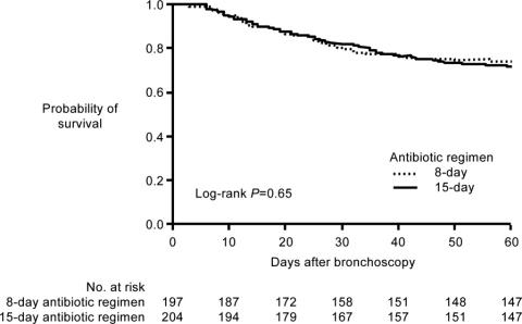
Initiation and Selection of Antibiotics
Early appropriate antimicrobial therapy is necessary to minimize the morbidity and mortality associated with hospital‐ or healthcare‐associated infections (HAIs). A number of studies have demonstrated that delayed or inadequate antimicrobial therapy leads to worse clinical outcomes and higher healthcare costs.1, 2 Inadequate antimicrobial therapy can also promote or enhance the development of resistance,2 with potential wide‐ranging impact beyond the immediate patient under care. Because delaying treatment until availability of culture results decreases the likelihood of a successful outcome, patients with a suspected invasive HAI commonly receive empiric therapy with a regimen expected to cover the most likely causative pathogens. Based on characteristics of the patient and healthcare facility or unit, likely pathogens may include bacteria or other pathogens resistant to 1 or more antimicrobial drug classes. This article discusses the various processes and factors that need to be considered when choosing empiric antibiotics in the hospital or other healthcare setting, and uses 3 case studies dealing with pneumonia, intra‐abdominal infection, and bacteremia, respectively, to illustrate points of interest.
IMPORTANCE OF EARLY ADEQUATE ANTIBIOTIC USE
The initial selection and early deployment of adequate antimicrobial therapy is critical for successful resolution of HAIs. The terms inadequate and inappropriate antimicrobial therapy are commonly used interchangeably in the literature, and can be defined as use of antimicrobial treatment without (sufficient) activity against the identified pathogen.2 Using an antibiotic for a fungal infection would be inadequate, as would using a drug or dosing regimen that is ineffective against the identified bacterial species due to resistance or a failure to achieve the drug's pharmacokinetic/pharmacodynamic target for efficacy against the pathogen. The complete absence of antimicrobial therapy is also considered inadequate therapy. Some investigators consider inappropriate therapy a more general term that includes excessive treatment as well as inadequate treatment.1 Others reserve the term inappropriate for use of an antimicrobial without activity against the identified pathogen, and the term inadequate for use of an insufficient regimen, either in terms of optimal dose, route of administration, timeliness, or failure to use combination therapy when appropriate.3 However, many or most research articles do not make the distinction, and the current article does not make a distinction.
In addition, some articles arbitrarily define inadequate therapy as either use (or absence) of a treatment without activity against the identified pathogen or a delay in appropriate or adequate treatment, eg, no patient exposure to adequate treatment within 24 hours of hospital admission. It is important to recognize this when evaluating articles in the literature. Other studies separate inadequate and delayed therapy as variables. However, when dealing with empiric therapy, the adequacy of initial empiric therapy cannot be fully determined until subsequent possession of the tissue/blood culture results.
Inadequate Antibiotic Treatment
A 1999 study by Kollef et al. identified inadequate antimicrobial treatment as the most important independent predictor of hospital mortality, in a group of patients with a nosocomial or community‐acquired infection, while in the medical or surgical intensive care unit (ICU).4 Infection sites included in the study were lung, bloodstream, urinary tract, gastrointestinal (GI) tract, and wound. Various other studies have confirmed an association between inadequate antibiotic therapy and increased hospital mortality, and some demonstrated a relationship between inadequate antibiotic therapy and longer hospital or ICU stays57 and higher hospital‐related costs.8 More specifically, initial inappropriate antibiotic therapy has been associated with increased mortality in patients with healthcare‐associated9 or ventilator‐associated pneumonia (VAP)5, 7, 8, 1014 and those with bacteremia/sepsis.6, 10, 1524 Inadequate empiric therapy has also been linked with worsened outcomes,19, 2529 longer hospital stays,2527, 29 and increased healthcare costs25, 29 in patients with infections of the GI tract.
With respect to specific bacterial pathogens, inappropriate antibiotic therapy has been shown to increase risk of hospital mortality for patients with VAP or bacteremia caused by Pseudomonas aeruginosa,20, 30 extended‐spectrum ‐lactamase (ESBL)‐producing or multidrug‐resistant (MDR) Klebsiella pneumoniae or Escherichia coli,21, 31, 32 and methicillin‐resistant Staphylococcus aureus (MRSA).15, 23 In fact, infection with resistant bacteria, and particularly MDR bacteria, is a principal risk factor for inadequate initial antibiotic therapy.14, 16, 33, 34 A recent study by Teixeira and coworkers showed that inadequate therapy was more than twice as common for additional episodes of VAP caused by MDR pathogens as for those involving drug‐susceptible pathogens (56% vs 25.5%).14 Moreover, VAP caused by MDR pathogens was identified as a significant independent predictor of inadequate antimicrobial therapy (odds ratio [OR], 3.07; 95% confidence interval [CI], 1.29‐7.30; P = 0.01). Infections caused by drug‐resistant versus susceptible bacteria have generally been associated with increased morbidity, longer hospital or ICU stays, and higher costs.24, 3437 At least part of the reason for these worsened outcomes appears to be an increased likelihood that initial therapy is inadequate for the causative agent. Because of this, it is particularly important to consider the probability of infection with resistant bacteria when initiating empiric antibiotic therapy.
Delayed Antibiotic Treatment
In addition to inadequate initial therapy, a delay in the onset of adequate therapy has also been shown to have negative impact on outcome in patients with VAP, bacteremia, or intra‐abdominal infections.12, 13, 27, 38, 39 For example, Iregui et al. identified administration of initially delayed appropriate antibiotic treatment (treatment delayed for 24 hours after initial diagnosis of VAP) as a significant predictor of hospital mortality (OR, 7.68; 95% CI, 4.50‐13.09; P 0.001) in patients with VAP at a US teaching hospital.38 Similarly, Lodise et al. identified delayed antibiotic treatment as an independent predictor of infection‐related mortality in patients with hospital‐acquired S aureus bacteremia (OR, 3.8; 95% CI, 1.3‐11.0; P = 0.01).39 Delayed versus early antibiotic therapy was also associated with significantly longer hospital stay (20.2 vs 14.3 days, P = 0.05). Classification and regression tree analysis identified 44.75 hours from the initial positive blood culture result to appropriate therapy as the breakpoint between delayed and early treatment for bacteremia.
Of particular interest, evidence suggests that the negative impact of initial delay or initial use of inadequate therapy often cannot be remedied by subsequent treatment alterations. For example, Luna et al. reported a significantly lower hospital mortality rate for VAP patients who received early adequate antibiotic therapy compared with those who received early inadequate therapy (38% vs 91%, P 0.001).13 In this study, early treatment referred to drug administration prior to bronchoscopy, which was performed within 24 hours of clinical diagnosis of VAP. A subset of patients only received treatment after bronchoscopy, and the mortality rate for VAP‐positive patients who received adequate antibiotic therapy after this initial delay was similar to that for VAP‐positive patients who received inadequate therapy postbronchoscopy (71% vs 70%). In other words, the negative impact of an initial delay in adequate therapy could not be subsequently overcome by using adequate antibiotic therapy later in the disease process. Similarly, a recent study by Zilberberg et al. of healthcare‐associated pneumonia reported that the negative effect of initial inadequate antibiotic therapy on hospital mortality could not be mitigated by subsequent escalation of adequate antibiotic therapy after reception of culture results.9 Finally, a study of inadequate initial empiric antibiotic therapy of postoperative intra‐abdominal infection (peritonitis) also showed that adverse outcomes could not be abrogated by changes in antibiotic therapy based on culture results.27 Taken together, the results from these studies emphasize the importance of early adequate antibiotic therapy.
PRACTICAL GUIDELINES FOR CHOOSING EMPIRIC ANTIBIOTICS
When choosing initial empiric therapy for a suspected hospital‐ or healthcare‐related bacterial infection, it is first important to determine if the patient has received prior antibiotic therapy, and if the patient has, then the clinician should consider choosing an antibiotic from a different drug class. This is because prior antibiotic therapy increases risk of infection with a pathogen resistant to the initial antibiotic drug and other members of its class. Also, depending on the site of the infection and likely pathogenic bacteria, the clinician will need to decide whether to initiate empiric therapy with a single antibiotic or combination of agents. A number of patient‐ and institution‐related factors can be utilized by clinicians to better identify the likely pathogen responsible for the infection, and it is critical to use this information when selecting initial empiric therapy. Finally, as is true whenever choosing antimicrobial or other drug therapies, clinicians need to consider and weigh the safety/tolerability profile, potential for drugdrug interactions, and relative cost of different treatment options. These will vary for individual patients receiving the same drug or drug combination.
MINIMIZING ANTIMICROBIAL RESISTANCE IN THE HOSPITAL OR HEALTHCARE SETTING
It is also important to consider the potential for development of antibiotic resistance when choosing initial empiric therapy. The current paradigm for treatment of serious hospital or healthcare infections is to prescribe broad‐spectrum antimicrobial therapy upfront while awaiting culture results, and to de‐escalate (or terminate) therapy once culture results are available4042or as 2 authors recently put it, get it right the first time, hit hard up front, and use large doses of broad‐spectrum antibiotics for a short period.41 The initial empiric antibiotic regimen should have a high likelihood of covering the most likely causative pathogens, including resistant species or strains. Furthermore, emergence of resistance is minimized when the initial regimen effectively covers the most likely causative pathogens, and subsequent culture results are utilized to streamline or narrow the initial regimen, when possible.40, 42 Emergence of resistance is also minimized by using the shortest duration of treatment with maximal clinical effect. (These latter 2 points are discussed in greater detail in the Kaye and File articles in this supplement.)
Factoring in Institution‐ and Patient‐Specific Factors
Local antibiograms are useful in determining the most likely infection‐causing pathogens, within different wards of the hospital, and their susceptibility or resistance to various antibiotics. Local patterns of pathogen susceptibility and resistance can differ markedly from national averages, so local antibiograms are more useful than national or even regional surveillance data when making choices about the initial agent and dosing regimen for initial empiric therapy.43 Hospitals are required by the Joint Commission to create antibiograms on at least an annual basis, although more frequent antibiograms are particularly useful, given that susceptibility or resistance patterns change over time. It is also important that hospital microbiologists create antibiograms specifically for different hospital wards or departments, as well as hospital‐wide. The incidence and susceptibility of pathogenic bacteria has been shown to vary across different wards within a hospital, as well as within different regions of a given country.4446
Patient‐specific factors should also be considered in the decision‐making process for selection of initial empiric therapy for a suspected bacterial infection. Relevant patient characteristics or factors may differ somewhat when examining risk for particular types of infection (eg, healthcare/hospital‐acquired pneumonia, VAP, or bacteremia) or particular antibiotic‐resistant pathogens (eg, MRSA, ESBL‐producing E coli or Klebsiella spp, P aeruginosa and MDR P aeruginosa, and carbapenem‐resistant Acinetobacter baumannii). Nonetheless, several risk factors appear to generally increase risk of infection with a resistant pathogen across these subcategories, including prior antibiotic treatment (with agents sometimes varying depending on the particular pathogen of interest); recent hospital admission or residence in a nursing home or extended‐care facility; prolonged hospital stay (particularly in the ICU); prior colonization with the pathogen; presence of an indwelling catheter (central venous, arterial, or urinary); and mechanical ventilation; among others.2, 47, 48 Patients who are immunocompromised, either due to their condition or immunosuppressive therapy, are also generally at increased risk of infection with resistant bacteria.47
Patient age and presence of comorbidities can also affect initial selection of empiric therapy. Cell‐mediated immunity tends to decline with age, and elderly individuals are also more likely to have conditions or comorbidities associated with diminished host immunity, both of which may contribute to increased susceptibility to infection and infection involving resistant bacteria.49 Furthermore, elderly individuals are more likely to have decline in renal or hepatic function or other physiologic changes that can alter drug pharmacokinetics and pharmacodynamics,50 and these factors need to be considered when selecting an initial empiric therapy regimen that covers likely pathogens, without increasing risk of drug toxicity. In addition, the increased number of comorbidities in elderly patients typically translates into polypharmacy, with potential for drugdrug interactions that need to be weighed when selecting initial empiric therapy.
Treat Infection, Not Contamination or Colonization
It is important to limit antimicrobial use to treatment of actual infections, and not for treatment of colonization or contamination. Treatment of colonization is a significant source of antimicrobial overuse. Hence, it is important that healthcare teams take appropriate steps to ensure they are treating pneumonia, bacteremia, or a urinary tract infection, not colonization of the tracheal aspirate, catheter tip or hub, or indwelling urinary catheter that is unassociated with actual infection. Strategies to employ when considering how to differentiate between true infection and colonization include using Gram stain in sputum specimens to look for evidence of polymorphonuclear leukocytes (inflammation), understanding that certain organisms, such as Enterococcus and Candida are not respiratory pathogens, recognizing that urinary catheters may be colonized in the absence of infection, and remaining vigilant regarding blood culture contamination. Since antibiotic use is generally linked to increased risk of resistance,51, 52 antibiotics should only be used when there is a clear clinical benefit associated with their use; treatment of colonization does not fit this description. When in doubt, an infectious diseases (ID) specialist consultation is recommended.
Similarly, overuse/misuse of antibiotics that occurs due to false‐positive culture results also increases development of resistance in hospitalized patients. In particular, contamination of blood cultures is relatively common in hospitalized patients, particularly in hospital emergency rooms,53 and frequently results in administration of antibiotics to treat an apparent infection that actually represents a contaminated culture. Antimicrobial treatment due to false‐positive blood culture results has been associated with prolonged hospitalization and elevated laboratory and hospital costs,54, 55 and provides an environment for development of antimicrobial resistance. Contamination of blood cultures often occurs at the point of blood collection via venipuncture or through indwelling catheters,56, 57 but can occur later in the process during laboratory handling or processing of specimens.58 Hence, it is important to use proper antisepsis when collecting blood or other cultures, to make sure it is blood and not skin or the catheter hub that is being cultured, and to make sure to use proper methods when processing all cultures.
Consult Infectious Diseases Experts
The 2007 guidelines from the Infectious Diseases Society of America and the Society for Healthcare Epidemiology of America (IDSA/SHEA) for the development of institutional antimicrobial stewardship programs recommend inclusion of an ID physician and a clinical pharmacist with ID training as core members of a multidisciplinary antimicrobial stewardship team.59 Consultation with an ID expert or inclusion of an ID specialist into an institutional antimicrobial stewardship program has been shown to improve antibiotic usage and reduce morbidity and mortality, length of hospital stay, healthcare costs, and resistance.6064 In hospitals without easy access to an ID specialist, hospitalists with ID training may be able to fulfill the role provided by ID physicians or clinical pharmacists with ID training.
CASE 1: HEALTHCARE‐ASSOCIATED PNEUMONIA
Table 1 provides the key initial data for Case 1. The patient has a history of hypertension, congestive heart failure (CHF), and myocardial infarction (MI), and is receiving medications consistent with such a history. In terms of her acute presentation, cough, fever, chills, dyspnea, lung crackles (rales), X‐ray evidence of lung infiltrate, reduced oxygen saturation, elevated white blood cell (WBC) count, and increased percentage of WBC bands are all consistent with a diagnosis of pneumonia of relatively recent origin. Progressive worsening of symptoms within the previous 36 hours is also consistent with infection of recent origin. There are no neurologic symptoms, and cardiac function appears relatively normal, with no evidence of MI or cardiac arrhythmia based on electrocardiogram or heart rate, although there is evidence of continuing hypertension and perhaps CHF.
| |
| History | A 72‐yr‐old woman recently hospitalized for congestive heart failure (CHF), returns to the emergency department from rehab with cough, fever, chills, shortness of breath, all progressively worsening over the past 36 hr |
| Past history of CHF (ejection fraction 44%), myocardial infarction 2 yr ago, hypertension, past smoking | |
| Medications: metoprolol 50 mg BID; furosemide 40 mg daily; aspirin 81 mg daily; enalapril 20 mg daily | |
| Physical | Vitals: BP 148/88, P 82, RR 16, T 101.7, O2 sat 92% on room air |
| Heart: S1, S2 no murmurs | |
| Lungs: crackles at R lung base | |
| Abdomen: bowel sounds present, non‐tender | |
| Extremities: trace edema bilaterally | |
| Neurologic: no focal findings | |
| Labs | EKG: NSR, no acute ST‐T changes |
| Chemistry, hemoglobin, platelets, within normal limits | |
| WBC: 14,700/mm3, 10% bands | |
| Cardiac enzymes negative | |
| ‐Natriuretic peptide within normal limits for age | |
| Chest X‐ray: right lower lung infiltrate | |
Given the patient's history of recent hospitalization, the clinician should consider that the pneumonia is most likely hospital‐ or healthcare‐acquired. Because she was described as developing the problem while in rehabilitation, it can be assumed that hospitalization occurred relatively recently. Hospital‐acquired pneumonia (HAP) is defined as pneumonia that occurs within 48 hours of hospital admission, and that was not incubating at the time of admission.47, 65 HAP accounts for approximately of 15% of all nosocomial/hospital‐acquired infections in the United States and up to 27% in the ICU,65, 66 and is a frequent cause of morbidity and mortality in this setting.65 In addition to hospitalization, other characteristics or risk factors for HAP include severe illness, hemodynamic compromise, depressed immune function, use of nasogastric tubes, and mechanical ventilation for the important subset of HAP patients with VAP.65 VAP is more precisely defined as HAP that arises >48‐72 hours after endotracheal intubation.47
Healthcare‐associated pneumonia (HCAP) is a more recently defined category that includes patients with HAP and VAP, and is characterized by hospitalization for 2 days in the preceding 90 days, or residence in a nursing home or extended‐care facility.47, 67, 68 Additional risk factors for HCAP include intravenous therapy at home (including antibiotics); intravenous chemotherapy or wound care within 30 days of the current infection; and recent attendance at a hospital or hemodialysis clinic.47 Most HAP or HCAP data have been derived from patients with VAP, and the 2005 American Thoracic Society (ATS)/IDSA guidelines for management of HAP, VAP, and HCAP recommend similar approaches for the initial treatment of patients with nonintubated HAP, VAP, and HCAP.47 There are general similarities between these 3 disease categories with respect to etiology, epidemiology of likely pathogens, and prognosis, and important differences compared with community‐acquired pneumonia (CAP), which in turn gives rise to different treatment strategies for HAP/VAP/HCAP and CAP.
Given an initial diagnosis of HAP or HCAP, the clinician should be considering the following questions: 1) What are the appropriate choices of antimicrobials? 2) What are the clinical parameters that should alert one to resistant organisms? 3) What are the appropriate cultures to order? 4) What is the role of Gram stain, if any?
Selection of Initial Empiric Therapy for Likely Pathogens
HAP is usually caused by bacterial pathogens, and much more rarely involves viruses or fungi in immunocompetent patients. Therefore, from the start, the focus should be on empiric therapy with an antibiotic or combination of antibiotics that has a high probability of covering the most likely pathogens. Empiric therapy is warranted because of the risk of mortality or other negative consequences when antibiotic therapy is delayed, particularly in an aged patient with a chronic illness like CHF (such as the case study here). Clues as to likely pathogensand hence most appropriate antibiotic regimencan be discerned by looking at the onset of HAP/VAP (early vs late) and whether the patient has risk factors for infection with a MDR or antibiotic‐susceptible bacterial pathogen. A significant proportion of patients with HAP, VAP, or HCAP are infected with MDR pathogens, and identifying these patients and providing them with appropriate broad‐spectrum empiric therapy is a key to successful management.
Early‐onset HAP/VAP (occurring 5 days after hospitalization) is more likely to be due to antibiotic‐sensitive bacteria than late‐onset HAP/VAP (occurring 5 days after hospitalization), which often occurs due to MDR species.47, 69, 70 Not surprisingly, risk of inappropriate initial antibiotic therapy14 is higher, and mortality is also higher in patients with late‐onset HAP/VAP.47, 71 Patients with early‐onset HAP/VAP who have received prior antibiotics or been hospitalized within the past 90 days are also at risk for infection due to MDR bacteria, and hence should be treated the same as patients with late‐onset HAP/VAP.47 Additional risk factors for infection with MDR pathogens include antimicrobial therapy in the preceding 90 days (particularly with broad‐spectrum agents), current hospitalization 5 days, high prevalence of antibiotic resistance in the specific hospital unit, immunosuppression, and presence of risk factors for HCAP (hospitalization 2 days in the preceding 90 days, residence in a nursing home or extended‐care facility, home infusion therapy, chronic dialysis within 30 days, home wound care, or family member with MDR pathogen).47 For VAP patients, duration of ventilator support 7 days is an additional risk factor for infection with a MDR pathogen.70, 72 A second episode of VAP is more likely to be due either to MRSA or P aeruginosa; therefore, these organisms need to be considered when selecting initial empiric therapy.
The most common bacterial causes of HAP, VAP, or HCAP include aerobic Gram‐negative bacilli, such as P aeruginosa, Acinetobacter spp, and Enterobacteriaceae (eg, K pneumoniae, E coli, Enterobacter spp), and Gram‐positive cocci, such as S aureus and Streptococcus pneumoniae.47, 73 MDR bacterial species are more likely when certain risk factors are present, and the ATS/IDSA guidelines recommend using a risk‐stratification process when selecting empiric antibiotic therapy for patients with suspected HAP, VAP, or HCAP.47 The guidelines also emphasize that local conditions can greatly impact whether a patient is infected with an antibiotic‐sensitive or antibiotic‐resistant species, regardless of other risk factors, thereby highlighting the importance of using recent hospital and hospital unit‐specific antibiograms when stratifying a patient based on risk.
Other guiding principles of initial empiric treatment, as outlined in the ATS/IDSA guidelines, include not delaying therapy while awaiting culture results and administration of therapy as soon as possible following diagnosis; making sure the dosing regimen as well as drug selection is appropriate/adequate for the patient and suspected pathogen; and using a different class of antibiotic for patients with prior antibiotic exposure.47 The guidelines further indicate that combination therapy is appropriate initial therapy in patients at high risk for infection with MDR bacteria, and that for patients receiving combination therapy including an aminoglycoside, the aminoglycoside can be stopped after 5‐7 days in responding patients. Table 2 highlights ATS/IDSA recommendations for initial empiric antibiotic therapy in patients with suspected HAP/VAP who have early‐onset disease and no risk factors for MDR pathogens.47 Table 3 highlights recommendations for initial empiric antibiotic therapy in patients with suspected HAP/VAP/HCAP who have late‐onset disease and/or risk factors for MDR pathogens.47
| Potential Pathogen | Recommended Antibiotic |
|---|---|
| |
| Streptococcus pneumoniae* | |
| Haemophilus influenzae | Ceftriaxone |
| Methicillin‐sensitive Staphylococcus aureus | or |
| Antibiotic‐sensitive enteric Gram‐negative bacilli | Levofloxacin, moxifloxacin, or ciprofloxacin |
| Escherichia coli | or |
| Klebsiella pneumoniae | Ampicillin/sulbactam |
| Enterobacter spp | or |
| Proteus spp | Ertapenem |
| Serratia marcescens | |
| Potential Pathogen | Combination Antibiotic Therapy |
|---|---|
| |
| Pathogens listed in Table 2, plus MDR pathogens | Antipseudomonal cephalosporin (cefepime, ceftazidime) |
| Pseudomonas aeruginosa | or |
| Klebsiella pneumoniae (ESBL‐positive)* | Antipseudomonal carbapenem (imipenem or meropenem) |
| Acinetobacter spp* | or |
| ‐Lactam/‐lactamase inhibitor (piperacillin‐tazobactam) | |
| plus | |
| Antipseudomonal fluoroquinolone* (ciprofloxacin or levofloxacin) | |
| or | |
| Aminoglycoside (amikacin, gentamicin, or tobramycin) | |
| plus | |
| Methicillin‐resistant Staphylococcus aureus (MRSA) | Linezolid or vancomycin |
| Legionella pneumophila* | |
Returning to Case 1, given a clinical diagnosis of HAP/HCAP and the patient's heightened risk for infection with MRSA or resistant Gram‐negative bacteria, she was initiated on a regimen consisting of piperacillin/tazobactam plus vancomycin and ciprofloxacin. The choice of 3 agents is consistent with ATS and IDSA guidelines to cover the potential for an ESBL‐producer or Acinetobacter, or Pseudomonas. Individual choices should be dictated by one's own institutional antibiogram or some knowledge of the rehabilitation facility from which the patient was transferred.
Guiding Principles for Culture Management
Culture collection and management plays an important role in diagnosis and subsequent treatment of HAP/VAP or HCAP. As outlined in the ATS/IDSA guidelines, patient management typically proceeds using either a clinical strategy or bacteriologic strategy, or a combination thereof.47 The clinical strategy makes use of cultures of endotracheal aspirates or sputum, with initial microscopic examination and Gram staining to identify bacterial growth and guide initiation of empiric antibiotic therapy. Cultures should always be performed before instituting antibiotic therapy. Microorganism growth is described as light, moderate, or heavy by microbiology laboratories using semiquantitative analysis. Gram staining should be performed only if the specimen is of good quality; most microbiology laboratories will do screening tests to ensure they are of good quality, or else will reject the specimen. When correlated with culture results, Gram staining can improve diagnostic accuracy.74
The bacteriologic strategy uses quantitative means to analyze and describe cultures of lower respiratory secretions or specimens obtained via endotracheal aspirates, mini‐bronchoalveolar lavage specimens (mini‐BAL), bronchoalveolar lavage, or protected‐specimen bronchial brushing, collected with or without a bronchoscope, ie, with or without invasive techniques. Hence, whereas the clinical strategy uses noninvasive tracheal aspirates to culture microorganisms for analysis, the bacteriologic strategy often employs a relatively noninvasive strategy like a mini‐BAL, or invasive (bronchoscopic) lower respiratory tract samples for quantitative culture analysis. Diagnosis of HAP/VAP or HCAP, and determination of the causative microorganism(s), requires growth above a certain threshold when using the semi‐quantitative analysis. The clinical approach is more sensitive, but can result in overtreatment, while the bacteriologic strategy is associated with risk of undertreatment due to false‐negative culture results. On the other hand, quantitative cultures increase the specificity of diagnosis.
CASE 2: INTRA‐ABDOMINAL INFECTION (DIVERTICULITIS)
Case 2 is a 56‐year‐old woman with no past medical history of note, who presented to her physician about 3 days ago, after 5 days of abdominal pain and fever (101.7F). She had an outpatient computed tomography (CT) scan, and the results suggested diverticulitis. After the CT scan, she was given amoxicillin/clavulanate. She now presents to the emergency department (3 days later) with worsening pain, fever, and severe weakness. A physical exam shows low blood pressure (84/58 mmHg) and tachycardia (132 bpm). Her respiratory rate is 22 breaths per minute. Oxygenation (O2 saturation 99% on room air) is normal, and the patient's lungs are clear. Abdominal examination reveals bowel sounds and diffuse tenderness, particularly at the left lower quadrant, and there is evidence of guarding and rebound. Her blood chemistry is generally normal, although the WBC count is elevated (15,200/mm3). No abnormalities are evident on chest X‐ray. The patient's blood pressure increases to 96/64 mmHg after she is infused with 2 liters of normal saline. She undergoes another CT scan and is admitted to the ICU. The CT scan shows diverticulitis with abscess and walled‐off perforation. An interventional radiologist inserts a pigtail catheter into the abscess for sample collection, and the samples are sent to the microbiology laboratory for culture.
The radiology results indicate that the patient has what may be considered a complicated intra‐abdominal infection (diverticulitis with abscess), community‐acquired. Because empiric therapy is usually necessary for patients with complicated or even uncomplicated intra‐abdominal infections, the clinician should now be asking: What is optimal empiric antimicrobial therapy for this patient? Both prior75 and current guidelines76 for the management of intra‐abdominal infection indicate that antimicrobial therapy should be initiated when a patient receives such a diagnosis or when such an infection is considered likely. In making the determination of initial empiric therapy, the clinician should also be considering whether there is likely involvement of resistant Gram‐negative bacteria, and if so, how that would change the choice of antibiotic therapy.
Enteric Gram‐negative bacilli such as E coli and K pneumoniae are the most common microorganisms isolated from patients with intra‐abdominal infections, although Gram‐positive cocci (Staphylococcus or Streptococcus spp, and less commonly, enterococci) and obligate anaerobic organisms (particularly, Bacteroides fragilis) are also frequent components of intra‐abdominal infections.77 The relative frequency of bacterial pathogens shifts in patients who acquired their intra‐abdominal infection in the hospital versus community setting, with greater prevalence of Enterobacter, P aeruginosa, and Enterococcus spp, and less frequent isolation of E coli and streptococci.77 Recent results from the Study for Monitoring Antimicrobial Resistance Trends (SMART) indicate a general increase in resistance among Gram‐negative bacilli isolated from patients with intra‐abdominal infections treated in medical centers, primarily due to acquisition of ESBLs.78, 79 This is true both in the United States79 and worldwide.78 Carbapenems continue to exhibit consistent activity against Gram‐negative bacilli isolated from intra‐abdominal infections, including ESBL‐producers.
Selection of initial empiric therapy should incorporate information from the literature and local antibiograms to determine the most likely causative pathogen(s), including ones with reduced susceptibility or resistance to commonly employed antibiotics. Then an antibiotic regimen should be selected that provides coverage of likely pathogens with minimal adverse events, including risk of collateral damage such as Clostridium difficile‐associated disease. Dose and dosing interval considerations are also important, particularly in patients with reduced renal or hepatic function. The general approach is to select an antibiotic or combination of antibiotic agents to provide coverage of the bacterial pathogens most commonly isolated from patients with intra‐abdominal infections, ie, aerobic/facultative anaerobic Gram‐negative bacilli, aerobic Gram‐positive cocci, and obligate anaerobic organisms.77 Table 4 presents the antibiotic treatment recommendations from the Surgical Infection Society and IDSA 2010 guidelines for management of patients with complicated intra‐abdominal infections.76 The guidelines are based on whether the patient has mild‐to‐moderate or high‐risk/severe community‐acquired complicated intra‐abdominal infections. Recommendations for the empiric treatment of hospital or healthcare‐associated complicated intra‐abdominal infections are largely based on local antibiogram (microbiologic) results, and include some similarities and differences compared with recommended treatment of high‐risk community‐acquired infections, as illustrated in Table 5.76 A patient with mild‐to‐moderate infection would be someone who does not require intensive care, and has community‐acquired intra‐abdominal infection due to secondary peritonitis. Severe intra‐abdominal infection would be defined by requiring intensive care, having sepsis, or having healthcare‐acquired peritonitis (such as a bowel leak following surgery). In line with these guidelines, and considering the case patient's risk profile, ciprofloxacin plus metronidazole was selected as initial empiric therapy, because she had not been hospitalized previously. Although she was hypotensive, the blood pressure was easily raised with fluids.
| Community‐Acquired Infection in Adults | ||
|---|---|---|
| Regimen | Mild‐to‐Moderate Severity* | High Risk or Severity |
| ||
| Single agent | Cefoxitin, ertapenem, moxifloxacin, tigecycline, and ticarcillin‐clavulanate | Imipenem‐cilastatin, meropenem, doripenem, and piperacillin‐tazobactam |
| Combination | Cefazolin, cefuroxime, ceftriaxone, cefotaxime, ciprofloxacin, or levofloxacin, each in combination with metronidazole | Cefepime, ceftazidime, ciprofloxacin, or levofloxacin, each in combination with metronidazole |
| Organisms Seen in the Hospital/Healthcare Infection at the Local Institution | Regimen | ||||
|---|---|---|---|---|---|
| Carbapenem* | Piperacillin‐Tazobactam | Ceftazidime or Cefepime + Metronidazole | Aminoglycoside | Vancomycin | |
| |||||
| 20% Resistant Pseudomonas aeruginosa, ESBL‐producing Enterobacteriaceae, Acinetobacter, or other multidrug‐resistant Gram‐negative bacteria | Recommended | Recommended | Recommended | Not recommended | Not recommended |
| ESBL‐producing Enterobacteriaceae | Recommended | Recommended | Not recommended | Recommended | Not recommended |
| P aeruginosa >20% resistant to ceftazidime | Recommended | Recommended | Not recommended | Recommended | Not recommended |
| Methicillin‐resistant Staphylococcus aureus | Not recommended | Not recommended | Not recommended | Not recommended | Recommended |
There are a number of controversies in intra‐abdominal sepsis management. These include whether or when initial empiric therapy should provide coverage of Enterococcus/vancomycin‐resistant enterococci or MRSA, when the regimen should include an antifungal to cover possible Candida spp infection, the role of resistant Bacteroides in intra‐abdominal infections, and when clinicians should be particularly concerned about ESBL‐producing or other resistant Gram‐negative bacteria. These topics are beyond the reach of the present article, but are and will continue to be important issues for clinicians to grapple with when selecting initial empiric therapy for patients with intra‐abdominal infections.
CASE 3: CENTRAL LINE‐ASSOCIATED BACTEREMIA
The third case is a 56‐year‐old man with epilepsy who presents to the emergency department with status epilepticus. Subsequent resuscitative efforts included intubation and placement of an internal jugular central line. The patient was admitted to the ICU, and aggressive treatment was initiated with repeated intravenous dosing of lorazepam and loading with fosphenytoin, which successfully broke the seizure. Subsequent imaging and laboratory tests failed to reveal any specific cause for the status epilepticus. The patient was extubated on day 4 and transferred out of the ICU. On day 5, he spiked a fever of 103.4F. He did not report any new symptoms, and there was no evidence of cough, sputum, shortness of breath, abdominal pain, diarrhea, or urinary symptoms. Physical examination revealed normal blood pressure (122/68 mmHg) and oxygen saturation (95% on room air), a normal respiratory rate (12 breaths per minute), clear lungs, no edema, no heart murmur, and normal neurologic findings. The patient's heart rate was somewhat high (102 bpm), and his temperature remained elevated (103.4F). Abdominal examination revealed no tenderness, and bowel sounds were present. Laboratory results were normal, except for an elevated WBC count (17,000/mm3 of blood). The chest X‐ray was clear.
This is an example of a patient with fever and leukocytosis of unknown origin. There are no focal findings indicative of a particular infection site or process. The patient was treated in the ICU, including use of a central catheter. Differential diagnosis of fever and leukocytosis without source, in a patient from the ICU with a central line, should consider catheter‐associated bacteremia; C difficile‐associated disease; a silent intra‐abdominal process, such as cholangitis or gangrenous cholecystitis; drug fever related to (in this patient) anticonvulsant therapy; urinary tract infection (no evidence for in this patient); or pulmonary embolism. The clinician needs to make a decision as to the relative benefits of empiric antibiotic or other antimicrobial treatment versus observation. If the patient is to be treated with an antibiotic, then a choice has to be made as to the best agent for the patient at hand.
In terms of the choice of antibiotics for a patient such as the one here, the clinician needs to assess the severity of illness and, when doing so, determine what infection site or sites should be covered, and the most likely sites of infection. A determination of likely pathogens also needs to be made. Cultures should be obtained prior to initiating therapy with a regimen providing broad coverage of the most likely pathogen(s), while allowing for the possibility of later de‐escalation based on clinical evaluation and culture results. This is similar to the situation for initial empiric treatment of pneumonia or intra‐abdominal infection. In general, the clinician should obtain blood, urine, and possibly sputum cultures to aid in future decision making. Furthermore, if the patient has diarrhea (which the current one does not), the clinician should obtain a stool for C difficile toxin analysis.
For the case illustrated here, the clinician determined catheter‐associated bacteremia was a strong possibility, and decided to initiate empiric therapy with vancomycin and piperacillin‐tazobactam to provide coverage of MRSA and resistant Gram‐negative bacteria. The most common causes of nosocomial or catheter‐associated bloodstream infections (BSIs) are coagulase‐negative staphylococci, S aureus, enterococci, and Candida spp,8082 but Gram‐negative bacilli like P aeruginosa, Klebsiella spp, and E coli (among others) are also frequently involved, particularly in patients with catheter‐associated BSIs.81 Moreover, significant and increasing percentages of Gram‐negative bacilli exhibit resistance to 1 or more antibiotic classes,8385 and >50% of S aureus are typically MRSA,82, 83, 85, 86 although there has been some decline in MRSA central line‐associated BSIs in US ICUs in recent years.87
Clinical practice guidelines from the IDSA recommend vancomycin (or daptomycin) for the management of MRSA bacteremia,88 while piperacillin‐tazobactam is frequently empirically added to Gram‐positive coverage for serious hospital‐acquired infections because of its broad activity against many pathogenic bacteria, including some ESBL‐producing Gram‐negative bacteria and P aeruginosa,89 which are significant causes of patient morbidity and mortality.36, 90 However, to be effective, both vancomycin and piperacillin‐tazobactam need to be properly dosed to maximize their pharmacodynamic properties. Guidelines from the American Society of Health‐System Pharmacists, IDSA, and Society of Infectious Diseases Pharmacists recommend vancomycin serum trough concentrations of 15‐20 mg/L for patients with bacteremia due to MRSA.91 These levels are recommended to improve penetration, increase the probability of obtaining optimal target serum concentrations, and improve clinical outcomes. To achieve these trough levels, the guidelines recommend doses of 15‐20 mg/kg of actual body weight given every 8‐12 hours for most patients with normal renal function, assuming a minimum inhibitory concentration (MIC) of 1 mg/L. In seriously ill patients, the guidelines recommend using a loading dose of 25‐30 mg/kg to facilitate rapid attainment of the target trough serum vancomycin level. (If the MIC is 2 mg/L, then the targeted pharmacodynamic parameter for vancomycin is unachievable, and an alternative therapy should be considered.)
The pharmacodynamic parameter that best predicts efficacy for ‐lactams like piperacillin is the duration of time that free drug concentrations remain above the MIC (T>MIC), with near maximal bactericidal effects for penicillins when the free drug concentrations remain above the MIC for 50% of the dosing interval.92 The target pharmacodynamic parameter for piperacillin‐tazobactam (50% T>MIC) may be better achieved with use of prolonged or extended infusion regimens than with intermittent, more rapidly infused, administration schedules. Lodise and coworkers recently reported that extended infusion (3.375 g intravenously [IV] for 4 hours every 8 hours) versus intermittent infusion of piperacillin‐tazobactam (3.375 g IV for 30 minutes every 4 to 6 hours) was associated with a significantly lower 14‐day mortality rate (12.2% vs 31.6%, P = 0.04) and median duration of hospital stay (21 vs 38 days, P = 0.02) in a cohort of hospitalized patients with a P aeruginosa infection.93 Based on data such as these, the case patient here was initiated on vancomycin (15‐20 mg/kg every 812 hours) and piperacillin‐tazobactam (3.375 g IV for 4 hours every 8 hours).
CONCLUSIONS
Successful treatment of patients with serious, life‐threatening hospital‐ or healthcare‐associated infections depends on early adequate antimicrobial treatment. To accomplish this, empiric therapy is typically employed with a broad‐spectrum regimen intended to cover likely causative pathogen(s) based on local antibiograms and risk factors for involvement of resistant microorganisms. Choice of empiric therapy should also be based on the site of infection, and make use of clinical practice guidelines, when available. Although this approach often means treatment with a regimen that is unnecessarily broad, based on subsequent culture findings, it is warranted based on the significant negative impact of initial inadequate/emnappropriate empiric therapy, and the inability to remedy this negative effect by later modification of antimicrobial therapy. The possibility of de‐escalating the initial broad‐spectrum regimen is revisited after the results from cultures collected prior to beginning empiric therapy become available, generally 2‐4 days after beginning the process. In this manner, both the dangers of initial inadequate empiric therapy and overuse or misuse of antimicrobials are minimized. To further minimize the risk of antimicrobial resistance linked to overuse or misuse of antimicrobial agents, care should be taken to avoid treatment of colonization or culture contamination.
- ,.Appropriate vs. inappropriate antimicrobial therapy.Clin Microbiol Infect.2008;14(suppl 3):15–21.
- .Inadequate antimicrobial treatment: an important determinant of outcome for hospitalized patients.Clin Infect Dis.2000;31(suppl 4):S131–S138.
- .Treatment options for nosocomial pneumonia due to MRSA.J Infect.2009;59(suppl 1):S25–S31.
- ,,,.Inadequate antimicrobial treatment of infections: a risk factor for hospital mortality among critically ill patients.Chest.1999;115:462–474.
- ,,,.Impact of appropriateness of initial antibiotic therapy on the outcome of ventilator‐associated pneumonia.Intensive Care Med.2001;27:355–362.
- ,,, et al.Mortality and morbidity attributable to inadequate empirical antimicrobial therapy in patients admitted to the ICU with sepsis: a matched cohort study.J Antimicrob Chemother.2008;61:436–441.
- ,,,,,.Effect from multiple episodes of inadequate empiric antibiotic therapy for ventilator‐associated pneumonia on morbidity and mortality among critically ill trauma patients.J Trauma.2005;58:94–101.
- ,,,,,.Predictors of 30‐day mortality and hospital costs in patients with ventilator‐associated pneumonia attributed to potentially antibiotic‐resistant gram‐negative bacteria.Chest.2008;134:281–287.
- ,,,,.Antimicrobial therapy escalation and hospital mortality among patients with health‐care‐associated pneumonia: a single‐center experience.Chest.2008;134:963–968.
- ,,.Impact of inappropriate antibiotic therapy on mortality in patients with ventilator‐associated pneumonia and blood stream infection: a meta‐analysis.J Crit Care.2008;23:91–100.
- ,,, et al.Ventilator‐associated pneumonia: breaking the vicious circle of antibiotic overuse.Crit Care Med.2007;35:379–385; quiz 386.
- ,,, et al.Appropriateness and delay to initiate therapy in ventilator‐associated pneumonia.Eur Respir J.2006;27:158–164.
- ,,, et al.Impact of BAL data on the therapy and outcome of ventilator‐associated pneumonia.Chest.1997;111:676–685.
- ,,,,.Inadequate treatment of ventilator‐associated pneumonia: risk factors and impact on outcomes.J Hosp Infect.2007;65:361–367.
- ,,, et al.Predictors of mortality in patients with methicillin‐resistant Staphylococcus aureus (MRSA) bacteraemia: the role of empiric antibiotic therapy.Eur J Clin Microbiol Infect Dis.2007;26:239–245.
- ,,,,,.Inappropriate initial antimicrobial therapy and its effect on survival in a clinical trial of immunomodulating therapy for severe sepsis.Am J Med.2003;115:529–535.
- ,,,,.The influence of inadequate antimicrobial treatment of bloodstream infections on patient outcomes in the ICU setting.Chest.2000;118:146–155.
- ,,, et al.Bloodstream infections caused by antibiotic‐resistant gram‐negative bacilli: risk factors for mortality and impact of inappropriate initial antimicrobial therapy on outcome.Antimicrob Agents Chemother.2005;49:760–766.
- ,,,,,.The benefit of appropriate empirical antibiotic treatment in patients with bloodstream infection.J Intern Med.1998;244:379–386.
- ,,,,,.Pseudomonas aeruginosa bloodstream infection: importance of appropriate initial antimicrobial treatment.Antimicrob Agents Chemother.2005;49:1306–1311.
- ,,, et al.Antibiotic therapy for Klebsiella pneumoniae bacteremia: implications of production of extended‐spectrum beta‐lactamases.Clin Infect Dis.2004;39:31–37.
- ,,, et al.Impact of inappropriate empirical therapy for sepsis due to health care‐associated methicillin‐resistant Staphylococcus aureus.J Infect.2009;58:131–137.
- ,,, et al.Influence of vancomycin minimum inhibitory concentration on the treatment of methicillin‐resistant Staphylococcus aureus bacteremia.Clin Infect Dis.2008;46:193–200.
- ,,, et al.Multidrug‐resistant Pseudomonas aeruginosa bloodstream infections: risk factors and mortality.Epidemiol Infect.2011; Jan 13:1–10.
- ,,,,,.Economic consequences of failure of initial antibiotic therapy in hospitalized adults with complicated intra‐abdominal infections.Surg Infect (Larchmt).2008;9:335–347.
- ,,, et al.Effect of inappropriate initial empiric antibiotic therapy on outcome of patients with community‐acquired intra‐abdominal infections requiring surgery.Eur J Clin Microbiol Infect Dis.2004;23:682–687.
- ,,,,,.Emergence of antibiotic‐resistant bacteria in cases of peritonitis after intraabdominal surgery affects the efficacy of empirical antimicrobial therapy.Clin Infect Dis.1996;23:486–494.
- ,,, et al.Antibiotic treatment for surgical peritonitis.Ann Surg.1991;214:543–549.
- ,,, et al.Inappropriate initial treatment of secondary intra‐abdominal infections leads to increased risk of clinical failure and costs.Br J Clin Pharmacol.2005;60:438–443.
- ,,, et al.Optimal management therapy for Pseudomonas aeruginosa ventilator‐associated pneumonia: an observational, multicenter study comparing monotherapy with combination antibiotic therapy.Crit Care Med.2007;35:1888–1895.
- ,,,,,.Risk factors for and outcomes of healthcare‐associated infection due to extended‐spectrum beta‐lactamase‐producing Escherichia coli or Klebsiella pneumoniae in Thailand.Infect Control Hosp Epidemiol.2007;28:873–876.
- ,,, et al.Impact of antibiotic resistance and of adequate empirical antibiotic treatment in the prognosis of patients with Escherichia coli bacteraemia.J Antimicrob Chemother.2007;60:855–863.
- .Predicting methicillin resistance and the effect of inadequate empiric therapy on survival in patients with Staphylococcus aureus bacteremia.Arch Intern Med.2000;160:1001–1004.
- ,,,,,.Impact of multiresistance of gram‐negative bacteria in bloodstream infection on mortality rates and length of stay.Infection.2008;36:31–35.
- ,,,.Clinical and economic impact of common multidrug‐resistant gram‐negative bacilli.Antimicrob Agents Chemother.2008;52:813–821.
- .Review of studies of the impact on Gram‐negative bacterial resistance on outcomes in the intensive care unit.Crit Care Med.2009;37:1463–1469.
- ,,,,,.Impact of multidrug‐resistant Pseudomonas aeruginosa bacteremia on patient outcomes.Antimicrob Agents Chemother.2010;54:3717–3722.
- ,,,,.Clinical importance of delays in the initiation of appropriate antibiotic treatment for ventilator‐associated pneumonia.Chest.2002;122:262–268.
- ,,,.Outcomes analysis of delayed antibiotic treatment for hospital‐acquired Staphylococcus aureus bacteremia.Clin Infect Dis.2003;36:1418–1423.
- .Broad‐spectrum antimicrobials and the treatment of serious bacterial infections: getting it right up front.Clin Infect Dis.2008;47(suppl 1):S3–S13.
- ,.A new paradigm for treating infections: “go hard and go home.”Crit Care Resusc.2009;11:276–281.
- .Use of broad‐spectrum antimicrobials for the treatment of pneumonia in seriously ill patients: maximizing clinical outcomes and minimizing selection of resistant organisms.Clin Infect Dis.2006;42(suppl 2):S72–S81.
- ,.Antibiotic resistance: location, location, location.Clin Microbiol Infect.2007;13(suppl 2):7–16.
- ,,,,,.Ventilator‐associated pneumonia in a multi‐hospital system: differences in microbiology by location.Infect Control Hosp Epidemiol.2003;24:853–858.
- ,,, et al.Incidence and susceptibility of pathogenic bacteria vary between intensive care units within a single hospital: implications for empiric antibiotic strategies.J Trauma.2000;49:638–645; discussion645–646.
- ,,,,.Variations in etiology of ventilator‐associated pneumonia across four treatment sites: implications for antimicrobial prescribing practices.Am J Respir Crit Care Med.1999;160:608–613.
- Hospital‐Acquired Pneumonia Guideline Committee of the American Thoracic Society and Infectious Diseases Society of America.Guidelines for the management of adults with hospital‐acquired, ventilator‐associated, and healthcare‐associated pneumonia.Am J Respir Crit Care Med.2005;171:388–416.
- .The dilemma of multidrug‐resistant gram‐negative bacteria.Am J Med Sci.2010;340:232–237.
- .Antimicrobial resistance and aging: beginning of the end of the antibiotic era?J Am Geriatr Soc.2002;50:S226–S229.
- ,.Principles of antimicrobial use in older adults.Clin Geriatr Med.2007;23:481–497,v.
- .Antimicrobial use: risk driver of multidrug resistant microorganisms in healthcare settings.Curr Opin Infect Dis.2009;22:352–358.
- .Collateral damage and what the future might hold. The need to balance prudent antibiotic utilization and stewardship with effective patient management.Int J Infect Dis.2006;10:S17–S24.
- ,,,.Blood culture contamination in Tanzania, Malawi, and the United States: a microbiological tale of three cities.J Clin Microbiol.2006;44:4425–4429.
- ,,, et al.Clinical and economic impact of contaminated blood cultures within the hospital setting.J Hosp Infect.2011;77:233–236.
- ,,.Contaminant blood cultures and resource utilization. The true consequences of false‐positive results.JAMA.1991;265:365–369.
- ,,,.Contamination of catheter‐drawn blood cultures.J Clin Microbiol.2001;39:3393–3394.
- ,,.Comparison of utility of blood cultures from intravascular catheters and peripheral veins: a systematic review and decision analysis.J Med Microbiol.2008;57:1–8.
- ,,.Contamination of blood cultures during venepuncture: fact or myth?Postgrad Med J.1990;66:1053–1058.
- ,,, et al.Infectious Diseases Society of America and the Society for Healthcare Epidemiology of America guidelines for developing an institutional program to enhance antimicrobial stewardship.Clin Infect Dis.2007;44:159–177.
- ,,,,.The value of infectious diseases consultation in Staphylococcus aureus bacteremia.Am J Med.2010;123:631–637.
- ,,,,.Infectious diseases consultation lowers mortality from Staphylococcus aureus bacteremia.Medicine (Baltimore).2009;88:263–267.
- ,,,,,.The value of infectious diseases specialists: non‐patient care activities.Clin Infect Dis.2008;47:1051–1063.
- ,,, et al.The value of an infectious diseases specialist.Clin Infect Dis.2003;36:1013–1017.
- ,,, et al.Mortality of S. aureus bacteremia and infectious diseases specialist consultation—a study of 521 patients in Germany.J Infect.2009;59:232–239.
- ,.Hospital‐acquired pneumonia: pathophysiology, diagnosis, and treatment.Surg Clin North Am.2009;89:439–461,ix.
- ,,,.Nosocomial infections in medical intensive care units in the United States. National Nosocomial Infections Surveillance System.Crit Care Med.1999;27:887–892.
- ,,.Healthcare‐associated pneumonia in adults: management principles to improve outcomes.Infect Dis Clin North Am.2004;18:939–962.
- ,,.Healthcare‐associated infections. A useful concept?Curr Opin Crit Care.2009;15:419–424.
- ,,,,.Ventilator‐associated pneumonia: incidence, risk factors, and microbiology.Semin Respir Infect.2000;15:272–279.
- ,,, et al.Ventilator‐associated pneumonia caused by potentially drug‐resistant bacteria.Am J Respir Crit Care Med.1998;157:531–539.
- ,,.A study of ventilator‐associated pneumonia: incidence, outcome, risk factors and measures to be taken for prevention.Indian J Anaesth.2010;54:535–540.
- ,,, et al.Risk factors for infection by Pseudomonas aeruginosa in patients with ventilator‐associated pneumonia.Intensive Care Med.1994;20:193–198.
- ,,,.Tackling empirical antibiotic therapy for ventilator‐associated pneumonia in your ICU: guidance for implementing the guidelines.Semin Respir Crit Care Med.2009;30:102–115.
- ,,,,,.Diagnosing pneumonia during mechanical ventilation: the clinical pulmonary infection score revisited.Am J Respir Crit Care Med.2003;168:173–179.
- ,,,,.Guidelines for clinical care: anti‐infective agents for intra‐abdominal infection. A Surgical Infection Society policy statement.Arch Surg.1992;127:83–89.
- ,,, et al.Diagnosis and management of complicated intra‐abdominal infection in adults and children: guidelines by the Surgical Infection Society and the Infectious Diseases Society of America.Clin Infect Dis.2010;50:133–164.
- ,.Intra‐abdominal infections.Surg Clin North Am.2009;89:421–437,ix.
- ,,,.In vitro susceptibilities of aerobic and facultative anaerobic Gram‐negative bacilli from patients with intra‐abdominal infections worldwide from 2005–2007: results from the SMART study.Int J Antimicrob Agents.2009;34:585–588.
- ,,,,,.Susceptibility of gram‐negative pathogens isolated from patients with complicated intra‐abdominal infections in the United States, 2007–2008: results of the Study for Monitoring Antimicrobial Resistance Trends (SMART).Antimicrob Agents Chemother.2010;54:3031–3034.
- ,,, et al.NHSN annual update: antimicrobial‐resistant pathogens associated with healthcare‐associated infections: annual summary of data reported to the National Healthcare Safety Network at the Centers for Disease Control and Prevention, 2006–2007.Infect Control Hosp Epidemiol.2008;29:996–1011.
- ,.Central line‐associated bloodstream infections: prevention and management.Infect Dis Clin North Am.2011;25:77–102.
- ,,,,,.Nosocomial bloodstream infections in US hospitals: analysis of 24,179 cases from a prospective nationwide surveillance study.Clin Infect Dis.2004;39:309–317.
- National Nosocomial Infections Surveillance (NNIS) system report,data summary from January 1992 through June 2004, issued October 2004.Am J Infect Control.2004;32:470–485.
- ,,,.Antimicrobial resistance trends of Escherichia coli bloodstream isolates: a population‐based study, 1998–2007.J Antimicrob Chemother.2009;64:169–174.
- ,,, et al.International Nosocomial Infection Control Consortium (INICC) report, data summary for 2003–2008, issued June 2009.Am J Infect Control.2010;38:95–104,e102.
- ,,.The impact of antimicrobial‐resistant, health care‐associated infections on mortality in the United States.Clin Infect Dis.2008;47:927–930.
- ,,,,.Methicillin‐resistant Staphylococcus aureus central line‐associated bloodstream infections in US intensive care units, 1997–2007.JAMA.2009;301:727–736.
- ,,, et al.Clinical practice guidelines by the Infectious Diseases Society of America for the treatment of methicillin‐resistant Staphylococcus aureus infections in adults and children: executive summary.Clin Infect Dis.2011;52:285–292.
- ,,,.Pharmacokinetic evaluation of piperacillin‐tazobactam.Expert Opin Drug Metab Toxicol.2010;6:1017–1031.
- ,,, et al.Morbidity associated with Pseudomonas aeruginosa bloodstream infections.Diagn Microbiol Infect Dis.2009;64:311–319.
- ,,, et al.Therapeutic monitoring of vancomycin in adult patients: a consensus review of the American Society of Health‐System Pharmacists, the Infectious Diseases Society of America, and the Society of Infectious Diseases Pharmacists.Am J Health Syst Pharm.2009;66:82–98.
- ,.Pharmacokinetic and pharmacodynamic parameters of antimicrobials: potential for providing dosing regimens that are less vulnerable to resistance.Clin Pharmacokinet.2009;48:517–528.
- ,,.Piperacillin‐tazobactam for Pseudomonas aeruginosa infection: clinical implications of an extended‐infusion dosing strategy.Clin Infect Dis.2007;44:357–363.
Early appropriate antimicrobial therapy is necessary to minimize the morbidity and mortality associated with hospital‐ or healthcare‐associated infections (HAIs). A number of studies have demonstrated that delayed or inadequate antimicrobial therapy leads to worse clinical outcomes and higher healthcare costs.1, 2 Inadequate antimicrobial therapy can also promote or enhance the development of resistance,2 with potential wide‐ranging impact beyond the immediate patient under care. Because delaying treatment until availability of culture results decreases the likelihood of a successful outcome, patients with a suspected invasive HAI commonly receive empiric therapy with a regimen expected to cover the most likely causative pathogens. Based on characteristics of the patient and healthcare facility or unit, likely pathogens may include bacteria or other pathogens resistant to 1 or more antimicrobial drug classes. This article discusses the various processes and factors that need to be considered when choosing empiric antibiotics in the hospital or other healthcare setting, and uses 3 case studies dealing with pneumonia, intra‐abdominal infection, and bacteremia, respectively, to illustrate points of interest.
IMPORTANCE OF EARLY ADEQUATE ANTIBIOTIC USE
The initial selection and early deployment of adequate antimicrobial therapy is critical for successful resolution of HAIs. The terms inadequate and inappropriate antimicrobial therapy are commonly used interchangeably in the literature, and can be defined as use of antimicrobial treatment without (sufficient) activity against the identified pathogen.2 Using an antibiotic for a fungal infection would be inadequate, as would using a drug or dosing regimen that is ineffective against the identified bacterial species due to resistance or a failure to achieve the drug's pharmacokinetic/pharmacodynamic target for efficacy against the pathogen. The complete absence of antimicrobial therapy is also considered inadequate therapy. Some investigators consider inappropriate therapy a more general term that includes excessive treatment as well as inadequate treatment.1 Others reserve the term inappropriate for use of an antimicrobial without activity against the identified pathogen, and the term inadequate for use of an insufficient regimen, either in terms of optimal dose, route of administration, timeliness, or failure to use combination therapy when appropriate.3 However, many or most research articles do not make the distinction, and the current article does not make a distinction.
In addition, some articles arbitrarily define inadequate therapy as either use (or absence) of a treatment without activity against the identified pathogen or a delay in appropriate or adequate treatment, eg, no patient exposure to adequate treatment within 24 hours of hospital admission. It is important to recognize this when evaluating articles in the literature. Other studies separate inadequate and delayed therapy as variables. However, when dealing with empiric therapy, the adequacy of initial empiric therapy cannot be fully determined until subsequent possession of the tissue/blood culture results.
Inadequate Antibiotic Treatment
A 1999 study by Kollef et al. identified inadequate antimicrobial treatment as the most important independent predictor of hospital mortality, in a group of patients with a nosocomial or community‐acquired infection, while in the medical or surgical intensive care unit (ICU).4 Infection sites included in the study were lung, bloodstream, urinary tract, gastrointestinal (GI) tract, and wound. Various other studies have confirmed an association between inadequate antibiotic therapy and increased hospital mortality, and some demonstrated a relationship between inadequate antibiotic therapy and longer hospital or ICU stays57 and higher hospital‐related costs.8 More specifically, initial inappropriate antibiotic therapy has been associated with increased mortality in patients with healthcare‐associated9 or ventilator‐associated pneumonia (VAP)5, 7, 8, 1014 and those with bacteremia/sepsis.6, 10, 1524 Inadequate empiric therapy has also been linked with worsened outcomes,19, 2529 longer hospital stays,2527, 29 and increased healthcare costs25, 29 in patients with infections of the GI tract.
With respect to specific bacterial pathogens, inappropriate antibiotic therapy has been shown to increase risk of hospital mortality for patients with VAP or bacteremia caused by Pseudomonas aeruginosa,20, 30 extended‐spectrum ‐lactamase (ESBL)‐producing or multidrug‐resistant (MDR) Klebsiella pneumoniae or Escherichia coli,21, 31, 32 and methicillin‐resistant Staphylococcus aureus (MRSA).15, 23 In fact, infection with resistant bacteria, and particularly MDR bacteria, is a principal risk factor for inadequate initial antibiotic therapy.14, 16, 33, 34 A recent study by Teixeira and coworkers showed that inadequate therapy was more than twice as common for additional episodes of VAP caused by MDR pathogens as for those involving drug‐susceptible pathogens (56% vs 25.5%).14 Moreover, VAP caused by MDR pathogens was identified as a significant independent predictor of inadequate antimicrobial therapy (odds ratio [OR], 3.07; 95% confidence interval [CI], 1.29‐7.30; P = 0.01). Infections caused by drug‐resistant versus susceptible bacteria have generally been associated with increased morbidity, longer hospital or ICU stays, and higher costs.24, 3437 At least part of the reason for these worsened outcomes appears to be an increased likelihood that initial therapy is inadequate for the causative agent. Because of this, it is particularly important to consider the probability of infection with resistant bacteria when initiating empiric antibiotic therapy.
Delayed Antibiotic Treatment
In addition to inadequate initial therapy, a delay in the onset of adequate therapy has also been shown to have negative impact on outcome in patients with VAP, bacteremia, or intra‐abdominal infections.12, 13, 27, 38, 39 For example, Iregui et al. identified administration of initially delayed appropriate antibiotic treatment (treatment delayed for 24 hours after initial diagnosis of VAP) as a significant predictor of hospital mortality (OR, 7.68; 95% CI, 4.50‐13.09; P 0.001) in patients with VAP at a US teaching hospital.38 Similarly, Lodise et al. identified delayed antibiotic treatment as an independent predictor of infection‐related mortality in patients with hospital‐acquired S aureus bacteremia (OR, 3.8; 95% CI, 1.3‐11.0; P = 0.01).39 Delayed versus early antibiotic therapy was also associated with significantly longer hospital stay (20.2 vs 14.3 days, P = 0.05). Classification and regression tree analysis identified 44.75 hours from the initial positive blood culture result to appropriate therapy as the breakpoint between delayed and early treatment for bacteremia.
Of particular interest, evidence suggests that the negative impact of initial delay or initial use of inadequate therapy often cannot be remedied by subsequent treatment alterations. For example, Luna et al. reported a significantly lower hospital mortality rate for VAP patients who received early adequate antibiotic therapy compared with those who received early inadequate therapy (38% vs 91%, P 0.001).13 In this study, early treatment referred to drug administration prior to bronchoscopy, which was performed within 24 hours of clinical diagnosis of VAP. A subset of patients only received treatment after bronchoscopy, and the mortality rate for VAP‐positive patients who received adequate antibiotic therapy after this initial delay was similar to that for VAP‐positive patients who received inadequate therapy postbronchoscopy (71% vs 70%). In other words, the negative impact of an initial delay in adequate therapy could not be subsequently overcome by using adequate antibiotic therapy later in the disease process. Similarly, a recent study by Zilberberg et al. of healthcare‐associated pneumonia reported that the negative effect of initial inadequate antibiotic therapy on hospital mortality could not be mitigated by subsequent escalation of adequate antibiotic therapy after reception of culture results.9 Finally, a study of inadequate initial empiric antibiotic therapy of postoperative intra‐abdominal infection (peritonitis) also showed that adverse outcomes could not be abrogated by changes in antibiotic therapy based on culture results.27 Taken together, the results from these studies emphasize the importance of early adequate antibiotic therapy.
PRACTICAL GUIDELINES FOR CHOOSING EMPIRIC ANTIBIOTICS
When choosing initial empiric therapy for a suspected hospital‐ or healthcare‐related bacterial infection, it is first important to determine if the patient has received prior antibiotic therapy, and if the patient has, then the clinician should consider choosing an antibiotic from a different drug class. This is because prior antibiotic therapy increases risk of infection with a pathogen resistant to the initial antibiotic drug and other members of its class. Also, depending on the site of the infection and likely pathogenic bacteria, the clinician will need to decide whether to initiate empiric therapy with a single antibiotic or combination of agents. A number of patient‐ and institution‐related factors can be utilized by clinicians to better identify the likely pathogen responsible for the infection, and it is critical to use this information when selecting initial empiric therapy. Finally, as is true whenever choosing antimicrobial or other drug therapies, clinicians need to consider and weigh the safety/tolerability profile, potential for drugdrug interactions, and relative cost of different treatment options. These will vary for individual patients receiving the same drug or drug combination.
MINIMIZING ANTIMICROBIAL RESISTANCE IN THE HOSPITAL OR HEALTHCARE SETTING
It is also important to consider the potential for development of antibiotic resistance when choosing initial empiric therapy. The current paradigm for treatment of serious hospital or healthcare infections is to prescribe broad‐spectrum antimicrobial therapy upfront while awaiting culture results, and to de‐escalate (or terminate) therapy once culture results are available4042or as 2 authors recently put it, get it right the first time, hit hard up front, and use large doses of broad‐spectrum antibiotics for a short period.41 The initial empiric antibiotic regimen should have a high likelihood of covering the most likely causative pathogens, including resistant species or strains. Furthermore, emergence of resistance is minimized when the initial regimen effectively covers the most likely causative pathogens, and subsequent culture results are utilized to streamline or narrow the initial regimen, when possible.40, 42 Emergence of resistance is also minimized by using the shortest duration of treatment with maximal clinical effect. (These latter 2 points are discussed in greater detail in the Kaye and File articles in this supplement.)
Factoring in Institution‐ and Patient‐Specific Factors
Local antibiograms are useful in determining the most likely infection‐causing pathogens, within different wards of the hospital, and their susceptibility or resistance to various antibiotics. Local patterns of pathogen susceptibility and resistance can differ markedly from national averages, so local antibiograms are more useful than national or even regional surveillance data when making choices about the initial agent and dosing regimen for initial empiric therapy.43 Hospitals are required by the Joint Commission to create antibiograms on at least an annual basis, although more frequent antibiograms are particularly useful, given that susceptibility or resistance patterns change over time. It is also important that hospital microbiologists create antibiograms specifically for different hospital wards or departments, as well as hospital‐wide. The incidence and susceptibility of pathogenic bacteria has been shown to vary across different wards within a hospital, as well as within different regions of a given country.4446
Patient‐specific factors should also be considered in the decision‐making process for selection of initial empiric therapy for a suspected bacterial infection. Relevant patient characteristics or factors may differ somewhat when examining risk for particular types of infection (eg, healthcare/hospital‐acquired pneumonia, VAP, or bacteremia) or particular antibiotic‐resistant pathogens (eg, MRSA, ESBL‐producing E coli or Klebsiella spp, P aeruginosa and MDR P aeruginosa, and carbapenem‐resistant Acinetobacter baumannii). Nonetheless, several risk factors appear to generally increase risk of infection with a resistant pathogen across these subcategories, including prior antibiotic treatment (with agents sometimes varying depending on the particular pathogen of interest); recent hospital admission or residence in a nursing home or extended‐care facility; prolonged hospital stay (particularly in the ICU); prior colonization with the pathogen; presence of an indwelling catheter (central venous, arterial, or urinary); and mechanical ventilation; among others.2, 47, 48 Patients who are immunocompromised, either due to their condition or immunosuppressive therapy, are also generally at increased risk of infection with resistant bacteria.47
Patient age and presence of comorbidities can also affect initial selection of empiric therapy. Cell‐mediated immunity tends to decline with age, and elderly individuals are also more likely to have conditions or comorbidities associated with diminished host immunity, both of which may contribute to increased susceptibility to infection and infection involving resistant bacteria.49 Furthermore, elderly individuals are more likely to have decline in renal or hepatic function or other physiologic changes that can alter drug pharmacokinetics and pharmacodynamics,50 and these factors need to be considered when selecting an initial empiric therapy regimen that covers likely pathogens, without increasing risk of drug toxicity. In addition, the increased number of comorbidities in elderly patients typically translates into polypharmacy, with potential for drugdrug interactions that need to be weighed when selecting initial empiric therapy.
Treat Infection, Not Contamination or Colonization
It is important to limit antimicrobial use to treatment of actual infections, and not for treatment of colonization or contamination. Treatment of colonization is a significant source of antimicrobial overuse. Hence, it is important that healthcare teams take appropriate steps to ensure they are treating pneumonia, bacteremia, or a urinary tract infection, not colonization of the tracheal aspirate, catheter tip or hub, or indwelling urinary catheter that is unassociated with actual infection. Strategies to employ when considering how to differentiate between true infection and colonization include using Gram stain in sputum specimens to look for evidence of polymorphonuclear leukocytes (inflammation), understanding that certain organisms, such as Enterococcus and Candida are not respiratory pathogens, recognizing that urinary catheters may be colonized in the absence of infection, and remaining vigilant regarding blood culture contamination. Since antibiotic use is generally linked to increased risk of resistance,51, 52 antibiotics should only be used when there is a clear clinical benefit associated with their use; treatment of colonization does not fit this description. When in doubt, an infectious diseases (ID) specialist consultation is recommended.
Similarly, overuse/misuse of antibiotics that occurs due to false‐positive culture results also increases development of resistance in hospitalized patients. In particular, contamination of blood cultures is relatively common in hospitalized patients, particularly in hospital emergency rooms,53 and frequently results in administration of antibiotics to treat an apparent infection that actually represents a contaminated culture. Antimicrobial treatment due to false‐positive blood culture results has been associated with prolonged hospitalization and elevated laboratory and hospital costs,54, 55 and provides an environment for development of antimicrobial resistance. Contamination of blood cultures often occurs at the point of blood collection via venipuncture or through indwelling catheters,56, 57 but can occur later in the process during laboratory handling or processing of specimens.58 Hence, it is important to use proper antisepsis when collecting blood or other cultures, to make sure it is blood and not skin or the catheter hub that is being cultured, and to make sure to use proper methods when processing all cultures.
Consult Infectious Diseases Experts
The 2007 guidelines from the Infectious Diseases Society of America and the Society for Healthcare Epidemiology of America (IDSA/SHEA) for the development of institutional antimicrobial stewardship programs recommend inclusion of an ID physician and a clinical pharmacist with ID training as core members of a multidisciplinary antimicrobial stewardship team.59 Consultation with an ID expert or inclusion of an ID specialist into an institutional antimicrobial stewardship program has been shown to improve antibiotic usage and reduce morbidity and mortality, length of hospital stay, healthcare costs, and resistance.6064 In hospitals without easy access to an ID specialist, hospitalists with ID training may be able to fulfill the role provided by ID physicians or clinical pharmacists with ID training.
CASE 1: HEALTHCARE‐ASSOCIATED PNEUMONIA
Table 1 provides the key initial data for Case 1. The patient has a history of hypertension, congestive heart failure (CHF), and myocardial infarction (MI), and is receiving medications consistent with such a history. In terms of her acute presentation, cough, fever, chills, dyspnea, lung crackles (rales), X‐ray evidence of lung infiltrate, reduced oxygen saturation, elevated white blood cell (WBC) count, and increased percentage of WBC bands are all consistent with a diagnosis of pneumonia of relatively recent origin. Progressive worsening of symptoms within the previous 36 hours is also consistent with infection of recent origin. There are no neurologic symptoms, and cardiac function appears relatively normal, with no evidence of MI or cardiac arrhythmia based on electrocardiogram or heart rate, although there is evidence of continuing hypertension and perhaps CHF.
| |
| History | A 72‐yr‐old woman recently hospitalized for congestive heart failure (CHF), returns to the emergency department from rehab with cough, fever, chills, shortness of breath, all progressively worsening over the past 36 hr |
| Past history of CHF (ejection fraction 44%), myocardial infarction 2 yr ago, hypertension, past smoking | |
| Medications: metoprolol 50 mg BID; furosemide 40 mg daily; aspirin 81 mg daily; enalapril 20 mg daily | |
| Physical | Vitals: BP 148/88, P 82, RR 16, T 101.7, O2 sat 92% on room air |
| Heart: S1, S2 no murmurs | |
| Lungs: crackles at R lung base | |
| Abdomen: bowel sounds present, non‐tender | |
| Extremities: trace edema bilaterally | |
| Neurologic: no focal findings | |
| Labs | EKG: NSR, no acute ST‐T changes |
| Chemistry, hemoglobin, platelets, within normal limits | |
| WBC: 14,700/mm3, 10% bands | |
| Cardiac enzymes negative | |
| ‐Natriuretic peptide within normal limits for age | |
| Chest X‐ray: right lower lung infiltrate | |
Given the patient's history of recent hospitalization, the clinician should consider that the pneumonia is most likely hospital‐ or healthcare‐acquired. Because she was described as developing the problem while in rehabilitation, it can be assumed that hospitalization occurred relatively recently. Hospital‐acquired pneumonia (HAP) is defined as pneumonia that occurs within 48 hours of hospital admission, and that was not incubating at the time of admission.47, 65 HAP accounts for approximately of 15% of all nosocomial/hospital‐acquired infections in the United States and up to 27% in the ICU,65, 66 and is a frequent cause of morbidity and mortality in this setting.65 In addition to hospitalization, other characteristics or risk factors for HAP include severe illness, hemodynamic compromise, depressed immune function, use of nasogastric tubes, and mechanical ventilation for the important subset of HAP patients with VAP.65 VAP is more precisely defined as HAP that arises >48‐72 hours after endotracheal intubation.47
Healthcare‐associated pneumonia (HCAP) is a more recently defined category that includes patients with HAP and VAP, and is characterized by hospitalization for 2 days in the preceding 90 days, or residence in a nursing home or extended‐care facility.47, 67, 68 Additional risk factors for HCAP include intravenous therapy at home (including antibiotics); intravenous chemotherapy or wound care within 30 days of the current infection; and recent attendance at a hospital or hemodialysis clinic.47 Most HAP or HCAP data have been derived from patients with VAP, and the 2005 American Thoracic Society (ATS)/IDSA guidelines for management of HAP, VAP, and HCAP recommend similar approaches for the initial treatment of patients with nonintubated HAP, VAP, and HCAP.47 There are general similarities between these 3 disease categories with respect to etiology, epidemiology of likely pathogens, and prognosis, and important differences compared with community‐acquired pneumonia (CAP), which in turn gives rise to different treatment strategies for HAP/VAP/HCAP and CAP.
Given an initial diagnosis of HAP or HCAP, the clinician should be considering the following questions: 1) What are the appropriate choices of antimicrobials? 2) What are the clinical parameters that should alert one to resistant organisms? 3) What are the appropriate cultures to order? 4) What is the role of Gram stain, if any?
Selection of Initial Empiric Therapy for Likely Pathogens
HAP is usually caused by bacterial pathogens, and much more rarely involves viruses or fungi in immunocompetent patients. Therefore, from the start, the focus should be on empiric therapy with an antibiotic or combination of antibiotics that has a high probability of covering the most likely pathogens. Empiric therapy is warranted because of the risk of mortality or other negative consequences when antibiotic therapy is delayed, particularly in an aged patient with a chronic illness like CHF (such as the case study here). Clues as to likely pathogensand hence most appropriate antibiotic regimencan be discerned by looking at the onset of HAP/VAP (early vs late) and whether the patient has risk factors for infection with a MDR or antibiotic‐susceptible bacterial pathogen. A significant proportion of patients with HAP, VAP, or HCAP are infected with MDR pathogens, and identifying these patients and providing them with appropriate broad‐spectrum empiric therapy is a key to successful management.
Early‐onset HAP/VAP (occurring 5 days after hospitalization) is more likely to be due to antibiotic‐sensitive bacteria than late‐onset HAP/VAP (occurring 5 days after hospitalization), which often occurs due to MDR species.47, 69, 70 Not surprisingly, risk of inappropriate initial antibiotic therapy14 is higher, and mortality is also higher in patients with late‐onset HAP/VAP.47, 71 Patients with early‐onset HAP/VAP who have received prior antibiotics or been hospitalized within the past 90 days are also at risk for infection due to MDR bacteria, and hence should be treated the same as patients with late‐onset HAP/VAP.47 Additional risk factors for infection with MDR pathogens include antimicrobial therapy in the preceding 90 days (particularly with broad‐spectrum agents), current hospitalization 5 days, high prevalence of antibiotic resistance in the specific hospital unit, immunosuppression, and presence of risk factors for HCAP (hospitalization 2 days in the preceding 90 days, residence in a nursing home or extended‐care facility, home infusion therapy, chronic dialysis within 30 days, home wound care, or family member with MDR pathogen).47 For VAP patients, duration of ventilator support 7 days is an additional risk factor for infection with a MDR pathogen.70, 72 A second episode of VAP is more likely to be due either to MRSA or P aeruginosa; therefore, these organisms need to be considered when selecting initial empiric therapy.
The most common bacterial causes of HAP, VAP, or HCAP include aerobic Gram‐negative bacilli, such as P aeruginosa, Acinetobacter spp, and Enterobacteriaceae (eg, K pneumoniae, E coli, Enterobacter spp), and Gram‐positive cocci, such as S aureus and Streptococcus pneumoniae.47, 73 MDR bacterial species are more likely when certain risk factors are present, and the ATS/IDSA guidelines recommend using a risk‐stratification process when selecting empiric antibiotic therapy for patients with suspected HAP, VAP, or HCAP.47 The guidelines also emphasize that local conditions can greatly impact whether a patient is infected with an antibiotic‐sensitive or antibiotic‐resistant species, regardless of other risk factors, thereby highlighting the importance of using recent hospital and hospital unit‐specific antibiograms when stratifying a patient based on risk.
Other guiding principles of initial empiric treatment, as outlined in the ATS/IDSA guidelines, include not delaying therapy while awaiting culture results and administration of therapy as soon as possible following diagnosis; making sure the dosing regimen as well as drug selection is appropriate/adequate for the patient and suspected pathogen; and using a different class of antibiotic for patients with prior antibiotic exposure.47 The guidelines further indicate that combination therapy is appropriate initial therapy in patients at high risk for infection with MDR bacteria, and that for patients receiving combination therapy including an aminoglycoside, the aminoglycoside can be stopped after 5‐7 days in responding patients. Table 2 highlights ATS/IDSA recommendations for initial empiric antibiotic therapy in patients with suspected HAP/VAP who have early‐onset disease and no risk factors for MDR pathogens.47 Table 3 highlights recommendations for initial empiric antibiotic therapy in patients with suspected HAP/VAP/HCAP who have late‐onset disease and/or risk factors for MDR pathogens.47
| Potential Pathogen | Recommended Antibiotic |
|---|---|
| |
| Streptococcus pneumoniae* | |
| Haemophilus influenzae | Ceftriaxone |
| Methicillin‐sensitive Staphylococcus aureus | or |
| Antibiotic‐sensitive enteric Gram‐negative bacilli | Levofloxacin, moxifloxacin, or ciprofloxacin |
| Escherichia coli | or |
| Klebsiella pneumoniae | Ampicillin/sulbactam |
| Enterobacter spp | or |
| Proteus spp | Ertapenem |
| Serratia marcescens | |
| Potential Pathogen | Combination Antibiotic Therapy |
|---|---|
| |
| Pathogens listed in Table 2, plus MDR pathogens | Antipseudomonal cephalosporin (cefepime, ceftazidime) |
| Pseudomonas aeruginosa | or |
| Klebsiella pneumoniae (ESBL‐positive)* | Antipseudomonal carbapenem (imipenem or meropenem) |
| Acinetobacter spp* | or |
| ‐Lactam/‐lactamase inhibitor (piperacillin‐tazobactam) | |
| plus | |
| Antipseudomonal fluoroquinolone* (ciprofloxacin or levofloxacin) | |
| or | |
| Aminoglycoside (amikacin, gentamicin, or tobramycin) | |
| plus | |
| Methicillin‐resistant Staphylococcus aureus (MRSA) | Linezolid or vancomycin |
| Legionella pneumophila* | |
Returning to Case 1, given a clinical diagnosis of HAP/HCAP and the patient's heightened risk for infection with MRSA or resistant Gram‐negative bacteria, she was initiated on a regimen consisting of piperacillin/tazobactam plus vancomycin and ciprofloxacin. The choice of 3 agents is consistent with ATS and IDSA guidelines to cover the potential for an ESBL‐producer or Acinetobacter, or Pseudomonas. Individual choices should be dictated by one's own institutional antibiogram or some knowledge of the rehabilitation facility from which the patient was transferred.
Guiding Principles for Culture Management
Culture collection and management plays an important role in diagnosis and subsequent treatment of HAP/VAP or HCAP. As outlined in the ATS/IDSA guidelines, patient management typically proceeds using either a clinical strategy or bacteriologic strategy, or a combination thereof.47 The clinical strategy makes use of cultures of endotracheal aspirates or sputum, with initial microscopic examination and Gram staining to identify bacterial growth and guide initiation of empiric antibiotic therapy. Cultures should always be performed before instituting antibiotic therapy. Microorganism growth is described as light, moderate, or heavy by microbiology laboratories using semiquantitative analysis. Gram staining should be performed only if the specimen is of good quality; most microbiology laboratories will do screening tests to ensure they are of good quality, or else will reject the specimen. When correlated with culture results, Gram staining can improve diagnostic accuracy.74
The bacteriologic strategy uses quantitative means to analyze and describe cultures of lower respiratory secretions or specimens obtained via endotracheal aspirates, mini‐bronchoalveolar lavage specimens (mini‐BAL), bronchoalveolar lavage, or protected‐specimen bronchial brushing, collected with or without a bronchoscope, ie, with or without invasive techniques. Hence, whereas the clinical strategy uses noninvasive tracheal aspirates to culture microorganisms for analysis, the bacteriologic strategy often employs a relatively noninvasive strategy like a mini‐BAL, or invasive (bronchoscopic) lower respiratory tract samples for quantitative culture analysis. Diagnosis of HAP/VAP or HCAP, and determination of the causative microorganism(s), requires growth above a certain threshold when using the semi‐quantitative analysis. The clinical approach is more sensitive, but can result in overtreatment, while the bacteriologic strategy is associated with risk of undertreatment due to false‐negative culture results. On the other hand, quantitative cultures increase the specificity of diagnosis.
CASE 2: INTRA‐ABDOMINAL INFECTION (DIVERTICULITIS)
Case 2 is a 56‐year‐old woman with no past medical history of note, who presented to her physician about 3 days ago, after 5 days of abdominal pain and fever (101.7F). She had an outpatient computed tomography (CT) scan, and the results suggested diverticulitis. After the CT scan, she was given amoxicillin/clavulanate. She now presents to the emergency department (3 days later) with worsening pain, fever, and severe weakness. A physical exam shows low blood pressure (84/58 mmHg) and tachycardia (132 bpm). Her respiratory rate is 22 breaths per minute. Oxygenation (O2 saturation 99% on room air) is normal, and the patient's lungs are clear. Abdominal examination reveals bowel sounds and diffuse tenderness, particularly at the left lower quadrant, and there is evidence of guarding and rebound. Her blood chemistry is generally normal, although the WBC count is elevated (15,200/mm3). No abnormalities are evident on chest X‐ray. The patient's blood pressure increases to 96/64 mmHg after she is infused with 2 liters of normal saline. She undergoes another CT scan and is admitted to the ICU. The CT scan shows diverticulitis with abscess and walled‐off perforation. An interventional radiologist inserts a pigtail catheter into the abscess for sample collection, and the samples are sent to the microbiology laboratory for culture.
The radiology results indicate that the patient has what may be considered a complicated intra‐abdominal infection (diverticulitis with abscess), community‐acquired. Because empiric therapy is usually necessary for patients with complicated or even uncomplicated intra‐abdominal infections, the clinician should now be asking: What is optimal empiric antimicrobial therapy for this patient? Both prior75 and current guidelines76 for the management of intra‐abdominal infection indicate that antimicrobial therapy should be initiated when a patient receives such a diagnosis or when such an infection is considered likely. In making the determination of initial empiric therapy, the clinician should also be considering whether there is likely involvement of resistant Gram‐negative bacteria, and if so, how that would change the choice of antibiotic therapy.
Enteric Gram‐negative bacilli such as E coli and K pneumoniae are the most common microorganisms isolated from patients with intra‐abdominal infections, although Gram‐positive cocci (Staphylococcus or Streptococcus spp, and less commonly, enterococci) and obligate anaerobic organisms (particularly, Bacteroides fragilis) are also frequent components of intra‐abdominal infections.77 The relative frequency of bacterial pathogens shifts in patients who acquired their intra‐abdominal infection in the hospital versus community setting, with greater prevalence of Enterobacter, P aeruginosa, and Enterococcus spp, and less frequent isolation of E coli and streptococci.77 Recent results from the Study for Monitoring Antimicrobial Resistance Trends (SMART) indicate a general increase in resistance among Gram‐negative bacilli isolated from patients with intra‐abdominal infections treated in medical centers, primarily due to acquisition of ESBLs.78, 79 This is true both in the United States79 and worldwide.78 Carbapenems continue to exhibit consistent activity against Gram‐negative bacilli isolated from intra‐abdominal infections, including ESBL‐producers.
Selection of initial empiric therapy should incorporate information from the literature and local antibiograms to determine the most likely causative pathogen(s), including ones with reduced susceptibility or resistance to commonly employed antibiotics. Then an antibiotic regimen should be selected that provides coverage of likely pathogens with minimal adverse events, including risk of collateral damage such as Clostridium difficile‐associated disease. Dose and dosing interval considerations are also important, particularly in patients with reduced renal or hepatic function. The general approach is to select an antibiotic or combination of antibiotic agents to provide coverage of the bacterial pathogens most commonly isolated from patients with intra‐abdominal infections, ie, aerobic/facultative anaerobic Gram‐negative bacilli, aerobic Gram‐positive cocci, and obligate anaerobic organisms.77 Table 4 presents the antibiotic treatment recommendations from the Surgical Infection Society and IDSA 2010 guidelines for management of patients with complicated intra‐abdominal infections.76 The guidelines are based on whether the patient has mild‐to‐moderate or high‐risk/severe community‐acquired complicated intra‐abdominal infections. Recommendations for the empiric treatment of hospital or healthcare‐associated complicated intra‐abdominal infections are largely based on local antibiogram (microbiologic) results, and include some similarities and differences compared with recommended treatment of high‐risk community‐acquired infections, as illustrated in Table 5.76 A patient with mild‐to‐moderate infection would be someone who does not require intensive care, and has community‐acquired intra‐abdominal infection due to secondary peritonitis. Severe intra‐abdominal infection would be defined by requiring intensive care, having sepsis, or having healthcare‐acquired peritonitis (such as a bowel leak following surgery). In line with these guidelines, and considering the case patient's risk profile, ciprofloxacin plus metronidazole was selected as initial empiric therapy, because she had not been hospitalized previously. Although she was hypotensive, the blood pressure was easily raised with fluids.
| Community‐Acquired Infection in Adults | ||
|---|---|---|
| Regimen | Mild‐to‐Moderate Severity* | High Risk or Severity |
| ||
| Single agent | Cefoxitin, ertapenem, moxifloxacin, tigecycline, and ticarcillin‐clavulanate | Imipenem‐cilastatin, meropenem, doripenem, and piperacillin‐tazobactam |
| Combination | Cefazolin, cefuroxime, ceftriaxone, cefotaxime, ciprofloxacin, or levofloxacin, each in combination with metronidazole | Cefepime, ceftazidime, ciprofloxacin, or levofloxacin, each in combination with metronidazole |
| Organisms Seen in the Hospital/Healthcare Infection at the Local Institution | Regimen | ||||
|---|---|---|---|---|---|
| Carbapenem* | Piperacillin‐Tazobactam | Ceftazidime or Cefepime + Metronidazole | Aminoglycoside | Vancomycin | |
| |||||
| 20% Resistant Pseudomonas aeruginosa, ESBL‐producing Enterobacteriaceae, Acinetobacter, or other multidrug‐resistant Gram‐negative bacteria | Recommended | Recommended | Recommended | Not recommended | Not recommended |
| ESBL‐producing Enterobacteriaceae | Recommended | Recommended | Not recommended | Recommended | Not recommended |
| P aeruginosa >20% resistant to ceftazidime | Recommended | Recommended | Not recommended | Recommended | Not recommended |
| Methicillin‐resistant Staphylococcus aureus | Not recommended | Not recommended | Not recommended | Not recommended | Recommended |
There are a number of controversies in intra‐abdominal sepsis management. These include whether or when initial empiric therapy should provide coverage of Enterococcus/vancomycin‐resistant enterococci or MRSA, when the regimen should include an antifungal to cover possible Candida spp infection, the role of resistant Bacteroides in intra‐abdominal infections, and when clinicians should be particularly concerned about ESBL‐producing or other resistant Gram‐negative bacteria. These topics are beyond the reach of the present article, but are and will continue to be important issues for clinicians to grapple with when selecting initial empiric therapy for patients with intra‐abdominal infections.
CASE 3: CENTRAL LINE‐ASSOCIATED BACTEREMIA
The third case is a 56‐year‐old man with epilepsy who presents to the emergency department with status epilepticus. Subsequent resuscitative efforts included intubation and placement of an internal jugular central line. The patient was admitted to the ICU, and aggressive treatment was initiated with repeated intravenous dosing of lorazepam and loading with fosphenytoin, which successfully broke the seizure. Subsequent imaging and laboratory tests failed to reveal any specific cause for the status epilepticus. The patient was extubated on day 4 and transferred out of the ICU. On day 5, he spiked a fever of 103.4F. He did not report any new symptoms, and there was no evidence of cough, sputum, shortness of breath, abdominal pain, diarrhea, or urinary symptoms. Physical examination revealed normal blood pressure (122/68 mmHg) and oxygen saturation (95% on room air), a normal respiratory rate (12 breaths per minute), clear lungs, no edema, no heart murmur, and normal neurologic findings. The patient's heart rate was somewhat high (102 bpm), and his temperature remained elevated (103.4F). Abdominal examination revealed no tenderness, and bowel sounds were present. Laboratory results were normal, except for an elevated WBC count (17,000/mm3 of blood). The chest X‐ray was clear.
This is an example of a patient with fever and leukocytosis of unknown origin. There are no focal findings indicative of a particular infection site or process. The patient was treated in the ICU, including use of a central catheter. Differential diagnosis of fever and leukocytosis without source, in a patient from the ICU with a central line, should consider catheter‐associated bacteremia; C difficile‐associated disease; a silent intra‐abdominal process, such as cholangitis or gangrenous cholecystitis; drug fever related to (in this patient) anticonvulsant therapy; urinary tract infection (no evidence for in this patient); or pulmonary embolism. The clinician needs to make a decision as to the relative benefits of empiric antibiotic or other antimicrobial treatment versus observation. If the patient is to be treated with an antibiotic, then a choice has to be made as to the best agent for the patient at hand.
In terms of the choice of antibiotics for a patient such as the one here, the clinician needs to assess the severity of illness and, when doing so, determine what infection site or sites should be covered, and the most likely sites of infection. A determination of likely pathogens also needs to be made. Cultures should be obtained prior to initiating therapy with a regimen providing broad coverage of the most likely pathogen(s), while allowing for the possibility of later de‐escalation based on clinical evaluation and culture results. This is similar to the situation for initial empiric treatment of pneumonia or intra‐abdominal infection. In general, the clinician should obtain blood, urine, and possibly sputum cultures to aid in future decision making. Furthermore, if the patient has diarrhea (which the current one does not), the clinician should obtain a stool for C difficile toxin analysis.
For the case illustrated here, the clinician determined catheter‐associated bacteremia was a strong possibility, and decided to initiate empiric therapy with vancomycin and piperacillin‐tazobactam to provide coverage of MRSA and resistant Gram‐negative bacteria. The most common causes of nosocomial or catheter‐associated bloodstream infections (BSIs) are coagulase‐negative staphylococci, S aureus, enterococci, and Candida spp,8082 but Gram‐negative bacilli like P aeruginosa, Klebsiella spp, and E coli (among others) are also frequently involved, particularly in patients with catheter‐associated BSIs.81 Moreover, significant and increasing percentages of Gram‐negative bacilli exhibit resistance to 1 or more antibiotic classes,8385 and >50% of S aureus are typically MRSA,82, 83, 85, 86 although there has been some decline in MRSA central line‐associated BSIs in US ICUs in recent years.87
Clinical practice guidelines from the IDSA recommend vancomycin (or daptomycin) for the management of MRSA bacteremia,88 while piperacillin‐tazobactam is frequently empirically added to Gram‐positive coverage for serious hospital‐acquired infections because of its broad activity against many pathogenic bacteria, including some ESBL‐producing Gram‐negative bacteria and P aeruginosa,89 which are significant causes of patient morbidity and mortality.36, 90 However, to be effective, both vancomycin and piperacillin‐tazobactam need to be properly dosed to maximize their pharmacodynamic properties. Guidelines from the American Society of Health‐System Pharmacists, IDSA, and Society of Infectious Diseases Pharmacists recommend vancomycin serum trough concentrations of 15‐20 mg/L for patients with bacteremia due to MRSA.91 These levels are recommended to improve penetration, increase the probability of obtaining optimal target serum concentrations, and improve clinical outcomes. To achieve these trough levels, the guidelines recommend doses of 15‐20 mg/kg of actual body weight given every 8‐12 hours for most patients with normal renal function, assuming a minimum inhibitory concentration (MIC) of 1 mg/L. In seriously ill patients, the guidelines recommend using a loading dose of 25‐30 mg/kg to facilitate rapid attainment of the target trough serum vancomycin level. (If the MIC is 2 mg/L, then the targeted pharmacodynamic parameter for vancomycin is unachievable, and an alternative therapy should be considered.)
The pharmacodynamic parameter that best predicts efficacy for ‐lactams like piperacillin is the duration of time that free drug concentrations remain above the MIC (T>MIC), with near maximal bactericidal effects for penicillins when the free drug concentrations remain above the MIC for 50% of the dosing interval.92 The target pharmacodynamic parameter for piperacillin‐tazobactam (50% T>MIC) may be better achieved with use of prolonged or extended infusion regimens than with intermittent, more rapidly infused, administration schedules. Lodise and coworkers recently reported that extended infusion (3.375 g intravenously [IV] for 4 hours every 8 hours) versus intermittent infusion of piperacillin‐tazobactam (3.375 g IV for 30 minutes every 4 to 6 hours) was associated with a significantly lower 14‐day mortality rate (12.2% vs 31.6%, P = 0.04) and median duration of hospital stay (21 vs 38 days, P = 0.02) in a cohort of hospitalized patients with a P aeruginosa infection.93 Based on data such as these, the case patient here was initiated on vancomycin (15‐20 mg/kg every 812 hours) and piperacillin‐tazobactam (3.375 g IV for 4 hours every 8 hours).
CONCLUSIONS
Successful treatment of patients with serious, life‐threatening hospital‐ or healthcare‐associated infections depends on early adequate antimicrobial treatment. To accomplish this, empiric therapy is typically employed with a broad‐spectrum regimen intended to cover likely causative pathogen(s) based on local antibiograms and risk factors for involvement of resistant microorganisms. Choice of empiric therapy should also be based on the site of infection, and make use of clinical practice guidelines, when available. Although this approach often means treatment with a regimen that is unnecessarily broad, based on subsequent culture findings, it is warranted based on the significant negative impact of initial inadequate/emnappropriate empiric therapy, and the inability to remedy this negative effect by later modification of antimicrobial therapy. The possibility of de‐escalating the initial broad‐spectrum regimen is revisited after the results from cultures collected prior to beginning empiric therapy become available, generally 2‐4 days after beginning the process. In this manner, both the dangers of initial inadequate empiric therapy and overuse or misuse of antimicrobials are minimized. To further minimize the risk of antimicrobial resistance linked to overuse or misuse of antimicrobial agents, care should be taken to avoid treatment of colonization or culture contamination.
Early appropriate antimicrobial therapy is necessary to minimize the morbidity and mortality associated with hospital‐ or healthcare‐associated infections (HAIs). A number of studies have demonstrated that delayed or inadequate antimicrobial therapy leads to worse clinical outcomes and higher healthcare costs.1, 2 Inadequate antimicrobial therapy can also promote or enhance the development of resistance,2 with potential wide‐ranging impact beyond the immediate patient under care. Because delaying treatment until availability of culture results decreases the likelihood of a successful outcome, patients with a suspected invasive HAI commonly receive empiric therapy with a regimen expected to cover the most likely causative pathogens. Based on characteristics of the patient and healthcare facility or unit, likely pathogens may include bacteria or other pathogens resistant to 1 or more antimicrobial drug classes. This article discusses the various processes and factors that need to be considered when choosing empiric antibiotics in the hospital or other healthcare setting, and uses 3 case studies dealing with pneumonia, intra‐abdominal infection, and bacteremia, respectively, to illustrate points of interest.
IMPORTANCE OF EARLY ADEQUATE ANTIBIOTIC USE
The initial selection and early deployment of adequate antimicrobial therapy is critical for successful resolution of HAIs. The terms inadequate and inappropriate antimicrobial therapy are commonly used interchangeably in the literature, and can be defined as use of antimicrobial treatment without (sufficient) activity against the identified pathogen.2 Using an antibiotic for a fungal infection would be inadequate, as would using a drug or dosing regimen that is ineffective against the identified bacterial species due to resistance or a failure to achieve the drug's pharmacokinetic/pharmacodynamic target for efficacy against the pathogen. The complete absence of antimicrobial therapy is also considered inadequate therapy. Some investigators consider inappropriate therapy a more general term that includes excessive treatment as well as inadequate treatment.1 Others reserve the term inappropriate for use of an antimicrobial without activity against the identified pathogen, and the term inadequate for use of an insufficient regimen, either in terms of optimal dose, route of administration, timeliness, or failure to use combination therapy when appropriate.3 However, many or most research articles do not make the distinction, and the current article does not make a distinction.
In addition, some articles arbitrarily define inadequate therapy as either use (or absence) of a treatment without activity against the identified pathogen or a delay in appropriate or adequate treatment, eg, no patient exposure to adequate treatment within 24 hours of hospital admission. It is important to recognize this when evaluating articles in the literature. Other studies separate inadequate and delayed therapy as variables. However, when dealing with empiric therapy, the adequacy of initial empiric therapy cannot be fully determined until subsequent possession of the tissue/blood culture results.
Inadequate Antibiotic Treatment
A 1999 study by Kollef et al. identified inadequate antimicrobial treatment as the most important independent predictor of hospital mortality, in a group of patients with a nosocomial or community‐acquired infection, while in the medical or surgical intensive care unit (ICU).4 Infection sites included in the study were lung, bloodstream, urinary tract, gastrointestinal (GI) tract, and wound. Various other studies have confirmed an association between inadequate antibiotic therapy and increased hospital mortality, and some demonstrated a relationship between inadequate antibiotic therapy and longer hospital or ICU stays57 and higher hospital‐related costs.8 More specifically, initial inappropriate antibiotic therapy has been associated with increased mortality in patients with healthcare‐associated9 or ventilator‐associated pneumonia (VAP)5, 7, 8, 1014 and those with bacteremia/sepsis.6, 10, 1524 Inadequate empiric therapy has also been linked with worsened outcomes,19, 2529 longer hospital stays,2527, 29 and increased healthcare costs25, 29 in patients with infections of the GI tract.
With respect to specific bacterial pathogens, inappropriate antibiotic therapy has been shown to increase risk of hospital mortality for patients with VAP or bacteremia caused by Pseudomonas aeruginosa,20, 30 extended‐spectrum ‐lactamase (ESBL)‐producing or multidrug‐resistant (MDR) Klebsiella pneumoniae or Escherichia coli,21, 31, 32 and methicillin‐resistant Staphylococcus aureus (MRSA).15, 23 In fact, infection with resistant bacteria, and particularly MDR bacteria, is a principal risk factor for inadequate initial antibiotic therapy.14, 16, 33, 34 A recent study by Teixeira and coworkers showed that inadequate therapy was more than twice as common for additional episodes of VAP caused by MDR pathogens as for those involving drug‐susceptible pathogens (56% vs 25.5%).14 Moreover, VAP caused by MDR pathogens was identified as a significant independent predictor of inadequate antimicrobial therapy (odds ratio [OR], 3.07; 95% confidence interval [CI], 1.29‐7.30; P = 0.01). Infections caused by drug‐resistant versus susceptible bacteria have generally been associated with increased morbidity, longer hospital or ICU stays, and higher costs.24, 3437 At least part of the reason for these worsened outcomes appears to be an increased likelihood that initial therapy is inadequate for the causative agent. Because of this, it is particularly important to consider the probability of infection with resistant bacteria when initiating empiric antibiotic therapy.
Delayed Antibiotic Treatment
In addition to inadequate initial therapy, a delay in the onset of adequate therapy has also been shown to have negative impact on outcome in patients with VAP, bacteremia, or intra‐abdominal infections.12, 13, 27, 38, 39 For example, Iregui et al. identified administration of initially delayed appropriate antibiotic treatment (treatment delayed for 24 hours after initial diagnosis of VAP) as a significant predictor of hospital mortality (OR, 7.68; 95% CI, 4.50‐13.09; P 0.001) in patients with VAP at a US teaching hospital.38 Similarly, Lodise et al. identified delayed antibiotic treatment as an independent predictor of infection‐related mortality in patients with hospital‐acquired S aureus bacteremia (OR, 3.8; 95% CI, 1.3‐11.0; P = 0.01).39 Delayed versus early antibiotic therapy was also associated with significantly longer hospital stay (20.2 vs 14.3 days, P = 0.05). Classification and regression tree analysis identified 44.75 hours from the initial positive blood culture result to appropriate therapy as the breakpoint between delayed and early treatment for bacteremia.
Of particular interest, evidence suggests that the negative impact of initial delay or initial use of inadequate therapy often cannot be remedied by subsequent treatment alterations. For example, Luna et al. reported a significantly lower hospital mortality rate for VAP patients who received early adequate antibiotic therapy compared with those who received early inadequate therapy (38% vs 91%, P 0.001).13 In this study, early treatment referred to drug administration prior to bronchoscopy, which was performed within 24 hours of clinical diagnosis of VAP. A subset of patients only received treatment after bronchoscopy, and the mortality rate for VAP‐positive patients who received adequate antibiotic therapy after this initial delay was similar to that for VAP‐positive patients who received inadequate therapy postbronchoscopy (71% vs 70%). In other words, the negative impact of an initial delay in adequate therapy could not be subsequently overcome by using adequate antibiotic therapy later in the disease process. Similarly, a recent study by Zilberberg et al. of healthcare‐associated pneumonia reported that the negative effect of initial inadequate antibiotic therapy on hospital mortality could not be mitigated by subsequent escalation of adequate antibiotic therapy after reception of culture results.9 Finally, a study of inadequate initial empiric antibiotic therapy of postoperative intra‐abdominal infection (peritonitis) also showed that adverse outcomes could not be abrogated by changes in antibiotic therapy based on culture results.27 Taken together, the results from these studies emphasize the importance of early adequate antibiotic therapy.
PRACTICAL GUIDELINES FOR CHOOSING EMPIRIC ANTIBIOTICS
When choosing initial empiric therapy for a suspected hospital‐ or healthcare‐related bacterial infection, it is first important to determine if the patient has received prior antibiotic therapy, and if the patient has, then the clinician should consider choosing an antibiotic from a different drug class. This is because prior antibiotic therapy increases risk of infection with a pathogen resistant to the initial antibiotic drug and other members of its class. Also, depending on the site of the infection and likely pathogenic bacteria, the clinician will need to decide whether to initiate empiric therapy with a single antibiotic or combination of agents. A number of patient‐ and institution‐related factors can be utilized by clinicians to better identify the likely pathogen responsible for the infection, and it is critical to use this information when selecting initial empiric therapy. Finally, as is true whenever choosing antimicrobial or other drug therapies, clinicians need to consider and weigh the safety/tolerability profile, potential for drugdrug interactions, and relative cost of different treatment options. These will vary for individual patients receiving the same drug or drug combination.
MINIMIZING ANTIMICROBIAL RESISTANCE IN THE HOSPITAL OR HEALTHCARE SETTING
It is also important to consider the potential for development of antibiotic resistance when choosing initial empiric therapy. The current paradigm for treatment of serious hospital or healthcare infections is to prescribe broad‐spectrum antimicrobial therapy upfront while awaiting culture results, and to de‐escalate (or terminate) therapy once culture results are available4042or as 2 authors recently put it, get it right the first time, hit hard up front, and use large doses of broad‐spectrum antibiotics for a short period.41 The initial empiric antibiotic regimen should have a high likelihood of covering the most likely causative pathogens, including resistant species or strains. Furthermore, emergence of resistance is minimized when the initial regimen effectively covers the most likely causative pathogens, and subsequent culture results are utilized to streamline or narrow the initial regimen, when possible.40, 42 Emergence of resistance is also minimized by using the shortest duration of treatment with maximal clinical effect. (These latter 2 points are discussed in greater detail in the Kaye and File articles in this supplement.)
Factoring in Institution‐ and Patient‐Specific Factors
Local antibiograms are useful in determining the most likely infection‐causing pathogens, within different wards of the hospital, and their susceptibility or resistance to various antibiotics. Local patterns of pathogen susceptibility and resistance can differ markedly from national averages, so local antibiograms are more useful than national or even regional surveillance data when making choices about the initial agent and dosing regimen for initial empiric therapy.43 Hospitals are required by the Joint Commission to create antibiograms on at least an annual basis, although more frequent antibiograms are particularly useful, given that susceptibility or resistance patterns change over time. It is also important that hospital microbiologists create antibiograms specifically for different hospital wards or departments, as well as hospital‐wide. The incidence and susceptibility of pathogenic bacteria has been shown to vary across different wards within a hospital, as well as within different regions of a given country.4446
Patient‐specific factors should also be considered in the decision‐making process for selection of initial empiric therapy for a suspected bacterial infection. Relevant patient characteristics or factors may differ somewhat when examining risk for particular types of infection (eg, healthcare/hospital‐acquired pneumonia, VAP, or bacteremia) or particular antibiotic‐resistant pathogens (eg, MRSA, ESBL‐producing E coli or Klebsiella spp, P aeruginosa and MDR P aeruginosa, and carbapenem‐resistant Acinetobacter baumannii). Nonetheless, several risk factors appear to generally increase risk of infection with a resistant pathogen across these subcategories, including prior antibiotic treatment (with agents sometimes varying depending on the particular pathogen of interest); recent hospital admission or residence in a nursing home or extended‐care facility; prolonged hospital stay (particularly in the ICU); prior colonization with the pathogen; presence of an indwelling catheter (central venous, arterial, or urinary); and mechanical ventilation; among others.2, 47, 48 Patients who are immunocompromised, either due to their condition or immunosuppressive therapy, are also generally at increased risk of infection with resistant bacteria.47
Patient age and presence of comorbidities can also affect initial selection of empiric therapy. Cell‐mediated immunity tends to decline with age, and elderly individuals are also more likely to have conditions or comorbidities associated with diminished host immunity, both of which may contribute to increased susceptibility to infection and infection involving resistant bacteria.49 Furthermore, elderly individuals are more likely to have decline in renal or hepatic function or other physiologic changes that can alter drug pharmacokinetics and pharmacodynamics,50 and these factors need to be considered when selecting an initial empiric therapy regimen that covers likely pathogens, without increasing risk of drug toxicity. In addition, the increased number of comorbidities in elderly patients typically translates into polypharmacy, with potential for drugdrug interactions that need to be weighed when selecting initial empiric therapy.
Treat Infection, Not Contamination or Colonization
It is important to limit antimicrobial use to treatment of actual infections, and not for treatment of colonization or contamination. Treatment of colonization is a significant source of antimicrobial overuse. Hence, it is important that healthcare teams take appropriate steps to ensure they are treating pneumonia, bacteremia, or a urinary tract infection, not colonization of the tracheal aspirate, catheter tip or hub, or indwelling urinary catheter that is unassociated with actual infection. Strategies to employ when considering how to differentiate between true infection and colonization include using Gram stain in sputum specimens to look for evidence of polymorphonuclear leukocytes (inflammation), understanding that certain organisms, such as Enterococcus and Candida are not respiratory pathogens, recognizing that urinary catheters may be colonized in the absence of infection, and remaining vigilant regarding blood culture contamination. Since antibiotic use is generally linked to increased risk of resistance,51, 52 antibiotics should only be used when there is a clear clinical benefit associated with their use; treatment of colonization does not fit this description. When in doubt, an infectious diseases (ID) specialist consultation is recommended.
Similarly, overuse/misuse of antibiotics that occurs due to false‐positive culture results also increases development of resistance in hospitalized patients. In particular, contamination of blood cultures is relatively common in hospitalized patients, particularly in hospital emergency rooms,53 and frequently results in administration of antibiotics to treat an apparent infection that actually represents a contaminated culture. Antimicrobial treatment due to false‐positive blood culture results has been associated with prolonged hospitalization and elevated laboratory and hospital costs,54, 55 and provides an environment for development of antimicrobial resistance. Contamination of blood cultures often occurs at the point of blood collection via venipuncture or through indwelling catheters,56, 57 but can occur later in the process during laboratory handling or processing of specimens.58 Hence, it is important to use proper antisepsis when collecting blood or other cultures, to make sure it is blood and not skin or the catheter hub that is being cultured, and to make sure to use proper methods when processing all cultures.
Consult Infectious Diseases Experts
The 2007 guidelines from the Infectious Diseases Society of America and the Society for Healthcare Epidemiology of America (IDSA/SHEA) for the development of institutional antimicrobial stewardship programs recommend inclusion of an ID physician and a clinical pharmacist with ID training as core members of a multidisciplinary antimicrobial stewardship team.59 Consultation with an ID expert or inclusion of an ID specialist into an institutional antimicrobial stewardship program has been shown to improve antibiotic usage and reduce morbidity and mortality, length of hospital stay, healthcare costs, and resistance.6064 In hospitals without easy access to an ID specialist, hospitalists with ID training may be able to fulfill the role provided by ID physicians or clinical pharmacists with ID training.
CASE 1: HEALTHCARE‐ASSOCIATED PNEUMONIA
Table 1 provides the key initial data for Case 1. The patient has a history of hypertension, congestive heart failure (CHF), and myocardial infarction (MI), and is receiving medications consistent with such a history. In terms of her acute presentation, cough, fever, chills, dyspnea, lung crackles (rales), X‐ray evidence of lung infiltrate, reduced oxygen saturation, elevated white blood cell (WBC) count, and increased percentage of WBC bands are all consistent with a diagnosis of pneumonia of relatively recent origin. Progressive worsening of symptoms within the previous 36 hours is also consistent with infection of recent origin. There are no neurologic symptoms, and cardiac function appears relatively normal, with no evidence of MI or cardiac arrhythmia based on electrocardiogram or heart rate, although there is evidence of continuing hypertension and perhaps CHF.
| |
| History | A 72‐yr‐old woman recently hospitalized for congestive heart failure (CHF), returns to the emergency department from rehab with cough, fever, chills, shortness of breath, all progressively worsening over the past 36 hr |
| Past history of CHF (ejection fraction 44%), myocardial infarction 2 yr ago, hypertension, past smoking | |
| Medications: metoprolol 50 mg BID; furosemide 40 mg daily; aspirin 81 mg daily; enalapril 20 mg daily | |
| Physical | Vitals: BP 148/88, P 82, RR 16, T 101.7, O2 sat 92% on room air |
| Heart: S1, S2 no murmurs | |
| Lungs: crackles at R lung base | |
| Abdomen: bowel sounds present, non‐tender | |
| Extremities: trace edema bilaterally | |
| Neurologic: no focal findings | |
| Labs | EKG: NSR, no acute ST‐T changes |
| Chemistry, hemoglobin, platelets, within normal limits | |
| WBC: 14,700/mm3, 10% bands | |
| Cardiac enzymes negative | |
| ‐Natriuretic peptide within normal limits for age | |
| Chest X‐ray: right lower lung infiltrate | |
Given the patient's history of recent hospitalization, the clinician should consider that the pneumonia is most likely hospital‐ or healthcare‐acquired. Because she was described as developing the problem while in rehabilitation, it can be assumed that hospitalization occurred relatively recently. Hospital‐acquired pneumonia (HAP) is defined as pneumonia that occurs within 48 hours of hospital admission, and that was not incubating at the time of admission.47, 65 HAP accounts for approximately of 15% of all nosocomial/hospital‐acquired infections in the United States and up to 27% in the ICU,65, 66 and is a frequent cause of morbidity and mortality in this setting.65 In addition to hospitalization, other characteristics or risk factors for HAP include severe illness, hemodynamic compromise, depressed immune function, use of nasogastric tubes, and mechanical ventilation for the important subset of HAP patients with VAP.65 VAP is more precisely defined as HAP that arises >48‐72 hours after endotracheal intubation.47
Healthcare‐associated pneumonia (HCAP) is a more recently defined category that includes patients with HAP and VAP, and is characterized by hospitalization for 2 days in the preceding 90 days, or residence in a nursing home or extended‐care facility.47, 67, 68 Additional risk factors for HCAP include intravenous therapy at home (including antibiotics); intravenous chemotherapy or wound care within 30 days of the current infection; and recent attendance at a hospital or hemodialysis clinic.47 Most HAP or HCAP data have been derived from patients with VAP, and the 2005 American Thoracic Society (ATS)/IDSA guidelines for management of HAP, VAP, and HCAP recommend similar approaches for the initial treatment of patients with nonintubated HAP, VAP, and HCAP.47 There are general similarities between these 3 disease categories with respect to etiology, epidemiology of likely pathogens, and prognosis, and important differences compared with community‐acquired pneumonia (CAP), which in turn gives rise to different treatment strategies for HAP/VAP/HCAP and CAP.
Given an initial diagnosis of HAP or HCAP, the clinician should be considering the following questions: 1) What are the appropriate choices of antimicrobials? 2) What are the clinical parameters that should alert one to resistant organisms? 3) What are the appropriate cultures to order? 4) What is the role of Gram stain, if any?
Selection of Initial Empiric Therapy for Likely Pathogens
HAP is usually caused by bacterial pathogens, and much more rarely involves viruses or fungi in immunocompetent patients. Therefore, from the start, the focus should be on empiric therapy with an antibiotic or combination of antibiotics that has a high probability of covering the most likely pathogens. Empiric therapy is warranted because of the risk of mortality or other negative consequences when antibiotic therapy is delayed, particularly in an aged patient with a chronic illness like CHF (such as the case study here). Clues as to likely pathogensand hence most appropriate antibiotic regimencan be discerned by looking at the onset of HAP/VAP (early vs late) and whether the patient has risk factors for infection with a MDR or antibiotic‐susceptible bacterial pathogen. A significant proportion of patients with HAP, VAP, or HCAP are infected with MDR pathogens, and identifying these patients and providing them with appropriate broad‐spectrum empiric therapy is a key to successful management.
Early‐onset HAP/VAP (occurring 5 days after hospitalization) is more likely to be due to antibiotic‐sensitive bacteria than late‐onset HAP/VAP (occurring 5 days after hospitalization), which often occurs due to MDR species.47, 69, 70 Not surprisingly, risk of inappropriate initial antibiotic therapy14 is higher, and mortality is also higher in patients with late‐onset HAP/VAP.47, 71 Patients with early‐onset HAP/VAP who have received prior antibiotics or been hospitalized within the past 90 days are also at risk for infection due to MDR bacteria, and hence should be treated the same as patients with late‐onset HAP/VAP.47 Additional risk factors for infection with MDR pathogens include antimicrobial therapy in the preceding 90 days (particularly with broad‐spectrum agents), current hospitalization 5 days, high prevalence of antibiotic resistance in the specific hospital unit, immunosuppression, and presence of risk factors for HCAP (hospitalization 2 days in the preceding 90 days, residence in a nursing home or extended‐care facility, home infusion therapy, chronic dialysis within 30 days, home wound care, or family member with MDR pathogen).47 For VAP patients, duration of ventilator support 7 days is an additional risk factor for infection with a MDR pathogen.70, 72 A second episode of VAP is more likely to be due either to MRSA or P aeruginosa; therefore, these organisms need to be considered when selecting initial empiric therapy.
The most common bacterial causes of HAP, VAP, or HCAP include aerobic Gram‐negative bacilli, such as P aeruginosa, Acinetobacter spp, and Enterobacteriaceae (eg, K pneumoniae, E coli, Enterobacter spp), and Gram‐positive cocci, such as S aureus and Streptococcus pneumoniae.47, 73 MDR bacterial species are more likely when certain risk factors are present, and the ATS/IDSA guidelines recommend using a risk‐stratification process when selecting empiric antibiotic therapy for patients with suspected HAP, VAP, or HCAP.47 The guidelines also emphasize that local conditions can greatly impact whether a patient is infected with an antibiotic‐sensitive or antibiotic‐resistant species, regardless of other risk factors, thereby highlighting the importance of using recent hospital and hospital unit‐specific antibiograms when stratifying a patient based on risk.
Other guiding principles of initial empiric treatment, as outlined in the ATS/IDSA guidelines, include not delaying therapy while awaiting culture results and administration of therapy as soon as possible following diagnosis; making sure the dosing regimen as well as drug selection is appropriate/adequate for the patient and suspected pathogen; and using a different class of antibiotic for patients with prior antibiotic exposure.47 The guidelines further indicate that combination therapy is appropriate initial therapy in patients at high risk for infection with MDR bacteria, and that for patients receiving combination therapy including an aminoglycoside, the aminoglycoside can be stopped after 5‐7 days in responding patients. Table 2 highlights ATS/IDSA recommendations for initial empiric antibiotic therapy in patients with suspected HAP/VAP who have early‐onset disease and no risk factors for MDR pathogens.47 Table 3 highlights recommendations for initial empiric antibiotic therapy in patients with suspected HAP/VAP/HCAP who have late‐onset disease and/or risk factors for MDR pathogens.47
| Potential Pathogen | Recommended Antibiotic |
|---|---|
| |
| Streptococcus pneumoniae* | |
| Haemophilus influenzae | Ceftriaxone |
| Methicillin‐sensitive Staphylococcus aureus | or |
| Antibiotic‐sensitive enteric Gram‐negative bacilli | Levofloxacin, moxifloxacin, or ciprofloxacin |
| Escherichia coli | or |
| Klebsiella pneumoniae | Ampicillin/sulbactam |
| Enterobacter spp | or |
| Proteus spp | Ertapenem |
| Serratia marcescens | |
| Potential Pathogen | Combination Antibiotic Therapy |
|---|---|
| |
| Pathogens listed in Table 2, plus MDR pathogens | Antipseudomonal cephalosporin (cefepime, ceftazidime) |
| Pseudomonas aeruginosa | or |
| Klebsiella pneumoniae (ESBL‐positive)* | Antipseudomonal carbapenem (imipenem or meropenem) |
| Acinetobacter spp* | or |
| ‐Lactam/‐lactamase inhibitor (piperacillin‐tazobactam) | |
| plus | |
| Antipseudomonal fluoroquinolone* (ciprofloxacin or levofloxacin) | |
| or | |
| Aminoglycoside (amikacin, gentamicin, or tobramycin) | |
| plus | |
| Methicillin‐resistant Staphylococcus aureus (MRSA) | Linezolid or vancomycin |
| Legionella pneumophila* | |
Returning to Case 1, given a clinical diagnosis of HAP/HCAP and the patient's heightened risk for infection with MRSA or resistant Gram‐negative bacteria, she was initiated on a regimen consisting of piperacillin/tazobactam plus vancomycin and ciprofloxacin. The choice of 3 agents is consistent with ATS and IDSA guidelines to cover the potential for an ESBL‐producer or Acinetobacter, or Pseudomonas. Individual choices should be dictated by one's own institutional antibiogram or some knowledge of the rehabilitation facility from which the patient was transferred.
Guiding Principles for Culture Management
Culture collection and management plays an important role in diagnosis and subsequent treatment of HAP/VAP or HCAP. As outlined in the ATS/IDSA guidelines, patient management typically proceeds using either a clinical strategy or bacteriologic strategy, or a combination thereof.47 The clinical strategy makes use of cultures of endotracheal aspirates or sputum, with initial microscopic examination and Gram staining to identify bacterial growth and guide initiation of empiric antibiotic therapy. Cultures should always be performed before instituting antibiotic therapy. Microorganism growth is described as light, moderate, or heavy by microbiology laboratories using semiquantitative analysis. Gram staining should be performed only if the specimen is of good quality; most microbiology laboratories will do screening tests to ensure they are of good quality, or else will reject the specimen. When correlated with culture results, Gram staining can improve diagnostic accuracy.74
The bacteriologic strategy uses quantitative means to analyze and describe cultures of lower respiratory secretions or specimens obtained via endotracheal aspirates, mini‐bronchoalveolar lavage specimens (mini‐BAL), bronchoalveolar lavage, or protected‐specimen bronchial brushing, collected with or without a bronchoscope, ie, with or without invasive techniques. Hence, whereas the clinical strategy uses noninvasive tracheal aspirates to culture microorganisms for analysis, the bacteriologic strategy often employs a relatively noninvasive strategy like a mini‐BAL, or invasive (bronchoscopic) lower respiratory tract samples for quantitative culture analysis. Diagnosis of HAP/VAP or HCAP, and determination of the causative microorganism(s), requires growth above a certain threshold when using the semi‐quantitative analysis. The clinical approach is more sensitive, but can result in overtreatment, while the bacteriologic strategy is associated with risk of undertreatment due to false‐negative culture results. On the other hand, quantitative cultures increase the specificity of diagnosis.
CASE 2: INTRA‐ABDOMINAL INFECTION (DIVERTICULITIS)
Case 2 is a 56‐year‐old woman with no past medical history of note, who presented to her physician about 3 days ago, after 5 days of abdominal pain and fever (101.7F). She had an outpatient computed tomography (CT) scan, and the results suggested diverticulitis. After the CT scan, she was given amoxicillin/clavulanate. She now presents to the emergency department (3 days later) with worsening pain, fever, and severe weakness. A physical exam shows low blood pressure (84/58 mmHg) and tachycardia (132 bpm). Her respiratory rate is 22 breaths per minute. Oxygenation (O2 saturation 99% on room air) is normal, and the patient's lungs are clear. Abdominal examination reveals bowel sounds and diffuse tenderness, particularly at the left lower quadrant, and there is evidence of guarding and rebound. Her blood chemistry is generally normal, although the WBC count is elevated (15,200/mm3). No abnormalities are evident on chest X‐ray. The patient's blood pressure increases to 96/64 mmHg after she is infused with 2 liters of normal saline. She undergoes another CT scan and is admitted to the ICU. The CT scan shows diverticulitis with abscess and walled‐off perforation. An interventional radiologist inserts a pigtail catheter into the abscess for sample collection, and the samples are sent to the microbiology laboratory for culture.
The radiology results indicate that the patient has what may be considered a complicated intra‐abdominal infection (diverticulitis with abscess), community‐acquired. Because empiric therapy is usually necessary for patients with complicated or even uncomplicated intra‐abdominal infections, the clinician should now be asking: What is optimal empiric antimicrobial therapy for this patient? Both prior75 and current guidelines76 for the management of intra‐abdominal infection indicate that antimicrobial therapy should be initiated when a patient receives such a diagnosis or when such an infection is considered likely. In making the determination of initial empiric therapy, the clinician should also be considering whether there is likely involvement of resistant Gram‐negative bacteria, and if so, how that would change the choice of antibiotic therapy.
Enteric Gram‐negative bacilli such as E coli and K pneumoniae are the most common microorganisms isolated from patients with intra‐abdominal infections, although Gram‐positive cocci (Staphylococcus or Streptococcus spp, and less commonly, enterococci) and obligate anaerobic organisms (particularly, Bacteroides fragilis) are also frequent components of intra‐abdominal infections.77 The relative frequency of bacterial pathogens shifts in patients who acquired their intra‐abdominal infection in the hospital versus community setting, with greater prevalence of Enterobacter, P aeruginosa, and Enterococcus spp, and less frequent isolation of E coli and streptococci.77 Recent results from the Study for Monitoring Antimicrobial Resistance Trends (SMART) indicate a general increase in resistance among Gram‐negative bacilli isolated from patients with intra‐abdominal infections treated in medical centers, primarily due to acquisition of ESBLs.78, 79 This is true both in the United States79 and worldwide.78 Carbapenems continue to exhibit consistent activity against Gram‐negative bacilli isolated from intra‐abdominal infections, including ESBL‐producers.
Selection of initial empiric therapy should incorporate information from the literature and local antibiograms to determine the most likely causative pathogen(s), including ones with reduced susceptibility or resistance to commonly employed antibiotics. Then an antibiotic regimen should be selected that provides coverage of likely pathogens with minimal adverse events, including risk of collateral damage such as Clostridium difficile‐associated disease. Dose and dosing interval considerations are also important, particularly in patients with reduced renal or hepatic function. The general approach is to select an antibiotic or combination of antibiotic agents to provide coverage of the bacterial pathogens most commonly isolated from patients with intra‐abdominal infections, ie, aerobic/facultative anaerobic Gram‐negative bacilli, aerobic Gram‐positive cocci, and obligate anaerobic organisms.77 Table 4 presents the antibiotic treatment recommendations from the Surgical Infection Society and IDSA 2010 guidelines for management of patients with complicated intra‐abdominal infections.76 The guidelines are based on whether the patient has mild‐to‐moderate or high‐risk/severe community‐acquired complicated intra‐abdominal infections. Recommendations for the empiric treatment of hospital or healthcare‐associated complicated intra‐abdominal infections are largely based on local antibiogram (microbiologic) results, and include some similarities and differences compared with recommended treatment of high‐risk community‐acquired infections, as illustrated in Table 5.76 A patient with mild‐to‐moderate infection would be someone who does not require intensive care, and has community‐acquired intra‐abdominal infection due to secondary peritonitis. Severe intra‐abdominal infection would be defined by requiring intensive care, having sepsis, or having healthcare‐acquired peritonitis (such as a bowel leak following surgery). In line with these guidelines, and considering the case patient's risk profile, ciprofloxacin plus metronidazole was selected as initial empiric therapy, because she had not been hospitalized previously. Although she was hypotensive, the blood pressure was easily raised with fluids.
| Community‐Acquired Infection in Adults | ||
|---|---|---|
| Regimen | Mild‐to‐Moderate Severity* | High Risk or Severity |
| ||
| Single agent | Cefoxitin, ertapenem, moxifloxacin, tigecycline, and ticarcillin‐clavulanate | Imipenem‐cilastatin, meropenem, doripenem, and piperacillin‐tazobactam |
| Combination | Cefazolin, cefuroxime, ceftriaxone, cefotaxime, ciprofloxacin, or levofloxacin, each in combination with metronidazole | Cefepime, ceftazidime, ciprofloxacin, or levofloxacin, each in combination with metronidazole |
| Organisms Seen in the Hospital/Healthcare Infection at the Local Institution | Regimen | ||||
|---|---|---|---|---|---|
| Carbapenem* | Piperacillin‐Tazobactam | Ceftazidime or Cefepime + Metronidazole | Aminoglycoside | Vancomycin | |
| |||||
| 20% Resistant Pseudomonas aeruginosa, ESBL‐producing Enterobacteriaceae, Acinetobacter, or other multidrug‐resistant Gram‐negative bacteria | Recommended | Recommended | Recommended | Not recommended | Not recommended |
| ESBL‐producing Enterobacteriaceae | Recommended | Recommended | Not recommended | Recommended | Not recommended |
| P aeruginosa >20% resistant to ceftazidime | Recommended | Recommended | Not recommended | Recommended | Not recommended |
| Methicillin‐resistant Staphylococcus aureus | Not recommended | Not recommended | Not recommended | Not recommended | Recommended |
There are a number of controversies in intra‐abdominal sepsis management. These include whether or when initial empiric therapy should provide coverage of Enterococcus/vancomycin‐resistant enterococci or MRSA, when the regimen should include an antifungal to cover possible Candida spp infection, the role of resistant Bacteroides in intra‐abdominal infections, and when clinicians should be particularly concerned about ESBL‐producing or other resistant Gram‐negative bacteria. These topics are beyond the reach of the present article, but are and will continue to be important issues for clinicians to grapple with when selecting initial empiric therapy for patients with intra‐abdominal infections.
CASE 3: CENTRAL LINE‐ASSOCIATED BACTEREMIA
The third case is a 56‐year‐old man with epilepsy who presents to the emergency department with status epilepticus. Subsequent resuscitative efforts included intubation and placement of an internal jugular central line. The patient was admitted to the ICU, and aggressive treatment was initiated with repeated intravenous dosing of lorazepam and loading with fosphenytoin, which successfully broke the seizure. Subsequent imaging and laboratory tests failed to reveal any specific cause for the status epilepticus. The patient was extubated on day 4 and transferred out of the ICU. On day 5, he spiked a fever of 103.4F. He did not report any new symptoms, and there was no evidence of cough, sputum, shortness of breath, abdominal pain, diarrhea, or urinary symptoms. Physical examination revealed normal blood pressure (122/68 mmHg) and oxygen saturation (95% on room air), a normal respiratory rate (12 breaths per minute), clear lungs, no edema, no heart murmur, and normal neurologic findings. The patient's heart rate was somewhat high (102 bpm), and his temperature remained elevated (103.4F). Abdominal examination revealed no tenderness, and bowel sounds were present. Laboratory results were normal, except for an elevated WBC count (17,000/mm3 of blood). The chest X‐ray was clear.
This is an example of a patient with fever and leukocytosis of unknown origin. There are no focal findings indicative of a particular infection site or process. The patient was treated in the ICU, including use of a central catheter. Differential diagnosis of fever and leukocytosis without source, in a patient from the ICU with a central line, should consider catheter‐associated bacteremia; C difficile‐associated disease; a silent intra‐abdominal process, such as cholangitis or gangrenous cholecystitis; drug fever related to (in this patient) anticonvulsant therapy; urinary tract infection (no evidence for in this patient); or pulmonary embolism. The clinician needs to make a decision as to the relative benefits of empiric antibiotic or other antimicrobial treatment versus observation. If the patient is to be treated with an antibiotic, then a choice has to be made as to the best agent for the patient at hand.
In terms of the choice of antibiotics for a patient such as the one here, the clinician needs to assess the severity of illness and, when doing so, determine what infection site or sites should be covered, and the most likely sites of infection. A determination of likely pathogens also needs to be made. Cultures should be obtained prior to initiating therapy with a regimen providing broad coverage of the most likely pathogen(s), while allowing for the possibility of later de‐escalation based on clinical evaluation and culture results. This is similar to the situation for initial empiric treatment of pneumonia or intra‐abdominal infection. In general, the clinician should obtain blood, urine, and possibly sputum cultures to aid in future decision making. Furthermore, if the patient has diarrhea (which the current one does not), the clinician should obtain a stool for C difficile toxin analysis.
For the case illustrated here, the clinician determined catheter‐associated bacteremia was a strong possibility, and decided to initiate empiric therapy with vancomycin and piperacillin‐tazobactam to provide coverage of MRSA and resistant Gram‐negative bacteria. The most common causes of nosocomial or catheter‐associated bloodstream infections (BSIs) are coagulase‐negative staphylococci, S aureus, enterococci, and Candida spp,8082 but Gram‐negative bacilli like P aeruginosa, Klebsiella spp, and E coli (among others) are also frequently involved, particularly in patients with catheter‐associated BSIs.81 Moreover, significant and increasing percentages of Gram‐negative bacilli exhibit resistance to 1 or more antibiotic classes,8385 and >50% of S aureus are typically MRSA,82, 83, 85, 86 although there has been some decline in MRSA central line‐associated BSIs in US ICUs in recent years.87
Clinical practice guidelines from the IDSA recommend vancomycin (or daptomycin) for the management of MRSA bacteremia,88 while piperacillin‐tazobactam is frequently empirically added to Gram‐positive coverage for serious hospital‐acquired infections because of its broad activity against many pathogenic bacteria, including some ESBL‐producing Gram‐negative bacteria and P aeruginosa,89 which are significant causes of patient morbidity and mortality.36, 90 However, to be effective, both vancomycin and piperacillin‐tazobactam need to be properly dosed to maximize their pharmacodynamic properties. Guidelines from the American Society of Health‐System Pharmacists, IDSA, and Society of Infectious Diseases Pharmacists recommend vancomycin serum trough concentrations of 15‐20 mg/L for patients with bacteremia due to MRSA.91 These levels are recommended to improve penetration, increase the probability of obtaining optimal target serum concentrations, and improve clinical outcomes. To achieve these trough levels, the guidelines recommend doses of 15‐20 mg/kg of actual body weight given every 8‐12 hours for most patients with normal renal function, assuming a minimum inhibitory concentration (MIC) of 1 mg/L. In seriously ill patients, the guidelines recommend using a loading dose of 25‐30 mg/kg to facilitate rapid attainment of the target trough serum vancomycin level. (If the MIC is 2 mg/L, then the targeted pharmacodynamic parameter for vancomycin is unachievable, and an alternative therapy should be considered.)
The pharmacodynamic parameter that best predicts efficacy for ‐lactams like piperacillin is the duration of time that free drug concentrations remain above the MIC (T>MIC), with near maximal bactericidal effects for penicillins when the free drug concentrations remain above the MIC for 50% of the dosing interval.92 The target pharmacodynamic parameter for piperacillin‐tazobactam (50% T>MIC) may be better achieved with use of prolonged or extended infusion regimens than with intermittent, more rapidly infused, administration schedules. Lodise and coworkers recently reported that extended infusion (3.375 g intravenously [IV] for 4 hours every 8 hours) versus intermittent infusion of piperacillin‐tazobactam (3.375 g IV for 30 minutes every 4 to 6 hours) was associated with a significantly lower 14‐day mortality rate (12.2% vs 31.6%, P = 0.04) and median duration of hospital stay (21 vs 38 days, P = 0.02) in a cohort of hospitalized patients with a P aeruginosa infection.93 Based on data such as these, the case patient here was initiated on vancomycin (15‐20 mg/kg every 812 hours) and piperacillin‐tazobactam (3.375 g IV for 4 hours every 8 hours).
CONCLUSIONS
Successful treatment of patients with serious, life‐threatening hospital‐ or healthcare‐associated infections depends on early adequate antimicrobial treatment. To accomplish this, empiric therapy is typically employed with a broad‐spectrum regimen intended to cover likely causative pathogen(s) based on local antibiograms and risk factors for involvement of resistant microorganisms. Choice of empiric therapy should also be based on the site of infection, and make use of clinical practice guidelines, when available. Although this approach often means treatment with a regimen that is unnecessarily broad, based on subsequent culture findings, it is warranted based on the significant negative impact of initial inadequate/emnappropriate empiric therapy, and the inability to remedy this negative effect by later modification of antimicrobial therapy. The possibility of de‐escalating the initial broad‐spectrum regimen is revisited after the results from cultures collected prior to beginning empiric therapy become available, generally 2‐4 days after beginning the process. In this manner, both the dangers of initial inadequate empiric therapy and overuse or misuse of antimicrobials are minimized. To further minimize the risk of antimicrobial resistance linked to overuse or misuse of antimicrobial agents, care should be taken to avoid treatment of colonization or culture contamination.
- ,.Appropriate vs. inappropriate antimicrobial therapy.Clin Microbiol Infect.2008;14(suppl 3):15–21.
- .Inadequate antimicrobial treatment: an important determinant of outcome for hospitalized patients.Clin Infect Dis.2000;31(suppl 4):S131–S138.
- .Treatment options for nosocomial pneumonia due to MRSA.J Infect.2009;59(suppl 1):S25–S31.
- ,,,.Inadequate antimicrobial treatment of infections: a risk factor for hospital mortality among critically ill patients.Chest.1999;115:462–474.
- ,,,.Impact of appropriateness of initial antibiotic therapy on the outcome of ventilator‐associated pneumonia.Intensive Care Med.2001;27:355–362.
- ,,, et al.Mortality and morbidity attributable to inadequate empirical antimicrobial therapy in patients admitted to the ICU with sepsis: a matched cohort study.J Antimicrob Chemother.2008;61:436–441.
- ,,,,,.Effect from multiple episodes of inadequate empiric antibiotic therapy for ventilator‐associated pneumonia on morbidity and mortality among critically ill trauma patients.J Trauma.2005;58:94–101.
- ,,,,,.Predictors of 30‐day mortality and hospital costs in patients with ventilator‐associated pneumonia attributed to potentially antibiotic‐resistant gram‐negative bacteria.Chest.2008;134:281–287.
- ,,,,.Antimicrobial therapy escalation and hospital mortality among patients with health‐care‐associated pneumonia: a single‐center experience.Chest.2008;134:963–968.
- ,,.Impact of inappropriate antibiotic therapy on mortality in patients with ventilator‐associated pneumonia and blood stream infection: a meta‐analysis.J Crit Care.2008;23:91–100.
- ,,, et al.Ventilator‐associated pneumonia: breaking the vicious circle of antibiotic overuse.Crit Care Med.2007;35:379–385; quiz 386.
- ,,, et al.Appropriateness and delay to initiate therapy in ventilator‐associated pneumonia.Eur Respir J.2006;27:158–164.
- ,,, et al.Impact of BAL data on the therapy and outcome of ventilator‐associated pneumonia.Chest.1997;111:676–685.
- ,,,,.Inadequate treatment of ventilator‐associated pneumonia: risk factors and impact on outcomes.J Hosp Infect.2007;65:361–367.
- ,,, et al.Predictors of mortality in patients with methicillin‐resistant Staphylococcus aureus (MRSA) bacteraemia: the role of empiric antibiotic therapy.Eur J Clin Microbiol Infect Dis.2007;26:239–245.
- ,,,,,.Inappropriate initial antimicrobial therapy and its effect on survival in a clinical trial of immunomodulating therapy for severe sepsis.Am J Med.2003;115:529–535.
- ,,,,.The influence of inadequate antimicrobial treatment of bloodstream infections on patient outcomes in the ICU setting.Chest.2000;118:146–155.
- ,,, et al.Bloodstream infections caused by antibiotic‐resistant gram‐negative bacilli: risk factors for mortality and impact of inappropriate initial antimicrobial therapy on outcome.Antimicrob Agents Chemother.2005;49:760–766.
- ,,,,,.The benefit of appropriate empirical antibiotic treatment in patients with bloodstream infection.J Intern Med.1998;244:379–386.
- ,,,,,.Pseudomonas aeruginosa bloodstream infection: importance of appropriate initial antimicrobial treatment.Antimicrob Agents Chemother.2005;49:1306–1311.
- ,,, et al.Antibiotic therapy for Klebsiella pneumoniae bacteremia: implications of production of extended‐spectrum beta‐lactamases.Clin Infect Dis.2004;39:31–37.
- ,,, et al.Impact of inappropriate empirical therapy for sepsis due to health care‐associated methicillin‐resistant Staphylococcus aureus.J Infect.2009;58:131–137.
- ,,, et al.Influence of vancomycin minimum inhibitory concentration on the treatment of methicillin‐resistant Staphylococcus aureus bacteremia.Clin Infect Dis.2008;46:193–200.
- ,,, et al.Multidrug‐resistant Pseudomonas aeruginosa bloodstream infections: risk factors and mortality.Epidemiol Infect.2011; Jan 13:1–10.
- ,,,,,.Economic consequences of failure of initial antibiotic therapy in hospitalized adults with complicated intra‐abdominal infections.Surg Infect (Larchmt).2008;9:335–347.
- ,,, et al.Effect of inappropriate initial empiric antibiotic therapy on outcome of patients with community‐acquired intra‐abdominal infections requiring surgery.Eur J Clin Microbiol Infect Dis.2004;23:682–687.
- ,,,,,.Emergence of antibiotic‐resistant bacteria in cases of peritonitis after intraabdominal surgery affects the efficacy of empirical antimicrobial therapy.Clin Infect Dis.1996;23:486–494.
- ,,, et al.Antibiotic treatment for surgical peritonitis.Ann Surg.1991;214:543–549.
- ,,, et al.Inappropriate initial treatment of secondary intra‐abdominal infections leads to increased risk of clinical failure and costs.Br J Clin Pharmacol.2005;60:438–443.
- ,,, et al.Optimal management therapy for Pseudomonas aeruginosa ventilator‐associated pneumonia: an observational, multicenter study comparing monotherapy with combination antibiotic therapy.Crit Care Med.2007;35:1888–1895.
- ,,,,,.Risk factors for and outcomes of healthcare‐associated infection due to extended‐spectrum beta‐lactamase‐producing Escherichia coli or Klebsiella pneumoniae in Thailand.Infect Control Hosp Epidemiol.2007;28:873–876.
- ,,, et al.Impact of antibiotic resistance and of adequate empirical antibiotic treatment in the prognosis of patients with Escherichia coli bacteraemia.J Antimicrob Chemother.2007;60:855–863.
- .Predicting methicillin resistance and the effect of inadequate empiric therapy on survival in patients with Staphylococcus aureus bacteremia.Arch Intern Med.2000;160:1001–1004.
- ,,,,,.Impact of multiresistance of gram‐negative bacteria in bloodstream infection on mortality rates and length of stay.Infection.2008;36:31–35.
- ,,,.Clinical and economic impact of common multidrug‐resistant gram‐negative bacilli.Antimicrob Agents Chemother.2008;52:813–821.
- .Review of studies of the impact on Gram‐negative bacterial resistance on outcomes in the intensive care unit.Crit Care Med.2009;37:1463–1469.
- ,,,,,.Impact of multidrug‐resistant Pseudomonas aeruginosa bacteremia on patient outcomes.Antimicrob Agents Chemother.2010;54:3717–3722.
- ,,,,.Clinical importance of delays in the initiation of appropriate antibiotic treatment for ventilator‐associated pneumonia.Chest.2002;122:262–268.
- ,,,.Outcomes analysis of delayed antibiotic treatment for hospital‐acquired Staphylococcus aureus bacteremia.Clin Infect Dis.2003;36:1418–1423.
- .Broad‐spectrum antimicrobials and the treatment of serious bacterial infections: getting it right up front.Clin Infect Dis.2008;47(suppl 1):S3–S13.
- ,.A new paradigm for treating infections: “go hard and go home.”Crit Care Resusc.2009;11:276–281.
- .Use of broad‐spectrum antimicrobials for the treatment of pneumonia in seriously ill patients: maximizing clinical outcomes and minimizing selection of resistant organisms.Clin Infect Dis.2006;42(suppl 2):S72–S81.
- ,.Antibiotic resistance: location, location, location.Clin Microbiol Infect.2007;13(suppl 2):7–16.
- ,,,,,.Ventilator‐associated pneumonia in a multi‐hospital system: differences in microbiology by location.Infect Control Hosp Epidemiol.2003;24:853–858.
- ,,, et al.Incidence and susceptibility of pathogenic bacteria vary between intensive care units within a single hospital: implications for empiric antibiotic strategies.J Trauma.2000;49:638–645; discussion645–646.
- ,,,,.Variations in etiology of ventilator‐associated pneumonia across four treatment sites: implications for antimicrobial prescribing practices.Am J Respir Crit Care Med.1999;160:608–613.
- Hospital‐Acquired Pneumonia Guideline Committee of the American Thoracic Society and Infectious Diseases Society of America.Guidelines for the management of adults with hospital‐acquired, ventilator‐associated, and healthcare‐associated pneumonia.Am J Respir Crit Care Med.2005;171:388–416.
- .The dilemma of multidrug‐resistant gram‐negative bacteria.Am J Med Sci.2010;340:232–237.
- .Antimicrobial resistance and aging: beginning of the end of the antibiotic era?J Am Geriatr Soc.2002;50:S226–S229.
- ,.Principles of antimicrobial use in older adults.Clin Geriatr Med.2007;23:481–497,v.
- .Antimicrobial use: risk driver of multidrug resistant microorganisms in healthcare settings.Curr Opin Infect Dis.2009;22:352–358.
- .Collateral damage and what the future might hold. The need to balance prudent antibiotic utilization and stewardship with effective patient management.Int J Infect Dis.2006;10:S17–S24.
- ,,,.Blood culture contamination in Tanzania, Malawi, and the United States: a microbiological tale of three cities.J Clin Microbiol.2006;44:4425–4429.
- ,,, et al.Clinical and economic impact of contaminated blood cultures within the hospital setting.J Hosp Infect.2011;77:233–236.
- ,,.Contaminant blood cultures and resource utilization. The true consequences of false‐positive results.JAMA.1991;265:365–369.
- ,,,.Contamination of catheter‐drawn blood cultures.J Clin Microbiol.2001;39:3393–3394.
- ,,.Comparison of utility of blood cultures from intravascular catheters and peripheral veins: a systematic review and decision analysis.J Med Microbiol.2008;57:1–8.
- ,,.Contamination of blood cultures during venepuncture: fact or myth?Postgrad Med J.1990;66:1053–1058.
- ,,, et al.Infectious Diseases Society of America and the Society for Healthcare Epidemiology of America guidelines for developing an institutional program to enhance antimicrobial stewardship.Clin Infect Dis.2007;44:159–177.
- ,,,,.The value of infectious diseases consultation in Staphylococcus aureus bacteremia.Am J Med.2010;123:631–637.
- ,,,,.Infectious diseases consultation lowers mortality from Staphylococcus aureus bacteremia.Medicine (Baltimore).2009;88:263–267.
- ,,,,,.The value of infectious diseases specialists: non‐patient care activities.Clin Infect Dis.2008;47:1051–1063.
- ,,, et al.The value of an infectious diseases specialist.Clin Infect Dis.2003;36:1013–1017.
- ,,, et al.Mortality of S. aureus bacteremia and infectious diseases specialist consultation—a study of 521 patients in Germany.J Infect.2009;59:232–239.
- ,.Hospital‐acquired pneumonia: pathophysiology, diagnosis, and treatment.Surg Clin North Am.2009;89:439–461,ix.
- ,,,.Nosocomial infections in medical intensive care units in the United States. National Nosocomial Infections Surveillance System.Crit Care Med.1999;27:887–892.
- ,,.Healthcare‐associated pneumonia in adults: management principles to improve outcomes.Infect Dis Clin North Am.2004;18:939–962.
- ,,.Healthcare‐associated infections. A useful concept?Curr Opin Crit Care.2009;15:419–424.
- ,,,,.Ventilator‐associated pneumonia: incidence, risk factors, and microbiology.Semin Respir Infect.2000;15:272–279.
- ,,, et al.Ventilator‐associated pneumonia caused by potentially drug‐resistant bacteria.Am J Respir Crit Care Med.1998;157:531–539.
- ,,.A study of ventilator‐associated pneumonia: incidence, outcome, risk factors and measures to be taken for prevention.Indian J Anaesth.2010;54:535–540.
- ,,, et al.Risk factors for infection by Pseudomonas aeruginosa in patients with ventilator‐associated pneumonia.Intensive Care Med.1994;20:193–198.
- ,,,.Tackling empirical antibiotic therapy for ventilator‐associated pneumonia in your ICU: guidance for implementing the guidelines.Semin Respir Crit Care Med.2009;30:102–115.
- ,,,,,.Diagnosing pneumonia during mechanical ventilation: the clinical pulmonary infection score revisited.Am J Respir Crit Care Med.2003;168:173–179.
- ,,,,.Guidelines for clinical care: anti‐infective agents for intra‐abdominal infection. A Surgical Infection Society policy statement.Arch Surg.1992;127:83–89.
- ,,, et al.Diagnosis and management of complicated intra‐abdominal infection in adults and children: guidelines by the Surgical Infection Society and the Infectious Diseases Society of America.Clin Infect Dis.2010;50:133–164.
- ,.Intra‐abdominal infections.Surg Clin North Am.2009;89:421–437,ix.
- ,,,.In vitro susceptibilities of aerobic and facultative anaerobic Gram‐negative bacilli from patients with intra‐abdominal infections worldwide from 2005–2007: results from the SMART study.Int J Antimicrob Agents.2009;34:585–588.
- ,,,,,.Susceptibility of gram‐negative pathogens isolated from patients with complicated intra‐abdominal infections in the United States, 2007–2008: results of the Study for Monitoring Antimicrobial Resistance Trends (SMART).Antimicrob Agents Chemother.2010;54:3031–3034.
- ,,, et al.NHSN annual update: antimicrobial‐resistant pathogens associated with healthcare‐associated infections: annual summary of data reported to the National Healthcare Safety Network at the Centers for Disease Control and Prevention, 2006–2007.Infect Control Hosp Epidemiol.2008;29:996–1011.
- ,.Central line‐associated bloodstream infections: prevention and management.Infect Dis Clin North Am.2011;25:77–102.
- ,,,,,.Nosocomial bloodstream infections in US hospitals: analysis of 24,179 cases from a prospective nationwide surveillance study.Clin Infect Dis.2004;39:309–317.
- National Nosocomial Infections Surveillance (NNIS) system report,data summary from January 1992 through June 2004, issued October 2004.Am J Infect Control.2004;32:470–485.
- ,,,.Antimicrobial resistance trends of Escherichia coli bloodstream isolates: a population‐based study, 1998–2007.J Antimicrob Chemother.2009;64:169–174.
- ,,, et al.International Nosocomial Infection Control Consortium (INICC) report, data summary for 2003–2008, issued June 2009.Am J Infect Control.2010;38:95–104,e102.
- ,,.The impact of antimicrobial‐resistant, health care‐associated infections on mortality in the United States.Clin Infect Dis.2008;47:927–930.
- ,,,,.Methicillin‐resistant Staphylococcus aureus central line‐associated bloodstream infections in US intensive care units, 1997–2007.JAMA.2009;301:727–736.
- ,,, et al.Clinical practice guidelines by the Infectious Diseases Society of America for the treatment of methicillin‐resistant Staphylococcus aureus infections in adults and children: executive summary.Clin Infect Dis.2011;52:285–292.
- ,,,.Pharmacokinetic evaluation of piperacillin‐tazobactam.Expert Opin Drug Metab Toxicol.2010;6:1017–1031.
- ,,, et al.Morbidity associated with Pseudomonas aeruginosa bloodstream infections.Diagn Microbiol Infect Dis.2009;64:311–319.
- ,,, et al.Therapeutic monitoring of vancomycin in adult patients: a consensus review of the American Society of Health‐System Pharmacists, the Infectious Diseases Society of America, and the Society of Infectious Diseases Pharmacists.Am J Health Syst Pharm.2009;66:82–98.
- ,.Pharmacokinetic and pharmacodynamic parameters of antimicrobials: potential for providing dosing regimens that are less vulnerable to resistance.Clin Pharmacokinet.2009;48:517–528.
- ,,.Piperacillin‐tazobactam for Pseudomonas aeruginosa infection: clinical implications of an extended‐infusion dosing strategy.Clin Infect Dis.2007;44:357–363.
- ,.Appropriate vs. inappropriate antimicrobial therapy.Clin Microbiol Infect.2008;14(suppl 3):15–21.
- .Inadequate antimicrobial treatment: an important determinant of outcome for hospitalized patients.Clin Infect Dis.2000;31(suppl 4):S131–S138.
- .Treatment options for nosocomial pneumonia due to MRSA.J Infect.2009;59(suppl 1):S25–S31.
- ,,,.Inadequate antimicrobial treatment of infections: a risk factor for hospital mortality among critically ill patients.Chest.1999;115:462–474.
- ,,,.Impact of appropriateness of initial antibiotic therapy on the outcome of ventilator‐associated pneumonia.Intensive Care Med.2001;27:355–362.
- ,,, et al.Mortality and morbidity attributable to inadequate empirical antimicrobial therapy in patients admitted to the ICU with sepsis: a matched cohort study.J Antimicrob Chemother.2008;61:436–441.
- ,,,,,.Effect from multiple episodes of inadequate empiric antibiotic therapy for ventilator‐associated pneumonia on morbidity and mortality among critically ill trauma patients.J Trauma.2005;58:94–101.
- ,,,,,.Predictors of 30‐day mortality and hospital costs in patients with ventilator‐associated pneumonia attributed to potentially antibiotic‐resistant gram‐negative bacteria.Chest.2008;134:281–287.
- ,,,,.Antimicrobial therapy escalation and hospital mortality among patients with health‐care‐associated pneumonia: a single‐center experience.Chest.2008;134:963–968.
- ,,.Impact of inappropriate antibiotic therapy on mortality in patients with ventilator‐associated pneumonia and blood stream infection: a meta‐analysis.J Crit Care.2008;23:91–100.
- ,,, et al.Ventilator‐associated pneumonia: breaking the vicious circle of antibiotic overuse.Crit Care Med.2007;35:379–385; quiz 386.
- ,,, et al.Appropriateness and delay to initiate therapy in ventilator‐associated pneumonia.Eur Respir J.2006;27:158–164.
- ,,, et al.Impact of BAL data on the therapy and outcome of ventilator‐associated pneumonia.Chest.1997;111:676–685.
- ,,,,.Inadequate treatment of ventilator‐associated pneumonia: risk factors and impact on outcomes.J Hosp Infect.2007;65:361–367.
- ,,, et al.Predictors of mortality in patients with methicillin‐resistant Staphylococcus aureus (MRSA) bacteraemia: the role of empiric antibiotic therapy.Eur J Clin Microbiol Infect Dis.2007;26:239–245.
- ,,,,,.Inappropriate initial antimicrobial therapy and its effect on survival in a clinical trial of immunomodulating therapy for severe sepsis.Am J Med.2003;115:529–535.
- ,,,,.The influence of inadequate antimicrobial treatment of bloodstream infections on patient outcomes in the ICU setting.Chest.2000;118:146–155.
- ,,, et al.Bloodstream infections caused by antibiotic‐resistant gram‐negative bacilli: risk factors for mortality and impact of inappropriate initial antimicrobial therapy on outcome.Antimicrob Agents Chemother.2005;49:760–766.
- ,,,,,.The benefit of appropriate empirical antibiotic treatment in patients with bloodstream infection.J Intern Med.1998;244:379–386.
- ,,,,,.Pseudomonas aeruginosa bloodstream infection: importance of appropriate initial antimicrobial treatment.Antimicrob Agents Chemother.2005;49:1306–1311.
- ,,, et al.Antibiotic therapy for Klebsiella pneumoniae bacteremia: implications of production of extended‐spectrum beta‐lactamases.Clin Infect Dis.2004;39:31–37.
- ,,, et al.Impact of inappropriate empirical therapy for sepsis due to health care‐associated methicillin‐resistant Staphylococcus aureus.J Infect.2009;58:131–137.
- ,,, et al.Influence of vancomycin minimum inhibitory concentration on the treatment of methicillin‐resistant Staphylococcus aureus bacteremia.Clin Infect Dis.2008;46:193–200.
- ,,, et al.Multidrug‐resistant Pseudomonas aeruginosa bloodstream infections: risk factors and mortality.Epidemiol Infect.2011; Jan 13:1–10.
- ,,,,,.Economic consequences of failure of initial antibiotic therapy in hospitalized adults with complicated intra‐abdominal infections.Surg Infect (Larchmt).2008;9:335–347.
- ,,, et al.Effect of inappropriate initial empiric antibiotic therapy on outcome of patients with community‐acquired intra‐abdominal infections requiring surgery.Eur J Clin Microbiol Infect Dis.2004;23:682–687.
- ,,,,,.Emergence of antibiotic‐resistant bacteria in cases of peritonitis after intraabdominal surgery affects the efficacy of empirical antimicrobial therapy.Clin Infect Dis.1996;23:486–494.
- ,,, et al.Antibiotic treatment for surgical peritonitis.Ann Surg.1991;214:543–549.
- ,,, et al.Inappropriate initial treatment of secondary intra‐abdominal infections leads to increased risk of clinical failure and costs.Br J Clin Pharmacol.2005;60:438–443.
- ,,, et al.Optimal management therapy for Pseudomonas aeruginosa ventilator‐associated pneumonia: an observational, multicenter study comparing monotherapy with combination antibiotic therapy.Crit Care Med.2007;35:1888–1895.
- ,,,,,.Risk factors for and outcomes of healthcare‐associated infection due to extended‐spectrum beta‐lactamase‐producing Escherichia coli or Klebsiella pneumoniae in Thailand.Infect Control Hosp Epidemiol.2007;28:873–876.
- ,,, et al.Impact of antibiotic resistance and of adequate empirical antibiotic treatment in the prognosis of patients with Escherichia coli bacteraemia.J Antimicrob Chemother.2007;60:855–863.
- .Predicting methicillin resistance and the effect of inadequate empiric therapy on survival in patients with Staphylococcus aureus bacteremia.Arch Intern Med.2000;160:1001–1004.
- ,,,,,.Impact of multiresistance of gram‐negative bacteria in bloodstream infection on mortality rates and length of stay.Infection.2008;36:31–35.
- ,,,.Clinical and economic impact of common multidrug‐resistant gram‐negative bacilli.Antimicrob Agents Chemother.2008;52:813–821.
- .Review of studies of the impact on Gram‐negative bacterial resistance on outcomes in the intensive care unit.Crit Care Med.2009;37:1463–1469.
- ,,,,,.Impact of multidrug‐resistant Pseudomonas aeruginosa bacteremia on patient outcomes.Antimicrob Agents Chemother.2010;54:3717–3722.
- ,,,,.Clinical importance of delays in the initiation of appropriate antibiotic treatment for ventilator‐associated pneumonia.Chest.2002;122:262–268.
- ,,,.Outcomes analysis of delayed antibiotic treatment for hospital‐acquired Staphylococcus aureus bacteremia.Clin Infect Dis.2003;36:1418–1423.
- .Broad‐spectrum antimicrobials and the treatment of serious bacterial infections: getting it right up front.Clin Infect Dis.2008;47(suppl 1):S3–S13.
- ,.A new paradigm for treating infections: “go hard and go home.”Crit Care Resusc.2009;11:276–281.
- .Use of broad‐spectrum antimicrobials for the treatment of pneumonia in seriously ill patients: maximizing clinical outcomes and minimizing selection of resistant organisms.Clin Infect Dis.2006;42(suppl 2):S72–S81.
- ,.Antibiotic resistance: location, location, location.Clin Microbiol Infect.2007;13(suppl 2):7–16.
- ,,,,,.Ventilator‐associated pneumonia in a multi‐hospital system: differences in microbiology by location.Infect Control Hosp Epidemiol.2003;24:853–858.
- ,,, et al.Incidence and susceptibility of pathogenic bacteria vary between intensive care units within a single hospital: implications for empiric antibiotic strategies.J Trauma.2000;49:638–645; discussion645–646.
- ,,,,.Variations in etiology of ventilator‐associated pneumonia across four treatment sites: implications for antimicrobial prescribing practices.Am J Respir Crit Care Med.1999;160:608–613.
- Hospital‐Acquired Pneumonia Guideline Committee of the American Thoracic Society and Infectious Diseases Society of America.Guidelines for the management of adults with hospital‐acquired, ventilator‐associated, and healthcare‐associated pneumonia.Am J Respir Crit Care Med.2005;171:388–416.
- .The dilemma of multidrug‐resistant gram‐negative bacteria.Am J Med Sci.2010;340:232–237.
- .Antimicrobial resistance and aging: beginning of the end of the antibiotic era?J Am Geriatr Soc.2002;50:S226–S229.
- ,.Principles of antimicrobial use in older adults.Clin Geriatr Med.2007;23:481–497,v.
- .Antimicrobial use: risk driver of multidrug resistant microorganisms in healthcare settings.Curr Opin Infect Dis.2009;22:352–358.
- .Collateral damage and what the future might hold. The need to balance prudent antibiotic utilization and stewardship with effective patient management.Int J Infect Dis.2006;10:S17–S24.
- ,,,.Blood culture contamination in Tanzania, Malawi, and the United States: a microbiological tale of three cities.J Clin Microbiol.2006;44:4425–4429.
- ,,, et al.Clinical and economic impact of contaminated blood cultures within the hospital setting.J Hosp Infect.2011;77:233–236.
- ,,.Contaminant blood cultures and resource utilization. The true consequences of false‐positive results.JAMA.1991;265:365–369.
- ,,,.Contamination of catheter‐drawn blood cultures.J Clin Microbiol.2001;39:3393–3394.
- ,,.Comparison of utility of blood cultures from intravascular catheters and peripheral veins: a systematic review and decision analysis.J Med Microbiol.2008;57:1–8.
- ,,.Contamination of blood cultures during venepuncture: fact or myth?Postgrad Med J.1990;66:1053–1058.
- ,,, et al.Infectious Diseases Society of America and the Society for Healthcare Epidemiology of America guidelines for developing an institutional program to enhance antimicrobial stewardship.Clin Infect Dis.2007;44:159–177.
- ,,,,.The value of infectious diseases consultation in Staphylococcus aureus bacteremia.Am J Med.2010;123:631–637.
- ,,,,.Infectious diseases consultation lowers mortality from Staphylococcus aureus bacteremia.Medicine (Baltimore).2009;88:263–267.
- ,,,,,.The value of infectious diseases specialists: non‐patient care activities.Clin Infect Dis.2008;47:1051–1063.
- ,,, et al.The value of an infectious diseases specialist.Clin Infect Dis.2003;36:1013–1017.
- ,,, et al.Mortality of S. aureus bacteremia and infectious diseases specialist consultation—a study of 521 patients in Germany.J Infect.2009;59:232–239.
- ,.Hospital‐acquired pneumonia: pathophysiology, diagnosis, and treatment.Surg Clin North Am.2009;89:439–461,ix.
- ,,,.Nosocomial infections in medical intensive care units in the United States. National Nosocomial Infections Surveillance System.Crit Care Med.1999;27:887–892.
- ,,.Healthcare‐associated pneumonia in adults: management principles to improve outcomes.Infect Dis Clin North Am.2004;18:939–962.
- ,,.Healthcare‐associated infections. A useful concept?Curr Opin Crit Care.2009;15:419–424.
- ,,,,.Ventilator‐associated pneumonia: incidence, risk factors, and microbiology.Semin Respir Infect.2000;15:272–279.
- ,,, et al.Ventilator‐associated pneumonia caused by potentially drug‐resistant bacteria.Am J Respir Crit Care Med.1998;157:531–539.
- ,,.A study of ventilator‐associated pneumonia: incidence, outcome, risk factors and measures to be taken for prevention.Indian J Anaesth.2010;54:535–540.
- ,,, et al.Risk factors for infection by Pseudomonas aeruginosa in patients with ventilator‐associated pneumonia.Intensive Care Med.1994;20:193–198.
- ,,,.Tackling empirical antibiotic therapy for ventilator‐associated pneumonia in your ICU: guidance for implementing the guidelines.Semin Respir Crit Care Med.2009;30:102–115.
- ,,,,,.Diagnosing pneumonia during mechanical ventilation: the clinical pulmonary infection score revisited.Am J Respir Crit Care Med.2003;168:173–179.
- ,,,,.Guidelines for clinical care: anti‐infective agents for intra‐abdominal infection. A Surgical Infection Society policy statement.Arch Surg.1992;127:83–89.
- ,,, et al.Diagnosis and management of complicated intra‐abdominal infection in adults and children: guidelines by the Surgical Infection Society and the Infectious Diseases Society of America.Clin Infect Dis.2010;50:133–164.
- ,.Intra‐abdominal infections.Surg Clin North Am.2009;89:421–437,ix.
- ,,,.In vitro susceptibilities of aerobic and facultative anaerobic Gram‐negative bacilli from patients with intra‐abdominal infections worldwide from 2005–2007: results from the SMART study.Int J Antimicrob Agents.2009;34:585–588.
- ,,,,,.Susceptibility of gram‐negative pathogens isolated from patients with complicated intra‐abdominal infections in the United States, 2007–2008: results of the Study for Monitoring Antimicrobial Resistance Trends (SMART).Antimicrob Agents Chemother.2010;54:3031–3034.
- ,,, et al.NHSN annual update: antimicrobial‐resistant pathogens associated with healthcare‐associated infections: annual summary of data reported to the National Healthcare Safety Network at the Centers for Disease Control and Prevention, 2006–2007.Infect Control Hosp Epidemiol.2008;29:996–1011.
- ,.Central line‐associated bloodstream infections: prevention and management.Infect Dis Clin North Am.2011;25:77–102.
- ,,,,,.Nosocomial bloodstream infections in US hospitals: analysis of 24,179 cases from a prospective nationwide surveillance study.Clin Infect Dis.2004;39:309–317.
- National Nosocomial Infections Surveillance (NNIS) system report,data summary from January 1992 through June 2004, issued October 2004.Am J Infect Control.2004;32:470–485.
- ,,,.Antimicrobial resistance trends of Escherichia coli bloodstream isolates: a population‐based study, 1998–2007.J Antimicrob Chemother.2009;64:169–174.
- ,,, et al.International Nosocomial Infection Control Consortium (INICC) report, data summary for 2003–2008, issued June 2009.Am J Infect Control.2010;38:95–104,e102.
- ,,.The impact of antimicrobial‐resistant, health care‐associated infections on mortality in the United States.Clin Infect Dis.2008;47:927–930.
- ,,,,.Methicillin‐resistant Staphylococcus aureus central line‐associated bloodstream infections in US intensive care units, 1997–2007.JAMA.2009;301:727–736.
- ,,, et al.Clinical practice guidelines by the Infectious Diseases Society of America for the treatment of methicillin‐resistant Staphylococcus aureus infections in adults and children: executive summary.Clin Infect Dis.2011;52:285–292.
- ,,,.Pharmacokinetic evaluation of piperacillin‐tazobactam.Expert Opin Drug Metab Toxicol.2010;6:1017–1031.
- ,,, et al.Morbidity associated with Pseudomonas aeruginosa bloodstream infections.Diagn Microbiol Infect Dis.2009;64:311–319.
- ,,, et al.Therapeutic monitoring of vancomycin in adult patients: a consensus review of the American Society of Health‐System Pharmacists, the Infectious Diseases Society of America, and the Society of Infectious Diseases Pharmacists.Am J Health Syst Pharm.2009;66:82–98.
- ,.Pharmacokinetic and pharmacodynamic parameters of antimicrobials: potential for providing dosing regimens that are less vulnerable to resistance.Clin Pharmacokinet.2009;48:517–528.
- ,,.Piperacillin‐tazobactam for Pseudomonas aeruginosa infection: clinical implications of an extended‐infusion dosing strategy.Clin Infect Dis.2007;44:357–363.
Improving inpatient care through antimicrobial stewardship: A case‐based approach to managing acute infections: Supplement to the Journal of Hospital Medicine
Estimated time to complete the activity: 3 hours 30 minutes
Jointly sponsored by the American Academy of CME
This activity is supported by an educational grant from Merck & Co., Inc.
There is no fee to participate in this CME‐certified activity.
Program Overview
Early and appropriate treatment of acute infections, especially in critically ill and immunocompromised patients, is paramount to successful outcomes. Appropriate empiric therapy often requires the use of multiple broad‐spectrum agents that must be used judiciously to preserve antimicrobial activity over time. Critical components of antimicrobial stewardship include the selection of appropriate antibiotics, de‐escalation of therapy after 2 or 3 days of empiric treatment, and a strategy for the duration and discontinuation of therapy. An evidence‐based approach to these essential stewardship factors will improve patient outcomes by decreasing unnecessary antimicrobial exposures and associated unwanted effects as well as reduce the risk for emergence of antimicrobial resistance.
The intent of this educational activity is to illustrate these components of antimicrobial stewardship in a practical, case‐based format. Since hospitalists and intensivists play a central role in the formation and operation of a successful antimicrobial stewardship program, special consideration will be given to strategies that they can apply in their daily practices.
Target Audience
This activity was designed to meet the needs of hospitalists and intensivists who are involved in the diagnosis, management, and treatment of infectious diseases in the hospital setting. Other healthcare professionals are also invited to participate.
Faculty and Topics
Empiric Antibiotic Selection Strategies for Healthcare‐Associated Pneumonia, Intra‐abdominal Infections, and Catheter‐Associated Bacteremia
David R. Snydman, MD, FACP, FIDSA
Chief, Division of Geographic Medicine and Infectious Diseases
Tufts Medical Center
Professor of Medicine
Tufts University School of Medicine
Boston, Massachusetts
After completing this article, learners should be better able to:
Differentiate between colonization and infection in their patients in order to devise optimal initial therapy strategies
Identify risk factors for the development of antimicrobial resistance
Select the appropriate therapeutic agent for their hospitalized patients based on the organism and site of infection
Antimicrobial De‐escalation Strategies in Hospitalized Patients with Pneumonia, Intra‐abdominal Infections, and Bacteremia
Keith S. Kaye, MD, MPH
Professor of Medicine
Wayne State University
Corporate Director, Infection Prevention, Epidemiology and Antimicrobial Stewardship
Detroit Medical Center
Detroit, Michigan
After completing this article, learners should be better able to:
Assess the rationale behind antimicrobial de‐escalation in healthcare settings and its potential healthcare benefits
Implement effective de‐escalation strategies for their patients that are pathogen‐specific and minimize the emergence of resistance
Identify common targets and opportunities for de‐escalation programs in their institution
Duration and Cessation of Antimicrobial Treatment
Thomas M. File, Jr., MD, MSc, MACP, FIDSA, FCCP
Professor, Internal Medicine
Head, Infectious Disease Section
Northeastern Ohio Universities College of Medicine and Pharmacy
Akron, Ohio
After completing this article, learners should be better able to:
Develop an evidence‐based approach to duration and cessation of antimicrobial therapy for their patients
Assess clinical data in support of a shorter course of antimicrobial therapy
Incorporate strategies for their patients to optimize antimicrobial choices, dosages, and durations of therapy in order to decrease the emergence of antimicrobial resistance
Infections, Bacterial Resistance, and Antimicrobial Stewardship: The Emerging Role of Hospitalists
David J. Rosenberg, MD, MPH, FACP, SFHM (Chairman)
Associate Chair for Hospital Operations Department of Medicine
Section Head, Hospital Medicine, Division of General Internal Medicine
North Shore University Hospital
Manhasset, New York
After completing this article, learners should be better able to:
Describe the role of the hospitalist in the successful implementation of an antimicrobial stewardship program to improve quality of care and outcomes
Identify the key elements of an antimicrobial stewardship program that promote the judicious use of antibiotics in hospital settings
Apply the critical antimicrobial stewardship elements to the care of patients in their hospital
Accreditation Statement
This activity has been planned and implemented in accordance with the Essential Areas and Policies of the Accreditation Council for Continuing Medical Education (ACCME) through the joint sponsorship of the American Academy of CME, Inc. and Global Education Exchange, Inc. American Academy of CME is accredited by the ACCME to provide continuing medical education for physicians.
Credit Designation
American Academy of CME designates this enduring material for a maximum of 3.5 AMA PRA Category 1 Credit(s). Physicians should claim only the credit commensurate with the extent of their participation in the activity.
Disclosure of Conflict of Interest
According to the disclosure policy of the American Academy of CME, all faculty, planning committee members, editors, managers, and other individuals who are in a position to control content are required to disclose any relevant relationships with any commercial interests related to this activity. The existence of these interests or relationships is not viewed as implying bias or decreasing the value of the presentation. All educational materials were reviewed for fair balance, scientific objectivity, and levels of evidence.
Academy planner John JD Juchniewicz, MCIS, CCMEP, and GLOBEX planners and editors Meri D. Pozo, PhD and Michael L. Coco, PhD reported no financial relationships or relationships to products or devices they or their spouse/life partner have with commercial interests related to the content of this CME activity.
The faculty reported the following financial relationships or affiliations with commercial interests during the past 12 months:
David J. Rosenberg, MD, MPH, FACP, SFHM
Advisory Boardfor scientific information: Canyon Pharmaceuticals
Consultantfor marketing purposes: UCB
Grant Recipient/Research Support (PI; funds paid to Feinstein Institute): Sanofi‐Aventis
Promotional Speaker's Bureau: Sanofi‐Aventis
David R. Snydman, MD, FACP, FIDSA
Advisory Boardfor scientific information: CSL Behring, Genentech, Genzyme, Millenium, Novartis
Consultantfor clinical trial design: CSL Behring
Grant Recipient/Research Support (PI; funds paid to Tufts Medical Center): Cubist, Forest Pharmaceuticals, Johnson & Johnson, Merck & Co., Inc., Pfizer
Promotional Speaker's Bureau: CSL Behring, Merck & Co., Inc.
Keith S. Kaye, MD, MPH
Advisory Boardfor scientific information: Forest Pharmaceuticals, Merck & Co., Inc., Ortho‐McNeil, Pfizer, TheraDoc
Grant Recipient/Research Support (PI; funds paid to Wayne State University): Merck & Co., Inc., Pfizer
Promotional Speaker's Bureau: Cubist, Merck & Co., Inc., Ortho‐McNeil, Pfizer
Thomas M. File, Jr., MD, MSc, MACP, FIDSA, FCCP
Consultantfor clinical trial design: Cerexa/Forest Pharmaceuticals, Glaxo SmithKline, Merck & Co., Inc., Nabriva Therapeutics, Ortho‐McNeil, Protez/Novartis, Pfizer, Rib‐X Pharmaceuticals, Shire, Tetraphase Pharmaceuticals
Grant Recipient/Research Support (PI; funds paid to Suma Health System): Boehringer Ingelheim, Cerexa/Forest Pharmaceuticals, Gilead, Ortho‐McNeil, Pfizer, Tibotec
Independent clinical peer‐reviewer:
David Alland, MD
Professor of Medicine
Chief, Division of Infectious Disease
Interim Director, Center for Emerging and Re‐Emerging Pathogens
Assistant Dean for Clinical Research
University of Medicine and Dentistry of New JerseyThe New Jersey Medical School
Newark, New Jersey
PI for NIH STTR grant to Cepheid (to develop TB diagnostics)grant ended 9/10
Member, group of patent holders related to molecular beacon licenses
Employee (spouse): Bristol‐Myers Squibb
Shareholder/Stock options (self and spouse): Bristol‐Myers Squibb
Disclosure of Unlabeled Use
This educational activity may contain discussion of published and/or investigational uses of agents that are not indicated by the FDA. Faculty have been asked to disclose off‐label and/or investigational uses where they are mentioned. American Academy of CME (Academy), Global Education Exchange, Inc. (GLOBEX) and Merck & Co., Inc. do not recommend the use of any agent outside of the labeled indications.
The opinions expressed in the educational activity are those of the faculty and do not necessarily represent the views of the Academy, GLOBEX, Merck & Co., Inc, or any other manufacturer of pharmaceuticals or devices. Before prescribing any medication, physicians should consult primary references and full prescribing information. Please refer to the official prescribing information for each product for discussion of approved indications, contraindications, and warnings. Further, learners should appraise the information presented critically and are encouraged to consult appropriate resources for any product or device mentioned in this activity.
In addition, the American Academy of CME requires all faculty/authors to note the level of evidence for any patient care recommendation they make.
Method of Participation:
There are no fees for participating and receiving CME credit for this activity. During the period January 9, 2012 through January 9, 2013, learners must 1) review the CME information including the learning objectives and disclosure statements; 2) study the educational content of the activity; 3) go online at
Media:
Journal supplement
Disclaimer
Participants have an implied responsibility to use the newly acquired information to enhance patient outcomes and their own professional development. The information presented in this activity is not meant to serve as a guideline for patient management. Any procedures, medications, or other courses of diagnosis or treatment discussed or suggested in this activity should not be used by clinicians without evaluation of their patient's conditions and possible contraindications on dangers in use, review of any applicable manufacturer's product information, and comparison with recommendations of other authorities.
Contact Info:
For questions or comments about this CME activity, contact:
John JD Juchniewicz, MCIS, CCMEP
American Academy of CME
jjuchniewicz@academycme.org
2012 American Academy of CME and Global Education Exchange, Inc.
Estimated time to complete the activity: 3 hours 30 minutes
Jointly sponsored by the American Academy of CME
This activity is supported by an educational grant from Merck & Co., Inc.
There is no fee to participate in this CME‐certified activity.
Program Overview
Early and appropriate treatment of acute infections, especially in critically ill and immunocompromised patients, is paramount to successful outcomes. Appropriate empiric therapy often requires the use of multiple broad‐spectrum agents that must be used judiciously to preserve antimicrobial activity over time. Critical components of antimicrobial stewardship include the selection of appropriate antibiotics, de‐escalation of therapy after 2 or 3 days of empiric treatment, and a strategy for the duration and discontinuation of therapy. An evidence‐based approach to these essential stewardship factors will improve patient outcomes by decreasing unnecessary antimicrobial exposures and associated unwanted effects as well as reduce the risk for emergence of antimicrobial resistance.
The intent of this educational activity is to illustrate these components of antimicrobial stewardship in a practical, case‐based format. Since hospitalists and intensivists play a central role in the formation and operation of a successful antimicrobial stewardship program, special consideration will be given to strategies that they can apply in their daily practices.
Target Audience
This activity was designed to meet the needs of hospitalists and intensivists who are involved in the diagnosis, management, and treatment of infectious diseases in the hospital setting. Other healthcare professionals are also invited to participate.
Faculty and Topics
Empiric Antibiotic Selection Strategies for Healthcare‐Associated Pneumonia, Intra‐abdominal Infections, and Catheter‐Associated Bacteremia
David R. Snydman, MD, FACP, FIDSA
Chief, Division of Geographic Medicine and Infectious Diseases
Tufts Medical Center
Professor of Medicine
Tufts University School of Medicine
Boston, Massachusetts
After completing this article, learners should be better able to:
Differentiate between colonization and infection in their patients in order to devise optimal initial therapy strategies
Identify risk factors for the development of antimicrobial resistance
Select the appropriate therapeutic agent for their hospitalized patients based on the organism and site of infection
Antimicrobial De‐escalation Strategies in Hospitalized Patients with Pneumonia, Intra‐abdominal Infections, and Bacteremia
Keith S. Kaye, MD, MPH
Professor of Medicine
Wayne State University
Corporate Director, Infection Prevention, Epidemiology and Antimicrobial Stewardship
Detroit Medical Center
Detroit, Michigan
After completing this article, learners should be better able to:
Assess the rationale behind antimicrobial de‐escalation in healthcare settings and its potential healthcare benefits
Implement effective de‐escalation strategies for their patients that are pathogen‐specific and minimize the emergence of resistance
Identify common targets and opportunities for de‐escalation programs in their institution
Duration and Cessation of Antimicrobial Treatment
Thomas M. File, Jr., MD, MSc, MACP, FIDSA, FCCP
Professor, Internal Medicine
Head, Infectious Disease Section
Northeastern Ohio Universities College of Medicine and Pharmacy
Akron, Ohio
After completing this article, learners should be better able to:
Develop an evidence‐based approach to duration and cessation of antimicrobial therapy for their patients
Assess clinical data in support of a shorter course of antimicrobial therapy
Incorporate strategies for their patients to optimize antimicrobial choices, dosages, and durations of therapy in order to decrease the emergence of antimicrobial resistance
Infections, Bacterial Resistance, and Antimicrobial Stewardship: The Emerging Role of Hospitalists
David J. Rosenberg, MD, MPH, FACP, SFHM (Chairman)
Associate Chair for Hospital Operations Department of Medicine
Section Head, Hospital Medicine, Division of General Internal Medicine
North Shore University Hospital
Manhasset, New York
After completing this article, learners should be better able to:
Describe the role of the hospitalist in the successful implementation of an antimicrobial stewardship program to improve quality of care and outcomes
Identify the key elements of an antimicrobial stewardship program that promote the judicious use of antibiotics in hospital settings
Apply the critical antimicrobial stewardship elements to the care of patients in their hospital
Accreditation Statement
This activity has been planned and implemented in accordance with the Essential Areas and Policies of the Accreditation Council for Continuing Medical Education (ACCME) through the joint sponsorship of the American Academy of CME, Inc. and Global Education Exchange, Inc. American Academy of CME is accredited by the ACCME to provide continuing medical education for physicians.
Credit Designation
American Academy of CME designates this enduring material for a maximum of 3.5 AMA PRA Category 1 Credit(s). Physicians should claim only the credit commensurate with the extent of their participation in the activity.
Disclosure of Conflict of Interest
According to the disclosure policy of the American Academy of CME, all faculty, planning committee members, editors, managers, and other individuals who are in a position to control content are required to disclose any relevant relationships with any commercial interests related to this activity. The existence of these interests or relationships is not viewed as implying bias or decreasing the value of the presentation. All educational materials were reviewed for fair balance, scientific objectivity, and levels of evidence.
Academy planner John JD Juchniewicz, MCIS, CCMEP, and GLOBEX planners and editors Meri D. Pozo, PhD and Michael L. Coco, PhD reported no financial relationships or relationships to products or devices they or their spouse/life partner have with commercial interests related to the content of this CME activity.
The faculty reported the following financial relationships or affiliations with commercial interests during the past 12 months:
David J. Rosenberg, MD, MPH, FACP, SFHM
Advisory Boardfor scientific information: Canyon Pharmaceuticals
Consultantfor marketing purposes: UCB
Grant Recipient/Research Support (PI; funds paid to Feinstein Institute): Sanofi‐Aventis
Promotional Speaker's Bureau: Sanofi‐Aventis
David R. Snydman, MD, FACP, FIDSA
Advisory Boardfor scientific information: CSL Behring, Genentech, Genzyme, Millenium, Novartis
Consultantfor clinical trial design: CSL Behring
Grant Recipient/Research Support (PI; funds paid to Tufts Medical Center): Cubist, Forest Pharmaceuticals, Johnson & Johnson, Merck & Co., Inc., Pfizer
Promotional Speaker's Bureau: CSL Behring, Merck & Co., Inc.
Keith S. Kaye, MD, MPH
Advisory Boardfor scientific information: Forest Pharmaceuticals, Merck & Co., Inc., Ortho‐McNeil, Pfizer, TheraDoc
Grant Recipient/Research Support (PI; funds paid to Wayne State University): Merck & Co., Inc., Pfizer
Promotional Speaker's Bureau: Cubist, Merck & Co., Inc., Ortho‐McNeil, Pfizer
Thomas M. File, Jr., MD, MSc, MACP, FIDSA, FCCP
Consultantfor clinical trial design: Cerexa/Forest Pharmaceuticals, Glaxo SmithKline, Merck & Co., Inc., Nabriva Therapeutics, Ortho‐McNeil, Protez/Novartis, Pfizer, Rib‐X Pharmaceuticals, Shire, Tetraphase Pharmaceuticals
Grant Recipient/Research Support (PI; funds paid to Suma Health System): Boehringer Ingelheim, Cerexa/Forest Pharmaceuticals, Gilead, Ortho‐McNeil, Pfizer, Tibotec
Independent clinical peer‐reviewer:
David Alland, MD
Professor of Medicine
Chief, Division of Infectious Disease
Interim Director, Center for Emerging and Re‐Emerging Pathogens
Assistant Dean for Clinical Research
University of Medicine and Dentistry of New JerseyThe New Jersey Medical School
Newark, New Jersey
PI for NIH STTR grant to Cepheid (to develop TB diagnostics)grant ended 9/10
Member, group of patent holders related to molecular beacon licenses
Employee (spouse): Bristol‐Myers Squibb
Shareholder/Stock options (self and spouse): Bristol‐Myers Squibb
Disclosure of Unlabeled Use
This educational activity may contain discussion of published and/or investigational uses of agents that are not indicated by the FDA. Faculty have been asked to disclose off‐label and/or investigational uses where they are mentioned. American Academy of CME (Academy), Global Education Exchange, Inc. (GLOBEX) and Merck & Co., Inc. do not recommend the use of any agent outside of the labeled indications.
The opinions expressed in the educational activity are those of the faculty and do not necessarily represent the views of the Academy, GLOBEX, Merck & Co., Inc, or any other manufacturer of pharmaceuticals or devices. Before prescribing any medication, physicians should consult primary references and full prescribing information. Please refer to the official prescribing information for each product for discussion of approved indications, contraindications, and warnings. Further, learners should appraise the information presented critically and are encouraged to consult appropriate resources for any product or device mentioned in this activity.
In addition, the American Academy of CME requires all faculty/authors to note the level of evidence for any patient care recommendation they make.
Method of Participation:
There are no fees for participating and receiving CME credit for this activity. During the period January 9, 2012 through January 9, 2013, learners must 1) review the CME information including the learning objectives and disclosure statements; 2) study the educational content of the activity; 3) go online at
Media:
Journal supplement
Disclaimer
Participants have an implied responsibility to use the newly acquired information to enhance patient outcomes and their own professional development. The information presented in this activity is not meant to serve as a guideline for patient management. Any procedures, medications, or other courses of diagnosis or treatment discussed or suggested in this activity should not be used by clinicians without evaluation of their patient's conditions and possible contraindications on dangers in use, review of any applicable manufacturer's product information, and comparison with recommendations of other authorities.
Contact Info:
For questions or comments about this CME activity, contact:
John JD Juchniewicz, MCIS, CCMEP
American Academy of CME
jjuchniewicz@academycme.org
2012 American Academy of CME and Global Education Exchange, Inc.
Estimated time to complete the activity: 3 hours 30 minutes
Jointly sponsored by the American Academy of CME
This activity is supported by an educational grant from Merck & Co., Inc.
There is no fee to participate in this CME‐certified activity.
Program Overview
Early and appropriate treatment of acute infections, especially in critically ill and immunocompromised patients, is paramount to successful outcomes. Appropriate empiric therapy often requires the use of multiple broad‐spectrum agents that must be used judiciously to preserve antimicrobial activity over time. Critical components of antimicrobial stewardship include the selection of appropriate antibiotics, de‐escalation of therapy after 2 or 3 days of empiric treatment, and a strategy for the duration and discontinuation of therapy. An evidence‐based approach to these essential stewardship factors will improve patient outcomes by decreasing unnecessary antimicrobial exposures and associated unwanted effects as well as reduce the risk for emergence of antimicrobial resistance.
The intent of this educational activity is to illustrate these components of antimicrobial stewardship in a practical, case‐based format. Since hospitalists and intensivists play a central role in the formation and operation of a successful antimicrobial stewardship program, special consideration will be given to strategies that they can apply in their daily practices.
Target Audience
This activity was designed to meet the needs of hospitalists and intensivists who are involved in the diagnosis, management, and treatment of infectious diseases in the hospital setting. Other healthcare professionals are also invited to participate.
Faculty and Topics
Empiric Antibiotic Selection Strategies for Healthcare‐Associated Pneumonia, Intra‐abdominal Infections, and Catheter‐Associated Bacteremia
David R. Snydman, MD, FACP, FIDSA
Chief, Division of Geographic Medicine and Infectious Diseases
Tufts Medical Center
Professor of Medicine
Tufts University School of Medicine
Boston, Massachusetts
After completing this article, learners should be better able to:
Differentiate between colonization and infection in their patients in order to devise optimal initial therapy strategies
Identify risk factors for the development of antimicrobial resistance
Select the appropriate therapeutic agent for their hospitalized patients based on the organism and site of infection
Antimicrobial De‐escalation Strategies in Hospitalized Patients with Pneumonia, Intra‐abdominal Infections, and Bacteremia
Keith S. Kaye, MD, MPH
Professor of Medicine
Wayne State University
Corporate Director, Infection Prevention, Epidemiology and Antimicrobial Stewardship
Detroit Medical Center
Detroit, Michigan
After completing this article, learners should be better able to:
Assess the rationale behind antimicrobial de‐escalation in healthcare settings and its potential healthcare benefits
Implement effective de‐escalation strategies for their patients that are pathogen‐specific and minimize the emergence of resistance
Identify common targets and opportunities for de‐escalation programs in their institution
Duration and Cessation of Antimicrobial Treatment
Thomas M. File, Jr., MD, MSc, MACP, FIDSA, FCCP
Professor, Internal Medicine
Head, Infectious Disease Section
Northeastern Ohio Universities College of Medicine and Pharmacy
Akron, Ohio
After completing this article, learners should be better able to:
Develop an evidence‐based approach to duration and cessation of antimicrobial therapy for their patients
Assess clinical data in support of a shorter course of antimicrobial therapy
Incorporate strategies for their patients to optimize antimicrobial choices, dosages, and durations of therapy in order to decrease the emergence of antimicrobial resistance
Infections, Bacterial Resistance, and Antimicrobial Stewardship: The Emerging Role of Hospitalists
David J. Rosenberg, MD, MPH, FACP, SFHM (Chairman)
Associate Chair for Hospital Operations Department of Medicine
Section Head, Hospital Medicine, Division of General Internal Medicine
North Shore University Hospital
Manhasset, New York
After completing this article, learners should be better able to:
Describe the role of the hospitalist in the successful implementation of an antimicrobial stewardship program to improve quality of care and outcomes
Identify the key elements of an antimicrobial stewardship program that promote the judicious use of antibiotics in hospital settings
Apply the critical antimicrobial stewardship elements to the care of patients in their hospital
Accreditation Statement
This activity has been planned and implemented in accordance with the Essential Areas and Policies of the Accreditation Council for Continuing Medical Education (ACCME) through the joint sponsorship of the American Academy of CME, Inc. and Global Education Exchange, Inc. American Academy of CME is accredited by the ACCME to provide continuing medical education for physicians.
Credit Designation
American Academy of CME designates this enduring material for a maximum of 3.5 AMA PRA Category 1 Credit(s). Physicians should claim only the credit commensurate with the extent of their participation in the activity.
Disclosure of Conflict of Interest
According to the disclosure policy of the American Academy of CME, all faculty, planning committee members, editors, managers, and other individuals who are in a position to control content are required to disclose any relevant relationships with any commercial interests related to this activity. The existence of these interests or relationships is not viewed as implying bias or decreasing the value of the presentation. All educational materials were reviewed for fair balance, scientific objectivity, and levels of evidence.
Academy planner John JD Juchniewicz, MCIS, CCMEP, and GLOBEX planners and editors Meri D. Pozo, PhD and Michael L. Coco, PhD reported no financial relationships or relationships to products or devices they or their spouse/life partner have with commercial interests related to the content of this CME activity.
The faculty reported the following financial relationships or affiliations with commercial interests during the past 12 months:
David J. Rosenberg, MD, MPH, FACP, SFHM
Advisory Boardfor scientific information: Canyon Pharmaceuticals
Consultantfor marketing purposes: UCB
Grant Recipient/Research Support (PI; funds paid to Feinstein Institute): Sanofi‐Aventis
Promotional Speaker's Bureau: Sanofi‐Aventis
David R. Snydman, MD, FACP, FIDSA
Advisory Boardfor scientific information: CSL Behring, Genentech, Genzyme, Millenium, Novartis
Consultantfor clinical trial design: CSL Behring
Grant Recipient/Research Support (PI; funds paid to Tufts Medical Center): Cubist, Forest Pharmaceuticals, Johnson & Johnson, Merck & Co., Inc., Pfizer
Promotional Speaker's Bureau: CSL Behring, Merck & Co., Inc.
Keith S. Kaye, MD, MPH
Advisory Boardfor scientific information: Forest Pharmaceuticals, Merck & Co., Inc., Ortho‐McNeil, Pfizer, TheraDoc
Grant Recipient/Research Support (PI; funds paid to Wayne State University): Merck & Co., Inc., Pfizer
Promotional Speaker's Bureau: Cubist, Merck & Co., Inc., Ortho‐McNeil, Pfizer
Thomas M. File, Jr., MD, MSc, MACP, FIDSA, FCCP
Consultantfor clinical trial design: Cerexa/Forest Pharmaceuticals, Glaxo SmithKline, Merck & Co., Inc., Nabriva Therapeutics, Ortho‐McNeil, Protez/Novartis, Pfizer, Rib‐X Pharmaceuticals, Shire, Tetraphase Pharmaceuticals
Grant Recipient/Research Support (PI; funds paid to Suma Health System): Boehringer Ingelheim, Cerexa/Forest Pharmaceuticals, Gilead, Ortho‐McNeil, Pfizer, Tibotec
Independent clinical peer‐reviewer:
David Alland, MD
Professor of Medicine
Chief, Division of Infectious Disease
Interim Director, Center for Emerging and Re‐Emerging Pathogens
Assistant Dean for Clinical Research
University of Medicine and Dentistry of New JerseyThe New Jersey Medical School
Newark, New Jersey
PI for NIH STTR grant to Cepheid (to develop TB diagnostics)grant ended 9/10
Member, group of patent holders related to molecular beacon licenses
Employee (spouse): Bristol‐Myers Squibb
Shareholder/Stock options (self and spouse): Bristol‐Myers Squibb
Disclosure of Unlabeled Use
This educational activity may contain discussion of published and/or investigational uses of agents that are not indicated by the FDA. Faculty have been asked to disclose off‐label and/or investigational uses where they are mentioned. American Academy of CME (Academy), Global Education Exchange, Inc. (GLOBEX) and Merck & Co., Inc. do not recommend the use of any agent outside of the labeled indications.
The opinions expressed in the educational activity are those of the faculty and do not necessarily represent the views of the Academy, GLOBEX, Merck & Co., Inc, or any other manufacturer of pharmaceuticals or devices. Before prescribing any medication, physicians should consult primary references and full prescribing information. Please refer to the official prescribing information for each product for discussion of approved indications, contraindications, and warnings. Further, learners should appraise the information presented critically and are encouraged to consult appropriate resources for any product or device mentioned in this activity.
In addition, the American Academy of CME requires all faculty/authors to note the level of evidence for any patient care recommendation they make.
Method of Participation:
There are no fees for participating and receiving CME credit for this activity. During the period January 9, 2012 through January 9, 2013, learners must 1) review the CME information including the learning objectives and disclosure statements; 2) study the educational content of the activity; 3) go online at
Media:
Journal supplement
Disclaimer
Participants have an implied responsibility to use the newly acquired information to enhance patient outcomes and their own professional development. The information presented in this activity is not meant to serve as a guideline for patient management. Any procedures, medications, or other courses of diagnosis or treatment discussed or suggested in this activity should not be used by clinicians without evaluation of their patient's conditions and possible contraindications on dangers in use, review of any applicable manufacturer's product information, and comparison with recommendations of other authorities.
Contact Info:
For questions or comments about this CME activity, contact:
John JD Juchniewicz, MCIS, CCMEP
American Academy of CME
jjuchniewicz@academycme.org
2012 American Academy of CME and Global Education Exchange, Inc.
Copyright © 2012 Society of Hospital Medicine
Preface
Antibiotic resistance is a particularly troublesome problem in healthcare institutions, and is clearly linked to antibiotic usage.12 The total annual cost of antimicrobial resistance was estimated to be as high as $35 billion or more in the United States in 1989,3 and much higher in current US dollars. Approximately 5 of every 100 patients admitted to a US hospital develops a nosocomial or hospital‐associated infection (HAI),4 and many of these infections involve bacteria resistant to 1 or more antibiotics.5 This is important because hospitalized patients infected with antibiotic‐resistant bacteria spend a longer time in the hospital, at increased cost, and are at higher risk of death compared to patients with similar infections due to antibiotic‐susceptible bacteria.6 Furthermore, choice of antimicrobial agents across all hospitalized patients is being driven by the concern for these resistant organisms, potentially contributing to medication costs and hospital length of stay.3
Awareness of the problem of HAIs and their frequent association with multidrug‐resistant pathogens, led The Joint Commission of the United States to identify reduction in risk of HAIs as one of their national patient safety goals.7 While The Joint Commission's primary focus is on infection control measures (eg, hand hygiene),7 antimicrobial stewardship can and should play a key role in reducing the emergence and subsequent transmission of antimicrobial‐resistant pathogens within the hospital or other healthcare settings.
Antimicrobial stewardship has been defined as the optimal selection, dose, and duration of an antimicrobial that results in the best clinical outcome for the treatment or prevention of infection, with minimal toxicity to the patient and minimal impact on subsequent resistance. Hospitalists will instinctively find the element of an antimicrobial stewardship to be inherently valuable to their clinical practice.8 By instituting and adhering to optimal antimicrobial usage within one's own practice and across their institutions, patient care is improved through better clinical outcomes, reduced microbial resistance, and shorter hospital stays.
This supplement of the Journal of Hospital Medicine examines key aspects of antimicrobial stewardship in 4 interrelated papers with respective focuses on appropriate initiation and selection of antibiotics (Dr Snydman), antimicrobial de‐escalation strategies (Dr Kaye), duration and cessation of treatment (Dr File), and the hospitalist's role in antimicrobial stewardship (Dr Rosenberg). Three case studies, interwoven through 3 of the 4 papers, are used to highlight the application of antimicrobial stewardship principles discussed in the respective papers, in patients commonly encountered in the hospital. The clinical cases deal with healthcare‐associated pneumonia, intra‐abdominal infections (diverticulitis), and central line‐associated bacteremia. The final paper by Rosenberg reviews trends in antimicrobial resistance, costs of hospital‐acquired infections, and lays out the argument for Hospitalist participation and, at times, leadership in antimicrobial stewardship programs.
- ,,, et al.Infectious Diseases Society of America and the Society for Healthcare Epidemiology of America guidelines for developing an institutional program to enhance antimicrobial stewardship.Clin Infect Dis.2007;44:159–177.
- .Antimicrobial use: risk driver of multidrug resistant microorganisms in healthcare settings.Curr Opin Infect Dis.2009;22:352–358.
- .Bug/drug resistance: sometimes less is more.Med Care.1989;27:194–203.
- .Nosocomial infection update.Emerg Infect Dis.1998;4:416–420.
- ,,, et al.NHSN annual update: antimicrobial‐resistant pathogens associated with healthcare‐associated infections: annual summary of data reported to the National Healthcare Safety Network at the Centers for Disease Control and Prevention, 2006–2007.Infect Control Hosp Epidemiol.2008;29:996–1011.
- .The relationship between antimicrobial resistance and patient outcomes: mortality, length of hospital stay, and health care costs.Clin Infect Dis.2006;42(suppl 2):S82–S89.
- The Joint Commission. Accreditation Program: Hospital. National Patient Safety Goals. Effective January 1, 2011. Available at http://www.jointcommission.org/assets/1/6/2011_NPSGs_HAP.pdf. Accessed January 24, 2011.
- .The search for good antimicrobial stewardship.Jt Comm J Qual Improv.2001;27:403–404.
Antibiotic resistance is a particularly troublesome problem in healthcare institutions, and is clearly linked to antibiotic usage.12 The total annual cost of antimicrobial resistance was estimated to be as high as $35 billion or more in the United States in 1989,3 and much higher in current US dollars. Approximately 5 of every 100 patients admitted to a US hospital develops a nosocomial or hospital‐associated infection (HAI),4 and many of these infections involve bacteria resistant to 1 or more antibiotics.5 This is important because hospitalized patients infected with antibiotic‐resistant bacteria spend a longer time in the hospital, at increased cost, and are at higher risk of death compared to patients with similar infections due to antibiotic‐susceptible bacteria.6 Furthermore, choice of antimicrobial agents across all hospitalized patients is being driven by the concern for these resistant organisms, potentially contributing to medication costs and hospital length of stay.3
Awareness of the problem of HAIs and their frequent association with multidrug‐resistant pathogens, led The Joint Commission of the United States to identify reduction in risk of HAIs as one of their national patient safety goals.7 While The Joint Commission's primary focus is on infection control measures (eg, hand hygiene),7 antimicrobial stewardship can and should play a key role in reducing the emergence and subsequent transmission of antimicrobial‐resistant pathogens within the hospital or other healthcare settings.
Antimicrobial stewardship has been defined as the optimal selection, dose, and duration of an antimicrobial that results in the best clinical outcome for the treatment or prevention of infection, with minimal toxicity to the patient and minimal impact on subsequent resistance. Hospitalists will instinctively find the element of an antimicrobial stewardship to be inherently valuable to their clinical practice.8 By instituting and adhering to optimal antimicrobial usage within one's own practice and across their institutions, patient care is improved through better clinical outcomes, reduced microbial resistance, and shorter hospital stays.
This supplement of the Journal of Hospital Medicine examines key aspects of antimicrobial stewardship in 4 interrelated papers with respective focuses on appropriate initiation and selection of antibiotics (Dr Snydman), antimicrobial de‐escalation strategies (Dr Kaye), duration and cessation of treatment (Dr File), and the hospitalist's role in antimicrobial stewardship (Dr Rosenberg). Three case studies, interwoven through 3 of the 4 papers, are used to highlight the application of antimicrobial stewardship principles discussed in the respective papers, in patients commonly encountered in the hospital. The clinical cases deal with healthcare‐associated pneumonia, intra‐abdominal infections (diverticulitis), and central line‐associated bacteremia. The final paper by Rosenberg reviews trends in antimicrobial resistance, costs of hospital‐acquired infections, and lays out the argument for Hospitalist participation and, at times, leadership in antimicrobial stewardship programs.
Antibiotic resistance is a particularly troublesome problem in healthcare institutions, and is clearly linked to antibiotic usage.12 The total annual cost of antimicrobial resistance was estimated to be as high as $35 billion or more in the United States in 1989,3 and much higher in current US dollars. Approximately 5 of every 100 patients admitted to a US hospital develops a nosocomial or hospital‐associated infection (HAI),4 and many of these infections involve bacteria resistant to 1 or more antibiotics.5 This is important because hospitalized patients infected with antibiotic‐resistant bacteria spend a longer time in the hospital, at increased cost, and are at higher risk of death compared to patients with similar infections due to antibiotic‐susceptible bacteria.6 Furthermore, choice of antimicrobial agents across all hospitalized patients is being driven by the concern for these resistant organisms, potentially contributing to medication costs and hospital length of stay.3
Awareness of the problem of HAIs and their frequent association with multidrug‐resistant pathogens, led The Joint Commission of the United States to identify reduction in risk of HAIs as one of their national patient safety goals.7 While The Joint Commission's primary focus is on infection control measures (eg, hand hygiene),7 antimicrobial stewardship can and should play a key role in reducing the emergence and subsequent transmission of antimicrobial‐resistant pathogens within the hospital or other healthcare settings.
Antimicrobial stewardship has been defined as the optimal selection, dose, and duration of an antimicrobial that results in the best clinical outcome for the treatment or prevention of infection, with minimal toxicity to the patient and minimal impact on subsequent resistance. Hospitalists will instinctively find the element of an antimicrobial stewardship to be inherently valuable to their clinical practice.8 By instituting and adhering to optimal antimicrobial usage within one's own practice and across their institutions, patient care is improved through better clinical outcomes, reduced microbial resistance, and shorter hospital stays.
This supplement of the Journal of Hospital Medicine examines key aspects of antimicrobial stewardship in 4 interrelated papers with respective focuses on appropriate initiation and selection of antibiotics (Dr Snydman), antimicrobial de‐escalation strategies (Dr Kaye), duration and cessation of treatment (Dr File), and the hospitalist's role in antimicrobial stewardship (Dr Rosenberg). Three case studies, interwoven through 3 of the 4 papers, are used to highlight the application of antimicrobial stewardship principles discussed in the respective papers, in patients commonly encountered in the hospital. The clinical cases deal with healthcare‐associated pneumonia, intra‐abdominal infections (diverticulitis), and central line‐associated bacteremia. The final paper by Rosenberg reviews trends in antimicrobial resistance, costs of hospital‐acquired infections, and lays out the argument for Hospitalist participation and, at times, leadership in antimicrobial stewardship programs.
- ,,, et al.Infectious Diseases Society of America and the Society for Healthcare Epidemiology of America guidelines for developing an institutional program to enhance antimicrobial stewardship.Clin Infect Dis.2007;44:159–177.
- .Antimicrobial use: risk driver of multidrug resistant microorganisms in healthcare settings.Curr Opin Infect Dis.2009;22:352–358.
- .Bug/drug resistance: sometimes less is more.Med Care.1989;27:194–203.
- .Nosocomial infection update.Emerg Infect Dis.1998;4:416–420.
- ,,, et al.NHSN annual update: antimicrobial‐resistant pathogens associated with healthcare‐associated infections: annual summary of data reported to the National Healthcare Safety Network at the Centers for Disease Control and Prevention, 2006–2007.Infect Control Hosp Epidemiol.2008;29:996–1011.
- .The relationship between antimicrobial resistance and patient outcomes: mortality, length of hospital stay, and health care costs.Clin Infect Dis.2006;42(suppl 2):S82–S89.
- The Joint Commission. Accreditation Program: Hospital. National Patient Safety Goals. Effective January 1, 2011. Available at http://www.jointcommission.org/assets/1/6/2011_NPSGs_HAP.pdf. Accessed January 24, 2011.
- .The search for good antimicrobial stewardship.Jt Comm J Qual Improv.2001;27:403–404.
- ,,, et al.Infectious Diseases Society of America and the Society for Healthcare Epidemiology of America guidelines for developing an institutional program to enhance antimicrobial stewardship.Clin Infect Dis.2007;44:159–177.
- .Antimicrobial use: risk driver of multidrug resistant microorganisms in healthcare settings.Curr Opin Infect Dis.2009;22:352–358.
- .Bug/drug resistance: sometimes less is more.Med Care.1989;27:194–203.
- .Nosocomial infection update.Emerg Infect Dis.1998;4:416–420.
- ,,, et al.NHSN annual update: antimicrobial‐resistant pathogens associated with healthcare‐associated infections: annual summary of data reported to the National Healthcare Safety Network at the Centers for Disease Control and Prevention, 2006–2007.Infect Control Hosp Epidemiol.2008;29:996–1011.
- .The relationship between antimicrobial resistance and patient outcomes: mortality, length of hospital stay, and health care costs.Clin Infect Dis.2006;42(suppl 2):S82–S89.
- The Joint Commission. Accreditation Program: Hospital. National Patient Safety Goals. Effective January 1, 2011. Available at http://www.jointcommission.org/assets/1/6/2011_NPSGs_HAP.pdf. Accessed January 24, 2011.
- .The search for good antimicrobial stewardship.Jt Comm J Qual Improv.2001;27:403–404.
Identifying and treating factors that put patients at risk for suicide
Managing community-acquired pneumonia during flu season
General internists need to be able to recognize community-acquired pneumonia (CAP) so that diagnostic and therapeutic interventions can be initiated promptly. It is also important to understand the most likely and possible causes of CAP so that appropriate initial antimicrobial therapy can be chosen. Especially during flu season, influenza can present as CAP and should be included in the differential diagnosis.
When managing a patient with CAP, the internist must decide which level of care, diagnostic tests, antimicrobial agents, and follow-up plans are needed. These topics will be reviewed in this article.
TWO TERMS TO REMEMBER
- CAP refers to pneumonia acquired outside a health care facility. It can be either bacterial or viral.
- CABP (community-acquired bacterial pneumonia) refers only to those cases caused by bacterial pathogens.
NUMBERS AND TRENDS
In the United States, CAP is the number-one cause of death from infection and the sixth leading cause of death overall.1 Each year, it is responsible for about 4.2 million outpatient visits, more than 60,000 deaths, and more than $17 billion in health care expenses.2
Community-acquired bacterial pneumonia: Common, serious
In a population-based US study in 1991, the incidence of CABP requiring hospitalization was 266.8 per 100,000 people.3
Estimates of overall mortality in CABP vary depending on the severity of illness and comorbid conditions. A meta-analysis published in 1996 found the overall mortality rate to be 13.7%, with a range of 5.1% to 36.5% depending on severity.4
In hospitalized patients, mortality rates and length of hospital stay appear to be declining over time. Between 1993 and 2005, the age-adjusted mortality rate decreased from 8.9% to 4.1%, and the average length of stay decreased from 7.5 to 5.7 days, with an overall reduction in hospital cost.5
CABP is more prevalent in older people than in the general population, and it increases with age from 18.2 cases per 1,000 patient-years in patients 60 to 69 years to 52.3 cases per 1,000 patient-years in those older than 85 years.6 Risk factors for pneumonia in the elderly include heart disease, chronic lung disease, immunosuppressive drugs, alcoholism, and increasing age.7 Similar to the trend in the general population, the mortality rate in elderly CABP patients appears to be decreasing over time, possibly thanks to rising rates of pneumococcal and influenza vaccination.8
Among the general population, risk factors for developing CABP also include smoking, occupational dust exposure, history of childhood pneumonia, unemployment, and single marital status.9 The incidence of CABP does not appear to be higher among pregnant women, although it is the most frequent cause of nonobstetric death in this population.10
The use of proton pump inhibitors may be an emerging risk factor for CABP.11 Also, use of nonsteroidal anti-inflammatory drugs among patients with CABP is associated with a blunted inflammatory response as well as a higher risk of pleuropulmonary complications and a delay in presentation.12
Influenza is also common, potentially severe
Influenza is also very common and potentially severe. It can cause a spectrum of disease, from mild upper respiratory tract symptoms to severe viral pneumonia that can be life-threatening and complicated by respiratory failure and the acute respiratory distress syndrome (ARDS).
Influenza infection can also be complicated by subsequent bacterial pneumonia. However, the epidemiology of influenza infection differs from that of CABP in that influenza occurs seasonally.
In the United States, seasonal influenza causes 36,000 deaths and 200,000 hospitalizations annually.13,14 As with CABP, the risk of death from influenza increases with age: it is 16 times greater in people age 85 and older than in those ages 65 to 69.13
During yearly seasonal epidemics, those at the highest risk of hospitalization and death are at the extremes of age. Risk factors for complicated influenza include heart disease, lung disease, diabetes, renal failure, rheumatologic conditions, dementia, and neurologic disease.15,16 During the 2009 H1N1 influenza pandemic, unexpected severity was seen in previously healthy young adults as well as those with obesity, neurodegenerative disease, pregnancy, and asthma.17
PATHOGENS: TYPICAL, ATYPICAL, VIRAL
Identifying the etiologic organism in CAP is confounded by limitations in the available diagnostic tests and also by poor-quality specimens that often are contaminated with bacteria that colonize the upper airways. Given these caveats, the primary pathogens responsible for CAP broadly include typical bacterial pathogens, atypical bacterial pathogens, and viruses.
Typical bacterial pathogens include Streptococcus pneumoniae, Haemophilus influenzae, Staphylococcus aureus, Moraxella catarrhalis, and, less commonly, a variety of aerobic and anaerobic gram-negative rods including Pseudomonas aeruginosa, Acinetobacter species, and Klebsiella pneumoniae.
Atypical bacterial pathogens include Mycoplasma pneumoniae, Chlamydia pneumoniae, and Legionella species.18
Viruses implicated in adult CAP include influenza A and B, parainfluenza viruses, respiratory syncytial virus, and adenovirus.19 More recently, human metapneumovirus has been described as a cause of adult CAP.20
Clues to uncommon microbes
Specific historic features or coexisting conditions that may suggest an uncommon microbiologic diagnosis include21:
- Recent travel to the southwestern United States or Southeast Asia
- Ill contacts
- Exposure to birds, bats, rabbits, or farm animals
- Alcoholism
- Chronic obstructive pulmonary disease
- Human immunodeficiency virus infection
- Structural lung disease
- Prolonged cough with whoop or posttussive vomiting
- Aspiration
- Bioterrorism.
In cases in which one or more of these conditions exist, CAP may also be caused by other agents not listed above, including Mycobacterium tuberculosis, oral anaerobes, atypical mycobacteria, Histoplasma capsulatum, Chlamydophila psittaci, Francisella tularensis, Coxiella burnettii, Pneumocystis jiroveci, Cryptococcus, Aspergillus, Coccidioides, Hantavirus, avian influenza, Burkholderia pseudomallei, severe acute respiratory syndrome virus, Bordetella pertussis, Bacillus anthracis, and Yersinia pestis.
HOW BACTERIA INVADE THE LUNGS
The pathophysiology of CABP involves both host defense and microbial virulence factors.
The airways are most commonly exposed to microbes by microaspiration of upper airway flora, although hematogenous seeding of the lungs in a bacteremic patient or contiguous spread of infection from an adjacent site can also occur.
Mucociliary clearance and the cough reflex are important initial defenses against infection and can be inhibited by neurologic diseases and conditions that impair the mucociliary mechanism. Mucosal immune cells, including macrophages and neutrophils, recognize invading pathogens and generate an antibody response.
Regulation of the host inflammatory response to infection depends on a complex interaction between immune cells, inflammatory cytokines (eg, tumor necrosis factor alpha, interleukin 1-beta, and interleukin 6), and anti-inflammatory cytokines such as interleukin 1 receptor antagonist and soluble tumor necrosis factor receptor type I.22
The interaction and timing of the inflammatory and anti-inflammatory response are essential in manifesting an appropriate host response to infection. An inadequate inflammatory response can lead to sepsis and death, but an excessive, late anti-inflammatory response can lead to a systemic inflammatory response such as ARDS. Polymorphisms within the genes coding for these factors may explain the variation in severity of illness among patients with CABP.23
HOW INFLUENZA DOES ITS DAMAGE
There are three types of influenza virus: A, B, and C. Type A causes most human infections. The influenza A virus envelope comprises a lipid bilayer that contains the projecting glycoproteins hemagglutinin and neuraminidase. Influenza viruses are named on the basis of these proteins and are designated with an H and an N, respectively, each followed by a number referring to the subtype.
Influenza infection begins when the virus makes contact with the epithelium. Hemagglutinin binds to the host cell and allows viral entry, where it begins replication. Neuraminidase prevents viral aggregation and facilitates the release of virus from infected cells.24
Mature virions are released and spread to neighboring host cells; this process is associated with desquamation and inflammation of the airways, causing cough, rhinorrhea, and sore throat. Systemic symptoms are associated with the induction of interferon, which causes fever and myalgia.25
Recovery and immunity to influenza infection occurs through both humoral and cell-mediated immunity, with antibodies directed against the specific hemagglutinin and neuraminidase antigens of the infecting virus. Immunity wanes over time and with antigenic drift of circulating viruses, making the host susceptible to recurrent influenza infection.24
Influenza is often complicated by bacterial superinfection
The influenza virus acts synergistically with certain bacteria to increase infectivity, and this may explain why influenza is often complicated by bacterial superinfection.
Mechanisms leading to bacterial superinfection include increased binding and invasion of bacteria, increased viral replication, and modification of the host inflammatory response. Some S aureus strains produce a protease that directly activates influenza virus hemagglutinin; other bacteria can activate plasminogen to promote influenza replication. The resulting increase in proteases in host tissues promotes activation of influenza through cleavage of hemagglutinin.26
The influenza virus also causes damage to the airway epithelial layer, leading to increased exposure of the binding sites necessary for adherence of S pneumoniae.27
CLINICAL PRESENTATION OF COMMUNITY-ACQUIRED PNEUMONIA
Although CAP is common, agreement on its essential clinical signs and symptoms is surprisingly limited, due in part to heterogeneous patient presentations and in part to interobserver variability. The reader is referred to two excellent reviews on this topic.28,29
The diagnosis of CAP is made on clinical grounds, based on a combination of signs and symptoms. Symptoms of pneumonia can include cough, fever, chills, sputum production, dyspnea, and pleuritic pain. Physical findings can include tachypnea, tachycardia, hypoxemia, and consolidation or rales on auscultation. Laboratory data may show leukocytosis or elevated C-reactive protein, and radiographic studies may show evidence of a new infiltrate.21,30,31
Clinical presentation of influenza
Seasonal influenza as a cause of CAP is difficult to distinguish from bacterial causes. The clinical presentation of seasonal influenza most commonly includes fever or subjective feverishness, cough, myalgia, and weakness.32 In a recent multivariate analysis, five clinical features were shown to be predictive of pandemic H1N1 influenza pneumonia rather than CABP: age younger than 65 years, absence of confusion, white blood cell count less than 12 × 109/L, temperature higher than 38°C (100.4°F), and bilateral opacities on radiography.32,33
Complicated influenza infection can be either primary viral pneumonia or bacterial superinfection.
During the 1918 influenza pandemic, which predated the ability to isolate viruses, two clinical syndromes emerged: an ARDS associated with the rapid onset of cyanosis, delirium, and frothy blood-tinged sputum; and an acute bronchopneumonia characterized by necrosis, hemorrhage, edema, and vasculitis.34,35 The first syndrome has subsequently been shown to be associated with primary viral pneumonia, while the second is caused by bacterial superinfection. Modern reexamination of 1918 data has shown that bacterial superinfection was likely the reason for the distinctly fulminant presentation of that pandemic.36,37
The 2009 H1N1 influenza pandemic caused relatively mild disease in most patients. However, those with severe pneumonia more commonly developed ARDS from primary influenza pneumonia than from bacterial superinfection.17
A third influenza-associated infection is secondary bacterial pneumonia, which follows influenza infection and mimics the presentation of CABP. A typical patient presents with a recent history of influenza-like illness, followed 4 to 14 days later by a recurrence of fever, dyspnea, productive cough, and consolidation on chest radiographs.38 Leukocytosis with an increased number of immature neutrophil forms, prolonged duration of fever, and elevated erythrocyte sedimentation rate are more likely in patients with secondary bacterial pneumonia.39 Isolates from sputum samples commonly include S pneumoniae, S aureus, H influenzae, and other gram-negative rods.40
In recent flu seasons, methicillin-resistant S aureus (MRSA) has emerged as a cause of severe secondary pneumonia. Most of these isolates carry genes for the toxin Panton-Valentine leukocidin; the associated mortality rate is as high as 33%.41,42 Although community-acquired MRSA pneumonia has only been reported in case series, distinct clinical features that have been described include severe pneumonia with high fever, hypotension, shock, respiratory failure, leukopenia, and multilobar and cavitary infiltrates.43
WHEN TO SUSPECT INFLUENZA
The triad of fever, cough, and abrupt onset are the best predictors of influenza, but no single combination of signs and symptoms predict influenza infection with 100% certainty. Therefore, an understanding of local epidemiologic data regarding circulating influenza is essential to maintain a high index of suspicion.44
It is appropriate to suspect influenza in:
- Anyone who is epidemiologically linked to a known outbreak of influenza
- Children, adults, and health care workers who have fever and abrupt onset of respiratory symptoms
- Patients with fever plus exacerbation of underlying pulmonary disease
- Severely ill patients with fever or hypothermia, especially during influenza season.45
DIAGNOSTIC TESTING
Once the diagnosis of pulmonary infection is suggested by clinical features, the initial evaluation should include measurement of vital signs, physical examination, and radiographic imaging of the chest. Additional diagnostic measures to consider include viral testing, blood culture, sputum culture, urinary antigen testing for Legionella and for S pneumoniae, fungal culture, and mycobacterial smear and culture.
Chest radiography (with posterior-anterior and lateral films) is the study that usually demonstrates the presence of a pulmonary infiltrate. If initial chest radiographs do not show an infiltrate, imaging can be repeated after treatment is started if the patient’s clinical presentation still suggests pneumonia. Chest radiographs are of limited value in predicting the pathogen, but they help to determine the extent of pneumonia and to detect parapneumonic effusion.46
A caveat: anterior-posterior, posterior-anterior, and lateral views can miss more than 10% of effusions large enough to warrant thoracentesis, especially when there is lower-lobe consolidation.47
Blood cultures are recommended for patients admitted to the intensive care unit and for those with cavitary infiltrates, leukopenia, alcohol abuse, severe liver disease, asplenia, positive pneumococcal urinary antigen testing, or a pleural effusion.21 However, blood cultures are positive in only 3% to 14% of hospitalized patients with CABP, and the impact of a positive blood culture on management decisions in CABP appears to be quite small.48–50
For the highest yield, blood culture results should be obtained before antibiotics are given. Not only is this good practice, but obtaining blood culture results before starting antibiotics is one of the quality measures evaluated by the Center for Medicare and Medicaid Services.51
Sputum culture is considered optional for outpatients and patients with less-severe pneumonia.21 While it can provide a rapid diagnosis in certain cases, a good-quality sputum sample is obtained in only 39% to 54% of patients with CABP, yields a predominant morphotype in only 45% of cases, and provides a useful microbiologic diagnosis in only 14.4%.52,53 Fungal and mycobacterial cultures are only indicated in certain situations such as cavitary infiltrates or immunosuppression.
Urinary antigen testing for Legionella and S pneumoniae should be done in patients with more severe illness and in those for whom outpatient therapy has failed.21S pneumoniae testing has been shown to allow early diagnosis of pneumococcal pneumonia in 26% more patients than with Gram staining, but it fails to identify 22% of the rapid diagnoses initially identified by Gram staining.54 Thus, a sequential approach is reasonable, with urinary antigen testing for patients at high risk without useful results from sputum Gram staining. Also, recent data suggest that the pneumococcal urinary antigen test may allow optimization of antimicrobial therapy with good clinical outcomes.55
Endotracheal tests. If the patient is intubated, collection of endotracheal aspirates, bronchoscopy, or nonbronchoscopic bronchial lavage (sometimes called “mini-BAL”) should be performed.
Thoracentesis and pleural fluid cultures should be done if a pleural effusion is found. Empyema, large or loculated effusions, and parapneumonic effusions with a pH lower than 7.20, glucose levels less than 3.4 mmol/L (60 mg/dL), or positive results on microbial staining or culture should be drained by chest tube or surgically.56
Testing for influenza should be done if it will change the clinical management, such as the choice of antibiotic or infection control practices. Specimens should be obtained with either a nasopharyngeal swab or aspirate and tested with reverse transcriptase polymerase chain reaction, immunofluorescent staining, or rapid antigen detection, depending on local availability.45
Inflammatory biomarkers such as C-reactive protein and procalcitonin have been receiving interest as ways to predict the etiology and prognosis of CAP and to guide therapy. Several studies have shown that C-reactive protein can help distinguish between CAP and bronchitis, with higher values suggesting more severe pneumonia and pneumonia caused by S pneumoniae or L pneumophila.57 Procalcitonin may help discriminate between severe lower respiratory tract infections of bacterial and 2009 H1N1 origin, although less effectively than C-reactive protein. Low procalcitonin values, particularly when combined with low C-reactive protein levels, suggest that bacterial infection is unlikely.58
RISK STRATIFICATION AND SITE-OF-CARE DECISIONS
Following a presumptive diagnosis of CAP, it is important to decide not only what treatment the patient will receive but whether he or she should be hospitalized. If the patient is to be admitted to the hospital, the clinician must also decide if his or her condition warrants intensive care.
Severity-of-illness scores
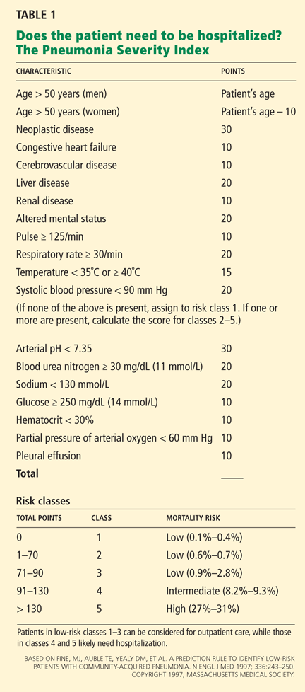
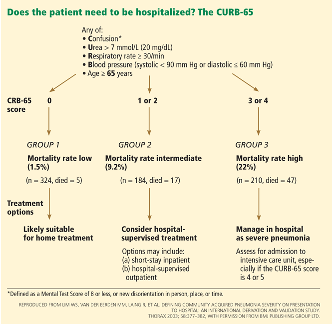
A recent meta-analysis compared the performance characteristics of the PSI and CURB-65 scores for predicting mortality in CAP and found no significant differences in overall test performance.61
Another meta-analysis found that the PSI was more sensitive than the CURB-65 and had a low false-negative rate, and so was better at showing which patients do not need to be hospitalized. Conversely, the CURB-65 was more specific and had a higher positive predictive value, and thus was more likely to correctly classify high-risk patients.62
Other scoring systems that aid in deciding about hospital admission and level of care include the CRB-6563 (which can be used instead of the CURB-65 if laboratory values are not available), SMART-COP,64 and SCAP.,65
Guidelines on when to admit to the intensive care unit
Guidelines from the Infectious Diseases Society of America (IDSA) and the American Thoracic Society (ATS) also provide guidance on when intensive care admission is advised,21 and their criteria were recently validated.66 The guidelines advocate direct admission to the intensive care unit for patients requiring vasopressors or mechanical ventilation, and intensive care unit or high-level monitoring for patients with three of the following criteria for severe CAP21:
- Respiratory rate ≥ 30
- Pao2/Fio2 ratio ≤ 250
- Multilobar infiltrates
- Confusion or disorientation
- Uremia (blood urea nitrogen ≥ 20 mg/dL)
- Leukopenia (white blood cell count < 4.0 × 10⁹/L)
- Thrombocytopenia (platelet count < 100 × 109/L)
- Hypothermia (core temperature < 36.0°C [96.8°F])
- Hypotension requiring aggressive fluid resuscitation.
None of these scoring systems or criteria is meant to replace clinical judgment. A recent study has suggested that an oxygen saturation of less than 92% is an appropriate threshold for hospital admission, in view of higher rates of adverse events in outpatients with saturations below this value.67
TREATMENT
Multiple studies have shown that treatment of CAP in accordance with guidelines has led to improved clinical outcomes.21,68–70
How fast must antibiotics be started?
Based on studies that showed a lower mortality rate when antibiotics were started sooner, Medicare and Medicaid adopted a quality measure calling for starting antibiotics within 4 hours in patients being admitted to the hospital.50,71 However, several subsequent studies showed that the diagnosis of pneumonia is often incorrect and that rapid administration of antibiotics could lead to misdiagnosis, overuse of antibiotics, and a higher risk of Clostridium difficile infection.72,73
The current IDSA/ATS guidelines21 recommend that the first antibiotic dose be given while the patient is still in the emergency department, but do not give a specific time within which it should be given. Medicare and Medicaid later updated their quality measure to antibiotic administration within 6 hours.
Which antibiotics should be used?
The selection of antimicrobial agent depends upon the patient’s severity of illness and comorbid conditions.
Although most studies of combination antibiotic therapy have been retrospective and observational, they suggest that a macrolide (ie, one of the “mycins”) added to a beta-lactam antibiotic is beneficial, possibly by covering atypical organisms or via anti-inflammatory action.74–76 The choice of one antibiotic over another appears to be less important, and a recent Cochrane review concluded that there was no significant difference in efficacy among five antibiotic pairs studied.77
Empiric outpatient treatment of a previously healthy patient with CAP and no risk factors for drug-resistant S pneumoniae should include either a macrolide (azithromycin [Zithromax], clarithromycin [Biaxin], or erythromycin) or doxycycline. If the patient has a chronic comorbid condition such as heart, lung, liver, or renal disease, diabetes mellitus, alcoholism, malignancy, asplenia, or immunosuppression or has received antimicrobials within the preceding 3 months, then treatment should include either a respiratory fluoroquinolone (moxifloxacin [Avelox] or levofloxacin [Levaquin]) or a beta-lactam plus a macrolide.21
Overall, published data suggest that the survival rate is about the same with fluoroquinolone monotherapy as with beta-lactam plus macrolide combination therapy, and better than with beta-lactam monotherapy.78
Selection of antibiotics for inpatient treatment of CAP is influenced by severity of illness. Inpatients who do not require intensive care should be treated with either a respiratory fluoroquinolone or combination therapy with a beta-lactam (cefotaxime [Claforan], ceftriaxone [Rocephin], ampicillin, or ertapenem [Invanz]) plus a macrolide or doxycycline.21,76,79
If a specific microbiologic diagnosis is made, then treatment can be narrowed. However in certain cases, such as invasive pneumococcal infection, combination therapy may still be superior.80,81 For patients who need intensive care, treatment should always include a beta-lactam plus either azithromycin or a respiratory fluoroquinolone.21 In certain situations, additional antibiotics may be added as well, such as agents to treat Pseudomonas, community-acquired MRSA, or both.
Switching to oral therapy; short-course therapy
In the interest of avoiding unnecessary antibiotics, numerous studies have addressed the issue of an “early switch” to oral antibiotics and “short-course” therapy for CAP. In general, once clinically stable, patients with CAP, including bacteremic S pneumoniae pneumonia, can be safely switched to oral antibiotics.82
The issue of short-course therapy is more complicated, and the appropriate length of therapy for CAP is not well established. However, 5 days of levofloxacin 750 mg was shown to be as successful as 7 to 10 days of levofloxacin 500 mg.83 In another study, in patients who improved after 3 days of intravenous therapy for CAP, there was no difference in clinical outcome between those who were changed to oral therapy for 5 more days and those who received an oral placebo.84
Most patients who achieve clinical stability in the first week do not need prolonged antibiotic therapy. However, certain conditions, such as S aureus bacteremic pneumonia, complicated pneumonia, and pneumonia due to unusual organisms, may require prolonged treatment.
Other therapies
Additional therapies studied in patients with pneumonia include early mobilization, adjunctive corticosteroids, and statin drugs.
Early mobilization was shown in one study to decrease hospital length of stay without increasing adverse effects.85
Corticosteroids are not supported as a standard of care for patients with severe CAP according to current available studies.86,87 Furthermore, a randomized, controlled trial showed that prednisolone daily for a week did not improve outcomes in hospitalized patients with CAP, and it was associated with increased late failure.88
Statin trials under way. Several observational studies have suggested that statins might be beneficial in managing sepsis through their effects on endothelial cell function, antioxidant effects, anti-inflammatory effects, and immunomodulatory effects.89 However, a recent large prospective multicenter cohort study of hospitalized patients with CAP did not find evidence of a protective effect of statins on clinically meaningful outcomes in CAP or significant differences in circulating biomarkers.90 Several randomized trials of statin therapy in patients with both ventilator-associated pneumonia and CAP are under way.
INFLUENZA TREATMENT: MOST EFFECTIVE WITHIN 48 HOURS
Treatment with antiviral drugs is most effective if started within 48 hours after symptom onset, although some patients with confirmed influenza who are either not improving or who are critically ill may still benefit from treatment started later.
Treatment should be considered in patients with laboratory-confirmed or suspected influenza who are at risk of developing complicated influenza and in otherwise healthy patients who wish to reduce the duration of illness or who have close contact with patients who are at high risk of complications.
Antiviral medications are oseltamivir (Tamiflu), zanamivir (Relenza), and the adamantines amantadine (Symmetrel) and rimantadine (Flumadine).
Due to evolving viral resistance patterns, the choice of antiviral drug depends on the strain. Seasonal H1N1 is best treated with zanamivir or an adamantine, while pandemic 2009 H1N1 and H3N2 are best treated with zanamivir or oseltamivir. When strain typing is not available, empiric therapy should be with either zanamivir monotherapy or a combination of oseltamivir plus rimantadine. Influenza B viruses are resistant to adamantines and should be treated only with either zanamivir or oseltamivir.45
FOLLOW-UP AND PREVENTION
Patients with CAP can generally be expected to improve within 3 to 7 days.91 However, it may be several weeks before they return to baseline.92
Follow-up plans may be guided by the time to clinical stability. For patients who do not achieve clinical stability until more than 72 hours after admission, more aggressive follow-up on discharge is indicated, since they are more likely to experience early readmission and death.93
Pneumococcal vaccination. Because S pneumoniae remains the most common cause of CAP, efforts should be made to vaccinate patients appropriately. The Advisory Committee on Immunization Practices (ACIP) and the US Centers for Disease Control and Prevention recommend that the pneumococcal polysaccharide vaccine (Pneumovax 23; PPSV23) be given to those over age 65. Those who were vaccinated before age 65 should receive another dose at age 65 or later if at least 5 years have passed since their previous dose. Those who receive it at or after age 65 should receive only a single dose. A second dose is recommended 5 years after the first dose for people age 19 to 64 years with functional or anatomic asplenia and for those who are immunocompromised.
Influenza vaccination for all. Of note, the ACIP updated its guidelines on influenza vaccination beginning with the 2010–2011 influenza season. It no longer advocates a risk-stratified approach. Instead, it recommends universal influenza vaccination for everybody more than 6 months old.94
Smoking cessation should be addressed. Smoking cessation is a Medicare and Medicaid quality measure and should be encouraged after an episode of CAP because quitting smoking reduces the risk of pneumococcal disease by approximately 14% each year thereafter.95
- Mortensen EM, Kapoor WN, Chang CC, Fine MJ. Assessment of mortality after long-term follow-up of patients with community-acquired pneumonia. Clin Infect Dis 2003; 37:1617–1624.
- File TM, Marrie TJ. Burden of community-acquired pneumonia in North American adults. Postgrad Med 2010; 122:130–141.
- Marston BJ, Plouffe JF, File TM, et al. Incidence of community-acquired pneumonia requiring hospitalization. Results of a population-based active surveillance study in Ohio. The Community-Based Pneumonia Incidence Study Group. Arch Intern Med 1997; 157:1709–1718.
- Fine MJ, Smith MA, Carson CA, et al. Prognosis and outcomes of patients with community-acquired pneumonia. JAMA 1996; 275:134–141.
- Ruhnke GW, Coca-Perraillon M, Kitch BT, Cutler DM. Trends in mortality and medical spending in patients hospitalized for community-acquired pneumonia: 1993–2005. Med Care 2010; 48:1111–1116.
- Jackson ML, Neuzil KM, Thompson WW, et al. The burden of community-acquired pneumonia in seniors: results of a population-based study. Clin Infect Dis 2004; 39:1642–1650.
- Koivula I, Sten M, Mäkelä PH. Risk factors for pneumonia in the elderly. Am J Med 1994; 96:313–320.
- Ruhnke GW, Coca-Perraillon M, Kitch BT, Cutler DM. Marked reduction in 30-day mortality among elderly patients with community-acquired pneumonia. Am J Med 2011; 124:171–178.
- Farr BM, Bartlett CL, Wadsworth J, Miller DL. Risk factors for community-acquired pneumonia diagnosed upon hospital admission. British Thoracic Society Pneumonia Study Group. Respir Med 2000; 94:954–963.
- Graves CR. Pneumonia in pregnancy. Clin Obstet Gynecol 2010; 53:329–336.
- Eom CS, Jeon CY, Lim JW, Cho EG, Park SM, Lee KS. Use of acid-suppressive drugs and risk of pneumonia: a systematic review and meta-analysis. CMAJ 2011; 183:310–319.
- Voiriot G, Dury S, Parrot A, Mayaud C, Fartoukh M. Nonsteroidal antiinflammatory drugs may affect the presentation and course of community-acquired pneumonia. Chest 2011; 139:387–394.
- Thompson WW, Shay DK, Weintraub E, et al. Mortality associated with influenza and respiratory syncytial virus in the United States. JAMA 2003; 289:179–186.
- Thompson WW, Shay DK, Weintraub E, et al. Influenza-associated hospitalizations in the United States. JAMA 2004; 292:1333–1340.
- Glezen WP, Decker M, Perrotta DM. Survey of underlying conditions of persons hospitalized with acute respiratory disease during influenza epidemics in Houston, 1978–1981. Am Rev Respir Dis 1987; 136:550–555.
- Izurieta HS, Thompson WW, Kramarz P, et al. Influenza and the rates of hospitalization for respiratory disease among infants and young children. N Engl J Med 2000; 342:232–239.
- Rothberg MB, Haessler SD. Complications of seasonal and pandemic influenza. Crit Care Med 2010; 38(suppl 4):e91–e97.
- Apisarnthanarak A, Mundy LM. Etiology of community-acquired pneumonia. Clin Chest Med 2005; 26:47–55.
- de Roux A, Marcos MA, Garcia E, et al. Viral community-acquired pneumonia in nonimmunocompromised adults. Chest 2004; 125:1343–1351.
- Hamelin ME, Côté S, Laforge J, et al. Human metapneumovirus infection in adults with community-acquired pneumonia and exacerbation of chronic obstructive pulmonary disease. Clin Infect Dis 2005; 41:498–502.
- Mandell LA, Wunderink RG, Anzueto A, et al; Infectious Diseases Society of America; American Thoracic Society. Infectious Diseases Society of America/American Thoracic Society consensus guidelines on the management of community-acquired pneumonia in adults. Clin Infect Dis 2007; 44(suppl 2):S27–S72.
- Kolling UK, Hansen F, Braun J, Rink L, Katus HA, Dalhoff K. Leucocyte response and anti-inflammatory cytokines in community acquired pneumonia. Thorax 2001; 56:121–125.
- Wunderink RG, Waterer GW. Community-acquired pneumonia: pathophysiology and host factors with focus on possible new approaches to management of lower respiratory tract infections. Infect Dis Clin North Am 2004; 18:743–759.
- Hilleman MR. Realities and enigmas of human viral influenza: pathogenesis, epidemiology and control. Vaccine 2002; 20:3068–3087.
- Bender BS, Small PA. Influenza: pathogenesis and host defense. Semin Respir Infect 1992; 7:38–45.
- Scheiblauer H, Reinacher M, Tashiro M, Rott R. Interactions between bacteria and influenza A virus in the development of influenza pneumonia. J Infect Dis 1992; 166:783–791.
- McCullers JA. Insights into the interaction between influenza virus and pneumococcus. Clin Microbiol Rev 2006; 19:571–582.
- Metlay JP, Kapoor WN, Fine MJ. Does this patient have community-acquired pneumonia? Diagnosing pneumonia by history and physical examination. JAMA 1997; 278:1440–1445.
- Benbassat J, Baumal R. Narrative review: should teaching of the respiratory physical examination be restricted only to signs with proven reliability and validity? J Gen Intern Med 2010; 25:865–872.
- Kolsuz M, Erginel S, Alatas O, et al. Acute phase reactants and cytokine levels in unilateral community-acquired pneumonia. Respiration 2003; 70:615–622.
- Alves DW, Kennedy MT. Community-acquired pneumonia in casualty: etiology, clinical features, diagnosis, and management (or a look at the “new” in pneumonia since 2002). Curr Opin Pulm Med 2004; 10:166–170.
- Monto AS, Gravenstein S, Elliott M, Colopy M, Schweinle J. Clinical signs and symptoms predicting influenza infection. Arch Intern Med 2000; 160:3243–3247.
- Bewick T, Myles P, Greenwood S, et al; Influenza Clinical Information Network. Clinical and laboratory features distinguishing pandemic H1N1 influenza-related pneumonia from interpandemic community-acquired pneumonia in adults. Thorax 2011; 66:247–252.
- Morens DM, Fauci AS. The 1918 influenza pandemic: insights for the 21st century. J Infect Dis 2007; 195:1018–1028.
- Starr I. Influenza in 1918: recollections of the epidemic in Philadelphia. 1976. Ann Intern Med 2006; 145:138–140.
- Morens DM, Taubenberger JK, Fauci AS. Predominant role of bacterial pneumonia as a cause of death in pandemic influenza: implications for pandemic influenza preparedness. J Infect Dis 2008; 198:962–970.
- Brundage JF, Shanks GD. Deaths from bacterial pneumonia during 1918–19 influenza pandemic. Emerg Infect Dis 2008; 14:1193–1199.
- Treanor J. Influenza virus. In: Mandell GL, Bennett JE, Dolin R, eds. Principles and Practice of Infectious Diseases. 6th ed. Philadelphia, PA: Elsevier/Churchill Livingstone; 2005:2060–2085.
- Jarstrand C, Tunevall G. The influence of bacterial superinfection on the clinical course of influenza. Studies from the influenza epidemics in Stockholm during the winters 1969–70 and 1971–72. Scand J Infect Dis 1975; 7:243–247.
- Schwarzmann SW, Adler JL, Sullivan RJ, Marine WM. Bacterial pneumonia during the Hong Kong influenza epidemic of 1968–1969. Arch Intern Med 1971; 127:1037–1041.
- Hageman JC, Uyeki TM, Francis JS, et al. Severe community-acquired pneumonia due to Staphylococcus aureus, 2003–04 influenza season. Emerg Infect Dis 2006; 12:894–899.
- Centers for Disease Control and Prevention (CDC). Severe methicillin-resistant Staphylococcus aureus community-acquired pneumonia associated with influenza—Louisiana and Georgia, December 2006–January 2007. MMWR Morb Mortal Wkly Rep 2007; 56:325–329.
- Hidron AI, Low CE, Honig EG, Blumberg HM. Emergence of community-acquired methicillin-resistant Staphylococcus aureus strain USA300 as a cause of necrotising community-onset pneumonia. Lancet Infect Dis 2009; 9:384–392.
- Call SA, Vollenweider MA, Hornung CA, Simel DL, McKinney WP. Does this patient have influenza? JAMA 2005; 293:987–997.
- Harper SA, Bradley JS, Englund JA, et al; Expert Panel of the Infectious Diseases Society of America. Seasonal influenza in adults and children—diagnosis, treatment, chemoprophylaxis, and institutional outbreak management: clinical practice guidelines of the Infectious Diseases Society of America. Clin Infect Dis 2009; 48:1003–1032.
- Boersma WG, Daniels JM, Löwenberg A, Boeve WJ, van de Jagt EJ. Reliability of radiographic findings and the relation to etiologic agents in community-acquired pneumonia. Respir Med 2006; 100:926–932.
- Brixey AG, Luo Y, Skouras V, Awdankiewicz A, Light RW. The efficacy of chest radiographs in detecting parapneumonic effusions. Respirology 2011; 16:1000–1004.
- Campbell SG, Marrie TJ, Anstey R, Dickinson G, Ackroyd-Stolarz S. The contribution of blood cultures to the clinical management of adult patients admitted to the hospital with community-acquired pneumonia: a prospective observational study. Chest 2003; 123:1142–1150.
- Waterer GW, Wunderink RG. The influence of the severity of community-acquired pneumonia on the usefulness of blood cultures. Respir Med 2001; 95:78–82.
- Houck PM, Bratzler DW, Nsa W, Ma A, Bartlett JG. Timing of antibiotic administration and outcomes for Medicare patients hospitalized with community-acquired pneumonia. Arch Intern Med 2004; 164:637–644.
- Information & Quality Healthcare. http://www.IQH.org/attachments/219_CoreMHelpBookletpg4_11_3.pdf. Accessed November 14, 2011.
- Rosón B, Carratalà J, Verdaguer R, Dorca J, Manresa F, Gudiol F. Prospective study of the usefulness of sputum Gram stain in the initial approach to community-acquired pneumonia requiring hospitalization. Clin Infect Dis 2000; 31:869–874.
- García-Vázquez E, Marcos MA, Mensa J, et al. Assessment of the usefulness of sputum culture for diagnosis of community-acquired pneumonia using the PORT predictive scoring system. Arch Intern Med 2004; 164:1807–1811.
- Rosón B, Fernández-Sabé N, Carratalà J, et al. Contribution of a urinary antigen assay (Binax NOW) to the early diagnosis of pneumococcal pneumonia. Clin Infect Dis 2004; 38:222–226.
- Sordé R, Falcó V, Lowak M, et al. Current and potential usefulness of pneumococcal urinary antigen detection in hospitalized patients with community-acquired pneumonia to guide antimicrobial therapy. Arch Intern Med 2011; 171:166–172.
- Koegelenberg CFN, Diacon AH, Bolliger CT. Parapneumonic pleural effusion and empyema. Respiration 2008; 75:241–250.
- Almirall J, Bolíbar I, Toran P, et al; Community-Acquired Pneumonia Maresme Study Group. Contribution of C-reactive protein to the diagnosis and assessment of severity of community-acquired pneumonia. Chest 2004; 125:1335–1342.
- Ingram PR, Inglis T, Moxon D, Speers D. Procalcitonin and C-reactive protein in severe 2009 H1N1 influenza infection. Intensive Care Med 2010; 36:528–532.
- Fine MJ, Auble TE, Yealy DM, et al. A prediction rule to identify low-risk patients with community-acquired pneumonia. N Engl J Med 1997; 336:243–250.
- Lim WS, van der Eerden MM, Laing R, et al. Defining community acquired pneumonia severity on presentation to hospital: an international derivation and validation study. Thorax 2003; 58:377–382.
- Chalmers JD, Singanayagam A, Akram AR, et al. Severity assessment tools for predicting mortality in hospitalised patients with community-acquired pneumonia. Systematic review and meta-analysis. Thorax 2010; 65:878–883.
- Loke YK, Kwok CS, Niruban A, Myint PK. Value of severity scales in predicting mortality from community-acquired pneumonia: systematic review and meta-analysis. Thorax 2010; 65:884–890.
- Capelastegui A, España PP, Quintana JM, et al. Validation of a predictive rule for the management of community-acquired pneumonia. Eur Respir J 2006; 27:151–157.
- Charles PG, Wolfe R, Whitby M, et al; Australian Community-Acquired Pneumonia Study Collaboration. SMART-COP: a tool for predicting the need for intensive respiratory or vasopressor support in community-acquired pneumonia. Clin Infect Dis 2008; 47:375–384.
- España PP, Capelastegui A, Gorordo I, et al. Development and validation of a clinical prediction rule for severe community-acquired pneumonia. Am J Respir Crit Care Med 2006; 174:1249–1256.
- Chalmers JD, Taylor JK, Mandal P, et al. Validation of the Infectious Diseases Society of America/American Thoracic Society minor criteria for intensive care unit admission in community-acquired pneumonia patients without major criteria or contraindications to intensive care unit care. Clin Infect Dis 2011; 53:503–511.
- Majumdar SR, Eurich DT, Gamble JM, Senthilselvan A, Marrie TJ. Oxygen saturations less than 92% are associated with major adverse events in outpatients with pneumonia: a population-based cohort study. Clin Infect Dis 2011; 52:325–331.
- Nathwani D, Rubinstein E, Barlow G, Davey P. Do guidelines for community-acquired pneumonia improve the cost-effectiveness of hospital care? Clin Infect Dis 2001; 32:728–741.
- Dean NC, Silver MP, Bateman KA, James B, Hadlock CJ, Hale D. Decreased mortality after implementation of a treatment guideline for community-acquired pneumonia. Am J Med 2001; 110:451–457.
- Capelastegui A, España PP, Quintana JM, et al. Improvement of process-of-care and outcomes after implementing a guideline for the management of community-acquired pneumonia: a controlled before-and-after design study. Clin Infect Dis 2004; 39:955–963.
- Silber SH, Garrett C, Singh R, et al. Early administration of antibiotics does not shorten time to clinical stability in patients with moderate-to-severe community-acquired pneumonia. Chest 2003; 124:1798–1804.
- Welker JA, Huston M, McCue JD. Antibiotic timing and errors in diagnosing pneumonia. Arch Intern Med 2008; 168:351–356.
- Polgreen PM, Chen YY, Cavanaugh JE, et al. An outbreak of severe Clostridium difficile-associated disease possibly related to inappropriate antimicrobial therapy for community-acquired pneumonia. Infect Control Hosp Epidemiol 2007; 28:212–214.
- Waterer GW, Somes GW, Wunderink RG. Monotherapy may be suboptimal for severe bacteremic pneumococcal pneumonia. Arch Intern Med 2001; 161:1837–1842.
- Lodise TP, Kwa A, Cosler L, Gupta R, Smith RP. Comparison of beta-lactam and macrolide combination therapy versus fluoroquinolone monotherapy in hospitalized Veterans Affairs patients with community-acquired pneumonia. Antimicrob Agents Chemother 2007; 51:3977–3982.
- Waterer GW, Rello J, Wunderink RG. Management of community-acquired pneumonia in adults. Am J Respir Crit Care Med 2011; 183:157–164.
- Bjerre LM, Verheij TJ, Kochen MM. Antibiotics for community acquired pneumonia in adult outpatients. Cochrane Database Syst Rev 2009; (4):CD002109.
- Frei CR, Labreche MJ, Attridge RT. Fluoroquinolones in community-acquired pneumonia: guide to selection and appropriate use. Drugs 2011; 71:757–770.
- Weiss K, Tillotson GS. The controversy of combination vs monotherapy in the treatment of hospitalized community-acquired pneumonia. Chest 2005; 128:940–946.
- Martínez JA, Horcajada JP, Almela M, et al. Addition of a macrolide to a beta-lactam-based empirical antibiotic regimen is associated with lower in-hospital mortality for patients with bacteremic pneumococcal pneumonia. Clin Infect Dis 2003; 36:389–395.
- Waterer GW, Somes GW, Wunderink RG. Monotherapy may be suboptimal for severe bacteremic pneumococcal pneumonia. Arch Intern Med 2001; 161:1837–1842.
- Ramirez JA, Bordon J. Early switch from intravenous to oral antibiotics in hospitalized patients with bacteremic community-acquired Streptococcus pneumoniae pneumonia. Arch Intern Med 2001; 161:848–850.
- Dunbar LM, Wunderink RG, Habib MP, et al. High-dose, short-course levofloxacin for community-acquired pneumonia: a new treatment paradigm. Clin Infect Dis 2003; 37:752–760.
- el Moussaoui R, de Borgie CA, van den Broek P, et al. Effectiveness of discontinuing antibiotic treatment after three days versus eight days in mild to moderate-severe community acquired pneumonia: randomised, double blind study. BMJ 2006; 332:1355.
- Mundy LM, Leet TL, Darst K, Schnitzler MA, Dunagan WC. Early mobilization of patients hospitalized with community-acquired pneumonia. Chest 2003; 124:883–889.
- Salluh JI, Póvoa P, Soares M, Castro-Faria-Neto HC, Bozza FA, Bozza PT. The role of corticosteroids in severe community-acquired pneumonia: a systematic review. Crit Care 2008; 12:R76.
- Mikami K, Suzuki M, Kitagawa H, et al. Efficacy of corticosteroids in the treatment of community-acquired pneumonia requiring hospitalization. Lung 2007; 185:249–255.
- Snijders D, Daniels JM, de Graaff CS, van der Werf TS, Boersma WG. Efficacy of corticosteroids in community-acquired pneumonia: a randomized double-blinded clinical trial. Am J Respir Crit Care Med 2010; 181:975–982.
- Chopra V, Flanders SA. Does statin use improve pneumonia outcomes? Chest 2009; 136:1381–1388.
- Yende S, Milbrandt EB, Kellum JA, et al. Understanding the potential role of statins in pneumonia and sepsis. Crit Care Med 2011; 39:1871–1878.
- Halm EA, Fine MJ, Marrie TJ, et al. Time to clinical stability in patients hospitalized with community-acquired pneumonia: implications for practice guidelines. JAMA 1998; 279:1452–1457.
- Marrie TJ, Lau CY, Wheeler SL, Wong CJ, Feagan BG. Predictors of symptom resolution in patients with community-acquired pneumonia. Clin Infect Dis 2000; 31:1362–1367.
- Aliberti S, Peyrani P, Filardo G, et al. Association between time to clinical stability and outcomes after discharge in hospitalized patients with community-acquired pneumonia. Chest 2011; 140:482–488.
- Fiore AE, Uyeki TM, Broder K, et al; Centers for Disease Control and Prevention (CDC). Prevention and control of influenza with vaccines: recommendations of the Advisory Committee on Immunization Practices (ACIP), 2010. MMWR Recomm Rep 2010; 59:1–62.
- Nuorti JP, Butler JC, Farley MM, et al. Cigarette smoking and invasive pneumococcal disease. Active Bacterial Core Surveillance Team. N Engl J Med 2000; 342:681–689.
General internists need to be able to recognize community-acquired pneumonia (CAP) so that diagnostic and therapeutic interventions can be initiated promptly. It is also important to understand the most likely and possible causes of CAP so that appropriate initial antimicrobial therapy can be chosen. Especially during flu season, influenza can present as CAP and should be included in the differential diagnosis.
When managing a patient with CAP, the internist must decide which level of care, diagnostic tests, antimicrobial agents, and follow-up plans are needed. These topics will be reviewed in this article.
TWO TERMS TO REMEMBER
- CAP refers to pneumonia acquired outside a health care facility. It can be either bacterial or viral.
- CABP (community-acquired bacterial pneumonia) refers only to those cases caused by bacterial pathogens.
NUMBERS AND TRENDS
In the United States, CAP is the number-one cause of death from infection and the sixth leading cause of death overall.1 Each year, it is responsible for about 4.2 million outpatient visits, more than 60,000 deaths, and more than $17 billion in health care expenses.2
Community-acquired bacterial pneumonia: Common, serious
In a population-based US study in 1991, the incidence of CABP requiring hospitalization was 266.8 per 100,000 people.3
Estimates of overall mortality in CABP vary depending on the severity of illness and comorbid conditions. A meta-analysis published in 1996 found the overall mortality rate to be 13.7%, with a range of 5.1% to 36.5% depending on severity.4
In hospitalized patients, mortality rates and length of hospital stay appear to be declining over time. Between 1993 and 2005, the age-adjusted mortality rate decreased from 8.9% to 4.1%, and the average length of stay decreased from 7.5 to 5.7 days, with an overall reduction in hospital cost.5
CABP is more prevalent in older people than in the general population, and it increases with age from 18.2 cases per 1,000 patient-years in patients 60 to 69 years to 52.3 cases per 1,000 patient-years in those older than 85 years.6 Risk factors for pneumonia in the elderly include heart disease, chronic lung disease, immunosuppressive drugs, alcoholism, and increasing age.7 Similar to the trend in the general population, the mortality rate in elderly CABP patients appears to be decreasing over time, possibly thanks to rising rates of pneumococcal and influenza vaccination.8
Among the general population, risk factors for developing CABP also include smoking, occupational dust exposure, history of childhood pneumonia, unemployment, and single marital status.9 The incidence of CABP does not appear to be higher among pregnant women, although it is the most frequent cause of nonobstetric death in this population.10
The use of proton pump inhibitors may be an emerging risk factor for CABP.11 Also, use of nonsteroidal anti-inflammatory drugs among patients with CABP is associated with a blunted inflammatory response as well as a higher risk of pleuropulmonary complications and a delay in presentation.12
Influenza is also common, potentially severe
Influenza is also very common and potentially severe. It can cause a spectrum of disease, from mild upper respiratory tract symptoms to severe viral pneumonia that can be life-threatening and complicated by respiratory failure and the acute respiratory distress syndrome (ARDS).
Influenza infection can also be complicated by subsequent bacterial pneumonia. However, the epidemiology of influenza infection differs from that of CABP in that influenza occurs seasonally.
In the United States, seasonal influenza causes 36,000 deaths and 200,000 hospitalizations annually.13,14 As with CABP, the risk of death from influenza increases with age: it is 16 times greater in people age 85 and older than in those ages 65 to 69.13
During yearly seasonal epidemics, those at the highest risk of hospitalization and death are at the extremes of age. Risk factors for complicated influenza include heart disease, lung disease, diabetes, renal failure, rheumatologic conditions, dementia, and neurologic disease.15,16 During the 2009 H1N1 influenza pandemic, unexpected severity was seen in previously healthy young adults as well as those with obesity, neurodegenerative disease, pregnancy, and asthma.17
PATHOGENS: TYPICAL, ATYPICAL, VIRAL
Identifying the etiologic organism in CAP is confounded by limitations in the available diagnostic tests and also by poor-quality specimens that often are contaminated with bacteria that colonize the upper airways. Given these caveats, the primary pathogens responsible for CAP broadly include typical bacterial pathogens, atypical bacterial pathogens, and viruses.
Typical bacterial pathogens include Streptococcus pneumoniae, Haemophilus influenzae, Staphylococcus aureus, Moraxella catarrhalis, and, less commonly, a variety of aerobic and anaerobic gram-negative rods including Pseudomonas aeruginosa, Acinetobacter species, and Klebsiella pneumoniae.
Atypical bacterial pathogens include Mycoplasma pneumoniae, Chlamydia pneumoniae, and Legionella species.18
Viruses implicated in adult CAP include influenza A and B, parainfluenza viruses, respiratory syncytial virus, and adenovirus.19 More recently, human metapneumovirus has been described as a cause of adult CAP.20
Clues to uncommon microbes
Specific historic features or coexisting conditions that may suggest an uncommon microbiologic diagnosis include21:
- Recent travel to the southwestern United States or Southeast Asia
- Ill contacts
- Exposure to birds, bats, rabbits, or farm animals
- Alcoholism
- Chronic obstructive pulmonary disease
- Human immunodeficiency virus infection
- Structural lung disease
- Prolonged cough with whoop or posttussive vomiting
- Aspiration
- Bioterrorism.
In cases in which one or more of these conditions exist, CAP may also be caused by other agents not listed above, including Mycobacterium tuberculosis, oral anaerobes, atypical mycobacteria, Histoplasma capsulatum, Chlamydophila psittaci, Francisella tularensis, Coxiella burnettii, Pneumocystis jiroveci, Cryptococcus, Aspergillus, Coccidioides, Hantavirus, avian influenza, Burkholderia pseudomallei, severe acute respiratory syndrome virus, Bordetella pertussis, Bacillus anthracis, and Yersinia pestis.
HOW BACTERIA INVADE THE LUNGS
The pathophysiology of CABP involves both host defense and microbial virulence factors.
The airways are most commonly exposed to microbes by microaspiration of upper airway flora, although hematogenous seeding of the lungs in a bacteremic patient or contiguous spread of infection from an adjacent site can also occur.
Mucociliary clearance and the cough reflex are important initial defenses against infection and can be inhibited by neurologic diseases and conditions that impair the mucociliary mechanism. Mucosal immune cells, including macrophages and neutrophils, recognize invading pathogens and generate an antibody response.
Regulation of the host inflammatory response to infection depends on a complex interaction between immune cells, inflammatory cytokines (eg, tumor necrosis factor alpha, interleukin 1-beta, and interleukin 6), and anti-inflammatory cytokines such as interleukin 1 receptor antagonist and soluble tumor necrosis factor receptor type I.22
The interaction and timing of the inflammatory and anti-inflammatory response are essential in manifesting an appropriate host response to infection. An inadequate inflammatory response can lead to sepsis and death, but an excessive, late anti-inflammatory response can lead to a systemic inflammatory response such as ARDS. Polymorphisms within the genes coding for these factors may explain the variation in severity of illness among patients with CABP.23
HOW INFLUENZA DOES ITS DAMAGE
There are three types of influenza virus: A, B, and C. Type A causes most human infections. The influenza A virus envelope comprises a lipid bilayer that contains the projecting glycoproteins hemagglutinin and neuraminidase. Influenza viruses are named on the basis of these proteins and are designated with an H and an N, respectively, each followed by a number referring to the subtype.
Influenza infection begins when the virus makes contact with the epithelium. Hemagglutinin binds to the host cell and allows viral entry, where it begins replication. Neuraminidase prevents viral aggregation and facilitates the release of virus from infected cells.24
Mature virions are released and spread to neighboring host cells; this process is associated with desquamation and inflammation of the airways, causing cough, rhinorrhea, and sore throat. Systemic symptoms are associated with the induction of interferon, which causes fever and myalgia.25
Recovery and immunity to influenza infection occurs through both humoral and cell-mediated immunity, with antibodies directed against the specific hemagglutinin and neuraminidase antigens of the infecting virus. Immunity wanes over time and with antigenic drift of circulating viruses, making the host susceptible to recurrent influenza infection.24
Influenza is often complicated by bacterial superinfection
The influenza virus acts synergistically with certain bacteria to increase infectivity, and this may explain why influenza is often complicated by bacterial superinfection.
Mechanisms leading to bacterial superinfection include increased binding and invasion of bacteria, increased viral replication, and modification of the host inflammatory response. Some S aureus strains produce a protease that directly activates influenza virus hemagglutinin; other bacteria can activate plasminogen to promote influenza replication. The resulting increase in proteases in host tissues promotes activation of influenza through cleavage of hemagglutinin.26
The influenza virus also causes damage to the airway epithelial layer, leading to increased exposure of the binding sites necessary for adherence of S pneumoniae.27
CLINICAL PRESENTATION OF COMMUNITY-ACQUIRED PNEUMONIA
Although CAP is common, agreement on its essential clinical signs and symptoms is surprisingly limited, due in part to heterogeneous patient presentations and in part to interobserver variability. The reader is referred to two excellent reviews on this topic.28,29
The diagnosis of CAP is made on clinical grounds, based on a combination of signs and symptoms. Symptoms of pneumonia can include cough, fever, chills, sputum production, dyspnea, and pleuritic pain. Physical findings can include tachypnea, tachycardia, hypoxemia, and consolidation or rales on auscultation. Laboratory data may show leukocytosis or elevated C-reactive protein, and radiographic studies may show evidence of a new infiltrate.21,30,31
Clinical presentation of influenza
Seasonal influenza as a cause of CAP is difficult to distinguish from bacterial causes. The clinical presentation of seasonal influenza most commonly includes fever or subjective feverishness, cough, myalgia, and weakness.32 In a recent multivariate analysis, five clinical features were shown to be predictive of pandemic H1N1 influenza pneumonia rather than CABP: age younger than 65 years, absence of confusion, white blood cell count less than 12 × 109/L, temperature higher than 38°C (100.4°F), and bilateral opacities on radiography.32,33
Complicated influenza infection can be either primary viral pneumonia or bacterial superinfection.
During the 1918 influenza pandemic, which predated the ability to isolate viruses, two clinical syndromes emerged: an ARDS associated with the rapid onset of cyanosis, delirium, and frothy blood-tinged sputum; and an acute bronchopneumonia characterized by necrosis, hemorrhage, edema, and vasculitis.34,35 The first syndrome has subsequently been shown to be associated with primary viral pneumonia, while the second is caused by bacterial superinfection. Modern reexamination of 1918 data has shown that bacterial superinfection was likely the reason for the distinctly fulminant presentation of that pandemic.36,37
The 2009 H1N1 influenza pandemic caused relatively mild disease in most patients. However, those with severe pneumonia more commonly developed ARDS from primary influenza pneumonia than from bacterial superinfection.17
A third influenza-associated infection is secondary bacterial pneumonia, which follows influenza infection and mimics the presentation of CABP. A typical patient presents with a recent history of influenza-like illness, followed 4 to 14 days later by a recurrence of fever, dyspnea, productive cough, and consolidation on chest radiographs.38 Leukocytosis with an increased number of immature neutrophil forms, prolonged duration of fever, and elevated erythrocyte sedimentation rate are more likely in patients with secondary bacterial pneumonia.39 Isolates from sputum samples commonly include S pneumoniae, S aureus, H influenzae, and other gram-negative rods.40
In recent flu seasons, methicillin-resistant S aureus (MRSA) has emerged as a cause of severe secondary pneumonia. Most of these isolates carry genes for the toxin Panton-Valentine leukocidin; the associated mortality rate is as high as 33%.41,42 Although community-acquired MRSA pneumonia has only been reported in case series, distinct clinical features that have been described include severe pneumonia with high fever, hypotension, shock, respiratory failure, leukopenia, and multilobar and cavitary infiltrates.43
WHEN TO SUSPECT INFLUENZA
The triad of fever, cough, and abrupt onset are the best predictors of influenza, but no single combination of signs and symptoms predict influenza infection with 100% certainty. Therefore, an understanding of local epidemiologic data regarding circulating influenza is essential to maintain a high index of suspicion.44
It is appropriate to suspect influenza in:
- Anyone who is epidemiologically linked to a known outbreak of influenza
- Children, adults, and health care workers who have fever and abrupt onset of respiratory symptoms
- Patients with fever plus exacerbation of underlying pulmonary disease
- Severely ill patients with fever or hypothermia, especially during influenza season.45
DIAGNOSTIC TESTING
Once the diagnosis of pulmonary infection is suggested by clinical features, the initial evaluation should include measurement of vital signs, physical examination, and radiographic imaging of the chest. Additional diagnostic measures to consider include viral testing, blood culture, sputum culture, urinary antigen testing for Legionella and for S pneumoniae, fungal culture, and mycobacterial smear and culture.
Chest radiography (with posterior-anterior and lateral films) is the study that usually demonstrates the presence of a pulmonary infiltrate. If initial chest radiographs do not show an infiltrate, imaging can be repeated after treatment is started if the patient’s clinical presentation still suggests pneumonia. Chest radiographs are of limited value in predicting the pathogen, but they help to determine the extent of pneumonia and to detect parapneumonic effusion.46
A caveat: anterior-posterior, posterior-anterior, and lateral views can miss more than 10% of effusions large enough to warrant thoracentesis, especially when there is lower-lobe consolidation.47
Blood cultures are recommended for patients admitted to the intensive care unit and for those with cavitary infiltrates, leukopenia, alcohol abuse, severe liver disease, asplenia, positive pneumococcal urinary antigen testing, or a pleural effusion.21 However, blood cultures are positive in only 3% to 14% of hospitalized patients with CABP, and the impact of a positive blood culture on management decisions in CABP appears to be quite small.48–50
For the highest yield, blood culture results should be obtained before antibiotics are given. Not only is this good practice, but obtaining blood culture results before starting antibiotics is one of the quality measures evaluated by the Center for Medicare and Medicaid Services.51
Sputum culture is considered optional for outpatients and patients with less-severe pneumonia.21 While it can provide a rapid diagnosis in certain cases, a good-quality sputum sample is obtained in only 39% to 54% of patients with CABP, yields a predominant morphotype in only 45% of cases, and provides a useful microbiologic diagnosis in only 14.4%.52,53 Fungal and mycobacterial cultures are only indicated in certain situations such as cavitary infiltrates or immunosuppression.
Urinary antigen testing for Legionella and S pneumoniae should be done in patients with more severe illness and in those for whom outpatient therapy has failed.21S pneumoniae testing has been shown to allow early diagnosis of pneumococcal pneumonia in 26% more patients than with Gram staining, but it fails to identify 22% of the rapid diagnoses initially identified by Gram staining.54 Thus, a sequential approach is reasonable, with urinary antigen testing for patients at high risk without useful results from sputum Gram staining. Also, recent data suggest that the pneumococcal urinary antigen test may allow optimization of antimicrobial therapy with good clinical outcomes.55
Endotracheal tests. If the patient is intubated, collection of endotracheal aspirates, bronchoscopy, or nonbronchoscopic bronchial lavage (sometimes called “mini-BAL”) should be performed.
Thoracentesis and pleural fluid cultures should be done if a pleural effusion is found. Empyema, large or loculated effusions, and parapneumonic effusions with a pH lower than 7.20, glucose levels less than 3.4 mmol/L (60 mg/dL), or positive results on microbial staining or culture should be drained by chest tube or surgically.56
Testing for influenza should be done if it will change the clinical management, such as the choice of antibiotic or infection control practices. Specimens should be obtained with either a nasopharyngeal swab or aspirate and tested with reverse transcriptase polymerase chain reaction, immunofluorescent staining, or rapid antigen detection, depending on local availability.45
Inflammatory biomarkers such as C-reactive protein and procalcitonin have been receiving interest as ways to predict the etiology and prognosis of CAP and to guide therapy. Several studies have shown that C-reactive protein can help distinguish between CAP and bronchitis, with higher values suggesting more severe pneumonia and pneumonia caused by S pneumoniae or L pneumophila.57 Procalcitonin may help discriminate between severe lower respiratory tract infections of bacterial and 2009 H1N1 origin, although less effectively than C-reactive protein. Low procalcitonin values, particularly when combined with low C-reactive protein levels, suggest that bacterial infection is unlikely.58
RISK STRATIFICATION AND SITE-OF-CARE DECISIONS
Following a presumptive diagnosis of CAP, it is important to decide not only what treatment the patient will receive but whether he or she should be hospitalized. If the patient is to be admitted to the hospital, the clinician must also decide if his or her condition warrants intensive care.
Severity-of-illness scores
A recent meta-analysis compared the performance characteristics of the PSI and CURB-65 scores for predicting mortality in CAP and found no significant differences in overall test performance.61
Another meta-analysis found that the PSI was more sensitive than the CURB-65 and had a low false-negative rate, and so was better at showing which patients do not need to be hospitalized. Conversely, the CURB-65 was more specific and had a higher positive predictive value, and thus was more likely to correctly classify high-risk patients.62
Other scoring systems that aid in deciding about hospital admission and level of care include the CRB-6563 (which can be used instead of the CURB-65 if laboratory values are not available), SMART-COP,64 and SCAP.,65
Guidelines on when to admit to the intensive care unit
Guidelines from the Infectious Diseases Society of America (IDSA) and the American Thoracic Society (ATS) also provide guidance on when intensive care admission is advised,21 and their criteria were recently validated.66 The guidelines advocate direct admission to the intensive care unit for patients requiring vasopressors or mechanical ventilation, and intensive care unit or high-level monitoring for patients with three of the following criteria for severe CAP21:
- Respiratory rate ≥ 30
- Pao2/Fio2 ratio ≤ 250
- Multilobar infiltrates
- Confusion or disorientation
- Uremia (blood urea nitrogen ≥ 20 mg/dL)
- Leukopenia (white blood cell count < 4.0 × 10⁹/L)
- Thrombocytopenia (platelet count < 100 × 109/L)
- Hypothermia (core temperature < 36.0°C [96.8°F])
- Hypotension requiring aggressive fluid resuscitation.
None of these scoring systems or criteria is meant to replace clinical judgment. A recent study has suggested that an oxygen saturation of less than 92% is an appropriate threshold for hospital admission, in view of higher rates of adverse events in outpatients with saturations below this value.67
TREATMENT
Multiple studies have shown that treatment of CAP in accordance with guidelines has led to improved clinical outcomes.21,68–70
How fast must antibiotics be started?
Based on studies that showed a lower mortality rate when antibiotics were started sooner, Medicare and Medicaid adopted a quality measure calling for starting antibiotics within 4 hours in patients being admitted to the hospital.50,71 However, several subsequent studies showed that the diagnosis of pneumonia is often incorrect and that rapid administration of antibiotics could lead to misdiagnosis, overuse of antibiotics, and a higher risk of Clostridium difficile infection.72,73
The current IDSA/ATS guidelines21 recommend that the first antibiotic dose be given while the patient is still in the emergency department, but do not give a specific time within which it should be given. Medicare and Medicaid later updated their quality measure to antibiotic administration within 6 hours.
Which antibiotics should be used?
The selection of antimicrobial agent depends upon the patient’s severity of illness and comorbid conditions.
Although most studies of combination antibiotic therapy have been retrospective and observational, they suggest that a macrolide (ie, one of the “mycins”) added to a beta-lactam antibiotic is beneficial, possibly by covering atypical organisms or via anti-inflammatory action.74–76 The choice of one antibiotic over another appears to be less important, and a recent Cochrane review concluded that there was no significant difference in efficacy among five antibiotic pairs studied.77
Empiric outpatient treatment of a previously healthy patient with CAP and no risk factors for drug-resistant S pneumoniae should include either a macrolide (azithromycin [Zithromax], clarithromycin [Biaxin], or erythromycin) or doxycycline. If the patient has a chronic comorbid condition such as heart, lung, liver, or renal disease, diabetes mellitus, alcoholism, malignancy, asplenia, or immunosuppression or has received antimicrobials within the preceding 3 months, then treatment should include either a respiratory fluoroquinolone (moxifloxacin [Avelox] or levofloxacin [Levaquin]) or a beta-lactam plus a macrolide.21
Overall, published data suggest that the survival rate is about the same with fluoroquinolone monotherapy as with beta-lactam plus macrolide combination therapy, and better than with beta-lactam monotherapy.78
Selection of antibiotics for inpatient treatment of CAP is influenced by severity of illness. Inpatients who do not require intensive care should be treated with either a respiratory fluoroquinolone or combination therapy with a beta-lactam (cefotaxime [Claforan], ceftriaxone [Rocephin], ampicillin, or ertapenem [Invanz]) plus a macrolide or doxycycline.21,76,79
If a specific microbiologic diagnosis is made, then treatment can be narrowed. However in certain cases, such as invasive pneumococcal infection, combination therapy may still be superior.80,81 For patients who need intensive care, treatment should always include a beta-lactam plus either azithromycin or a respiratory fluoroquinolone.21 In certain situations, additional antibiotics may be added as well, such as agents to treat Pseudomonas, community-acquired MRSA, or both.
Switching to oral therapy; short-course therapy
In the interest of avoiding unnecessary antibiotics, numerous studies have addressed the issue of an “early switch” to oral antibiotics and “short-course” therapy for CAP. In general, once clinically stable, patients with CAP, including bacteremic S pneumoniae pneumonia, can be safely switched to oral antibiotics.82
The issue of short-course therapy is more complicated, and the appropriate length of therapy for CAP is not well established. However, 5 days of levofloxacin 750 mg was shown to be as successful as 7 to 10 days of levofloxacin 500 mg.83 In another study, in patients who improved after 3 days of intravenous therapy for CAP, there was no difference in clinical outcome between those who were changed to oral therapy for 5 more days and those who received an oral placebo.84
Most patients who achieve clinical stability in the first week do not need prolonged antibiotic therapy. However, certain conditions, such as S aureus bacteremic pneumonia, complicated pneumonia, and pneumonia due to unusual organisms, may require prolonged treatment.
Other therapies
Additional therapies studied in patients with pneumonia include early mobilization, adjunctive corticosteroids, and statin drugs.
Early mobilization was shown in one study to decrease hospital length of stay without increasing adverse effects.85
Corticosteroids are not supported as a standard of care for patients with severe CAP according to current available studies.86,87 Furthermore, a randomized, controlled trial showed that prednisolone daily for a week did not improve outcomes in hospitalized patients with CAP, and it was associated with increased late failure.88
Statin trials under way. Several observational studies have suggested that statins might be beneficial in managing sepsis through their effects on endothelial cell function, antioxidant effects, anti-inflammatory effects, and immunomodulatory effects.89 However, a recent large prospective multicenter cohort study of hospitalized patients with CAP did not find evidence of a protective effect of statins on clinically meaningful outcomes in CAP or significant differences in circulating biomarkers.90 Several randomized trials of statin therapy in patients with both ventilator-associated pneumonia and CAP are under way.
INFLUENZA TREATMENT: MOST EFFECTIVE WITHIN 48 HOURS
Treatment with antiviral drugs is most effective if started within 48 hours after symptom onset, although some patients with confirmed influenza who are either not improving or who are critically ill may still benefit from treatment started later.
Treatment should be considered in patients with laboratory-confirmed or suspected influenza who are at risk of developing complicated influenza and in otherwise healthy patients who wish to reduce the duration of illness or who have close contact with patients who are at high risk of complications.
Antiviral medications are oseltamivir (Tamiflu), zanamivir (Relenza), and the adamantines amantadine (Symmetrel) and rimantadine (Flumadine).
Due to evolving viral resistance patterns, the choice of antiviral drug depends on the strain. Seasonal H1N1 is best treated with zanamivir or an adamantine, while pandemic 2009 H1N1 and H3N2 are best treated with zanamivir or oseltamivir. When strain typing is not available, empiric therapy should be with either zanamivir monotherapy or a combination of oseltamivir plus rimantadine. Influenza B viruses are resistant to adamantines and should be treated only with either zanamivir or oseltamivir.45
FOLLOW-UP AND PREVENTION
Patients with CAP can generally be expected to improve within 3 to 7 days.91 However, it may be several weeks before they return to baseline.92
Follow-up plans may be guided by the time to clinical stability. For patients who do not achieve clinical stability until more than 72 hours after admission, more aggressive follow-up on discharge is indicated, since they are more likely to experience early readmission and death.93
Pneumococcal vaccination. Because S pneumoniae remains the most common cause of CAP, efforts should be made to vaccinate patients appropriately. The Advisory Committee on Immunization Practices (ACIP) and the US Centers for Disease Control and Prevention recommend that the pneumococcal polysaccharide vaccine (Pneumovax 23; PPSV23) be given to those over age 65. Those who were vaccinated before age 65 should receive another dose at age 65 or later if at least 5 years have passed since their previous dose. Those who receive it at or after age 65 should receive only a single dose. A second dose is recommended 5 years after the first dose for people age 19 to 64 years with functional or anatomic asplenia and for those who are immunocompromised.
Influenza vaccination for all. Of note, the ACIP updated its guidelines on influenza vaccination beginning with the 2010–2011 influenza season. It no longer advocates a risk-stratified approach. Instead, it recommends universal influenza vaccination for everybody more than 6 months old.94
Smoking cessation should be addressed. Smoking cessation is a Medicare and Medicaid quality measure and should be encouraged after an episode of CAP because quitting smoking reduces the risk of pneumococcal disease by approximately 14% each year thereafter.95
General internists need to be able to recognize community-acquired pneumonia (CAP) so that diagnostic and therapeutic interventions can be initiated promptly. It is also important to understand the most likely and possible causes of CAP so that appropriate initial antimicrobial therapy can be chosen. Especially during flu season, influenza can present as CAP and should be included in the differential diagnosis.
When managing a patient with CAP, the internist must decide which level of care, diagnostic tests, antimicrobial agents, and follow-up plans are needed. These topics will be reviewed in this article.
TWO TERMS TO REMEMBER
- CAP refers to pneumonia acquired outside a health care facility. It can be either bacterial or viral.
- CABP (community-acquired bacterial pneumonia) refers only to those cases caused by bacterial pathogens.
NUMBERS AND TRENDS
In the United States, CAP is the number-one cause of death from infection and the sixth leading cause of death overall.1 Each year, it is responsible for about 4.2 million outpatient visits, more than 60,000 deaths, and more than $17 billion in health care expenses.2
Community-acquired bacterial pneumonia: Common, serious
In a population-based US study in 1991, the incidence of CABP requiring hospitalization was 266.8 per 100,000 people.3
Estimates of overall mortality in CABP vary depending on the severity of illness and comorbid conditions. A meta-analysis published in 1996 found the overall mortality rate to be 13.7%, with a range of 5.1% to 36.5% depending on severity.4
In hospitalized patients, mortality rates and length of hospital stay appear to be declining over time. Between 1993 and 2005, the age-adjusted mortality rate decreased from 8.9% to 4.1%, and the average length of stay decreased from 7.5 to 5.7 days, with an overall reduction in hospital cost.5
CABP is more prevalent in older people than in the general population, and it increases with age from 18.2 cases per 1,000 patient-years in patients 60 to 69 years to 52.3 cases per 1,000 patient-years in those older than 85 years.6 Risk factors for pneumonia in the elderly include heart disease, chronic lung disease, immunosuppressive drugs, alcoholism, and increasing age.7 Similar to the trend in the general population, the mortality rate in elderly CABP patients appears to be decreasing over time, possibly thanks to rising rates of pneumococcal and influenza vaccination.8
Among the general population, risk factors for developing CABP also include smoking, occupational dust exposure, history of childhood pneumonia, unemployment, and single marital status.9 The incidence of CABP does not appear to be higher among pregnant women, although it is the most frequent cause of nonobstetric death in this population.10
The use of proton pump inhibitors may be an emerging risk factor for CABP.11 Also, use of nonsteroidal anti-inflammatory drugs among patients with CABP is associated with a blunted inflammatory response as well as a higher risk of pleuropulmonary complications and a delay in presentation.12
Influenza is also common, potentially severe
Influenza is also very common and potentially severe. It can cause a spectrum of disease, from mild upper respiratory tract symptoms to severe viral pneumonia that can be life-threatening and complicated by respiratory failure and the acute respiratory distress syndrome (ARDS).
Influenza infection can also be complicated by subsequent bacterial pneumonia. However, the epidemiology of influenza infection differs from that of CABP in that influenza occurs seasonally.
In the United States, seasonal influenza causes 36,000 deaths and 200,000 hospitalizations annually.13,14 As with CABP, the risk of death from influenza increases with age: it is 16 times greater in people age 85 and older than in those ages 65 to 69.13
During yearly seasonal epidemics, those at the highest risk of hospitalization and death are at the extremes of age. Risk factors for complicated influenza include heart disease, lung disease, diabetes, renal failure, rheumatologic conditions, dementia, and neurologic disease.15,16 During the 2009 H1N1 influenza pandemic, unexpected severity was seen in previously healthy young adults as well as those with obesity, neurodegenerative disease, pregnancy, and asthma.17
PATHOGENS: TYPICAL, ATYPICAL, VIRAL
Identifying the etiologic organism in CAP is confounded by limitations in the available diagnostic tests and also by poor-quality specimens that often are contaminated with bacteria that colonize the upper airways. Given these caveats, the primary pathogens responsible for CAP broadly include typical bacterial pathogens, atypical bacterial pathogens, and viruses.
Typical bacterial pathogens include Streptococcus pneumoniae, Haemophilus influenzae, Staphylococcus aureus, Moraxella catarrhalis, and, less commonly, a variety of aerobic and anaerobic gram-negative rods including Pseudomonas aeruginosa, Acinetobacter species, and Klebsiella pneumoniae.
Atypical bacterial pathogens include Mycoplasma pneumoniae, Chlamydia pneumoniae, and Legionella species.18
Viruses implicated in adult CAP include influenza A and B, parainfluenza viruses, respiratory syncytial virus, and adenovirus.19 More recently, human metapneumovirus has been described as a cause of adult CAP.20
Clues to uncommon microbes
Specific historic features or coexisting conditions that may suggest an uncommon microbiologic diagnosis include21:
- Recent travel to the southwestern United States or Southeast Asia
- Ill contacts
- Exposure to birds, bats, rabbits, or farm animals
- Alcoholism
- Chronic obstructive pulmonary disease
- Human immunodeficiency virus infection
- Structural lung disease
- Prolonged cough with whoop or posttussive vomiting
- Aspiration
- Bioterrorism.
In cases in which one or more of these conditions exist, CAP may also be caused by other agents not listed above, including Mycobacterium tuberculosis, oral anaerobes, atypical mycobacteria, Histoplasma capsulatum, Chlamydophila psittaci, Francisella tularensis, Coxiella burnettii, Pneumocystis jiroveci, Cryptococcus, Aspergillus, Coccidioides, Hantavirus, avian influenza, Burkholderia pseudomallei, severe acute respiratory syndrome virus, Bordetella pertussis, Bacillus anthracis, and Yersinia pestis.
HOW BACTERIA INVADE THE LUNGS
The pathophysiology of CABP involves both host defense and microbial virulence factors.
The airways are most commonly exposed to microbes by microaspiration of upper airway flora, although hematogenous seeding of the lungs in a bacteremic patient or contiguous spread of infection from an adjacent site can also occur.
Mucociliary clearance and the cough reflex are important initial defenses against infection and can be inhibited by neurologic diseases and conditions that impair the mucociliary mechanism. Mucosal immune cells, including macrophages and neutrophils, recognize invading pathogens and generate an antibody response.
Regulation of the host inflammatory response to infection depends on a complex interaction between immune cells, inflammatory cytokines (eg, tumor necrosis factor alpha, interleukin 1-beta, and interleukin 6), and anti-inflammatory cytokines such as interleukin 1 receptor antagonist and soluble tumor necrosis factor receptor type I.22
The interaction and timing of the inflammatory and anti-inflammatory response are essential in manifesting an appropriate host response to infection. An inadequate inflammatory response can lead to sepsis and death, but an excessive, late anti-inflammatory response can lead to a systemic inflammatory response such as ARDS. Polymorphisms within the genes coding for these factors may explain the variation in severity of illness among patients with CABP.23
HOW INFLUENZA DOES ITS DAMAGE
There are three types of influenza virus: A, B, and C. Type A causes most human infections. The influenza A virus envelope comprises a lipid bilayer that contains the projecting glycoproteins hemagglutinin and neuraminidase. Influenza viruses are named on the basis of these proteins and are designated with an H and an N, respectively, each followed by a number referring to the subtype.
Influenza infection begins when the virus makes contact with the epithelium. Hemagglutinin binds to the host cell and allows viral entry, where it begins replication. Neuraminidase prevents viral aggregation and facilitates the release of virus from infected cells.24
Mature virions are released and spread to neighboring host cells; this process is associated with desquamation and inflammation of the airways, causing cough, rhinorrhea, and sore throat. Systemic symptoms are associated with the induction of interferon, which causes fever and myalgia.25
Recovery and immunity to influenza infection occurs through both humoral and cell-mediated immunity, with antibodies directed against the specific hemagglutinin and neuraminidase antigens of the infecting virus. Immunity wanes over time and with antigenic drift of circulating viruses, making the host susceptible to recurrent influenza infection.24
Influenza is often complicated by bacterial superinfection
The influenza virus acts synergistically with certain bacteria to increase infectivity, and this may explain why influenza is often complicated by bacterial superinfection.
Mechanisms leading to bacterial superinfection include increased binding and invasion of bacteria, increased viral replication, and modification of the host inflammatory response. Some S aureus strains produce a protease that directly activates influenza virus hemagglutinin; other bacteria can activate plasminogen to promote influenza replication. The resulting increase in proteases in host tissues promotes activation of influenza through cleavage of hemagglutinin.26
The influenza virus also causes damage to the airway epithelial layer, leading to increased exposure of the binding sites necessary for adherence of S pneumoniae.27
CLINICAL PRESENTATION OF COMMUNITY-ACQUIRED PNEUMONIA
Although CAP is common, agreement on its essential clinical signs and symptoms is surprisingly limited, due in part to heterogeneous patient presentations and in part to interobserver variability. The reader is referred to two excellent reviews on this topic.28,29
The diagnosis of CAP is made on clinical grounds, based on a combination of signs and symptoms. Symptoms of pneumonia can include cough, fever, chills, sputum production, dyspnea, and pleuritic pain. Physical findings can include tachypnea, tachycardia, hypoxemia, and consolidation or rales on auscultation. Laboratory data may show leukocytosis or elevated C-reactive protein, and radiographic studies may show evidence of a new infiltrate.21,30,31
Clinical presentation of influenza
Seasonal influenza as a cause of CAP is difficult to distinguish from bacterial causes. The clinical presentation of seasonal influenza most commonly includes fever or subjective feverishness, cough, myalgia, and weakness.32 In a recent multivariate analysis, five clinical features were shown to be predictive of pandemic H1N1 influenza pneumonia rather than CABP: age younger than 65 years, absence of confusion, white blood cell count less than 12 × 109/L, temperature higher than 38°C (100.4°F), and bilateral opacities on radiography.32,33
Complicated influenza infection can be either primary viral pneumonia or bacterial superinfection.
During the 1918 influenza pandemic, which predated the ability to isolate viruses, two clinical syndromes emerged: an ARDS associated with the rapid onset of cyanosis, delirium, and frothy blood-tinged sputum; and an acute bronchopneumonia characterized by necrosis, hemorrhage, edema, and vasculitis.34,35 The first syndrome has subsequently been shown to be associated with primary viral pneumonia, while the second is caused by bacterial superinfection. Modern reexamination of 1918 data has shown that bacterial superinfection was likely the reason for the distinctly fulminant presentation of that pandemic.36,37
The 2009 H1N1 influenza pandemic caused relatively mild disease in most patients. However, those with severe pneumonia more commonly developed ARDS from primary influenza pneumonia than from bacterial superinfection.17
A third influenza-associated infection is secondary bacterial pneumonia, which follows influenza infection and mimics the presentation of CABP. A typical patient presents with a recent history of influenza-like illness, followed 4 to 14 days later by a recurrence of fever, dyspnea, productive cough, and consolidation on chest radiographs.38 Leukocytosis with an increased number of immature neutrophil forms, prolonged duration of fever, and elevated erythrocyte sedimentation rate are more likely in patients with secondary bacterial pneumonia.39 Isolates from sputum samples commonly include S pneumoniae, S aureus, H influenzae, and other gram-negative rods.40
In recent flu seasons, methicillin-resistant S aureus (MRSA) has emerged as a cause of severe secondary pneumonia. Most of these isolates carry genes for the toxin Panton-Valentine leukocidin; the associated mortality rate is as high as 33%.41,42 Although community-acquired MRSA pneumonia has only been reported in case series, distinct clinical features that have been described include severe pneumonia with high fever, hypotension, shock, respiratory failure, leukopenia, and multilobar and cavitary infiltrates.43
WHEN TO SUSPECT INFLUENZA
The triad of fever, cough, and abrupt onset are the best predictors of influenza, but no single combination of signs and symptoms predict influenza infection with 100% certainty. Therefore, an understanding of local epidemiologic data regarding circulating influenza is essential to maintain a high index of suspicion.44
It is appropriate to suspect influenza in:
- Anyone who is epidemiologically linked to a known outbreak of influenza
- Children, adults, and health care workers who have fever and abrupt onset of respiratory symptoms
- Patients with fever plus exacerbation of underlying pulmonary disease
- Severely ill patients with fever or hypothermia, especially during influenza season.45
DIAGNOSTIC TESTING
Once the diagnosis of pulmonary infection is suggested by clinical features, the initial evaluation should include measurement of vital signs, physical examination, and radiographic imaging of the chest. Additional diagnostic measures to consider include viral testing, blood culture, sputum culture, urinary antigen testing for Legionella and for S pneumoniae, fungal culture, and mycobacterial smear and culture.
Chest radiography (with posterior-anterior and lateral films) is the study that usually demonstrates the presence of a pulmonary infiltrate. If initial chest radiographs do not show an infiltrate, imaging can be repeated after treatment is started if the patient’s clinical presentation still suggests pneumonia. Chest radiographs are of limited value in predicting the pathogen, but they help to determine the extent of pneumonia and to detect parapneumonic effusion.46
A caveat: anterior-posterior, posterior-anterior, and lateral views can miss more than 10% of effusions large enough to warrant thoracentesis, especially when there is lower-lobe consolidation.47
Blood cultures are recommended for patients admitted to the intensive care unit and for those with cavitary infiltrates, leukopenia, alcohol abuse, severe liver disease, asplenia, positive pneumococcal urinary antigen testing, or a pleural effusion.21 However, blood cultures are positive in only 3% to 14% of hospitalized patients with CABP, and the impact of a positive blood culture on management decisions in CABP appears to be quite small.48–50
For the highest yield, blood culture results should be obtained before antibiotics are given. Not only is this good practice, but obtaining blood culture results before starting antibiotics is one of the quality measures evaluated by the Center for Medicare and Medicaid Services.51
Sputum culture is considered optional for outpatients and patients with less-severe pneumonia.21 While it can provide a rapid diagnosis in certain cases, a good-quality sputum sample is obtained in only 39% to 54% of patients with CABP, yields a predominant morphotype in only 45% of cases, and provides a useful microbiologic diagnosis in only 14.4%.52,53 Fungal and mycobacterial cultures are only indicated in certain situations such as cavitary infiltrates or immunosuppression.
Urinary antigen testing for Legionella and S pneumoniae should be done in patients with more severe illness and in those for whom outpatient therapy has failed.21S pneumoniae testing has been shown to allow early diagnosis of pneumococcal pneumonia in 26% more patients than with Gram staining, but it fails to identify 22% of the rapid diagnoses initially identified by Gram staining.54 Thus, a sequential approach is reasonable, with urinary antigen testing for patients at high risk without useful results from sputum Gram staining. Also, recent data suggest that the pneumococcal urinary antigen test may allow optimization of antimicrobial therapy with good clinical outcomes.55
Endotracheal tests. If the patient is intubated, collection of endotracheal aspirates, bronchoscopy, or nonbronchoscopic bronchial lavage (sometimes called “mini-BAL”) should be performed.
Thoracentesis and pleural fluid cultures should be done if a pleural effusion is found. Empyema, large or loculated effusions, and parapneumonic effusions with a pH lower than 7.20, glucose levels less than 3.4 mmol/L (60 mg/dL), or positive results on microbial staining or culture should be drained by chest tube or surgically.56
Testing for influenza should be done if it will change the clinical management, such as the choice of antibiotic or infection control practices. Specimens should be obtained with either a nasopharyngeal swab or aspirate and tested with reverse transcriptase polymerase chain reaction, immunofluorescent staining, or rapid antigen detection, depending on local availability.45
Inflammatory biomarkers such as C-reactive protein and procalcitonin have been receiving interest as ways to predict the etiology and prognosis of CAP and to guide therapy. Several studies have shown that C-reactive protein can help distinguish between CAP and bronchitis, with higher values suggesting more severe pneumonia and pneumonia caused by S pneumoniae or L pneumophila.57 Procalcitonin may help discriminate between severe lower respiratory tract infections of bacterial and 2009 H1N1 origin, although less effectively than C-reactive protein. Low procalcitonin values, particularly when combined with low C-reactive protein levels, suggest that bacterial infection is unlikely.58
RISK STRATIFICATION AND SITE-OF-CARE DECISIONS
Following a presumptive diagnosis of CAP, it is important to decide not only what treatment the patient will receive but whether he or she should be hospitalized. If the patient is to be admitted to the hospital, the clinician must also decide if his or her condition warrants intensive care.
Severity-of-illness scores
A recent meta-analysis compared the performance characteristics of the PSI and CURB-65 scores for predicting mortality in CAP and found no significant differences in overall test performance.61
Another meta-analysis found that the PSI was more sensitive than the CURB-65 and had a low false-negative rate, and so was better at showing which patients do not need to be hospitalized. Conversely, the CURB-65 was more specific and had a higher positive predictive value, and thus was more likely to correctly classify high-risk patients.62
Other scoring systems that aid in deciding about hospital admission and level of care include the CRB-6563 (which can be used instead of the CURB-65 if laboratory values are not available), SMART-COP,64 and SCAP.,65
Guidelines on when to admit to the intensive care unit
Guidelines from the Infectious Diseases Society of America (IDSA) and the American Thoracic Society (ATS) also provide guidance on when intensive care admission is advised,21 and their criteria were recently validated.66 The guidelines advocate direct admission to the intensive care unit for patients requiring vasopressors or mechanical ventilation, and intensive care unit or high-level monitoring for patients with three of the following criteria for severe CAP21:
- Respiratory rate ≥ 30
- Pao2/Fio2 ratio ≤ 250
- Multilobar infiltrates
- Confusion or disorientation
- Uremia (blood urea nitrogen ≥ 20 mg/dL)
- Leukopenia (white blood cell count < 4.0 × 10⁹/L)
- Thrombocytopenia (platelet count < 100 × 109/L)
- Hypothermia (core temperature < 36.0°C [96.8°F])
- Hypotension requiring aggressive fluid resuscitation.
None of these scoring systems or criteria is meant to replace clinical judgment. A recent study has suggested that an oxygen saturation of less than 92% is an appropriate threshold for hospital admission, in view of higher rates of adverse events in outpatients with saturations below this value.67
TREATMENT
Multiple studies have shown that treatment of CAP in accordance with guidelines has led to improved clinical outcomes.21,68–70
How fast must antibiotics be started?
Based on studies that showed a lower mortality rate when antibiotics were started sooner, Medicare and Medicaid adopted a quality measure calling for starting antibiotics within 4 hours in patients being admitted to the hospital.50,71 However, several subsequent studies showed that the diagnosis of pneumonia is often incorrect and that rapid administration of antibiotics could lead to misdiagnosis, overuse of antibiotics, and a higher risk of Clostridium difficile infection.72,73
The current IDSA/ATS guidelines21 recommend that the first antibiotic dose be given while the patient is still in the emergency department, but do not give a specific time within which it should be given. Medicare and Medicaid later updated their quality measure to antibiotic administration within 6 hours.
Which antibiotics should be used?
The selection of antimicrobial agent depends upon the patient’s severity of illness and comorbid conditions.
Although most studies of combination antibiotic therapy have been retrospective and observational, they suggest that a macrolide (ie, one of the “mycins”) added to a beta-lactam antibiotic is beneficial, possibly by covering atypical organisms or via anti-inflammatory action.74–76 The choice of one antibiotic over another appears to be less important, and a recent Cochrane review concluded that there was no significant difference in efficacy among five antibiotic pairs studied.77
Empiric outpatient treatment of a previously healthy patient with CAP and no risk factors for drug-resistant S pneumoniae should include either a macrolide (azithromycin [Zithromax], clarithromycin [Biaxin], or erythromycin) or doxycycline. If the patient has a chronic comorbid condition such as heart, lung, liver, or renal disease, diabetes mellitus, alcoholism, malignancy, asplenia, or immunosuppression or has received antimicrobials within the preceding 3 months, then treatment should include either a respiratory fluoroquinolone (moxifloxacin [Avelox] or levofloxacin [Levaquin]) or a beta-lactam plus a macrolide.21
Overall, published data suggest that the survival rate is about the same with fluoroquinolone monotherapy as with beta-lactam plus macrolide combination therapy, and better than with beta-lactam monotherapy.78
Selection of antibiotics for inpatient treatment of CAP is influenced by severity of illness. Inpatients who do not require intensive care should be treated with either a respiratory fluoroquinolone or combination therapy with a beta-lactam (cefotaxime [Claforan], ceftriaxone [Rocephin], ampicillin, or ertapenem [Invanz]) plus a macrolide or doxycycline.21,76,79
If a specific microbiologic diagnosis is made, then treatment can be narrowed. However in certain cases, such as invasive pneumococcal infection, combination therapy may still be superior.80,81 For patients who need intensive care, treatment should always include a beta-lactam plus either azithromycin or a respiratory fluoroquinolone.21 In certain situations, additional antibiotics may be added as well, such as agents to treat Pseudomonas, community-acquired MRSA, or both.
Switching to oral therapy; short-course therapy
In the interest of avoiding unnecessary antibiotics, numerous studies have addressed the issue of an “early switch” to oral antibiotics and “short-course” therapy for CAP. In general, once clinically stable, patients with CAP, including bacteremic S pneumoniae pneumonia, can be safely switched to oral antibiotics.82
The issue of short-course therapy is more complicated, and the appropriate length of therapy for CAP is not well established. However, 5 days of levofloxacin 750 mg was shown to be as successful as 7 to 10 days of levofloxacin 500 mg.83 In another study, in patients who improved after 3 days of intravenous therapy for CAP, there was no difference in clinical outcome between those who were changed to oral therapy for 5 more days and those who received an oral placebo.84
Most patients who achieve clinical stability in the first week do not need prolonged antibiotic therapy. However, certain conditions, such as S aureus bacteremic pneumonia, complicated pneumonia, and pneumonia due to unusual organisms, may require prolonged treatment.
Other therapies
Additional therapies studied in patients with pneumonia include early mobilization, adjunctive corticosteroids, and statin drugs.
Early mobilization was shown in one study to decrease hospital length of stay without increasing adverse effects.85
Corticosteroids are not supported as a standard of care for patients with severe CAP according to current available studies.86,87 Furthermore, a randomized, controlled trial showed that prednisolone daily for a week did not improve outcomes in hospitalized patients with CAP, and it was associated with increased late failure.88
Statin trials under way. Several observational studies have suggested that statins might be beneficial in managing sepsis through their effects on endothelial cell function, antioxidant effects, anti-inflammatory effects, and immunomodulatory effects.89 However, a recent large prospective multicenter cohort study of hospitalized patients with CAP did not find evidence of a protective effect of statins on clinically meaningful outcomes in CAP or significant differences in circulating biomarkers.90 Several randomized trials of statin therapy in patients with both ventilator-associated pneumonia and CAP are under way.
INFLUENZA TREATMENT: MOST EFFECTIVE WITHIN 48 HOURS
Treatment with antiviral drugs is most effective if started within 48 hours after symptom onset, although some patients with confirmed influenza who are either not improving or who are critically ill may still benefit from treatment started later.
Treatment should be considered in patients with laboratory-confirmed or suspected influenza who are at risk of developing complicated influenza and in otherwise healthy patients who wish to reduce the duration of illness or who have close contact with patients who are at high risk of complications.
Antiviral medications are oseltamivir (Tamiflu), zanamivir (Relenza), and the adamantines amantadine (Symmetrel) and rimantadine (Flumadine).
Due to evolving viral resistance patterns, the choice of antiviral drug depends on the strain. Seasonal H1N1 is best treated with zanamivir or an adamantine, while pandemic 2009 H1N1 and H3N2 are best treated with zanamivir or oseltamivir. When strain typing is not available, empiric therapy should be with either zanamivir monotherapy or a combination of oseltamivir plus rimantadine. Influenza B viruses are resistant to adamantines and should be treated only with either zanamivir or oseltamivir.45
FOLLOW-UP AND PREVENTION
Patients with CAP can generally be expected to improve within 3 to 7 days.91 However, it may be several weeks before they return to baseline.92
Follow-up plans may be guided by the time to clinical stability. For patients who do not achieve clinical stability until more than 72 hours after admission, more aggressive follow-up on discharge is indicated, since they are more likely to experience early readmission and death.93
Pneumococcal vaccination. Because S pneumoniae remains the most common cause of CAP, efforts should be made to vaccinate patients appropriately. The Advisory Committee on Immunization Practices (ACIP) and the US Centers for Disease Control and Prevention recommend that the pneumococcal polysaccharide vaccine (Pneumovax 23; PPSV23) be given to those over age 65. Those who were vaccinated before age 65 should receive another dose at age 65 or later if at least 5 years have passed since their previous dose. Those who receive it at or after age 65 should receive only a single dose. A second dose is recommended 5 years after the first dose for people age 19 to 64 years with functional or anatomic asplenia and for those who are immunocompromised.
Influenza vaccination for all. Of note, the ACIP updated its guidelines on influenza vaccination beginning with the 2010–2011 influenza season. It no longer advocates a risk-stratified approach. Instead, it recommends universal influenza vaccination for everybody more than 6 months old.94
Smoking cessation should be addressed. Smoking cessation is a Medicare and Medicaid quality measure and should be encouraged after an episode of CAP because quitting smoking reduces the risk of pneumococcal disease by approximately 14% each year thereafter.95
- Mortensen EM, Kapoor WN, Chang CC, Fine MJ. Assessment of mortality after long-term follow-up of patients with community-acquired pneumonia. Clin Infect Dis 2003; 37:1617–1624.
- File TM, Marrie TJ. Burden of community-acquired pneumonia in North American adults. Postgrad Med 2010; 122:130–141.
- Marston BJ, Plouffe JF, File TM, et al. Incidence of community-acquired pneumonia requiring hospitalization. Results of a population-based active surveillance study in Ohio. The Community-Based Pneumonia Incidence Study Group. Arch Intern Med 1997; 157:1709–1718.
- Fine MJ, Smith MA, Carson CA, et al. Prognosis and outcomes of patients with community-acquired pneumonia. JAMA 1996; 275:134–141.
- Ruhnke GW, Coca-Perraillon M, Kitch BT, Cutler DM. Trends in mortality and medical spending in patients hospitalized for community-acquired pneumonia: 1993–2005. Med Care 2010; 48:1111–1116.
- Jackson ML, Neuzil KM, Thompson WW, et al. The burden of community-acquired pneumonia in seniors: results of a population-based study. Clin Infect Dis 2004; 39:1642–1650.
- Koivula I, Sten M, Mäkelä PH. Risk factors for pneumonia in the elderly. Am J Med 1994; 96:313–320.
- Ruhnke GW, Coca-Perraillon M, Kitch BT, Cutler DM. Marked reduction in 30-day mortality among elderly patients with community-acquired pneumonia. Am J Med 2011; 124:171–178.
- Farr BM, Bartlett CL, Wadsworth J, Miller DL. Risk factors for community-acquired pneumonia diagnosed upon hospital admission. British Thoracic Society Pneumonia Study Group. Respir Med 2000; 94:954–963.
- Graves CR. Pneumonia in pregnancy. Clin Obstet Gynecol 2010; 53:329–336.
- Eom CS, Jeon CY, Lim JW, Cho EG, Park SM, Lee KS. Use of acid-suppressive drugs and risk of pneumonia: a systematic review and meta-analysis. CMAJ 2011; 183:310–319.
- Voiriot G, Dury S, Parrot A, Mayaud C, Fartoukh M. Nonsteroidal antiinflammatory drugs may affect the presentation and course of community-acquired pneumonia. Chest 2011; 139:387–394.
- Thompson WW, Shay DK, Weintraub E, et al. Mortality associated with influenza and respiratory syncytial virus in the United States. JAMA 2003; 289:179–186.
- Thompson WW, Shay DK, Weintraub E, et al. Influenza-associated hospitalizations in the United States. JAMA 2004; 292:1333–1340.
- Glezen WP, Decker M, Perrotta DM. Survey of underlying conditions of persons hospitalized with acute respiratory disease during influenza epidemics in Houston, 1978–1981. Am Rev Respir Dis 1987; 136:550–555.
- Izurieta HS, Thompson WW, Kramarz P, et al. Influenza and the rates of hospitalization for respiratory disease among infants and young children. N Engl J Med 2000; 342:232–239.
- Rothberg MB, Haessler SD. Complications of seasonal and pandemic influenza. Crit Care Med 2010; 38(suppl 4):e91–e97.
- Apisarnthanarak A, Mundy LM. Etiology of community-acquired pneumonia. Clin Chest Med 2005; 26:47–55.
- de Roux A, Marcos MA, Garcia E, et al. Viral community-acquired pneumonia in nonimmunocompromised adults. Chest 2004; 125:1343–1351.
- Hamelin ME, Côté S, Laforge J, et al. Human metapneumovirus infection in adults with community-acquired pneumonia and exacerbation of chronic obstructive pulmonary disease. Clin Infect Dis 2005; 41:498–502.
- Mandell LA, Wunderink RG, Anzueto A, et al; Infectious Diseases Society of America; American Thoracic Society. Infectious Diseases Society of America/American Thoracic Society consensus guidelines on the management of community-acquired pneumonia in adults. Clin Infect Dis 2007; 44(suppl 2):S27–S72.
- Kolling UK, Hansen F, Braun J, Rink L, Katus HA, Dalhoff K. Leucocyte response and anti-inflammatory cytokines in community acquired pneumonia. Thorax 2001; 56:121–125.
- Wunderink RG, Waterer GW. Community-acquired pneumonia: pathophysiology and host factors with focus on possible new approaches to management of lower respiratory tract infections. Infect Dis Clin North Am 2004; 18:743–759.
- Hilleman MR. Realities and enigmas of human viral influenza: pathogenesis, epidemiology and control. Vaccine 2002; 20:3068–3087.
- Bender BS, Small PA. Influenza: pathogenesis and host defense. Semin Respir Infect 1992; 7:38–45.
- Scheiblauer H, Reinacher M, Tashiro M, Rott R. Interactions between bacteria and influenza A virus in the development of influenza pneumonia. J Infect Dis 1992; 166:783–791.
- McCullers JA. Insights into the interaction between influenza virus and pneumococcus. Clin Microbiol Rev 2006; 19:571–582.
- Metlay JP, Kapoor WN, Fine MJ. Does this patient have community-acquired pneumonia? Diagnosing pneumonia by history and physical examination. JAMA 1997; 278:1440–1445.
- Benbassat J, Baumal R. Narrative review: should teaching of the respiratory physical examination be restricted only to signs with proven reliability and validity? J Gen Intern Med 2010; 25:865–872.
- Kolsuz M, Erginel S, Alatas O, et al. Acute phase reactants and cytokine levels in unilateral community-acquired pneumonia. Respiration 2003; 70:615–622.
- Alves DW, Kennedy MT. Community-acquired pneumonia in casualty: etiology, clinical features, diagnosis, and management (or a look at the “new” in pneumonia since 2002). Curr Opin Pulm Med 2004; 10:166–170.
- Monto AS, Gravenstein S, Elliott M, Colopy M, Schweinle J. Clinical signs and symptoms predicting influenza infection. Arch Intern Med 2000; 160:3243–3247.
- Bewick T, Myles P, Greenwood S, et al; Influenza Clinical Information Network. Clinical and laboratory features distinguishing pandemic H1N1 influenza-related pneumonia from interpandemic community-acquired pneumonia in adults. Thorax 2011; 66:247–252.
- Morens DM, Fauci AS. The 1918 influenza pandemic: insights for the 21st century. J Infect Dis 2007; 195:1018–1028.
- Starr I. Influenza in 1918: recollections of the epidemic in Philadelphia. 1976. Ann Intern Med 2006; 145:138–140.
- Morens DM, Taubenberger JK, Fauci AS. Predominant role of bacterial pneumonia as a cause of death in pandemic influenza: implications for pandemic influenza preparedness. J Infect Dis 2008; 198:962–970.
- Brundage JF, Shanks GD. Deaths from bacterial pneumonia during 1918–19 influenza pandemic. Emerg Infect Dis 2008; 14:1193–1199.
- Treanor J. Influenza virus. In: Mandell GL, Bennett JE, Dolin R, eds. Principles and Practice of Infectious Diseases. 6th ed. Philadelphia, PA: Elsevier/Churchill Livingstone; 2005:2060–2085.
- Jarstrand C, Tunevall G. The influence of bacterial superinfection on the clinical course of influenza. Studies from the influenza epidemics in Stockholm during the winters 1969–70 and 1971–72. Scand J Infect Dis 1975; 7:243–247.
- Schwarzmann SW, Adler JL, Sullivan RJ, Marine WM. Bacterial pneumonia during the Hong Kong influenza epidemic of 1968–1969. Arch Intern Med 1971; 127:1037–1041.
- Hageman JC, Uyeki TM, Francis JS, et al. Severe community-acquired pneumonia due to Staphylococcus aureus, 2003–04 influenza season. Emerg Infect Dis 2006; 12:894–899.
- Centers for Disease Control and Prevention (CDC). Severe methicillin-resistant Staphylococcus aureus community-acquired pneumonia associated with influenza—Louisiana and Georgia, December 2006–January 2007. MMWR Morb Mortal Wkly Rep 2007; 56:325–329.
- Hidron AI, Low CE, Honig EG, Blumberg HM. Emergence of community-acquired methicillin-resistant Staphylococcus aureus strain USA300 as a cause of necrotising community-onset pneumonia. Lancet Infect Dis 2009; 9:384–392.
- Call SA, Vollenweider MA, Hornung CA, Simel DL, McKinney WP. Does this patient have influenza? JAMA 2005; 293:987–997.
- Harper SA, Bradley JS, Englund JA, et al; Expert Panel of the Infectious Diseases Society of America. Seasonal influenza in adults and children—diagnosis, treatment, chemoprophylaxis, and institutional outbreak management: clinical practice guidelines of the Infectious Diseases Society of America. Clin Infect Dis 2009; 48:1003–1032.
- Boersma WG, Daniels JM, Löwenberg A, Boeve WJ, van de Jagt EJ. Reliability of radiographic findings and the relation to etiologic agents in community-acquired pneumonia. Respir Med 2006; 100:926–932.
- Brixey AG, Luo Y, Skouras V, Awdankiewicz A, Light RW. The efficacy of chest radiographs in detecting parapneumonic effusions. Respirology 2011; 16:1000–1004.
- Campbell SG, Marrie TJ, Anstey R, Dickinson G, Ackroyd-Stolarz S. The contribution of blood cultures to the clinical management of adult patients admitted to the hospital with community-acquired pneumonia: a prospective observational study. Chest 2003; 123:1142–1150.
- Waterer GW, Wunderink RG. The influence of the severity of community-acquired pneumonia on the usefulness of blood cultures. Respir Med 2001; 95:78–82.
- Houck PM, Bratzler DW, Nsa W, Ma A, Bartlett JG. Timing of antibiotic administration and outcomes for Medicare patients hospitalized with community-acquired pneumonia. Arch Intern Med 2004; 164:637–644.
- Information & Quality Healthcare. http://www.IQH.org/attachments/219_CoreMHelpBookletpg4_11_3.pdf. Accessed November 14, 2011.
- Rosón B, Carratalà J, Verdaguer R, Dorca J, Manresa F, Gudiol F. Prospective study of the usefulness of sputum Gram stain in the initial approach to community-acquired pneumonia requiring hospitalization. Clin Infect Dis 2000; 31:869–874.
- García-Vázquez E, Marcos MA, Mensa J, et al. Assessment of the usefulness of sputum culture for diagnosis of community-acquired pneumonia using the PORT predictive scoring system. Arch Intern Med 2004; 164:1807–1811.
- Rosón B, Fernández-Sabé N, Carratalà J, et al. Contribution of a urinary antigen assay (Binax NOW) to the early diagnosis of pneumococcal pneumonia. Clin Infect Dis 2004; 38:222–226.
- Sordé R, Falcó V, Lowak M, et al. Current and potential usefulness of pneumococcal urinary antigen detection in hospitalized patients with community-acquired pneumonia to guide antimicrobial therapy. Arch Intern Med 2011; 171:166–172.
- Koegelenberg CFN, Diacon AH, Bolliger CT. Parapneumonic pleural effusion and empyema. Respiration 2008; 75:241–250.
- Almirall J, Bolíbar I, Toran P, et al; Community-Acquired Pneumonia Maresme Study Group. Contribution of C-reactive protein to the diagnosis and assessment of severity of community-acquired pneumonia. Chest 2004; 125:1335–1342.
- Ingram PR, Inglis T, Moxon D, Speers D. Procalcitonin and C-reactive protein in severe 2009 H1N1 influenza infection. Intensive Care Med 2010; 36:528–532.
- Fine MJ, Auble TE, Yealy DM, et al. A prediction rule to identify low-risk patients with community-acquired pneumonia. N Engl J Med 1997; 336:243–250.
- Lim WS, van der Eerden MM, Laing R, et al. Defining community acquired pneumonia severity on presentation to hospital: an international derivation and validation study. Thorax 2003; 58:377–382.
- Chalmers JD, Singanayagam A, Akram AR, et al. Severity assessment tools for predicting mortality in hospitalised patients with community-acquired pneumonia. Systematic review and meta-analysis. Thorax 2010; 65:878–883.
- Loke YK, Kwok CS, Niruban A, Myint PK. Value of severity scales in predicting mortality from community-acquired pneumonia: systematic review and meta-analysis. Thorax 2010; 65:884–890.
- Capelastegui A, España PP, Quintana JM, et al. Validation of a predictive rule for the management of community-acquired pneumonia. Eur Respir J 2006; 27:151–157.
- Charles PG, Wolfe R, Whitby M, et al; Australian Community-Acquired Pneumonia Study Collaboration. SMART-COP: a tool for predicting the need for intensive respiratory or vasopressor support in community-acquired pneumonia. Clin Infect Dis 2008; 47:375–384.
- España PP, Capelastegui A, Gorordo I, et al. Development and validation of a clinical prediction rule for severe community-acquired pneumonia. Am J Respir Crit Care Med 2006; 174:1249–1256.
- Chalmers JD, Taylor JK, Mandal P, et al. Validation of the Infectious Diseases Society of America/American Thoracic Society minor criteria for intensive care unit admission in community-acquired pneumonia patients without major criteria or contraindications to intensive care unit care. Clin Infect Dis 2011; 53:503–511.
- Majumdar SR, Eurich DT, Gamble JM, Senthilselvan A, Marrie TJ. Oxygen saturations less than 92% are associated with major adverse events in outpatients with pneumonia: a population-based cohort study. Clin Infect Dis 2011; 52:325–331.
- Nathwani D, Rubinstein E, Barlow G, Davey P. Do guidelines for community-acquired pneumonia improve the cost-effectiveness of hospital care? Clin Infect Dis 2001; 32:728–741.
- Dean NC, Silver MP, Bateman KA, James B, Hadlock CJ, Hale D. Decreased mortality after implementation of a treatment guideline for community-acquired pneumonia. Am J Med 2001; 110:451–457.
- Capelastegui A, España PP, Quintana JM, et al. Improvement of process-of-care and outcomes after implementing a guideline for the management of community-acquired pneumonia: a controlled before-and-after design study. Clin Infect Dis 2004; 39:955–963.
- Silber SH, Garrett C, Singh R, et al. Early administration of antibiotics does not shorten time to clinical stability in patients with moderate-to-severe community-acquired pneumonia. Chest 2003; 124:1798–1804.
- Welker JA, Huston M, McCue JD. Antibiotic timing and errors in diagnosing pneumonia. Arch Intern Med 2008; 168:351–356.
- Polgreen PM, Chen YY, Cavanaugh JE, et al. An outbreak of severe Clostridium difficile-associated disease possibly related to inappropriate antimicrobial therapy for community-acquired pneumonia. Infect Control Hosp Epidemiol 2007; 28:212–214.
- Waterer GW, Somes GW, Wunderink RG. Monotherapy may be suboptimal for severe bacteremic pneumococcal pneumonia. Arch Intern Med 2001; 161:1837–1842.
- Lodise TP, Kwa A, Cosler L, Gupta R, Smith RP. Comparison of beta-lactam and macrolide combination therapy versus fluoroquinolone monotherapy in hospitalized Veterans Affairs patients with community-acquired pneumonia. Antimicrob Agents Chemother 2007; 51:3977–3982.
- Waterer GW, Rello J, Wunderink RG. Management of community-acquired pneumonia in adults. Am J Respir Crit Care Med 2011; 183:157–164.
- Bjerre LM, Verheij TJ, Kochen MM. Antibiotics for community acquired pneumonia in adult outpatients. Cochrane Database Syst Rev 2009; (4):CD002109.
- Frei CR, Labreche MJ, Attridge RT. Fluoroquinolones in community-acquired pneumonia: guide to selection and appropriate use. Drugs 2011; 71:757–770.
- Weiss K, Tillotson GS. The controversy of combination vs monotherapy in the treatment of hospitalized community-acquired pneumonia. Chest 2005; 128:940–946.
- Martínez JA, Horcajada JP, Almela M, et al. Addition of a macrolide to a beta-lactam-based empirical antibiotic regimen is associated with lower in-hospital mortality for patients with bacteremic pneumococcal pneumonia. Clin Infect Dis 2003; 36:389–395.
- Waterer GW, Somes GW, Wunderink RG. Monotherapy may be suboptimal for severe bacteremic pneumococcal pneumonia. Arch Intern Med 2001; 161:1837–1842.
- Ramirez JA, Bordon J. Early switch from intravenous to oral antibiotics in hospitalized patients with bacteremic community-acquired Streptococcus pneumoniae pneumonia. Arch Intern Med 2001; 161:848–850.
- Dunbar LM, Wunderink RG, Habib MP, et al. High-dose, short-course levofloxacin for community-acquired pneumonia: a new treatment paradigm. Clin Infect Dis 2003; 37:752–760.
- el Moussaoui R, de Borgie CA, van den Broek P, et al. Effectiveness of discontinuing antibiotic treatment after three days versus eight days in mild to moderate-severe community acquired pneumonia: randomised, double blind study. BMJ 2006; 332:1355.
- Mundy LM, Leet TL, Darst K, Schnitzler MA, Dunagan WC. Early mobilization of patients hospitalized with community-acquired pneumonia. Chest 2003; 124:883–889.
- Salluh JI, Póvoa P, Soares M, Castro-Faria-Neto HC, Bozza FA, Bozza PT. The role of corticosteroids in severe community-acquired pneumonia: a systematic review. Crit Care 2008; 12:R76.
- Mikami K, Suzuki M, Kitagawa H, et al. Efficacy of corticosteroids in the treatment of community-acquired pneumonia requiring hospitalization. Lung 2007; 185:249–255.
- Snijders D, Daniels JM, de Graaff CS, van der Werf TS, Boersma WG. Efficacy of corticosteroids in community-acquired pneumonia: a randomized double-blinded clinical trial. Am J Respir Crit Care Med 2010; 181:975–982.
- Chopra V, Flanders SA. Does statin use improve pneumonia outcomes? Chest 2009; 136:1381–1388.
- Yende S, Milbrandt EB, Kellum JA, et al. Understanding the potential role of statins in pneumonia and sepsis. Crit Care Med 2011; 39:1871–1878.
- Halm EA, Fine MJ, Marrie TJ, et al. Time to clinical stability in patients hospitalized with community-acquired pneumonia: implications for practice guidelines. JAMA 1998; 279:1452–1457.
- Marrie TJ, Lau CY, Wheeler SL, Wong CJ, Feagan BG. Predictors of symptom resolution in patients with community-acquired pneumonia. Clin Infect Dis 2000; 31:1362–1367.
- Aliberti S, Peyrani P, Filardo G, et al. Association between time to clinical stability and outcomes after discharge in hospitalized patients with community-acquired pneumonia. Chest 2011; 140:482–488.
- Fiore AE, Uyeki TM, Broder K, et al; Centers for Disease Control and Prevention (CDC). Prevention and control of influenza with vaccines: recommendations of the Advisory Committee on Immunization Practices (ACIP), 2010. MMWR Recomm Rep 2010; 59:1–62.
- Nuorti JP, Butler JC, Farley MM, et al. Cigarette smoking and invasive pneumococcal disease. Active Bacterial Core Surveillance Team. N Engl J Med 2000; 342:681–689.
- Mortensen EM, Kapoor WN, Chang CC, Fine MJ. Assessment of mortality after long-term follow-up of patients with community-acquired pneumonia. Clin Infect Dis 2003; 37:1617–1624.
- File TM, Marrie TJ. Burden of community-acquired pneumonia in North American adults. Postgrad Med 2010; 122:130–141.
- Marston BJ, Plouffe JF, File TM, et al. Incidence of community-acquired pneumonia requiring hospitalization. Results of a population-based active surveillance study in Ohio. The Community-Based Pneumonia Incidence Study Group. Arch Intern Med 1997; 157:1709–1718.
- Fine MJ, Smith MA, Carson CA, et al. Prognosis and outcomes of patients with community-acquired pneumonia. JAMA 1996; 275:134–141.
- Ruhnke GW, Coca-Perraillon M, Kitch BT, Cutler DM. Trends in mortality and medical spending in patients hospitalized for community-acquired pneumonia: 1993–2005. Med Care 2010; 48:1111–1116.
- Jackson ML, Neuzil KM, Thompson WW, et al. The burden of community-acquired pneumonia in seniors: results of a population-based study. Clin Infect Dis 2004; 39:1642–1650.
- Koivula I, Sten M, Mäkelä PH. Risk factors for pneumonia in the elderly. Am J Med 1994; 96:313–320.
- Ruhnke GW, Coca-Perraillon M, Kitch BT, Cutler DM. Marked reduction in 30-day mortality among elderly patients with community-acquired pneumonia. Am J Med 2011; 124:171–178.
- Farr BM, Bartlett CL, Wadsworth J, Miller DL. Risk factors for community-acquired pneumonia diagnosed upon hospital admission. British Thoracic Society Pneumonia Study Group. Respir Med 2000; 94:954–963.
- Graves CR. Pneumonia in pregnancy. Clin Obstet Gynecol 2010; 53:329–336.
- Eom CS, Jeon CY, Lim JW, Cho EG, Park SM, Lee KS. Use of acid-suppressive drugs and risk of pneumonia: a systematic review and meta-analysis. CMAJ 2011; 183:310–319.
- Voiriot G, Dury S, Parrot A, Mayaud C, Fartoukh M. Nonsteroidal antiinflammatory drugs may affect the presentation and course of community-acquired pneumonia. Chest 2011; 139:387–394.
- Thompson WW, Shay DK, Weintraub E, et al. Mortality associated with influenza and respiratory syncytial virus in the United States. JAMA 2003; 289:179–186.
- Thompson WW, Shay DK, Weintraub E, et al. Influenza-associated hospitalizations in the United States. JAMA 2004; 292:1333–1340.
- Glezen WP, Decker M, Perrotta DM. Survey of underlying conditions of persons hospitalized with acute respiratory disease during influenza epidemics in Houston, 1978–1981. Am Rev Respir Dis 1987; 136:550–555.
- Izurieta HS, Thompson WW, Kramarz P, et al. Influenza and the rates of hospitalization for respiratory disease among infants and young children. N Engl J Med 2000; 342:232–239.
- Rothberg MB, Haessler SD. Complications of seasonal and pandemic influenza. Crit Care Med 2010; 38(suppl 4):e91–e97.
- Apisarnthanarak A, Mundy LM. Etiology of community-acquired pneumonia. Clin Chest Med 2005; 26:47–55.
- de Roux A, Marcos MA, Garcia E, et al. Viral community-acquired pneumonia in nonimmunocompromised adults. Chest 2004; 125:1343–1351.
- Hamelin ME, Côté S, Laforge J, et al. Human metapneumovirus infection in adults with community-acquired pneumonia and exacerbation of chronic obstructive pulmonary disease. Clin Infect Dis 2005; 41:498–502.
- Mandell LA, Wunderink RG, Anzueto A, et al; Infectious Diseases Society of America; American Thoracic Society. Infectious Diseases Society of America/American Thoracic Society consensus guidelines on the management of community-acquired pneumonia in adults. Clin Infect Dis 2007; 44(suppl 2):S27–S72.
- Kolling UK, Hansen F, Braun J, Rink L, Katus HA, Dalhoff K. Leucocyte response and anti-inflammatory cytokines in community acquired pneumonia. Thorax 2001; 56:121–125.
- Wunderink RG, Waterer GW. Community-acquired pneumonia: pathophysiology and host factors with focus on possible new approaches to management of lower respiratory tract infections. Infect Dis Clin North Am 2004; 18:743–759.
- Hilleman MR. Realities and enigmas of human viral influenza: pathogenesis, epidemiology and control. Vaccine 2002; 20:3068–3087.
- Bender BS, Small PA. Influenza: pathogenesis and host defense. Semin Respir Infect 1992; 7:38–45.
- Scheiblauer H, Reinacher M, Tashiro M, Rott R. Interactions between bacteria and influenza A virus in the development of influenza pneumonia. J Infect Dis 1992; 166:783–791.
- McCullers JA. Insights into the interaction between influenza virus and pneumococcus. Clin Microbiol Rev 2006; 19:571–582.
- Metlay JP, Kapoor WN, Fine MJ. Does this patient have community-acquired pneumonia? Diagnosing pneumonia by history and physical examination. JAMA 1997; 278:1440–1445.
- Benbassat J, Baumal R. Narrative review: should teaching of the respiratory physical examination be restricted only to signs with proven reliability and validity? J Gen Intern Med 2010; 25:865–872.
- Kolsuz M, Erginel S, Alatas O, et al. Acute phase reactants and cytokine levels in unilateral community-acquired pneumonia. Respiration 2003; 70:615–622.
- Alves DW, Kennedy MT. Community-acquired pneumonia in casualty: etiology, clinical features, diagnosis, and management (or a look at the “new” in pneumonia since 2002). Curr Opin Pulm Med 2004; 10:166–170.
- Monto AS, Gravenstein S, Elliott M, Colopy M, Schweinle J. Clinical signs and symptoms predicting influenza infection. Arch Intern Med 2000; 160:3243–3247.
- Bewick T, Myles P, Greenwood S, et al; Influenza Clinical Information Network. Clinical and laboratory features distinguishing pandemic H1N1 influenza-related pneumonia from interpandemic community-acquired pneumonia in adults. Thorax 2011; 66:247–252.
- Morens DM, Fauci AS. The 1918 influenza pandemic: insights for the 21st century. J Infect Dis 2007; 195:1018–1028.
- Starr I. Influenza in 1918: recollections of the epidemic in Philadelphia. 1976. Ann Intern Med 2006; 145:138–140.
- Morens DM, Taubenberger JK, Fauci AS. Predominant role of bacterial pneumonia as a cause of death in pandemic influenza: implications for pandemic influenza preparedness. J Infect Dis 2008; 198:962–970.
- Brundage JF, Shanks GD. Deaths from bacterial pneumonia during 1918–19 influenza pandemic. Emerg Infect Dis 2008; 14:1193–1199.
- Treanor J. Influenza virus. In: Mandell GL, Bennett JE, Dolin R, eds. Principles and Practice of Infectious Diseases. 6th ed. Philadelphia, PA: Elsevier/Churchill Livingstone; 2005:2060–2085.
- Jarstrand C, Tunevall G. The influence of bacterial superinfection on the clinical course of influenza. Studies from the influenza epidemics in Stockholm during the winters 1969–70 and 1971–72. Scand J Infect Dis 1975; 7:243–247.
- Schwarzmann SW, Adler JL, Sullivan RJ, Marine WM. Bacterial pneumonia during the Hong Kong influenza epidemic of 1968–1969. Arch Intern Med 1971; 127:1037–1041.
- Hageman JC, Uyeki TM, Francis JS, et al. Severe community-acquired pneumonia due to Staphylococcus aureus, 2003–04 influenza season. Emerg Infect Dis 2006; 12:894–899.
- Centers for Disease Control and Prevention (CDC). Severe methicillin-resistant Staphylococcus aureus community-acquired pneumonia associated with influenza—Louisiana and Georgia, December 2006–January 2007. MMWR Morb Mortal Wkly Rep 2007; 56:325–329.
- Hidron AI, Low CE, Honig EG, Blumberg HM. Emergence of community-acquired methicillin-resistant Staphylococcus aureus strain USA300 as a cause of necrotising community-onset pneumonia. Lancet Infect Dis 2009; 9:384–392.
- Call SA, Vollenweider MA, Hornung CA, Simel DL, McKinney WP. Does this patient have influenza? JAMA 2005; 293:987–997.
- Harper SA, Bradley JS, Englund JA, et al; Expert Panel of the Infectious Diseases Society of America. Seasonal influenza in adults and children—diagnosis, treatment, chemoprophylaxis, and institutional outbreak management: clinical practice guidelines of the Infectious Diseases Society of America. Clin Infect Dis 2009; 48:1003–1032.
- Boersma WG, Daniels JM, Löwenberg A, Boeve WJ, van de Jagt EJ. Reliability of radiographic findings and the relation to etiologic agents in community-acquired pneumonia. Respir Med 2006; 100:926–932.
- Brixey AG, Luo Y, Skouras V, Awdankiewicz A, Light RW. The efficacy of chest radiographs in detecting parapneumonic effusions. Respirology 2011; 16:1000–1004.
- Campbell SG, Marrie TJ, Anstey R, Dickinson G, Ackroyd-Stolarz S. The contribution of blood cultures to the clinical management of adult patients admitted to the hospital with community-acquired pneumonia: a prospective observational study. Chest 2003; 123:1142–1150.
- Waterer GW, Wunderink RG. The influence of the severity of community-acquired pneumonia on the usefulness of blood cultures. Respir Med 2001; 95:78–82.
- Houck PM, Bratzler DW, Nsa W, Ma A, Bartlett JG. Timing of antibiotic administration and outcomes for Medicare patients hospitalized with community-acquired pneumonia. Arch Intern Med 2004; 164:637–644.
- Information & Quality Healthcare. http://www.IQH.org/attachments/219_CoreMHelpBookletpg4_11_3.pdf. Accessed November 14, 2011.
- Rosón B, Carratalà J, Verdaguer R, Dorca J, Manresa F, Gudiol F. Prospective study of the usefulness of sputum Gram stain in the initial approach to community-acquired pneumonia requiring hospitalization. Clin Infect Dis 2000; 31:869–874.
- García-Vázquez E, Marcos MA, Mensa J, et al. Assessment of the usefulness of sputum culture for diagnosis of community-acquired pneumonia using the PORT predictive scoring system. Arch Intern Med 2004; 164:1807–1811.
- Rosón B, Fernández-Sabé N, Carratalà J, et al. Contribution of a urinary antigen assay (Binax NOW) to the early diagnosis of pneumococcal pneumonia. Clin Infect Dis 2004; 38:222–226.
- Sordé R, Falcó V, Lowak M, et al. Current and potential usefulness of pneumococcal urinary antigen detection in hospitalized patients with community-acquired pneumonia to guide antimicrobial therapy. Arch Intern Med 2011; 171:166–172.
- Koegelenberg CFN, Diacon AH, Bolliger CT. Parapneumonic pleural effusion and empyema. Respiration 2008; 75:241–250.
- Almirall J, Bolíbar I, Toran P, et al; Community-Acquired Pneumonia Maresme Study Group. Contribution of C-reactive protein to the diagnosis and assessment of severity of community-acquired pneumonia. Chest 2004; 125:1335–1342.
- Ingram PR, Inglis T, Moxon D, Speers D. Procalcitonin and C-reactive protein in severe 2009 H1N1 influenza infection. Intensive Care Med 2010; 36:528–532.
- Fine MJ, Auble TE, Yealy DM, et al. A prediction rule to identify low-risk patients with community-acquired pneumonia. N Engl J Med 1997; 336:243–250.
- Lim WS, van der Eerden MM, Laing R, et al. Defining community acquired pneumonia severity on presentation to hospital: an international derivation and validation study. Thorax 2003; 58:377–382.
- Chalmers JD, Singanayagam A, Akram AR, et al. Severity assessment tools for predicting mortality in hospitalised patients with community-acquired pneumonia. Systematic review and meta-analysis. Thorax 2010; 65:878–883.
- Loke YK, Kwok CS, Niruban A, Myint PK. Value of severity scales in predicting mortality from community-acquired pneumonia: systematic review and meta-analysis. Thorax 2010; 65:884–890.
- Capelastegui A, España PP, Quintana JM, et al. Validation of a predictive rule for the management of community-acquired pneumonia. Eur Respir J 2006; 27:151–157.
- Charles PG, Wolfe R, Whitby M, et al; Australian Community-Acquired Pneumonia Study Collaboration. SMART-COP: a tool for predicting the need for intensive respiratory or vasopressor support in community-acquired pneumonia. Clin Infect Dis 2008; 47:375–384.
- España PP, Capelastegui A, Gorordo I, et al. Development and validation of a clinical prediction rule for severe community-acquired pneumonia. Am J Respir Crit Care Med 2006; 174:1249–1256.
- Chalmers JD, Taylor JK, Mandal P, et al. Validation of the Infectious Diseases Society of America/American Thoracic Society minor criteria for intensive care unit admission in community-acquired pneumonia patients without major criteria or contraindications to intensive care unit care. Clin Infect Dis 2011; 53:503–511.
- Majumdar SR, Eurich DT, Gamble JM, Senthilselvan A, Marrie TJ. Oxygen saturations less than 92% are associated with major adverse events in outpatients with pneumonia: a population-based cohort study. Clin Infect Dis 2011; 52:325–331.
- Nathwani D, Rubinstein E, Barlow G, Davey P. Do guidelines for community-acquired pneumonia improve the cost-effectiveness of hospital care? Clin Infect Dis 2001; 32:728–741.
- Dean NC, Silver MP, Bateman KA, James B, Hadlock CJ, Hale D. Decreased mortality after implementation of a treatment guideline for community-acquired pneumonia. Am J Med 2001; 110:451–457.
- Capelastegui A, España PP, Quintana JM, et al. Improvement of process-of-care and outcomes after implementing a guideline for the management of community-acquired pneumonia: a controlled before-and-after design study. Clin Infect Dis 2004; 39:955–963.
- Silber SH, Garrett C, Singh R, et al. Early administration of antibiotics does not shorten time to clinical stability in patients with moderate-to-severe community-acquired pneumonia. Chest 2003; 124:1798–1804.
- Welker JA, Huston M, McCue JD. Antibiotic timing and errors in diagnosing pneumonia. Arch Intern Med 2008; 168:351–356.
- Polgreen PM, Chen YY, Cavanaugh JE, et al. An outbreak of severe Clostridium difficile-associated disease possibly related to inappropriate antimicrobial therapy for community-acquired pneumonia. Infect Control Hosp Epidemiol 2007; 28:212–214.
- Waterer GW, Somes GW, Wunderink RG. Monotherapy may be suboptimal for severe bacteremic pneumococcal pneumonia. Arch Intern Med 2001; 161:1837–1842.
- Lodise TP, Kwa A, Cosler L, Gupta R, Smith RP. Comparison of beta-lactam and macrolide combination therapy versus fluoroquinolone monotherapy in hospitalized Veterans Affairs patients with community-acquired pneumonia. Antimicrob Agents Chemother 2007; 51:3977–3982.
- Waterer GW, Rello J, Wunderink RG. Management of community-acquired pneumonia in adults. Am J Respir Crit Care Med 2011; 183:157–164.
- Bjerre LM, Verheij TJ, Kochen MM. Antibiotics for community acquired pneumonia in adult outpatients. Cochrane Database Syst Rev 2009; (4):CD002109.
- Frei CR, Labreche MJ, Attridge RT. Fluoroquinolones in community-acquired pneumonia: guide to selection and appropriate use. Drugs 2011; 71:757–770.
- Weiss K, Tillotson GS. The controversy of combination vs monotherapy in the treatment of hospitalized community-acquired pneumonia. Chest 2005; 128:940–946.
- Martínez JA, Horcajada JP, Almela M, et al. Addition of a macrolide to a beta-lactam-based empirical antibiotic regimen is associated with lower in-hospital mortality for patients with bacteremic pneumococcal pneumonia. Clin Infect Dis 2003; 36:389–395.
- Waterer GW, Somes GW, Wunderink RG. Monotherapy may be suboptimal for severe bacteremic pneumococcal pneumonia. Arch Intern Med 2001; 161:1837–1842.
- Ramirez JA, Bordon J. Early switch from intravenous to oral antibiotics in hospitalized patients with bacteremic community-acquired Streptococcus pneumoniae pneumonia. Arch Intern Med 2001; 161:848–850.
- Dunbar LM, Wunderink RG, Habib MP, et al. High-dose, short-course levofloxacin for community-acquired pneumonia: a new treatment paradigm. Clin Infect Dis 2003; 37:752–760.
- el Moussaoui R, de Borgie CA, van den Broek P, et al. Effectiveness of discontinuing antibiotic treatment after three days versus eight days in mild to moderate-severe community acquired pneumonia: randomised, double blind study. BMJ 2006; 332:1355.
- Mundy LM, Leet TL, Darst K, Schnitzler MA, Dunagan WC. Early mobilization of patients hospitalized with community-acquired pneumonia. Chest 2003; 124:883–889.
- Salluh JI, Póvoa P, Soares M, Castro-Faria-Neto HC, Bozza FA, Bozza PT. The role of corticosteroids in severe community-acquired pneumonia: a systematic review. Crit Care 2008; 12:R76.
- Mikami K, Suzuki M, Kitagawa H, et al. Efficacy of corticosteroids in the treatment of community-acquired pneumonia requiring hospitalization. Lung 2007; 185:249–255.
- Snijders D, Daniels JM, de Graaff CS, van der Werf TS, Boersma WG. Efficacy of corticosteroids in community-acquired pneumonia: a randomized double-blinded clinical trial. Am J Respir Crit Care Med 2010; 181:975–982.
- Chopra V, Flanders SA. Does statin use improve pneumonia outcomes? Chest 2009; 136:1381–1388.
- Yende S, Milbrandt EB, Kellum JA, et al. Understanding the potential role of statins in pneumonia and sepsis. Crit Care Med 2011; 39:1871–1878.
- Halm EA, Fine MJ, Marrie TJ, et al. Time to clinical stability in patients hospitalized with community-acquired pneumonia: implications for practice guidelines. JAMA 1998; 279:1452–1457.
- Marrie TJ, Lau CY, Wheeler SL, Wong CJ, Feagan BG. Predictors of symptom resolution in patients with community-acquired pneumonia. Clin Infect Dis 2000; 31:1362–1367.
- Aliberti S, Peyrani P, Filardo G, et al. Association between time to clinical stability and outcomes after discharge in hospitalized patients with community-acquired pneumonia. Chest 2011; 140:482–488.
- Fiore AE, Uyeki TM, Broder K, et al; Centers for Disease Control and Prevention (CDC). Prevention and control of influenza with vaccines: recommendations of the Advisory Committee on Immunization Practices (ACIP), 2010. MMWR Recomm Rep 2010; 59:1–62.
- Nuorti JP, Butler JC, Farley MM, et al. Cigarette smoking and invasive pneumococcal disease. Active Bacterial Core Surveillance Team. N Engl J Med 2000; 342:681–689.
KEY POINTS
- Especially during flu season, clinicians should consider influenza in patients with respiratory symptoms.
- The diagnosis of CAP is based primarily on clinical factors: a combination of signs and symptoms such as cough, fever, chills, sputum production, dyspnea, pleuritic pain, tachypnea, tachycardia, hypoxemia, consolidation or rales on auscultation, and a new infiltrate on chest imaging.
- Empiric outpatient treatment of a previously healthy patient with CABP should include either a macrolide or doxycycline. A fluoroquinolone or beta-lactam plus a macrolide should be used for patients with comorbid conditions.
- Several indices have been validated for use in deciding on inpatient vs outpatient treatment and whether a patient with pneumonia should be admitted to an intensive care unit.
Overcoming barriers to hypertension control in African Americans
High blood pressure takes a devastating toll on African Americans. Better control can go a long way to closing the “mortality gap” between African Americans and white Americans. But which strategies are best to address this complex problem?
In this report, we review the evidence on practice-based approaches to improving blood pressure control, from new styles of patient education to home blood pressure monitoring, focusing on studies in African Americans (Table 1).1–11
BETTER CONTROL IS NEEDED
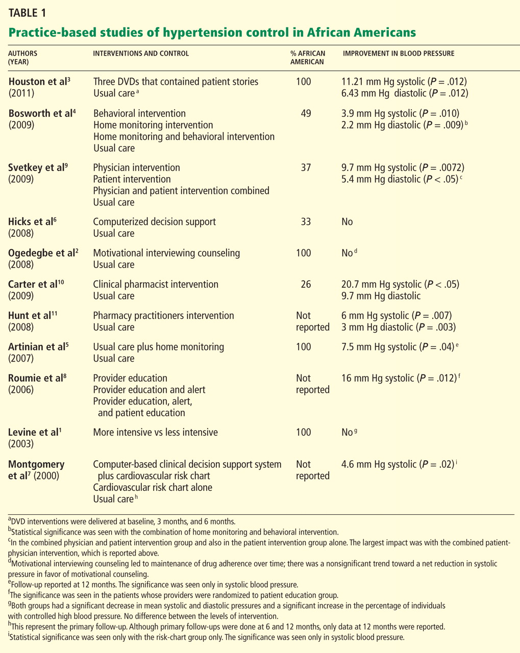
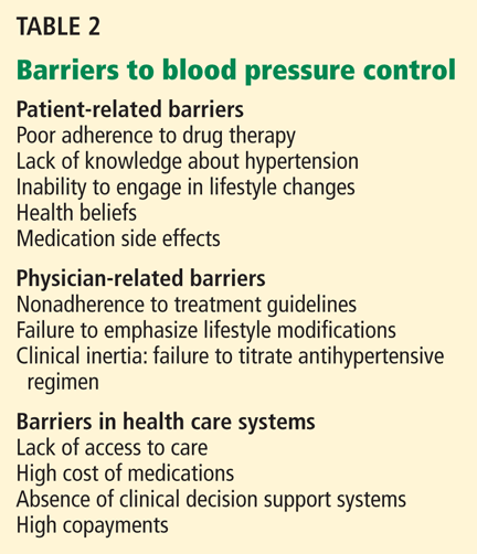
PATIENT-RELATED BARRIERS
Patient-related barriers24–40 include:
- Poor knowledge about hypertension and its consequences31,32
- Poor adherence to drug therapy (a major factor,24–26 as African Americans have poorer adherence rates than whites,27–29 which may explain some of the racial disparity in blood pressure control30)
- False health beliefs34–37
- Inability to change one’s lifestyle
- Side effects of antihypertensive drugs32
- Unrealistic expectations of treatment (eg, a cure33)
- Demographic factors (eg, socioeconomic status, educational level, age, sex).24,38–40
Perhaps the most salient and easily modifiable of these factors are patients’ reluctance to modify their lifestyle and their misconceptions about the causes, treatment, and prevention of hypertension. Patients whose beliefs are discordant with traditional biomedical concepts of hypertension have poorer blood pressure control than those whose beliefs are concordant.41 This may be more relevant to African Americans, since they are known to have cultural health beliefs that differ from those of Western culture (eg, that hypertension is a curable rather than a chronic illness, and that hypertension is a disease of nerves that often affects the blood and clogs the arteries).42
PHYSICIAN-RELATED BARRIERS
Barriers to effective blood pressure control at the physician level43–48 include:
- Nonadherence to treatment guidelines44
- Failure to intensify the regimen if goals are not met45
- Failure to emphasize therapeutic lifestyle changes.43,46–48
When primary care physicians do not follow evidence-based guidelines, the reason may be that they are not aware of them or that they do not understand them. In a national survey of 1,029 physicians that was designed to explore how well physicians know the indications for specific antihypertensive drugs and how closely their opinions and practice agreed with national guidelines, only 37.3% correctly answered all of the knowledge-related questions.49
Other reasons for nonadherence are that physicians may disagree with the guidelines, may not be able to follow the guidelines, may not believe that following them will achieve the desired effect, or may have no motivation to change their practice.50
Whatever the reason, Hyman et al51 reported that as many as 30% of physicians did not recommend treatment for patients with diastolic blood pressures of 90 to 100 mm Hg, and a higher proportion did not treat patients with systolic blood pressures of 140 to 160 mm Hg.
BARRIERS IN HEALTH CARE SYSTEMS
Although health care systems present barriers to optimal blood pressure control,20,27,31,52 there is evidence that most cases of uncontrolled hypertension occur in patients with good access to care.32,53,54 For example, an NHANES study53 suggested that most patients with uncontrolled hypertension had in fact seen a physician on average at least three times in the previous year. And this may be more pervasive in African Americans: one survey found hypertension was uncontrolled in 75% of hypertensive African American patients despite free access to care, free medications, and regular follow-up visits.41
Thus, the most significant barriers to blood pressure control appear to be patient-related and physician-related.
INTERVENTIONS AIMED AT PATIENTS
The most common approaches to improving blood pressure control at the patient level, regardless of race, are patient education,55–61 home blood pressure monitoring,62–67 and behavioral counseling to address misconceptions about hypertension,68 to improve adherence to drug therapy,69–73 and to encourage lifestyle modifications.74–78
Patient education
Patient education can improve blood pressure control.58,79–82 Its aims are to increase patients’ understanding of the disease83 and to encourage them to be more active in their own care.80,84,85
Patient education has a moderate effect on blood pressure control. The average proportion of patients whose hypertension was under control in community-based trials of various interventions ranged from 60% to 70%, compared with 38% to 46% with usual care.56,80,81
However, these strategies largely did not address misconceptions patients have about hypertension. This issue is especially critical in African Americans, who may have different perceptions of hypertension and different expectations for care41: beliefs that hypertension is “curable,” not chronic, and that medication is needed only for hypertension-related symptoms may translate to poorer rates of medication adherence.
Levine et al1 evaluated the efficacy of home visits by trained community health advisory board workers in a neighborhood in Baltimore, MD, with a high prevalence of hypertension. Participants were randomized to receive either one visit or five visits during the 40-month study period. Both groups had a statistically significant reduction in blood pressure, and in both groups the proportion of patients with adequate blood pressure control increased significantly. The results support the use of a practice- and community-based partnership to improve blood pressure control in African American patients.
Ogedegbe et al2 randomized 190 hypertensive African American patients to receive usual care or quarterly counseling sessions that used motivational interviewing focused on medication adherence. The counseled patients stayed adherent to their medications, whereas adherence declined significantly in those receiving usual care. This effect was associated with a modest, nonsignificant trend toward a net reduction in systolic blood pressure with motivational interviewing.
A novel method of health education is the use of narrative communication—ie, storytelling. It has a good amount of evidence to support it, as culturally appropriate storytelling may allow patients to identify with a story as it relates to their own lives.86–89 Examples of educational storytelling include:
- A woman with hypertension discussing what it means to have high blood pressure, and the benefits of controlling it, such as living long enough to see her grandchildren grow up
- A man discussing the importance of involving family and friends to help control blood pressure, and how dietary modifications can be made to ensure that salt alternatives are used when the family does the cooking.
Storytelling should be done in a culturally appropriate context. For example, storytellers should have the same background as the patient (ie, similar socioeconomic status and ethnic background): patients are more likely to be influenced if they identify with the storyteller and imagine themselves in a similar situation.
Houston et al3 randomized 299 hypertensive African Americans to view either three DVDs that featured patients with hypertension or three “attention-control DVDs” on topics not related to hypertension. The intervention group’s DVDs focused on storytelling and “learning more.” In the storytelling section, patients told personal stories about what it meant to have hypertension and gave advice on how to best interact with health care providers and methods to improve medication adherence. A “learning more” section focused on what high blood pressure is, addressed therapeutic lifestyle changes, and encouraged patients to communicate with their health care providers. The patients who viewed the patient narratives had significantly lower blood pressure at 3 months than those assigned to usual care. Although blood pressure subsequently increased in both groups, the benefits of the intervention still existed at the end of follow-up.
Important to note about two of the above three studies1,3 is that the interventions were done by people other than physicians, thus emphasizing the importance of a team approach to blood pressure control.
Behavioral counseling
The effectiveness of lifestyle modifications such as diet, weight loss, and physical activity in preventing and treating hypertension is well established.74–78 For example:
- In the Dietary Approaches to Stop Hypertension (DASH) trial,76 a healthy diet lowered blood pressure about as much as single drugs do, particularly in African Americans.
- The Trial of Nonpharmacologic Interventions in the Elderly (TONE)74 showed that exercise can lower blood pressure in obese hypertensive patients.
- The PREMIER trial (Lifestyle Interventions for Blood Pressure Control)75 showed that a single brief counseling session could produce substantial decreases in blood pressure in patients with stage 1 hypertension or high-normal blood pressure.
Unfortunately, these results have been hard to translate into primary care practice, especially for African American patients. Several studies have evaluated the impact of lifestyle interventions on blood pressure control in primary care practices with a large population of African American patients.
Bosworth et al,4 in a study of a practice in which almost half the patients were African American, randomized patients to receive usual care, nurse-administered tailored behavioral telephone counseling, home blood pressure monitoring, or home monitoring plus tailored behavioral telephone counseling. The combination of home monitoring and tailored behavioral telephone counseling led to a statistically significant improvement at 24 months compared with baseline.
Home blood pressure monitoring
The effectiveness of self-monitoring in improving blood pressure control is also well documented.62,63,65–67,90–95
Pickering et al62 studied patients with poorly controlled hypertension in a managed-care setting and found a reduction of 7 mm Hg systolic and 5 mm Hg diastolic pressure after 3 to 6 months of home monitoring compared with usual care.
Mengden et al,94 in a similar study, found average blood pressure reductions at 6 months of 19.3/11.9 mm Hg in the home-monitoring group vs 10.6/8.8 mm Hg in the usual-care group.
The effect of home blood pressure monitoring may be greater in African Americans.
Rogers et al93 found it to be more effective at lowering blood pressure than usual care in a group of 121 patients with poorly controlled hypertension followed in primary care practices, and these reductions were twice as large in African American patients than in white patients.93
Bondmass,92 in a study of 33 African American patients with poorly controlled hypertension, reported a 53% control rate within 4 weeks of home monitoring. All patients in the study had uncontrolled blood pressure at baseline (> 140/90 mm Hg).
Artinian et al5 evaluated the effect of nurse-managed telemonitoring on blood pressure control vs enhanced usual care. All participants were African American. The monitored group had a significantly greater reduction in systolic pressure at 12 months compared with those who received enhanced usual care.
PHYSICIAN-LEVEL INTERVENTIONS
Most interventions to improve how physicians manage patients with hypertension are designed to improve adherence to treatment guidelines. In most cases, these interventions are based on continuous quality improvement and disease management concepts such as physician education and academic detailing, reminders, feedback on performance measures, and risk-assessment tools.96,97
Physician education
Interest is increasing in physician educational interventions for blood pressure control.24,98
Inui et al,99 in an early study in a primary care practice, found that patients of physicians who received tutorials on hypertension management were more compliant with their drug regimens and had better blood pressure control than patients of physicians in the control group.
Jennett et al,100 in a similar randomized clinical trial, found that physicians who participated in an education activity were more adherent to treatment guidelines at 6 and 12 months compared with those who did not participate.
Maue et al101 showed that rates of blood pressure control improved from 41% to 52% after a 6-month educational intervention for physicians in a managed-care setting.
Tu et al102 reviewed 12 studies in which seven different physician educational interventions were used either alone or in combination and concluded that physician education improves compliance with guidelines for managing hypertension.
Unfortunately, these studies did not report outcomes separately for African American and white patients.
Hicks et al6 found that disease management approaches that target physicians whose patients with hypertension are mostly African American did not yield clinically relevant improvement in these patients, and that minority patients were significantly less likely to have their blood pressure controlled at the end of the study compared with their non-Hispanic white counterparts.
Feedback to providers
Several studies have shown that, given reminders and feedback systems, physicians will change their practice.103–106
Mashru and Lant104 combined chart audits and physician education in primary care practices and found they improved physician performance measures such as accuracy of diagnosis, number of patients who received cardiovascular risk assessment, and number of patients whose treatment was based on clinical laboratory assessments.
Feedback takes many forms but consists mostly of computerized information107 or peer-to-peer academic detailing with opinion leaders.108–110
Dickinson et al,106 for instance, showed that computer-generated listings of patients’ blood pressures combined with a physician education program on clinical management of hypertension led to increased knowledge and better follow-up on their patients.
Again, however, these studies did not distinguish between African American and white patients, which makes it difficult to judge whether or not these approaches work differently for physicians with a large proportion of African American patients.
Computerized decision-support systems
Computerized decision-support systems have proliferated in primary care practices.111
McAlister et al103 found that general practitioners randomized to manage hypertension with the assistance of a computer obtained better outcomes than with usual care.
Montgomery and Fahey,107 in a systematic review, found improved blood pressure control in two of the three trials that compared computer-generated feedback reports and reminders to usual care. Specifically, 51% of patients whose physicians received reminders either had controlled blood pressure or were at least receiving treatment vs 33% in the control group at 12 months. This difference was even higher at 24 months.
Montgomery et al7 later randomized primary care practices to use a computer-based decision-support system and a cardiovascular risk chart, the risk chart alone, or to continue as usual. Results indicated no reduction in cardiovascular risk in the computer-system or the chart-only group, whereas patients in the chart-only group had a significant reduction in systolic pressure and were prescribed more cardiovascular drugs. This study indicates that use of a computerized decision-support system is not superior to chart review and audit feedback alone.
Evidence that computerized decision systems improve blood pressure control in African Americans is scant. However, when one looks at the evidence from studies of African Americans, the outcomes do not seem to differ between African American and white patients.
Hicks et al6 examined the effectiveness of computerized decision support in improving hypertension care in a racially diverse population. Physicians were randomized to receive computerized decision support or to provide usual care without computerized support. Both groups improved significantly in prescribing appropriate drugs but not in overall blood pressure control. Furthermore, the study showed no reduction in racial disparities of care and blood pressure control.
A potential explanation for the lack of improvement in blood pressure was that the intervention dealt with making sure the appropriate drugs were prescribed rather than making sure physicians also appropriately intensified antihypertensive management when necessary.
INTERVENTIONS TARGETING PATIENTS AND PHYSICIANS
Several studies have targeted both patient and physician-level barriers to blood pressure control in practice-based settings.
Roumie et al8 randomized physicians to one of three intervention groups:
- “Provider education” consisting of an email message with a Web-based link to the Seventh Report of the Joint National Committee on Prevention, Detection, Evaluation, and Treatment of High Blood Pressure (JNC-7)
- Provider education plus a computer alert with information about their patient’s blood pressure
- Provider education, a computer alert, and patient education (ie, patients received a letter encouraging adherence to drug therapy, changing their lifestyle, and talking with their doctor about their blood pressure).
Patients whose providers were randomized to the third group had better blood pressure control. The report did not differentiate African American vs white patients. The data, however, did show the effectiveness of adding patient education to provider education to improve blood pressure control.
Bosworth et al,112 in a study in which 40% of patients were African American, randomized patients to usual care or to bimonthly nurse-delivered behavioral telephone counseling. They also randomized providers either to receive computer-generated decision support designed to improve adherence to guidelines or to receive no support.
There were no significant differences in rates of blood pressure control in the intervention groups compared with a control group. Although differences in blood pressure control between groups were not significant, patients randomized to behavioral intervention had significantly better blood pressure control at the 24-month follow-up than at baseline.
Svetkey et al9 evaluated the effects of physician intervention, patient intervention, and physician intervention plus patient intervention compared with control on systolic blood pressure at 6 months. They found that an intensive behavioral lifestyle intervention led to a significant reduction in systolic pressure at 6 months. By itself, the physician intervention did not have a meaningful effect, but patients in the combined physician-and-patient-intervention group experienced the greatest reduction (9.7 ± 12.7 mm Hg).
It takes a team
Physicians should not be the only focus in helping patients achieve blood pressure control. Although physician and patient factors need to be addressed to improve blood pressure control in African Americans, emphasis should also be placed on interdisciplinary, team-based care utilizing health care providers such as nurses, physician assistants, and pharmacists. Team-based care has been shown to have the greatest impact of all the strategies for improving blood pressure control.113 There is a good amount of evidence involving interventions with a focus on health care providers other than physicians, although the data lack a sufficient focus on African Americans.
Carter et al,10 in a randomized controlled trial in which 26.3% of the patients were African American, found that an intervention consisting of clinical pharmacists giving physicians drug therapy recommendations based on national guidelines resulted in a significantly lower blood pressure compared with a control group: the mean reduction was 20.7/9.7 mm Hg in the intervention group vs 6.8/4.5 mm Hg in the control group.
Carter et al114 performed a meta-analysis of 37 studies and found that two strategies led to a significant reduction in blood pressure: a pharmacist-led intervention with treatment recommendations to physicians resulted in a systolic pressure reduction of 9.30 mm Hg; and nurse-led interventions resulted in a systolic pressure reduction of 4.80 mm Hg. Again, many of the studies cited in this meta-analysis lacked a focus on African Americans.
Hunt et al11 conducted a randomized controlled trial in which pharmacists actively participated in the management of blood pressure. They were involved with every aspect of care, including reviewing medications and adverse drug reactions, assessing lifestyle behaviors and barriers to adherence, making dosing adjustments, and adding medications. Patients randomized to the intervention group achieved significantly lower systolic and diastolic pressures (137/75 vs 143/78 mm Hg in the control group). However, information about race was not included.
The above studies are just a few out of a large body of evidence demonstrating the value of team-based care to improve blood pressure control. It has yet to be determined whether these models can improve blood pressure control specifically in African Americans, since so many of these trials lacked a focus on this group. Promising is an ongoing randomized prospective trial by Carter et al115 evaluating a model of collaboration between physicians and pharmacists, with a focus on patients in underrepresented minorities.
SO WHAT WORKS?
Although there is a growing body of literature on interventions to try to reduce disparities in hypertension and blood pressure control between African Americans and whites, only a few randomized controlled trials have focused on African Americans, and several have not reported their results.116 So the question remains: How should we interpret the available data, which are aggregated across racial groups, and put it into practice when caring for hypertensive African American patients?
Patient education. In trying to overcome patient-related barriers, emphasis should be on patient education, in particular addressing misconceptions about hypertension and promoting adherence to antihypertensive therapy. This is evident from the narrative storytelling intervention by Houston et al.3 Although this is the first study of its kind, this strategy may be something to consider if future studies replicate these findings. Culturally appropriate storytelling may allow patients to identify with the stories as they relate to their own personal lives. It can be an effective way to address patient education and change behaviors.
Self-monitoring with a home blood pressure monitor has also proven effective in the management of hypertension in African Americans. Indeed, the few studies that reported findings in African Americans showed impressive reductions in blood pressure. The benefits of home monitoring are well documented, and the effect on physician-related barriers such as clinical inertia are also quite impressive.117 However, most of these studies did not assess the long-term impact or cost-effectiveness of home monitoring on blood pressure control.
Behavioral counseling. Although we have good evidence of the effectiveness of behavioral counseling, whether this is sustained long-term has been less studied in African Americans. Thus, while interventions that targeted African Americans have reported impressive reductions in blood pressure, the effect tends to be greatest during the first few months of implementation, with the benefits disappearing over time.
Physician-related interventions. With regard to physician-level interventions, research has focused on physician education, utilizing alerts and computerized clinical decision-support systems. Evidence is scant on whether the use of computerized systems results in improves hypertension care in African Americans. However, a closer look at the data from studies that report outcomes in African American and white patients shows that the results do not seem to differ between these groups. Still, there is insufficient information about the impact on hypertensive African Americans.6
Strategies that address both patient- and physician-related barriers can improve overall blood pressure control; however, there is a lack of data comparing outcomes in hypertensive African Americans with those of whites, making it difficult to know if this would be an effective strategy in African American patients alone.
More studies needed that focus on African Americans
Developing interventions to improve blood pressure control in African Americans should be an ongoing priority for research if we intend to address racial disparities in cardiovascular disease. Although it is reassuring that there is a growing body of evidence and research with this focus,118–121 more research is needed to determine effective strategies that address barriers related to physician practice and to the health care system overall as they relate to blood pressure control in African Americans. More importantly, these strategies should also emphasize a team-based approach that includes nurses, pharmacists, and physician assistants. Developing targeted interventions for hypertensive African Americans will help reduce disparities in the rates of cardiovascular illness and death in this patient population.
- Levine DM, Bone LR, Hill MN, et al. The effectiveness of a community/academic health center partnership in decreasing the level of blood pressure in an urban African-American population. Ethn Dis 2003; 13:354–361.
- Ogedegbe G, Chaplin W, Schoenthaler A, et al. A practice-based trial of motivational interviewing and adherence in hypertensive African Americans. Am J Hypertens 2008; 21:1137–1143.
- Houston TK, Allison JJ, Sussman M, et al. Culturally appropriate storytelling to improve blood pressure: a randomized trial. Ann Intern Med 2011; 154:77–84.
- Bosworth HB, Olsen MK, Grubber JM, et al. Two self-management interventions to improve hypertension control: a randomized trial. Ann Intern Med 2009; 151:687–695.
- Artinian NT, Flack JM, Nordstrom CK, et al. Effects of nurse-managed telemonitoring on blood pressure at 12-month follow-up among urban African Americans. Nurs Res 2007; 56:312–322.
- Hicks LS, Sequist TD, Ayanian JZ, et al. Impact of computerized decision support on blood pressure management and control: a randomized controlled trial. J Gen Intern Med 2008; 23:429–441.
- Montgomery AA, Fahey T, Peters TJ, MacIntosh C, Sharp DJ. Evaluation of computer based clinical decision support system and risk chart for management of hypertension in primary care: randomised controlled trial. BMJ 2000; 320:686–690.
- Roumie CL, Elasy TA, Greevy R, et al. Improving blood pressure control through provider education, provider alerts, and patient education: a cluster randomized trial. Ann Intern Med 2006; 145:165–175.
- Svetkey LP, Pollak KI, Yancy WS, et al. Hypertension improvement project: randomized trial of quality improvement for physicians and lifestyle modification for patients. Hypertension 2009; 54:1226–1233.
- Carter BL, Ardery G, Dawson JD, et al. Physician and pharmacist collaboration to improve blood pressure control. Arch Intern Med 2009; 169:1996–2002.
- Hunt JS, Siemienczuk J, Pape G, et al. A randomized controlled trial of team-based care: impact of physician-pharmacist collaboration on uncontrolled hypertension. J Gen Intern Med 2008; 23:1966–1972.
- US Department of Health and Human Services: Office of Disease Prevention and Health Promotion—Healthy People 2010. Nasnewsletter 2000; 15:3.
- Egan BM, Zhao Y, Axon RN. US trends in prevalence, awareness, treatment, and control of hypertension, 1988–2008. JAMA 2010; 303:2043–2050.
- US Centers for Disease Control and Prevention. Age-specific excess deaths associated with stroke among racial/ethnic minority populations–United States, 1997. JAMA 2000; 283:2382–2383.
- Giles WH, Kittner SJ, Hebel JR, Losonczy KG, Sherwin RW. Determinants of black-white differences in the risk of cerebral infarction. The National Health and Nutrition Examination Survey Epidemiologic Follow-up Study. Arch Intern Med 1995; 155:1319–1324.
- Klag MJ, Whelton PK, Randall BL, Neaton JD, Brancati FL, Stamler J. End-stage renal disease in African-American and white men. 16-year MRFIT findings. JAMA 1997; 277:1293–1298.
- Pavlik VN, Hyman DJ, Vallbona C, Toronjo C, Louis K. Hypertension awareness and control in an inner-city African-American sample. J Hum Hypertens 1997; 11:277–283.
- Roger VL, Go AS, Lloyd-Jones DM, et al. Heart Disease and Stroke Statistics—2011 Update: A Report From the American Heart Association. Circulation Feb 1; 123( 4):e18–e209.
- Chobanian AV, Bakris GL, Black HR, et al., Joint National Committee on Prevention, Detection, Evaluation, and Treatment of High Blood Pressure; National Heart, Lung, and Blood Institute; National High Blood Pressure Education Program Coordinating Committee. Seventh report of the Joint National Committee on Prevention, Detection, Evaluation, and Treatment of High Blood Pressure. Hypertension 2003; 42:1206–1252.
- Bone LR, Hill MN, Stallings R, et al. Community health survey in an urban African-American neighborhood: distribution and correlates of elevated blood pressure. Ethn Dis 2000; 10:87–95.
- Weber MA. Strategies for improving blood pressure control. Am J Hypertens 1998; 11:897–899.
- Hill MN, Sutton BS. Barriers to hypertension care and control. Curr Hypertens Rep 2000; 2:445–450.
- Alderman MH. Barriers to blood pressure control. Am J Hypertens 1999; 12:1268–1269.
- Chobanian AV. Control of hypertension—an important national priority. N Engl J Med 2001; 345:534–535.
- Miller NH, Hill M, Kottke T, Ockene IS. The multilevel compliance challenge: recommendations for a call to action. A statement for healthcare professionals. Circulation 1997; 95:1085–1090.
- Sackett DL, Snow JC. The magnitude of compliance and noncompliance. In:Haynes RB, Taylor DW, Sackett DL, eds. Compliance in Health Care. Baltimore, MD: John Hopkins University Press; 1979:11–22.
- Shea S, Misra D, Ehrlich MH, Field L, Francis CK. Correlates of nonadherence to hypertension treatment in an inner-city minority population. Am J Public Health 1992; 82:1607–1612.
- Kirscht JP, Rosenstock IM. Patient adherence to antihypertensive medical regimens. J Community Health. 1977; 3:115–124.
- Hershey JC, Morton BG, Davis JB, Reichgott MJ. Patient compliance with antihypertensive medication. Am J Public Health 1980; 70:1081–1089.
- Bosworth HB, Powers B, Grubber JM, et al. Racial differences in blood pressure control: potential explanatory factors. J Gen Intern Med 2008; 23:692–698.
- Douglas JG, Ferdinand KC, Bakris GL, Sowers JR. Barriers to blood pressure control in African Americans. Overcoming obstacles is challenging, but target goals can be attained. Postgrad Med 2002; 112:51–52,55,59–62passim.
- Knight EL, Bohn RL, Wang PS, Glynn RJ, Mogun H, Avorn J. Predictors of uncontrolled hypertension in ambulatory patients. Hypertension 2001; 38:809–814.
- Ogedegbe G, Mancuso CA, Allegrante JP. Expectations of blood pressure management in hypertensive African-American patients: a qualitative study. J Natl Med Assoc 2004; 96:442–449.
- Blumhagen D. Hypertension: a folk illness with a medical name. Cult Med Psychiatry 1980; 4:197–224.
- Meyer D, Leventhal H, Gutmann M. Common-sense models of illness: the example of hypertension. Health Psychol 1985; 4:115–135.
- Nelson EC, Stason WB, Neutra RR, Solomon HS, McArdle PJ. Impact of patient perceptions on compliance with treatment for hypertension. Med Care 1978; 16:893–906.
- Heurtin-Roberts S. ‘High-pertension’—the uses of a chronic folk illness for personal adaptation. Soc Sci Med 1993; 37:285–294.
- Lang T. Social and economic factors as obstacles to blood pressure control. Am J Hypertens 1998; 11:900–902.
- Lang T. Factors that appear as obstacles to the control of high blood pressure. Ethn Dis 2000; 10:125–130.
- Stamler R, Shipley M, Elliott P, Dyer A, Sans S, Stamler J. Higher blood pressure in adults with less education. Some explanations from INTERSALT. Hypertension 1992; 19:237–241.
- Heurtin-Roberts S, Reisin E. The relation of culturally influenced lay models of hypertension to compliance with treatment. Am J Hypertens 1992; 5:787–792.
- Snow LF. Folk medical beliefs and their implications for care of patients. A review bases on studies among black Americans. Ann Intern Med 1974; 81:82–96.
- Hicks LS, Shaykevich S, Bates DW, Ayanian JZ. Determinants of racial/ethnic differences in blood pressure management among hypertensive patients. BMC Cardiovasc Disord 2005; 5:16.
- Mehta SS, Wilcox CS, Schulman KA. Treatment of hypertension in patients with comorbidities: results from the study of hypertensive prescribing practices (SHyPP). Am J Hypertens 1999; 12:333–340.
- Ballard DJ, Strogatz DS, Wagner EH, et al. Hypertension control in a rural southern community: medical care process and dropping out. Am J Prev Med 1988; 4:133–139.
- Hajjar I, Miller K, Hirth V. Age-related bias in the management of hypertension: a national survey of physicians’ opinions on hypertension in elderly adults. J Gerontol A Biol Sci Med Sci 2002; 57:M487–M491.
- McAlister FA, Laupacis A, Teo KK, Hamilton PG, Montague TJ. A survey of clinician attitudes and management practices in hypertension. J Hum Hypertens 1997; 11:413–419.
- Trilling JS, Froom J. The urgent need to improve hypertension care. Arch Fam Med 2000; 9:794–801.
- Huse DM, Roht LH, Alpert JS, Hartz SC. Physicians’ knowledge, attitudes, and practice of pharmacologic treatment of hypertension. Ann Pharmacother 2001; 35:1173–1179.
- Cabana MD, Rand CS, Powe NR, et al. Why don’t physicians follow clinical practice guidelines? A framework for improvement. JAMA 1999; 282:1458–1465.
- Hyman DJ, Pavlik VN, Vallbona C. Physician role in lack of awareness and control of hypertension. J Clin Hypertens (Greenwich) 2000; 2:324–330.
- Morley Kotchen J, Walker WE, Kotchen TA. Rationale for a community approach to hypertension control among inner city minority populations. Heart Dis Stroke 1994; 3:61–62.
- Hyman DJ, Pavlik VN. Characteristics of patients with uncontrolled hypertension in the United States. N Engl J Med 2001; 345:479–486.
- Lang T. Factors that appear as obstacles to the control of high blood pressure. Ethn Dis 2000; 10:125–130.
- Pierce JP, Watson DS, Knights S, Gliddon T, Williams S, Watson R. A controlled trial of health education in the physician’s office. Prev Med 1984; 13:185–194.
- Morisky DE, DeMuth NM, Field-Fass M, Green LW, Levine DM. Evaluation of family health education to build social support for long-term control of high blood pressure. Health Educ Q 1985; 12:35–50.
- Lorgelly P, Siatis I, Brooks A, et al. Is ambulatory blood pressure monitoring cost-effective in the routine surveillance of treated hypertensive patients in primary care? Br J Gen Pract 2003; 53:794–796.
- Green LW, Levine DM, Wolle J, Deeds S. Development of randomized patient education experiments with urban poor hypertensives. Patient Couns Health Educ 1979; 1:106–111.
- Gruesser M, Hartmann P, Schlottmann N, Lohmann FW, Sawicki PT, Joergens V. Structured patient education for out-patients with hypertension in general practice: a model project in Germany. J Hum Hypertens 1997; 11:501–506.
- Mühlhauser I, Sawicki PT, Didjurgeit U, Jörgens V, Trampisch HJ, Berger M. Evaluation of a structured treatment and teaching programme on hypertension in general practice. Clin Exp Hypertens 1993; 15:125–142.
- Roca B, Nadal E, Rovira RE, Valls S, Lapuebla C, Lloría N. Usefulness of a hypertension education program. South Med J 2003; 96:1133–1137.
- Pickering TG, Gerin W, Holland JK. Home blood pressure teletransmission for better diagnosis and treatment. Curr Hypertens Rep 1999; 1:489–494.
- Yarows SA, Julius S, Pickering TG. Home blood pressure monitoring. Arch Intern Med 2000; 160:1251–1257.
- Haynes RB, Sackett DL, Gibson ES, et al. Improvement of medication compliance in uncontrolled hypertension. Lancet 1976; 1:1265–1268.
- Johnson AL, Taylor DW, Sackett DL, Dunnett CW, Shimizu AG. Self-recording of blood pressure in the management of hypertension. Can Med Assoc J 1978; 119:1034–1039.
- Carnahan JE, Nugent CA. The effects of self-monitoring by patients on the control of hypertension. Am J Med Sci 1975; 269:69–73.
- Stahl SM, Kelley CR, Neill PJ, Grim CE, Mamlin J. Effects of home blood pressure measurement on long-term BP control. Am J Public Health 1984; 74:704–709.
- Boulware LE, Daumit GL, Frick KD, Minkovitz CS, Lawrence RS, Powe NR. An evidence-based review of patient-centered behavioral interventions for hypertension. Am J Prev Med 2001; 21:221–232.
- Haynes RB, Mattson ME, Engebretson TO. Patient compliance to prescribed antihypertensive medication regimens: a report to the National Heart, Lung, and Blood institute. Bethesda, MD: US Department of Health and Human Services, Public Health Service, National Institutes of Health, 1980. NIH publication 81-2102.
- Burke LE, Dunbar-Jacob JM, Hill MN. Compliance with cardiovascular disease prevention strategies: a review of the research. Ann Behav Med 1997; 19:239–263.
- Dunbar-Jacob J, Dwyer K, Dunning EJ. Compliance with antihypertensive regimen: a review of the research in the 1980s. Ann Behav Med 1991; 13:31–39.
- Haynes RB, Montague P, Oliver T, McKibbon KA, Brouwers MC, Kanani R. Interventions for helping patients to follow prescriptions for medications. Cochrane Database Syst Rev 2000; ( 2):CD000011.
- Roter DL, Hall JA, Merisca R, Nordstrom B, Cretin D, Svarstad B. Effectiveness of interventions to improve patient compliance: a meta-analysis. Med Care 1998; 36:1138–1161.
- Appel LJ, Espeland MA, Easter L, Wilson AC, Folmar S, Lacy CR. Effects of reduced sodium intake on hypertension control in older individuals: results from the Trial of Nonpharmacologic Interventions in the Elderly (TONE). Arch Intern Med 2001; 161:685–693.
- Appel LJ, Champagne CM, Harsha DW, et al; Writing Group of the PREMIER Collaborative Research Group. Effects of comprehensive lifestyle modification on blood pressure control: main results of the PREMIER clinical trial. JAMA 2003; 289:2083–2093.
- Appel LJ, Moore TJ, Obarzanek E, et al. A clinical trial of the effects of dietary patterns on blood pressure. DASH Collaborative Research Group. N Engl J Med 1997; 336:1117–1124.
- Moore TJ, Conlin PR, Ard J, Svetkey LP. DASH (Dietary Approaches to Stop Hypertension) diet is effective treatment for stage 1 isolated systolic hypertension. Hypertension 2001; 38:155–158.
- Stevens VJ, Obarzanek E, Cook NR, et al; Trials for the Hypertension Prevention Research Group. Long-term weight loss and changes in blood pressure: results of the Trials of Hypertension Prevention, phase II. Ann Intern Med 2001; 134:1–11.
- Sawicki PT, Mühlhauser I, Didjurgeit U, Berger M. Improvement of hypertension care by a structured treatment and teaching programme. J Hum Hypertens 1993; 7:571–573.
- Morisky DE, Bowler MH, Finlay JS. An educational and behavioral approach toward increasing patient activation in hypertension management. J Community Health 1982; 7:171–182.
- Levine DM, Green LW, Deeds SG, Chwalow J, Russell RP, Finlay J. Health education for hypertensive patients. JAMA 1979; 241:1700–1703.
- Iso H, Shimamoto T, Yokota K, Sankai T, Jacobs DR, Komachi Y. Community-based education classes for hypertension control. A 1.5-year randomized controlled trial. Hypertension 1996; 27:968–974.
- Cuspidi C, Sampieri L, Macca G, et al. Improvement of patients’ knowledge by a single educational meeting on hypertension. J Hum Hypertens 2001; 15:57–61.
- Nessman DG, Carnahan JE, Nugent CA. Increasing compliance. Patient-operated hypertension groups. Arch Intern Med 1980; 140:1427–1430.
- Casasanta L, Patel S. Outcomes of an educational component of a disease management program for hypertension. Manag Care Interface 1999; 12:70–73.
- McAdams DP. The Stories We Live By: Personal Myths and the Making of the Self. New York NY: The Guilford Press; 1993.
- Bruner J. Acts of Meaning. Cambridge, MA: Harvard Univ Pr; 1990.
- Slater MD, Rouner D. Entertainment—education and elaboration likelihood: Understanding the processing of narrative persuasion. Commun Theory 2002; 12:173–191.
- Dal CS, Zanna MP, Fong GT. Narrative persuasion and overcoming resistance. In:Knowles ES, Linn J, eds. Resistance and Persuasion. Mahwah, NJ: Lawrence Erlbaum Assoc; 2004:175–191.
- Artinian NT, Washington OG, Templin TN. Effects of home telemonitoring and community-based monitoring on blood pressure control in urban African Americans: a pilot study. Heart Lung 2001; 30:191–199.
- Bailey B, Carney SL, Gillies AA, Smith AJ. Antihypertensive drug treatment: a comparison of usual care with self blood pressure measurement. J Hum Hypertens 1999; 13:147–150.
- Bondmass M. The effect of home monitoring and telemanagement on blood pressure control among African Americans. Telemed J 2000; 6:15–23.
- Rogers MA, Small D, Buchan DA, et al. Home monitoring service improves mean arterial pressure in patients with essential hypertension. A randomized, controlled trial. Ann Intern Med 2001; 134:1024–1032.
- Mengden T, Uen S, Baulmann J, Vetter H. Significance of blood pressure self-measurement as compared with office blood pressure measurement and ambulatory 24-hour blood pressure measurement in pharmacological studies. Blood Press Monit 2003; 8:169–172.
- Friedman RH, Kazis LE, Jette A, et al. A telecommunications system for monitoring and counseling patients with hypertension. Impact on medication adherence and blood pressure control. Am J Hypertens 1996; 9:285–292.
- Oxman AD, Thomson MA, Davis DA, Haynes RB. No magic bullets: a systematic review of 102 trials of interventions to improve professional practice. CMAJ 1995; 153:1423–1431.
- Wensing M, van der Weijden T, Grol R. Implementing guidelines and innovations in general practice: which interventions are effective? Br J Gen Pract 1998; 48:991–997.
- Davis DA, Thomson MA, Oxman AD, Haynes RB. Changing physician performance. A systematic review of the effect of continuing medical education strategies. JAMA 1995; 274:700–705.
- Inui TS, Yourtee EL, Williamson JW. Improved outcomes in hypertension after physician tutorials. A controlled trial. Ann Intern Med 1976; 84:646–651.
- Jennett PA, Wilson TW, Hayton RC, Mainprize GW, Laxdal OE. Desirable behaviours in the office management of hypertension addressed through continuing medical education. Can J Public Health 1989; 80:359–362.
- Maue SK, Rivo ML, Weiss B, Farrelly EW, Brower-Stenger S. Effect of a primary care physician-focused, population-based approach to blood pressure control. Fam Med 2002; 34:508–513.
- Tu K, Davis D. Can we alter physician behavior by educational methods? Lessons learned from studies of the management and follow-up of hypertension. J Contin Educ Health Prof 2002; 22:11–22.
- McAlister NH, Covvey HD, Tong C, Lee A, Wigle ED. Randomised controlled trial of computer assisted management of hypertension in primary care. Br Med J (Clin Res Ed) 1986; 293:670–674.
- Mashru M, Lant A. Interpractice audit of diagnosis and management of hypertension in primary care: educational intervention and review of medical records. BMJ 1997; 314:942–946.
- Degoulet P, Menard J, Berger C, Plouin PF, Devries C, Hirel JC. Hypertension management: the computer as a participant. Am J Med 1980; 68:559–567.
- Dickinson JC, Warshaw GA, Gehlbach SH, Bobula JA, Muhlbaier LH, Parkerson GR. Improving hypertension control: impact of computer feedback and physician education. Med Care 1981; 19:843–854.
- Montgomery AA, Fahey T. A systematic review of the use of computers in the management of hypertension. J Epidemiol Community Health 1998; 52:520–525.
- Coleman MT, Lott JA, Sharma S. Use of continuous quality improvement to identify barriers in the management of hypertension. Am J Med Qual 2000; 15:72–77.
- Goldberg HI, Wagner EH, Fihn SD, et al. A randomized controlled trial of CQI teams and academic detailing: can they alter compliance with guidelines? Jt Comm J Qual Improv 1998; 24:130–142.
- Horowitz CR, Goldberg HI, Martin DP, et al. Conducting a randomized controlled trial of CQI and academic detailing to implement clinical guidelines. Jt Comm J Qual Improv 1996; 22:734–750.
- Johnson B, McNair D, Kailasam K, et al. Discern—an integrated prospective decision support system. Proc Annu Symp Comput Appl Med Care 1994; 969.
- Bosworth HB, Olsen MK, Dudley T, et al. Patient education and provider decision support to control blood pressure in primary care: a cluster randomized trial. Am Heart J 2009; 157:450–456.
- Walsh JM, McDonald KM, Shojania KG, et al. Quality improvement strategies for hypertension management: a systematic review. Med Care 2006; 44:646–657.
- Carter BL, Rogers M, Daly J, Zheng S, James PA. The potency of team-based care interventions for hypertension: a meta-analysis. Arch Intern Med 2009; 169:1748–1755.
- Carter BL, Clarke W, Ardery G, et al; Collaboration Among Pharmacists Physicians To Improve Outcomes Now (CAPTION) Trial Investigators. A cluster-randomized effectiveness trial of a physician-pharmacist collaborative model to improve blood pressure control. Circ Cardiovasc Qual Outcomes 2010; 3:418–423.
- Einhorn PT. National heart, lung, and blood institute-initiated program “interventions to improve hypertension control rates in African Americans”: background and implementation. Circ Cardiovasc Qual Outcomes 2009; 2:236–240.
- Agarwal R, Bills JE, Hecht TJ, Light RP. Role of home blood pressure monitoring in overcoming therapeutic inertia and improving hypertension control: a systematic review and meta-analysis. Hypertension 2011; 57:29–38.
- Bosworth HB, Olsen MK, Neary A, et al. Take Control of Your Blood Pressure (TCYB) study: a multifactorial tailored behavioral and educational intervention for achieving blood pressure control. Patient Educ Couns 2008; 70:338–347.
- Bosworth HB, Olsen MK, Goldstein MK, et al. The veterans’ study to improve the control of hypertension (V-STITCH): design and methodology. Contemp Clin Trials 2005; 26:155–168.
- Ogedegbe G, Tobin JN, Fernandez S, et al. Counseling African Americans to Control Hypertension (CAATCH) trial: a multi-level intervention to improve blood pressure control in hypertensive blacks. Circ Cardiovasc Qual Outcomes 2009; 2:249–256.
- Bosworth HB, Almirall D, Weiner BJ, et al. The implementation of a translational study involving a primary care based behavioral program to improve blood pressure control: The HTN-IMPROVE study protocol (01295). Implement Sci 2010; 5:54.
High blood pressure takes a devastating toll on African Americans. Better control can go a long way to closing the “mortality gap” between African Americans and white Americans. But which strategies are best to address this complex problem?
In this report, we review the evidence on practice-based approaches to improving blood pressure control, from new styles of patient education to home blood pressure monitoring, focusing on studies in African Americans (Table 1).1–11
BETTER CONTROL IS NEEDED
PATIENT-RELATED BARRIERS
Patient-related barriers24–40 include:
- Poor knowledge about hypertension and its consequences31,32
- Poor adherence to drug therapy (a major factor,24–26 as African Americans have poorer adherence rates than whites,27–29 which may explain some of the racial disparity in blood pressure control30)
- False health beliefs34–37
- Inability to change one’s lifestyle
- Side effects of antihypertensive drugs32
- Unrealistic expectations of treatment (eg, a cure33)
- Demographic factors (eg, socioeconomic status, educational level, age, sex).24,38–40
Perhaps the most salient and easily modifiable of these factors are patients’ reluctance to modify their lifestyle and their misconceptions about the causes, treatment, and prevention of hypertension. Patients whose beliefs are discordant with traditional biomedical concepts of hypertension have poorer blood pressure control than those whose beliefs are concordant.41 This may be more relevant to African Americans, since they are known to have cultural health beliefs that differ from those of Western culture (eg, that hypertension is a curable rather than a chronic illness, and that hypertension is a disease of nerves that often affects the blood and clogs the arteries).42
PHYSICIAN-RELATED BARRIERS
Barriers to effective blood pressure control at the physician level43–48 include:
- Nonadherence to treatment guidelines44
- Failure to intensify the regimen if goals are not met45
- Failure to emphasize therapeutic lifestyle changes.43,46–48
When primary care physicians do not follow evidence-based guidelines, the reason may be that they are not aware of them or that they do not understand them. In a national survey of 1,029 physicians that was designed to explore how well physicians know the indications for specific antihypertensive drugs and how closely their opinions and practice agreed with national guidelines, only 37.3% correctly answered all of the knowledge-related questions.49
Other reasons for nonadherence are that physicians may disagree with the guidelines, may not be able to follow the guidelines, may not believe that following them will achieve the desired effect, or may have no motivation to change their practice.50
Whatever the reason, Hyman et al51 reported that as many as 30% of physicians did not recommend treatment for patients with diastolic blood pressures of 90 to 100 mm Hg, and a higher proportion did not treat patients with systolic blood pressures of 140 to 160 mm Hg.
BARRIERS IN HEALTH CARE SYSTEMS
Although health care systems present barriers to optimal blood pressure control,20,27,31,52 there is evidence that most cases of uncontrolled hypertension occur in patients with good access to care.32,53,54 For example, an NHANES study53 suggested that most patients with uncontrolled hypertension had in fact seen a physician on average at least three times in the previous year. And this may be more pervasive in African Americans: one survey found hypertension was uncontrolled in 75% of hypertensive African American patients despite free access to care, free medications, and regular follow-up visits.41
Thus, the most significant barriers to blood pressure control appear to be patient-related and physician-related.
INTERVENTIONS AIMED AT PATIENTS
The most common approaches to improving blood pressure control at the patient level, regardless of race, are patient education,55–61 home blood pressure monitoring,62–67 and behavioral counseling to address misconceptions about hypertension,68 to improve adherence to drug therapy,69–73 and to encourage lifestyle modifications.74–78
Patient education
Patient education can improve blood pressure control.58,79–82 Its aims are to increase patients’ understanding of the disease83 and to encourage them to be more active in their own care.80,84,85
Patient education has a moderate effect on blood pressure control. The average proportion of patients whose hypertension was under control in community-based trials of various interventions ranged from 60% to 70%, compared with 38% to 46% with usual care.56,80,81
However, these strategies largely did not address misconceptions patients have about hypertension. This issue is especially critical in African Americans, who may have different perceptions of hypertension and different expectations for care41: beliefs that hypertension is “curable,” not chronic, and that medication is needed only for hypertension-related symptoms may translate to poorer rates of medication adherence.
Levine et al1 evaluated the efficacy of home visits by trained community health advisory board workers in a neighborhood in Baltimore, MD, with a high prevalence of hypertension. Participants were randomized to receive either one visit or five visits during the 40-month study period. Both groups had a statistically significant reduction in blood pressure, and in both groups the proportion of patients with adequate blood pressure control increased significantly. The results support the use of a practice- and community-based partnership to improve blood pressure control in African American patients.
Ogedegbe et al2 randomized 190 hypertensive African American patients to receive usual care or quarterly counseling sessions that used motivational interviewing focused on medication adherence. The counseled patients stayed adherent to their medications, whereas adherence declined significantly in those receiving usual care. This effect was associated with a modest, nonsignificant trend toward a net reduction in systolic blood pressure with motivational interviewing.
A novel method of health education is the use of narrative communication—ie, storytelling. It has a good amount of evidence to support it, as culturally appropriate storytelling may allow patients to identify with a story as it relates to their own lives.86–89 Examples of educational storytelling include:
- A woman with hypertension discussing what it means to have high blood pressure, and the benefits of controlling it, such as living long enough to see her grandchildren grow up
- A man discussing the importance of involving family and friends to help control blood pressure, and how dietary modifications can be made to ensure that salt alternatives are used when the family does the cooking.
Storytelling should be done in a culturally appropriate context. For example, storytellers should have the same background as the patient (ie, similar socioeconomic status and ethnic background): patients are more likely to be influenced if they identify with the storyteller and imagine themselves in a similar situation.
Houston et al3 randomized 299 hypertensive African Americans to view either three DVDs that featured patients with hypertension or three “attention-control DVDs” on topics not related to hypertension. The intervention group’s DVDs focused on storytelling and “learning more.” In the storytelling section, patients told personal stories about what it meant to have hypertension and gave advice on how to best interact with health care providers and methods to improve medication adherence. A “learning more” section focused on what high blood pressure is, addressed therapeutic lifestyle changes, and encouraged patients to communicate with their health care providers. The patients who viewed the patient narratives had significantly lower blood pressure at 3 months than those assigned to usual care. Although blood pressure subsequently increased in both groups, the benefits of the intervention still existed at the end of follow-up.
Important to note about two of the above three studies1,3 is that the interventions were done by people other than physicians, thus emphasizing the importance of a team approach to blood pressure control.
Behavioral counseling
The effectiveness of lifestyle modifications such as diet, weight loss, and physical activity in preventing and treating hypertension is well established.74–78 For example:
- In the Dietary Approaches to Stop Hypertension (DASH) trial,76 a healthy diet lowered blood pressure about as much as single drugs do, particularly in African Americans.
- The Trial of Nonpharmacologic Interventions in the Elderly (TONE)74 showed that exercise can lower blood pressure in obese hypertensive patients.
- The PREMIER trial (Lifestyle Interventions for Blood Pressure Control)75 showed that a single brief counseling session could produce substantial decreases in blood pressure in patients with stage 1 hypertension or high-normal blood pressure.
Unfortunately, these results have been hard to translate into primary care practice, especially for African American patients. Several studies have evaluated the impact of lifestyle interventions on blood pressure control in primary care practices with a large population of African American patients.
Bosworth et al,4 in a study of a practice in which almost half the patients were African American, randomized patients to receive usual care, nurse-administered tailored behavioral telephone counseling, home blood pressure monitoring, or home monitoring plus tailored behavioral telephone counseling. The combination of home monitoring and tailored behavioral telephone counseling led to a statistically significant improvement at 24 months compared with baseline.
Home blood pressure monitoring
The effectiveness of self-monitoring in improving blood pressure control is also well documented.62,63,65–67,90–95
Pickering et al62 studied patients with poorly controlled hypertension in a managed-care setting and found a reduction of 7 mm Hg systolic and 5 mm Hg diastolic pressure after 3 to 6 months of home monitoring compared with usual care.
Mengden et al,94 in a similar study, found average blood pressure reductions at 6 months of 19.3/11.9 mm Hg in the home-monitoring group vs 10.6/8.8 mm Hg in the usual-care group.
The effect of home blood pressure monitoring may be greater in African Americans.
Rogers et al93 found it to be more effective at lowering blood pressure than usual care in a group of 121 patients with poorly controlled hypertension followed in primary care practices, and these reductions were twice as large in African American patients than in white patients.93
Bondmass,92 in a study of 33 African American patients with poorly controlled hypertension, reported a 53% control rate within 4 weeks of home monitoring. All patients in the study had uncontrolled blood pressure at baseline (> 140/90 mm Hg).
Artinian et al5 evaluated the effect of nurse-managed telemonitoring on blood pressure control vs enhanced usual care. All participants were African American. The monitored group had a significantly greater reduction in systolic pressure at 12 months compared with those who received enhanced usual care.
PHYSICIAN-LEVEL INTERVENTIONS
Most interventions to improve how physicians manage patients with hypertension are designed to improve adherence to treatment guidelines. In most cases, these interventions are based on continuous quality improvement and disease management concepts such as physician education and academic detailing, reminders, feedback on performance measures, and risk-assessment tools.96,97
Physician education
Interest is increasing in physician educational interventions for blood pressure control.24,98
Inui et al,99 in an early study in a primary care practice, found that patients of physicians who received tutorials on hypertension management were more compliant with their drug regimens and had better blood pressure control than patients of physicians in the control group.
Jennett et al,100 in a similar randomized clinical trial, found that physicians who participated in an education activity were more adherent to treatment guidelines at 6 and 12 months compared with those who did not participate.
Maue et al101 showed that rates of blood pressure control improved from 41% to 52% after a 6-month educational intervention for physicians in a managed-care setting.
Tu et al102 reviewed 12 studies in which seven different physician educational interventions were used either alone or in combination and concluded that physician education improves compliance with guidelines for managing hypertension.
Unfortunately, these studies did not report outcomes separately for African American and white patients.
Hicks et al6 found that disease management approaches that target physicians whose patients with hypertension are mostly African American did not yield clinically relevant improvement in these patients, and that minority patients were significantly less likely to have their blood pressure controlled at the end of the study compared with their non-Hispanic white counterparts.
Feedback to providers
Several studies have shown that, given reminders and feedback systems, physicians will change their practice.103–106
Mashru and Lant104 combined chart audits and physician education in primary care practices and found they improved physician performance measures such as accuracy of diagnosis, number of patients who received cardiovascular risk assessment, and number of patients whose treatment was based on clinical laboratory assessments.
Feedback takes many forms but consists mostly of computerized information107 or peer-to-peer academic detailing with opinion leaders.108–110
Dickinson et al,106 for instance, showed that computer-generated listings of patients’ blood pressures combined with a physician education program on clinical management of hypertension led to increased knowledge and better follow-up on their patients.
Again, however, these studies did not distinguish between African American and white patients, which makes it difficult to judge whether or not these approaches work differently for physicians with a large proportion of African American patients.
Computerized decision-support systems
Computerized decision-support systems have proliferated in primary care practices.111
McAlister et al103 found that general practitioners randomized to manage hypertension with the assistance of a computer obtained better outcomes than with usual care.
Montgomery and Fahey,107 in a systematic review, found improved blood pressure control in two of the three trials that compared computer-generated feedback reports and reminders to usual care. Specifically, 51% of patients whose physicians received reminders either had controlled blood pressure or were at least receiving treatment vs 33% in the control group at 12 months. This difference was even higher at 24 months.
Montgomery et al7 later randomized primary care practices to use a computer-based decision-support system and a cardiovascular risk chart, the risk chart alone, or to continue as usual. Results indicated no reduction in cardiovascular risk in the computer-system or the chart-only group, whereas patients in the chart-only group had a significant reduction in systolic pressure and were prescribed more cardiovascular drugs. This study indicates that use of a computerized decision-support system is not superior to chart review and audit feedback alone.
Evidence that computerized decision systems improve blood pressure control in African Americans is scant. However, when one looks at the evidence from studies of African Americans, the outcomes do not seem to differ between African American and white patients.
Hicks et al6 examined the effectiveness of computerized decision support in improving hypertension care in a racially diverse population. Physicians were randomized to receive computerized decision support or to provide usual care without computerized support. Both groups improved significantly in prescribing appropriate drugs but not in overall blood pressure control. Furthermore, the study showed no reduction in racial disparities of care and blood pressure control.
A potential explanation for the lack of improvement in blood pressure was that the intervention dealt with making sure the appropriate drugs were prescribed rather than making sure physicians also appropriately intensified antihypertensive management when necessary.
INTERVENTIONS TARGETING PATIENTS AND PHYSICIANS
Several studies have targeted both patient and physician-level barriers to blood pressure control in practice-based settings.
Roumie et al8 randomized physicians to one of three intervention groups:
- “Provider education” consisting of an email message with a Web-based link to the Seventh Report of the Joint National Committee on Prevention, Detection, Evaluation, and Treatment of High Blood Pressure (JNC-7)
- Provider education plus a computer alert with information about their patient’s blood pressure
- Provider education, a computer alert, and patient education (ie, patients received a letter encouraging adherence to drug therapy, changing their lifestyle, and talking with their doctor about their blood pressure).
Patients whose providers were randomized to the third group had better blood pressure control. The report did not differentiate African American vs white patients. The data, however, did show the effectiveness of adding patient education to provider education to improve blood pressure control.
Bosworth et al,112 in a study in which 40% of patients were African American, randomized patients to usual care or to bimonthly nurse-delivered behavioral telephone counseling. They also randomized providers either to receive computer-generated decision support designed to improve adherence to guidelines or to receive no support.
There were no significant differences in rates of blood pressure control in the intervention groups compared with a control group. Although differences in blood pressure control between groups were not significant, patients randomized to behavioral intervention had significantly better blood pressure control at the 24-month follow-up than at baseline.
Svetkey et al9 evaluated the effects of physician intervention, patient intervention, and physician intervention plus patient intervention compared with control on systolic blood pressure at 6 months. They found that an intensive behavioral lifestyle intervention led to a significant reduction in systolic pressure at 6 months. By itself, the physician intervention did not have a meaningful effect, but patients in the combined physician-and-patient-intervention group experienced the greatest reduction (9.7 ± 12.7 mm Hg).
It takes a team
Physicians should not be the only focus in helping patients achieve blood pressure control. Although physician and patient factors need to be addressed to improve blood pressure control in African Americans, emphasis should also be placed on interdisciplinary, team-based care utilizing health care providers such as nurses, physician assistants, and pharmacists. Team-based care has been shown to have the greatest impact of all the strategies for improving blood pressure control.113 There is a good amount of evidence involving interventions with a focus on health care providers other than physicians, although the data lack a sufficient focus on African Americans.
Carter et al,10 in a randomized controlled trial in which 26.3% of the patients were African American, found that an intervention consisting of clinical pharmacists giving physicians drug therapy recommendations based on national guidelines resulted in a significantly lower blood pressure compared with a control group: the mean reduction was 20.7/9.7 mm Hg in the intervention group vs 6.8/4.5 mm Hg in the control group.
Carter et al114 performed a meta-analysis of 37 studies and found that two strategies led to a significant reduction in blood pressure: a pharmacist-led intervention with treatment recommendations to physicians resulted in a systolic pressure reduction of 9.30 mm Hg; and nurse-led interventions resulted in a systolic pressure reduction of 4.80 mm Hg. Again, many of the studies cited in this meta-analysis lacked a focus on African Americans.
Hunt et al11 conducted a randomized controlled trial in which pharmacists actively participated in the management of blood pressure. They were involved with every aspect of care, including reviewing medications and adverse drug reactions, assessing lifestyle behaviors and barriers to adherence, making dosing adjustments, and adding medications. Patients randomized to the intervention group achieved significantly lower systolic and diastolic pressures (137/75 vs 143/78 mm Hg in the control group). However, information about race was not included.
The above studies are just a few out of a large body of evidence demonstrating the value of team-based care to improve blood pressure control. It has yet to be determined whether these models can improve blood pressure control specifically in African Americans, since so many of these trials lacked a focus on this group. Promising is an ongoing randomized prospective trial by Carter et al115 evaluating a model of collaboration between physicians and pharmacists, with a focus on patients in underrepresented minorities.
SO WHAT WORKS?
Although there is a growing body of literature on interventions to try to reduce disparities in hypertension and blood pressure control between African Americans and whites, only a few randomized controlled trials have focused on African Americans, and several have not reported their results.116 So the question remains: How should we interpret the available data, which are aggregated across racial groups, and put it into practice when caring for hypertensive African American patients?
Patient education. In trying to overcome patient-related barriers, emphasis should be on patient education, in particular addressing misconceptions about hypertension and promoting adherence to antihypertensive therapy. This is evident from the narrative storytelling intervention by Houston et al.3 Although this is the first study of its kind, this strategy may be something to consider if future studies replicate these findings. Culturally appropriate storytelling may allow patients to identify with the stories as they relate to their own personal lives. It can be an effective way to address patient education and change behaviors.
Self-monitoring with a home blood pressure monitor has also proven effective in the management of hypertension in African Americans. Indeed, the few studies that reported findings in African Americans showed impressive reductions in blood pressure. The benefits of home monitoring are well documented, and the effect on physician-related barriers such as clinical inertia are also quite impressive.117 However, most of these studies did not assess the long-term impact or cost-effectiveness of home monitoring on blood pressure control.
Behavioral counseling. Although we have good evidence of the effectiveness of behavioral counseling, whether this is sustained long-term has been less studied in African Americans. Thus, while interventions that targeted African Americans have reported impressive reductions in blood pressure, the effect tends to be greatest during the first few months of implementation, with the benefits disappearing over time.
Physician-related interventions. With regard to physician-level interventions, research has focused on physician education, utilizing alerts and computerized clinical decision-support systems. Evidence is scant on whether the use of computerized systems results in improves hypertension care in African Americans. However, a closer look at the data from studies that report outcomes in African American and white patients shows that the results do not seem to differ between these groups. Still, there is insufficient information about the impact on hypertensive African Americans.6
Strategies that address both patient- and physician-related barriers can improve overall blood pressure control; however, there is a lack of data comparing outcomes in hypertensive African Americans with those of whites, making it difficult to know if this would be an effective strategy in African American patients alone.
More studies needed that focus on African Americans
Developing interventions to improve blood pressure control in African Americans should be an ongoing priority for research if we intend to address racial disparities in cardiovascular disease. Although it is reassuring that there is a growing body of evidence and research with this focus,118–121 more research is needed to determine effective strategies that address barriers related to physician practice and to the health care system overall as they relate to blood pressure control in African Americans. More importantly, these strategies should also emphasize a team-based approach that includes nurses, pharmacists, and physician assistants. Developing targeted interventions for hypertensive African Americans will help reduce disparities in the rates of cardiovascular illness and death in this patient population.
High blood pressure takes a devastating toll on African Americans. Better control can go a long way to closing the “mortality gap” between African Americans and white Americans. But which strategies are best to address this complex problem?
In this report, we review the evidence on practice-based approaches to improving blood pressure control, from new styles of patient education to home blood pressure monitoring, focusing on studies in African Americans (Table 1).1–11
BETTER CONTROL IS NEEDED
PATIENT-RELATED BARRIERS
Patient-related barriers24–40 include:
- Poor knowledge about hypertension and its consequences31,32
- Poor adherence to drug therapy (a major factor,24–26 as African Americans have poorer adherence rates than whites,27–29 which may explain some of the racial disparity in blood pressure control30)
- False health beliefs34–37
- Inability to change one’s lifestyle
- Side effects of antihypertensive drugs32
- Unrealistic expectations of treatment (eg, a cure33)
- Demographic factors (eg, socioeconomic status, educational level, age, sex).24,38–40
Perhaps the most salient and easily modifiable of these factors are patients’ reluctance to modify their lifestyle and their misconceptions about the causes, treatment, and prevention of hypertension. Patients whose beliefs are discordant with traditional biomedical concepts of hypertension have poorer blood pressure control than those whose beliefs are concordant.41 This may be more relevant to African Americans, since they are known to have cultural health beliefs that differ from those of Western culture (eg, that hypertension is a curable rather than a chronic illness, and that hypertension is a disease of nerves that often affects the blood and clogs the arteries).42
PHYSICIAN-RELATED BARRIERS
Barriers to effective blood pressure control at the physician level43–48 include:
- Nonadherence to treatment guidelines44
- Failure to intensify the regimen if goals are not met45
- Failure to emphasize therapeutic lifestyle changes.43,46–48
When primary care physicians do not follow evidence-based guidelines, the reason may be that they are not aware of them or that they do not understand them. In a national survey of 1,029 physicians that was designed to explore how well physicians know the indications for specific antihypertensive drugs and how closely their opinions and practice agreed with national guidelines, only 37.3% correctly answered all of the knowledge-related questions.49
Other reasons for nonadherence are that physicians may disagree with the guidelines, may not be able to follow the guidelines, may not believe that following them will achieve the desired effect, or may have no motivation to change their practice.50
Whatever the reason, Hyman et al51 reported that as many as 30% of physicians did not recommend treatment for patients with diastolic blood pressures of 90 to 100 mm Hg, and a higher proportion did not treat patients with systolic blood pressures of 140 to 160 mm Hg.
BARRIERS IN HEALTH CARE SYSTEMS
Although health care systems present barriers to optimal blood pressure control,20,27,31,52 there is evidence that most cases of uncontrolled hypertension occur in patients with good access to care.32,53,54 For example, an NHANES study53 suggested that most patients with uncontrolled hypertension had in fact seen a physician on average at least three times in the previous year. And this may be more pervasive in African Americans: one survey found hypertension was uncontrolled in 75% of hypertensive African American patients despite free access to care, free medications, and regular follow-up visits.41
Thus, the most significant barriers to blood pressure control appear to be patient-related and physician-related.
INTERVENTIONS AIMED AT PATIENTS
The most common approaches to improving blood pressure control at the patient level, regardless of race, are patient education,55–61 home blood pressure monitoring,62–67 and behavioral counseling to address misconceptions about hypertension,68 to improve adherence to drug therapy,69–73 and to encourage lifestyle modifications.74–78
Patient education
Patient education can improve blood pressure control.58,79–82 Its aims are to increase patients’ understanding of the disease83 and to encourage them to be more active in their own care.80,84,85
Patient education has a moderate effect on blood pressure control. The average proportion of patients whose hypertension was under control in community-based trials of various interventions ranged from 60% to 70%, compared with 38% to 46% with usual care.56,80,81
However, these strategies largely did not address misconceptions patients have about hypertension. This issue is especially critical in African Americans, who may have different perceptions of hypertension and different expectations for care41: beliefs that hypertension is “curable,” not chronic, and that medication is needed only for hypertension-related symptoms may translate to poorer rates of medication adherence.
Levine et al1 evaluated the efficacy of home visits by trained community health advisory board workers in a neighborhood in Baltimore, MD, with a high prevalence of hypertension. Participants were randomized to receive either one visit or five visits during the 40-month study period. Both groups had a statistically significant reduction in blood pressure, and in both groups the proportion of patients with adequate blood pressure control increased significantly. The results support the use of a practice- and community-based partnership to improve blood pressure control in African American patients.
Ogedegbe et al2 randomized 190 hypertensive African American patients to receive usual care or quarterly counseling sessions that used motivational interviewing focused on medication adherence. The counseled patients stayed adherent to their medications, whereas adherence declined significantly in those receiving usual care. This effect was associated with a modest, nonsignificant trend toward a net reduction in systolic blood pressure with motivational interviewing.
A novel method of health education is the use of narrative communication—ie, storytelling. It has a good amount of evidence to support it, as culturally appropriate storytelling may allow patients to identify with a story as it relates to their own lives.86–89 Examples of educational storytelling include:
- A woman with hypertension discussing what it means to have high blood pressure, and the benefits of controlling it, such as living long enough to see her grandchildren grow up
- A man discussing the importance of involving family and friends to help control blood pressure, and how dietary modifications can be made to ensure that salt alternatives are used when the family does the cooking.
Storytelling should be done in a culturally appropriate context. For example, storytellers should have the same background as the patient (ie, similar socioeconomic status and ethnic background): patients are more likely to be influenced if they identify with the storyteller and imagine themselves in a similar situation.
Houston et al3 randomized 299 hypertensive African Americans to view either three DVDs that featured patients with hypertension or three “attention-control DVDs” on topics not related to hypertension. The intervention group’s DVDs focused on storytelling and “learning more.” In the storytelling section, patients told personal stories about what it meant to have hypertension and gave advice on how to best interact with health care providers and methods to improve medication adherence. A “learning more” section focused on what high blood pressure is, addressed therapeutic lifestyle changes, and encouraged patients to communicate with their health care providers. The patients who viewed the patient narratives had significantly lower blood pressure at 3 months than those assigned to usual care. Although blood pressure subsequently increased in both groups, the benefits of the intervention still existed at the end of follow-up.
Important to note about two of the above three studies1,3 is that the interventions were done by people other than physicians, thus emphasizing the importance of a team approach to blood pressure control.
Behavioral counseling
The effectiveness of lifestyle modifications such as diet, weight loss, and physical activity in preventing and treating hypertension is well established.74–78 For example:
- In the Dietary Approaches to Stop Hypertension (DASH) trial,76 a healthy diet lowered blood pressure about as much as single drugs do, particularly in African Americans.
- The Trial of Nonpharmacologic Interventions in the Elderly (TONE)74 showed that exercise can lower blood pressure in obese hypertensive patients.
- The PREMIER trial (Lifestyle Interventions for Blood Pressure Control)75 showed that a single brief counseling session could produce substantial decreases in blood pressure in patients with stage 1 hypertension or high-normal blood pressure.
Unfortunately, these results have been hard to translate into primary care practice, especially for African American patients. Several studies have evaluated the impact of lifestyle interventions on blood pressure control in primary care practices with a large population of African American patients.
Bosworth et al,4 in a study of a practice in which almost half the patients were African American, randomized patients to receive usual care, nurse-administered tailored behavioral telephone counseling, home blood pressure monitoring, or home monitoring plus tailored behavioral telephone counseling. The combination of home monitoring and tailored behavioral telephone counseling led to a statistically significant improvement at 24 months compared with baseline.
Home blood pressure monitoring
The effectiveness of self-monitoring in improving blood pressure control is also well documented.62,63,65–67,90–95
Pickering et al62 studied patients with poorly controlled hypertension in a managed-care setting and found a reduction of 7 mm Hg systolic and 5 mm Hg diastolic pressure after 3 to 6 months of home monitoring compared with usual care.
Mengden et al,94 in a similar study, found average blood pressure reductions at 6 months of 19.3/11.9 mm Hg in the home-monitoring group vs 10.6/8.8 mm Hg in the usual-care group.
The effect of home blood pressure monitoring may be greater in African Americans.
Rogers et al93 found it to be more effective at lowering blood pressure than usual care in a group of 121 patients with poorly controlled hypertension followed in primary care practices, and these reductions were twice as large in African American patients than in white patients.93
Bondmass,92 in a study of 33 African American patients with poorly controlled hypertension, reported a 53% control rate within 4 weeks of home monitoring. All patients in the study had uncontrolled blood pressure at baseline (> 140/90 mm Hg).
Artinian et al5 evaluated the effect of nurse-managed telemonitoring on blood pressure control vs enhanced usual care. All participants were African American. The monitored group had a significantly greater reduction in systolic pressure at 12 months compared with those who received enhanced usual care.
PHYSICIAN-LEVEL INTERVENTIONS
Most interventions to improve how physicians manage patients with hypertension are designed to improve adherence to treatment guidelines. In most cases, these interventions are based on continuous quality improvement and disease management concepts such as physician education and academic detailing, reminders, feedback on performance measures, and risk-assessment tools.96,97
Physician education
Interest is increasing in physician educational interventions for blood pressure control.24,98
Inui et al,99 in an early study in a primary care practice, found that patients of physicians who received tutorials on hypertension management were more compliant with their drug regimens and had better blood pressure control than patients of physicians in the control group.
Jennett et al,100 in a similar randomized clinical trial, found that physicians who participated in an education activity were more adherent to treatment guidelines at 6 and 12 months compared with those who did not participate.
Maue et al101 showed that rates of blood pressure control improved from 41% to 52% after a 6-month educational intervention for physicians in a managed-care setting.
Tu et al102 reviewed 12 studies in which seven different physician educational interventions were used either alone or in combination and concluded that physician education improves compliance with guidelines for managing hypertension.
Unfortunately, these studies did not report outcomes separately for African American and white patients.
Hicks et al6 found that disease management approaches that target physicians whose patients with hypertension are mostly African American did not yield clinically relevant improvement in these patients, and that minority patients were significantly less likely to have their blood pressure controlled at the end of the study compared with their non-Hispanic white counterparts.
Feedback to providers
Several studies have shown that, given reminders and feedback systems, physicians will change their practice.103–106
Mashru and Lant104 combined chart audits and physician education in primary care practices and found they improved physician performance measures such as accuracy of diagnosis, number of patients who received cardiovascular risk assessment, and number of patients whose treatment was based on clinical laboratory assessments.
Feedback takes many forms but consists mostly of computerized information107 or peer-to-peer academic detailing with opinion leaders.108–110
Dickinson et al,106 for instance, showed that computer-generated listings of patients’ blood pressures combined with a physician education program on clinical management of hypertension led to increased knowledge and better follow-up on their patients.
Again, however, these studies did not distinguish between African American and white patients, which makes it difficult to judge whether or not these approaches work differently for physicians with a large proportion of African American patients.
Computerized decision-support systems
Computerized decision-support systems have proliferated in primary care practices.111
McAlister et al103 found that general practitioners randomized to manage hypertension with the assistance of a computer obtained better outcomes than with usual care.
Montgomery and Fahey,107 in a systematic review, found improved blood pressure control in two of the three trials that compared computer-generated feedback reports and reminders to usual care. Specifically, 51% of patients whose physicians received reminders either had controlled blood pressure or were at least receiving treatment vs 33% in the control group at 12 months. This difference was even higher at 24 months.
Montgomery et al7 later randomized primary care practices to use a computer-based decision-support system and a cardiovascular risk chart, the risk chart alone, or to continue as usual. Results indicated no reduction in cardiovascular risk in the computer-system or the chart-only group, whereas patients in the chart-only group had a significant reduction in systolic pressure and were prescribed more cardiovascular drugs. This study indicates that use of a computerized decision-support system is not superior to chart review and audit feedback alone.
Evidence that computerized decision systems improve blood pressure control in African Americans is scant. However, when one looks at the evidence from studies of African Americans, the outcomes do not seem to differ between African American and white patients.
Hicks et al6 examined the effectiveness of computerized decision support in improving hypertension care in a racially diverse population. Physicians were randomized to receive computerized decision support or to provide usual care without computerized support. Both groups improved significantly in prescribing appropriate drugs but not in overall blood pressure control. Furthermore, the study showed no reduction in racial disparities of care and blood pressure control.
A potential explanation for the lack of improvement in blood pressure was that the intervention dealt with making sure the appropriate drugs were prescribed rather than making sure physicians also appropriately intensified antihypertensive management when necessary.
INTERVENTIONS TARGETING PATIENTS AND PHYSICIANS
Several studies have targeted both patient and physician-level barriers to blood pressure control in practice-based settings.
Roumie et al8 randomized physicians to one of three intervention groups:
- “Provider education” consisting of an email message with a Web-based link to the Seventh Report of the Joint National Committee on Prevention, Detection, Evaluation, and Treatment of High Blood Pressure (JNC-7)
- Provider education plus a computer alert with information about their patient’s blood pressure
- Provider education, a computer alert, and patient education (ie, patients received a letter encouraging adherence to drug therapy, changing their lifestyle, and talking with their doctor about their blood pressure).
Patients whose providers were randomized to the third group had better blood pressure control. The report did not differentiate African American vs white patients. The data, however, did show the effectiveness of adding patient education to provider education to improve blood pressure control.
Bosworth et al,112 in a study in which 40% of patients were African American, randomized patients to usual care or to bimonthly nurse-delivered behavioral telephone counseling. They also randomized providers either to receive computer-generated decision support designed to improve adherence to guidelines or to receive no support.
There were no significant differences in rates of blood pressure control in the intervention groups compared with a control group. Although differences in blood pressure control between groups were not significant, patients randomized to behavioral intervention had significantly better blood pressure control at the 24-month follow-up than at baseline.
Svetkey et al9 evaluated the effects of physician intervention, patient intervention, and physician intervention plus patient intervention compared with control on systolic blood pressure at 6 months. They found that an intensive behavioral lifestyle intervention led to a significant reduction in systolic pressure at 6 months. By itself, the physician intervention did not have a meaningful effect, but patients in the combined physician-and-patient-intervention group experienced the greatest reduction (9.7 ± 12.7 mm Hg).
It takes a team
Physicians should not be the only focus in helping patients achieve blood pressure control. Although physician and patient factors need to be addressed to improve blood pressure control in African Americans, emphasis should also be placed on interdisciplinary, team-based care utilizing health care providers such as nurses, physician assistants, and pharmacists. Team-based care has been shown to have the greatest impact of all the strategies for improving blood pressure control.113 There is a good amount of evidence involving interventions with a focus on health care providers other than physicians, although the data lack a sufficient focus on African Americans.
Carter et al,10 in a randomized controlled trial in which 26.3% of the patients were African American, found that an intervention consisting of clinical pharmacists giving physicians drug therapy recommendations based on national guidelines resulted in a significantly lower blood pressure compared with a control group: the mean reduction was 20.7/9.7 mm Hg in the intervention group vs 6.8/4.5 mm Hg in the control group.
Carter et al114 performed a meta-analysis of 37 studies and found that two strategies led to a significant reduction in blood pressure: a pharmacist-led intervention with treatment recommendations to physicians resulted in a systolic pressure reduction of 9.30 mm Hg; and nurse-led interventions resulted in a systolic pressure reduction of 4.80 mm Hg. Again, many of the studies cited in this meta-analysis lacked a focus on African Americans.
Hunt et al11 conducted a randomized controlled trial in which pharmacists actively participated in the management of blood pressure. They were involved with every aspect of care, including reviewing medications and adverse drug reactions, assessing lifestyle behaviors and barriers to adherence, making dosing adjustments, and adding medications. Patients randomized to the intervention group achieved significantly lower systolic and diastolic pressures (137/75 vs 143/78 mm Hg in the control group). However, information about race was not included.
The above studies are just a few out of a large body of evidence demonstrating the value of team-based care to improve blood pressure control. It has yet to be determined whether these models can improve blood pressure control specifically in African Americans, since so many of these trials lacked a focus on this group. Promising is an ongoing randomized prospective trial by Carter et al115 evaluating a model of collaboration between physicians and pharmacists, with a focus on patients in underrepresented minorities.
SO WHAT WORKS?
Although there is a growing body of literature on interventions to try to reduce disparities in hypertension and blood pressure control between African Americans and whites, only a few randomized controlled trials have focused on African Americans, and several have not reported their results.116 So the question remains: How should we interpret the available data, which are aggregated across racial groups, and put it into practice when caring for hypertensive African American patients?
Patient education. In trying to overcome patient-related barriers, emphasis should be on patient education, in particular addressing misconceptions about hypertension and promoting adherence to antihypertensive therapy. This is evident from the narrative storytelling intervention by Houston et al.3 Although this is the first study of its kind, this strategy may be something to consider if future studies replicate these findings. Culturally appropriate storytelling may allow patients to identify with the stories as they relate to their own personal lives. It can be an effective way to address patient education and change behaviors.
Self-monitoring with a home blood pressure monitor has also proven effective in the management of hypertension in African Americans. Indeed, the few studies that reported findings in African Americans showed impressive reductions in blood pressure. The benefits of home monitoring are well documented, and the effect on physician-related barriers such as clinical inertia are also quite impressive.117 However, most of these studies did not assess the long-term impact or cost-effectiveness of home monitoring on blood pressure control.
Behavioral counseling. Although we have good evidence of the effectiveness of behavioral counseling, whether this is sustained long-term has been less studied in African Americans. Thus, while interventions that targeted African Americans have reported impressive reductions in blood pressure, the effect tends to be greatest during the first few months of implementation, with the benefits disappearing over time.
Physician-related interventions. With regard to physician-level interventions, research has focused on physician education, utilizing alerts and computerized clinical decision-support systems. Evidence is scant on whether the use of computerized systems results in improves hypertension care in African Americans. However, a closer look at the data from studies that report outcomes in African American and white patients shows that the results do not seem to differ between these groups. Still, there is insufficient information about the impact on hypertensive African Americans.6
Strategies that address both patient- and physician-related barriers can improve overall blood pressure control; however, there is a lack of data comparing outcomes in hypertensive African Americans with those of whites, making it difficult to know if this would be an effective strategy in African American patients alone.
More studies needed that focus on African Americans
Developing interventions to improve blood pressure control in African Americans should be an ongoing priority for research if we intend to address racial disparities in cardiovascular disease. Although it is reassuring that there is a growing body of evidence and research with this focus,118–121 more research is needed to determine effective strategies that address barriers related to physician practice and to the health care system overall as they relate to blood pressure control in African Americans. More importantly, these strategies should also emphasize a team-based approach that includes nurses, pharmacists, and physician assistants. Developing targeted interventions for hypertensive African Americans will help reduce disparities in the rates of cardiovascular illness and death in this patient population.
- Levine DM, Bone LR, Hill MN, et al. The effectiveness of a community/academic health center partnership in decreasing the level of blood pressure in an urban African-American population. Ethn Dis 2003; 13:354–361.
- Ogedegbe G, Chaplin W, Schoenthaler A, et al. A practice-based trial of motivational interviewing and adherence in hypertensive African Americans. Am J Hypertens 2008; 21:1137–1143.
- Houston TK, Allison JJ, Sussman M, et al. Culturally appropriate storytelling to improve blood pressure: a randomized trial. Ann Intern Med 2011; 154:77–84.
- Bosworth HB, Olsen MK, Grubber JM, et al. Two self-management interventions to improve hypertension control: a randomized trial. Ann Intern Med 2009; 151:687–695.
- Artinian NT, Flack JM, Nordstrom CK, et al. Effects of nurse-managed telemonitoring on blood pressure at 12-month follow-up among urban African Americans. Nurs Res 2007; 56:312–322.
- Hicks LS, Sequist TD, Ayanian JZ, et al. Impact of computerized decision support on blood pressure management and control: a randomized controlled trial. J Gen Intern Med 2008; 23:429–441.
- Montgomery AA, Fahey T, Peters TJ, MacIntosh C, Sharp DJ. Evaluation of computer based clinical decision support system and risk chart for management of hypertension in primary care: randomised controlled trial. BMJ 2000; 320:686–690.
- Roumie CL, Elasy TA, Greevy R, et al. Improving blood pressure control through provider education, provider alerts, and patient education: a cluster randomized trial. Ann Intern Med 2006; 145:165–175.
- Svetkey LP, Pollak KI, Yancy WS, et al. Hypertension improvement project: randomized trial of quality improvement for physicians and lifestyle modification for patients. Hypertension 2009; 54:1226–1233.
- Carter BL, Ardery G, Dawson JD, et al. Physician and pharmacist collaboration to improve blood pressure control. Arch Intern Med 2009; 169:1996–2002.
- Hunt JS, Siemienczuk J, Pape G, et al. A randomized controlled trial of team-based care: impact of physician-pharmacist collaboration on uncontrolled hypertension. J Gen Intern Med 2008; 23:1966–1972.
- US Department of Health and Human Services: Office of Disease Prevention and Health Promotion—Healthy People 2010. Nasnewsletter 2000; 15:3.
- Egan BM, Zhao Y, Axon RN. US trends in prevalence, awareness, treatment, and control of hypertension, 1988–2008. JAMA 2010; 303:2043–2050.
- US Centers for Disease Control and Prevention. Age-specific excess deaths associated with stroke among racial/ethnic minority populations–United States, 1997. JAMA 2000; 283:2382–2383.
- Giles WH, Kittner SJ, Hebel JR, Losonczy KG, Sherwin RW. Determinants of black-white differences in the risk of cerebral infarction. The National Health and Nutrition Examination Survey Epidemiologic Follow-up Study. Arch Intern Med 1995; 155:1319–1324.
- Klag MJ, Whelton PK, Randall BL, Neaton JD, Brancati FL, Stamler J. End-stage renal disease in African-American and white men. 16-year MRFIT findings. JAMA 1997; 277:1293–1298.
- Pavlik VN, Hyman DJ, Vallbona C, Toronjo C, Louis K. Hypertension awareness and control in an inner-city African-American sample. J Hum Hypertens 1997; 11:277–283.
- Roger VL, Go AS, Lloyd-Jones DM, et al. Heart Disease and Stroke Statistics—2011 Update: A Report From the American Heart Association. Circulation Feb 1; 123( 4):e18–e209.
- Chobanian AV, Bakris GL, Black HR, et al., Joint National Committee on Prevention, Detection, Evaluation, and Treatment of High Blood Pressure; National Heart, Lung, and Blood Institute; National High Blood Pressure Education Program Coordinating Committee. Seventh report of the Joint National Committee on Prevention, Detection, Evaluation, and Treatment of High Blood Pressure. Hypertension 2003; 42:1206–1252.
- Bone LR, Hill MN, Stallings R, et al. Community health survey in an urban African-American neighborhood: distribution and correlates of elevated blood pressure. Ethn Dis 2000; 10:87–95.
- Weber MA. Strategies for improving blood pressure control. Am J Hypertens 1998; 11:897–899.
- Hill MN, Sutton BS. Barriers to hypertension care and control. Curr Hypertens Rep 2000; 2:445–450.
- Alderman MH. Barriers to blood pressure control. Am J Hypertens 1999; 12:1268–1269.
- Chobanian AV. Control of hypertension—an important national priority. N Engl J Med 2001; 345:534–535.
- Miller NH, Hill M, Kottke T, Ockene IS. The multilevel compliance challenge: recommendations for a call to action. A statement for healthcare professionals. Circulation 1997; 95:1085–1090.
- Sackett DL, Snow JC. The magnitude of compliance and noncompliance. In:Haynes RB, Taylor DW, Sackett DL, eds. Compliance in Health Care. Baltimore, MD: John Hopkins University Press; 1979:11–22.
- Shea S, Misra D, Ehrlich MH, Field L, Francis CK. Correlates of nonadherence to hypertension treatment in an inner-city minority population. Am J Public Health 1992; 82:1607–1612.
- Kirscht JP, Rosenstock IM. Patient adherence to antihypertensive medical regimens. J Community Health. 1977; 3:115–124.
- Hershey JC, Morton BG, Davis JB, Reichgott MJ. Patient compliance with antihypertensive medication. Am J Public Health 1980; 70:1081–1089.
- Bosworth HB, Powers B, Grubber JM, et al. Racial differences in blood pressure control: potential explanatory factors. J Gen Intern Med 2008; 23:692–698.
- Douglas JG, Ferdinand KC, Bakris GL, Sowers JR. Barriers to blood pressure control in African Americans. Overcoming obstacles is challenging, but target goals can be attained. Postgrad Med 2002; 112:51–52,55,59–62passim.
- Knight EL, Bohn RL, Wang PS, Glynn RJ, Mogun H, Avorn J. Predictors of uncontrolled hypertension in ambulatory patients. Hypertension 2001; 38:809–814.
- Ogedegbe G, Mancuso CA, Allegrante JP. Expectations of blood pressure management in hypertensive African-American patients: a qualitative study. J Natl Med Assoc 2004; 96:442–449.
- Blumhagen D. Hypertension: a folk illness with a medical name. Cult Med Psychiatry 1980; 4:197–224.
- Meyer D, Leventhal H, Gutmann M. Common-sense models of illness: the example of hypertension. Health Psychol 1985; 4:115–135.
- Nelson EC, Stason WB, Neutra RR, Solomon HS, McArdle PJ. Impact of patient perceptions on compliance with treatment for hypertension. Med Care 1978; 16:893–906.
- Heurtin-Roberts S. ‘High-pertension’—the uses of a chronic folk illness for personal adaptation. Soc Sci Med 1993; 37:285–294.
- Lang T. Social and economic factors as obstacles to blood pressure control. Am J Hypertens 1998; 11:900–902.
- Lang T. Factors that appear as obstacles to the control of high blood pressure. Ethn Dis 2000; 10:125–130.
- Stamler R, Shipley M, Elliott P, Dyer A, Sans S, Stamler J. Higher blood pressure in adults with less education. Some explanations from INTERSALT. Hypertension 1992; 19:237–241.
- Heurtin-Roberts S, Reisin E. The relation of culturally influenced lay models of hypertension to compliance with treatment. Am J Hypertens 1992; 5:787–792.
- Snow LF. Folk medical beliefs and their implications for care of patients. A review bases on studies among black Americans. Ann Intern Med 1974; 81:82–96.
- Hicks LS, Shaykevich S, Bates DW, Ayanian JZ. Determinants of racial/ethnic differences in blood pressure management among hypertensive patients. BMC Cardiovasc Disord 2005; 5:16.
- Mehta SS, Wilcox CS, Schulman KA. Treatment of hypertension in patients with comorbidities: results from the study of hypertensive prescribing practices (SHyPP). Am J Hypertens 1999; 12:333–340.
- Ballard DJ, Strogatz DS, Wagner EH, et al. Hypertension control in a rural southern community: medical care process and dropping out. Am J Prev Med 1988; 4:133–139.
- Hajjar I, Miller K, Hirth V. Age-related bias in the management of hypertension: a national survey of physicians’ opinions on hypertension in elderly adults. J Gerontol A Biol Sci Med Sci 2002; 57:M487–M491.
- McAlister FA, Laupacis A, Teo KK, Hamilton PG, Montague TJ. A survey of clinician attitudes and management practices in hypertension. J Hum Hypertens 1997; 11:413–419.
- Trilling JS, Froom J. The urgent need to improve hypertension care. Arch Fam Med 2000; 9:794–801.
- Huse DM, Roht LH, Alpert JS, Hartz SC. Physicians’ knowledge, attitudes, and practice of pharmacologic treatment of hypertension. Ann Pharmacother 2001; 35:1173–1179.
- Cabana MD, Rand CS, Powe NR, et al. Why don’t physicians follow clinical practice guidelines? A framework for improvement. JAMA 1999; 282:1458–1465.
- Hyman DJ, Pavlik VN, Vallbona C. Physician role in lack of awareness and control of hypertension. J Clin Hypertens (Greenwich) 2000; 2:324–330.
- Morley Kotchen J, Walker WE, Kotchen TA. Rationale for a community approach to hypertension control among inner city minority populations. Heart Dis Stroke 1994; 3:61–62.
- Hyman DJ, Pavlik VN. Characteristics of patients with uncontrolled hypertension in the United States. N Engl J Med 2001; 345:479–486.
- Lang T. Factors that appear as obstacles to the control of high blood pressure. Ethn Dis 2000; 10:125–130.
- Pierce JP, Watson DS, Knights S, Gliddon T, Williams S, Watson R. A controlled trial of health education in the physician’s office. Prev Med 1984; 13:185–194.
- Morisky DE, DeMuth NM, Field-Fass M, Green LW, Levine DM. Evaluation of family health education to build social support for long-term control of high blood pressure. Health Educ Q 1985; 12:35–50.
- Lorgelly P, Siatis I, Brooks A, et al. Is ambulatory blood pressure monitoring cost-effective in the routine surveillance of treated hypertensive patients in primary care? Br J Gen Pract 2003; 53:794–796.
- Green LW, Levine DM, Wolle J, Deeds S. Development of randomized patient education experiments with urban poor hypertensives. Patient Couns Health Educ 1979; 1:106–111.
- Gruesser M, Hartmann P, Schlottmann N, Lohmann FW, Sawicki PT, Joergens V. Structured patient education for out-patients with hypertension in general practice: a model project in Germany. J Hum Hypertens 1997; 11:501–506.
- Mühlhauser I, Sawicki PT, Didjurgeit U, Jörgens V, Trampisch HJ, Berger M. Evaluation of a structured treatment and teaching programme on hypertension in general practice. Clin Exp Hypertens 1993; 15:125–142.
- Roca B, Nadal E, Rovira RE, Valls S, Lapuebla C, Lloría N. Usefulness of a hypertension education program. South Med J 2003; 96:1133–1137.
- Pickering TG, Gerin W, Holland JK. Home blood pressure teletransmission for better diagnosis and treatment. Curr Hypertens Rep 1999; 1:489–494.
- Yarows SA, Julius S, Pickering TG. Home blood pressure monitoring. Arch Intern Med 2000; 160:1251–1257.
- Haynes RB, Sackett DL, Gibson ES, et al. Improvement of medication compliance in uncontrolled hypertension. Lancet 1976; 1:1265–1268.
- Johnson AL, Taylor DW, Sackett DL, Dunnett CW, Shimizu AG. Self-recording of blood pressure in the management of hypertension. Can Med Assoc J 1978; 119:1034–1039.
- Carnahan JE, Nugent CA. The effects of self-monitoring by patients on the control of hypertension. Am J Med Sci 1975; 269:69–73.
- Stahl SM, Kelley CR, Neill PJ, Grim CE, Mamlin J. Effects of home blood pressure measurement on long-term BP control. Am J Public Health 1984; 74:704–709.
- Boulware LE, Daumit GL, Frick KD, Minkovitz CS, Lawrence RS, Powe NR. An evidence-based review of patient-centered behavioral interventions for hypertension. Am J Prev Med 2001; 21:221–232.
- Haynes RB, Mattson ME, Engebretson TO. Patient compliance to prescribed antihypertensive medication regimens: a report to the National Heart, Lung, and Blood institute. Bethesda, MD: US Department of Health and Human Services, Public Health Service, National Institutes of Health, 1980. NIH publication 81-2102.
- Burke LE, Dunbar-Jacob JM, Hill MN. Compliance with cardiovascular disease prevention strategies: a review of the research. Ann Behav Med 1997; 19:239–263.
- Dunbar-Jacob J, Dwyer K, Dunning EJ. Compliance with antihypertensive regimen: a review of the research in the 1980s. Ann Behav Med 1991; 13:31–39.
- Haynes RB, Montague P, Oliver T, McKibbon KA, Brouwers MC, Kanani R. Interventions for helping patients to follow prescriptions for medications. Cochrane Database Syst Rev 2000; ( 2):CD000011.
- Roter DL, Hall JA, Merisca R, Nordstrom B, Cretin D, Svarstad B. Effectiveness of interventions to improve patient compliance: a meta-analysis. Med Care 1998; 36:1138–1161.
- Appel LJ, Espeland MA, Easter L, Wilson AC, Folmar S, Lacy CR. Effects of reduced sodium intake on hypertension control in older individuals: results from the Trial of Nonpharmacologic Interventions in the Elderly (TONE). Arch Intern Med 2001; 161:685–693.
- Appel LJ, Champagne CM, Harsha DW, et al; Writing Group of the PREMIER Collaborative Research Group. Effects of comprehensive lifestyle modification on blood pressure control: main results of the PREMIER clinical trial. JAMA 2003; 289:2083–2093.
- Appel LJ, Moore TJ, Obarzanek E, et al. A clinical trial of the effects of dietary patterns on blood pressure. DASH Collaborative Research Group. N Engl J Med 1997; 336:1117–1124.
- Moore TJ, Conlin PR, Ard J, Svetkey LP. DASH (Dietary Approaches to Stop Hypertension) diet is effective treatment for stage 1 isolated systolic hypertension. Hypertension 2001; 38:155–158.
- Stevens VJ, Obarzanek E, Cook NR, et al; Trials for the Hypertension Prevention Research Group. Long-term weight loss and changes in blood pressure: results of the Trials of Hypertension Prevention, phase II. Ann Intern Med 2001; 134:1–11.
- Sawicki PT, Mühlhauser I, Didjurgeit U, Berger M. Improvement of hypertension care by a structured treatment and teaching programme. J Hum Hypertens 1993; 7:571–573.
- Morisky DE, Bowler MH, Finlay JS. An educational and behavioral approach toward increasing patient activation in hypertension management. J Community Health 1982; 7:171–182.
- Levine DM, Green LW, Deeds SG, Chwalow J, Russell RP, Finlay J. Health education for hypertensive patients. JAMA 1979; 241:1700–1703.
- Iso H, Shimamoto T, Yokota K, Sankai T, Jacobs DR, Komachi Y. Community-based education classes for hypertension control. A 1.5-year randomized controlled trial. Hypertension 1996; 27:968–974.
- Cuspidi C, Sampieri L, Macca G, et al. Improvement of patients’ knowledge by a single educational meeting on hypertension. J Hum Hypertens 2001; 15:57–61.
- Nessman DG, Carnahan JE, Nugent CA. Increasing compliance. Patient-operated hypertension groups. Arch Intern Med 1980; 140:1427–1430.
- Casasanta L, Patel S. Outcomes of an educational component of a disease management program for hypertension. Manag Care Interface 1999; 12:70–73.
- McAdams DP. The Stories We Live By: Personal Myths and the Making of the Self. New York NY: The Guilford Press; 1993.
- Bruner J. Acts of Meaning. Cambridge, MA: Harvard Univ Pr; 1990.
- Slater MD, Rouner D. Entertainment—education and elaboration likelihood: Understanding the processing of narrative persuasion. Commun Theory 2002; 12:173–191.
- Dal CS, Zanna MP, Fong GT. Narrative persuasion and overcoming resistance. In:Knowles ES, Linn J, eds. Resistance and Persuasion. Mahwah, NJ: Lawrence Erlbaum Assoc; 2004:175–191.
- Artinian NT, Washington OG, Templin TN. Effects of home telemonitoring and community-based monitoring on blood pressure control in urban African Americans: a pilot study. Heart Lung 2001; 30:191–199.
- Bailey B, Carney SL, Gillies AA, Smith AJ. Antihypertensive drug treatment: a comparison of usual care with self blood pressure measurement. J Hum Hypertens 1999; 13:147–150.
- Bondmass M. The effect of home monitoring and telemanagement on blood pressure control among African Americans. Telemed J 2000; 6:15–23.
- Rogers MA, Small D, Buchan DA, et al. Home monitoring service improves mean arterial pressure in patients with essential hypertension. A randomized, controlled trial. Ann Intern Med 2001; 134:1024–1032.
- Mengden T, Uen S, Baulmann J, Vetter H. Significance of blood pressure self-measurement as compared with office blood pressure measurement and ambulatory 24-hour blood pressure measurement in pharmacological studies. Blood Press Monit 2003; 8:169–172.
- Friedman RH, Kazis LE, Jette A, et al. A telecommunications system for monitoring and counseling patients with hypertension. Impact on medication adherence and blood pressure control. Am J Hypertens 1996; 9:285–292.
- Oxman AD, Thomson MA, Davis DA, Haynes RB. No magic bullets: a systematic review of 102 trials of interventions to improve professional practice. CMAJ 1995; 153:1423–1431.
- Wensing M, van der Weijden T, Grol R. Implementing guidelines and innovations in general practice: which interventions are effective? Br J Gen Pract 1998; 48:991–997.
- Davis DA, Thomson MA, Oxman AD, Haynes RB. Changing physician performance. A systematic review of the effect of continuing medical education strategies. JAMA 1995; 274:700–705.
- Inui TS, Yourtee EL, Williamson JW. Improved outcomes in hypertension after physician tutorials. A controlled trial. Ann Intern Med 1976; 84:646–651.
- Jennett PA, Wilson TW, Hayton RC, Mainprize GW, Laxdal OE. Desirable behaviours in the office management of hypertension addressed through continuing medical education. Can J Public Health 1989; 80:359–362.
- Maue SK, Rivo ML, Weiss B, Farrelly EW, Brower-Stenger S. Effect of a primary care physician-focused, population-based approach to blood pressure control. Fam Med 2002; 34:508–513.
- Tu K, Davis D. Can we alter physician behavior by educational methods? Lessons learned from studies of the management and follow-up of hypertension. J Contin Educ Health Prof 2002; 22:11–22.
- McAlister NH, Covvey HD, Tong C, Lee A, Wigle ED. Randomised controlled trial of computer assisted management of hypertension in primary care. Br Med J (Clin Res Ed) 1986; 293:670–674.
- Mashru M, Lant A. Interpractice audit of diagnosis and management of hypertension in primary care: educational intervention and review of medical records. BMJ 1997; 314:942–946.
- Degoulet P, Menard J, Berger C, Plouin PF, Devries C, Hirel JC. Hypertension management: the computer as a participant. Am J Med 1980; 68:559–567.
- Dickinson JC, Warshaw GA, Gehlbach SH, Bobula JA, Muhlbaier LH, Parkerson GR. Improving hypertension control: impact of computer feedback and physician education. Med Care 1981; 19:843–854.
- Montgomery AA, Fahey T. A systematic review of the use of computers in the management of hypertension. J Epidemiol Community Health 1998; 52:520–525.
- Coleman MT, Lott JA, Sharma S. Use of continuous quality improvement to identify barriers in the management of hypertension. Am J Med Qual 2000; 15:72–77.
- Goldberg HI, Wagner EH, Fihn SD, et al. A randomized controlled trial of CQI teams and academic detailing: can they alter compliance with guidelines? Jt Comm J Qual Improv 1998; 24:130–142.
- Horowitz CR, Goldberg HI, Martin DP, et al. Conducting a randomized controlled trial of CQI and academic detailing to implement clinical guidelines. Jt Comm J Qual Improv 1996; 22:734–750.
- Johnson B, McNair D, Kailasam K, et al. Discern—an integrated prospective decision support system. Proc Annu Symp Comput Appl Med Care 1994; 969.
- Bosworth HB, Olsen MK, Dudley T, et al. Patient education and provider decision support to control blood pressure in primary care: a cluster randomized trial. Am Heart J 2009; 157:450–456.
- Walsh JM, McDonald KM, Shojania KG, et al. Quality improvement strategies for hypertension management: a systematic review. Med Care 2006; 44:646–657.
- Carter BL, Rogers M, Daly J, Zheng S, James PA. The potency of team-based care interventions for hypertension: a meta-analysis. Arch Intern Med 2009; 169:1748–1755.
- Carter BL, Clarke W, Ardery G, et al; Collaboration Among Pharmacists Physicians To Improve Outcomes Now (CAPTION) Trial Investigators. A cluster-randomized effectiveness trial of a physician-pharmacist collaborative model to improve blood pressure control. Circ Cardiovasc Qual Outcomes 2010; 3:418–423.
- Einhorn PT. National heart, lung, and blood institute-initiated program “interventions to improve hypertension control rates in African Americans”: background and implementation. Circ Cardiovasc Qual Outcomes 2009; 2:236–240.
- Agarwal R, Bills JE, Hecht TJ, Light RP. Role of home blood pressure monitoring in overcoming therapeutic inertia and improving hypertension control: a systematic review and meta-analysis. Hypertension 2011; 57:29–38.
- Bosworth HB, Olsen MK, Neary A, et al. Take Control of Your Blood Pressure (TCYB) study: a multifactorial tailored behavioral and educational intervention for achieving blood pressure control. Patient Educ Couns 2008; 70:338–347.
- Bosworth HB, Olsen MK, Goldstein MK, et al. The veterans’ study to improve the control of hypertension (V-STITCH): design and methodology. Contemp Clin Trials 2005; 26:155–168.
- Ogedegbe G, Tobin JN, Fernandez S, et al. Counseling African Americans to Control Hypertension (CAATCH) trial: a multi-level intervention to improve blood pressure control in hypertensive blacks. Circ Cardiovasc Qual Outcomes 2009; 2:249–256.
- Bosworth HB, Almirall D, Weiner BJ, et al. The implementation of a translational study involving a primary care based behavioral program to improve blood pressure control: The HTN-IMPROVE study protocol (01295). Implement Sci 2010; 5:54.
- Levine DM, Bone LR, Hill MN, et al. The effectiveness of a community/academic health center partnership in decreasing the level of blood pressure in an urban African-American population. Ethn Dis 2003; 13:354–361.
- Ogedegbe G, Chaplin W, Schoenthaler A, et al. A practice-based trial of motivational interviewing and adherence in hypertensive African Americans. Am J Hypertens 2008; 21:1137–1143.
- Houston TK, Allison JJ, Sussman M, et al. Culturally appropriate storytelling to improve blood pressure: a randomized trial. Ann Intern Med 2011; 154:77–84.
- Bosworth HB, Olsen MK, Grubber JM, et al. Two self-management interventions to improve hypertension control: a randomized trial. Ann Intern Med 2009; 151:687–695.
- Artinian NT, Flack JM, Nordstrom CK, et al. Effects of nurse-managed telemonitoring on blood pressure at 12-month follow-up among urban African Americans. Nurs Res 2007; 56:312–322.
- Hicks LS, Sequist TD, Ayanian JZ, et al. Impact of computerized decision support on blood pressure management and control: a randomized controlled trial. J Gen Intern Med 2008; 23:429–441.
- Montgomery AA, Fahey T, Peters TJ, MacIntosh C, Sharp DJ. Evaluation of computer based clinical decision support system and risk chart for management of hypertension in primary care: randomised controlled trial. BMJ 2000; 320:686–690.
- Roumie CL, Elasy TA, Greevy R, et al. Improving blood pressure control through provider education, provider alerts, and patient education: a cluster randomized trial. Ann Intern Med 2006; 145:165–175.
- Svetkey LP, Pollak KI, Yancy WS, et al. Hypertension improvement project: randomized trial of quality improvement for physicians and lifestyle modification for patients. Hypertension 2009; 54:1226–1233.
- Carter BL, Ardery G, Dawson JD, et al. Physician and pharmacist collaboration to improve blood pressure control. Arch Intern Med 2009; 169:1996–2002.
- Hunt JS, Siemienczuk J, Pape G, et al. A randomized controlled trial of team-based care: impact of physician-pharmacist collaboration on uncontrolled hypertension. J Gen Intern Med 2008; 23:1966–1972.
- US Department of Health and Human Services: Office of Disease Prevention and Health Promotion—Healthy People 2010. Nasnewsletter 2000; 15:3.
- Egan BM, Zhao Y, Axon RN. US trends in prevalence, awareness, treatment, and control of hypertension, 1988–2008. JAMA 2010; 303:2043–2050.
- US Centers for Disease Control and Prevention. Age-specific excess deaths associated with stroke among racial/ethnic minority populations–United States, 1997. JAMA 2000; 283:2382–2383.
- Giles WH, Kittner SJ, Hebel JR, Losonczy KG, Sherwin RW. Determinants of black-white differences in the risk of cerebral infarction. The National Health and Nutrition Examination Survey Epidemiologic Follow-up Study. Arch Intern Med 1995; 155:1319–1324.
- Klag MJ, Whelton PK, Randall BL, Neaton JD, Brancati FL, Stamler J. End-stage renal disease in African-American and white men. 16-year MRFIT findings. JAMA 1997; 277:1293–1298.
- Pavlik VN, Hyman DJ, Vallbona C, Toronjo C, Louis K. Hypertension awareness and control in an inner-city African-American sample. J Hum Hypertens 1997; 11:277–283.
- Roger VL, Go AS, Lloyd-Jones DM, et al. Heart Disease and Stroke Statistics—2011 Update: A Report From the American Heart Association. Circulation Feb 1; 123( 4):e18–e209.
- Chobanian AV, Bakris GL, Black HR, et al., Joint National Committee on Prevention, Detection, Evaluation, and Treatment of High Blood Pressure; National Heart, Lung, and Blood Institute; National High Blood Pressure Education Program Coordinating Committee. Seventh report of the Joint National Committee on Prevention, Detection, Evaluation, and Treatment of High Blood Pressure. Hypertension 2003; 42:1206–1252.
- Bone LR, Hill MN, Stallings R, et al. Community health survey in an urban African-American neighborhood: distribution and correlates of elevated blood pressure. Ethn Dis 2000; 10:87–95.
- Weber MA. Strategies for improving blood pressure control. Am J Hypertens 1998; 11:897–899.
- Hill MN, Sutton BS. Barriers to hypertension care and control. Curr Hypertens Rep 2000; 2:445–450.
- Alderman MH. Barriers to blood pressure control. Am J Hypertens 1999; 12:1268–1269.
- Chobanian AV. Control of hypertension—an important national priority. N Engl J Med 2001; 345:534–535.
- Miller NH, Hill M, Kottke T, Ockene IS. The multilevel compliance challenge: recommendations for a call to action. A statement for healthcare professionals. Circulation 1997; 95:1085–1090.
- Sackett DL, Snow JC. The magnitude of compliance and noncompliance. In:Haynes RB, Taylor DW, Sackett DL, eds. Compliance in Health Care. Baltimore, MD: John Hopkins University Press; 1979:11–22.
- Shea S, Misra D, Ehrlich MH, Field L, Francis CK. Correlates of nonadherence to hypertension treatment in an inner-city minority population. Am J Public Health 1992; 82:1607–1612.
- Kirscht JP, Rosenstock IM. Patient adherence to antihypertensive medical regimens. J Community Health. 1977; 3:115–124.
- Hershey JC, Morton BG, Davis JB, Reichgott MJ. Patient compliance with antihypertensive medication. Am J Public Health 1980; 70:1081–1089.
- Bosworth HB, Powers B, Grubber JM, et al. Racial differences in blood pressure control: potential explanatory factors. J Gen Intern Med 2008; 23:692–698.
- Douglas JG, Ferdinand KC, Bakris GL, Sowers JR. Barriers to blood pressure control in African Americans. Overcoming obstacles is challenging, but target goals can be attained. Postgrad Med 2002; 112:51–52,55,59–62passim.
- Knight EL, Bohn RL, Wang PS, Glynn RJ, Mogun H, Avorn J. Predictors of uncontrolled hypertension in ambulatory patients. Hypertension 2001; 38:809–814.
- Ogedegbe G, Mancuso CA, Allegrante JP. Expectations of blood pressure management in hypertensive African-American patients: a qualitative study. J Natl Med Assoc 2004; 96:442–449.
- Blumhagen D. Hypertension: a folk illness with a medical name. Cult Med Psychiatry 1980; 4:197–224.
- Meyer D, Leventhal H, Gutmann M. Common-sense models of illness: the example of hypertension. Health Psychol 1985; 4:115–135.
- Nelson EC, Stason WB, Neutra RR, Solomon HS, McArdle PJ. Impact of patient perceptions on compliance with treatment for hypertension. Med Care 1978; 16:893–906.
- Heurtin-Roberts S. ‘High-pertension’—the uses of a chronic folk illness for personal adaptation. Soc Sci Med 1993; 37:285–294.
- Lang T. Social and economic factors as obstacles to blood pressure control. Am J Hypertens 1998; 11:900–902.
- Lang T. Factors that appear as obstacles to the control of high blood pressure. Ethn Dis 2000; 10:125–130.
- Stamler R, Shipley M, Elliott P, Dyer A, Sans S, Stamler J. Higher blood pressure in adults with less education. Some explanations from INTERSALT. Hypertension 1992; 19:237–241.
- Heurtin-Roberts S, Reisin E. The relation of culturally influenced lay models of hypertension to compliance with treatment. Am J Hypertens 1992; 5:787–792.
- Snow LF. Folk medical beliefs and their implications for care of patients. A review bases on studies among black Americans. Ann Intern Med 1974; 81:82–96.
- Hicks LS, Shaykevich S, Bates DW, Ayanian JZ. Determinants of racial/ethnic differences in blood pressure management among hypertensive patients. BMC Cardiovasc Disord 2005; 5:16.
- Mehta SS, Wilcox CS, Schulman KA. Treatment of hypertension in patients with comorbidities: results from the study of hypertensive prescribing practices (SHyPP). Am J Hypertens 1999; 12:333–340.
- Ballard DJ, Strogatz DS, Wagner EH, et al. Hypertension control in a rural southern community: medical care process and dropping out. Am J Prev Med 1988; 4:133–139.
- Hajjar I, Miller K, Hirth V. Age-related bias in the management of hypertension: a national survey of physicians’ opinions on hypertension in elderly adults. J Gerontol A Biol Sci Med Sci 2002; 57:M487–M491.
- McAlister FA, Laupacis A, Teo KK, Hamilton PG, Montague TJ. A survey of clinician attitudes and management practices in hypertension. J Hum Hypertens 1997; 11:413–419.
- Trilling JS, Froom J. The urgent need to improve hypertension care. Arch Fam Med 2000; 9:794–801.
- Huse DM, Roht LH, Alpert JS, Hartz SC. Physicians’ knowledge, attitudes, and practice of pharmacologic treatment of hypertension. Ann Pharmacother 2001; 35:1173–1179.
- Cabana MD, Rand CS, Powe NR, et al. Why don’t physicians follow clinical practice guidelines? A framework for improvement. JAMA 1999; 282:1458–1465.
- Hyman DJ, Pavlik VN, Vallbona C. Physician role in lack of awareness and control of hypertension. J Clin Hypertens (Greenwich) 2000; 2:324–330.
- Morley Kotchen J, Walker WE, Kotchen TA. Rationale for a community approach to hypertension control among inner city minority populations. Heart Dis Stroke 1994; 3:61–62.
- Hyman DJ, Pavlik VN. Characteristics of patients with uncontrolled hypertension in the United States. N Engl J Med 2001; 345:479–486.
- Lang T. Factors that appear as obstacles to the control of high blood pressure. Ethn Dis 2000; 10:125–130.
- Pierce JP, Watson DS, Knights S, Gliddon T, Williams S, Watson R. A controlled trial of health education in the physician’s office. Prev Med 1984; 13:185–194.
- Morisky DE, DeMuth NM, Field-Fass M, Green LW, Levine DM. Evaluation of family health education to build social support for long-term control of high blood pressure. Health Educ Q 1985; 12:35–50.
- Lorgelly P, Siatis I, Brooks A, et al. Is ambulatory blood pressure monitoring cost-effective in the routine surveillance of treated hypertensive patients in primary care? Br J Gen Pract 2003; 53:794–796.
- Green LW, Levine DM, Wolle J, Deeds S. Development of randomized patient education experiments with urban poor hypertensives. Patient Couns Health Educ 1979; 1:106–111.
- Gruesser M, Hartmann P, Schlottmann N, Lohmann FW, Sawicki PT, Joergens V. Structured patient education for out-patients with hypertension in general practice: a model project in Germany. J Hum Hypertens 1997; 11:501–506.
- Mühlhauser I, Sawicki PT, Didjurgeit U, Jörgens V, Trampisch HJ, Berger M. Evaluation of a structured treatment and teaching programme on hypertension in general practice. Clin Exp Hypertens 1993; 15:125–142.
- Roca B, Nadal E, Rovira RE, Valls S, Lapuebla C, Lloría N. Usefulness of a hypertension education program. South Med J 2003; 96:1133–1137.
- Pickering TG, Gerin W, Holland JK. Home blood pressure teletransmission for better diagnosis and treatment. Curr Hypertens Rep 1999; 1:489–494.
- Yarows SA, Julius S, Pickering TG. Home blood pressure monitoring. Arch Intern Med 2000; 160:1251–1257.
- Haynes RB, Sackett DL, Gibson ES, et al. Improvement of medication compliance in uncontrolled hypertension. Lancet 1976; 1:1265–1268.
- Johnson AL, Taylor DW, Sackett DL, Dunnett CW, Shimizu AG. Self-recording of blood pressure in the management of hypertension. Can Med Assoc J 1978; 119:1034–1039.
- Carnahan JE, Nugent CA. The effects of self-monitoring by patients on the control of hypertension. Am J Med Sci 1975; 269:69–73.
- Stahl SM, Kelley CR, Neill PJ, Grim CE, Mamlin J. Effects of home blood pressure measurement on long-term BP control. Am J Public Health 1984; 74:704–709.
- Boulware LE, Daumit GL, Frick KD, Minkovitz CS, Lawrence RS, Powe NR. An evidence-based review of patient-centered behavioral interventions for hypertension. Am J Prev Med 2001; 21:221–232.
- Haynes RB, Mattson ME, Engebretson TO. Patient compliance to prescribed antihypertensive medication regimens: a report to the National Heart, Lung, and Blood institute. Bethesda, MD: US Department of Health and Human Services, Public Health Service, National Institutes of Health, 1980. NIH publication 81-2102.
- Burke LE, Dunbar-Jacob JM, Hill MN. Compliance with cardiovascular disease prevention strategies: a review of the research. Ann Behav Med 1997; 19:239–263.
- Dunbar-Jacob J, Dwyer K, Dunning EJ. Compliance with antihypertensive regimen: a review of the research in the 1980s. Ann Behav Med 1991; 13:31–39.
- Haynes RB, Montague P, Oliver T, McKibbon KA, Brouwers MC, Kanani R. Interventions for helping patients to follow prescriptions for medications. Cochrane Database Syst Rev 2000; ( 2):CD000011.
- Roter DL, Hall JA, Merisca R, Nordstrom B, Cretin D, Svarstad B. Effectiveness of interventions to improve patient compliance: a meta-analysis. Med Care 1998; 36:1138–1161.
- Appel LJ, Espeland MA, Easter L, Wilson AC, Folmar S, Lacy CR. Effects of reduced sodium intake on hypertension control in older individuals: results from the Trial of Nonpharmacologic Interventions in the Elderly (TONE). Arch Intern Med 2001; 161:685–693.
- Appel LJ, Champagne CM, Harsha DW, et al; Writing Group of the PREMIER Collaborative Research Group. Effects of comprehensive lifestyle modification on blood pressure control: main results of the PREMIER clinical trial. JAMA 2003; 289:2083–2093.
- Appel LJ, Moore TJ, Obarzanek E, et al. A clinical trial of the effects of dietary patterns on blood pressure. DASH Collaborative Research Group. N Engl J Med 1997; 336:1117–1124.
- Moore TJ, Conlin PR, Ard J, Svetkey LP. DASH (Dietary Approaches to Stop Hypertension) diet is effective treatment for stage 1 isolated systolic hypertension. Hypertension 2001; 38:155–158.
- Stevens VJ, Obarzanek E, Cook NR, et al; Trials for the Hypertension Prevention Research Group. Long-term weight loss and changes in blood pressure: results of the Trials of Hypertension Prevention, phase II. Ann Intern Med 2001; 134:1–11.
- Sawicki PT, Mühlhauser I, Didjurgeit U, Berger M. Improvement of hypertension care by a structured treatment and teaching programme. J Hum Hypertens 1993; 7:571–573.
- Morisky DE, Bowler MH, Finlay JS. An educational and behavioral approach toward increasing patient activation in hypertension management. J Community Health 1982; 7:171–182.
- Levine DM, Green LW, Deeds SG, Chwalow J, Russell RP, Finlay J. Health education for hypertensive patients. JAMA 1979; 241:1700–1703.
- Iso H, Shimamoto T, Yokota K, Sankai T, Jacobs DR, Komachi Y. Community-based education classes for hypertension control. A 1.5-year randomized controlled trial. Hypertension 1996; 27:968–974.
- Cuspidi C, Sampieri L, Macca G, et al. Improvement of patients’ knowledge by a single educational meeting on hypertension. J Hum Hypertens 2001; 15:57–61.
- Nessman DG, Carnahan JE, Nugent CA. Increasing compliance. Patient-operated hypertension groups. Arch Intern Med 1980; 140:1427–1430.
- Casasanta L, Patel S. Outcomes of an educational component of a disease management program for hypertension. Manag Care Interface 1999; 12:70–73.
- McAdams DP. The Stories We Live By: Personal Myths and the Making of the Self. New York NY: The Guilford Press; 1993.
- Bruner J. Acts of Meaning. Cambridge, MA: Harvard Univ Pr; 1990.
- Slater MD, Rouner D. Entertainment—education and elaboration likelihood: Understanding the processing of narrative persuasion. Commun Theory 2002; 12:173–191.
- Dal CS, Zanna MP, Fong GT. Narrative persuasion and overcoming resistance. In:Knowles ES, Linn J, eds. Resistance and Persuasion. Mahwah, NJ: Lawrence Erlbaum Assoc; 2004:175–191.
- Artinian NT, Washington OG, Templin TN. Effects of home telemonitoring and community-based monitoring on blood pressure control in urban African Americans: a pilot study. Heart Lung 2001; 30:191–199.
- Bailey B, Carney SL, Gillies AA, Smith AJ. Antihypertensive drug treatment: a comparison of usual care with self blood pressure measurement. J Hum Hypertens 1999; 13:147–150.
- Bondmass M. The effect of home monitoring and telemanagement on blood pressure control among African Americans. Telemed J 2000; 6:15–23.
- Rogers MA, Small D, Buchan DA, et al. Home monitoring service improves mean arterial pressure in patients with essential hypertension. A randomized, controlled trial. Ann Intern Med 2001; 134:1024–1032.
- Mengden T, Uen S, Baulmann J, Vetter H. Significance of blood pressure self-measurement as compared with office blood pressure measurement and ambulatory 24-hour blood pressure measurement in pharmacological studies. Blood Press Monit 2003; 8:169–172.
- Friedman RH, Kazis LE, Jette A, et al. A telecommunications system for monitoring and counseling patients with hypertension. Impact on medication adherence and blood pressure control. Am J Hypertens 1996; 9:285–292.
- Oxman AD, Thomson MA, Davis DA, Haynes RB. No magic bullets: a systematic review of 102 trials of interventions to improve professional practice. CMAJ 1995; 153:1423–1431.
- Wensing M, van der Weijden T, Grol R. Implementing guidelines and innovations in general practice: which interventions are effective? Br J Gen Pract 1998; 48:991–997.
- Davis DA, Thomson MA, Oxman AD, Haynes RB. Changing physician performance. A systematic review of the effect of continuing medical education strategies. JAMA 1995; 274:700–705.
- Inui TS, Yourtee EL, Williamson JW. Improved outcomes in hypertension after physician tutorials. A controlled trial. Ann Intern Med 1976; 84:646–651.
- Jennett PA, Wilson TW, Hayton RC, Mainprize GW, Laxdal OE. Desirable behaviours in the office management of hypertension addressed through continuing medical education. Can J Public Health 1989; 80:359–362.
- Maue SK, Rivo ML, Weiss B, Farrelly EW, Brower-Stenger S. Effect of a primary care physician-focused, population-based approach to blood pressure control. Fam Med 2002; 34:508–513.
- Tu K, Davis D. Can we alter physician behavior by educational methods? Lessons learned from studies of the management and follow-up of hypertension. J Contin Educ Health Prof 2002; 22:11–22.
- McAlister NH, Covvey HD, Tong C, Lee A, Wigle ED. Randomised controlled trial of computer assisted management of hypertension in primary care. Br Med J (Clin Res Ed) 1986; 293:670–674.
- Mashru M, Lant A. Interpractice audit of diagnosis and management of hypertension in primary care: educational intervention and review of medical records. BMJ 1997; 314:942–946.
- Degoulet P, Menard J, Berger C, Plouin PF, Devries C, Hirel JC. Hypertension management: the computer as a participant. Am J Med 1980; 68:559–567.
- Dickinson JC, Warshaw GA, Gehlbach SH, Bobula JA, Muhlbaier LH, Parkerson GR. Improving hypertension control: impact of computer feedback and physician education. Med Care 1981; 19:843–854.
- Montgomery AA, Fahey T. A systematic review of the use of computers in the management of hypertension. J Epidemiol Community Health 1998; 52:520–525.
- Coleman MT, Lott JA, Sharma S. Use of continuous quality improvement to identify barriers in the management of hypertension. Am J Med Qual 2000; 15:72–77.
- Goldberg HI, Wagner EH, Fihn SD, et al. A randomized controlled trial of CQI teams and academic detailing: can they alter compliance with guidelines? Jt Comm J Qual Improv 1998; 24:130–142.
- Horowitz CR, Goldberg HI, Martin DP, et al. Conducting a randomized controlled trial of CQI and academic detailing to implement clinical guidelines. Jt Comm J Qual Improv 1996; 22:734–750.
- Johnson B, McNair D, Kailasam K, et al. Discern—an integrated prospective decision support system. Proc Annu Symp Comput Appl Med Care 1994; 969.
- Bosworth HB, Olsen MK, Dudley T, et al. Patient education and provider decision support to control blood pressure in primary care: a cluster randomized trial. Am Heart J 2009; 157:450–456.
- Walsh JM, McDonald KM, Shojania KG, et al. Quality improvement strategies for hypertension management: a systematic review. Med Care 2006; 44:646–657.
- Carter BL, Rogers M, Daly J, Zheng S, James PA. The potency of team-based care interventions for hypertension: a meta-analysis. Arch Intern Med 2009; 169:1748–1755.
- Carter BL, Clarke W, Ardery G, et al; Collaboration Among Pharmacists Physicians To Improve Outcomes Now (CAPTION) Trial Investigators. A cluster-randomized effectiveness trial of a physician-pharmacist collaborative model to improve blood pressure control. Circ Cardiovasc Qual Outcomes 2010; 3:418–423.
- Einhorn PT. National heart, lung, and blood institute-initiated program “interventions to improve hypertension control rates in African Americans”: background and implementation. Circ Cardiovasc Qual Outcomes 2009; 2:236–240.
- Agarwal R, Bills JE, Hecht TJ, Light RP. Role of home blood pressure monitoring in overcoming therapeutic inertia and improving hypertension control: a systematic review and meta-analysis. Hypertension 2011; 57:29–38.
- Bosworth HB, Olsen MK, Neary A, et al. Take Control of Your Blood Pressure (TCYB) study: a multifactorial tailored behavioral and educational intervention for achieving blood pressure control. Patient Educ Couns 2008; 70:338–347.
- Bosworth HB, Olsen MK, Goldstein MK, et al. The veterans’ study to improve the control of hypertension (V-STITCH): design and methodology. Contemp Clin Trials 2005; 26:155–168.
- Ogedegbe G, Tobin JN, Fernandez S, et al. Counseling African Americans to Control Hypertension (CAATCH) trial: a multi-level intervention to improve blood pressure control in hypertensive blacks. Circ Cardiovasc Qual Outcomes 2009; 2:249–256.
- Bosworth HB, Almirall D, Weiner BJ, et al. The implementation of a translational study involving a primary care based behavioral program to improve blood pressure control: The HTN-IMPROVE study protocol (01295). Implement Sci 2010; 5:54.
KEY POINTS
- Rates of cardiovascular disease and related death are disparately high in African Americans.
- Ways to improve how physicians manage blood pressure in this patient population may include chart audit with feedback, a computerized clinical decision-support system, and keeping up-to-date with treatment guidelines. However, more data are needed to determine the effectiveness of these interventions.
- A novel method of health education is the use of narrative communication—ie, storytelling. Culturally appropriate storytelling may allow patients to identify with a story as it relates to their own lives.
- A team-based approach to blood pressure control that involves nurses, pharmacists, and physician assistants should be emphasized, even though studies that have shown positive results did not focus specifically on African Americans.
Addressing disparities in health care
In the united states, minority populations are rapidly increasing. In 1970, minorities—ie, African American, Hispanic, Asian, and Native American—accounted for 12.3% of the US population, but they now account for 25%. And this growth is expected to continue, so that by 2050 one of every two Americans will be African American, Hispanic, Asian, Pacific Islander, or Native American.1
Also, while advances in medicine over the past several decades have reduced death rates from cancer and coronary artery disease and have contributed to a longer life expectancy for Americans, minority populations have not benefited equally from these improvements.2 In fact, the growing minority populations suffer from disparities in health care compared with white patients: minority patients have a higher incidence and burden of disease, and poorer health outcomes, contributing to shorter life expectancy.
Clearly, there is an urgent need for physicians, other health care providers, health systems, and medical researchers to increase their awareness of disparities in health care and their impact on patients, as well as on the US health system and the US economy. Now more than ever, we need to equip ourselves to more effectively engage minorities and to deliver culturally competent health care that improves outcomes in our minority patients.
A MULTIFACTORIAL PROBLEM
Disparities in health care are often thought to be the result of poverty and a related lack of access to quality health care. But clinical experience and research show that this is overly simplistic. In fact, disparities result from a variety of factors. Patient-related factors can include culturally related beliefs,1 dietary preferences, and health-seeking behaviors (perhaps influenced by a distrust of doctors, researchers, and the health care system), in addition to poor health literacy. Physician-related factors include poor cultural competency, which leads to poor communication with the patient. Other factors are a continuing lack of representation of minority patients in clinical research trials, as well as biologic factors.3
TAKING ACTION
In view of the disparities in health care that affect racial and ethnic minorities, and the many factors underlying the problem, the US Department of Health and Human Services launched the initiative Healthy People 2020, a continuation of the previous 10-year Healthy People initiatives. Healthy People 2020 calls for health providers and health systems to devise effective ways to eliminate health disparities.4 It outlines high-priority health issues, sets 10-year goals for improving the health of all Americans, and suggests specific actions to take to address health disparities.4
On another front, in 2010 the National Institutes of Health formally established its National Institute of Health and Health Disparities, which funds research into the pathogenesis of health disparities in racial and ethnic minorities.5 Clearly, racial, ethnic, and cultural factors need to be considered for health care to result in better outcomes in minority populations.
OUR NEW SERIES
In this issue of the Cleveland Clinic Journal of Medicine, we launch a series we hope will provide practical tools for physicians to address the disparities in our health care system. The first installment, by Odesosu et al (page 46), addresses barriers to optimal hypertension control in African Americans by outlining potential tactics for both patients and physicians. Future articles will address the challenge of health literacy and cultural issues in medicine, slowing the progression of renal disease in African Americans (especially the complex issue of which antihypertensive agents to use), and the challenges of diabetes in Hispanics. We also plan articles on kidney transplantation in African Americans and on prostate cancer, heart failure, lupus, and diabetes.
We look forward to your comments on this series as well as suggestions for future topics. We believe that as physicians, other health providers, health systems, health insurers and policy-makers become more aware of the disparities in health care, they will embrace ways in which to deliver or promote personalized, culturally competent health care. We hope this series will provide practical tools for physicians to address these complex issues.
- Modlin CS. Culture, race, and disparities in health care. Cleve Clin J Med 2003; 70:283–288.
- Centers for Disease Control and Prevention life expectancy data. www.cdc.gov/nchs/fastats/lifexpec.htm. Accessed December 5, 2011.
- Klein JB, Nguyen CT, Saffore L, Modlin C, Modlin CS. Racial disparities in urologic health care. J Natl Med Assoc 2010: 102:108–117.
- Healthy People 2020. www.healthypeople.gov/2020/. Accessed December 5, 2011.
- National Institutes of Health. NIH announces Institute on minority health and health disparities. www.nih.gov/news/health/sep2010/nimhd-27.htm. Accessed December 5, 2011.
In the united states, minority populations are rapidly increasing. In 1970, minorities—ie, African American, Hispanic, Asian, and Native American—accounted for 12.3% of the US population, but they now account for 25%. And this growth is expected to continue, so that by 2050 one of every two Americans will be African American, Hispanic, Asian, Pacific Islander, or Native American.1
Also, while advances in medicine over the past several decades have reduced death rates from cancer and coronary artery disease and have contributed to a longer life expectancy for Americans, minority populations have not benefited equally from these improvements.2 In fact, the growing minority populations suffer from disparities in health care compared with white patients: minority patients have a higher incidence and burden of disease, and poorer health outcomes, contributing to shorter life expectancy.
Clearly, there is an urgent need for physicians, other health care providers, health systems, and medical researchers to increase their awareness of disparities in health care and their impact on patients, as well as on the US health system and the US economy. Now more than ever, we need to equip ourselves to more effectively engage minorities and to deliver culturally competent health care that improves outcomes in our minority patients.
A MULTIFACTORIAL PROBLEM
Disparities in health care are often thought to be the result of poverty and a related lack of access to quality health care. But clinical experience and research show that this is overly simplistic. In fact, disparities result from a variety of factors. Patient-related factors can include culturally related beliefs,1 dietary preferences, and health-seeking behaviors (perhaps influenced by a distrust of doctors, researchers, and the health care system), in addition to poor health literacy. Physician-related factors include poor cultural competency, which leads to poor communication with the patient. Other factors are a continuing lack of representation of minority patients in clinical research trials, as well as biologic factors.3
TAKING ACTION
In view of the disparities in health care that affect racial and ethnic minorities, and the many factors underlying the problem, the US Department of Health and Human Services launched the initiative Healthy People 2020, a continuation of the previous 10-year Healthy People initiatives. Healthy People 2020 calls for health providers and health systems to devise effective ways to eliminate health disparities.4 It outlines high-priority health issues, sets 10-year goals for improving the health of all Americans, and suggests specific actions to take to address health disparities.4
On another front, in 2010 the National Institutes of Health formally established its National Institute of Health and Health Disparities, which funds research into the pathogenesis of health disparities in racial and ethnic minorities.5 Clearly, racial, ethnic, and cultural factors need to be considered for health care to result in better outcomes in minority populations.
OUR NEW SERIES
In this issue of the Cleveland Clinic Journal of Medicine, we launch a series we hope will provide practical tools for physicians to address the disparities in our health care system. The first installment, by Odesosu et al (page 46), addresses barriers to optimal hypertension control in African Americans by outlining potential tactics for both patients and physicians. Future articles will address the challenge of health literacy and cultural issues in medicine, slowing the progression of renal disease in African Americans (especially the complex issue of which antihypertensive agents to use), and the challenges of diabetes in Hispanics. We also plan articles on kidney transplantation in African Americans and on prostate cancer, heart failure, lupus, and diabetes.
We look forward to your comments on this series as well as suggestions for future topics. We believe that as physicians, other health providers, health systems, health insurers and policy-makers become more aware of the disparities in health care, they will embrace ways in which to deliver or promote personalized, culturally competent health care. We hope this series will provide practical tools for physicians to address these complex issues.
In the united states, minority populations are rapidly increasing. In 1970, minorities—ie, African American, Hispanic, Asian, and Native American—accounted for 12.3% of the US population, but they now account for 25%. And this growth is expected to continue, so that by 2050 one of every two Americans will be African American, Hispanic, Asian, Pacific Islander, or Native American.1
Also, while advances in medicine over the past several decades have reduced death rates from cancer and coronary artery disease and have contributed to a longer life expectancy for Americans, minority populations have not benefited equally from these improvements.2 In fact, the growing minority populations suffer from disparities in health care compared with white patients: minority patients have a higher incidence and burden of disease, and poorer health outcomes, contributing to shorter life expectancy.
Clearly, there is an urgent need for physicians, other health care providers, health systems, and medical researchers to increase their awareness of disparities in health care and their impact on patients, as well as on the US health system and the US economy. Now more than ever, we need to equip ourselves to more effectively engage minorities and to deliver culturally competent health care that improves outcomes in our minority patients.
A MULTIFACTORIAL PROBLEM
Disparities in health care are often thought to be the result of poverty and a related lack of access to quality health care. But clinical experience and research show that this is overly simplistic. In fact, disparities result from a variety of factors. Patient-related factors can include culturally related beliefs,1 dietary preferences, and health-seeking behaviors (perhaps influenced by a distrust of doctors, researchers, and the health care system), in addition to poor health literacy. Physician-related factors include poor cultural competency, which leads to poor communication with the patient. Other factors are a continuing lack of representation of minority patients in clinical research trials, as well as biologic factors.3
TAKING ACTION
In view of the disparities in health care that affect racial and ethnic minorities, and the many factors underlying the problem, the US Department of Health and Human Services launched the initiative Healthy People 2020, a continuation of the previous 10-year Healthy People initiatives. Healthy People 2020 calls for health providers and health systems to devise effective ways to eliminate health disparities.4 It outlines high-priority health issues, sets 10-year goals for improving the health of all Americans, and suggests specific actions to take to address health disparities.4
On another front, in 2010 the National Institutes of Health formally established its National Institute of Health and Health Disparities, which funds research into the pathogenesis of health disparities in racial and ethnic minorities.5 Clearly, racial, ethnic, and cultural factors need to be considered for health care to result in better outcomes in minority populations.
OUR NEW SERIES
In this issue of the Cleveland Clinic Journal of Medicine, we launch a series we hope will provide practical tools for physicians to address the disparities in our health care system. The first installment, by Odesosu et al (page 46), addresses barriers to optimal hypertension control in African Americans by outlining potential tactics for both patients and physicians. Future articles will address the challenge of health literacy and cultural issues in medicine, slowing the progression of renal disease in African Americans (especially the complex issue of which antihypertensive agents to use), and the challenges of diabetes in Hispanics. We also plan articles on kidney transplantation in African Americans and on prostate cancer, heart failure, lupus, and diabetes.
We look forward to your comments on this series as well as suggestions for future topics. We believe that as physicians, other health providers, health systems, health insurers and policy-makers become more aware of the disparities in health care, they will embrace ways in which to deliver or promote personalized, culturally competent health care. We hope this series will provide practical tools for physicians to address these complex issues.
- Modlin CS. Culture, race, and disparities in health care. Cleve Clin J Med 2003; 70:283–288.
- Centers for Disease Control and Prevention life expectancy data. www.cdc.gov/nchs/fastats/lifexpec.htm. Accessed December 5, 2011.
- Klein JB, Nguyen CT, Saffore L, Modlin C, Modlin CS. Racial disparities in urologic health care. J Natl Med Assoc 2010: 102:108–117.
- Healthy People 2020. www.healthypeople.gov/2020/. Accessed December 5, 2011.
- National Institutes of Health. NIH announces Institute on minority health and health disparities. www.nih.gov/news/health/sep2010/nimhd-27.htm. Accessed December 5, 2011.
- Modlin CS. Culture, race, and disparities in health care. Cleve Clin J Med 2003; 70:283–288.
- Centers for Disease Control and Prevention life expectancy data. www.cdc.gov/nchs/fastats/lifexpec.htm. Accessed December 5, 2011.
- Klein JB, Nguyen CT, Saffore L, Modlin C, Modlin CS. Racial disparities in urologic health care. J Natl Med Assoc 2010: 102:108–117.
- Healthy People 2020. www.healthypeople.gov/2020/. Accessed December 5, 2011.
- National Institutes of Health. NIH announces Institute on minority health and health disparities. www.nih.gov/news/health/sep2010/nimhd-27.htm. Accessed December 5, 2011.
Is niacin ineffective? Or did AIM-HIGH miss its target?
The recent publication of the AIM-HIGH trial (Atherothrombosis Intervention in Metabolic Syndrome With Low HDL/High Triglycerides: Impact on Global Health Outcomes)1 has thrown the use of niacin as a lipid-modifying therapy into question. The trial was stopped early because an interim analysis found that the patients who took extended-release niacin had no clinical benefit. In addition, it found a trend toward more ischemic strokes, though this finding was later found not to be statistically significant.
Complicating the interpretation, while both the treatment group and the control group in the study received statin therapy, the researchers attempted to keep low-density lipoprotein cholesterol (LDL-C) levels equal, meaning that patients in the control group received more intensive statin therapy than those in the treatment group. And the placebo that the control patients received was actually a low dose of niacin, to induce flushing and thus to blind study participants and their physicians to which drug they were taking.
In the article that follows, I will explore the background, design, findings, and implications of this key trial and try to untangle the many questions about how to interpret it.
LOWERING LDL-C REDUCES RISK, BUT DOES NOT ELIMINATE IT
Large randomized controlled trials have consistently shown that lowering the level of LDL-C reduces cardiovascular event rates by 25% to 45% both in people who are known to have coronary artery disease and in those who are not.2–4 As a result, guidelines for preventing cardiovascular disease have increasingly emphasized maintaining low LDL-C levels. This has led to a proliferation in the use of inhibitors of 3-hydroxy-3-methylglutaryl coenzyme A reductase (statins) in patients at high cardiovascular risk.
However, these agents only reduce the risk—they do not eliminate it. Needed are additional therapies to complement existing LDL-C-lowering approaches to lower the cardiovascular risk even further.
Raising HDL-C: The next frontier
One such strategy for further lowering cardiovascular risk that has received considerable interest is to promote the biological activity of the “good” cholesterol.
Studies have consistently shown that the higher the plasma level of high-density lipoprotein cholesterol (HDL-C), the lower the risk of cardiovascular events, suggesting that raising HDL-C may be beneficial.5 Studies in animals with atherosclerosis show that raising HDL-C via genetic modification of the animal or direct infusion of the molecule has a favorable impact on both the size and the structure of experimental plaque.6,7
Accordingly, much activity has focused on developing new therapies that raise HDL-C more effectively than current ones.
Why niacin should protect the heart
For more than 50 years, niacin has been used to manage dyslipidemia.
In addition to raising HDL-C levels more effectively than any other agent available today, niacin also lowers the levels of LDL-C, triglycerides, and lipoprotein (a).8 Before statins were available, the Coronary Drug Project found that niacin reduced the rate of nonfatal myocardial infarction and the 15-year mortality rate.9 In addition, niacin has been shown to slow the progression of carotid intimal-medial thickness and coronary atherosclerosis, and even to reverse these processes in some trials.10–12
However, a number of issues remain about using niacin to prevent cardiovascular events. Nearly all patients who take it experience flushing, which limits its tolerability and, thus, our ability to titrate doses to levels needed for adequate lipid changes. While a number of modifications of niacin administration have been developed (eg, extended-release formulations and products that inhibit flushing), no large study has tested the clinical efficacy of these strategies. Furthermore, until AIM-HIGH, no large-scale trial had directly evaluated the impact of niacin therapy on a background of statin therapy.
AIM-HIGH STUDY DESIGN
The intent of the AIM-HIGH trial was to determine whether extended-release niacin (Niaspan) would reduce the risk of cardiovascular events when added to therapy with a statin—in this case, simvastatin (Zocor) supplemented with ezetimibe (Zetia).1
The trial was funded by the National Heart, Lung, and Blood Institute (NHLBI) and by Abbott Laboratories, which also supplied the extended-release niacin and the ezetimibe. Merck donated the simvastatin.
Patient characteristics
The patients were all at least 45 years of age with established, stable coronary heart disease, cerebrovascular or carotid arterial disease, or peripheral arterial disease. They also had to have low levels of HDL-C (< 40 mg/dL in men, < 50 mg/dL in women), elevated triglycerides (150–400 mg/dL), and LDL-C levels lower than 180 mg/dL if they were not taking a statin at entry.
The mean age of the patients was 64 years, 85% were men, and 92% were white. They had a high prevalence of cardiovascular risk factors: 34% had diabetes, 71% had hypertension, and 81% had metabolic syndrome. Nearly all (94%) of the patients were taking a statin at entry; 76% had been taking one for more than 1 year, and 40% had been taking one for more than 5 years.1
Simvastatin, ezetimibe, and either niacin or placebo
All lipid-modifying agents except statins and ezetimibe were stopped for least 4 weeks after enrollment.
All patients then entered a 4- to 8-week open-label period, during which they took simvastatin 40 mg daily and extended-release niacin starting at 500 mg and increased weekly up to 2,000 mg daily. Patients who could tolerate at least 1,500 mg daily were randomly assigned to treatment with either niacin 1,500 to 2,000 mg or matching placebo. Both groups continued to receive simvastatin. The placebo contained a small dose of immediate-release niacin (50 mg) in each tablet to induce flushing and to maintain blinding of treatment.
Given that niacin also lowers LDL-C, an algorithm was used to try to keep LDL-C levels roughly the same in both treatment groups. This involved adjusting the simvastatin dose and permitting the use of ezetimibe 10 mg to keep the LDL-C level between 40 and 80 mg/dL. Accordingly, participating physicians were told their patients’ LDL-C levels but were blinded to their HDL-C and triglyceride levels throughout the study.
Every 6 months, patients had a follow-up visit in the clinic, and midway through each 6-month interval they received a phone call from the investigators.1
AIM-HIGH end points
The primary end point was the composite of the first event of death due to coronary heart disease, nonfatal myocardial infarction, ischemic stroke, hospitalization for acute coronary syndrome, or symptom-driven revascularization of the coronary or cerebral arteries.
Secondary end points were:
- Death from coronary heart disease, nonfatal myocardial infarction, ischemic stroke, or hospitalization for acute coronary syndrome
- Death from coronary heart disease, nonfatal myocardial infarction, or ischemic stroke
- Death from cardiovascular causes.
Tertiary end points included:
- Death from any cause
- Individual components of the primary end point
- Prespecified subgroups according to sex, history or no history of diabetes, and presence or absence of the metabolic syndrome.1
All clinical events were adjudicated by a central committee.
STUDY HALTED EARLY
The study was planned to run for a mean of 4.6 years, during which 800 primary end point events were expected. With these numbers, the investigators calculated that the study had 85% power to detect a 25% reduction in the primary end point, at a one-sided alpha level of 0.025.
The plan called for an interim analysis when 50% of the anticipated events had occurred, with prespecified stopping boundaries based on either efficacy or futility. The boundary for lack of efficacy required an observed hazard ratio of at least 1.02 with a probability of less than .001.
In the interim analysis, after a median follow-up of only 3 years, the data and safety monitoring board recommended stopping the study early because the boundary for futility had been crossed and, unexpectedly, the rate of ischemic stroke was higher in the niacin-treated patients than in those receiving placebo.
MAJOR FINDINGS OF AIM-HIGH
Of 4,273 patients who began open-label treatment with niacin, 3,414 were randomized to treatment with niacin or placebo.1
HDL-C levels went up in both groups
At 2 years:
- HDL-C levels had increased by 25.0% (to 42 mg/dL) in the niacin group and by 9.8% (to 38 mg/dL) in the placebo group
- Triglycerides had decreased by 28.6% with niacin and by 8.1% with placebo
- LDL-C had decreased by 12.0% with niacin and by 5.5% with placebo.
Patients in the placebo group were more likely to have subsequently received the maximum dose of simvastatin, ie, 80 mg/day (24.7% vs 17.5%), and to have received ezetimibe (21.5% vs 9.5%). More patients in the niacin group required either dose reduction of the study drug (6.3% vs 3.4%) or drug discontinuation (25.4% vs 20.1%).1
No difference in the primary end point
There was no difference between the two treatment groups in the rate of the primary end point, which occurred in 282 (16.4%) of the 1,718 patients in the niacin group and 272 (16.2%) of the 1,696 patients in the placebo group (P = .79; hazard ratio 1.02, 95% confidence interval 0.87–1.21).1
However, more patients in the niacin group than in the placebo group who reached the primary end point did so by having a first ischemic stroke: 27 patients (1.6%) vs 15 patients (0.9%). Eight of these patients, all in the niacin group, had their stroke between 2 months and 4 years after they had stopped taking the study drug.
Further analysis that included all ischemic strokes revealed the same trend: 29 vs 18 patients (P = .11).1
No benefit was observed for niacin-treated patients in terms of any of the secondary or tertiary end points.
Subgroup analysis revealed no evidence of statistical heterogeneity: ie, niacin seemed to lack efficacy in all the prespecified subgroups studied (age 65 and older vs younger, men vs women, and those with or without diabetes, metabolic syndrome, prior myocardial infarction, or statin use at entry).
In general, niacin was well tolerated in the active-treatment group, with a low incidence of liver and muscle abnormalities.
PUTTING AIM-HIGH IN CONTEXT
How should practicing clinicians interpret these outcomes?
Ever since the NHLBI reported (in an urgent press release) that it was stopping the study early due to futility and a potential excess of strokes,13 there has been considerable debate as to which factors contributed to these outcomes. In the wake of the publication of more detailed information about the trial,1 this debate is likely to continue.
The AIM-HIGH results can be interpreted in several ways:
- Perhaps niacin is no good as a preventive agent
- Perhaps raising HDL-C is flawed as a preventive strategy
- Perhaps AIM-HIGH had methodologic flaws, such as looking at the wrong patient cohort or using a treatment protocol that set itself up for failure
- Perhaps statins are so good that, once you prescribe one, anything else you give provides no additional benefit.
Which of these is correct?
Is niacin no good?
In its most simple form, AIM-HIGH has always been seen as a clinical trial of niacin. While the early trials of immediate-release niacin were encouraging in terms of its effects on lipids, atherosclerotic plaque, and cardiovascular outcomes, using it in clinical practice has always been challenging, largely because many patients cannot tolerate it in doses high enough to be effective. A number of developments have improved niacin’s tolerability, but its clinical impact in the statin era has not been evaluated.
Niacin’s lack of efficacy in this trial will ultimately be viewed as a failure of the drug itself, but is this the case?
AIM-HIGH was not simply a direct comparison of niacin vs placebo on top of standard medical practice. The investigators recognized that niacin has additional effects—in particular, lowering levels of atherogenic lipids—and they attempted to control for these effects by titrating the other LDL-C-lowering therapies during the study. As a result, the trial was actually a comparison between niacin plus low-dose simvastatin on the one hand, and placebo plus high-dose simvastatin (and, more often, also ezetimibe) on the other.
Furthermore, the placebo-treated patients received small doses of immediate-release niacin to induce flushing and maintain blinding. It is therefore hard to conclude that this clinical trial was a direct evaluation of the impact of niacin.
In contrast, the Heart Protection Study 2-Treatment of HDL to Reduce the Incidence of Vascular Events (HPS2-THRIVE) study is currently evaluating extended-release niacin in combination with laropiprant, a prostaglandin receptor antagonist, vs placebo in more than 24,000 statin-treated patients.14 Without any in-trial titration of lipids, this study provides a more direct comparison of the effects of niacin in the statin era.
Niacin continues to attract interest, largely because it can raise HDL-C by 20% to 30% when given at doses of 1,500 mg or more. Also, consistent observations from population studies of an inverse relationship between HDL-C levels and cardiovascular risk5 have stimulated interest in developing novel agents that substantially raise HDL-C.
Is raising HDL-C a flawed strategy?
The failure of HDL-C-raising therapies in clinical trials15,16 has fueled concern that HDL may not be the magic elixir that many have sought. Given that niacin is the most effective HDL-C-raising agent currently available, its lack of efficacy in AIM-HIGH could be perceived as another nail in the coffin of the hypothesis that raising the HDL-C level with pharmacologic agents is beneficial.
AIM-HIGH was designed to examine the effects of raising HDL-C. To this end, it was performed exclusively in patients with low HDL-C levels, and the investigators tried to isolate the potential effects of raising HDL-C by equalizing the LDL-C levels in the treatment groups.
However, the HDL-C changes observed in AIM-HIGH are likely to have undermined the study objective. While niacin predictably increased HDL-C levels by 25%, an unexpected increase in HDL-C of 9.8% in the placebo-treated patients resulted in a difference in achieved HDL-C levels of only 4 mg/dL between the groups. This was far less than anticipated, and it likely had a major impact on an already underpowered study.
AIM-HIGH was designed to have 85% power to demonstrate a 25% reduction in clinical events, which was an optimistic estimate. On the basis of population studies, a difference of 4 mg/dL in HDL-C would be anticipated to result in no more than a 10% lower rate of clinical events, far beyond AIM-HIGH’s limit of detection.
The reasons for the increase in HDL-C in the placebo group are unknown, but they likely reflect the use of higher doses of simvastatin, some regression to the mean, and, possibly, the small doses of immediate-release niacin that the placebo contained. (Contrary to the belief of the investigators, there have been some reports of lipid changes with such doses,17 which may have contributed to the observed HDL-C-raising.)
Given that the HDL-C difference between the groups was relatively small and that niacin has additional effects beyond raising HDL-C and lowering LDL-C, it is unlikely that the futility of AIM-HIGH reflects a major indictment of HDL-C-raising. For the time being, the jury is still out on this question.
Was AIM-HIGH methodologically flawed?
A number of methodologic issues may have affected AIM-HIGH’s ability to adequately address its objectives.
The wrong cohort? In planning a study such as AIM-HIGH, the need for a relatively small sample size and the need to detect the greatest relative risk reduction with niacin would require enrollment of patients at the highest risk of cardiovascular events despite the use of statins. These needs were satisfied by only including patients who had atherosclerotic cardiovascular disease and low HDL-C levels. The inclusion of patients with low levels of HDL-C was also expected to promote greater increases in this lipid, and potentially event reduction, with niacin.
But no benefit was observed. It remains to be determined whether the inclusion of a high proportion of patients with the metabolic syndrome adversely affected the ability to detect a benefit with niacin. While post hoc analyses of studies of carotid intimal-medial thickness demonstrated no relationship between raising HDL-C with niacin and slowing of disease progression in patients with the metabolic syndrome,18 it remains to be determined whether this would translate to any effect on cardiovascular event rates.
Inadequate statistical power? An underpowered study would leave very little room for error, a pertinent point given the variability in therapeutic response in both actively treated and placebo-treated patients typically encountered in clinical trials. Giving low doses of immediate-release niacin and titrating the simvastatin dose to control LDL-C, resulting in imbalances in lipid-modifying therapies, represent additional flaws in the study design.
Stopped too soon? The early cessation of the study was somewhat questionable. The study crossed the prespecified boundary for lack of efficacy at the time of the interim analysis, and initial review by the data and safety monitoring board suggested an excess rate of ischemic stroke with niacin. The inclusion of this latter finding in the press release prompted considerable speculation regarding potential mechanisms and also concern among patients currently taking niacin. The subsequent finding that this signal was not statistically significant serves as an important warning for those conducting clinical trials not to prematurely overstate preliminary observations.
The implications for agents used in clinical practice are considerable: negative findings should not be overemphasized without robust evidence.
Do statins make everything else irrelevant?
The final factor to consider is the relative modifiability of residual clinical risk in statin-treated patients.
While residual risk is often cited as the reason to develop new antiatherosclerotic therapies, it is unknown how many of these ongoing events can be prevented. Several nonmodifiable factors such as age and concomitant disease are likely to contribute to these clinical events, which may limit our ability to further reduce event rates in patients who have already achieved low LDL-C levels with statin therapy. This may underscore the observation that no major clinical trial has demonstrated clinical benefit of an antiatherosclerotic agent on top of background medical care that included statins.
The finding that atherosclerosis continues to progress in many patients even though they take statins in high doses or achieve low LDL-C levels suggests that there is still room for improvement.
WHAT FUTURE FOR NIACIN?
So what does the future hold for niacin? The ongoing HPS2-THRIVE study provides another opportunity to evaluate the potential clinical efficacy of niacin in statin-treated patients. For now, we must wait for the results of this study.
In the meantime, it would seem reasonable to continue treatment with niacin in patients who need it for its multiple lipid-modifying effects. Whether clinicians will be less likely to initiate niacin therapy until there is clear evidence of clinical benefit remains uncertain. As for HDL-C, it remains to be determined whether any therapy targeting either quantitative or qualitative changes will be beneficial.
Over the last 3 decades, clinical trials have provided important insights into the prevention of cardiovascular events and have had a profound impact on clinical practice. Such studies simply evaluate whether one strategy is better or worse than the existing standard of care. They do not provide mechanistic insights, and when attempts have been made to address mechanisms in the study design, the trial, as in the case of AIM-HIGH, leaves more questions than answers.
Future trials will provide more clarity as to the optimal way to treat patients, but they must be based on a robust design that permits the study question to be adequately addressed.
- The AIM-HIGH Investigators. Niacin in patients with low HDL cholesterol levels receiving intensive statin therapy. N Engl J Med 2011; 365:2255–2267.
- Randomised trial of cholesterol lowering in 4444 patients with coronary heart disease: the Scandinavian Simvastatin Survival Study (4S). Lancet 1994; 344:1383–1389.
- MRC/BHF Heart Protection Study of cholesterol lowering with simvastatin in 20,536 high-risk individuals: a randomised placebo-controlled trial. Lancet 2002; 360:7–22.
- Ridker PM, Danielson E, Fonseca FA, et al. Rosuvastatin to prevent vascular events in men and women with elevated C-reactive protein. N Engl J Med 2008; 359:2195–2207.
- Gordon T, Castelli WP, Hjortland MC, Kannel WB, Dawber TR. High density lipoprotein as a protective factor against coronary heart disease. The Framingham Study. Am J Med 1977; 62:707–714.
- Rubin EM, Krauss RM, Spangler EA, Verstuyft JG, Clift SM. Inhibition of early atherogenesis in transgenic mice by human apolipoprotein AI. Nature 1991; 353:265–267.
- Nicholls SJ, Cutri B, Worthley SG, et al. Impact of short-term administration of high-density lipoproteins and atorvastatin on atherosclerosis in rabbits. Arterioscler Thromb Vasc Biol 2005; 25:2416–2421.
- deLemos AS, Wolfe ML, Long CJ, Sivapackianathan R, Rader DJ. Identification of genetic variants in endothelial lipase in persons with elevated high-density lipoprotein cholesterol. Circulation 2002; 106:1321–1326.
- Canner PL, Berge KG, Wenger NK, et al. Fifteen year mortality in Coronary Drug Project patients: long-term benefit with niacin. J Am Coll Cardiol 1986; 8:1245–1255.
- Taylor AJ, Sullenberger LE, Lee HJ, Lee JK, Grace KA. Arterial Biology for the Investigation of the Treatment Effects of Reducing Cholesterol (ARBITER) 2: a double-blind, placebo-controlled study of extended-release niacin on atherosclerosis progression in secondary prevention patients treated with statins. Circulation 2004; 110:3512–3517.
- Taylor AJ, Lee HJ, Sullenberger LE. The effect of 24 months of combination statin and extended-release niacin on carotid intima-media thickness: ARBITER 3. Curr Med Res Opin 2006; 22:2243–2250.
- Brown BG, Zhao X-Q, Chait A, et al. Simvastatin and niacin, antioxidant vitamins, or the combination for the prevention of coronary disease. N Engl J Med 2001; 345:1583–1592.
- US Department of Health and Human Services. NIH stops clinical trial on combination cholesterol treatment. http://public.nhlbi.nih.gov/newsroom/home/GetPressRelease.aspx?id=2792. Accessed November 30, 2011.
- Brown BG, Zhao XQ. Nicotinic acid, alone and in combinations, for reduction of cardiovascular risk. Am J Cardiol 2008; 101:58B–62B.
- Barter PJ, Caulfield M, Eriksson M, et al. Effects of torcetrapib in patients at high risk for coronary events. N Engl J Med 2007; 357:2109–2122.
- Ginsberg HN, Elam MB, Lovato LC, et al. Effects of combination lipid therapy in type 2 diabetes mellitus. N Engl J Med 2010; 362:1563–1574.
- Luria MH, Sapoznikov D. Raising HDL cholesterol with low-dose nicotinic acid and bezafibrate: preliminary experience. Postgrad Med J 1993; 69:296–299.
- Taylor AJ, Zhu D, Sullenberger LE, Lee HJ, Lee JK, Grace KA. Relationship between glycemic status and progression of carotid intima-media thickness during treatment with combined statin and extended-release niacin in ARBITER 2. Vasc Health Risk Manag 2007; 3:159–164.
The recent publication of the AIM-HIGH trial (Atherothrombosis Intervention in Metabolic Syndrome With Low HDL/High Triglycerides: Impact on Global Health Outcomes)1 has thrown the use of niacin as a lipid-modifying therapy into question. The trial was stopped early because an interim analysis found that the patients who took extended-release niacin had no clinical benefit. In addition, it found a trend toward more ischemic strokes, though this finding was later found not to be statistically significant.
Complicating the interpretation, while both the treatment group and the control group in the study received statin therapy, the researchers attempted to keep low-density lipoprotein cholesterol (LDL-C) levels equal, meaning that patients in the control group received more intensive statin therapy than those in the treatment group. And the placebo that the control patients received was actually a low dose of niacin, to induce flushing and thus to blind study participants and their physicians to which drug they were taking.
In the article that follows, I will explore the background, design, findings, and implications of this key trial and try to untangle the many questions about how to interpret it.
LOWERING LDL-C REDUCES RISK, BUT DOES NOT ELIMINATE IT
Large randomized controlled trials have consistently shown that lowering the level of LDL-C reduces cardiovascular event rates by 25% to 45% both in people who are known to have coronary artery disease and in those who are not.2–4 As a result, guidelines for preventing cardiovascular disease have increasingly emphasized maintaining low LDL-C levels. This has led to a proliferation in the use of inhibitors of 3-hydroxy-3-methylglutaryl coenzyme A reductase (statins) in patients at high cardiovascular risk.
However, these agents only reduce the risk—they do not eliminate it. Needed are additional therapies to complement existing LDL-C-lowering approaches to lower the cardiovascular risk even further.
Raising HDL-C: The next frontier
One such strategy for further lowering cardiovascular risk that has received considerable interest is to promote the biological activity of the “good” cholesterol.
Studies have consistently shown that the higher the plasma level of high-density lipoprotein cholesterol (HDL-C), the lower the risk of cardiovascular events, suggesting that raising HDL-C may be beneficial.5 Studies in animals with atherosclerosis show that raising HDL-C via genetic modification of the animal or direct infusion of the molecule has a favorable impact on both the size and the structure of experimental plaque.6,7
Accordingly, much activity has focused on developing new therapies that raise HDL-C more effectively than current ones.
Why niacin should protect the heart
For more than 50 years, niacin has been used to manage dyslipidemia.
In addition to raising HDL-C levels more effectively than any other agent available today, niacin also lowers the levels of LDL-C, triglycerides, and lipoprotein (a).8 Before statins were available, the Coronary Drug Project found that niacin reduced the rate of nonfatal myocardial infarction and the 15-year mortality rate.9 In addition, niacin has been shown to slow the progression of carotid intimal-medial thickness and coronary atherosclerosis, and even to reverse these processes in some trials.10–12
However, a number of issues remain about using niacin to prevent cardiovascular events. Nearly all patients who take it experience flushing, which limits its tolerability and, thus, our ability to titrate doses to levels needed for adequate lipid changes. While a number of modifications of niacin administration have been developed (eg, extended-release formulations and products that inhibit flushing), no large study has tested the clinical efficacy of these strategies. Furthermore, until AIM-HIGH, no large-scale trial had directly evaluated the impact of niacin therapy on a background of statin therapy.
AIM-HIGH STUDY DESIGN
The intent of the AIM-HIGH trial was to determine whether extended-release niacin (Niaspan) would reduce the risk of cardiovascular events when added to therapy with a statin—in this case, simvastatin (Zocor) supplemented with ezetimibe (Zetia).1
The trial was funded by the National Heart, Lung, and Blood Institute (NHLBI) and by Abbott Laboratories, which also supplied the extended-release niacin and the ezetimibe. Merck donated the simvastatin.
Patient characteristics
The patients were all at least 45 years of age with established, stable coronary heart disease, cerebrovascular or carotid arterial disease, or peripheral arterial disease. They also had to have low levels of HDL-C (< 40 mg/dL in men, < 50 mg/dL in women), elevated triglycerides (150–400 mg/dL), and LDL-C levels lower than 180 mg/dL if they were not taking a statin at entry.
The mean age of the patients was 64 years, 85% were men, and 92% were white. They had a high prevalence of cardiovascular risk factors: 34% had diabetes, 71% had hypertension, and 81% had metabolic syndrome. Nearly all (94%) of the patients were taking a statin at entry; 76% had been taking one for more than 1 year, and 40% had been taking one for more than 5 years.1
Simvastatin, ezetimibe, and either niacin or placebo
All lipid-modifying agents except statins and ezetimibe were stopped for least 4 weeks after enrollment.
All patients then entered a 4- to 8-week open-label period, during which they took simvastatin 40 mg daily and extended-release niacin starting at 500 mg and increased weekly up to 2,000 mg daily. Patients who could tolerate at least 1,500 mg daily were randomly assigned to treatment with either niacin 1,500 to 2,000 mg or matching placebo. Both groups continued to receive simvastatin. The placebo contained a small dose of immediate-release niacin (50 mg) in each tablet to induce flushing and to maintain blinding of treatment.
Given that niacin also lowers LDL-C, an algorithm was used to try to keep LDL-C levels roughly the same in both treatment groups. This involved adjusting the simvastatin dose and permitting the use of ezetimibe 10 mg to keep the LDL-C level between 40 and 80 mg/dL. Accordingly, participating physicians were told their patients’ LDL-C levels but were blinded to their HDL-C and triglyceride levels throughout the study.
Every 6 months, patients had a follow-up visit in the clinic, and midway through each 6-month interval they received a phone call from the investigators.1
AIM-HIGH end points
The primary end point was the composite of the first event of death due to coronary heart disease, nonfatal myocardial infarction, ischemic stroke, hospitalization for acute coronary syndrome, or symptom-driven revascularization of the coronary or cerebral arteries.
Secondary end points were:
- Death from coronary heart disease, nonfatal myocardial infarction, ischemic stroke, or hospitalization for acute coronary syndrome
- Death from coronary heart disease, nonfatal myocardial infarction, or ischemic stroke
- Death from cardiovascular causes.
Tertiary end points included:
- Death from any cause
- Individual components of the primary end point
- Prespecified subgroups according to sex, history or no history of diabetes, and presence or absence of the metabolic syndrome.1
All clinical events were adjudicated by a central committee.
STUDY HALTED EARLY
The study was planned to run for a mean of 4.6 years, during which 800 primary end point events were expected. With these numbers, the investigators calculated that the study had 85% power to detect a 25% reduction in the primary end point, at a one-sided alpha level of 0.025.
The plan called for an interim analysis when 50% of the anticipated events had occurred, with prespecified stopping boundaries based on either efficacy or futility. The boundary for lack of efficacy required an observed hazard ratio of at least 1.02 with a probability of less than .001.
In the interim analysis, after a median follow-up of only 3 years, the data and safety monitoring board recommended stopping the study early because the boundary for futility had been crossed and, unexpectedly, the rate of ischemic stroke was higher in the niacin-treated patients than in those receiving placebo.
MAJOR FINDINGS OF AIM-HIGH
Of 4,273 patients who began open-label treatment with niacin, 3,414 were randomized to treatment with niacin or placebo.1
HDL-C levels went up in both groups
At 2 years:
- HDL-C levels had increased by 25.0% (to 42 mg/dL) in the niacin group and by 9.8% (to 38 mg/dL) in the placebo group
- Triglycerides had decreased by 28.6% with niacin and by 8.1% with placebo
- LDL-C had decreased by 12.0% with niacin and by 5.5% with placebo.
Patients in the placebo group were more likely to have subsequently received the maximum dose of simvastatin, ie, 80 mg/day (24.7% vs 17.5%), and to have received ezetimibe (21.5% vs 9.5%). More patients in the niacin group required either dose reduction of the study drug (6.3% vs 3.4%) or drug discontinuation (25.4% vs 20.1%).1
No difference in the primary end point
There was no difference between the two treatment groups in the rate of the primary end point, which occurred in 282 (16.4%) of the 1,718 patients in the niacin group and 272 (16.2%) of the 1,696 patients in the placebo group (P = .79; hazard ratio 1.02, 95% confidence interval 0.87–1.21).1
However, more patients in the niacin group than in the placebo group who reached the primary end point did so by having a first ischemic stroke: 27 patients (1.6%) vs 15 patients (0.9%). Eight of these patients, all in the niacin group, had their stroke between 2 months and 4 years after they had stopped taking the study drug.
Further analysis that included all ischemic strokes revealed the same trend: 29 vs 18 patients (P = .11).1
No benefit was observed for niacin-treated patients in terms of any of the secondary or tertiary end points.
Subgroup analysis revealed no evidence of statistical heterogeneity: ie, niacin seemed to lack efficacy in all the prespecified subgroups studied (age 65 and older vs younger, men vs women, and those with or without diabetes, metabolic syndrome, prior myocardial infarction, or statin use at entry).
In general, niacin was well tolerated in the active-treatment group, with a low incidence of liver and muscle abnormalities.
PUTTING AIM-HIGH IN CONTEXT
How should practicing clinicians interpret these outcomes?
Ever since the NHLBI reported (in an urgent press release) that it was stopping the study early due to futility and a potential excess of strokes,13 there has been considerable debate as to which factors contributed to these outcomes. In the wake of the publication of more detailed information about the trial,1 this debate is likely to continue.
The AIM-HIGH results can be interpreted in several ways:
- Perhaps niacin is no good as a preventive agent
- Perhaps raising HDL-C is flawed as a preventive strategy
- Perhaps AIM-HIGH had methodologic flaws, such as looking at the wrong patient cohort or using a treatment protocol that set itself up for failure
- Perhaps statins are so good that, once you prescribe one, anything else you give provides no additional benefit.
Which of these is correct?
Is niacin no good?
In its most simple form, AIM-HIGH has always been seen as a clinical trial of niacin. While the early trials of immediate-release niacin were encouraging in terms of its effects on lipids, atherosclerotic plaque, and cardiovascular outcomes, using it in clinical practice has always been challenging, largely because many patients cannot tolerate it in doses high enough to be effective. A number of developments have improved niacin’s tolerability, but its clinical impact in the statin era has not been evaluated.
Niacin’s lack of efficacy in this trial will ultimately be viewed as a failure of the drug itself, but is this the case?
AIM-HIGH was not simply a direct comparison of niacin vs placebo on top of standard medical practice. The investigators recognized that niacin has additional effects—in particular, lowering levels of atherogenic lipids—and they attempted to control for these effects by titrating the other LDL-C-lowering therapies during the study. As a result, the trial was actually a comparison between niacin plus low-dose simvastatin on the one hand, and placebo plus high-dose simvastatin (and, more often, also ezetimibe) on the other.
Furthermore, the placebo-treated patients received small doses of immediate-release niacin to induce flushing and maintain blinding. It is therefore hard to conclude that this clinical trial was a direct evaluation of the impact of niacin.
In contrast, the Heart Protection Study 2-Treatment of HDL to Reduce the Incidence of Vascular Events (HPS2-THRIVE) study is currently evaluating extended-release niacin in combination with laropiprant, a prostaglandin receptor antagonist, vs placebo in more than 24,000 statin-treated patients.14 Without any in-trial titration of lipids, this study provides a more direct comparison of the effects of niacin in the statin era.
Niacin continues to attract interest, largely because it can raise HDL-C by 20% to 30% when given at doses of 1,500 mg or more. Also, consistent observations from population studies of an inverse relationship between HDL-C levels and cardiovascular risk5 have stimulated interest in developing novel agents that substantially raise HDL-C.
Is raising HDL-C a flawed strategy?
The failure of HDL-C-raising therapies in clinical trials15,16 has fueled concern that HDL may not be the magic elixir that many have sought. Given that niacin is the most effective HDL-C-raising agent currently available, its lack of efficacy in AIM-HIGH could be perceived as another nail in the coffin of the hypothesis that raising the HDL-C level with pharmacologic agents is beneficial.
AIM-HIGH was designed to examine the effects of raising HDL-C. To this end, it was performed exclusively in patients with low HDL-C levels, and the investigators tried to isolate the potential effects of raising HDL-C by equalizing the LDL-C levels in the treatment groups.
However, the HDL-C changes observed in AIM-HIGH are likely to have undermined the study objective. While niacin predictably increased HDL-C levels by 25%, an unexpected increase in HDL-C of 9.8% in the placebo-treated patients resulted in a difference in achieved HDL-C levels of only 4 mg/dL between the groups. This was far less than anticipated, and it likely had a major impact on an already underpowered study.
AIM-HIGH was designed to have 85% power to demonstrate a 25% reduction in clinical events, which was an optimistic estimate. On the basis of population studies, a difference of 4 mg/dL in HDL-C would be anticipated to result in no more than a 10% lower rate of clinical events, far beyond AIM-HIGH’s limit of detection.
The reasons for the increase in HDL-C in the placebo group are unknown, but they likely reflect the use of higher doses of simvastatin, some regression to the mean, and, possibly, the small doses of immediate-release niacin that the placebo contained. (Contrary to the belief of the investigators, there have been some reports of lipid changes with such doses,17 which may have contributed to the observed HDL-C-raising.)
Given that the HDL-C difference between the groups was relatively small and that niacin has additional effects beyond raising HDL-C and lowering LDL-C, it is unlikely that the futility of AIM-HIGH reflects a major indictment of HDL-C-raising. For the time being, the jury is still out on this question.
Was AIM-HIGH methodologically flawed?
A number of methodologic issues may have affected AIM-HIGH’s ability to adequately address its objectives.
The wrong cohort? In planning a study such as AIM-HIGH, the need for a relatively small sample size and the need to detect the greatest relative risk reduction with niacin would require enrollment of patients at the highest risk of cardiovascular events despite the use of statins. These needs were satisfied by only including patients who had atherosclerotic cardiovascular disease and low HDL-C levels. The inclusion of patients with low levels of HDL-C was also expected to promote greater increases in this lipid, and potentially event reduction, with niacin.
But no benefit was observed. It remains to be determined whether the inclusion of a high proportion of patients with the metabolic syndrome adversely affected the ability to detect a benefit with niacin. While post hoc analyses of studies of carotid intimal-medial thickness demonstrated no relationship between raising HDL-C with niacin and slowing of disease progression in patients with the metabolic syndrome,18 it remains to be determined whether this would translate to any effect on cardiovascular event rates.
Inadequate statistical power? An underpowered study would leave very little room for error, a pertinent point given the variability in therapeutic response in both actively treated and placebo-treated patients typically encountered in clinical trials. Giving low doses of immediate-release niacin and titrating the simvastatin dose to control LDL-C, resulting in imbalances in lipid-modifying therapies, represent additional flaws in the study design.
Stopped too soon? The early cessation of the study was somewhat questionable. The study crossed the prespecified boundary for lack of efficacy at the time of the interim analysis, and initial review by the data and safety monitoring board suggested an excess rate of ischemic stroke with niacin. The inclusion of this latter finding in the press release prompted considerable speculation regarding potential mechanisms and also concern among patients currently taking niacin. The subsequent finding that this signal was not statistically significant serves as an important warning for those conducting clinical trials not to prematurely overstate preliminary observations.
The implications for agents used in clinical practice are considerable: negative findings should not be overemphasized without robust evidence.
Do statins make everything else irrelevant?
The final factor to consider is the relative modifiability of residual clinical risk in statin-treated patients.
While residual risk is often cited as the reason to develop new antiatherosclerotic therapies, it is unknown how many of these ongoing events can be prevented. Several nonmodifiable factors such as age and concomitant disease are likely to contribute to these clinical events, which may limit our ability to further reduce event rates in patients who have already achieved low LDL-C levels with statin therapy. This may underscore the observation that no major clinical trial has demonstrated clinical benefit of an antiatherosclerotic agent on top of background medical care that included statins.
The finding that atherosclerosis continues to progress in many patients even though they take statins in high doses or achieve low LDL-C levels suggests that there is still room for improvement.
WHAT FUTURE FOR NIACIN?
So what does the future hold for niacin? The ongoing HPS2-THRIVE study provides another opportunity to evaluate the potential clinical efficacy of niacin in statin-treated patients. For now, we must wait for the results of this study.
In the meantime, it would seem reasonable to continue treatment with niacin in patients who need it for its multiple lipid-modifying effects. Whether clinicians will be less likely to initiate niacin therapy until there is clear evidence of clinical benefit remains uncertain. As for HDL-C, it remains to be determined whether any therapy targeting either quantitative or qualitative changes will be beneficial.
Over the last 3 decades, clinical trials have provided important insights into the prevention of cardiovascular events and have had a profound impact on clinical practice. Such studies simply evaluate whether one strategy is better or worse than the existing standard of care. They do not provide mechanistic insights, and when attempts have been made to address mechanisms in the study design, the trial, as in the case of AIM-HIGH, leaves more questions than answers.
Future trials will provide more clarity as to the optimal way to treat patients, but they must be based on a robust design that permits the study question to be adequately addressed.
The recent publication of the AIM-HIGH trial (Atherothrombosis Intervention in Metabolic Syndrome With Low HDL/High Triglycerides: Impact on Global Health Outcomes)1 has thrown the use of niacin as a lipid-modifying therapy into question. The trial was stopped early because an interim analysis found that the patients who took extended-release niacin had no clinical benefit. In addition, it found a trend toward more ischemic strokes, though this finding was later found not to be statistically significant.
Complicating the interpretation, while both the treatment group and the control group in the study received statin therapy, the researchers attempted to keep low-density lipoprotein cholesterol (LDL-C) levels equal, meaning that patients in the control group received more intensive statin therapy than those in the treatment group. And the placebo that the control patients received was actually a low dose of niacin, to induce flushing and thus to blind study participants and their physicians to which drug they were taking.
In the article that follows, I will explore the background, design, findings, and implications of this key trial and try to untangle the many questions about how to interpret it.
LOWERING LDL-C REDUCES RISK, BUT DOES NOT ELIMINATE IT
Large randomized controlled trials have consistently shown that lowering the level of LDL-C reduces cardiovascular event rates by 25% to 45% both in people who are known to have coronary artery disease and in those who are not.2–4 As a result, guidelines for preventing cardiovascular disease have increasingly emphasized maintaining low LDL-C levels. This has led to a proliferation in the use of inhibitors of 3-hydroxy-3-methylglutaryl coenzyme A reductase (statins) in patients at high cardiovascular risk.
However, these agents only reduce the risk—they do not eliminate it. Needed are additional therapies to complement existing LDL-C-lowering approaches to lower the cardiovascular risk even further.
Raising HDL-C: The next frontier
One such strategy for further lowering cardiovascular risk that has received considerable interest is to promote the biological activity of the “good” cholesterol.
Studies have consistently shown that the higher the plasma level of high-density lipoprotein cholesterol (HDL-C), the lower the risk of cardiovascular events, suggesting that raising HDL-C may be beneficial.5 Studies in animals with atherosclerosis show that raising HDL-C via genetic modification of the animal or direct infusion of the molecule has a favorable impact on both the size and the structure of experimental plaque.6,7
Accordingly, much activity has focused on developing new therapies that raise HDL-C more effectively than current ones.
Why niacin should protect the heart
For more than 50 years, niacin has been used to manage dyslipidemia.
In addition to raising HDL-C levels more effectively than any other agent available today, niacin also lowers the levels of LDL-C, triglycerides, and lipoprotein (a).8 Before statins were available, the Coronary Drug Project found that niacin reduced the rate of nonfatal myocardial infarction and the 15-year mortality rate.9 In addition, niacin has been shown to slow the progression of carotid intimal-medial thickness and coronary atherosclerosis, and even to reverse these processes in some trials.10–12
However, a number of issues remain about using niacin to prevent cardiovascular events. Nearly all patients who take it experience flushing, which limits its tolerability and, thus, our ability to titrate doses to levels needed for adequate lipid changes. While a number of modifications of niacin administration have been developed (eg, extended-release formulations and products that inhibit flushing), no large study has tested the clinical efficacy of these strategies. Furthermore, until AIM-HIGH, no large-scale trial had directly evaluated the impact of niacin therapy on a background of statin therapy.
AIM-HIGH STUDY DESIGN
The intent of the AIM-HIGH trial was to determine whether extended-release niacin (Niaspan) would reduce the risk of cardiovascular events when added to therapy with a statin—in this case, simvastatin (Zocor) supplemented with ezetimibe (Zetia).1
The trial was funded by the National Heart, Lung, and Blood Institute (NHLBI) and by Abbott Laboratories, which also supplied the extended-release niacin and the ezetimibe. Merck donated the simvastatin.
Patient characteristics
The patients were all at least 45 years of age with established, stable coronary heart disease, cerebrovascular or carotid arterial disease, or peripheral arterial disease. They also had to have low levels of HDL-C (< 40 mg/dL in men, < 50 mg/dL in women), elevated triglycerides (150–400 mg/dL), and LDL-C levels lower than 180 mg/dL if they were not taking a statin at entry.
The mean age of the patients was 64 years, 85% were men, and 92% were white. They had a high prevalence of cardiovascular risk factors: 34% had diabetes, 71% had hypertension, and 81% had metabolic syndrome. Nearly all (94%) of the patients were taking a statin at entry; 76% had been taking one for more than 1 year, and 40% had been taking one for more than 5 years.1
Simvastatin, ezetimibe, and either niacin or placebo
All lipid-modifying agents except statins and ezetimibe were stopped for least 4 weeks after enrollment.
All patients then entered a 4- to 8-week open-label period, during which they took simvastatin 40 mg daily and extended-release niacin starting at 500 mg and increased weekly up to 2,000 mg daily. Patients who could tolerate at least 1,500 mg daily were randomly assigned to treatment with either niacin 1,500 to 2,000 mg or matching placebo. Both groups continued to receive simvastatin. The placebo contained a small dose of immediate-release niacin (50 mg) in each tablet to induce flushing and to maintain blinding of treatment.
Given that niacin also lowers LDL-C, an algorithm was used to try to keep LDL-C levels roughly the same in both treatment groups. This involved adjusting the simvastatin dose and permitting the use of ezetimibe 10 mg to keep the LDL-C level between 40 and 80 mg/dL. Accordingly, participating physicians were told their patients’ LDL-C levels but were blinded to their HDL-C and triglyceride levels throughout the study.
Every 6 months, patients had a follow-up visit in the clinic, and midway through each 6-month interval they received a phone call from the investigators.1
AIM-HIGH end points
The primary end point was the composite of the first event of death due to coronary heart disease, nonfatal myocardial infarction, ischemic stroke, hospitalization for acute coronary syndrome, or symptom-driven revascularization of the coronary or cerebral arteries.
Secondary end points were:
- Death from coronary heart disease, nonfatal myocardial infarction, ischemic stroke, or hospitalization for acute coronary syndrome
- Death from coronary heart disease, nonfatal myocardial infarction, or ischemic stroke
- Death from cardiovascular causes.
Tertiary end points included:
- Death from any cause
- Individual components of the primary end point
- Prespecified subgroups according to sex, history or no history of diabetes, and presence or absence of the metabolic syndrome.1
All clinical events were adjudicated by a central committee.
STUDY HALTED EARLY
The study was planned to run for a mean of 4.6 years, during which 800 primary end point events were expected. With these numbers, the investigators calculated that the study had 85% power to detect a 25% reduction in the primary end point, at a one-sided alpha level of 0.025.
The plan called for an interim analysis when 50% of the anticipated events had occurred, with prespecified stopping boundaries based on either efficacy or futility. The boundary for lack of efficacy required an observed hazard ratio of at least 1.02 with a probability of less than .001.
In the interim analysis, after a median follow-up of only 3 years, the data and safety monitoring board recommended stopping the study early because the boundary for futility had been crossed and, unexpectedly, the rate of ischemic stroke was higher in the niacin-treated patients than in those receiving placebo.
MAJOR FINDINGS OF AIM-HIGH
Of 4,273 patients who began open-label treatment with niacin, 3,414 were randomized to treatment with niacin or placebo.1
HDL-C levels went up in both groups
At 2 years:
- HDL-C levels had increased by 25.0% (to 42 mg/dL) in the niacin group and by 9.8% (to 38 mg/dL) in the placebo group
- Triglycerides had decreased by 28.6% with niacin and by 8.1% with placebo
- LDL-C had decreased by 12.0% with niacin and by 5.5% with placebo.
Patients in the placebo group were more likely to have subsequently received the maximum dose of simvastatin, ie, 80 mg/day (24.7% vs 17.5%), and to have received ezetimibe (21.5% vs 9.5%). More patients in the niacin group required either dose reduction of the study drug (6.3% vs 3.4%) or drug discontinuation (25.4% vs 20.1%).1
No difference in the primary end point
There was no difference between the two treatment groups in the rate of the primary end point, which occurred in 282 (16.4%) of the 1,718 patients in the niacin group and 272 (16.2%) of the 1,696 patients in the placebo group (P = .79; hazard ratio 1.02, 95% confidence interval 0.87–1.21).1
However, more patients in the niacin group than in the placebo group who reached the primary end point did so by having a first ischemic stroke: 27 patients (1.6%) vs 15 patients (0.9%). Eight of these patients, all in the niacin group, had their stroke between 2 months and 4 years after they had stopped taking the study drug.
Further analysis that included all ischemic strokes revealed the same trend: 29 vs 18 patients (P = .11).1
No benefit was observed for niacin-treated patients in terms of any of the secondary or tertiary end points.
Subgroup analysis revealed no evidence of statistical heterogeneity: ie, niacin seemed to lack efficacy in all the prespecified subgroups studied (age 65 and older vs younger, men vs women, and those with or without diabetes, metabolic syndrome, prior myocardial infarction, or statin use at entry).
In general, niacin was well tolerated in the active-treatment group, with a low incidence of liver and muscle abnormalities.
PUTTING AIM-HIGH IN CONTEXT
How should practicing clinicians interpret these outcomes?
Ever since the NHLBI reported (in an urgent press release) that it was stopping the study early due to futility and a potential excess of strokes,13 there has been considerable debate as to which factors contributed to these outcomes. In the wake of the publication of more detailed information about the trial,1 this debate is likely to continue.
The AIM-HIGH results can be interpreted in several ways:
- Perhaps niacin is no good as a preventive agent
- Perhaps raising HDL-C is flawed as a preventive strategy
- Perhaps AIM-HIGH had methodologic flaws, such as looking at the wrong patient cohort or using a treatment protocol that set itself up for failure
- Perhaps statins are so good that, once you prescribe one, anything else you give provides no additional benefit.
Which of these is correct?
Is niacin no good?
In its most simple form, AIM-HIGH has always been seen as a clinical trial of niacin. While the early trials of immediate-release niacin were encouraging in terms of its effects on lipids, atherosclerotic plaque, and cardiovascular outcomes, using it in clinical practice has always been challenging, largely because many patients cannot tolerate it in doses high enough to be effective. A number of developments have improved niacin’s tolerability, but its clinical impact in the statin era has not been evaluated.
Niacin’s lack of efficacy in this trial will ultimately be viewed as a failure of the drug itself, but is this the case?
AIM-HIGH was not simply a direct comparison of niacin vs placebo on top of standard medical practice. The investigators recognized that niacin has additional effects—in particular, lowering levels of atherogenic lipids—and they attempted to control for these effects by titrating the other LDL-C-lowering therapies during the study. As a result, the trial was actually a comparison between niacin plus low-dose simvastatin on the one hand, and placebo plus high-dose simvastatin (and, more often, also ezetimibe) on the other.
Furthermore, the placebo-treated patients received small doses of immediate-release niacin to induce flushing and maintain blinding. It is therefore hard to conclude that this clinical trial was a direct evaluation of the impact of niacin.
In contrast, the Heart Protection Study 2-Treatment of HDL to Reduce the Incidence of Vascular Events (HPS2-THRIVE) study is currently evaluating extended-release niacin in combination with laropiprant, a prostaglandin receptor antagonist, vs placebo in more than 24,000 statin-treated patients.14 Without any in-trial titration of lipids, this study provides a more direct comparison of the effects of niacin in the statin era.
Niacin continues to attract interest, largely because it can raise HDL-C by 20% to 30% when given at doses of 1,500 mg or more. Also, consistent observations from population studies of an inverse relationship between HDL-C levels and cardiovascular risk5 have stimulated interest in developing novel agents that substantially raise HDL-C.
Is raising HDL-C a flawed strategy?
The failure of HDL-C-raising therapies in clinical trials15,16 has fueled concern that HDL may not be the magic elixir that many have sought. Given that niacin is the most effective HDL-C-raising agent currently available, its lack of efficacy in AIM-HIGH could be perceived as another nail in the coffin of the hypothesis that raising the HDL-C level with pharmacologic agents is beneficial.
AIM-HIGH was designed to examine the effects of raising HDL-C. To this end, it was performed exclusively in patients with low HDL-C levels, and the investigators tried to isolate the potential effects of raising HDL-C by equalizing the LDL-C levels in the treatment groups.
However, the HDL-C changes observed in AIM-HIGH are likely to have undermined the study objective. While niacin predictably increased HDL-C levels by 25%, an unexpected increase in HDL-C of 9.8% in the placebo-treated patients resulted in a difference in achieved HDL-C levels of only 4 mg/dL between the groups. This was far less than anticipated, and it likely had a major impact on an already underpowered study.
AIM-HIGH was designed to have 85% power to demonstrate a 25% reduction in clinical events, which was an optimistic estimate. On the basis of population studies, a difference of 4 mg/dL in HDL-C would be anticipated to result in no more than a 10% lower rate of clinical events, far beyond AIM-HIGH’s limit of detection.
The reasons for the increase in HDL-C in the placebo group are unknown, but they likely reflect the use of higher doses of simvastatin, some regression to the mean, and, possibly, the small doses of immediate-release niacin that the placebo contained. (Contrary to the belief of the investigators, there have been some reports of lipid changes with such doses,17 which may have contributed to the observed HDL-C-raising.)
Given that the HDL-C difference between the groups was relatively small and that niacin has additional effects beyond raising HDL-C and lowering LDL-C, it is unlikely that the futility of AIM-HIGH reflects a major indictment of HDL-C-raising. For the time being, the jury is still out on this question.
Was AIM-HIGH methodologically flawed?
A number of methodologic issues may have affected AIM-HIGH’s ability to adequately address its objectives.
The wrong cohort? In planning a study such as AIM-HIGH, the need for a relatively small sample size and the need to detect the greatest relative risk reduction with niacin would require enrollment of patients at the highest risk of cardiovascular events despite the use of statins. These needs were satisfied by only including patients who had atherosclerotic cardiovascular disease and low HDL-C levels. The inclusion of patients with low levels of HDL-C was also expected to promote greater increases in this lipid, and potentially event reduction, with niacin.
But no benefit was observed. It remains to be determined whether the inclusion of a high proportion of patients with the metabolic syndrome adversely affected the ability to detect a benefit with niacin. While post hoc analyses of studies of carotid intimal-medial thickness demonstrated no relationship between raising HDL-C with niacin and slowing of disease progression in patients with the metabolic syndrome,18 it remains to be determined whether this would translate to any effect on cardiovascular event rates.
Inadequate statistical power? An underpowered study would leave very little room for error, a pertinent point given the variability in therapeutic response in both actively treated and placebo-treated patients typically encountered in clinical trials. Giving low doses of immediate-release niacin and titrating the simvastatin dose to control LDL-C, resulting in imbalances in lipid-modifying therapies, represent additional flaws in the study design.
Stopped too soon? The early cessation of the study was somewhat questionable. The study crossed the prespecified boundary for lack of efficacy at the time of the interim analysis, and initial review by the data and safety monitoring board suggested an excess rate of ischemic stroke with niacin. The inclusion of this latter finding in the press release prompted considerable speculation regarding potential mechanisms and also concern among patients currently taking niacin. The subsequent finding that this signal was not statistically significant serves as an important warning for those conducting clinical trials not to prematurely overstate preliminary observations.
The implications for agents used in clinical practice are considerable: negative findings should not be overemphasized without robust evidence.
Do statins make everything else irrelevant?
The final factor to consider is the relative modifiability of residual clinical risk in statin-treated patients.
While residual risk is often cited as the reason to develop new antiatherosclerotic therapies, it is unknown how many of these ongoing events can be prevented. Several nonmodifiable factors such as age and concomitant disease are likely to contribute to these clinical events, which may limit our ability to further reduce event rates in patients who have already achieved low LDL-C levels with statin therapy. This may underscore the observation that no major clinical trial has demonstrated clinical benefit of an antiatherosclerotic agent on top of background medical care that included statins.
The finding that atherosclerosis continues to progress in many patients even though they take statins in high doses or achieve low LDL-C levels suggests that there is still room for improvement.
WHAT FUTURE FOR NIACIN?
So what does the future hold for niacin? The ongoing HPS2-THRIVE study provides another opportunity to evaluate the potential clinical efficacy of niacin in statin-treated patients. For now, we must wait for the results of this study.
In the meantime, it would seem reasonable to continue treatment with niacin in patients who need it for its multiple lipid-modifying effects. Whether clinicians will be less likely to initiate niacin therapy until there is clear evidence of clinical benefit remains uncertain. As for HDL-C, it remains to be determined whether any therapy targeting either quantitative or qualitative changes will be beneficial.
Over the last 3 decades, clinical trials have provided important insights into the prevention of cardiovascular events and have had a profound impact on clinical practice. Such studies simply evaluate whether one strategy is better or worse than the existing standard of care. They do not provide mechanistic insights, and when attempts have been made to address mechanisms in the study design, the trial, as in the case of AIM-HIGH, leaves more questions than answers.
Future trials will provide more clarity as to the optimal way to treat patients, but they must be based on a robust design that permits the study question to be adequately addressed.
- The AIM-HIGH Investigators. Niacin in patients with low HDL cholesterol levels receiving intensive statin therapy. N Engl J Med 2011; 365:2255–2267.
- Randomised trial of cholesterol lowering in 4444 patients with coronary heart disease: the Scandinavian Simvastatin Survival Study (4S). Lancet 1994; 344:1383–1389.
- MRC/BHF Heart Protection Study of cholesterol lowering with simvastatin in 20,536 high-risk individuals: a randomised placebo-controlled trial. Lancet 2002; 360:7–22.
- Ridker PM, Danielson E, Fonseca FA, et al. Rosuvastatin to prevent vascular events in men and women with elevated C-reactive protein. N Engl J Med 2008; 359:2195–2207.
- Gordon T, Castelli WP, Hjortland MC, Kannel WB, Dawber TR. High density lipoprotein as a protective factor against coronary heart disease. The Framingham Study. Am J Med 1977; 62:707–714.
- Rubin EM, Krauss RM, Spangler EA, Verstuyft JG, Clift SM. Inhibition of early atherogenesis in transgenic mice by human apolipoprotein AI. Nature 1991; 353:265–267.
- Nicholls SJ, Cutri B, Worthley SG, et al. Impact of short-term administration of high-density lipoproteins and atorvastatin on atherosclerosis in rabbits. Arterioscler Thromb Vasc Biol 2005; 25:2416–2421.
- deLemos AS, Wolfe ML, Long CJ, Sivapackianathan R, Rader DJ. Identification of genetic variants in endothelial lipase in persons with elevated high-density lipoprotein cholesterol. Circulation 2002; 106:1321–1326.
- Canner PL, Berge KG, Wenger NK, et al. Fifteen year mortality in Coronary Drug Project patients: long-term benefit with niacin. J Am Coll Cardiol 1986; 8:1245–1255.
- Taylor AJ, Sullenberger LE, Lee HJ, Lee JK, Grace KA. Arterial Biology for the Investigation of the Treatment Effects of Reducing Cholesterol (ARBITER) 2: a double-blind, placebo-controlled study of extended-release niacin on atherosclerosis progression in secondary prevention patients treated with statins. Circulation 2004; 110:3512–3517.
- Taylor AJ, Lee HJ, Sullenberger LE. The effect of 24 months of combination statin and extended-release niacin on carotid intima-media thickness: ARBITER 3. Curr Med Res Opin 2006; 22:2243–2250.
- Brown BG, Zhao X-Q, Chait A, et al. Simvastatin and niacin, antioxidant vitamins, or the combination for the prevention of coronary disease. N Engl J Med 2001; 345:1583–1592.
- US Department of Health and Human Services. NIH stops clinical trial on combination cholesterol treatment. http://public.nhlbi.nih.gov/newsroom/home/GetPressRelease.aspx?id=2792. Accessed November 30, 2011.
- Brown BG, Zhao XQ. Nicotinic acid, alone and in combinations, for reduction of cardiovascular risk. Am J Cardiol 2008; 101:58B–62B.
- Barter PJ, Caulfield M, Eriksson M, et al. Effects of torcetrapib in patients at high risk for coronary events. N Engl J Med 2007; 357:2109–2122.
- Ginsberg HN, Elam MB, Lovato LC, et al. Effects of combination lipid therapy in type 2 diabetes mellitus. N Engl J Med 2010; 362:1563–1574.
- Luria MH, Sapoznikov D. Raising HDL cholesterol with low-dose nicotinic acid and bezafibrate: preliminary experience. Postgrad Med J 1993; 69:296–299.
- Taylor AJ, Zhu D, Sullenberger LE, Lee HJ, Lee JK, Grace KA. Relationship between glycemic status and progression of carotid intima-media thickness during treatment with combined statin and extended-release niacin in ARBITER 2. Vasc Health Risk Manag 2007; 3:159–164.
- The AIM-HIGH Investigators. Niacin in patients with low HDL cholesterol levels receiving intensive statin therapy. N Engl J Med 2011; 365:2255–2267.
- Randomised trial of cholesterol lowering in 4444 patients with coronary heart disease: the Scandinavian Simvastatin Survival Study (4S). Lancet 1994; 344:1383–1389.
- MRC/BHF Heart Protection Study of cholesterol lowering with simvastatin in 20,536 high-risk individuals: a randomised placebo-controlled trial. Lancet 2002; 360:7–22.
- Ridker PM, Danielson E, Fonseca FA, et al. Rosuvastatin to prevent vascular events in men and women with elevated C-reactive protein. N Engl J Med 2008; 359:2195–2207.
- Gordon T, Castelli WP, Hjortland MC, Kannel WB, Dawber TR. High density lipoprotein as a protective factor against coronary heart disease. The Framingham Study. Am J Med 1977; 62:707–714.
- Rubin EM, Krauss RM, Spangler EA, Verstuyft JG, Clift SM. Inhibition of early atherogenesis in transgenic mice by human apolipoprotein AI. Nature 1991; 353:265–267.
- Nicholls SJ, Cutri B, Worthley SG, et al. Impact of short-term administration of high-density lipoproteins and atorvastatin on atherosclerosis in rabbits. Arterioscler Thromb Vasc Biol 2005; 25:2416–2421.
- deLemos AS, Wolfe ML, Long CJ, Sivapackianathan R, Rader DJ. Identification of genetic variants in endothelial lipase in persons with elevated high-density lipoprotein cholesterol. Circulation 2002; 106:1321–1326.
- Canner PL, Berge KG, Wenger NK, et al. Fifteen year mortality in Coronary Drug Project patients: long-term benefit with niacin. J Am Coll Cardiol 1986; 8:1245–1255.
- Taylor AJ, Sullenberger LE, Lee HJ, Lee JK, Grace KA. Arterial Biology for the Investigation of the Treatment Effects of Reducing Cholesterol (ARBITER) 2: a double-blind, placebo-controlled study of extended-release niacin on atherosclerosis progression in secondary prevention patients treated with statins. Circulation 2004; 110:3512–3517.
- Taylor AJ, Lee HJ, Sullenberger LE. The effect of 24 months of combination statin and extended-release niacin on carotid intima-media thickness: ARBITER 3. Curr Med Res Opin 2006; 22:2243–2250.
- Brown BG, Zhao X-Q, Chait A, et al. Simvastatin and niacin, antioxidant vitamins, or the combination for the prevention of coronary disease. N Engl J Med 2001; 345:1583–1592.
- US Department of Health and Human Services. NIH stops clinical trial on combination cholesterol treatment. http://public.nhlbi.nih.gov/newsroom/home/GetPressRelease.aspx?id=2792. Accessed November 30, 2011.
- Brown BG, Zhao XQ. Nicotinic acid, alone and in combinations, for reduction of cardiovascular risk. Am J Cardiol 2008; 101:58B–62B.
- Barter PJ, Caulfield M, Eriksson M, et al. Effects of torcetrapib in patients at high risk for coronary events. N Engl J Med 2007; 357:2109–2122.
- Ginsberg HN, Elam MB, Lovato LC, et al. Effects of combination lipid therapy in type 2 diabetes mellitus. N Engl J Med 2010; 362:1563–1574.
- Luria MH, Sapoznikov D. Raising HDL cholesterol with low-dose nicotinic acid and bezafibrate: preliminary experience. Postgrad Med J 1993; 69:296–299.
- Taylor AJ, Zhu D, Sullenberger LE, Lee HJ, Lee JK, Grace KA. Relationship between glycemic status and progression of carotid intima-media thickness during treatment with combined statin and extended-release niacin in ARBITER 2. Vasc Health Risk Manag 2007; 3:159–164.
KEY POINTS
- The study was stopped early because of the concerns raised by the interim analysis.
- The AIM-HIGH results can be interpreted in several ways: perhaps niacin is no good as a preventive agent; perhaps raising levels of high-density lipoprotein cholesterol (HDL-C) is flawed as a preventive strategy; perhaps AIM-HIGH had methodologic flaws; or perhaps statins are so good that, once you prescribe one, anything else you do will not make much of a difference.
- It seems reasonable to continue niacin treatment in patients who need its multiple lipid-modifying effects. It is uncertain if clinicians will be less likely to prescribe niacin therapy until we have clear evidence of clinical benefit. As for HDL-C, it remains to be determined whether any therapy targeting quantitative or qualitative changes will be beneficial.