User login
IL-6 Receptor Inhibitors Show Early Promise for CPPD
Interleukin-6 receptor (IL-6r) inhibition is a promising approach for the treatment of calcium pyrophosphate deposition disease (CPPD), although no prospective studies have been conducted to date. Nevertheless, a retrospective analysis of patients treated with the IL-6r inhibitor tocilizumab, presented at American College of Rheumatology (ACR) 2024 Annual Meeting, showed improved CPPD control in more than two thirds of patients who had failed or could not tolerate usual therapies.
Both the monosodium urate (MSU) crystals associated with gout and CPP crystals induce inflammation dependent on IL-1 beta, but IL-1 beta inhibitors have been investigated more as treatments for gout than for CPPD, and they are recommended by both ACR and European Alliance of Associations for Rheumatology (EULAR) guidelines for patients with gout who have flares despite efforts to treat with colchicine, nonsteroidal anti-inflammatory drugs, and corticosteroids. However, IL-1 beta inhibitors are sometimes used off label in CPPD.
There are similarities among the various crystal types that induce arthritis, typically producing similar clinical features of acute arthritis and severe pain and local inflammation and tending to self-resolve within days to weeks. Those shared clinical features suggest common inflammatory mechanisms, likely stemming from the innate immune system, said Augustin Latourte, MD, PhD, of Lariboisière Hospital, Paris, France, during a talk on the topic at the annual research symposium of the Gout Hyperuricemia and Crystal-Associated Disease Network (G-CAN).
CPPD management is generally derived from strategies developed for gout, but there is little evidence supporting IL-1 beta inhibitors outside of case reports, he said. One clinical trial published in 2020 showed efficacy of the IL-1 inhibitor anakinra, but the study was halted due to low patient recruitment, resulting in a small study population. In that study, “anakinra seems to have a faster onset of action than prednisone and could be useful in specific situations regarding acute CPPD arthritis. But it’s not relevant for chronic CPPD arthritis when you have persistent polyarthritis requiring chronic treatment. Anakinra requires daily injections and may not be appropriate in this situation,” he said.
IL-6r inhibition has been studied since IL-6 was first discovered in 1989 as a mediator of inflammatory responses in gout and CPPD, when it was shown that both CPP and MSU crystals can stimulate its production. In monocytes, IL-6 is expressed at higher levels than IL-1 beta in response to both CPP and MSU crystals. IL-6 production in monocytes in response to crystals is dependent on IL-1, and IL-1 inhibition reduces IL-6 production. “So the hypothesis is that IL-1 beta is the first event, and the production of IL-6 and the amplification of crystal inflammation occurs downstream before the self-limitation of the crystal-induced arthritis. IL-6 may be a very important event in the onset of crystal-induced inflammation,” Latourte said.
Building on Mechanistic Insights to Test Off-Label Use of Tocilizumab
Inspired by this insight, Latourte’s group tested tocilizumab in a 28-year-old man with a familial ANKH mutation who had not responded to anakinra and other conventional treatments. The patient experienced a reduction in flare intensity in the first month after the initial treatment and no flares after the second tocilizumab infusion. The group went on to test tocilizumab in 10 additional patients with CPPD (median age, 62.5 years), including 6 with idiopathic CPPD, 3 with Gitelman syndrome, and 1 with ANKH mutation. The clinical presentation included four with recurrent acute arthritis and six with chronic polyarthritis, and all had x-ray–proven disease, with a median visual analog scale (VAS) of 60 mm out of 100 mm. Tocilizumab was administered intravenously or subcutaneously. At 3 months, there was a median improvement of 30 mm in the VAS. Treatment efficacy continued for a median follow-up of 5.5 months at the time of publication, and the researchers have noted ongoing efficacy out to 50 months for some patients.
Tocilizumab has gone on to more frequent use in Europe as a second- or third-line therapy for CPPD, which led Latourte and his colleagues to perform the retrospective analysis that they presented at the ACR meeting. It included 55 patients who received tocilizumab for chronic inflammatory CPPD at two university hospitals. Participants had a median age of 72 years, and 67.3% were women. The patient group included 39 with chronic CPPD, 14 with recurrent acute CPPD (who experienced 0-4 attacks per month), and two patients with mixed CPPD. All participants had been treated with colchicine, and 20 had been treated also with prednisone and 24 with anakinra. Patients had stopped anakinra because it was either ineffective (n = 13) or poorly tolerated (n = 11).
Tocilizumab was administered intravenously in 46 patients and subcutaneously in nine patients for a median duration of 16.5 months (range, 0.8-76.4 months). The median VAS for pain (0- to 100-mm scale) dropped from 60 mm at baseline to 40 mm at 3 months and 30 mm at 6 months. There were 21 adverse events, including 8 cytopenias, 6 transaminase elevations, 4 infections (two severe), and 3 injection-site reactions. After a median of 7.8 months, 26 patients discontinued tocilizumab because of lack of efficacy in 15 patients and intolerance in 11. Among those who continued on tocilizumab, the median length of treatment was 26.0 months (range, 3-76.5 months).
Comments on the Study
The study population had some unusual characteristics, according to G-CAN President Robert Terkeltaub, MD, who was asked to comment on the study. “Almost half the patients had received anakinra and then, basically, they failed. In about half of the people who got anakinra, it didn’t work, and in the other half, it wasn’t well tolerated. It’s just rather odd. I find anakinra reasonably well tolerated by people, but we’re dealing with an older population of patients, and the subcutaneous administration of anakinra sometimes can give you injection site reactions, but people started off with a pain level that was close to what we register as severe pain, and the pain level decreased,” he said.
Treatment decisions can be difficult in patients with chronic or acute recurrent CPPD, said Terkeltaub, professor of medicine at the University of California, San Diego. “What’s the lesser evil? Putting people on chronic prednisone is really hard on patients or using a biologic that’s more of an immunologic scalpel here, and more selective, and trying to get people through a long course of therapy. If you have chronic arthritis, it took a while for it to get chronic, and it generally doesn’t go away overnight.”
Terkeltaub also pointed out the gastrointestinal side effects of IL-6r inhibitors, which can include diverticulitis, but there are also concerns over infections and lipid and liver abnormalities. Subcutaneously injected tocilizumab also has a longer half-life than something like anakinra.
Beyond the retrospective nature of the study and the limits it imposes on conclusions that can be drawn, Terkeltaub noted a lack of data on the number of “inflamed joints [in each patient] and what the functioning of the patients was.”
Still, the findings are encouraging. “What I can glean from this study is that the first biologic drug might be an IL-6 inhibitor, but you really need prospective, controlled, blinded clinical trials to know, and it’s hard. It’s just hard to do those trials” because patients tend to be of advanced age, Terkeltaub said.
Latourte said a randomized controlled trial of tocilizumab vs placebo, called TociCCAre, is planned to begin in France in 2025.
Latourte has financial relationships with Fresenius Kabi, Roche Chugai, AbbVie, Arsylab, Celltrion, Janssen, Nordic, Pfizer, UCB, Amgen, Biogen, Galapagos, and Eli Lilly. Terkeltaub had no relevant financial disclosures.
A version of this article appeared on Medscape.com.
FROM ACR 2024 AND G-CAN 2024
Special Considerations Needed in Applying Lupus Nephritis Guideline to Children
WASHINGTON — When the American College of Rheumatology (ACR) released its updated guideline for management of lupus nephritis (LN) at its 2024 Annual Meeting, they included recommendations for managing pediatric LN for the first time.
The pediatric recommendations use the same classification criteria, outcome measures, and treatments as in adults — including the first-line triple therapy recommendation — but there remain important differences between pediatric and adult LN, Mary Beth Son, MD, clinical chief of immunology and section chief of rheumatology at Boston Children’s Hospital in Massachusetts, and an associate professor of pediatrics at Harvard Medical School, also in Boston, told attendees.
“In general, kids and adolescents with lupus are sicker,” Son said. They are more likely to have renal manifestations and neuropsychiatric lupus at diagnosis, compared with adults. Further, “although the disease is the same, it’s happening to kids and adolescents who are undergoing critical periods of growth and development.”
Medication risk profiles also shift for younger patients, Son noted.
“Importantly, they’re at risk for higher cumulative dosing of both glucocorticoids and cyclophosphamide,” Son said. “When we give an adolescent a course of cyclophosphamide, we have to be aware that this might be the first of a few courses over the course of the lifetime disease, and with increasing numbers of cyclophosphamide courses, you have increased risk for infertility and malignancy.”
Son also acknowledged challenges of pediatric literature, including differences in definitions of pediatric lupus, very few randomized controlled trials, and fewer pediatric studies in general, with fewer participants. Given these research gaps, the guideline panels included pediatric rheumatologists and nephrologists, and the patient panel included several patients with childhood-onset disease.
Son also addressed differences in pediatric drug development. Dosing studies also do not always directly translate from adults to children because children have larger drug volume distribution and differences in drug clearance, and they may need different formulations, she said. Children tend to tolerate medications better than adults because they usually have fewer comorbidities, but the assessment of a drug’s safety must take its impact on growth and development into consideration.
During a press conference after the session where the guideline was presented, Linda Hiraki, MD, ScD, a clinician-scientist in rheumatology at the Hospital for Sick Children, Toronto, Ontario, Canada, said the panel took into consideration that pediatric patients receive their diagnosis during a critical time of development, so considerations of medication risks include the fact that children “have much more life to live.”
Triple Therapy Recommended
As with adults, the pediatric LN guideline recommends a triple therapy approach: glucocorticoids plus mycophenolate mofetil and belimumab, in addition to the usual renin-angiotensin-aldosterone system inhibitors and hydroxychloroquine. But Son acknowledged limitations of applying the new guideline to children. For one, voclosporin has not been studied in or approved for pediatric patients, although there exists modest evidence for other calcineurin inhibitors, mainly tacrolimus, in children.
“The other important consideration is that the lower dose of prednisone that’s being offered by the guidelines of 40 mg per day as a starting dose has not been studied in pediatric lupus nephritis patients,” Son said. “However, I would offer that, given that we know that kids get higher doses and longer courses, it’s even more important to consider a lower dose to begin with in the setting of other immunosuppressants.”
Good Practice Statements for Pediatric LN
Son also reviewed three good practice statements for pediatric LN. First, “glucocorticoid regimens should use pediatric-appropriate doses for children, as reduction of human glucocorticoid dosing is critically important given the early age of pediatric lupus onset and attendant comorbidities,” she said.
That statement is based on both common sense and some literature, including awareness that children are more likely to receive higher doses of steroids and that children’s higher damage scores are driven in part by steroid-related toxicity, such as avascular necrosis and cataracts. In addition, glucocorticoids can have profound effects on body mass index, mood, and height attainment.
“This is during a period of emerging self-identity and struggles with appearance; steroids exacerbate that” as well as mood issues already associated with puberty, Son said.
The second good practice statement recommends that clinicians monitor patients “for delayed pubertal onset and decreased growth velocity that can result from disease activity and glucocorticoid treatment and consider referral to pediatric endocrinology if indicated.” The third states that “a structured, intentional transition from pediatric to adult rheumatology care is indicated to avoid poor outcomes during this vulnerable period.”
During the press conference, Hiraki said that pediatric rheumatologists already recognize the need for discussions about transfer to adult care to begin very early, even years before patients are ready to transfer.
“The transition from being a pediatric patient to being an adult patient is very challenging for a number of reasons,” starting with loss of insurance coverage, added Bonnie Bermas, MD, a professor of internal medicine at UT Southwestern Medical Center in Dallas, Texas. When adult rheumatologists take on these patients, they may not have had care for 2 or 3 years, she said.
Rebecca Sadun, MD, PhD, an associate professor of pediatrics in rheumatology at Duke University School of Medicine, Durham, North Carolina, and vice-chair of the Systemic Lupus Erythematosus Committee for the Childhood Arthritis and Rheumatology Research Alliance, was not involved in the guideline development process but reviewed the new guideline.
“We appreciate that the ACR took care to involve pediatric rheumatologists, pediatric nephrologists, and patients with childhood-onset lupus in the development of the newest lupus nephritis treatment guidelines,” she said in an interview. She also noted, however, that “the dearth of pediatric-specific clinical trial data means that we continue to wonder when it is appropriate to extrapolate from adult data regarding the efficacy, safety, and dosing of certain medications, including steroids and voclosporin.” She also noted that voclosporin use can increase pill burden and therefore be difficult to use in pediatrics.
“Children, adolescents, and young adults are a unique population with unique challenges, including significant struggles with adherence to complex medication regimens,” she said. Sadun drew attention to two themes from the guideline that she found particularly applicable to management of pediatric LN.
“First, we must remain wary of the serious consequences of long-term, high-dose glucocorticoids, and we should continue to look towards steroid-sparing strategies that will reduce reliance on glucocorticoids,” Sadun said. “Second, we are likely to see better outcomes, including better renal response, when we take advantage of combination immunosuppression earlier in the disease course.”
Son, Bermas, and Sadun had no disclosures. Hiraki has consulted for Janssen. The guideline development did not involve outside funding.
A version of this article first appeared on Medscape.com.
FROM ACR 2024
Project’s Improvement in JIA Outcome Disparities Sets Stage for Further Interventions
WASHINGTON — A quality improvement project aimed at reducing racial disparities in juvenile idiopathic arthritis (JIA) led to a modest reduction in the overall clinical Juvenile Arthritis Disease Activity Score (cJADAS) and a 17% reduction in the disparity gap between Black and White patients, according to a study presented at the annual meeting of the American College of Rheumatology.
“Our work has led to initial progress in all groups, but we did not fully close the gap in outcomes,” Dori Abel, MD, MSHP, an attending rheumatologist at Children’s Hospital of Philadelphia in Pennsylvania, told attendees. But the project still revealed that it’s feasible to improve outcomes and reduce disparities with a “multipronged, equity-driven approach,” she said. “Stratifying data by demographic variables can reveal important differences in health care delivery and outcomes, catalyzing improvement efforts.”
Giya Harry, MD, MPH, MSc, an associate professor of pediatric rheumatology at Wake Forest University School of Medicine in Winston-Salem, North Carolina, was not involved in the study but praised both the effort and the progress made.
“The results are promising and suggest that with additional interventions targeting other key drivers, the team may be successful in completely eliminating the disparity in outcomes,” Harry said in an interview. “I applaud the hard work of Dr Abel and the other members of the team for doing the important work of characterizing the very complex issue of disparities in JIA outcomes across different race groups.”
It will now be important to build upon what the physicians learned during this process, said Harry, also the chair of the Diversity, Equity, Inclusion, and Accessibility committee of the Childhood Arthritis and Rheumatology Research Alliance.
“Patience is needed as they cycle through interventions with an emphasis on other key drivers” of disparities, Harry said.
Targeting Factors That Clinicians Can Potentially Influence
In her presentation, Abel discussed the various barriers that interfere with patients’ ability to move up the “JIA escalator” of getting referred and diagnosed, starting treatment and getting control of the disease, and monitoring and managing the disease and flares. These barriers include difficulties with access, trust, finances, insurance, caregivers’ missed work, medication burden, side effects, system barriers, and exhaustion and depression among caregivers and patients.
These barriers then contribute to disparities in JIA outcomes. In the STOP-JIA study, for example, Black children had greater polyarthritis disease activity in the first year and greater odds of radiographic damage, Abel noted. At her own institution, despite a mean cJADAS of 2.9 for the whole population of patients with JIA, the average was 5.0 for non-Hispanic Black patients, compared with 2.6 for non-Hispanic White patients.
The team therefore developed and implemented a quality improvement initiative aimed at improving the overall mean cJADAS and narrowing the gap between Black and White patients. The goal was to reduce the mean cJADAS to 2.7 by July 2024 and simultaneously reduce the cJADAS in Black patients by 1.2 units, or 50% of the baseline disparity gap, without increasing the existing gap.
The team first explored the many overlapping and interacting drivers of disparities within the realms of community characteristics, JIA treatment course, patient/family characteristics, organizational infrastructure, divisional infrastructure, and provider characteristics. While many of the individual factors driving disparities are outside clinicians’ control, “there are some domains clinicians may be able to directly influence, such as provider characteristics, JIA treatment course, and possibly divisional infrastructure,” Harry noted, and the team appeared to choose goals that fell under domains within clinicians’ potential influence.
The research team focused their efforts on four areas: Consistent outcome documentation, application of JIA best practices, providing access to at-risk patients, and team awareness and agency.
As part of improving consistent outcome documentation, they integrated outcome metrics into data visualization tools so that gaps were more evident. Applying JIA best practices included standardizing their approach to assessing medication adherence and barriers, with changes to the JIA note templates in the electronic health record and updates to medication adherence handouts.
Providing access to at-risk patients included several components:
- Creating a population management team
- Defining a target population to engage with for earlier follow-up
- Using a monthly batch outreach to defined patients
- Having a coordinator or social worker reach out to those who don’t make appointments
- Using a new JIA/high disease activity video follow-up program.
Finally, team awareness and agency involved giving physicians monthly access to mean cJADAS values for their own patients and at the division level. They also held quarterly disparity mitigation workshops.
Although the institution’s JIA population grew 13%, from 776 to 878 patients, over the course of the study, from January 2023 to May 2024, there was minimal change in the characteristics of the patient population. By May 2024, two thirds of patients (68%) were women, and 23% had public insurance. The population included 67% non-Hispanic White, 9% Hispanic/Latino, 7% non-Hispanic Black, and 4% Asian patients.
One third of the patients (32%) had the oligoarticular subtype, and other subtypes included enthesitis-related at 16%, polyarticular rheumatoid factor (RF)–negative at 15%, systemic at 7%, psoriatic at 6%, undifferentiated at 5%, and polyarticular RF-positive at 4%; data on subtype were unavailable for 14%. Most of their patients (71%) were in a high or very high quintile of the Childhood Opportunity Index, and 12% were in a low or very low quintile.
Results of the Quality Improvement Project
As of May 2024, the team had reached most of the goals they had set in terms of individual metrics. They met their goal of having a complete cJADAS calculated in more than 80% of JIA visits each month. With a goal of having over 90% of JIA monthly visits include disease activity target attestations, they reached 95% by May.
They aimed to have over half of JIA monthly visits include documentation of medication adherence/barrier assessment, and 75% of monthly visits had one. For their monthly outreach goal for overdue visits, they aimed to contact more than 75% of patients within 30 days if they were newly overdue for a follow-up visit but had only reached 47% by May 2024. The team had also completed 154 Maintenance of Certification assessments by May.
From initiation of project planning in January 2023 through May 2024, the overall JIA patient population experienced an improvement in cJADAS from 2.9 to 2.54. In individual cJADAS components, the mean patient global score improved from 1.71 to 1.47, the physician global score improved from 0.81 to 0.75, and the joint count score improved from 0.71 to 0.68.
In the non-Hispanic Black population, the mean cJADAS improved from 5.06 in January 2023 to 4.31 in May 2024. Mean cJADAS in the non-Hispanic White population fell from 2.63 to 2.29. With a difference of 0.4 points fewer between the Black and White populations, the disparity gap closed by 17%.
One of the team’s next steps will be to focus on the Hispanic population in 2024-2025 by optimizing language services, working toward greater family involvement to better understand barriers to care, and ongoing population management.
Abel and Harry had no disclosures. No external funding was noted.
A version of this article appeared on Medscape.com.
FROM ACR 2024
Could Biomarkers Help to Detect Lung Disease Earlier in Systemic JIA?
WASHINGTON — Children who have systemic juvenile idiopathic arthritis with lung disease (sJIA-LD) have distinct biomarker profiles that may hold potential in eventually detecting LD earlier and identifying personalized treatment, according to research presented at the American College of Rheumatology (ACR) 2024 Annual Meeting.
Established risk factors for LD, which affects up to 1 in every 20 patients with sJIA, include being of a younger age, having more macrophage activation syndrome (MAS) episodes, and having more adverse reactions to biologics, Esraa Eloseily, MD, MS, an assistant professor of pediatrics at UT Southwestern Children’s Medical Center, Dallas, told attendees.
“The pathophysiology remains unclear and debate centers around elevated IL-18 [interleukin 18] and T-cell activation in association with HLA-DRB1*15/DRESS [drug reaction with eosinophilia and systemic symptoms] reactions to biologics, and thus, we have urgent unmet needs to understand the prevalence, the pathogenesis, disease biomarkers, and influence of biologics,” Eloseily said.
Their study confirmed that patients with LD tended to be younger and have more MAS. The researchers also found lower hemoglobin and higher white blood cell counts and platelets in patients with sJIA-LD, as well as a higher medication burden, particularly with steroids, biologics, and Janus kinase (JAK) inhibitors.
Randy Cron, MD, PhD, director of the Division of Pediatric Rheumatology at the University of Alabama at Birmingham, attended the presentation and noted that every additional piece of information is helpful in filling out the picture of what we know and can predict about sJIA-LD development.
“We’re all learning as we go, so the more people that study this, the better,” Cron told Medscape Medical News. “Even if it’s just seeing things that other groups have seen or really solidifying the risk factors for the development of lung disease, I think, at this point, that’s one of the most clinically relevant things: Do we screen? Who do we screen? Certainly, kids who have very young age of onset, children who develop macrophage activation syndrome, children who have high IL-18 levels.”
Study Results
The study compared 37 patients with sJIA-LD from 16 Childhood Arthritis and Rheumatology Research Alliance (CARRA) Registry sites with 141 patients with sJIA but without LD who had similar follow-up durations in the CARRA Registry.
Disease duration for patients with sJIA-LD was defined as the time from their initial sJIA diagnosis to their baseline sJIA-LD cohort visit, which was considered their index visit. In patients without LD, duration was from their enrollment in the CARRA Registry to their last CARRA Registry visit, which was considered their index visit.
The patients with sJIA-LD were significantly younger — a median age of 1 year — at onset of sJIA than those without LD, who had a median age of 5 years (P < .0019). The patients with sJIA-LD were also younger (median age, 7 years) at their index visit than those without LD (median age, 10 years) (P < .0001).
There were also significant differences in medication usage between those with and without LD. While 40.5% of patients with sJIA-LD were using steroids at their index visit, only 11.4% of those without LD were using steroids (P < .0001). Yet the mean dose of steroids was significantly lower in those with LD (5.45 mg/d) than in those without (20.7 mg/d). In addition, nearly half the patients with sJIA-LD had ever used cyclosporin A (45.7%) compared with 2.8% of those without LD (P < .0001), and 17.1% of patients with sJIA-LD had used mycophenolate mofetil compared with 0.7% of those without LD (P = .0002).
Similar disparities were seen for usage of biologics and JAK inhibitors: Anakinra (82.9% vs 56.7%; P = .0036), abatacept (8.6% vs 1.4%; P = .053), tofacitinib (57.1% vs 5.7%; P < .0001), ruxolitinib (25.7% vs 0%; P < .0001), baricitinib (8.6% vs 0%; P = .007), and emapalumab (23% vs 0.7%; P < .0001). Further, 5.7% of those with sJIA-LD had taken etoposide and 22.9% had received intravenous immunoglobulin compared with 0% and 4.3%, respectively, in those without LD (P = .04 and P = .001).
Laboratory parameters of patients with sJIA-LD were also significantly different from those of patients without LD, including a higher white blood cell count (8.8 × 10⁹/L vs 8.1 × 10⁹/L; P = .01), higher platelets (316.5 × 10⁹/L vs 311.2 × 10⁹/L; P = .03), and lower hemoglobin (11.5 g/dL vs 12.6 g/dL; P = .007). Ferritin levels trended nonsignificantly higher in patients with sJIA-LD (506 ng/mL vs 173.2 ng/mL; P = .09), and aspartate aminotransferase levels were significantly higher (37 U/L vs 28.72 U/L; P < .0001).
Patients’ overall well-being was “unexpectedly” higher in patients with sJIA-LD (P = .007), Eloseily noted, including the parent/patient rating (P = .027). However, more of the patients without LD had an excellent (61%) or very good (20.4%) health-related quality of life compared with those with LD (13% and 39%), and nearly one third of patients with sJIA-LD (30.4%) had only fair health-related quality of life compared with 5.5% without LD (P = .0002).
In line with known risk factors, most of the patients with sJIA-LD had a prior MAS episode (67.6%) compared with 10.6% of those without LD (P < .0001). Mortality was also higher in those with LD, two of whom died, whereas none without LD died (P = .04).
While existing biomarkers have been reported, they lack independent validation, Eloseily told attendees. Among the known key biomarkers are IL-18/interferon (IFN)-gamma axis, which are known to drive MAS and pulmonary inflammation, especially in those with MAS and LD; ICAM-5 and MMP-7, which is linked to fibrosis and tissue remodeling; and CCL11, CCL17, and GDF-15, which is linked to fibrosis and inflammation.
Because the CARRA Registry has limited availability of biosamples for most patients, the researchers used plasma samples from the FROST study for 27 patients with sJIA-LD and 46 patients without LD. When they compared 23 biomarkers between the groups, most of the known key biomarkers, as well as several other biomarkers, were significantly elevated in those with LD compared with in those without:
- MMP-7 (P = .001)
- IFN gamma (P = .008)
- CCL11 (P < .0001)
- CCL17 (P = .002)
- CCL15 (P < .0001)
- MCP-1 (P = .0003)
- MCP-3 (P = .02)
- CCL25 (P < .0001)
- CD25 (P < .0001)
- GDF-15 (P < .0001)
- TRAIL (P < .0001)
- IL-23 (P = .002)
They found that IL-18 only trended higher (P = .07), and there were not adequate data for ICAM-5.
The study was limited by the differences in disease duration between the compared groups and the limited availability of biosamples, which they only had from patients enrolled in the FROST study.
The research was funded by CARRA and the Arthritis Foundation. Eloseily reported no disclosures. Cron reported serving as an adviser for AbbVie/Abbott and Sobi, receiving grant funding and speaking and consulting fees from Pfizer, and receiving royalties from Springer.
A version of this article appeared on Medscape.com.
FROM ACR 2024
Could a Urinary Biomarker Panel Be a ‘Game Changer’ for Lupus Nephritis Management?
WASHINGTON — An investigational 12-protein panel of urinary biomarkers predicted histologically active lupus nephritis (LN) with 86% accuracy, according to research presented at the American College of Rheumatology (ACR) 2024 Annual Meeting.
The noninvasive biomarker panel “robustly predicts meaningful and actionable histological findings” in patients with active proliferative LN, Andrea Fava, MD, assistant professor of medicine in the Division of Rheumatology at Johns Hopkins Medicine in Baltimore, told attendees.
“In contrast to proteinuria, which can’t differentiate inflammation from damage, this panel for histological activity includes a set of 12 proteins linked to intrarenal inflammation,” said Fava, director of Lupus Translational Research at Johns Hopkins. A decline in the biomarker score at 3 months predicted a clinical response at 1 year, and persistent elevation of the score at 1 year predicted permanent loss of kidney function, “which makes it tempting as a treatment endpoint,” Fava said. “Upon further validation, this biomarker panel could aid in the diagnosis of lupus nephritis and guide treatment decisions.”
Alfred Kim, MD, PhD, an associate professor of medicine at Washington University in St. Louis, was not involved in the research but noted the potential value of a reliable biomarker panel.
“If we have urinary biomarkers that strongly associate with histologic activity, this would be a game changer in the management of LN,” Kim told Medscape Medical News. “Right now, the gold standard is to perform another kidney biopsy to determine if therapy is working. But this is invasive, and many patients do not want to do another kidney biopsy. Conversely, the easiest way to assess lupus nephritis activity is through a urinalysis, focusing on urinary protein levels,” but relying on proteinuria has limitations as well.
“The most important [limitation] is that proteinuria cannot distinguish treatable inflammation from chronic damage,” Fava said. Persistent histologic activity in patients without proteinuria predicts flares, but tracking histologic activity, as Kim noted, requires repeat biopsies.
“So we need better biomarkers because biomarkers that can reflect tissue biology in real time can guide personalized treatment, and that’s one of the main goals of the Accelerating Medicines Partnership [AMP],” he said. The AMP is a public-private partnership between the National Institutes of Health (NIH), the US Food and Drug Administration (FDA), multiple biopharmaceutical and life science companies, and nonprofit and other organizations. Lupus is one of the AMP’s funded projects.
Kim agreed that “effective biomarkers are a huge unmet need in LN.” Further, he said, “imagine a world where the diagnosis of LN can be made just through urinary biomarkers and obviate the need for biopsy. Both patients and providers will be ecstatic at this possibility.”
Fava described the background for how his research team determined what biomarkers to test. They had previously enrolled 225 patients with LN undergoing a clinically indicated kidney biopsy and collected urine samples from them at baseline and at 12, 24, and 52 weeks after their biopsy.
Of the 225 patients included, 9% with only mesangial LN (class I-II), 25% with pure membranous LN (class V), 24% with mixed LN (class III or IV with or without V), 38% with proliferative LN (class III or IV), and 4% with advanced sclerosis LN (class VI). From these samples, they quantified 1200 proteins and looked at how they correlated with histologic activity.
“What was interesting was that in patients who were classified as responders after 1 year, there were many of these proteins that declined as early as 3 months, suggesting that effective immunosuppression is reducing intrarenal inflammation, and we can capture it in real time,” Fava said.
Biomarker Panel Predicts Histologically Active LN
So they set to determining whether they could develop a urinary biomarker for histologically active LN that could be useful in clinical decision-making. They focused on one that could detect active proliferative LN with an NIH activity index score > 2. Their 179 participants included 47.5% Black, 27.9% White, and 14.5% Asian participants, with 10.1% of other races. The predominantly female (86.6%) cohort had an average age of 37 years. Among the LN classes, about one third (34.6%) had pure proliferative disease, 17.9% had mixed proliferative, 27.9% had pure membranous, 11.7% had class I or II, and 5% had class VI. Just over half the participants (55.7%) had not responded to treatment at 12 months, whereas 25% had a complete response, and 19.3% had a partial response.
However, both the 78 participants with an NIH activity index score > 2 and the 101 with a score ≤ 2 had a median score of 3 on the NIH chronicity index. And the urine protein-to-creatinine ratio — 2.8 in the group with an NIH activity index score > 2 and 2.4 in the other group — was nearly indistinguishable between the two groups, Fava said.
They then trained multiple algorithms on 80% of the data to find the best performing set of proteins (with an area under the curve [AUC] of 90%) for predicting an NIH activity index score > 2. They reduced the number of proteins to maximize practicality and performance of the panel, Fava said, and ultimately identified a 12-protein panel that was highly predictive of an NIH activity index score > 2. Then, they validated that panel using the other 20% of the data. The training set had an AUC of 90%, and the test set was validated with an AUC of 93%.
The 12-protein panel score outperformed anti-dsDNA, C3 complement, and proteinuria, with a sensitivity of 81%, a specificity of 90%, a positive predictive value of 87%, a negative predictive value of 86%, and an accuracy of 86%. The proteins with the greatest relative importance were CD163, cathepsin S, FOLR2, and CEACAM-1.
“In contrast to proteinuria, these proteins were related to inflammatory processes found in the kidneys in patients with lupus nephritis, such as activation of macrophages, neutrophils and monocytes, lymphocytes, and complement,” Fava said.
When they looked at the trajectories of the probabilities from the biomarker panel at 3, 6, and 12 months, the probability of the NIH activity index score remaining > 2 stayed high in the nonresponders over 1 year, but the trajectory declined at 3 months in the responders, indicating a decrease in kidney inflammation (P < .001).
Can the Biomarker Panel Serve as a Treatment Endpoint?
Then, to determine whether the panel could act as a reliable treatment endpoint, the researchers followed the patients for up to 7 years. One third of the patients lost more than 40% of their kidney function during the follow-up. They found that a high urinary biomarker score at 12 months predicted future glomerular filtration rate loss, independent of proteinuria.
This panel was tested specifically for proliferative LN, so “we may need distinct panels for each [LN type] to capture most of these patients,” Kim said. “I think that’s where the gold mine is: A personalized medicine approach where a large biomarker panel identifies which smaller panel that patient best fits, then use that for monitoring.”
Kim did note an important potential limitation in the study regarding how samples are used in biomarker discovery and validation vs in clinical practice. “Most samples in research studies are frozen, then thawed, while urine is assayed within a couple hours after collection in the clinical setting,” he said. “Do sample processing differences create a situation where a biomarker works in a research project but not in the clinical setting?” But more likely, he said, the opposite may be the case, where frozen samples allow for more degradation of proteins and potentially useful LN biomarker candidates are never detected.
Another challenge, Kim added, albeit unrelated to the study findings, is that diagnostic companies are finding it difficult to get payers to cover new tests, so that could become a challenge if the panel undergoes further validation and then FDA qualification.
The research was funded by Exagen. Fava reported disclosures with Arctiva, AstraZeneca, Exagen, Novartis, UCB, Bristol Myers Squibb, Annexon Bio, and Bain Capital. His coauthors reported financial relationships with numerous pharmaceutical and life science companies, including Exagen, and some are employees of Exagen.
Kim reported research agreements with AstraZeneca, Bristol Myers Squibb, Novartis, and CRISPR Therapeutics; receiving royalties from Kypha; and receiving consulting/speaking fees from AbbVie, Amgen, Atara Bio, Aurinia, Cargo Tx, Exagen, GlaxoSmithKline, Hinge Bio, Kypha, and UpToDate.
A version of this article appeared on Medscape.com.
FROM ACR 2024
Drugs Targeting Osteoarthritis Pain: What’s in Development?
WASHINGTON — Investigational treatments aimed specifically at reducing pain in knee osteoarthritis (OA) are moving forward in parallel with disease-modifying approaches.
“We still have very few treatments for the pain of osteoarthritis…It worries me that people think the only way forward is structure modification. I think while we’re waiting for some drugs to be structure modifying, we still need more pain relief. About 70% of people can’t tolerate or shouldn’t be on a [nonsteroidal anti-inflammatory drug], and that leaves a large number of people with pain,” Philip Conaghan, MBBS, PhD, Chair of Musculoskeletal Medicine at the University of Leeds in England, said in an interview.
At the annual meeting of the American College of Rheumatology, Conaghan, who is also honorary consultant rheumatologist for the Leeds Teaching Hospitals NHS Trust, presented new data for two novel approaches, both targeting peripheral nociceptive pain signaling.
In a late-breaking poster, he presented phase 2 trial data on RTX-GRT7039 (resiniferatoxin [RTX]), an agonist of the transient receptor potential vanilloid 1 that is a driver of OA pain. The trial investigated the efficacy and safety of a single intra-articular injection of RTX-GRT7039 in people with knee OA.
And separately, in a late-breaking oral abstract session, Conaghan presented phase 2 trial safety and efficacy data for another investigational agent called LEVI-04, a first-in-class neurotrophin receptor fusion protein (p75NTR-Fc) that supplements the endogenous protein and provides analgesia via inhibition of NT-3 activity.
“I think both have potential to provide good pain relief, through slightly different mechanisms,” Conaghan said in an interview.
Asked to comment, session moderator Gregory C. Gardner, MD, emeritus professor in the Division of Rheumatology at the University of Washington, Seattle, said in an interview: “I think the results are really exciting terms of the ability to control pain to a significant degree in patients with osteoarthritis.”
However, Gardner also said, “The molecules can be very expensive ... so who do we give them to? Will insurance companies pay for this simply for OA pain? They improve function ... so clearly, [they] will be a boon to treating osteoarthritis, but do we give them to people with only more advanced forms of osteoarthritis or earlier on?”
Moreover, Gardner said, “One of my concerns about treating osteoarthritis is I don’t want to do too good of a job treating pain in somebody who has a biomechanically abnormal joint. ... You’ve got a knee that’s worn out some of the cartilage, and now you feel like you can go out and play soccer again. That’s not a good thing. That joint will wear out very quickly, even though it doesn’t feel pain.”
Another OA expert, Matlock Jeffries, MD, director of the Arthritis Research Unit at the Oklahoma Medical Research Foundation, Oklahoma City, said in an interview, “I think we don’t focus nearly enough on pain, and that’s [partly] because the [Food and Drug Administration] has defined endpoints for knee OA trials that are radiographic. ... Patients do not care what their joint space narrowing is. They care what their pain is. And joint space changes and pain do not correlate in knee OA. ... About 20% or 30% of patients who have completely normal x-rays have a lot of pain…I hope that we’ll have some new OA pain therapeutics in the future because that’s what patients actually care about.”
But Jeffries noted that it will be very important to ensure that these agents don’t produce significant side effects, as had been seen previously in several large industry-sponsored trials of drugs targeting nerve growth factors.
“The big concern that we have in the field ... is that the nerve growth factor antibody trials were all stopped because there was a low but persistent risk of rapidly progressive OA in a small percent of patients. I think one of the questions in the field is whether targeting other things having to do with OA pain is going to result in similar bad outcomes. I think the answer is probably not, but that’s one thing that people do worry about, and they never really figured out why the [rapidly progressive OA] was happening.”
‘Potential to Provide Meaningful and Sustained Analgesia’
The phase 2 trial of RTX-GRT7039, funded by manufacturer Grünenthal, enrolled 40 patients with a baseline visual analog pain score (VAS) of > 40 mm on motion for average joint pain in the target knee over the past 2 days with or without analgesic medication and Kellgren-Lawrence grades 2-4.
They were randomized to receive a single intra-articular injection of 2 mg or 4 mg RTX-GRT7039 within 1 minute after receiving 5 mL ropivacaine (0.5%) or 4 mg or 8 mg RTX-GRT7039 administered 15 minutes after 5 mL ropivacaine pretreatment, or equivalent placebo treatments plus ropivacaine.
Plasma samples were collected for up to 2 hours, and VAS pain scores were collected for up to 3 hours post injection.
Reductions in VAS scores from baseline in the treated knee were seen in all RTX treatment groups as early as day 8 post injection and were maintained up to 6 months, while no reductions in VAS pain on motion scores were seen in the placebo group.
At 3 months, the absolute baseline-adjusted reductions in VAS scores were similar for RTX 2 mg (–39.75), RTX 4 mg (–40.20), and RTX 8 mg (–30.25), while the reduction in the placebo group was just –8.50. At 6 months, the mean absolute reduction in VAS score was numerically greater in the RTX 2-mg (–46.49), RTX 4-mg (–43.40), and RTX 8-mg (–38.60) groups vs the group that received RTX 4 mg within 1 minute after receiving ropivacaine (–22.00).
At both 3 and 6 months, a higher proportion of patients receiving any dose of RTX-GRT7039 achieved ≥ 50% and ≥ 70% reduction in pain on motion, compared with those who received placebo. All RTX-GRT7039 treatment groups reported a greater improvement in Western Ontario and McMaster Universities Osteoarthritis Index (WOMAC) total score than the placebo group at both 3 and 6 months.
Rates of treatment-emergent adverse events were similar between the RTX groups (85.7%-90.9%) and placebo (85.7%) and slightly lower in the group that received RTX 4 mg within 1 minute of receiving ropivacaine (60.0%).
There was a trend toward greater procedural/injection site pain in the RTX treatment groups, compared with placebo, most commonly arthralgia (37.5%), headache (17.5%), and back pain (10%). This tended to peak around 0.5 hours post injection and resolve by 1.5-3.0 hours.
No treatment-related serious adverse events occurred, and no treatment-emergent adverse events led to discontinuation or death.
“This early-phase trial indicates that RTX-GRT7039 has the potential to provide meaningful and sustained analgesia for patients with knee OA pain,” Conaghan and colleagues wrote in their poster.
The drug is now being evaluated in three phase 3 trials (NCT05248386, NCT05449132, and NCT05377489).
LEVI-04: Modulation of NT-3 Appears to Work Safely
LEVI-04 was evaluated in a phase 2, 20-week, 13-center (Europe and Hong Kong) randomized, double-blind, placebo-controlled trial in 518 people with knee OA who had WOMAC pain subscale scores ≥ 20, mean average daily pain numeric rating scale score of 4-9, and radiographic Kellgren-Lawrence grade ≥ 2.
They were randomized to a total of five infusions of placebo or 0.3 mg/kg, 0.1 mg/kg, or 2 mg/kg LEVI-04 from baseline through week 16, with safety follow-up to week 30.
The primary endpoint, change in WOMAC pain from baseline to weeks 5 and 17, was met for all three doses. At 17 weeks, those were –2.79, –2.89, and –3.08 for 0.3 mg, 1.0 mg, and 2 mg, respectively, vs –2.28 for placebo (all P < .05).
Secondary endpoints, including WOMAC physical function, WOMAC stiffness, and Patient Global Assessment, and > 50% pain responders, were also all met at weeks 5 and 17. More than 50% of the LEVI-04–treated patients reported ≥ 50% reduction in pain, and > 25% reported ≥ 75% reduction at weeks 5 and 17.
“So, this modulation of NT-3 is working,” Conaghan commented.
There were no increased incidences of severe adverse events, treatment-emergent adverse events, or joint pathologies, including rapidly progressive OA, compared with placebo.
There were more paresthesias reported with the active drug, 2-4 vs 1 with placebo. “That says to me that the drug is working and that it’s having an effect on peripheral nerves, but luckily these were all mild or moderate and didn’t lead to any study withdrawal or discontinuation,” Conaghan said.
Phase 3 trials are in the planning stages, he noted.
Other Approaches to Treating OA Pain
Other approaches to treating OA pain have included methotrexate, for which Conaghan was also a coauthor on one paper that came out earlier in 2024. “This presumably works by treating inflammation, but it’s not clear if that is within-joint inflammation or systemic inflammation,” he said in an interview.
Another approach, using the weight loss drug semaglutide, was presented in April 2024 at the 2024 World Congress on Osteoarthritis annual meeting and published in October 2024 in The New England Journal of Medicine
The trial involving RTX-GRT7039 was funded by Grünenthal, and some study coauthors are employees of the company. The trial involving LEVI-04 was funded by Levicept, and some study coauthors are employees of the company. Conaghan is a consultant and/or speaker for Eli Lilly, Eupraxia Pharmaceuticals, Formation Bio, Galapagos, Genascence, GlaxoSmithKline, Grünenthal, Janssen Pharmaceuticals, Kolon TissueGene, Levicept, Medipost, Moebius, Novartis, Pacira, Sandoz, Stryker Corporation, and Takeda. Gardner and Jeffries had no disclosures.
A version of this article appeared on Medscape.com.
FROM ACR 2024
Triple Therapy Now Advised for Lupus Nephritis in Updated Guideline
WASHINGTON — A new guideline for management of lupus nephritis (LN) was unveiled at the annual meeting of the American College of Rheumatology (ACR), updating the 2012 LN guideline to recommend a more aggressive first-line approach to treating the disease.
“The biggest differences are that we are recommending what we’re calling triple therapy, where we incorporate the glucocorticoid therapy with baseline conventional immunosuppressants, usually mycophenolate with cyclophosphamide, and the addition of one of the newer agents more recently approved by the FDA [Food and Drug Administration] — belimumab, voclosporin, or another CNI [calcineurin inhibitor],” said Lisa Sammaritano, MD, director of the Rheumatology Reproductive Health Program of the Barbara Volcker Center for Women and Rheumatic Diseases at the Hospital for Special Surgery and professor of clinical medicine at Weill Cornell Medical College, both in New York City.
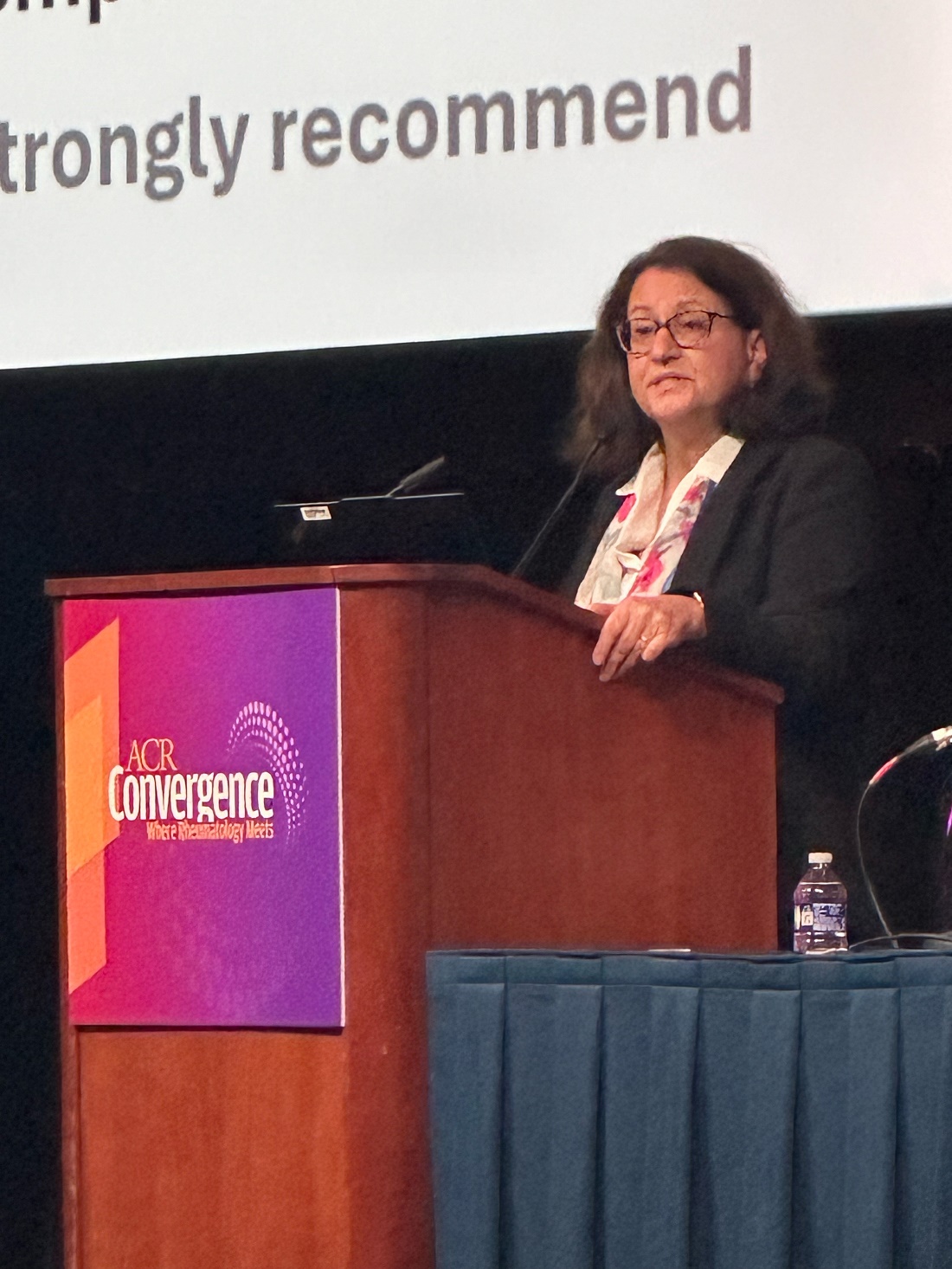
“This is a bit of a change from not only our previous guideline but some of the other guidelines out there, and it is based on the fact that we have very convincing evidence that starting with triple therapy yields to better long-term outcomes for our patients than starting with only two agents and waiting to see if they respond before escalating therapy,” she said. Other key updates include recommending use of pulse glucocorticoid therapy with a lower dose and more rapid steroid taper and treating patients with the recommended therapy for 3-5 years.
The guiding principles of the guideline are not only to preserve kidney function and minimize morbidity and mortality but also to ensure collaborative care with nephrology, to utilize shared decision-making that includes patients’ values and preferences, to reduce healthcare disparities, and to consider pediatric and geriatric populations. The guidelines are based on a quantitative synthesis of 105 studies that yielded 7 strong recommendations, 21 conditional recommendations, and 13 good practice statements — those commonly accepted as beneficial or practical advice even if there is little direct evidence to support them. The voting panel of 19 members included not only 3 nephrologists and 2 pediatric rheumatologists but also 2 patient representatives with LN.
The recommendations are just that, “a recommendation, not an order,” Sammaritano said, and strong recommendations are those “where we think, unequivocally, almost everybody should follow that recommendation. When we feel that we cannot make a strong recommendation, then we call our recommendation conditional, and it is conditional on looking at different things,” she said.
“Patients are different, especially lupus patients, and so one lupus nephritis patient may have different clinical characteristics, different thoughts about what therapy will work for them in their lives, or what therapy they really do not want to pursue,” Sammaritano said. “Maybe they can’t conceive of coming to the hospital once a month for intravenous therapy. Maybe they’re concerned about pill burden, which is something that our patient panel really emphasized to us. So, conditional recommendation means this voting panel thought that this was the best overall for most patients and most circumstances, recognizing there will still be a significant number of people, clinicians and patients, who may feel differently for that particular situation. So, that’s where you know the patient-clinician discussion can help with decision-making.”
What Are the Recommendations?
All patients with systemic lupus erythematosus (SLE) are strongly recommended to undergo proteinuria screening every 6-12 months or at the time of a flare. Those suspected of having LN should receive a prompt kidney biopsy and treatment with glucocorticoids while awaiting the biopsy and results. Two conditional recommendations for kidney biopsy include patients with SLE with unexplained impaired kidney function or a protein to creatinine ratio > 0.5 g/g, and patients with LN with a suspected flare after initial response or a lack of response or worsening after 6 months of therapy.
The guidelines include a strong recommendation for all patients with SLE to receive hydroxychloroquine and a conditional recommendation for all patients with elevated proteinuria (> 0.5 g/g) to receive renin-angiotensin-aldosterone system inhibitors (RAAS-I). Dosages in patients with LN with decreased glomerular filtration rate (GFR) should be adjusted as needed.
Sammaritano then reviewed the specifics on medication treatment. The glucocorticoid therapy in all patients with LN should begin with Pulse IV Therapy at 250-1000 mg/d for 1-3 days, followed by oral prednisone ≤ 0.5 mg/kg per day up to 40 mg/d, then tapered to a target dose > 5 mg/d within 6 months. The justification for this course comes from a 2024 systematic review finding pulse followed by oral glucocorticoids maximized complete renal response while minimizing toxicities, Sammaritano said.
“We have all become acutely aware of the very high risk of prolonged high dose of glucocorticoids for our patients,” she said, “and importantly, our patient panel participants strongly emphasized their preference for minimizing glucocorticoids dose.”
In addition to the recommendation of all patients receiving hydroxychloroquine and RAAS-I, first-line treatment of active, new-onset, or flaring LN should begin with triple therapy — glucocorticoids with two additional immunosuppressive agents. For patients with class III/IV LN, triple therapy includes the glucocorticoids course with a mycophenolic acid analog (MPAA) and either belimumab or a CNI. Conditional recommendations support MPAA with belimumab for significant extrarenal manifestations and MPAA with CNI for proteinuria ≥ 3 g/g.
An alternative triple therapy for class III/IV is glucocorticoids with low-dose cyclophosphamide and belimumab, but MPAA at 2-3 g/d is preferred over cyclophosphamide. The preferred regimen for cyclophosphamide is derived from the Euro-Lupus Nephritis Trial: Intravenous 500 mg every 2 weeks for six doses and then MPAA. Sammaritano noted that there are some limited data on using cyclophosphamide with belimumab, but “we do not specifically recommend cyclophosphamide with a CNI as one of our options because this combination has not been studied in randomized controlled trials.”
There are less data supporting class V recommendations, Sammaritano said, but for those with proteinuria of at least 1 g/g, the panel still recommends triple therapy with glucocorticoids, a MPAA, and a CNI. A CNI is preferred over belimumab because of its stabilizing effects on the podocyte cytoskeleton. Two alternative triple therapies for class V–only patients are glucocorticoids with belimumab and either low-dose cyclophosphamide or MPAA.
Dual therapy is only recommended if triple therapy is not available or not tolerated. The voting panel chose to recommend triple therapy over dual therapy with escalation for two reasons. First, the BLISS-LN and AURORA 1 trials showed improved outcomes with initial triple therapy over initial dual therapies.
Second, “nephron loss proceeds throughout a person’s lifetime even for those who do not have lupus nephritis, and every case of lupus nephritis or every period of time with uncontrolled lupus nephritis changes the course of that decline for the worse,” Sammaritano said. “So, we feel we can’t wait for nephron loss to implement what has been shown to be the most efficacious therapy. We want to gain rapid control of inflammation using the most effective regimen to prevent further damage and flare and maintain survival.”
Therapy is conditionally recommended for at least 3-5 years because “not only do we want to gain rapid control of disease activity [but we also] want to maintain control of disease activity until there’s sustained inactive disease,” Sammaritano said. “Repeat kidney biopsies show that immunologic activity persists in the kidneys for several years, and the withdrawal of immunosuppression when there is histologic activity predisposes patients to flare.” But immunosuppressive therapy can be tapered over time as determined by renal disease activity and medication tolerability.
For patients with refractory disease, consider additional factors that could be affecting the disease, such as adherence, the presence of other diagnoses, or advanced chronicity.
“If true refractory nephritis is present,” she said, “we recommend escalation to a more intensive regimen,” including the addition of anti-CD20 agents, combination therapy with three immunosuppressives, or referral for investigational therapy.
“We also emphasize the importance of other adjunctive therapies preventing comorbidities, such as cardiovascular disease, changes in bone health, or infection risk,” she said. In older patients, avoid polypharmacy as much as possible and be mindful of age-related GFR, she added.
A strong recommendation supported monitoring patients with LN and proteinuria at least every 3 months if they have not achieved complete renal response and every 3-6 months after sustained complete renal response.
Last, in patients with LN and end-stage kidney disease (ESKD), the voting panel strongly recommends transplant over dialysis and conditionally recommends proceeding to the transplant without requiring a complete clinical or serologic remission as long as no other organs are involved. In patients with LN at risk for ESKD, the guideline conditionally recommends consideration of a preemptive transplant, and patients on dialysis or post transplant are strongly recommended to regularly follow up with rheumatology.
Gabriel Kirsch, MD, a resident rheumatologist at the University of Florida College of Medicine, Jacksonville, said he found the guidelines helpful, “especially the guidance on the dichotomy between using belimumab and voclosporin and the clinical and patient preference that help you make that decision.”
Kirsch had hoped, however, to hear more about the impact of therapeutic drug monitoring of hydroxychloroquine on LN outcomes. He also noted a clinical scenario he’s come across that wasn’t addressed.
“When you’re checking GFR on these folks, a lot of our eGFR calculators are creatinine based, and creatinine at the extremes of muscle mass can be inaccurate,” such as getting artificially low creatinine readings from pediatric patients because of their low muscle mass or from patients with muscle atrophy caused by a lot of glucocorticoid exposure. “I was hoping for some more guidance on that,” he said.
Ellen Ginzler, MD, MPH, chief of rheumatology at SUNY Health Science Center in Brooklyn, New York, said the guidelines were pretty much what she expected them to be. She agreed with the panel’s advice that, when deciding between belimumab or voclosporin, “if it’s pure proteinuria, then you add voclosporin. If the patient has extra renal manifestations, you go with belimumab first.”
“They really made it quite clear that, despite the fact that people really want to reduce the amount of immunosuppression — and I agree you should taper steroids quickly — you really need to keep the immunosuppression for a prolonged period of time because all of the studies that have been done for years show that the longer you’re on immunosuppression after you achieve remission or a low disease activity state, the better your chance of not flaring,” Ginzler said. “Rapid tapering or discontinuation really increases the risk of flare.”
Sammaritano, Kirsch, and Ginzler had no disclosures. No external funding was used.
A version of this article appeared on Medscape.com.
WASHINGTON — A new guideline for management of lupus nephritis (LN) was unveiled at the annual meeting of the American College of Rheumatology (ACR), updating the 2012 LN guideline to recommend a more aggressive first-line approach to treating the disease.
“The biggest differences are that we are recommending what we’re calling triple therapy, where we incorporate the glucocorticoid therapy with baseline conventional immunosuppressants, usually mycophenolate with cyclophosphamide, and the addition of one of the newer agents more recently approved by the FDA [Food and Drug Administration] — belimumab, voclosporin, or another CNI [calcineurin inhibitor],” said Lisa Sammaritano, MD, director of the Rheumatology Reproductive Health Program of the Barbara Volcker Center for Women and Rheumatic Diseases at the Hospital for Special Surgery and professor of clinical medicine at Weill Cornell Medical College, both in New York City.
“This is a bit of a change from not only our previous guideline but some of the other guidelines out there, and it is based on the fact that we have very convincing evidence that starting with triple therapy yields to better long-term outcomes for our patients than starting with only two agents and waiting to see if they respond before escalating therapy,” she said. Other key updates include recommending use of pulse glucocorticoid therapy with a lower dose and more rapid steroid taper and treating patients with the recommended therapy for 3-5 years.
The guiding principles of the guideline are not only to preserve kidney function and minimize morbidity and mortality but also to ensure collaborative care with nephrology, to utilize shared decision-making that includes patients’ values and preferences, to reduce healthcare disparities, and to consider pediatric and geriatric populations. The guidelines are based on a quantitative synthesis of 105 studies that yielded 7 strong recommendations, 21 conditional recommendations, and 13 good practice statements — those commonly accepted as beneficial or practical advice even if there is little direct evidence to support them. The voting panel of 19 members included not only 3 nephrologists and 2 pediatric rheumatologists but also 2 patient representatives with LN.
The recommendations are just that, “a recommendation, not an order,” Sammaritano said, and strong recommendations are those “where we think, unequivocally, almost everybody should follow that recommendation. When we feel that we cannot make a strong recommendation, then we call our recommendation conditional, and it is conditional on looking at different things,” she said.
“Patients are different, especially lupus patients, and so one lupus nephritis patient may have different clinical characteristics, different thoughts about what therapy will work for them in their lives, or what therapy they really do not want to pursue,” Sammaritano said. “Maybe they can’t conceive of coming to the hospital once a month for intravenous therapy. Maybe they’re concerned about pill burden, which is something that our patient panel really emphasized to us. So, conditional recommendation means this voting panel thought that this was the best overall for most patients and most circumstances, recognizing there will still be a significant number of people, clinicians and patients, who may feel differently for that particular situation. So, that’s where you know the patient-clinician discussion can help with decision-making.”
What Are the Recommendations?
All patients with systemic lupus erythematosus (SLE) are strongly recommended to undergo proteinuria screening every 6-12 months or at the time of a flare. Those suspected of having LN should receive a prompt kidney biopsy and treatment with glucocorticoids while awaiting the biopsy and results. Two conditional recommendations for kidney biopsy include patients with SLE with unexplained impaired kidney function or a protein to creatinine ratio > 0.5 g/g, and patients with LN with a suspected flare after initial response or a lack of response or worsening after 6 months of therapy.
The guidelines include a strong recommendation for all patients with SLE to receive hydroxychloroquine and a conditional recommendation for all patients with elevated proteinuria (> 0.5 g/g) to receive renin-angiotensin-aldosterone system inhibitors (RAAS-I). Dosages in patients with LN with decreased glomerular filtration rate (GFR) should be adjusted as needed.
Sammaritano then reviewed the specifics on medication treatment. The glucocorticoid therapy in all patients with LN should begin with Pulse IV Therapy at 250-1000 mg/d for 1-3 days, followed by oral prednisone ≤ 0.5 mg/kg per day up to 40 mg/d, then tapered to a target dose > 5 mg/d within 6 months. The justification for this course comes from a 2024 systematic review finding pulse followed by oral glucocorticoids maximized complete renal response while minimizing toxicities, Sammaritano said.
“We have all become acutely aware of the very high risk of prolonged high dose of glucocorticoids for our patients,” she said, “and importantly, our patient panel participants strongly emphasized their preference for minimizing glucocorticoids dose.”
In addition to the recommendation of all patients receiving hydroxychloroquine and RAAS-I, first-line treatment of active, new-onset, or flaring LN should begin with triple therapy — glucocorticoids with two additional immunosuppressive agents. For patients with class III/IV LN, triple therapy includes the glucocorticoids course with a mycophenolic acid analog (MPAA) and either belimumab or a CNI. Conditional recommendations support MPAA with belimumab for significant extrarenal manifestations and MPAA with CNI for proteinuria ≥ 3 g/g.
An alternative triple therapy for class III/IV is glucocorticoids with low-dose cyclophosphamide and belimumab, but MPAA at 2-3 g/d is preferred over cyclophosphamide. The preferred regimen for cyclophosphamide is derived from the Euro-Lupus Nephritis Trial: Intravenous 500 mg every 2 weeks for six doses and then MPAA. Sammaritano noted that there are some limited data on using cyclophosphamide with belimumab, but “we do not specifically recommend cyclophosphamide with a CNI as one of our options because this combination has not been studied in randomized controlled trials.”
There are less data supporting class V recommendations, Sammaritano said, but for those with proteinuria of at least 1 g/g, the panel still recommends triple therapy with glucocorticoids, a MPAA, and a CNI. A CNI is preferred over belimumab because of its stabilizing effects on the podocyte cytoskeleton. Two alternative triple therapies for class V–only patients are glucocorticoids with belimumab and either low-dose cyclophosphamide or MPAA.
Dual therapy is only recommended if triple therapy is not available or not tolerated. The voting panel chose to recommend triple therapy over dual therapy with escalation for two reasons. First, the BLISS-LN and AURORA 1 trials showed improved outcomes with initial triple therapy over initial dual therapies.
Second, “nephron loss proceeds throughout a person’s lifetime even for those who do not have lupus nephritis, and every case of lupus nephritis or every period of time with uncontrolled lupus nephritis changes the course of that decline for the worse,” Sammaritano said. “So, we feel we can’t wait for nephron loss to implement what has been shown to be the most efficacious therapy. We want to gain rapid control of inflammation using the most effective regimen to prevent further damage and flare and maintain survival.”
Therapy is conditionally recommended for at least 3-5 years because “not only do we want to gain rapid control of disease activity [but we also] want to maintain control of disease activity until there’s sustained inactive disease,” Sammaritano said. “Repeat kidney biopsies show that immunologic activity persists in the kidneys for several years, and the withdrawal of immunosuppression when there is histologic activity predisposes patients to flare.” But immunosuppressive therapy can be tapered over time as determined by renal disease activity and medication tolerability.
For patients with refractory disease, consider additional factors that could be affecting the disease, such as adherence, the presence of other diagnoses, or advanced chronicity.
“If true refractory nephritis is present,” she said, “we recommend escalation to a more intensive regimen,” including the addition of anti-CD20 agents, combination therapy with three immunosuppressives, or referral for investigational therapy.
“We also emphasize the importance of other adjunctive therapies preventing comorbidities, such as cardiovascular disease, changes in bone health, or infection risk,” she said. In older patients, avoid polypharmacy as much as possible and be mindful of age-related GFR, she added.
A strong recommendation supported monitoring patients with LN and proteinuria at least every 3 months if they have not achieved complete renal response and every 3-6 months after sustained complete renal response.
Last, in patients with LN and end-stage kidney disease (ESKD), the voting panel strongly recommends transplant over dialysis and conditionally recommends proceeding to the transplant without requiring a complete clinical or serologic remission as long as no other organs are involved. In patients with LN at risk for ESKD, the guideline conditionally recommends consideration of a preemptive transplant, and patients on dialysis or post transplant are strongly recommended to regularly follow up with rheumatology.
Gabriel Kirsch, MD, a resident rheumatologist at the University of Florida College of Medicine, Jacksonville, said he found the guidelines helpful, “especially the guidance on the dichotomy between using belimumab and voclosporin and the clinical and patient preference that help you make that decision.”
Kirsch had hoped, however, to hear more about the impact of therapeutic drug monitoring of hydroxychloroquine on LN outcomes. He also noted a clinical scenario he’s come across that wasn’t addressed.
“When you’re checking GFR on these folks, a lot of our eGFR calculators are creatinine based, and creatinine at the extremes of muscle mass can be inaccurate,” such as getting artificially low creatinine readings from pediatric patients because of their low muscle mass or from patients with muscle atrophy caused by a lot of glucocorticoid exposure. “I was hoping for some more guidance on that,” he said.
Ellen Ginzler, MD, MPH, chief of rheumatology at SUNY Health Science Center in Brooklyn, New York, said the guidelines were pretty much what she expected them to be. She agreed with the panel’s advice that, when deciding between belimumab or voclosporin, “if it’s pure proteinuria, then you add voclosporin. If the patient has extra renal manifestations, you go with belimumab first.”
“They really made it quite clear that, despite the fact that people really want to reduce the amount of immunosuppression — and I agree you should taper steroids quickly — you really need to keep the immunosuppression for a prolonged period of time because all of the studies that have been done for years show that the longer you’re on immunosuppression after you achieve remission or a low disease activity state, the better your chance of not flaring,” Ginzler said. “Rapid tapering or discontinuation really increases the risk of flare.”
Sammaritano, Kirsch, and Ginzler had no disclosures. No external funding was used.
A version of this article appeared on Medscape.com.
FROM ACR 2024
Kidney, Cardiovascular Benefits Seen With GLP-1 RA Drugs in SLE, Lupus Nephritis
WASHINGTON — Glucagon-like peptide 1 receptor agonist (GLP-1 RA) medications appear beneficial for people with systemic lupus erythematosus (SLE) and lupus nephritis, two new studies suggest.
“The risk of cardiovascular disease is thought to be at least double that for people with lupus ... and we know the risk of progressing to end-stage renal disease [ESKD] for patients with lupus nephritis can be as high as 10%-30%, so there’s clearly a major unmet need for new treatments and approaches to improve these outcomes, perhaps with adjunctive treatment beyond our typical immunosuppressive therapy,” April Jorge, MD, of Massachusetts General Hospital, Boston, said at the annual meeting of the American College of Rheumatology (ACR).
The GLP-1 RAs are approved for the treatment of type 2 diabetes (T2D) and obesity. They also have proven cardiovascular benefit, along with emerging data suggesting kidney protection independent of glucose lowering. Jorge presented findings from a study using data from the US multicenter electronic health record database TriNetX, showing that, among patients who had both T2D and SLE, those using GLP-1 RAs had lower risks for major adverse cardiac events (MACE), venous thrombosis, kidney disease progression, and all-cause mortality, compared with those using a different class of T2D medication.
A second study using TriNetX, presented at the same ACR meeting session by Anna-Kay Palmer, MD, a third-year internal medicine resident at Jefferson Einstein Hospital, Philadelphia, Pennsylvania, showed that GLP-1 RAs reduced the risk of progression to ESKD in patients with lupus nephritis, possibly caused by reductions in pro-inflammatory mediators.
Asked to comment, session moderator Diane L. Kamen, MD, professor of medicine at the Medical University of South Carolina Division of Rheumatology, Charleston, said in an interview that she definitely supports the use of GLP-1 RAs for patients who have SLE and/or lupus nephritis and also a drug label indication, either T2D or obesity. “[The GLP-1 RA prescriber] will usually run it by rheumatology to make sure that it doesn’t conflict with any of their other medical treatment, and it’s very reassuring to know that they could actually get a win-win.”
But as far as prescribing off-label for those with SLE/lupus nephritis who don’t have other GLP-1 RA indications, Kamen said, “that’s a black hole at this point. We need to do those prospective studies. But if they have another indication, yes.”
Cardiovascular, Kidney Benefits of GLP-1 RAs
Jorge noted that patients with lupus were excluded from the randomized clinical trials of GLP-1 RAs, so the current study was designed to investigate the potential impact of these medications on cardiovascular and kidney outcomes in patients with SLE and lupus nephritis.
From TriNetX data for 46 healthcare organizations nationwide, a total of 96,511 patients with both SLE and T2D but not ESKD had initiated either a GLP-1 RA or another diabetes drug class, dipeptidyl peptidase 4 inhibitors (DPP4i), between October 2006 and August 2021. Of those, 29,177 had lupus nephritis.
Propensity score matching for factors such as demographics, lupus severity, comorbidities, and medication use was used to emulate a randomized trial. This yielded 25,838 with SLE and T2D, of whom 910 initiated a GLP-1 RA and 1004 started a DPP4i, and 12,387 with lupus nephritis and T2D, including 267 on a GLP-1 RA and 324 on a DPP4i. After matching, the mean age was 55 years, more than 90% were women, and just under half were White individuals. About one third had chronic kidney disease stages ≥ 3, and about 15% had heart failure.
Over an average follow-up time of 1.2-1.4 years among those with SLE, the hazard ratio (HR) for MACE (a composite of myocardial infarction, stroke, and heart failure) for those taking a GLP-1 RA vs a DPP4i was 0.66, a significant difference. And for venous thrombosis, the HR was also significant at 0.49.
Kidney disease progression, defined as an estimated glomerular filtration rate decline of 30% or more or new ESKD, was significantly less likely in the GLP-1 RA group, with a HR of 0.77. All-cause mortality also was dramatically reduced (HR, 0.26). As expected, there was no difference in control outcome, genital infections (HR, 1.02).
In the subgroup with lupus nephritis, there were also lower risks for both MACE (HR, 0.64) and for renal progression (HR, 0.70). “The findings suggest similar cardiac and kidney benefits among patients with SLE and lupus nephritis as have been observed in other populations,” Jorge concluded.
Kamen commented that the study design “was pretty brilliant, because you wouldn’t be able to do a placebo-controlled trial since the indication was diabetes ... but the fact is you do see that the GLP-1 RA gets the benefit whereas the other drug does not.”
Next steps, Jorge said, will be mechanistic studies to better understand the effects of GLP-1 RAs in lupus and other rheumatic diseases, prospective studies of GLP-1 RAs in SLE and lupus nephritis without diabetes, and clarification of ideal timing for GLP-1 RA use in SLE and lupus nephritis.
“Ideally, with our prospective studies with these patients we can try to isolate the effect on patients with lupus and also better understand whether there might be an impact on disease activity through the anti-inflammatory effects of these medications, rather than just the cardioprotective and nephroprotective benefits,” she said.
In Those With Lupus Nephritis, Kidney Protection Seen
In her presentation, Palmer noted that, despite immunosuppressive therapies for SLE, 10%-20% of patients who develop lupus nephritis will progress to ESKD within 5 years of diagnosis.
She added that GLP-1 RAs have been shown to reduce albuminuria in people with diabetes and have been hypothesized to reduce inflammation through multiple pathways, thereby potentially reducing kidney disease independently of the presence of diabetes or weight loss. These pathways include modulating immune cell signaling and reducing pro-inflammatory cytokines.
Based on all this, Palmer and colleagues used International Classification of Diseases – 10th edition diagnostic codes in TriNetX to identify 839 patients who had been diagnosed with lupus nephritis between 2014 and 2024 and who were prescribed liraglutide, dulaglutide, semaglutide, or exenatide for any time after the lupus nephritis diagnosis. Another 29,840 patients with lupus nephritis had not used GLP-1 RAs.
After 1:1 propensity score matching for age, sex, race, ethnicity, presence of hypertension, diabetes, use of immunosuppressive and diabetes medication, smoking, obesity, and statin use, there were 735 individuals in each group. About two thirds in each had diabetes, whereas the rest had been prescribed the GLP-1 RAs for other indications.
Patients who were not on GLP-1 RAs were twice as likely to develop ESKD or dialysis (8.88% vs 3.971%; odds ratio, 2.35; P = .001).
Kamen pointed out that not including the use of angiotensin-converting enzyme inhibitors and angiotensin receptor blockers was a study flaw. On the other hand, the fact that not everyone in this study had diabetes was an advantage.
Jorge received grant/research support from Bristol-Myers Squibb, Cabaletta Bio, and the Lupus Clinical Investigator Network. Kamen is an adviser/review panel member for Alpine Immune Sciences. Palmer had no disclosures.
A version of this article appeared on Medscape.com.
FROM ACR 2024
RA Assessment Via Automated Ultrasound Scanner With AI Saves Time, Performs as Well as Rheumatologists
WASHINGTON — A fully automated ultrasound scanning system combined with artificial intelligence–based disease activity scoring performed as well as expert rheumatologists in hand joint assessment of patients with rheumatoid arthritis (RA), new research found.
The system, made by a Danish company called ROPCA, comprises an ultrasound scanner called ARTHUR (RA Ultrasound Robot) that interacts directly with the patient and scans 11 joints per hand and a neural network–based software system, DIANA (Diagnosis Aid Network for RA), that evaluates the images and monitors RA activity.
The combined system classifies the degree of RA according to the joint European Alliance of Associations for Rheumatology (EULAR)–Outcome Measures in Rheumatology (OMERACT) standards for RA diagnosis. It received a CE Mark in Europe in 2022 and is currently in use in six rheumatology clinics in Denmark, Germany, Switzerland, and Austria, with more to come, ROPCA Co-founder and Chief Medical Officer Søren A. Just, MD, said in an interview.
“Automated systems could help rheumatologists in the early detection and monitoring of arthritis diseases. Systems can be placed or move in areas with insufficient rheumatological expertise,” Just said during a special late-breaker session presentation at the annual meeting of the American College of Rheumatology (ACR).
He said in an interview: “Currently, there are so many people referred and few and fewer rheumatologists. So we need to think differently. We need good automated assistants.” As a screening tool, the system can determine whether a person with hand pain has RA or just osteoarthritis “and also can give the patient an immediate answer, instead of waiting sometimes up to 6 months to get the information.”
Just, who is also a senior physician in the Department of Internal Medicine at Odense University Hospital in Denmark, said that his department is also using the system to assess flares in patients with established RA. “They can have a blood sample taken. They’re scanned by the robot, and you can see if there is any disease activity. But I think that screening of patients with joint pain is the beginning.”
Asked to comment, session moderator Gregory C. Gardner, MD, Emeritus Professor in the Division of Rheumatology at the University of Washington, Seattle, and a member of the ACR conference program committee, said in an interview “one of the reasons we chose to feature this abstract is because we’re interested in science at the convergence. We really thought this was a potential way to move the field forward for rheumatologists.”
Gardner said it’s an advantage that the patient could potentially have an ARTHUR scan with a DIANA report and get blood tests done prior to a visit with the rheumatologist. “It’s really time-consuming for a human to do these studies, so if you automate it, that’s a step forward in terms of having the data available for the rheumatologist to view and use sequentially to follow how patients are doing.”
When introducing Just’s presentation, Gardner called it “the coolest abstract of the meeting.”
Both DIANA and ARTHUR Performed At Least as Well as Human Rheumatologists
In the study, 30 patients with RA underwent two scans by ARTHUR, followed by a scan from a rheumatologist specialist in musculoskeletal ultrasound. The scans were sent to DIANA, who graded the images according to the Global OMERACT-EULAR Synovitis Score, as did the human rheumatologist.
A “ground truth” was established by another human expert who evaluated both ARTHUR’s and the other rheumatologist’s images, blinded to the scanning method. The image with the highest disease activity was deemed “ground truth,” and agreement with that was assessed for the two individual methods.

Just showed a video of a patient being scanned by ARTHUR. The machine verbally guided her through removing her jewelry, applying the gel, and placing her hand on the screen under the scanner. ARTHUR’s arm moved around on the patient’s hand, locating the best angles to take grayscale images and Doppler images and Doppler video. The scan takes 15-20 minutes, and the images are stored, Just said.
The study patients had a mean age of 65 years, and 23 of the 30 were men. Their average disease duration was 11 years, and mean Disease Activity Score in 28 joints using C-reactive protein was 3.86, indicating moderate disease. A majority (73%) of patients were taking disease-modifying antirheumatic drugs, and about one third were taking biologics. ARTHUR scanned a total of 660 joints, and 564 scans were successful.
For repeatability between the two ARTHUR scans, percent exact agreement was 63% for synovial hypertrophy, 75% for Doppler activity, and 60% combined. Percent close (within a point) agreements were 93%, 94%, and 92%, respectively. Binary agreements as to whether the joint was healthy vs diseased were 88%, 91%, and 85%, respectively.
At the joint level, ARTHUR and DIANA’s percent exact agreement with ground truth was 49% for synovial hypertrophy, 63% for Doppler activity, and 48% combined. Binary agreements with disease vs healthy were 80%, 88%, and 78%, respectively.
The human rheumatologists scored very similarly. Percent exact agreement with ground truth was 51% for synovial hypertrophy, 64% for Doppler activity, and 50% combined. Percent close agreements were 94%, 94%, and 92%, respectively. And binary agreements with diseased vs healthy were 83%, 91%, and 80%, respectively.
At the patient level (all joints combined), ARTHUR and DIANA’s binary disease assessment of healthy vs disease showed agreement with the ground truth of 87% for synovial hypertrophy, 83% for Doppler activity, and 87% combined. Here, the rheumatologists scored lower, at 53%, 67%, and 60%, respectively.
“In this study, we think the precision of ARTHUR and DIANA was comparable to that of an experienced rheumatologist, at both the joint and patient level,” Just said.
Gardner pointed out another advantage of the system. “DIANA doesn’t get fatigued. ... With human reading, the precision may change based on the time of day or stress level. ... But with DIANA, you’re going to get consistent information.”
Just said that the Arthritis Foundation in Germany recently put ARTHUR and DIANA on a bus and took it to cities that lacked a rheumatologist. Patients lined up, answered a questionnaire, had blood drawn, and received their scans. A rheumatologist on the bus then interpreted the data and consulted with the individuals about their RA risk. “In the last trip, we screened 800 patients in 6 days. So there are definitely possibilities here.”
Just is co-owner of ROPCA. Gardner had no disclosures.
A version of this article appeared on Medscape.com.
WASHINGTON — A fully automated ultrasound scanning system combined with artificial intelligence–based disease activity scoring performed as well as expert rheumatologists in hand joint assessment of patients with rheumatoid arthritis (RA), new research found.
The system, made by a Danish company called ROPCA, comprises an ultrasound scanner called ARTHUR (RA Ultrasound Robot) that interacts directly with the patient and scans 11 joints per hand and a neural network–based software system, DIANA (Diagnosis Aid Network for RA), that evaluates the images and monitors RA activity.
The combined system classifies the degree of RA according to the joint European Alliance of Associations for Rheumatology (EULAR)–Outcome Measures in Rheumatology (OMERACT) standards for RA diagnosis. It received a CE Mark in Europe in 2022 and is currently in use in six rheumatology clinics in Denmark, Germany, Switzerland, and Austria, with more to come, ROPCA Co-founder and Chief Medical Officer Søren A. Just, MD, said in an interview.
“Automated systems could help rheumatologists in the early detection and monitoring of arthritis diseases. Systems can be placed or move in areas with insufficient rheumatological expertise,” Just said during a special late-breaker session presentation at the annual meeting of the American College of Rheumatology (ACR).
He said in an interview: “Currently, there are so many people referred and few and fewer rheumatologists. So we need to think differently. We need good automated assistants.” As a screening tool, the system can determine whether a person with hand pain has RA or just osteoarthritis “and also can give the patient an immediate answer, instead of waiting sometimes up to 6 months to get the information.”
Just, who is also a senior physician in the Department of Internal Medicine at Odense University Hospital in Denmark, said that his department is also using the system to assess flares in patients with established RA. “They can have a blood sample taken. They’re scanned by the robot, and you can see if there is any disease activity. But I think that screening of patients with joint pain is the beginning.”
Asked to comment, session moderator Gregory C. Gardner, MD, Emeritus Professor in the Division of Rheumatology at the University of Washington, Seattle, and a member of the ACR conference program committee, said in an interview “one of the reasons we chose to feature this abstract is because we’re interested in science at the convergence. We really thought this was a potential way to move the field forward for rheumatologists.”
Gardner said it’s an advantage that the patient could potentially have an ARTHUR scan with a DIANA report and get blood tests done prior to a visit with the rheumatologist. “It’s really time-consuming for a human to do these studies, so if you automate it, that’s a step forward in terms of having the data available for the rheumatologist to view and use sequentially to follow how patients are doing.”
When introducing Just’s presentation, Gardner called it “the coolest abstract of the meeting.”
Both DIANA and ARTHUR Performed At Least as Well as Human Rheumatologists
In the study, 30 patients with RA underwent two scans by ARTHUR, followed by a scan from a rheumatologist specialist in musculoskeletal ultrasound. The scans were sent to DIANA, who graded the images according to the Global OMERACT-EULAR Synovitis Score, as did the human rheumatologist.
A “ground truth” was established by another human expert who evaluated both ARTHUR’s and the other rheumatologist’s images, blinded to the scanning method. The image with the highest disease activity was deemed “ground truth,” and agreement with that was assessed for the two individual methods.
Just showed a video of a patient being scanned by ARTHUR. The machine verbally guided her through removing her jewelry, applying the gel, and placing her hand on the screen under the scanner. ARTHUR’s arm moved around on the patient’s hand, locating the best angles to take grayscale images and Doppler images and Doppler video. The scan takes 15-20 minutes, and the images are stored, Just said.
The study patients had a mean age of 65 years, and 23 of the 30 were men. Their average disease duration was 11 years, and mean Disease Activity Score in 28 joints using C-reactive protein was 3.86, indicating moderate disease. A majority (73%) of patients were taking disease-modifying antirheumatic drugs, and about one third were taking biologics. ARTHUR scanned a total of 660 joints, and 564 scans were successful.
For repeatability between the two ARTHUR scans, percent exact agreement was 63% for synovial hypertrophy, 75% for Doppler activity, and 60% combined. Percent close (within a point) agreements were 93%, 94%, and 92%, respectively. Binary agreements as to whether the joint was healthy vs diseased were 88%, 91%, and 85%, respectively.
At the joint level, ARTHUR and DIANA’s percent exact agreement with ground truth was 49% for synovial hypertrophy, 63% for Doppler activity, and 48% combined. Binary agreements with disease vs healthy were 80%, 88%, and 78%, respectively.
The human rheumatologists scored very similarly. Percent exact agreement with ground truth was 51% for synovial hypertrophy, 64% for Doppler activity, and 50% combined. Percent close agreements were 94%, 94%, and 92%, respectively. And binary agreements with diseased vs healthy were 83%, 91%, and 80%, respectively.
At the patient level (all joints combined), ARTHUR and DIANA’s binary disease assessment of healthy vs disease showed agreement with the ground truth of 87% for synovial hypertrophy, 83% for Doppler activity, and 87% combined. Here, the rheumatologists scored lower, at 53%, 67%, and 60%, respectively.
“In this study, we think the precision of ARTHUR and DIANA was comparable to that of an experienced rheumatologist, at both the joint and patient level,” Just said.
Gardner pointed out another advantage of the system. “DIANA doesn’t get fatigued. ... With human reading, the precision may change based on the time of day or stress level. ... But with DIANA, you’re going to get consistent information.”
Just said that the Arthritis Foundation in Germany recently put ARTHUR and DIANA on a bus and took it to cities that lacked a rheumatologist. Patients lined up, answered a questionnaire, had blood drawn, and received their scans. A rheumatologist on the bus then interpreted the data and consulted with the individuals about their RA risk. “In the last trip, we screened 800 patients in 6 days. So there are definitely possibilities here.”
Just is co-owner of ROPCA. Gardner had no disclosures.
A version of this article appeared on Medscape.com.
FROM ACR 2024
Phase 3 Lupus Trial Shows Promising Results for Dapirolizumab Pegol
WASHINGTON — The investigational anti-CD40 ligand agent dapirolizumab pegol (DZP) outperformed placebo in improving disease activity and reducing high-dose corticosteroid use in patients with systemic lupus erythematosus (SLE) in the phase 3 PHOENYCS GO trial.
“We really think that dapirolizumab pegol may represent a novel treatment for lupus, particularly given its broad immune modulatory effects,” said Megan Clowse, MD, MPH, associate professor of medicine and chief of the Division of Rheumatology and Immunology at Duke University School of Medicine in Durham, North Carolina. She presented the study in a late-breaking poster session at the American College of Rheumatology (ACR) 2024 Annual Meeting.
There is a “huge unmet need” for drugs for lupus, Clowse told this news organization. Patients with SLE continue to have high disease burden, including ongoing symptoms often driven by inflammation. Corticosteroids are often the best medications to control disease activity, she said, but they can result in long-term toxicity.
What Makes DZP Unique?
Through CD40 ligand signaling, DZP has been shown to reduce B- and T-cell activation and to downregulate interferon pathways. Previous antibodies targeting the CD40 ligand have been associated with an increased risk for thromboembolic events. However, DZP lacks the Fc portion of the antibody, which can bind to platelets and cause clotting. Data from phase 1, 2, and 3 trials thus far do not show an elevated risk for these events, Clowse explained. In fact, safety signals were strong enough that patients with antiphospholipid antibodies — a key driver for blood clots in patients with SLE — were included in the trial.
In PHOENYCS GO, investigators enrolled 321 patients with moderate to severe SLE with persistently active or frequently flaring/relapsing-remitting disease activity despite stable standard of care (SOC) medications such as antimalarials, corticosteroids, and immunosuppressants.
Patients were randomized 2:1 to receive intravenous DZP (24 mg/kg) plus SOC or intravenous placebo plus SOC every 4 weeks, with patients and investigators blinded to treatment assignments.
Patients taking a corticosteroid dose > 7.5 mg/day began a mandatory steroid taper by week 8 of the trial, with the goal of reducing that to < 7.5 mg/day. The tapering regimen was at the discretion of providers and was adapted to each patient’s individual disease activity.
The primary endpoint was British Isles Lupus Assessment Group–based Composite Lupus Assessment (BICLA) response at week 48.
Patients in the DZP and placebo groups were on average 43.5 and 41.5 years old, respectively. More than 90% of patients were women, all on concomitant SLE medications. About half of the participants took a daily corticosteroid dose > 7.5 mg.
At 48 weeks, half of the DZP group (49.5%) achieved BICLA response compared with 34.6% in the placebo group (P = .0110). A higher proportion of patients taking DZP achieved SLE Responder Index-4 response than those taking placebo (60.1% vs 41.1%, respectively; P = .0014), and the rate of severe British Isles Lupus Assessment Group flares in the DZP group was half that of the placebo group (11.6% vs 23.4%; P = .0257). In the subgroup of patients who underwent corticosteroid tapering, 72.4% receiving DZP and 52.9% taking placebo reduced their dose to < 7.5 mg/day by 48 weeks (P = .0404).
DZP was generally well tolerated. Over 48 weeks, 82.6% of the DZP group and 75% of the placebo group reported treatment-emergent adverse events, but serious occurrences were more common in the placebo group (14.8%) than in the DZP group (9.9%). Herpes viral infections were higher in the placebo group, although there were three ophthalmic herpes cases in the DZP group. There was one case of acute myocardial infarction and one death linked to gangrene-related sepsis in patients receiving DZP.
A ‘Mild to Moderate’ Response
Although these are definitely positive results, they show a “mild to moderate response” to DZP, commented Gregory Gardner, MD, an emeritus professor in the Division of Rheumatology at the University of Washington, Seattle, and chair of the American College of Rheumatology’s annual meeting planning committee. He moderated the session where the research was presented. Although DZP showed efficacy among some patients, he noted, “there were still 51% patients that it didn’t work for.”
The drug uses an alternative pathway to current lupus drugs, Gardner added, and more research is needed to understand how best to use this medication in practice.
Clowse noted that DZP could be particularly beneficial for patients with SLE who want to get pregnant. Many drugs used to treat the disease are teratogenic; however, “because of the lack of Fc portion on this drug, it very likely does not cross the placenta in any kind of significant amount,” she said. Although there are not yet any reproductive safety data on DZP, she added, “that is a great potential niche.”
Biogen and UCB, which are jointly developing DZP, aim to start a second phase 3 trial of DZP in patients with SLE, called PHOENYCS FLY, in 2024.
The trial was sponsored by UCB. Clowse is a consultant and has received grant/research support from GSK and UCB. Gardner had no relevant disclosures.
A version of this article appeared on Medscape.com.
FROM ACR 2024