User login
M. Alexander Otto began his reporting career early in 1999 covering the pharmaceutical industry for a national pharmacists' magazine and freelancing for the Washington Post and other newspapers. He then joined BNA, now part of Bloomberg News, covering health law and the protection of people and animals in medical research. Alex next worked for the McClatchy Company. Based on his work, Alex won a year-long Knight Science Journalism Fellowship to MIT in 2008-2009. He joined the company shortly thereafter. Alex has a newspaper journalism degree from Syracuse (N.Y.) University and a master's degree in medical science -- a physician assistant degree -- from George Washington University. Alex is based in Seattle.
Mobilize subarachnoid hemorrhage patients as soon as possible
SAN DIEGO – Aneurysmal subarachnoid hemorrhage patients left the ICU a mean of 3 days earlier when they were mobilized quickly after their stroke, according to a retrospective study from the Capital Institute for Neurosciences in Trenton, N.J.
Investigators there compared functional outcomes for 38 historical subarachnoid hemorrhage (SAH) controls, and 55 SAH patients after an early mobilization program was started in the neuro-ICU about 5 years ago.
The groups were matched for demographics, clip vs. coil ligation, and Hunt and Hess severity grade, among other things. Patients were generally in their 40s and 50s.
The early mobilization group got out of bed quicker (mean 4.2 days vs. 6.4 days), walked 50 feet sooner (mean 6.4 days vs. 10.5 days), and left the ICU earlier (mean 12.8 days vs. 15.7 days). The findings were all statistically significant; there was also a possible trend towards more discharges to the community (60% vs. 50%; P = .481).
"What we focused on was getting patients sitting in a chair. We were very aggressive; some of these patients were post-op day 1. Even with patients who were almost comatose, we did something, even just help them sit on the edge of the bed; the upright position facilitates arousal. We at least give it a shot to see if it helped," said investigator and physical therapist Melissa Arcaro, who presented the findings at the International Stroke Conference, sponsored by the American Heart Association.
Patients were assessed daily by physicians to see if they were hemodynamically and neurologically stable enough to participate. They had to be able to open their eyes and move one extremity on command. Transcranial Doppler ultrasound was performed before each session, and patients were excused for the day if their Lindegaard ratios were greater than 3.
Tachycardia and orthostatic issues were the main problems. "We didn’t have any patients who started to rebleed or had vasospasms that resulted in infarcts," Ms. Arcaro said.
Early mobilization is now standard practice at her ICU. "Patients are stuck in bed all the time, so they look forward to us coming in and helping them get up and brush their teeth or use the toilet. The biggest comment I get is, ‘Oh my God, I feel like a normal person again,’ " she said.
Prolonged bed rest and immobility are known to be bad for hospital patients, but "mobilization in the ICU, and the neuro-ICU in particular, is a new area. There’s not a whole lot of information out there," she said.
The investigators have no disclosures, and did not report outside funding.
AT THE INTERNATIONAL STROKE CONFERENCE
Major finding: Aneurysmal subarachnoid hemorrhage patients mobilized as early as post-op day 1 left the ICU sooner than did those who were left in bed (mean 12.8 vs. 15.7 days).
Data source: A retrospective functional outcomes review of 55 patients mobilized early in the neuro-ICU, and 38 controls.
Disclosures: The investigators have no disclosures and did not report their funding source.
Beta-blocker use associated with better outcomes after insular cortex infarcts
SAN DIEGO – Prior beta-blocker use may improve outcomes following insular ischemic strokes, according to a prospective, observational study of 1,014 consecutive stroke patients at Massachusetts General Hospital in Boston.
Favorable outcomes, defined by modified Rankin Scale (mRS) scores of 0-2 at 3 months – occurred with significantly greater likelihood among patients with infarcts of the insular or surrounding opercular cortex who used beta-blockers (BBs) within 24 hours before stroke onset, after researchers controlled for underlying cardiovascular and other risk factors on multivariate analysis (odds ratio, 2.21; 95% confidence interval, 1.03-4.75; P = .04).
"A lot of people have studied the effect" of BBs on stroke, and have found that "indiscriminate use in acute stroke patients is not beneficial, and may be associated with harm," said senior author Dr. Hakan Ay of the departments of neurology and radiology at Massachusetts General.
But previous studies lumped strokes together, regardless of infarct location. "This is the first study that links response to a specific brain site. What this tells us is if you find the right patient, you can get benefit" from BBs, although the finding must be confirmed by randomized trial, Dr. Ay said at the International Stroke Conference, sponsored by the American Heart Association.
The 402 BB users and 612 nonusers in the study, about evenly split between men and women, were admitted within 3 days of stroke onset.
BB patients were older (mean 74 years vs. 65 years) and more often had risk factors such as coronary artery disease (33% vs. 13%), atrial fibrillation (40% vs. 17%), and heart failure (12% vs. 4%).
The National Institutes of Health Stroke Scale score was higher in the BB group (mean 5 vs. 3). Twenty-three percent of BB patients and 18% of nonusers got intravenous thrombolysis (P = .06).
Overall, about two-thirds of patients in the study did well, with mRS scores of 0-2 at 3 months. BB patients did a bit worse, with just 57% achieving the score. Univariate analyses revealed a decreased probability of good outcome in the BB patients, as expected given their poorer baseline health (OR 0.72; 95% CI 0.55-0.93).
After adjustment for that baseline difference on multivariate analysis, prior BB use did not predict good or bad outcomes when all strokes were considered (OR 1.33; 95% CI 0.90-1.96; P = .16), which might indicate that while insular patients seemed to benefit, prior BB use may have contributed to worse outcomes in other stroke types, Dr. Ay said.
The benefit emerged when multivariate analysis was limited to BB patients and nonusers who had insular strokes. The investigators did not report how many were in each group.
There’s biological plausibility for the finding; insular injury is associated with autonomic dysregulation, leading to increases in blood pressure and other problems that likely affect outcomes. BBs might moderate the effect, Dr. Ay said.
The investigators had no relevant disclosures. The work was funded by the National Institute of Neurological Disorders and Stroke.
AT THE INTERNATIONAL STROKE CONFERENCE
Major finding: Beta-blocker use within 24 hours before stroke onset significantly predicts favorable outcomes in patients with infarcts of the insular or surrounding opercular cortex (odds ratio, 2.21; 95% confidence interval, 1.03-4.75; P = .04).
Data Source: Prospective, observational study of 1,014 consecutive stroke patients
Disclosures: The investigators had no relevant disclosures. The work was funded by the National Institute of Neurological Disorders and Stroke.
Continuous AF monitors raise questions about when to intervene
SAN DIEGO – Medtronic’s Reveal XT heart monitor implant detected atrial fibrillation missed by older techniques in a randomized trial.
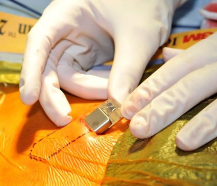
The company’s North American and European CRYSTAL-AF (Cryptogenic Stroke and Underlying Atrial Fibrillation) trial randomized 221 patients to the device after cryptogenic ischemic strokes or, less commonly, transient ischemic attacks, and 220 others to usual monitoring with ECGs or 24-hour Holter monitors. The Reveal XT device, about the size of a computer thumb drive, is slipped under the flesh on the left side of the chest with local anesthesia, and transmits data wirelessly.
After 6 months, the device picked up atrial fibrillation (AF) in 19 patients (8.6%). Standard monitoring detected AF in three patients, or 1.4% (hazard ratio, 6.4; 95% confidence interval, 1.9-21.7; P = .0006).
Fourteen (74%) Reveal XT AF patients were asymptomatic, and 18 (94.7%) were switched from antiplatelet therapy to oral anticoagulants. One control group AF patient was asymptomatic, and two were switched to oral anticoagulants. Every patient in the trial was eligible for anticoagulants because of past embolic events and subsequent CHADS2 scores of 2 or more, but few were on anticoagulants when the trial started.
It’s probably not a surprise that Reveal XT and other modern implantable devices are good at finding AF, but it’s less clear sometimes about how to handle it. The investigators did not report the frequency of AF or the number of strokes, deaths, and anticoagulation complications in the trial.
"If someone had a stroke, and then is found to have AF, it’s hard to ignore that," said presenter and investigator Dr. Rod Passman, a cardiologist and professor of medicine at Northwestern University in Chicago.
There may be less to worry about with younger patients and those with few stroke risks. The mean age in the trial was 62 years, and most of the subjects were men.
However, "we are learning that anticoagulation is effective" in high-risk patients. "The studies were done on patients with a lot of AF, but even small episodes may increase the long-term stroke risk. We are not clear about what the threshold is, but I do think we are honing in [on it]. The question will be if there’s a subgroup at a particularly high risk," he said at the International Stroke Conference, sponsored by the American Heart Association.
After a year, Reveal XT had detected AF in 29 patients (13.1%), most of them asymptomatic. Standard monitoring found AF in four (1.8%), two of whom were asymptomatic (HR, 7.3; 95% CI, 2.6-20.8; P less than .0001). Detecting AF in the four control arm patients took 121 ECGs, 32 Holter monitors, and one event recorder. Almost all of the AF patients in both groups were switched to anticoagulants.
About 27 (93%) of the device AF patients had a maximum 1-day AF duration of more than 6 minutes; about 13 (45%) had episodes of 12-24 hours.
The device continued to outperform older methods at 3 years, the life of its battery (HR, 8.8; 95% CI, 3.5-22.2; P less than .0001).
It was taken out of five patients (2.3%) because of infections or pocket erosions.
Dr. Passman reported payments from Medtronic to work on the trial and significant research grants and speakers’ bureau and consultant fees from the company. The other 11 investigators disclosed personal payments from the company. Three were Medtronic employees. The company funded the trial.
 |
|
Long-term cardiac monitoring is coming into play, and [overtreatment of atrial fibrillation] is a legitimate concern. There’re many questions about where to draw the line between risks and benefits.
But a lot of people in this trial had episodes lasting 12-24 hours, so the probability is high that AF caused their initial stroke. If it turns out that continual monitoring identifies AF cases with the same high stroke risk as the much smaller group we are picking up now, the implications are enormous because we have effective treatments: oral anticoagulants.
Dr. Steven Greenberg is a professor of neurology at Harvard Medical School in Boston. He said he had no relevant financial disclosures.
SAN DIEGO – Medtronic’s Reveal XT heart monitor implant detected atrial fibrillation missed by older techniques in a randomized trial.
The company’s North American and European CRYSTAL-AF (Cryptogenic Stroke and Underlying Atrial Fibrillation) trial randomized 221 patients to the device after cryptogenic ischemic strokes or, less commonly, transient ischemic attacks, and 220 others to usual monitoring with ECGs or 24-hour Holter monitors. The Reveal XT device, about the size of a computer thumb drive, is slipped under the flesh on the left side of the chest with local anesthesia, and transmits data wirelessly.
After 6 months, the device picked up atrial fibrillation (AF) in 19 patients (8.6%). Standard monitoring detected AF in three patients, or 1.4% (hazard ratio, 6.4; 95% confidence interval, 1.9-21.7; P = .0006).
Fourteen (74%) Reveal XT AF patients were asymptomatic, and 18 (94.7%) were switched from antiplatelet therapy to oral anticoagulants. One control group AF patient was asymptomatic, and two were switched to oral anticoagulants. Every patient in the trial was eligible for anticoagulants because of past embolic events and subsequent CHADS2 scores of 2 or more, but few were on anticoagulants when the trial started.
It’s probably not a surprise that Reveal XT and other modern implantable devices are good at finding AF, but it’s less clear sometimes about how to handle it. The investigators did not report the frequency of AF or the number of strokes, deaths, and anticoagulation complications in the trial.
"If someone had a stroke, and then is found to have AF, it’s hard to ignore that," said presenter and investigator Dr. Rod Passman, a cardiologist and professor of medicine at Northwestern University in Chicago.
There may be less to worry about with younger patients and those with few stroke risks. The mean age in the trial was 62 years, and most of the subjects were men.
However, "we are learning that anticoagulation is effective" in high-risk patients. "The studies were done on patients with a lot of AF, but even small episodes may increase the long-term stroke risk. We are not clear about what the threshold is, but I do think we are honing in [on it]. The question will be if there’s a subgroup at a particularly high risk," he said at the International Stroke Conference, sponsored by the American Heart Association.
After a year, Reveal XT had detected AF in 29 patients (13.1%), most of them asymptomatic. Standard monitoring found AF in four (1.8%), two of whom were asymptomatic (HR, 7.3; 95% CI, 2.6-20.8; P less than .0001). Detecting AF in the four control arm patients took 121 ECGs, 32 Holter monitors, and one event recorder. Almost all of the AF patients in both groups were switched to anticoagulants.
About 27 (93%) of the device AF patients had a maximum 1-day AF duration of more than 6 minutes; about 13 (45%) had episodes of 12-24 hours.
The device continued to outperform older methods at 3 years, the life of its battery (HR, 8.8; 95% CI, 3.5-22.2; P less than .0001).
It was taken out of five patients (2.3%) because of infections or pocket erosions.
Dr. Passman reported payments from Medtronic to work on the trial and significant research grants and speakers’ bureau and consultant fees from the company. The other 11 investigators disclosed personal payments from the company. Three were Medtronic employees. The company funded the trial.
AT THE INTERNATIONAL STROKE CONFERENCE
Major finding: After 6 months, Medtronic’s Reveal XT heart monitor implant detected atrial fibrillation in 19 patients (8.6%). Standard monitoring detected AF in three (1.4%).
Data Source: A randomized prospective trial in mostly cryptogenic stroke patients.
Disclosures: The presenter, also an investigator, reported payments from Medtronic to work on the trial, and significant research grants and speakers’ bureau and consultant fees from the company. The other 11 investigators disclosed personal payments from the company. Three of the authors are employed by Medtronic. The company funded the trial.
Vasodilator cocktail beats single-agent infusion for subarachnoid hemorrhage vasospasm
SAN DIEGO – Cerebral vasospasms open up, on average, 34.9% more when patients are infused with an intra-arterial cocktail of nicardipine, verapamil, and nitroglycerin, instead of the usual approach of nicardipine or verapamil alone, according to a retrospective study from the University of Texas, Houston.
Investigators there compared the cocktail to single-agent infusions in patients with vasospasms due to aneurysmal subarachnoid hemorrhages, after the offending aneurysms had been clipped or coiled.
Fifty-four patients with 116 spasmed vessels were infused with verapamil 10 mg or nicardipine 5 mg per vascular territory. Another 50 patients with 106 spasmed vessels were infused with verapamil 10 mg, nicardipine 5 mg, and nitroglycerin 200 mcg per vascular territory. The patients underwent repeat cerebral angiography at least 15 minutes after treatment.
In addition to having greater average dilation, the cocktail group had significantly greater improvement in the ratio of arterial lumen diameter before and after treatment than did the single-agent group (45.8% vs. 10.9%, respectively). The effect was independent of age.
There was a trend toward more modified Rankin Scale (mRS) scores of 0-2 in the multiple-agent group, but it was not statistically significant. Lead investigator Dr. Peng Roc Chen, a cerebrovascular neurosurgeon at the University of Texas, Houston, has launched a prospective randomized trial with his colleagues to investigate the matter further. Nine medical centers have signed up so far, and they are looking for more.
Single-agent infusion is standard practice in the United States, usually with verapamil, but it doesn’t work well, "and there’s no conclusive literature suggesting the best intra-arterial infusion regimen for vasospasm, particularly when balloon angioplasty is not feasible," Dr. Chen said at the International Stroke Conference, sponsored by the American Heart Association.
The team hoped for synergistic effects by combining commonly used intra-arterial vasodilators with different mechanisms of action, while avoiding the cardiovascular instability that comes with high-dose infusion of single agents. Verapamil and nicardipine are both calcium-channel blockers, but they work on different receptors, he said.
At discharge, 24 (44.4%) patients in the single-agent group had an mRS score of 0-2, 28 (51.9%) had an mRS score of 3-5, and 2 (3.7%) had died. At 3 months, 25 of the 34 patients not lost to follow-up (73.5%) had an mRS score of 0-2, seven (20.6%) had an mRS score of 3-5, and 2 (5.9%) had died.
In the multiagent group, 31 (62%) had an mRS score of 0-2 at discharge, 16 (32%) had an mRS score of 3-5, and 3 patients (6%) had died. At 3 months, 29 of the 36 patients not lost to follow-up (80.6%) had an mRS score of 0-2, 4 (11.1%) had an mRS score of 3-5, and 3 (8.3%) had died.
Small numbers and follow-up loss may have contributed to the lack of outcome significance. It’s also possible that subarachnoid hemorrhage drove outcomes, regardless of vasospasm treatment. In any case, multiagent infusion is now the standard approach at Dr. Chen’s medical center; the single-agent patients were historical controls, he said.
Sixteen (29.6%) patients in the single-agent arm needed additional treatment, either repeat infusions or balloon angioplasties. Twenty-two (44%) needed additional treatment in the multiple-agent group (P = .128).
Dr. Chen did not report patient demographics, but said there were no differences in post-treatment blood pressures, heart rate changes, or intracranial pressures between the two treatment groups. Patients who had balloon angioplasties before vasodilation were excluded from the study.
Verapamil and nicardipine were infused at a rate of 1 mg/min and nitroglycerin, at a rate of 100 mcg/min. In the single-agent arm, the investigators found no differences in vessel diameter change between verapamil and nicardipine.
The investigators reported having no relevant financial disclosures.
 |
|
We’ve been learning that combining therapies seems to be better than giving a single agent. Whether this will translate into something we’ll be using down the line remains to be seen. We have to look at the safety of giving the three together. There’s a possibility, perhaps, that you could increase the diameter of the vessel too much, to the point where the patient will have edema, or you drop the pressure too much. Those issues may not have been picked up retrospectively.
Dr. Jose Suarez is head of the section of vascular neurology and neurocritical care at Baylor College of Medicine, Houston. He reported having no relevant financial disclosures.
SAN DIEGO – Cerebral vasospasms open up, on average, 34.9% more when patients are infused with an intra-arterial cocktail of nicardipine, verapamil, and nitroglycerin, instead of the usual approach of nicardipine or verapamil alone, according to a retrospective study from the University of Texas, Houston.
Investigators there compared the cocktail to single-agent infusions in patients with vasospasms due to aneurysmal subarachnoid hemorrhages, after the offending aneurysms had been clipped or coiled.
Fifty-four patients with 116 spasmed vessels were infused with verapamil 10 mg or nicardipine 5 mg per vascular territory. Another 50 patients with 106 spasmed vessels were infused with verapamil 10 mg, nicardipine 5 mg, and nitroglycerin 200 mcg per vascular territory. The patients underwent repeat cerebral angiography at least 15 minutes after treatment.
In addition to having greater average dilation, the cocktail group had significantly greater improvement in the ratio of arterial lumen diameter before and after treatment than did the single-agent group (45.8% vs. 10.9%, respectively). The effect was independent of age.
There was a trend toward more modified Rankin Scale (mRS) scores of 0-2 in the multiple-agent group, but it was not statistically significant. Lead investigator Dr. Peng Roc Chen, a cerebrovascular neurosurgeon at the University of Texas, Houston, has launched a prospective randomized trial with his colleagues to investigate the matter further. Nine medical centers have signed up so far, and they are looking for more.
Single-agent infusion is standard practice in the United States, usually with verapamil, but it doesn’t work well, "and there’s no conclusive literature suggesting the best intra-arterial infusion regimen for vasospasm, particularly when balloon angioplasty is not feasible," Dr. Chen said at the International Stroke Conference, sponsored by the American Heart Association.
The team hoped for synergistic effects by combining commonly used intra-arterial vasodilators with different mechanisms of action, while avoiding the cardiovascular instability that comes with high-dose infusion of single agents. Verapamil and nicardipine are both calcium-channel blockers, but they work on different receptors, he said.
At discharge, 24 (44.4%) patients in the single-agent group had an mRS score of 0-2, 28 (51.9%) had an mRS score of 3-5, and 2 (3.7%) had died. At 3 months, 25 of the 34 patients not lost to follow-up (73.5%) had an mRS score of 0-2, seven (20.6%) had an mRS score of 3-5, and 2 (5.9%) had died.
In the multiagent group, 31 (62%) had an mRS score of 0-2 at discharge, 16 (32%) had an mRS score of 3-5, and 3 patients (6%) had died. At 3 months, 29 of the 36 patients not lost to follow-up (80.6%) had an mRS score of 0-2, 4 (11.1%) had an mRS score of 3-5, and 3 (8.3%) had died.
Small numbers and follow-up loss may have contributed to the lack of outcome significance. It’s also possible that subarachnoid hemorrhage drove outcomes, regardless of vasospasm treatment. In any case, multiagent infusion is now the standard approach at Dr. Chen’s medical center; the single-agent patients were historical controls, he said.
Sixteen (29.6%) patients in the single-agent arm needed additional treatment, either repeat infusions or balloon angioplasties. Twenty-two (44%) needed additional treatment in the multiple-agent group (P = .128).
Dr. Chen did not report patient demographics, but said there were no differences in post-treatment blood pressures, heart rate changes, or intracranial pressures between the two treatment groups. Patients who had balloon angioplasties before vasodilation were excluded from the study.
Verapamil and nicardipine were infused at a rate of 1 mg/min and nitroglycerin, at a rate of 100 mcg/min. In the single-agent arm, the investigators found no differences in vessel diameter change between verapamil and nicardipine.
The investigators reported having no relevant financial disclosures.
AT THE INTERNATIONAL STROKE CONFERENCE
Major finding: Patients who received nicardipine, verapamil, and nitroglycerin had significantly greater improvement in the ratio of arterial lumen diameter before and after treatment than did those who received verapamil or nicardipine alone (45.8% vs. 10.9%, respectively).
Data Source: A retrospective study of 50 patients infused with an intra-arterial vasodilator cocktail and 54 infused with verapamil or nicardipine.
Disclosures: The investigators reported having no relevant financial disclosures.
.
Depression, not cognitive deficits, improves with sleep loss in bipolar I
Total sleep deprivation and light therapy relieve depression in bipolar patients but do not improve overall cognitive function, according to an Italian study published in the Journal of Affective Disorders.
In addition, bipolar patients "do not experience the well-known worsening of performance observed in healthy controls after sleep loss," said Sara Poletti, Ph.D., of the Scientific Institute and University Vita-Salute San Raffaele Turro, Milan, and her associates (J. Affect. Disord. 2014;156:144-9 [doi:10.1016/j.jad.2013.11.023]).
The investigators administered the Brief Assessment of Cognition in Schizophrenia (BACS) to 100 depressed bipolar I patients (DSM-IV) and 100 healthy controls, and then retested 42 subjects with bipolar disorder who underwent total sleep deprivation (TSD) and light therapy (LT), during which they were kept awake for three 36-hour periods over the course of a week, with bright lights shone on them for 30 minutes at 3 a.m. on TSD nights and in the morning after recovery sleep. The Hamilton Depression Rating Scale (HDRS) was administered to assess depression.
The mean age was 47 years in the bipolar group and 44 years in the control group. Both groups were made up of a majority of women. A quarter of the patients with bipolar disorder reported previous psychotic symptoms. Most of the TSD/LT patients were taking lithium, and some were taking other mood stabilizers, benzodiazepines, and antidepressants.
As expected from previous investigations, the 100 bipolar patients had significantly lower baseline cognitive function scores, showing impairment in verbal memory, working memory (digit sequencing), psychomotor coordination (token motor task), verbal fluency, selective attention (symbol coding), and executive function (Wisconsin Card Sorting Test).
Also in keeping with past studies, TSD/LT treatment caused an overall significant decrease in HDRS scores: 31 of the patients with bipolar (74%) achieved the strict remission criterion of HDRS score of less than 8 at day 7 and could be rated as full responders to treatment.
Regarding cognitive function, TSD and light therapy "showed a significant difference only for symbol coding (P less than 0.004). No significant differences were found for the remaining variables," Dr. Poletti and her associates said.
"Although most of the patients responded to TSD treatment reporting a clinical improvement of depressive symptomatology, cognitive deficits persisted in almost each function. The only improvement we observed was in symbol coding, confirming a positive effect of TSD on speed-of-information processing in bipolar patients," they wrote.
Also "in agreement with the literature, we found that as medication load decreases, cognitive performance improves. However, medication effects alone are not likely to fully account for the deficits described in these patients. We can hypothesize that TSD and LT act on cognitive functions through their effect on brain structures and neurotransmitter system[s]. Studies are needed to understand if remediation strategies such as those used for schizophrenia treatment could be introduced for bipolar patients," the investigators said.
Among the study limitations is that "the use and reporting of medications varied between patients, and it was very difficult to control for the potentially negative effect of medication on neurocognitive function, especially as the control group was not taking any medication," Dr. Poletti and her associates noted.
The study received no outside funding. The investigators said that they had no financial conflicts.
FROM THE JOURNAL OF AFFECTIVE DISORDERS
Major finding: Total sleep deprivation and light therapy showed a significant improvement for symbol coding in bipolar I patients (P less than 0.004) but no improvements in other cognitive domains.
Data source: Cognitive function testing in 100 depressed patients with bipolar I and 100 age-matched healthy controls, followed by repeat testing in 42 patients after sleep deprivation and light therapy.
Disclosures: The study received no outside funding. The investigators said that they had no financial conflicts.
Hypomania less than 4 days does not rule out bipolar II disorder
The DSM criteria requiring a duration of 4 or more days for the diagnosis of a hypomanic episode are unnecessary for the clinical definition of bipolar II disorder. These criteria probably also exclude patients who truly have the condition, according to an Australian study published in the Journal of Affective Disorders.
The investigators compared responses on the Mood Swings Questionnaire (MSQ) and the Mood Disorders Questionnaire (MDQ) from 315 adult patients diagnosed with bipolar II (BP II) disorder whose hypomanic episodes lasted less than 4 days, and 186 whose episodes met the DSM criteria of lasting 4 or more days. The mean age of both groups was about 35 years, and about 60% of each were women (J. Affect. Disord. 2014;156:87-91).
The mean total MSQ score was 49.6 in the brief-episode group and 57.0 in the standard-duration group, with the brief group also reporting slightly lower scores on most MSQ subscales. Results were similar on the MDQ among those who completed it, with the brief-duration group returning significantly lower scores on 6 of the 13 items, and a somewhat lower total score (9.5 vs. 10.3; P less than .05).
Overall, patients whose hypomania lasts less than 4 days scored 14% lower on the MSQ and 8% lower on the MDQ, "which, while formally significant, are relatively slight differences ... . A plausible explanation is that those with BP II disorder experiencing longer episodes tend to also have somewhat more severe hypomanic episodes," said Dr. Gordon Parker, of the University of New South Wales in Sydney, Australia, and his associates.
"Our study findings are strongly consistent with previous studies arguing that the clinical phenotype of BP II disorder ... is not dependent on a minimum duration of 4 days as imposed by DSM-IV and DSM-5, but further advanced by validation against a number of clinical correlates and not simply by examining phenomenological expression," Dr. Parker and his associates wrote.
"The current DSM-5 imposition therefore continues to risk those with a true bipolar disorder being excluded from receiving such a diagnosis. If our sample is indicative, the duration criterion may exclude the majority – in that 63% of our sample comprised the ‘brief’ group."
The two groups did not differ significantly in age of onset of depression and hypomania, age at formal diagnosis, and other key variables. Rates of bipolar disorder in first-degree relatives were comparable across the two groups, at a bit under 40% in each. "We interpret [that] finding ... as being a distinctive one, and supporting the validity of brief BP II states," the investigators said.
However, "the possibility of false positive BP II diagnoses, especially with brief hypomanic episodes, must be conceded while our examination of clinical symptoms was limited to two measures," they wrote, citing one of several limitations.
The authors said they had no relevant financial disclosures. The study was funded by the National Health and Medical Research Council of Australia and the New South Wales Department of Health.
FROM THE JOURNAL OF AFFECTIVE DISORDERS
Major finding: Compared with bipolar II disorder patients whose hypomania lasts at least 4 days, patients with episodes of shorter duration scored 14% lower on the Mood Swings Questionnaire and 8% lower on the Mood Disorders Questionnaire, significant but slight differences.
Data source: Questionnaire responses from 501 bipolar II patients
Disclosures: The authors said they had no relevant financial disclosures. The study was funded by the National Health and Medical Research Council of Australia and the New South Wales Department of Health.
Affordable Care Act requires insurers to cover clinical trials
The Affordable Care Act now requires health insurance companies to cover routine costs for clinical trial participants, the American Society of Clinical Oncology reminded clinicians in educational materials about the provision it released Feb. 4.
The group is concerned, it said, because "the federal government has not yet issued regulations to guide implementation of the new law," and, at least so far, has characterized it as a "self-implementing" statute that insurers are "expected to implement ... using a good faith, reasonable interpretation of the law."
"While much of the statutory language is clear, in the absence of federal guidance, payers will likely vary on the legal interpretation of each element of the provision. It is likely that securing compliance with the law may require considerable negotiations with some insurers or health plans. There is no assurance that all parties will agree on the legal interpretation of each element of the provision," the group said.
To help, the American Society of Clinical Oncology (ASCO) issued a detailed explanation of the measure, plus educational materials for patients and a form investigators can fill out to demonstrate that a trial and potential subject meet the law’s requirements.
"ASCO and other groups fought long and hard for this law requiring insurers nationwide to cover the routine costs of care for individuals participating in clinical trials," ASCO president Dr. Clifford Hudis noted in a statement.
The hope is to counter poor study enrollment, the main reason that about 20% of cancer trials are never completed, according to a study reported at the 2014 Genitourinary Cancers Symposium earlier this year. Sometimes patients simply can’t afford to participate, because "some health plans have denied coverage ... of routine costs that are offered as part of the clinical trial." The new law might help patients afford clinical trial participation, the group said.
The law applies to plans newly issued or renewed after Jan. 1, 2014. Routine costs include all items and services that an insurance company would cover for a patient not enrolled in a clinical trial. Plans cannot prohibit participation in clinical trials; deny or limit coverage of routine patient costs for items and services furnished in connection with participation in a trial; or discriminate against an individual because they are enrolled in a trial.
The provision covers studies that are either federally funded, conducted under an Investigational New Drug Application, or exempt from an Investigational New Drug Application.
It does not apply to Medicaid plans, and payers are only required to cover routine costs delivered by out-of-network providers if out-of-network benefits are part of a patient’s insurance plan. "An insurer may attempt to deny coverage on the grounds that the service or item is ‘clearly inconsistent with the established standard of care.’ Providers may consider requesting that the insurers prove that the item or service is inconsistent with the standard of care," ASCO noted.
Parents with bipolar who understand condition watch for it in their children
Parents with bipolar disorder generally know that their children have an increased risk for the condition, but whether they actively monitor their children for the disorder depends to some extent on how well they manage it themselves, according to a web survey of 266 parents.
Most of the survey respondents were white. Most were female (83.7%), and nearly two-thirds (63.7%) had more than one child. The median age range of the respondents was 41-45.
They rated themselves against statements signaling active monitoring for nascent bipolar disorder, such as "I watch my child’s moods," "I teach my child what to do if his/her moods become bad or unstable," and "I plan for what I would do if I notice symptoms in my child."
In addition, they responded to statements signaling what the researchers called "cognitive distancing" from the possibility that their children might develop bipolar disorder, including "my child’s personality makes him/her less likely to develop a mood disorder" and "the home environment my child has grown up in makes him/her less likely to develop a mood disorder." The parents answered questions about their own illness, as well (Soc. Sci. Med. 2014;104:194-200).
The degree to which parents with bipolar disorder paid attention to their children’s mood was associated with perceived control over the child’s well-being (P less than .005), coping with their own illness (P = .001), and a family history of bipolar disorder (P = .001), among other factors. Cognitive distancing was negatively associated with current mania (P = .007), perceiving bipolar disorder as genetic (P less than .001), and having more children (P = .004), perhaps because parents were more confident that they would spot a problem if they could compare one child to another. The findings were statistically significant.
The investigators said the main limitation of their study is that it used a sample with bipolar disorder that was self-reported. In addition, reports of relatives affected by bipolar were "unexpectedly high, indicating a biased study sample," an unclear survey question, and/or an overreporting of illness in relatives."
Still, the results mean that "health care providers need to move away from a model of parents with mental illness as vehicles of risk to offspring. They should capitalize on parents’ strengths, and help them identify and evaluate their coping strategies," said Holly Landrum Peay of the National Human Genome Research Institute in Bethesda, Md., and her associates.
The National Institutes of Health funded the study. The investigators reported that they have no financial conflicts.
FROM SOCIAL SCIENCE & MEDICINE
Major finding: Parents with bipolar disorder are more likely to monitor their children for the disorder if they cope well with it themselves (P = .001).
Data source: A web survey of 266 parents with bipolar disorders, mostly mothers.
Disclosures: The National Institutes of Health funded the study. The investigators reported that they have no financial conflicts.
Increased functional connectivity found on brain MRIs of euthymic bipolar patients
Investigators might have detected brain circuitry dysfunction in euthymic bipolar II patients, according to an imaging study published online in Progress in Neuro-Psychopharmacology & Biological Psychiatry.
The team compared functional MRIs of 19 adult patients euthymic for at least 2 weeks with 18 adult controls with no personal or family history of mental illness. The subjects ranged in age from 21 to 45. Participants with bipolar disorder scored significantly lower on the Wechsler Test of Adult Reading, which suggested that they had lower IQs and socioeconomic status. None of the study participants with bipolar disorder reported ever experiencing a manic or mixed episode, or catatonic symptoms.
On the day of the scan, the subjects’ mood and psychomotor symptoms were assessed using the Youth Mania Rating Scale for hypomanic symptoms, the Montgomery-Åsberg Depression Rating Scale for depressive symptoms, and the CORE for psychomotorsymptoms, reported Dr. William R. Marchand, a psychiatrist at the Veterans Affairs Medical Center in Salt Lake City, and his colleagues (Prog. Neuropsychopharmacol. Biol. Psychiatry. 2014 Jan. 16 [doi:10.1016/j.pnpbp.2014.01.004]).
"We found [statistically significant] increased functional connectivity among bipolar subjects compared to healthy controls in two [cortical midline structure] circuits," reported Dr. Marchand, who also is affiliated with the departments of psychiatry and psychology at the University of Utah. "One circuit included the medial aspect of the left superior frontal gyrus and the dorsolateral region of the left superior frontal gyrus. The other included the medial aspect of the right superior frontal gyrus, the dorsolateral region of the left superior frontal gyrus, and the right medial frontal gyrus and surrounding region."
Specifically, in the left medial superior frontal region, significantly increased connectivity was found (cluster size 628 voxels; P = .0008), compared with controls. In the dorsolateral region of the left superior frontal gyrus, in one cluster, the investigators also found significantly increased connectivity (cluster size 673 voxels; P = .0004) among the subjects with bipolar disorder, compared with controls.
The investigators said cortical midline structure (CMS) circuitry is thought to play a key role in the neurobiology of affective illness because the "medial cortex is involved in emotional regulation, self-referential thinking, the default mode network and may mediate the relationship between aberrant self-referential thinking and negative affect in mood disorders."
Previously, the investigators identified similar problems in depressed patients with bipolar disorder. That the problems persist even when patients are doing well indicates that the findings "may represent trait pathology." Among the implications, they said, is that "nonmedication approaches that impact medical cortical function, such as mindfulness-based interventions, may warrant evaluation as adjunctive treatments for relapse prevention."
One limitation of the study cited by the investigators is the relatively small sample size. They suggested that future studies focus on whether CMS circuits affect cognitive processes.
No financial disclosures were reported. The study was funded by the Department of Veterans Affairs.
FROM PROGRESS IN NEURO-PSYCHOPHARMACOLOGY & BIOLOGICAL PSYCHIATRY
Major finding: Analyses found increased functional connectivity among patients with bipolar II disorder. In the left medial superior frontal region, for example, significantly increased connectivity was found (cluster size 628 voxels; P = .0008), compared with controls. In the dorsolateral region of the left superior frontal gyrus, in one cluster, the investigators also found significantly increased connectivity (cluster size 673 voxels; P = .0004) among the subjects with bipolar disorder, compared with controls.
Data source: Functional MRIs of 19 subjects with bipolar II disorder and 18 controls.
Disclosures: No financial disclosures were reported. The study was funded by the Department of Veterans Affairs.
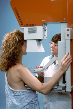
Biennial mammography keeps women safe and saves billions of dollars
Following United States Preventive Services Task Force mammography screening guidelines would save about $4.4 billion annually, without sacrificing quality of care, according to a study published online Feb. 3 in the Annals of Internal Medicine.
Taking the costs of mammography, computer-aided detection, recalls, biopsies, and other variables into account, the investigators estimated that it would have cost $3.5 billion to screen 85% of women in 2010 who met the USPSTF criteria: Screen women aged 50-74 years every other year, plus high-risk women aged 40-49 years and women aged 75-85 years with few comorbidities biennially (Ann. Intern. Med. 2014 Feb. 3;160:145-53 [doi:10.7326/M13-1217]).
Instead, $7.8 billion was actually spent in 2010 to screen just 70% of women using the mélange of screening practices in place today in the United States, a byproduct of the country’s uncertainty about the right way to proceed, the researchers noted.
Meanwhile, following the advice of the American Cancer Society and other groups for annual screenings of women aged 40-84 years would have cost $10.1 billion if 85% had come into the clinic. Screening 85% of women aged 50-69 years biennially – in keeping with the European approach – would have cost $2.6 billion.
Previous studies have found fewer false-positives and recalls with biennial screening, and no significant increase in late-stage cancer detections, said Dr. Cristina O’Donoghue of the University of Illinois at Chicago and her associates.
"Following mammography screening guidelines, such as those from the USPSTF, [which] optimize frequency on the basis of best-available evidence will ... improve screening and save billions of dollars," money that’s better spent on expanding mammography access and increasing risk-based screening, breast-cancer prevention, and mammography reading by experts less likely to make mistakes, among other things, the researchers said.
Even so, "in the United States, there has been resistance to reducing frequency or modifying the age-range for mammography. Those who advocate annual screening should justify the increased costs of nearly $7 billion per year, compared with biennial policies," they said.
The project was a modeling exercise to price out various approaches to breast cancer screening using Medicare reimbursement rates; census figures; Breast Cancer Surveillance Consortium records; and other data sources. The researchers stated that they thought the cost information would help doctors, women, and policy makers find the right approach, especially as the Affordable Care Act improves mammography access and increases screening rates.
The U.S. task force’s biennial approach "is in line with our national goals of advancing health care delivery while improving cost-efficiency," the researchers said.
They concluded that screening frequency is the main driver of costs in mammography, followed by the percentage of women screened, cost per screen, percentage of women screened with digital mammography, and percentage of mammography recalls.
The researchers had no financial conflicts to disclose. The study was funded by the University of California and the Safeway Foundation.
We applaud [Dr.] O’Donoghue and [her] colleagues for meticulously assessing the total cost of breast cancer screening in the United States. Although there is often cause to be skeptical about simulation models because results are based on numerous assumptions, we find the article by O’Donoghue and colleagues to be reasonable and conservative.
Integrating cost into the cancer screening conversation is a challenge. Providers and patients are not only shielded from cost information, but some may raise concerns that the mere mention of costs is a step down the road to rationing. However, both advocates and skeptics should know the costs associated with different breast-cancer screening strategies, particularly when there is so much debate about which approach is most effective.
Costs, including out-of-pocket costs, should be part of the conversation because women with high-deductible health plans may find themselves facing a hefty bill for adjunctive imaging tests and procedures. At the societal level, costs should be integrated into our national dialogue about screening. It is unsustainable to remain ignorant of the costs associated with any health intervention, even breast-cancer screening.
The approximate $8 billion difference among breast-cancer screening strategies is roughly twice as large as the entire annual budget of the National Cancer Institute.
Dr. Joann Elmore and Dr. Cary Gross made the above comments in an editorial accompanying the study (Ann. Intern. Med. 2014 Feb. 3;160:203-4). Dr. Elmore is a professor of medicine at the University of Washington in Seattle. Dr. Gross is a professor of medicine at Yale University in New Haven, Conn., and director of the school’s Cancer Outcomes Public Policy and Effectiveness Research Center. Dr. Gross is a paid Fair Health Inc. board member and disclosed grants from Medtronic and 21st Century Oncology.
Following United States Preventive Services Task Force mammography screening guidelines would save about $4.4 billion annually, without sacrificing quality of care, according to a study published online Feb. 3 in the Annals of Internal Medicine.
Taking the costs of mammography, computer-aided detection, recalls, biopsies, and other variables into account, the investigators estimated that it would have cost $3.5 billion to screen 85% of women in 2010 who met the USPSTF criteria: Screen women aged 50-74 years every other year, plus high-risk women aged 40-49 years and women aged 75-85 years with few comorbidities biennially (Ann. Intern. Med. 2014 Feb. 3;160:145-53 [doi:10.7326/M13-1217]).
Instead, $7.8 billion was actually spent in 2010 to screen just 70% of women using the mélange of screening practices in place today in the United States, a byproduct of the country’s uncertainty about the right way to proceed, the researchers noted.
Meanwhile, following the advice of the American Cancer Society and other groups for annual screenings of women aged 40-84 years would have cost $10.1 billion if 85% had come into the clinic. Screening 85% of women aged 50-69 years biennially – in keeping with the European approach – would have cost $2.6 billion.
Previous studies have found fewer false-positives and recalls with biennial screening, and no significant increase in late-stage cancer detections, said Dr. Cristina O’Donoghue of the University of Illinois at Chicago and her associates.
"Following mammography screening guidelines, such as those from the USPSTF, [which] optimize frequency on the basis of best-available evidence will ... improve screening and save billions of dollars," money that’s better spent on expanding mammography access and increasing risk-based screening, breast-cancer prevention, and mammography reading by experts less likely to make mistakes, among other things, the researchers said.
Even so, "in the United States, there has been resistance to reducing frequency or modifying the age-range for mammography. Those who advocate annual screening should justify the increased costs of nearly $7 billion per year, compared with biennial policies," they said.
The project was a modeling exercise to price out various approaches to breast cancer screening using Medicare reimbursement rates; census figures; Breast Cancer Surveillance Consortium records; and other data sources. The researchers stated that they thought the cost information would help doctors, women, and policy makers find the right approach, especially as the Affordable Care Act improves mammography access and increases screening rates.
The U.S. task force’s biennial approach "is in line with our national goals of advancing health care delivery while improving cost-efficiency," the researchers said.
They concluded that screening frequency is the main driver of costs in mammography, followed by the percentage of women screened, cost per screen, percentage of women screened with digital mammography, and percentage of mammography recalls.
The researchers had no financial conflicts to disclose. The study was funded by the University of California and the Safeway Foundation.
FROM THE ANNALS OF INTERNAL MEDICINE
Major finding: Following USPSTF mammography recommendations – which call for biennial screenings – would have saved the U.S. health care system approximately $4.4 billion in 2010.
Data source: Estimate of 2010 mammography costs based on modeling of Medicare and other expenses.
Disclosures: The authors had no disclosures. The study was funded by the University of California and the Safeway Foundation.
