User login
PARP inhibitor prolongs PFS in mCRPC
BARCELONA – PARP inhibitors may be able to do for men with advanced castration-resistant prostate cancer what they currently do for women with breast or ovarian cancers linked to BRCA mutations, investigators report.
Among men with metastatic castration-resistant prostate cancer (mCRPC) that had progressed on prior therapy with either abiraterone (Zytiga) or enzalutamide (Xtandi) and that bore DNA-repair mutations (BRCA1, BRCA2, or ATM), those who were randomized in the PROfound trial to receive olaparib (Lynparza) had significant improvement in radiographic progression-free survival (rPFS) compared with patients assigned to the physician’s choice of a new hormonal agent, reported Maha Hussain, MD, of the Robert H. Lurie Comprehensive Cancer Center at Northwestern University in Chicago.
“PROfound is the first positive biomarker-selected phase 3 study evaluating a molecularly-targeted therapy in men with metastatic castration-resistant prostate cancer, highlighting the importance of genomic testing in this population, and also importantly highlighting the feasibility of precision-medicine trials in this disease,” she said at the European Society for Medical Oncology (ESMO) Congress.
Approximately 25% of men with mCRPC have loss-of-function mutations or alterations in homologous recombinant repair (HRR) genes, especially BRCA1, BRCA2, and ATM. Breast and ovarian cancers bearing these mutations are known to be sensitive to PARP (poly ADP ribose polymerase) inhibitors such as olaparib.
To see whether men with mCRPC could derive a similar benefit, the investigators enrolled patients who had experienced disease progression on abiraterone or enzalutamiude and whose tumors had one or more alterations in any qualifying gene with direct or indirect role in homologous recombinant DNA repair.
Patients were stratified by previous taxane use and measurable disease, and then two cohorts were enrolled. Cohort A included 245 men with BRCA1, BRCA2, or ATM mutations, and cohort B included 142 men with other alterations (in BARD1, BIRP1, CDK12, CHEK1, CHEK2, FANCL, PALB2, PPP2R2A, RAD15B, RAD15C, RAD15D, or RAD54L).
The median age in cohort A was 68, and in cohort B it was 67, with men 86 years of age at the upper end of the range.
“I think it’s remarkable that men in their late 80s and even early 90s were eligible, were able to be enter this study and be treated on it, and I think that’s an important point here,” Dr. Hussain said.
Nearly one-fourth of patients in the trial had metastatic disease at their initial diagnosis, she noted.
Patients in each cohort were randomized on a 2:1 basis to receive either open-label olaparib 300 mg twice daily, or the treating physician’s choice of abireraterone or enzalutamide, plus predisone.
Upon blinded independent central review (BICR) showing disease progression, patients were allowed to cross over to the olaparib arm, which more than 80% of patients eventually did.
Radiographic PFS in the BRCA1, BRCA2 and ATM cohort according to BICR, the primary endpoint, was a median of 7.39 months with olaparib, compared with 3.55 months with the other therapies, for a hazard ratio (HR) for progression on olaparib of 0.34 (P less than .0001).
A somewhat smaller but still significant benefit was seen for olaparib in the overall population (both cohorts), with a median rPFS of 5.82 months vs. 3.52 months, respectively (HR 0.49, P less than .0001).
Among patients in cohort A, the objective response rate was 33.3% with olaparib, compared with 2.3% for the other agents, resulting in an odds ratio for response of 20.86 (P less than .0001)
Olaparib was also associated with longer time to pain progression in patients in cohort A, with the median not reached compared with 9.92 months with the hormonal agents (HR 0.44, P = .0192).
Among patients in the physician’s choice arm who had disease progression, 80.6% in cohort A and 84.6% in cohort B were crossed over to olaparib.
At this interim analysis, median overall survival was 18.5 months with olaparib, compared with 15.11 months with the other agents, but this difference was not statistically significant. Further follow-up will be needed before a difference in overall survival becomes evident, Dr. Hussain said.
“I think this study has demonstrated that now prostate cancer can be treated with a targeted therapy approach,” said Ignacio Duran, MD, of Hospital Universitario Marques de Valdecilla in Santander, Spain. He was the invited discussant and moderator of a briefing where Dr. Hussain outlined the study details prior to presentation in a symposium.
“Not all prostate cancer patients have the same tumors, and this is the first time we’ve been able to identify that we can more precisely characterize the molecular biology, the genetic background of these tumors, and that is going to determine how we treat them,” he said.
The PROfound trial made “a double hit: superiority in terms of efficacy, and it has proved a new concept that in prostate cancer has not been proved before,” he added.
“This is a truly practice-changing study, not just for our practice and our patients, but also for the study design,” said Eleni Efstathiou, MD, PhD, of the University of Texas MD Anderson Cancer Center in Houston, who was the invited discussant at the symposium where Dr. Hussain presented the full trial data.
She lauded the use of a validated genomic testing tissue-based assay (FoundationOne CDx next-generation sequencing test) to identify patients who might benefit from olaparib.
She said that the PARP inhibition-specific strategy of the trial appears to have paid off, with statistically significant, clinically meaningful improvement in outcomes and an acceptable safety profile.
“And when it comes to a prostate cancer therapy strategry? Well, we’re starting to enter into the targeted therapy era,” she said.
The PROfound trial was sponsored by AstraZeneca and is part of an alliance between AstraZeneca and Merck. Dr. Hussain disclosed travel and/or accommodation support, honoraria, consulting/advisory fees and research support from AstraZeneca and others. Dr. Duran disclosed advisory board fees from Roche and BMS, and speaker honoraria from Roche, Bristol-Myers Squibb, and Merck. Dr. Efstathiou disclosed research support and honoraria from various companies, not including AstraZeneca or Merck.
SOURCE: Hussain M et al. ESMO 2019, Abstract LBA-12.
REPORTING FROM ESMO 2019
Immunotherapy duo extends survival in first line for advanced NSCLC
BARCELONA – Patients with than patients treated with chemotherapy, reported investigators in the Checkmate 227 trial.
Among 793 patients with treatment-naive metastatic NSCLC with programmed death-ligand 1 (PD-L1) expression of at least 1%, median overall survival (OS) was 17.1 months for patients treated with the combination versus 14.9 months for patients treated with chemotherapy, a difference that translated into a hazard ratio with the combination of 0.79 (P = .007). Two-year overall survival rates were 40% and 32.8%, respectively.
The improvement in OS with the combination occurred regardless of PD-L1 status, suggesting that it can be a chemotherapy-free option for patients with previously untreated metastatic NSCLC, Solange Peters, MD, of Centre Hospitalier Universitaire Vaudois, Lausanne, Switzerland, said at the European Society for Medical Oncology Congress.
Checkmate 227 “is the first phase 3 study proving the concept in an evidence-based fashion of the combination of CTLA4 and anti–PD-1/PD-L1 in non–small cell lung cancer,” Dr. Peters said at a briefing prior to her presentation of the data in a symposium.
CheckMate 227 investigators enrolled patients with stage IV or recurrent NSCLC who had not received any treatment previously. Part one of the multipart study was designed to compare different nivolumab-based regimens versus chemotherapy in patients with PD-L1 expression of 1% or greater, or less than 1%.
The trial has two independent primary endpoints, the first of which, progression-free survival (PFS) with nivolumab and ipilimumab versus chemotherapy, was reported at the 2018 annual meeting of the American Association for Cancer Research.
Dr. Peters presented the overall survival endpoint at ESMO 2019. Results of the study were published simultaneously in the New England Journal of Medicine (2019 Sep 28. doi: 10.1056/NEJMoa1910231).
In part one of the trial, patients with PD-L1 expression of at least 1% (1,189 patients) were randomized on a 1:1:1 basis to one of three treatment options: nivolumab plus low-dose ipilimumab, nivolumab alone, or histology-based chemotherapy. Patients with PD-L1 expression less than 1% (550 patients) were randomized to nivolumab plus low-dose ipilimumab, nivolumab plus chemotherapy, or chemotherapy.
The survival benefit seen with the immunotherapy combination was durable, with 2-year OS rates of 40% for nivolumab/ipilimumab versus 32.8% for chemotherapy. The median durations of response were 23.2 months vs. 6.2 months, respectively.
Patients with low or no PD-L1 expression levels also benefited from the immunotherapy combination, with a median OS of 17.2 months with nivolumab plus ipilimumab, compared with 12.2 months with chemotherapy.
Among all enrolled patients, the median OS duration was 17.1 months with nivolumab plus ipilimumab versus 13.9 months with chemotherapy.
Grade 3 or 4 treatment-related adverse events in the overall study population occurred in 32.8% of patients treated with combined immunotherapy and in 36% of those treated with chemotherapy.
“A key question remains: Is the addition of ipilimumab providing additional benefit on top of nivolumab?” Dr. Peters asked. The answer to that question appears to be “yes,” she said, pointing to a comparison of OS among patients with PD-L1 expression below 1%. Although this subanalysis was not powered for statistical significance, the survival curves indicated a significant advantage for the combination, compared with nivolumab alone or chemotherapy, with respective median OS rates of 17.2, 15.2, and 12.2 months.
“What have we learned from Checkmate 227 so far? Caveat emptor – buyer beware. Beware of the adverse event profile for this doublet chemotherapy combination,” said Sanjay Popat, MD, PhD, of the Royal Marsden Hospital in London, who was the invited discussant at the symposium.
He noted that among PD-L1–positive patients, the OS benefit appears to be driven by patients with high levels of PD-L1 expression (50% or greater).
Additional questions that need addressing, he said, include how immune-related adverse events evolve over time; whether the combination can be made less toxic without impairing efficacy; and what is the optimum duration, dosing, and scheduling of ipilimumab.
“What is the promise of [Checkmate] 227? The promise of the 227 is to have long-lasting responses. That means long life for our patients with a chemo-sparing regimen,” commented Marina Garassino, MD, from the Istituto Nazionale dei Tumori in Milan, who was the invited discussant at the presymposium briefing.
“At the same time, we have to go back to the bench, to the scientists, and to ask the scientists to [help us] understand who are the patients to be treated with the combination of immunotherapy and immunotherapy, with the combination of chemotherapy and immunotherapy, and just with a single agent,” she said.
The study was funded by Bristol-Myers Squibb. Dr. Peters reported a consultant/advisory role, speakers bureau activity, and research support from BMS and others. Dr. Popat disclosed honoraria from BMS and others. Dr. Garrassino disclosed personal fees from BMS and others.
SOURCE: Hellmann MD et al. ESMO 2019. N Engl J Med. 2019 Sep 28. doi: 10.1056/NEJMoa1910231.
REPORTING FROM ESMO 2019
Osimertinib improves survival in advanced NSCLC
BARCELONA – In patients with , compared with other agents targeted against NSCLC with epidermal growth factor–receptor (EGFR) mutations, investigators for the FLAURA trial reported.

After median follow-up ranging from 27 to 35.8 months, the median overall survival was 38.6 months for patients randomized to osimertinib, compared with 31.8 months for patients assigned to either of two comparator tyrosine kinase inhibitors (TKIs), gefitinib (Iressa) or erlotinib (Tarceva).
The hazard ratio for death with osimertinib was 0.799 (P = .0462), reported Suresh Ramalingam, MD, director of the lung cancer program at Winship Cancer Institute of Emory University, Atlanta.
“I’m excited that the new milestone accomplished with osimertinib in this trial will serve as the platform to build on in our efforts to improve the lives of patients with lung cancer,” he said at the European Society for Medical Oncology Congress.
Osimertinib is the first TKI to show improvement in overall survival over another TKI in the treatment of advanced stage cancers, he noted.
Overall survival was a secondary endpoint of the FLAURA trial. As previously reported, FLAURA met its primary endpoint of improvement in progression-free survival (PFS) in an interim analysis presented at ESMO 2017. In that analysis, osimertinib cut the risk of disease progression by 54%, compared with gefitinib or erlotinib.
Among 279 patients with EGFR-mutated locally advanced or metastatic NSCLC treated with osimertinib, the median PFS was 18.9 months, compared with 10.2 months for 277 patients treated with the standard of care, which translated into a HR of 0.46 (P less than .0001).
The FLAURA results supported Food and Drug Administration approval of osimertinib in April 2018 for first-line treatment of patients with metastatic NSCLC with EGFR mutations as detected by an FDA-approved test.
The current overall survival analysis, although not powered to show differences among patient subgroups, showed trends favoring osimertinib over a comparator TKI among both men and women, older and younger patients, patients with central nervous system metastases at trial entry, and patients with the EGFR exon 19 deletion at randomization.
The 31.8 month median overall survival for the control (comparator-TKI) arm is among the highest reported for patients with EGFR-mutated NSCLC, Dr. Ramalingam noted.
“That is because a lot of patients crossed over from the control group to receive osimertinib on progression,” he said, adding that the magnitude of benefit from osimertinib was greater among non-Asian patients, compared with Asians.
FLAURA details
In the phase 3 FLAURA trial, investigators stratified patients with previously untreated NSCLC positive for EGFR resistance mutations according to mutation status (exon 19 deletion or the L858R amino acid substitution in exon 21) and race (Asian or non-Asian).
Patients were randomly assigned to treatment with either oral osimertinib 80 mg daily or an EGFR TKI, either oral gefitinib 250 mg or erlotinib 150 mg daily.
The patients were assessed by Response Evaluation Criteria in Solid Tumors (RECIST) every 6 weeks until objective disease progression.
Patients assigned to the standard-of-care arm who had central confirmation of progression and T790M positivity were allowed to cross over to open-label osimertinib.
PFS, the primary endpoint, was also significantly better with osimertinib than with either of the comparator TKIs in patients with and without central nervous system metastases at study entry (HR, 0.47; P = .0009 for patients with CNS metastases; HR, 0.46; P less than .0001 for patients with no CNS metastases).
“For clinicians, for patients, and also for our health authorities, the results in terms of overall survival are really relevant, and this is why this study is so important, knowing this secondary endpoint from a statistical point of view. The study is statistically significant and clinically relevant,” commented Pilar Garrido, MD, from the department of medical oncology, Hospital Universitario Ramón y Cajal in Madrid, the invited discussant at a briefing where Dr. Ramalingam outlined the study findings prior to his presentation of the data in a symposium.
“What’s the future of EGFR mutant lung cancer? Well, I think we should be done with single-agent EGFR-TKI comparisons: We have a clear agent that’s associated with an improvement in survival. I think our focus needs to shift to building on or adding to osimertinib,” commented Pasi A Jänne, MD, PhD, director of the Lowe Center for Thoracic Oncology at Dana-Farber Cancer Institute in Boston, the invited discussant at the symposium.
He said that the challenge for clinicians will be to identify high- and low-risk EGFR-mutant NSCLC, and to determine which patients could be treated with a single agent, and which may require a combination therapy approach.
FLAURA was sponsored by AstraZeneca. Dr. Ramalingam disclosed honoraria, an advisory or consulting role, and research funding from that company and others. Dr. Garrido disclosed a speaker and advisory role for AstraZeneca and others. Dr. Jänne disclosed prior consulting for AstraZeneca.
SOURCE: Ramalingam S et al. ESMO 2019. Abstract LBA5_PR.
BARCELONA – In patients with , compared with other agents targeted against NSCLC with epidermal growth factor–receptor (EGFR) mutations, investigators for the FLAURA trial reported.
After median follow-up ranging from 27 to 35.8 months, the median overall survival was 38.6 months for patients randomized to osimertinib, compared with 31.8 months for patients assigned to either of two comparator tyrosine kinase inhibitors (TKIs), gefitinib (Iressa) or erlotinib (Tarceva).
The hazard ratio for death with osimertinib was 0.799 (P = .0462), reported Suresh Ramalingam, MD, director of the lung cancer program at Winship Cancer Institute of Emory University, Atlanta.
“I’m excited that the new milestone accomplished with osimertinib in this trial will serve as the platform to build on in our efforts to improve the lives of patients with lung cancer,” he said at the European Society for Medical Oncology Congress.
Osimertinib is the first TKI to show improvement in overall survival over another TKI in the treatment of advanced stage cancers, he noted.
Overall survival was a secondary endpoint of the FLAURA trial. As previously reported, FLAURA met its primary endpoint of improvement in progression-free survival (PFS) in an interim analysis presented at ESMO 2017. In that analysis, osimertinib cut the risk of disease progression by 54%, compared with gefitinib or erlotinib.
Among 279 patients with EGFR-mutated locally advanced or metastatic NSCLC treated with osimertinib, the median PFS was 18.9 months, compared with 10.2 months for 277 patients treated with the standard of care, which translated into a HR of 0.46 (P less than .0001).
The FLAURA results supported Food and Drug Administration approval of osimertinib in April 2018 for first-line treatment of patients with metastatic NSCLC with EGFR mutations as detected by an FDA-approved test.
The current overall survival analysis, although not powered to show differences among patient subgroups, showed trends favoring osimertinib over a comparator TKI among both men and women, older and younger patients, patients with central nervous system metastases at trial entry, and patients with the EGFR exon 19 deletion at randomization.
The 31.8 month median overall survival for the control (comparator-TKI) arm is among the highest reported for patients with EGFR-mutated NSCLC, Dr. Ramalingam noted.
“That is because a lot of patients crossed over from the control group to receive osimertinib on progression,” he said, adding that the magnitude of benefit from osimertinib was greater among non-Asian patients, compared with Asians.
FLAURA details
In the phase 3 FLAURA trial, investigators stratified patients with previously untreated NSCLC positive for EGFR resistance mutations according to mutation status (exon 19 deletion or the L858R amino acid substitution in exon 21) and race (Asian or non-Asian).
Patients were randomly assigned to treatment with either oral osimertinib 80 mg daily or an EGFR TKI, either oral gefitinib 250 mg or erlotinib 150 mg daily.
The patients were assessed by Response Evaluation Criteria in Solid Tumors (RECIST) every 6 weeks until objective disease progression.
Patients assigned to the standard-of-care arm who had central confirmation of progression and T790M positivity were allowed to cross over to open-label osimertinib.
PFS, the primary endpoint, was also significantly better with osimertinib than with either of the comparator TKIs in patients with and without central nervous system metastases at study entry (HR, 0.47; P = .0009 for patients with CNS metastases; HR, 0.46; P less than .0001 for patients with no CNS metastases).
“For clinicians, for patients, and also for our health authorities, the results in terms of overall survival are really relevant, and this is why this study is so important, knowing this secondary endpoint from a statistical point of view. The study is statistically significant and clinically relevant,” commented Pilar Garrido, MD, from the department of medical oncology, Hospital Universitario Ramón y Cajal in Madrid, the invited discussant at a briefing where Dr. Ramalingam outlined the study findings prior to his presentation of the data in a symposium.
“What’s the future of EGFR mutant lung cancer? Well, I think we should be done with single-agent EGFR-TKI comparisons: We have a clear agent that’s associated with an improvement in survival. I think our focus needs to shift to building on or adding to osimertinib,” commented Pasi A Jänne, MD, PhD, director of the Lowe Center for Thoracic Oncology at Dana-Farber Cancer Institute in Boston, the invited discussant at the symposium.
He said that the challenge for clinicians will be to identify high- and low-risk EGFR-mutant NSCLC, and to determine which patients could be treated with a single agent, and which may require a combination therapy approach.
FLAURA was sponsored by AstraZeneca. Dr. Ramalingam disclosed honoraria, an advisory or consulting role, and research funding from that company and others. Dr. Garrido disclosed a speaker and advisory role for AstraZeneca and others. Dr. Jänne disclosed prior consulting for AstraZeneca.
SOURCE: Ramalingam S et al. ESMO 2019. Abstract LBA5_PR.
REPORTING FROM ESMO 2019
Adjuvant radiotherapy no better than salvage post prostatectomy
BARCELONA – , results of a large randomized trial and separate meta-analysis indicate.
Among nearly 1,400 men with postoperative prostate-specific antigen (PSA) levels below 0.2 ng/mL and one or more risk factors, followed for a median of 5 years, there were no significant differences in any of the secondary outcomes between men randomized to radiotherapy and observation alone, reported Chris Parker, MD, from the Royal Marsden Hospital in London.
“In comparison with a policy of early salvage radiotherapy, adjuvant radiotherapy did not improve biochemical progression-free survival and did not delay the further use of hormone therapy,” he said at the European Society for Medical Oncology Congress, on behalf of colleagues in the RADICALS-RT trial.
Results of the RADICAL-RT trial were also pooled with results from two other large trials in a collaborative series of meta-analyses of long-term prostate cancer outcomes – dubbed ARTISTIC – which found no significant differences in event-free survival (EFS) for men randomized to either adjuvant or salvage radiotherapy.
“We don’t see any evidence from the ARTISTIC results that adjuvant radiotherapy improves event-free survival, compared to early salvage radiotherapy, and our best estimate is of a small, 1% difference in event-free survival at 5 years,” said Claire Vale, PhD, a research fellow at University College, London.
RADICALS RT details
Dr. Parker presented results from an early analysis of the secondary endpoint of biochemical progression-free survival (PFS), conducted in cooperation with the ARTISTIC investigators.
In RADICALS RT, investigators in the United Kingdom, Denmark, Canada, and Ireland enrolled 1,369 men following radical prostatectomy and after stratification by Gleason score, margin status, treatment center, and radiotherapy schedule (52.5 Gy delivered in 20 fractions, or 66 Gy delivered), randomly assigned them to either postoperative radiotherapy or observation with radiotherapy.
The patients enrolled had postoperative PSAs less than 2 ng/mL and one or more risk factors, either pathologic stage 3/4, Gleason score 7-10, positive surgical margins, or preoperative PSA of 10 ng/mL or greater.
The trigger for radiotherapy in men assigned to observation was PSA failure, defined as a PSA level of at least 0.1 ng/mL or 3 consecutive PSA rises.
At the median 5-year follow-up, PFS rates were 85% for patients assigned to adjuvant radiotherapy, and 88% for those assigned to observation, translating to a hazard ratio of 1.10, which was not statistically significant.
However, there were significant differences between the groups in both self-reported urinary incontinence (5.3% of patients in the radiotherapy group vs. 2.7% in the observation group, P = .008) and grade 3 or 4 urethral stricture at any time (8% vs. 5%, respectively, P = .03).
Results of the primary outcome, freedom from distant metastases will require longer follow-up, Dr. Parker said.
ARTISTIC meta-analysis
Dr. Vale presented results of the ARTISTIC collaborative meta-analysis, which included data from three randomized trials, including RADICALS, GETUG-AFU 17, and RAVES.
The meta-analysis was designed to include a consistent definition of PSA-driven EFS, prior to the unblinding of trial results.
The definition of EFS used in the trial was time from randomization until the first evidence of either PSA of 0.4 ng/mL or greater and rising after completion of radiotherapy, clinical/radiological progression, initiation on nontrial treatment, death from prostate cancer following completion of radiotherapy, or PSA level of 2.0 ng/mL or greater any time after randomization.
They analyzed data on a total of 1,074 men assigned to adjuvant radiotherapy and 1,077 assigned to salvage radiotherapy.
In each of the three trials included in the meta-analysis, there was a nonsignificant trend toward better PSA-driven EFS with salvage radiotherapy vs. adjuvant radiotherapy. The overall hazard ratio was 1.12, with the 95% confidence interval crossing 1, indicating a nonsignificant result.
Dr. Vale noted that early salvage radiotherapy spares many men from potentially unnecessary radiation and its negative consequences. Of the men included in the trials, more than 60% of those randomized to observation with salvage radiotherapy have yet to start radiotherapy.
She noted that investigators still need to assess long-term definitive outcomes such as metastases and survival, and whether some patients may derive benefit from adjuvant radiotherapy.
Invited discussant Gert De Meerleer, MD, PhD, a radiation oncologist at University Hospitals Leuven in Belgium, acknowledged that he agreed with most of the findings of the RADICALS-RT and ARTISTIC investigators.
An important take-home message, he said, is that “cure is the aim, and radiotherapy the keystone. If you have PSA relapse after prostatectomy, you can say, ‘I don’t treat the patient’ – fair enough. But if you treat the patient, only giving systemic therapy is the wrong way to go, you have to add radiotherapy,” he said.
He also agreed that “salvage is probably the modality of choice, and it is also in a lot of European countries, provided it is early.”
The RADICALS-RT trial is supported by Cancer Research UK and the Medical Research Council UK. Dr. Parker said he had no disclosures relevant to the trial. ARTISTIC is funded by the Medical Research Council. Dr. Vale reported having no disclosures. Dr. De Meerleer reported speaker fees, advisory board activities, and scientific grants not related to the studies.
SOURCE: Parker C et al. and Vale C et al. ESMO 2019. Abstracts LBA49_PR and LBA48_PR.
REPORTING FROM ESMO 2019

Worse PFS when radiation is withheld in early-stage Hodgkin lymphoma
Radiotherapy appears to be an essential component of the optimal treatment regimen for adults with early-stage favorable Hodgkin lymphoma, investigators in a randomized phase 3 trial asserted.
Among more than 600 patients with early-stage Hodgkin lymphoma who were positron-emission tomography (PET)–negative after two cycles of standard chemotherapy, 5-year progression-free survival was significantly better for patients who had also received involved-field radiotherapy, compared with those who had received chemotherapy alone.
The HD16 trial was designed to show whether using PET findings to opt for consolidation radiotherapy could be noninferior to the use of combined modality therapy (CMT) with ABVD (doxorubicin, bleomycin, vinblastine and dacarbazine) and radiation for all patients with early-stage favorable-risk disease.
“However, we failed to meet the primary objective of the trial as PET-guided omission of radiotherapy results in poorer tumor control compared with CMT. We therefore recommend proceeding with consolidation radiotherapy as a standard of care for patients achieving a metabolic response after two cycles of ABVD,” Michael Fuchs, MD, from the University Hospital of Cologne, Germany, and colleagues in the German Hodgkin Study Group wrote in the Journal of Clinical Oncology.
The investigators also found that patients who remained PET-positive after two cycles of ABVD were at high risk for treatment failure, particularly when a Deauville score of 4 was used as the minimum threshold for positivity.
Although CMT is associated with high cure rates for patients with Hodgkin lymphoma, clinicians are concerned about long-term toxicities and risk for second malignancies, which prompted investigators to see whether radiotherapy could be safely eliminated in some cases.
The HD16 investigators enrolled 1,150 patients aged 18-75 years with early-stage favorable Hodgkin lymphoma, and randomly assigned them to receive two cycles of ABVD with either 20 Gy of involved-field radiotherapy or PET-guided treatment in which involved-field radiation was eliminated for those patients who were PET-negative after chemotherapy, with PET negativity defined as a Deauville score less than 3.
After a median follow-up of 47 months, the 5-year progression-free survival rates among 628 PET-negative patients were 93.4% for those assigned to CMT, versus 86.1% for patients assigned to chemotherapy alone, which translated into a hazard ratio HR of 1.78 (95% confidence interval, 1.02-3.12; P = .040). The upper limit of the confidence interval exceeds the predefined noninferiority margin of 3.01, which indicates that eliminating radiation was clinically inferior.
The difference in progression-free survival rates between the treatment arms was primarily caused by a significant increase in disease recurrence in what would have been the involved field for patients in the ABVD-alone group (in-field recurrence rate, 9% vs. 2% for patients who received CMT; P = .0003). In contrast, there were no significant differences between the groups in out-of-field recurrences (5% vs. 4%, respectively).
Five-year overall survival rates were virtually identical between the treatment arms among PET-negative patients.
When the investigators compared all PET-negative with PET-positive patients (Deauville score 4), they saw that 5-year estimated progression-free survival rates were 93.1% vs. 80.9%, respectively, an absolute difference of 12.1% that translated into a HR of 2.94 (P less than .001). There were no significant differences by PET status in 5-year overall survival, however.
“We assume that the small radiation fields and doses used in our HD16 trial will induce fewer late adverse events than those reported in the literature. However, we cannot exclude an increased risk for certain late effects, such as breast cancer in very young women, as the risk for this specific second malignancy increases with younger age,” the researchers wrote.
The study was supported by grants from Deutsche Krebshilfe and the Swiss State Secretariat for Education, Research, and Innovation. Dr. Fuchs reported honoraria from Amgen, Affimed, Celgene, and Takeda. Multiple coauthors reported industry funding.
SOURCE: Fuchs M et al. J Clin Oncol. 2019 Sep 10. doi: 10.1200/JCO.19.00964.
FROM THE JOURNAL OF CLINICAL ONCOLOGY
PBRM1 mutations not quite up to snuff as ccRCC biomarkers
Loss-of-function mutations in the PBRM1 gene are more common among patients with metastatic clear cell renal cell carcinoma (ccRCC) who have clinical responses to immune checkpoint inhibitors (ICIs), but the mutations fall a little short of serving as clinical biomarkers, investigators say.
Among patients with metastatic ccRCC enrolled in a randomized, phase 3 clinical trial comparing nivolumab (Opdivo) with everolimus (Afinitor) in patients who had previously received antiangiogenic therapy, patients with responses to nivolumab were more than twice as likely as nonresponders to have a truncating loss-of-function mutation in PBRM1, reported Toni K. Choueiri, MD, and colleagues from the Dana-Farber Cancer Institute in Boston.
Patients with PBRM1 mutations were also more likely to have a clinical benefit (complete/partial response, or stable disease with tumor shrinkage and progression-free survival [PFS] of at least 6 months) as well as better PFS and overall survival than patients without mutations.
“The association of PBRM1 truncating mutations with response to anti–[programmed death-1] therapy was confirmed in an independent ccRCC cohort. However, key limitations restrict use of PBRM1 mutations as a clinical biomarker,” they wrote in a research letter to JAMA Oncology.
Those limitations include an only modest effect of mutations on response and survival, a lack of evidence for a PBRM1 mutation effect in the first-line setting, and a possible association between mutations and benefit from prior antiangiogenic therapies, they acknowledged.
Dr. Choueiri was a coauthor of a previous study of genomic correlates of response to ICIs in ccRCC, which found that PBRM1 loss may influence response to checkpoint inhibitors by altering global tumor-cell expression.
In the current study, he and his colleagues looked at archival tumor tissue from an independent cohort of 382 patients who were part of a larger phase 3 trial. Of this group, 189 were treated with nivolumab, and 193 were treated with everolimus. PBRM1 mutations were identified in 55 of the nivolumab-treated patients (29%) and in 45 of the everolimus-treated patients (23%).
When they looked at clinical responses, they found that 15 of 38 patients with response to nivolumab (39%) had truncating PBRM1 mutations, compared with 16 of 74 nonresponding patients (22%), which translated into an odds ratio for response of 2.34 (P = .04).
Similarly, PBRM1 mutations among nivolumab-treated patients were significantly associated with clinical benefit (OR, 2.14; P = .0497), PFS (hazard ratio for progression, 0.67; P = .03), and overall survival (HR, 0.65; P = .03).
In contrast, there were no significant associations in everolimus-treated patients between PBRM1 mutations and either response, PFS, or overall survival.
“The concomitant presence of other cellular or molecular features may further influence the findings described herein. Nonetheless, this validated association between PBRM1 alterations and ICI response in a large randomized study represents a further step toward the development of genomic predictors for immunotherapies in advanced RCC,” the investigators concluded.
The study was supported by Department of Defense Congressionally Directed Medical Research Programs and Bristol-Myers Squibb. Dr. Choueiri disclosed personal fees from Bristol-Myers Squibb and fees and grants from other companies. One coauthor is a Bristol-Myers Squibb employee and shareholder.
SOURCE: Choueiri TK et al. JAMA Oncol. 2019 Sep 5. doi: 10.1001/jamaoncol.2019.3158.
FROM JAMA ONCOLOGY
Stem cells gene edited to be HIV resistant treat ALL, but not HIV
Gene editing of donor stem cells prior to transplantation into a patient with both HIV infection and acute lymphoblastic leukemia (ALL) was safe and effectively treated the patient’s leukemia, but failed to resolve his HIV, investigators reported.

The 27-year-old man received an HLA-matched transplant of hematopoietic stem and progenitor cells (HSPCs) that had been genetically engineered to lack CCR5, a key gateway for HIV entry into cells.
Although the transplant resulted in complete remission of leukemia with full donor chimerism, only about 9% of the posttransplant lymphocytes showed disruption of CCR5, and during a brief trial of antiretroviral therapy interruption his HIV viral load rebounded, reported Hongkui Deng, PhD, and colleagues from Peking University in China.
Although the experiment did not meet its goal of a drug-free HIV remission, it serves as a proof of concept for the use of CRISPR-Cas9 (clustered regularly interspaced palindromic repeats/CRISPR-associated protein 9) gene editing to treat HIV infection, the authors contend.
“These results show the proof of principle that transplantation and long-term engraftment of CRISPR-edited allogeneic HSPCs can be achieved; however, the efficiency of the response was not adequate to achieve the target of cure of HIV-1 infection,” they wrote in a brief report published in the New England Journal of Medicine.
As previously reported, other research groups have investigated genetic editing to mimic a naturally occurring mutation that effectively disables the CCR5 HIV coreceptor, preventing the retrovirus from entering healthy cells. The mutation was first identified in a man named Timothy Brown who came to be known as “the Berlin patient” after he was apparently cured of HIV infection after a bone marrow transplant from a donor who had the mutation.
Dr. Deng and colleagues took advantage of HSPC transplantation, a standard therapy for ALL to see whether it could also have beneficial effects on concomitant HIV infection.
They treated donor HSPCs with CRISPR-Cas9 to ablate CCR5 and then delivered them to the patient along with additional CD34-depleted donor cells from mobilized peripheral blood.
The transplant was a success, with neutrophil engraftment on day 13 and platelet engraftment on day 27, and the leukemia was in morphologic complete remission at week 4 following transplantation. The patient remained in complete remission from leukemia throughout the 19-month follow-up period, with full donor chimerism .
However, when a planned interruption of antiretroviral therapy was carried out at 7 months post transplant, the serum viral load increased to 3 × 10⁷ copies/ml at week 4 following interruption, and the patient was restarted on the drug. His viral levels gradually decreased to undetectable level during the subsequent months.
The investigators noted that 2 weeks after the drug interruption trial was started there was a small increase in the percentage of CCR5 insertion/deletions.
“The low efficiency of gene editing in the patient may be due to the competitive engraftment of the coinfused HSPCs in CD34-depleted cells and the persistence of donor T cells. To further clarify the anti-HIV effect of CCR5-ablated HSPCs, it will be essential to increase the gene-editing efficiency of our CRISPR-Cas9 system and improve the transplantation protocol,” they wrote.
The study was funded by the Beijing Municipal Science and Technology Commission and others (unspecified). All authors reported having nothing to disclose.
SOURCE: Xu L et al. N Engl J Med. 2019. doi: 10.1056/NEJMoa1817426.
Gene editing of donor stem cells prior to transplantation into a patient with both HIV infection and acute lymphoblastic leukemia (ALL) was safe and effectively treated the patient’s leukemia, but failed to resolve his HIV, investigators reported.
The 27-year-old man received an HLA-matched transplant of hematopoietic stem and progenitor cells (HSPCs) that had been genetically engineered to lack CCR5, a key gateway for HIV entry into cells.
Although the transplant resulted in complete remission of leukemia with full donor chimerism, only about 9% of the posttransplant lymphocytes showed disruption of CCR5, and during a brief trial of antiretroviral therapy interruption his HIV viral load rebounded, reported Hongkui Deng, PhD, and colleagues from Peking University in China.
Although the experiment did not meet its goal of a drug-free HIV remission, it serves as a proof of concept for the use of CRISPR-Cas9 (clustered regularly interspaced palindromic repeats/CRISPR-associated protein 9) gene editing to treat HIV infection, the authors contend.
“These results show the proof of principle that transplantation and long-term engraftment of CRISPR-edited allogeneic HSPCs can be achieved; however, the efficiency of the response was not adequate to achieve the target of cure of HIV-1 infection,” they wrote in a brief report published in the New England Journal of Medicine.
As previously reported, other research groups have investigated genetic editing to mimic a naturally occurring mutation that effectively disables the CCR5 HIV coreceptor, preventing the retrovirus from entering healthy cells. The mutation was first identified in a man named Timothy Brown who came to be known as “the Berlin patient” after he was apparently cured of HIV infection after a bone marrow transplant from a donor who had the mutation.
Dr. Deng and colleagues took advantage of HSPC transplantation, a standard therapy for ALL to see whether it could also have beneficial effects on concomitant HIV infection.
They treated donor HSPCs with CRISPR-Cas9 to ablate CCR5 and then delivered them to the patient along with additional CD34-depleted donor cells from mobilized peripheral blood.
The transplant was a success, with neutrophil engraftment on day 13 and platelet engraftment on day 27, and the leukemia was in morphologic complete remission at week 4 following transplantation. The patient remained in complete remission from leukemia throughout the 19-month follow-up period, with full donor chimerism .
However, when a planned interruption of antiretroviral therapy was carried out at 7 months post transplant, the serum viral load increased to 3 × 10⁷ copies/ml at week 4 following interruption, and the patient was restarted on the drug. His viral levels gradually decreased to undetectable level during the subsequent months.
The investigators noted that 2 weeks after the drug interruption trial was started there was a small increase in the percentage of CCR5 insertion/deletions.
“The low efficiency of gene editing in the patient may be due to the competitive engraftment of the coinfused HSPCs in CD34-depleted cells and the persistence of donor T cells. To further clarify the anti-HIV effect of CCR5-ablated HSPCs, it will be essential to increase the gene-editing efficiency of our CRISPR-Cas9 system and improve the transplantation protocol,” they wrote.
The study was funded by the Beijing Municipal Science and Technology Commission and others (unspecified). All authors reported having nothing to disclose.
SOURCE: Xu L et al. N Engl J Med. 2019. doi: 10.1056/NEJMoa1817426.
FROM NEW ENGLAND JOURNAL OF MEDICINE
Key clinical point: Donor cells depleted of the HIV coreceptor CCR5 effectively treated ALL, but not HIV.
Major finding: The patient had a sustained complete remission of ALL, but HIV persisted after transplantation.
Study details: Case report of a 27-year-old man with ALL and HIV.
Disclosures: The study was funded by the Beijing Municipal Science and Technology Commission and others (unspecified). All authors reported having nothing to disclose.
Source: Xu L et al. N Engl J Med. 2019. doi: 10.1056/NEJMoa1817426.
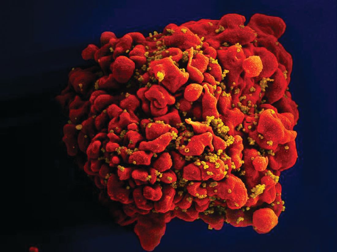
Many experimental drugs veer off course when targeting cancer
Clinical trials of novel cancer drugs miss their marks far more often than they hit them, in part because the drugs themselves may be aimed at the wrong targets or the targets themselves may not be that important in the first place, investigators have found.
Using CRISPR (clustered regularly interspaced palindromic repeats) gene editing to study the effects of 10 drugs that are in development targeting six proteins ostensibly crucial to the health or survival of cancer cells, Jason Sheltzer, PhD, of Cold Spring Harbor (N.Y.) Laboratory and colleagues found that the cancer cells could get along just fine without the targeted proteins, suggesting that the drugs’ alleged efficacy in the lab dish was because of other, off-target effects.
“It seemed like these genes that encode proteins that are being targeted by putative precision agents in clinical trials aren’t actually essential at all for cancer cell growth, so that was one surprise that we found,” Dr. Sheltzer said in a telephone briefing for reporters held prior to publication of the study in Science Translational Medicine.
“The second surprise was that we took the drugs that were supposed to be specific for these proteins and then we treated cancer cells with them, and we found that the drugs continued to kill the cancer cells that totally lacked the target protein expression,” he said.
But the investigators also made a serendipitous discovery that one of the drugs they tested, OTS964, was not – as originally thought – an inhibitor of the PBK protein but instead was an inhibitor of cyclin-dependent kinase (CDK) 11, making it a molecular relative of drugs such as the CDK4/6 inhibitors ribociclib (Kisqali) and palbociclib (Ibrance), both potent inhibitors of hormone receptor–positive, HER2-negative metastatic breast cancer.
Drug development insights
Their findings also indicate that less-precise candidate-drug identification techniques using RNA interference (RNAi) to knock down protein expression may have led earlier investigators down the garden path, resulting in errors that can lead to the all-too-familiar scenario of a seemingly promising compound flourishing in the early drug development process, only to wither on the vine in clinical trials.
“Everyone knows that it’s really hard to make new cancer drugs, but what we’ve been finding out is that, once a new drug is even made, it can be just as difficult to really understand how that drug is working to kill cancer cells,” said coauthor Chris Giuliano, currently a doctoral candidate at the Massachusetts Institute of Technology in Cambridge, who also spoke at the briefing.
“Our study showed us that a potential problem with the cancer drug development pipeline is that the way in which some of these new cancer drugs work is incompletely understood. Ten years ago many of these studies were developed with a tool known as RNAi, which while being the best available tool at the time ultimately led many researchers to arrive at the wrong conclusions about how some of these drug targets actually work,” he added.
Sour on MELK
The investigators had previously found that MELK (maternal embryonic leucine zipper kinase), a protein previously identified as essential to survival in multiple cancer types, could be eliminated from cancer cells using CRISPR gene editing without significant harm to the cells, and that a drug targeted against MELK in phase 2 clinical trials (OTS167) continued to kill the knockout cells in a lab dish with no loss of potency. This finding alerted the researchers to the possibility that drugs in development could be targeting the wrong protein, accounting for at least some of the high failure rate in cancer drug development, and potentially explaining some of the toxicities seen with experimental agents.
“Moreover, clinical trials that use a biomarker to select patients for trial inclusion are about twice as likely to succeed as those without one. Misidentifying a drug’s mechanism of action could hamper efforts to uncover a biomarker capable of predicting therapeutic responses, further decreasing the success rate of clinical trials,” they wrote.
Other false targets
In the current study, the investigators tested whether other drugs were designed to point toward nonessential or “superfluous” targets, and whether the mechanisms of action of the drugs had been mischaracterized.
They focused on five proteins that were thought to be so important to cancer cells that their loss would inhibit or block cell proliferation (HDAC6, MAPK14/p38, PAK4, PBK, and PIM1) and one (CASP3/caspase-3) that was thought to induce apoptosis when activated by a small molecule.
First, the investigators determined that the putative targets – the five proteins listed before – may not be required for actual cancer cell growth or survival, and then found evidence to suggest that misidentification of the proteins may have been caused by the uncertainties of RNAi.
They then used CRISPR to assess the mechanism of action of each of the 10 drugs, and whether the effects they induced were on or off target. They found that PAC-1, a putative caspase-3 activator currently in three clinical trials, actually works in a caspase-3–independent manner, and that all 10 anticancer drugs “exhibited clear evidence of target-independent cell killing in every [knockout] cell line that we examined.”
Finally, as noted before, they determined that the actual mechanism of action of OTS964 was not PBK inhibition, but inhibition of CDK11, a protein that appears to be vital for mitosis in human cancers.
“We think that CDK11 is an exciting target for future therapeutic development, and we found it specifically by looking for the true targets of these mischaracterized agents,” Dr. Sheltzer said.
The investigators acknowledged that their study was limited by the use of well-established cancer cell lines that may not fully reflect how cancer acts in the human body, and they could not rule out that the superfluous proteins they identified might be important for the survival of rare cancers.
“Additionally, we’re not saying that these targets offer no therapeutic potential,” said lead author Ann Lin, who is currently a Fulbright Fellow at the University of Oslo.
“It might be that there are other, unrelated proteins in the cell taking over its role and targeting of both proteins in combination is needed to kill the cancer cells. Furthermore, removal of these proteins may reveal a weakness in the cancer that can be targeted by a second drug, so our experiments showed that uniquely targeting these proteins alone showed little efficacy,” she said.
Research in the Sheltzer Lab is supported by an National Institutes of Health Early Independence Award, a Breast Cancer Alliance Young Investigator Award, a Damon Runyon-Rachleff Innovation Award, a Gates Foundation Innovative Technology Solutions grant, and a CSHL-Northwell Translational Cancer Research grant. The authors reported that they have no competing interests.
SOURCE: Lin A et al. Sci Transl Med. 2019 Sep 11. doi: 10.1126/scitranslmed.aaw8412 .
FROM SCIENCE TRANSLATIONAL MEDICINE
Key clinical point: Cancer drug developers need more stringent methods for verifying targets before advancing drugs to clinical trials.
Major finding: Ten drugs in development against cancer have mechanisms of action independent of their targets.
Study details: In vitro studies of cancer drugs and their targets using CRISPR gene-editing techniques.
Disclosures: Research in the Sheltzer Lab is supported by a National Institutes of Health Early Independence Award, a Breast Cancer Alliance Young Investigator Award, a Damon Runyon-Rachleff Innovation Award, a Gates Foundation Innovative Technology Solutions grant, and a CSHL-Northwell Translational Cancer Research grant. The authors reported that they have no competing interests.
Source: Lin A et al. Sci Transl Med. 2019 Sep 11. doi: 10.1126/scitranslmed.aaw8412.
HER2-mutant NSCLC confers high brain metastases risk
Although only a small percentage of non–small cell lung cancers are positive for HER2 mutations, HER2-mutant lung cancers are associated with a high incidence of brain metastases, investigators have found.
Among 98 consecutive patients with HER2-mutant non–small cell lung cancer (NSCLC), the incidence of brain metastases at initial diagnosis was comparable with or slightly lower than that seen with other, more common mutations, but the incidence during treatment was significantly higher than in patients with KRAS-mutant NSCLC, and trended higher than the incidence rate of brain metastases in patients with epidermal growth factor receptor (EGFR)–mutated NSCLC, reported Michael Offin, MD, and colleagues from Memorial Sloan Kettering Cancer in New York.
“This finding provides a framework for CNS surveillance and treatment strategies, including radiotherapy, for patients with HER2-mutant lung cancers and underlines the urgent need for the development of novel HER2-targeted agents with activity in the CNS,” they wrote in Cancer.
NSCLC with oncogenic driver mutations in HER2 account for 2% of adenocarcinomas of the lung, but relatively little is known about the risk for brain metastases in patients HER2-mutant lung cancer, the authors wrote.
“Because HER2-amplified breast cancers are more likely to develop brain metastases through constitutive HER2 signaling, we hypothesize that HER2-mutant lung cancers are also more apt to develop brain metastases in comparison with lung cancers driven by other oncogenes,” they wrote.
To explore this hypothesis, they conducted a retrospective record review of 98 patients treated at their center with metastatic HER2-mutant NSCLC, and compared them with 200 patients with metastatic KRAS-mutant NSCLC, and 200 with metastatic, sensitizing EGFR-mutant NSCLC.
They found that at the initial diagnosis of metastatic disease, the percentage of patients with brain metastases was similar between patients with HER2 mutations and those with KRAS mutations (19% vs. 24%; odds ratio for HER2 vs. KRAS, 0.7; P = .33). However significantly more patients with EGFR-mutant tumors had brain metastases at diagnosis, compared with patients HER2 mutations (31% vs. 19%; OR, 0.5; P = .03).
Interestingly, significantly more patients with HER2-mutant tumors developed brain metastases on treatment than patients with KRAS-mutant tumors (28% vs. 8%; hazard ratio, 5.2; P less than .001). There was also a trend toward more on-treatment brain metastases among patients with HER2 mutations, compared with patients with EGFR mutation (28% vs .16%; HR, 1.7), but this difference was not statistically significant.
The risk for on-treatment brain metastases was even higher among patients with a HER2 mutation characterized by a 12 base–pair, in-frame insertion in exon 20 (HER2 YVMA). In these patients, the OR for brain metastases developing during treatment versus patients with KRAS mutations was 5.9 (P less than .001).
The median overall survival was worse for patients with KRAS-mutant NSCLC (1.1 years) and HER2-mutant cancers (1.6 years) versus 3 years for patients with EFGR-mutant cancers (P less than .001 for KRAS; P = .002 for HER2 vs. EGFR).
The use of HER2-targeted therapies did not have an effect on either the development of brain metastases or survival, and overall survival was slightly but significantly worse for patients with HER2-mutant tumors who had radiotherapy of the brain.
The study was supported by the National Institutes of Health through a National Cancer Institute Cancer Center support grant and by the Eloise Briskin Foundation. Dr. Offin reported honoraria from Bristol-Myers Squibb, Merck, PharmaMar, Novartis, and Targeted Oncology. Multiple coauthors had similar disclosures.
SOURCE: Offin M et al. Cancer. 2019 Aug 30. doi: 10.1002/cncr.32461.
The report by Offin et al. adds to the growing body of literature supporting the potential value of surveillance brain imaging in a subset of patients with lung cancer. The optimal frequency of surveillance brain imaging in patients with non–small cell lung cancer (NSCLC) after the initial diagnosis of metastatic disease has not been established, and practices vary widely. For example, in our own practice, we routinely perform surveillance brain imaging for patients with ALK fusion–positive lung cancer even in the absence of new neurologic symptoms or signs because of a relatively high cumulative incidence of brain metastases within this subgroup in retrospective studies. Whether a similar approach should be taken for HER2-mutant patients awaits further data. Ultimately, in this era of rapidly evolving therapies for advanced NSCLC, early detection of relatively small, asymptomatic brain metastases could translate into the ability to avoid potentially morbid local CNS-directed therapies (such as surgical resection or whole-brain radiotherapy) while allowing the prompt initiation of CNS-active systemic therapies. At the same time, surveillance strategies will need to be data driven and balanced against health utilization considerations and patient preferences.
Overall, the study by Offin et al. offers important new insights into the incidence of brain metastases in HER2-mutant NSCLC. The finding of a relatively high cumulative incidence of brain metastases in this patient population and the shorter survival resulting therein underscore the urgency of developing better HER2-targeted therapeutics with CNS efficacy. Finally, further investigation will be needed to continue to elucidate potential differences in the CNS tropism of distinct oncogenic drivers in NSCLC and to understand the biological mechanisms underlying these differences.
Remarks by Jessica J. Lin, MD, and Justin F. Gainor, MD, from the Massachusetts General Hospital Cancer Center in Boston are adapted and condensed from an editorial accompanying the study by Offin et al. published online in Cancer. No funding source was reported. Dr. Lin reported serving as a compensated consultant for or receiving honoraria from Chugai and Boehringer Ingelheim, and receiving institutional research funding from Loxo Oncology and Novartis. Dr. Gainor has served as a compensated consultant for or received honoraria or research support from numerous pharmaceutical companies and has an immediate family member who is an employee of Ironwood Pharmaceuticals.
Although only a small percentage of non–small cell lung cancers are positive for HER2 mutations, HER2-mutant lung cancers are associated with a high incidence of brain metastases, investigators have found.
Among 98 consecutive patients with HER2-mutant non–small cell lung cancer (NSCLC), the incidence of brain metastases at initial diagnosis was comparable with or slightly lower than that seen with other, more common mutations, but the incidence during treatment was significantly higher than in patients with KRAS-mutant NSCLC, and trended higher than the incidence rate of brain metastases in patients with epidermal growth factor receptor (EGFR)–mutated NSCLC, reported Michael Offin, MD, and colleagues from Memorial Sloan Kettering Cancer in New York.
“This finding provides a framework for CNS surveillance and treatment strategies, including radiotherapy, for patients with HER2-mutant lung cancers and underlines the urgent need for the development of novel HER2-targeted agents with activity in the CNS,” they wrote in Cancer.
NSCLC with oncogenic driver mutations in HER2 account for 2% of adenocarcinomas of the lung, but relatively little is known about the risk for brain metastases in patients HER2-mutant lung cancer, the authors wrote.
“Because HER2-amplified breast cancers are more likely to develop brain metastases through constitutive HER2 signaling, we hypothesize that HER2-mutant lung cancers are also more apt to develop brain metastases in comparison with lung cancers driven by other oncogenes,” they wrote.
To explore this hypothesis, they conducted a retrospective record review of 98 patients treated at their center with metastatic HER2-mutant NSCLC, and compared them with 200 patients with metastatic KRAS-mutant NSCLC, and 200 with metastatic, sensitizing EGFR-mutant NSCLC.
They found that at the initial diagnosis of metastatic disease, the percentage of patients with brain metastases was similar between patients with HER2 mutations and those with KRAS mutations (19% vs. 24%; odds ratio for HER2 vs. KRAS, 0.7; P = .33). However significantly more patients with EGFR-mutant tumors had brain metastases at diagnosis, compared with patients HER2 mutations (31% vs. 19%; OR, 0.5; P = .03).
Interestingly, significantly more patients with HER2-mutant tumors developed brain metastases on treatment than patients with KRAS-mutant tumors (28% vs. 8%; hazard ratio, 5.2; P less than .001). There was also a trend toward more on-treatment brain metastases among patients with HER2 mutations, compared with patients with EGFR mutation (28% vs .16%; HR, 1.7), but this difference was not statistically significant.
The risk for on-treatment brain metastases was even higher among patients with a HER2 mutation characterized by a 12 base–pair, in-frame insertion in exon 20 (HER2 YVMA). In these patients, the OR for brain metastases developing during treatment versus patients with KRAS mutations was 5.9 (P less than .001).
The median overall survival was worse for patients with KRAS-mutant NSCLC (1.1 years) and HER2-mutant cancers (1.6 years) versus 3 years for patients with EFGR-mutant cancers (P less than .001 for KRAS; P = .002 for HER2 vs. EGFR).
The use of HER2-targeted therapies did not have an effect on either the development of brain metastases or survival, and overall survival was slightly but significantly worse for patients with HER2-mutant tumors who had radiotherapy of the brain.
The study was supported by the National Institutes of Health through a National Cancer Institute Cancer Center support grant and by the Eloise Briskin Foundation. Dr. Offin reported honoraria from Bristol-Myers Squibb, Merck, PharmaMar, Novartis, and Targeted Oncology. Multiple coauthors had similar disclosures.
SOURCE: Offin M et al. Cancer. 2019 Aug 30. doi: 10.1002/cncr.32461.
FROM CANCER
Protons safe, effective for breast cancer control
In women with nonmetastatic breast cancer, postoperative regional node irradiation with proton-beam radiation was associated with low rates of toxicity and with disease control rates similar to those reported with conventional photon-based radiation therapy (RT), investigators in a small prospective clinical study reported.
Among 62 survivors of a cohort of 69 women with nonmetastatic breast cancer who required postoperative radiation to the internal mammary nodes (IMN) and for whom conventional radiation was thought to pose excess risks of toxicity, there were no cases of the primary endpoint of grade 3 or greater radiation pneumonitis and no grade 4 toxicities within 3 months of therapy, reported Rachel B. Jimenez, MD, of the Massachusetts General Hospital Cancer Center in Boston and colleagues.
“In our prospective trial of women with locally advanced breast cancer who required treatment of the IMNs, proton beam RT was safe and effective. Future research will provide needed information about the potential long-term normal tissue–sparing benefits of this complex treatment modality compared with conventional radiation,” they wrote in the Journal of Clinical Oncology.
Protons offer the theoretic advantage over photons of minimizing radiation dose to the heart and lungs when treating the breast, chest wall, or regional lymph nodes. Protons, unlike photons, deliver their maximum ionizing energy to tissues immediately before they come to rest, allowing for a more precise dose of radiation to local tissues.
“Multiple dosimetric planning studies that compared proton RT with photon RT have demonstrated superior delivery to targeted areas while sparing more of the heart and lungs. However, prospective clinical data are lacking to support the safety and efficacy of proton RT for breast cancer,” the investigators wrote.
To rectify this, they enrolled adult patients with nonmetastatic breast cancer who required postoperative radiation therapy inclusive of the IMNs and for whom the treating physician determined that either breast reconstruction would prevent adequate target coverage or conventional radiation would deliver 20 Gy or more to 5% or more of the heart and/or the left anterior descending artery (LAD).
A total of 69 patients (median age 45 years) were evaluable for the primary endpoint. In all, 63 of the 70 enrolled patients had left-sided breast cancer, 5 had right-sided breast cancer, and 2 had bilateral breast cancer. The majority of patients (65) had stage II-III disease. All but 2 patients underwent systemic chemotherapy, and 50 underwent immediate reconstruction.
The median of the mean proton radiation dose to the chest wall/breast was 49.7 Gy (relative biological effectiveness) and to the IMN was 48.8 Gy (relative biological effectiveness), indicating comprehensive coverage. The mean heart dose was a median of 0.50 Gy, and the mean dose to the LAD was a median of 1.16 Gy.
After a median follow-up of 55 months, the 5-year rate for locoregional failure among 62 surviving patients was 1.5%
As noted before, there were no cases of grade 3 radiation pneumonitis and no grade 4 toxicities of any type within 3 months of radiation therapy. One patient developed grade 2 radiation pneumonitis 4 months after therapy and was successfully treated with oral corticosteroids, and one developed a severe infection of the bilateral chest wall 4 months after radiation to the left-side chest wall. She was treated with intravenous antibiotics.
There were no significant changes in either echocardiography or cardiac biomarkers after radiotherapy.
The authors noted that the toxicity and disease control rates compared favorably with those of historical data on conventional radiation therapy from two studies published in 2015 (N Engl J Med. 2015;373:307-16; N Engl J Med. 2015;373:317-27).
The study was supported by a grant from the National Institutes of Health. Dr. Jimenez disclosed research funding from Focal Therapeutics.
SOURCE: Jimenez RB et al. J Clin Oncol 2019 Aug 26. doi: 10.1200/JCO.18.02366.
FROM JOURNAL OF CLINICAL ONCOLOGY