User login
Gut bacteria influenced response to checkpoint inhibitors
The gut microbome may influence responses to immune checkpoint inhibitors, based on results from two studies, and one of the investigators is now gearing up for the next step - evaluating in a clinical trial whether altering the microflora will actually improve responses.
In the first study, investigators carried out a series of experiments using fecal microbiome samples from patients with metastatic melanoma embarking on therapy with a PD-1 (programmed cell death protein 1) inhibitor.
“In melanoma patients, there were differential signals in the gut microbiome of responders versus nonresponders, and I think the clincher was when we transplanted fecal samples from responders to nonresponders in germ-free mice, essentially reconstituting the microbiome and showing that it equally affected the systemic immunity and antitumor immunity when we implanted tumors, as well as response to checkpoint blockade,” lead author Jennifer A. Wargo, MD, MMSc, of the University of Texas MD Anderson Cancer Center in Houston, said in an interview.
Dr. Wargo and her colleagues first collected buccal and fecal microbiome samples from 112 patients with metastatic melanoma before they began therapy with a PD-1 inhibitor. After performing taxonomic profiling on all samples, they found that there was a clustering effect by response status in the gut microbiome, but not the oral microbiome, and because changes in the oral microbiome did not appear to be related to treatment response, they focused on the gut.
When Dr. Wargo and her colleagues studied the posttherapy microbiomes of 43 patients (30 responders and 13 nonresponders) according to Response Evaluation Criteria in Solid Tumors (RECIST 1.1), they found that the responders had a significantly higher degree of alpha diversity, a measure of species diversity within a specific environment, compared with nonresponders (P less than .01). In addition, responders had a relative abundance of Ruminococcaceae, commonly occurring gut microbes that break down complex carbohydrates, the investigators reported (Science. 2017 Nov. 2. doi: 10.1126/science.aan4236).
They found that patients whose microbiomes were diverse in general, and in particular were enriched with Faecalibacterium and Clostridiales species, were more likely to respond to immunotherapy with a PD-1 inhibitor and have a longer duration of progression-free survival. In contrast, patients whose microbiomes were more enriched with Bacteroidales species were more likely to be nonresponders.
To get a better understanding of the mechanisms whereby gut bacteria may influence response to PD-1 inhibitors, they performed metagenomic analysis on samples from 14 responders and 11 nonresponders, and found that responders had micro-organisms predominantly associated with anabolic functions that may support host immunity, whereas nonresponders had microbiomes where catabolic functions were more common.
The investigators next performed immune profiling, and found that both systemic immunity and local immunity in the tumor microenvironment in responders were associated with the aforementioned favorable gut microbiome.
The researchers then transplanted feces from the human donors into germ-free mice and then injected tumor cells into the mice, and found that tumor growth was significantly reduced, and response to PD-1 inhibition was significantly enhanced, in mice who received feces from responders.
“An obvious next step is to run a clinical trial to test the hypothesis that by modulating the microbiome, you can actually enhance responses to therapy,” Dr. Wargo said. Details of the clinical trial are still being worked out, but will likely involve fecal transfers and other mechanisms for modulating the microbiome in hopes of improving responses to PD-1 inhibitors.
“It’s going to be a very biomarker-heavy trial,” she said. “We’re going to look, certainly, for changes in the microbiome, and will also do a lot of profiling in the blood, the tumor, and in the microbiome to see if there are changes that occur by modulating that microbiome. Then of course we’ll look for differences in response rates in patients as well.”
Bacteria also affect epithelial cancers
In a separate study, also published in Science, investigators led by Bertrand Routy, MD, of the Gustave Roussy Cancer Institute in Villejuif, France, reported that patients with non–small cell lung cancer and urothelial carcinoma who had previously used systemic antibiotics had reduced survival when treated with a PD-1 inhibitor, compared with patients who had never taken antibiotics (Science. 2017 Nov. 2 doi: 10.1126/science.aan3706).
Analysis of the gut microbiome in these patients showed that higher levels of Akkermansia muciniphila were associated with the best clinical outcomes, with the species detectable in the microbiome of 69% of patients who had partial responses to anti–PD-1 therapy, and in 58% of those with stable disease. In contrast, the bacterium was detectable in only 34% of patients who experienced disease progression.
As in the experiments by Dr. Wargo and her associates, when the French investigators first treated mice with antibiotics and then gave them oral supplements containing the bacteria, the supplements restored response to PD-1 blockade,
“We conclude from the study that the gut microbiome markedly influences the outcome of PD-1 blockade in mice and patients,” Dr. Routy and his associates wrote.
They acknowledged that the mechanism whereby a common organism such as Akkermansia muciniphila might have an immunomodulatory effect is still unknown,
“Irrespective of these remaining questions, our findings suggest that the microbiome governs the cancer-immune set point of cancer-bearing individuals and offer[s] novel avenues for manipulating the gut ecosystem to circumvent primary resistance to [immune checkpoint inhibitors],” they wrote.
The study by Dr. Wargo and her colleagues was supported by contributions to the University of Texas MD Anderson Melanoma Moon Shots program. Dr. Wargo is supported by the Binational Science Foundation, Melanoma Research Alliance, Stand Up to Cancer, and the MDACC Melanoma Moon Shots Program. The work by Dr. Routy and his associates was supported by the Goustave Roussy Cancer Institute and McGill University. Coauthors were supported by the National Cancer Institute of France and other agencies and philanthropies.
FROM SCIENCE
Key clinical point: Modulating the gut microbome may improve responses to immune checkpoint inhibitors in patients with advanced melanoma, non–small cell lung cancer, and urothelial carcinoma.
Major finding: Responders to a checkpoint inhibitor had a significantly higher degree of alpha diversity, a measure of species diversity within a specific environment, compared with nonresponders (P less than .01).
Data source: A series of studies using microbiome samples from cancer patients receiving immune checkpoint inhibitors.
Disclosures: The study by Dr. Wargo and her colleagues was supported by contributions to the University of Texas MD Anderson Melanoma Moon Shots Program. Dr. Wargo is supported by the Binational Science Foundation, Melanoma Research Alliance, Stand Up to Cancer, and the MDACC Melanoma Moon Shots Program. The work by Dr. Routy and his colleagues was supported by the Goustave Roussy Cancer Institute and McGill University. Coauthors were supported by the National Cancer Institute of France and other agencies and philanthropies.
Neoantigen profiling predicts response to immunotherapy
In antitumor immunity and immunotherapy, quality and fitness count.
Specifically, the quality and fitness of neoantigens – tumor-specific mutated peptides on the surface of cancer cells – can influence a patient’s response to immune checkpoint inhibitors, and mathematical models of neoantigen fitness can serve as biomarkers for response to immunotherapy, according to investigators of two separate but related studies published in Nature.
In one study, Marta Łuksza, PhD, from the Simons Center for Systems Biology at the Institute for Advanced Study in Princeton, N.J., and colleagues propose a neoantigen fitness model that can predict tumor response to checkpoint blockade immunotherapy.
Importantly, low-fitness neoantigens identified by our method may be leveraged for developing novel immunotherapies,” they wrote (Nature. 2017 Nov 8. doi: 10.1038/nature24473).
In a related study, Vinod P. Balachandran, MD, from the David M. Rubinstein Center for Pancreatic Cancer Research at Memorial Sloan Kettering Cancer Center in New York and colleagues, including Dr. Łuksza and others, looked at T-cell antigens in long-term survivors of pancreatic cancer and identified specific neoantigens as T-cell targets.
“More broadly, we identify neoantigen quality as a biomarker for immunogenic tumors that may guide the application of immunotherapies,” Dr. Balachandran and colleagues wrote (Nature. 2017 Nov 8. doi: 10.1038/nature24462).
Proof of concept
The studies provide a proof of concept that mathematical modeling of tumor evolution and the interactions of tumors with the immune system may soon provide clinicians with valuable and actionable information about responses to immunotherapy, Benjamin Greenbaum, PhD, senior author on the study by Łuksza et al., and a coauthor on the pancreatic cancer study said in an interview.
“We’re trying to come up with measures that take into account what we think the underlying processes are and what lies behind therapy response, and that should lead to better predictive models associated with response in the future,” said Dr. Greenbaum, of the Tisch Cancer Institute, Icahn School of Medicine at Mount Sinai Medical Center, New York.
One of the key findings of the studies is that neoantigen quality – the ability of neoantigens to spark T-cell recognition – seems to be as or more important than neoantigen quantity for influencing immune responses during tumor evolution.
“The general logic behind the idea that mutational burden can be a good predictor of response is that the more mutations you have, the more likely that you have a neoantigen, a peptide generated by a tumor mutation, that elicits productive T-cell recognition. We tried to model that process that might lead to productive T-cell recognition, to assign a kind of number to every neoantigen to provide some estimate of how likely it was to undergo a productive process,” Dr. Greenbaum explained.
Melanoma and lung cancer survivors
In the study by Łuksza et al., the investigators created a mathematical fitness model that can predict how tumors respond to immunotherapy based on how neoantigens interact with the immune system and applied the model to data on three previously reported patient cohorts, including two groups of patients with malignant melanoma treated with a cytotoxic T-lymphocyte associated protein 4 (CTLA4) immune checkpoint such as ipilimumab (Yervoy), and one group of patients with non–small cell lung cancer treated with a programmed death-1 (PD-1) inhibitor (for example, nivolumab [Opdivo]).
They found that their proposed model is more accurate than genomic biomarkers for predicting how a specific tumor may respond to immunotherapy.
“Importantly, low-fitness neoantigens identified by our method may be leveraged for developing novel immunotherapies. By using an immune fitness model to study immunotherapy, we reveal broad similarities between the evolution of tumors and rapidly evolving pathogens,” they wrote.
Pancreatic cancer survivors
Fewer than 7% of patients diagnosed with pancreatic ductal adenocarcinoma (PDAC) survive more than 5 years, despite the best surgical and medical therapy. But a few lucky patients are long-term survivors, and Dr. Balachandran and associates sought to examine what aspects of T-cell immunity contributed to their longevity.
Rather than relying on genomic analysis of tumor samples, however, they used a combination of genetic, immunohistochemical, and transcriptional immunoprofiling, as well as computational biophysics and function to identify T-cell antigens in the long-term survivors.
When they compared surgically resected patients matched by tumor stage, they found that tumors from those with a median overall survival (OS) of 6 years had a 3-fold greater density of CD8-positive T cells and a 12-fold greater density of cytolytic CD8-positive cells, as well as more mature dendritic cells, regulatory T cells, and macrophages, but decreased numbers of CD4-positive T cells, compared with patients with a more typical course of survival (median OS, 0.8 years). There were no differences between long- and short-term survivors in either B cells or major histocompatibility complex (MHC) class I–positive cells.
They then performed whole-exome sequencing on tumor samples to determine the frequency of neoantigens and found a median of 38 predicted neoantigens per tumor.
“Notably, patients with both the highest predicted neoantigen number and either the greatest CD3+, CD8+, or polyclonal T-cell repertoire, but neither alone, exhibited the longest survival,” they wrote.
When they looked for qualities of neoantigens responsible for promoting T-cell activation in the long-term survivors, they found that the tumors from the survivors, compared with others, were enriched in neoantigen qualities that could be described by a mathematical fitness model.
“Our results provide insight into the heterogeneous immunobiology of PDAC, a presumed poorly immunogenic and checkpoint blockade–refractory tumor, demonstrating that neoantigens may be T-cell targets in [long-term survivors]”, they wrote.
The investigators propose that immunity to neoantigens that are generated during the outgrowth of a primary tumor could at least partially explain the lower incidence of relapse and prolonged survival of a small minority of patients with pancreatic cancer.
“Our findings support the development of strategies to harness neoantigen-specific immunity to treat checkpoint blockade–refractory cancers, and the identification of immunogenic hot spots for directed neoantigen targeting,” they concluded.
The studies were supported by grants from Stand Up to Cancer, American Cancer Society, National Science Foundation, Lustgarten Foundation, Janssen Research & Development, the STARR Cancer Consortium, the Pershing Square Sohn Cancer Research Alliance, the National Institutes of Health, the V Foundation, Swim Across America, Ludwig Institute for Cancer Research, the Parker Institute for Cancer Immunotherapy, a National Cancer Institute Career Development Award, and a Memorial Sloan Kettering Cancer Center core grant. Dr. Łuksza and Dr. Greenbaum disclosed consulting for Merck. Dr. Balachandran disclosed research funding from Bristol-Myers Squibb.
FROM NATURE
Key clinical point: Proof-of-concept studies show that mathematical modeling of neoantigens can be used to predict tumor responses to immune checkpoint inhibitors.
Major finding: Neoantigen quality may be a better biomarker for guiding immunotherapy than tumor genomic profiling.
Data source: Basic science reports focusing on neoantigens and their potential influence on tumor interactions with the immune system.
Disclosures: The studies were supported by grants from Stand Up to Cancer, American Cancer Society, National Science Foundation, Lustgarten Foundation, Janssen Research & Development, the STARR Cancer Consortium, the Pershing Square Sohn Cancer Research Alliance, the National Institutes of Health, the V Foundation, Swim Across America, Ludwig Institute for Cancer Research, the Parker Institute for Cancer Immunotherapy, a National Cancer Institute Career Development Award, and a Memorial Sloan Kettering Cancer Center core grant. Dr. Łuksza and Dr. Greenbaum disclosed consulting for Merck. Dr. Balachandran disclosed research funding from Bristol-Myers Squibb.
Idelalisib efficacy against CLL tarnished by toxicity
NEW YORK – PI3K inhibitors are highly active against B-cell malignancies, but this class of drugs, led by
Idelalisib is a potent inhibitor of the delta isoform of phosphatidylinositol 3-kinase (PI3K) that in a phase 1 trial was associated at higher dose levels with a median progression-free survival (PFS) of 32 months in patients with CLL who had received a median of five prior lines of therapy, noted Jennifer R. Brown, MD, PhD, director of the CLL center at the Dana-Farber Cancer Institute in Boston.
“This is really a very effective drug. So what’s happened? Why aren’t we using it more?” she asked rhetorically at an international congress on hematologic malignancies.
“This relates to a pattern of toxicities that has becoming increasingly familiar to us,” she added.
There is increasing evidence to suggest that the toxicities associated with idelalisib are immune mediated, indicating both the need for caution among clinicians who think about prescribing the drug, and a potential future use for this and other PI3K inhibitors as immunomodulatory agents, Dr. Brown said.
Registration trial toxicities
Among 760 patients enrolled in trials for the idelalisib registration programs, grade 3 or greater diarrhea and/or colitis and transaminitis each occurred in 14% of patients, rash occurred in 6%, and pneumonitis of any grade was seen in 3%.
Among patients with relapsed disease, transaminitis was often self-limiting and usually resolved when the drug was withheld, and about 75% of patients were successfully restarted on idelalisib at the same or lower dose, Dr. Brown noted.
Rashes, which can occur any time with therapy, were also successfully managed by withholding drug and then rechallenging, with the addition of corticosteroids as necessary.
Patients who developed drug-related pneumonitis were less likely than those with other toxicities to be rechallenged, and most required steroids until the infections resolved.
“The steroid responsiveness of many of these side effects suggested that they were autoimmune,” Dr. Brown said.
Drugs only work when you take them
The toxicities seen with idelalisib have had a marked effect on the use of the drug. In registration trials for idelalisib in combination with rituximab or ofatumumab (Arzerra), each of which had at least 2 years of follow-up, only 22.5% of 369 patients remained on idelalisib, primarily because of toxicities rather than disease progression. The combined 2-year progression in these trials was 13.3% In contrast, 40.7% of patients discontinued idelalisib because of adverse events.
Out to about 7 months, survival rates for patients who discontinued idelalisib because of disease progression or adverse events were roughly similar, but survival for the patients who stopped because of side effects began to plateau out to 2 years, Dr. Brown noted.
As of March 2016, 23.2% of patients who received idelalisib in clinical trials in combination with other agents as second- or third-line therapy had died, compared with 31% of controls, indicating a clear survival benefit with the drug.
“This is probably because the benefit of disease control in that setting overwhelmed the adverse event or infections problem,” she said.
Many of the deaths in registration trials were related to opportunistic infections, including Pneumocystis jiroveci pneumonia, fungal infection, and cytomegalovirus.
“Idelalisib, I think, is a prototypical delta inhibitor with a pattern of immune-mediated toxicity that remains unpredictable and can be severe. We now have pretty good data, based on the Gilead [sponsor] trials, that younger age and less prior therapy predispose to this toxicity,” Dr. Brown said.
Evidence is less robust, but growing, that mutated IGHV and a decrease in regulatory T cells may be also be risk factors for immune-mediated toxicities with idelalisib. Immune modulation with the drug may also account for associated neutropenia, sepsis, and opportunistic infections seen with idelalisib therapy, she added.
So how to use it?
Currently, the best uses for idelalisib and other PI3K inhibitors in CLL appear to be in single-agent therapy in patients with relapsed disease who cannot tolerate a Bruton’s tyrosine kinase (BTK) inhibitor such as ibrutinib (Imbruvica) or in patients whose disease has progressed on a BTK inhibitor.
“Where I think about this drug is in older, more heavily pretreated patients, who are generally at less risk for toxicities, and if they have significant comorbidities that may impact BTK-inhibitor tolerability, usually cardiac,” Dr. Brown said.
Future expansion of PI3K inhibitors in B-cell malignancies may require identifying a biomarker for tolerance, alternative dosing schedules, or identification of an idelalisib/drug X combination that might mitigate the toxicity, she said.
The immune-activation properties of PI3K-delta inhibitors suggests that they might also play a role as antitumor immunomodulatory agents in treatment of both hematologic malignancies and solid tumors, Dr. Brown concluded.
Idelalisib trials were sponsored by Gilead Sciences. Dr. Brown disclosed serving as a consultant for Gilead and other companies.
EXPERT ANALYSIS FROM LYMPHOMA & MYELOMA
Keynote 040: Pembrolizumab misses efficacy endpoint in advanced HNSCC
MADRID – Although pembrolizumab (Keytruda) was associated with a 19% reduction in the risk of death compared with the standard of care in patients with relapsed or metastatic head and neck squamous cell carcinoma (HNSCC) in the KEYNOTE 040 trial, the immune checkpoint inhibitor just missed meeting its primary efficacy endpoint of an improvement in overall survival.
The fault may lie in the confounding of overall survival results when patients who were initially assigned to standard of care were crossed over to subsequent therapy with a checkpoint inhibitor after the study ended, said lead investigator Ezra Cohen, MD, of Moores Cancer Center at UC San Diego Health Sciences in La Jolla, Calif.
“This was a trial that clearly did not meet its primary endpoint, but was felt to confer some benefit – at least in the opinion of this investigator – to pembrolizumab vs. standard of care,” he said at the European Society for Medical Oncology Congress.
The byzantine statistical design of the trial, while it may warm a mathematician’s heart, imposed stringent restrictions on the data that may also have led to the ultimate failure of the programmed death-1 (PD-1) inhibitor to meet the efficacy endpoint, he said.
In July 2017, Merck, which markets pembrolizumab, announced the failure in a press release, promising to present the data in a future medical meeting. Dr. Cohen’s ESMO presentation was the fulfillment of that promise.
In the trial, patients with squamous cell carcinomas of the oral cavity, oropharynx, hypopharynx, or larynx with disease progression after a platinum-containing chemotherapy regimen, or recurrence/disease progress within 3-6 months of multimodal therapy using platinum, were randomly assigned to therapy with either pembrolizumab 200 mg intravenously every 3 weeks for 2 years, or to standard of care at the investigator’s choice: either methotrexate 40 mg/m2 weekly, docetaxel 75 mg/m2 every 3 weeks, or cetuximab (Erbitux) 250 mg/m2 weekly after a loading dose of 400 mg per m2.
Some math required
Dr. Cohen explained that the statistical design of the trial involved a multiplicity strategy using a family-wise alpha strictly controlled at 0.025. The alpha was allocated in a stepwise fashion. The study hypothesis was that pembrolizumab would have an overall survival (OS) advantage with a one-side alpha, and if that was met, OS would then be looked at in specific cohorts according to expression of the PD ligand 1 (PD-L1) on cells, followed by evaluation of objective response rates and progression-free survival in each subgroup.
The final analysis was to be performed after 380 OS events had occurred among 495 randomized patients. The prespecified efficacy boundary for OS in the intention-to-treat (ITT) population was .0175, translating into a hazard ratio (HR) of 0.80.
Median OS in the ITT population was 8.4 months for the pembrolizumab arm, compared with 7.1 months for the standard-of-care arm. This translated into an HR of 0.81 (P = .0204), which do not reach the aforementioned efficacy boundary.
Rates of 1-year overall survival were 37% in the experimental arm, vs. 27% in the standard-of-care arm.
“When one looks at the biomarker-specific cohorts, we can see that these differences are further exaggerated in favor of pembrolizumab, Dr. Cohen said.
An analysis stratified by patients with PD-L1 expression on 1% or more of cells vs. less than 1% showed median overall OS of 8.7 months for those with higher levels of expression, vs. 7.1 months for expression levels below 1% (HR 0.75, P = .0078).
Similarly, an analysis comparing patients with a tumor proportion score (TPS; expression of PD-L1 in the membranes of 50% or more of tumor cells) with patients whose tumors had lower TPS levels showed a median OS of 11.6 months with pembrolizumab, vs. 7.9 months with standard of care (HR 0.54, P = .0017).
Objective response rates were also significantly higher with pembrolizumab in the patients with higher levels of both overall PD-L1 expression (17.3% vs. 9.9% with standard care), and in patients with 50% or more of tumor cells expressing membrane PD-L1 (26.6% vs. 65%, P = .0009).
In an exploratory analysis, the investigators also found that among patients in the standard of care arm who went on to receive a checkpoint inhibitor as subsequent therapy, the median OS was 20.1 months, compared with 9.8 months for patients who received other subsequent therapies, and 4.8 months for those who did not receive subsequent therapy, suggesting that crossover to a checkpoint inhibitor may have diluted overall survival differences between the trial arms, Dr. Cohen said.
Treatment-related adverse events of any grade were more frequent in the standard of care arm, except for hypothyroidism, which was substantially more frequent with pembrolizumab. The incidence of immune-mediated adverse events other than hypothyroidism was generally similar between the treatment arms.
“I think pembrolizumab, despite not meeting the primary endpoint of overall survival, showed evidence of activity and a [good] safety profile. So I think this study is borderline possible, since there is a 19% reduction in the risk of death, with a hazard ratio borderline to the statistical hypothesis that was initially planned,” said invited discussant Sandrine Faivre, MD, PhD, of Bichat-Beaujon University Hospitals in Paris.
Although pembrolizumab did not reach the primary efficacy endpoint in the ITT population, among patients with TPS of 50% or greater, the benefit with the PD-1 inhibitor was “exquisite” she said, adding that this subgroup of patients made up only 26% of the ITT population.
The study was funded by Merck. Dr. Cohen disclosed stock ownership in Human Longevity, Inc, and being an advisory board member for AstraZeneca, Bristol-Myers Squibb, Human Longevity, Merck, and Pfizer. Dr. Faivre disclosed unspecified relationships with Bayer Pharma, Bristol-Myers Squibb, Eli Lilly, Ipsen, Merck, Serono, and Novartis.
AT ESMO 2017
Key clinical point: Pembrolizumab was associated with a 19% reduction in the risk of death in patients with advanced HNSCC, but was not significantly better than standard of care.
Major finding: Median overall survival in the ITT population was 8.4 months with pembrolizumab arm, vs 7.1 months for the standard-of-care arm. The difference did not reach the prespecified efficacy boundary.
Data source: Randomized phase 3 trial of 495 patients with relapsed or metastatic head and neck squamous cell carcinoma.
Disclosures: Merck funded the study. Dr. Cohen disclosed stock ownership in Human Longevity, and being an advisory board member for AstraZeneca, Bristol-Myers Squibb, Human Longevity, Merck, and Pfizer. Dr. Faivre disclosed unspecified relationships with Bayer Pharma, Bristol-Myers Squibb, Eli Lilly, Ipsen, Merck, Serono, and Novartis.
High EZH2 expression a marker for death risk in RCC
In patients with localized clear cell renal cell carcinoma (RCC), tumor levels of the oncogenic protein EZH2 (enhancer of zeste homolog 2) were predictive of risk of RCC-specific death, including in patients considered at low or intermediate risk by a standard prognostic model.
Among nearly 2,000 tumors from patients with RCC in three different cohorts, the risks of both all-cause mortality and RCC-specific death were approximately double for patients with tumors that had high expression of EZH2 compared with those whose tumors expressed only low levels, reported Thai Huu Ho, MD, PhD, from the Mayo Clinic in Phoenix, and colleagues.
Among patients deemed to be at low risk according to the Mayo Clinic stage, size, grade, and necrosis (SSIGN) score, high levels of EZH2 were associated with a sixfold increase in risk of death, the investigators wrote (J Clin Oncol. 2017 Oct 4. doi: 10.1200/JCO.2017.73.3238).
“With the increasing incidence of small RCC tumors detected by cross-sectional imaging, our study emphasizes the clinical utility of a biomarker that is compatible with a single FFPE [formalin-fixed, paraffin-embedded] slide that accurately predicts risk of RCC death beyond existing clinicopathologic models” they wrote.
EZH2 is a chromatin remodeler, a member of a family of proteins that are involved in epigenetic gene silencing. Although previous studies have explored potential associations between EZH2 expression and RCC outcomes, results have been conflicting, Dr. Ho and associates noted.
In hopes of getting a more definitive picture of the potential role of EZH2 as a prognostic biomarker for RCC, the investigators looked at the association between EZH2 expression and survival in tumors from 532 patients in the Cancer Genome Atlas (CGA) cohort, 122 patients from a University of Texas Southwestern Medical Center (Dallas) cohort, and from 1,338 patients in a Mayo Clinic cohort.
In a model adjusted for age and SSIGN score, patients in the CGA cohort whose tumors had high levels of EZH2 expression had a hazard ratio (HR) for worse overall survival of 1.54 (P less than .028) compared with patients with low expression. Respective HRs for overall survival in the UT Southwestern and Mayo Cohorts were 2.16 (P = .034) and 1.43 (P = .00026).
When the researchers looked at RCC-specific survival in patients in the Mayo cohort, they found that those with the highest levels of EZH2 expression had a twofold risk for death vs. those with the lowest levels (HR 1.97, P less than .001).
They also found that patients with a low-risk SSIGN score who had high levels of EZH2 protein expression had an HR for RCC-specific death of 6.14, and that patients with intermediate-risk SSIGN scores has an HR for RCC-related death of 2.12 (P less than .001 for both comparisons).
The investigators noted that EZH2 enzymatic activity in RCC could potentially be targeted by EZH2 inhibitors such as tazemetostat.
“Further studies are required to determine how to better incorporate molecular biomarkers with prognostic information into surveillance guidelines and adjuvant clinical trials,” they concluded.
The study was supported by the Mayo Clinic, Gerstner Family Career Development Award, National Cancer Institute, and Cancer Prevention Research Institute of Texas. Dr. Ho and seven coauthors reported no relationships to disclose. The remaining investigators reported relationships with various companies.
FROM JOURNAL OF CLINICAL ONCOLOGY
Key clinical point: High levels of EZH2 were associated with worse survival of clear cell renal cell carcinoma (RCC).
Major finding: Patients with RCC who had high levels of EZH2 expression in tumors had about a 1.5-fold risk for all-cause mortality, and twofold risk for RCC-specific death.
Data source: Analysis of EZH2 gene and protein expression in tumors from 1,192 patients with RCC in three cohorts.
Disclosures: The study was supported by the Mayo Clinic, Gerstner Family Career Development Award National Cancer Institute and Cancer Prevention Research Institute of Texas. Dr. Ho and seven coauthors reported no relationships to disclose. The remaining investigators reported relationships with various companies.
PFS better with first-line pazopanib vs. sorafenib in mRCC
MADRID – The drug sequence matters when it comes to the treatment of advanced or metastatic renal cell carcinoma (mRCC) investigators have found.
The median progression-free survival (PFS) for patients treated with first-line pazopanib (Votrient) followed by sorafenib (Nexavar) as a second-line therapy at the time of first progression was 12.9 months, compared with 8.6 months for patients started on sorafenib and then crossed over to pazopanib at progression, reported Margitta Retz, MD, of the Technical University of Munich.
The trial design specified that to show noninferiority for the sorafenib-pazopanib combination, the upper limit of the one-sided 95% confidence interval of the hazard ratio would have to be less than 1.225, but the upper limit was instead 1.68.
However, two key secondary endpoints – progression-free survival during first line-therapy and disease-control rate – clearly favored the pazopanib-first sequence, although there were no significant differences in overall survival (OS), Dr. Retz said at the European Society for Medical Oncology Congress.
SWITCH-2 was a phase 3, randomized, open-label crossover trial designed to test the combination and sequencing of sorafenib and pazopanib, each of which is approved for the treatment of mRCC.
In the SWITCH-1 trial, investigators looked at sorafenib followed by sunitinib (Sutent) or vice-versa and found no significant differences in either total PFS or OS. Based on these results, they conducted a similar trial using sorafenib and pazopanib.
A total of 377 patients in Germany, Austria, and the Netherlands with mRCC who were not good candidates for cytokines and had no prior systemic therapies were enrolled and randomly assigned to sorafenib 400 mg twice daily or pazopanib 800 mg once daily until progression or intolerable toxicity. In each arm, patients were crossed over to the other drug at the time of first progression.
After 42 months of follow-up, the hazard ratio for the primary endpoint of total PFS was 1.36 trending in favor of pazopanib.
For the secondary endpoint of first-line PFS, pazopanib-first was clearly superior, with a median PFS of 9.3 months, compared with 5.6 months for sorafenib-first (HR, 1.56, P = .0017). There was no difference in second-line PFS, at 2.2 vs. 2.9 months, respectively.
Overall survival trended in favor of up-front pazopanib, with a median of 28 months vs. a median of 22.7 months for up-front sorafenib, but this difference, as noted before, was not significant.
An analysis of tumor response and disease-control rates showed that in the first line, the pazopanib-sorafenib sequence was associated with a disease-control rate (composite of complete and partial responses and stable disease) of 77.7%, compared with 67.7% for sorafenib-pazopanib (P = .0304).
In the second line, however, the disease control rate favored the sorafenib-followed-by-pazopanib arm, at 56.6% vs. 43.6% (P = .0112).
A subgroup analysis showed that, in terms of total PFS, the pazopanib-sorafenib sequence offered greater benefit to patients older than 65, those with favorable Memorial Sloan Kettering Cancer Center (MSKCC/Motzer) scores, patients with good Karnofsky Performance Status, and patients whose tumors had a non–clear cell histology.
In each study arm, adverse events were more commonly seen during first-line therapy. With sorafenib, the most frequent adverse events were hand-foot skin reaction, alopecia, and rash. For pazopanib, the most common adverse events were fatigue, hypertension, nausea, abdominal pain, and elevation of liver enzymes.
Although the investigators reported no differences in overall survival, “I question that: I think half a year of survival is a meaningful difference. Although it’s statistically insignificant, it might be important for the patients,” he said.
He suggested that ESMO guidelines regarding treatment of patients with mRCC should be revised to reflect data from this and other trials suggesting that sorafenib should be dropped as a treatment option in either first- or second-line therapy.
The trial was supported by grants from Bayer and Novartis. Dr. Retz disclosed honoraria from those companies and others, and advisory board participation for other drug makers. Dr. Staehler disclosed but did not specify relationships with Bayer, Novartis, and others.
MADRID – The drug sequence matters when it comes to the treatment of advanced or metastatic renal cell carcinoma (mRCC) investigators have found.
The median progression-free survival (PFS) for patients treated with first-line pazopanib (Votrient) followed by sorafenib (Nexavar) as a second-line therapy at the time of first progression was 12.9 months, compared with 8.6 months for patients started on sorafenib and then crossed over to pazopanib at progression, reported Margitta Retz, MD, of the Technical University of Munich.
The trial design specified that to show noninferiority for the sorafenib-pazopanib combination, the upper limit of the one-sided 95% confidence interval of the hazard ratio would have to be less than 1.225, but the upper limit was instead 1.68.
However, two key secondary endpoints – progression-free survival during first line-therapy and disease-control rate – clearly favored the pazopanib-first sequence, although there were no significant differences in overall survival (OS), Dr. Retz said at the European Society for Medical Oncology Congress.
SWITCH-2 was a phase 3, randomized, open-label crossover trial designed to test the combination and sequencing of sorafenib and pazopanib, each of which is approved for the treatment of mRCC.
In the SWITCH-1 trial, investigators looked at sorafenib followed by sunitinib (Sutent) or vice-versa and found no significant differences in either total PFS or OS. Based on these results, they conducted a similar trial using sorafenib and pazopanib.
A total of 377 patients in Germany, Austria, and the Netherlands with mRCC who were not good candidates for cytokines and had no prior systemic therapies were enrolled and randomly assigned to sorafenib 400 mg twice daily or pazopanib 800 mg once daily until progression or intolerable toxicity. In each arm, patients were crossed over to the other drug at the time of first progression.
After 42 months of follow-up, the hazard ratio for the primary endpoint of total PFS was 1.36 trending in favor of pazopanib.
For the secondary endpoint of first-line PFS, pazopanib-first was clearly superior, with a median PFS of 9.3 months, compared with 5.6 months for sorafenib-first (HR, 1.56, P = .0017). There was no difference in second-line PFS, at 2.2 vs. 2.9 months, respectively.
Overall survival trended in favor of up-front pazopanib, with a median of 28 months vs. a median of 22.7 months for up-front sorafenib, but this difference, as noted before, was not significant.
An analysis of tumor response and disease-control rates showed that in the first line, the pazopanib-sorafenib sequence was associated with a disease-control rate (composite of complete and partial responses and stable disease) of 77.7%, compared with 67.7% for sorafenib-pazopanib (P = .0304).
In the second line, however, the disease control rate favored the sorafenib-followed-by-pazopanib arm, at 56.6% vs. 43.6% (P = .0112).
A subgroup analysis showed that, in terms of total PFS, the pazopanib-sorafenib sequence offered greater benefit to patients older than 65, those with favorable Memorial Sloan Kettering Cancer Center (MSKCC/Motzer) scores, patients with good Karnofsky Performance Status, and patients whose tumors had a non–clear cell histology.
In each study arm, adverse events were more commonly seen during first-line therapy. With sorafenib, the most frequent adverse events were hand-foot skin reaction, alopecia, and rash. For pazopanib, the most common adverse events were fatigue, hypertension, nausea, abdominal pain, and elevation of liver enzymes.
Although the investigators reported no differences in overall survival, “I question that: I think half a year of survival is a meaningful difference. Although it’s statistically insignificant, it might be important for the patients,” he said.
He suggested that ESMO guidelines regarding treatment of patients with mRCC should be revised to reflect data from this and other trials suggesting that sorafenib should be dropped as a treatment option in either first- or second-line therapy.
The trial was supported by grants from Bayer and Novartis. Dr. Retz disclosed honoraria from those companies and others, and advisory board participation for other drug makers. Dr. Staehler disclosed but did not specify relationships with Bayer, Novartis, and others.
MADRID – The drug sequence matters when it comes to the treatment of advanced or metastatic renal cell carcinoma (mRCC) investigators have found.
The median progression-free survival (PFS) for patients treated with first-line pazopanib (Votrient) followed by sorafenib (Nexavar) as a second-line therapy at the time of first progression was 12.9 months, compared with 8.6 months for patients started on sorafenib and then crossed over to pazopanib at progression, reported Margitta Retz, MD, of the Technical University of Munich.
The trial design specified that to show noninferiority for the sorafenib-pazopanib combination, the upper limit of the one-sided 95% confidence interval of the hazard ratio would have to be less than 1.225, but the upper limit was instead 1.68.
However, two key secondary endpoints – progression-free survival during first line-therapy and disease-control rate – clearly favored the pazopanib-first sequence, although there were no significant differences in overall survival (OS), Dr. Retz said at the European Society for Medical Oncology Congress.
SWITCH-2 was a phase 3, randomized, open-label crossover trial designed to test the combination and sequencing of sorafenib and pazopanib, each of which is approved for the treatment of mRCC.
In the SWITCH-1 trial, investigators looked at sorafenib followed by sunitinib (Sutent) or vice-versa and found no significant differences in either total PFS or OS. Based on these results, they conducted a similar trial using sorafenib and pazopanib.
A total of 377 patients in Germany, Austria, and the Netherlands with mRCC who were not good candidates for cytokines and had no prior systemic therapies were enrolled and randomly assigned to sorafenib 400 mg twice daily or pazopanib 800 mg once daily until progression or intolerable toxicity. In each arm, patients were crossed over to the other drug at the time of first progression.
After 42 months of follow-up, the hazard ratio for the primary endpoint of total PFS was 1.36 trending in favor of pazopanib.
For the secondary endpoint of first-line PFS, pazopanib-first was clearly superior, with a median PFS of 9.3 months, compared with 5.6 months for sorafenib-first (HR, 1.56, P = .0017). There was no difference in second-line PFS, at 2.2 vs. 2.9 months, respectively.
Overall survival trended in favor of up-front pazopanib, with a median of 28 months vs. a median of 22.7 months for up-front sorafenib, but this difference, as noted before, was not significant.
An analysis of tumor response and disease-control rates showed that in the first line, the pazopanib-sorafenib sequence was associated with a disease-control rate (composite of complete and partial responses and stable disease) of 77.7%, compared with 67.7% for sorafenib-pazopanib (P = .0304).
In the second line, however, the disease control rate favored the sorafenib-followed-by-pazopanib arm, at 56.6% vs. 43.6% (P = .0112).
A subgroup analysis showed that, in terms of total PFS, the pazopanib-sorafenib sequence offered greater benefit to patients older than 65, those with favorable Memorial Sloan Kettering Cancer Center (MSKCC/Motzer) scores, patients with good Karnofsky Performance Status, and patients whose tumors had a non–clear cell histology.
In each study arm, adverse events were more commonly seen during first-line therapy. With sorafenib, the most frequent adverse events were hand-foot skin reaction, alopecia, and rash. For pazopanib, the most common adverse events were fatigue, hypertension, nausea, abdominal pain, and elevation of liver enzymes.
Although the investigators reported no differences in overall survival, “I question that: I think half a year of survival is a meaningful difference. Although it’s statistically insignificant, it might be important for the patients,” he said.
He suggested that ESMO guidelines regarding treatment of patients with mRCC should be revised to reflect data from this and other trials suggesting that sorafenib should be dropped as a treatment option in either first- or second-line therapy.
The trial was supported by grants from Bayer and Novartis. Dr. Retz disclosed honoraria from those companies and others, and advisory board participation for other drug makers. Dr. Staehler disclosed but did not specify relationships with Bayer, Novartis, and others.
AT ESMO 2017
Key clinical point: than when the drug sequence was reversed.
Major finding: Total PFS with sorafenib-pazopanib did not meet its primary endpoint of noninferiority to pazopanib-sorafenib.
Data source: Randomized, sequential open-label, phase 3 trial in 377 patients with advanced/metastatic RCC.
Disclosures: The trial was supported by grants from Bayer and Novartis. Dr. Retz disclosed honoraria from those companies and others, and advisory board participation for other drug makers. Dr. Staehler disclosed but did not specify relationships with Bayer, Novartis, and others.
ExteNET: Benefit of extended neratinib in HER2+ breast cancer sustained
MADRID – who received postoperative trastuzumab (Herceptin) and chemotherapy, long-term follow-up results from the ExteNET trial show.
In a planned intention-to-treat analysis at 5 years of follow-up, extended adjuvant therapy with the tyrosine kinase inhibitor neratinib was associated with a small but significant improvement in invasive disease-free survival (iDFS), compared with placebo, with most of the benefit occurring in women with hormone receptor–positive disease, reported Miguel Martin, MD, of the Gregorio Marañón Health Research Institute in Madrid.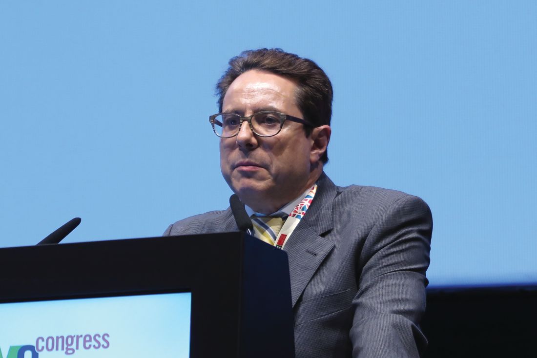
Data from an earlier analysis of the trial supported the Food and Drug Administration’s decision to approve neratinib in the extended adjuvant setting in July 2017.
In the ExteNET trial, 2,840 women with early HER2-positive breast cancer who had undergone surgery and adjuvant treatment with trastuzumab and chemotherapy were stratified by nodal and hormone receptor status and by concurrent vs. sequential chemotherapy and trastuzumab, and were then randomly assigned to receive oral neratinib 240 mg/day for 1 year, or placebo. Analyses of iDFS were planned for 2 and 5 years, and an overall survival analysis was planned after 248 patient deaths had occurred. Overall survival data have not matured as yet, Dr. Martin noted.
Results of an unspecified 3-year analysis of the trial, presented at the San Antonio Breast Cancer Symposium in 2015, showed a continued benefit for the addition of neratinib, a finding that has now been extended out to 5 years.
At ESMO 2017, Dr. Martin presented data on all efficacy endpoints except overall survival in the intention-to-treat population.
By the cutoff date in March 2017, 2,117 of the original 2,840 patients (76%) gave consent for collection of additional data, including 1,028 who had been assigned to neratinib, and 1,089 assigned to placebo.
The 5-year iDFS rate was 90.2% for patients assigned to neratinib, compared with 87.7% for those assigned to placebo, an absolute difference of 2.5%. This translated into a hazard ratio favoring neratinib of 0.73 (P = .008). Neratinib was also significantly better than placebo for DFS in patients with ductal carcinoma in situ (89.7% vs. 86.8, HR, 0.71, P = .004).
However, there were no significant differences at 5 years between trial arms in either distant DFS, time to distant recurrence, or central nervous system recurrences. Dr. Martin noted that the although there were fewer CNS recurrences with neratinib (1.30% vs. 1.82%), the total number of cases was too small to detect a possible difference.
In a subgroup analysis, neratinib trended toward better performance in all categories, but was significantly better than placebo only among patients from Asia, Eastern Europe, and South America, and among patients with four or more positive lymph nodes.
An analysis of iDFS by hormone receptor status showed that for HR-positive patients, the 5-year iDFS rate was 91.2% with neratinib vs. 86.8% with placebo, translating into a hazard ratio of 0.60, P = .002). In contrast, iDFS rates were nearly identical among HR-negative patients, at 88.9% vs. 88.8%, respectively.
Following treatment discontinuation, there was no evidence of increased symptomatic cardiotoxicity or second primary malignancies vs. placebo, and no late-term consequences of neratinib-associated diarrhea, Dr. Martin said.
A separate poster on health-related quality of life, also presented at ESMO 2017, showed that patients assigned to neratinib had a drop in quality-of-life measures during the first month of treatment, possibly because of diarrhea, but then had a steady improvement toward baseline. There is an ongoing study to evaluate whether loperamide-based regimens can reduce or prevent neratinib-associated diarrhea, the investigators noted.
“In ExteNET, we’ve seen continued demonstration of clinically significant benefit, particularly in higher-risk, hormone receptor–positive disease, despite many limitations, with change in sponsor and initial plan for only 2 years of follow-up,” said Hope S. Rugo, MD, from the University of California, San Francisco, the invited discussant.
“Survival data is pending, and we’re looking forward to seeing that in 2019, but the reduction in distant events, although small, is still encouraging,” she said.
The trial is sponsored by Puma Biotechnology. Dr. Martin disclosed honoraria from Roche/Genentech, Novartis, Amgen, AstraZeneca, Pfizer, PharmaMar, and Lilly, and research grants from Roche and Novartis. Dr Rugo disclosed travel support from PUMA and Mylan, research support from Genentech/Roche, and honoraria from Biotheranostics. She also serves on the Oncology Practice Advisory Board.
MADRID – who received postoperative trastuzumab (Herceptin) and chemotherapy, long-term follow-up results from the ExteNET trial show.
In a planned intention-to-treat analysis at 5 years of follow-up, extended adjuvant therapy with the tyrosine kinase inhibitor neratinib was associated with a small but significant improvement in invasive disease-free survival (iDFS), compared with placebo, with most of the benefit occurring in women with hormone receptor–positive disease, reported Miguel Martin, MD, of the Gregorio Marañón Health Research Institute in Madrid.
Data from an earlier analysis of the trial supported the Food and Drug Administration’s decision to approve neratinib in the extended adjuvant setting in July 2017.
In the ExteNET trial, 2,840 women with early HER2-positive breast cancer who had undergone surgery and adjuvant treatment with trastuzumab and chemotherapy were stratified by nodal and hormone receptor status and by concurrent vs. sequential chemotherapy and trastuzumab, and were then randomly assigned to receive oral neratinib 240 mg/day for 1 year, or placebo. Analyses of iDFS were planned for 2 and 5 years, and an overall survival analysis was planned after 248 patient deaths had occurred. Overall survival data have not matured as yet, Dr. Martin noted.
Results of an unspecified 3-year analysis of the trial, presented at the San Antonio Breast Cancer Symposium in 2015, showed a continued benefit for the addition of neratinib, a finding that has now been extended out to 5 years.
At ESMO 2017, Dr. Martin presented data on all efficacy endpoints except overall survival in the intention-to-treat population.
By the cutoff date in March 2017, 2,117 of the original 2,840 patients (76%) gave consent for collection of additional data, including 1,028 who had been assigned to neratinib, and 1,089 assigned to placebo.
The 5-year iDFS rate was 90.2% for patients assigned to neratinib, compared with 87.7% for those assigned to placebo, an absolute difference of 2.5%. This translated into a hazard ratio favoring neratinib of 0.73 (P = .008). Neratinib was also significantly better than placebo for DFS in patients with ductal carcinoma in situ (89.7% vs. 86.8, HR, 0.71, P = .004).
However, there were no significant differences at 5 years between trial arms in either distant DFS, time to distant recurrence, or central nervous system recurrences. Dr. Martin noted that the although there were fewer CNS recurrences with neratinib (1.30% vs. 1.82%), the total number of cases was too small to detect a possible difference.
In a subgroup analysis, neratinib trended toward better performance in all categories, but was significantly better than placebo only among patients from Asia, Eastern Europe, and South America, and among patients with four or more positive lymph nodes.
An analysis of iDFS by hormone receptor status showed that for HR-positive patients, the 5-year iDFS rate was 91.2% with neratinib vs. 86.8% with placebo, translating into a hazard ratio of 0.60, P = .002). In contrast, iDFS rates were nearly identical among HR-negative patients, at 88.9% vs. 88.8%, respectively.
Following treatment discontinuation, there was no evidence of increased symptomatic cardiotoxicity or second primary malignancies vs. placebo, and no late-term consequences of neratinib-associated diarrhea, Dr. Martin said.
A separate poster on health-related quality of life, also presented at ESMO 2017, showed that patients assigned to neratinib had a drop in quality-of-life measures during the first month of treatment, possibly because of diarrhea, but then had a steady improvement toward baseline. There is an ongoing study to evaluate whether loperamide-based regimens can reduce or prevent neratinib-associated diarrhea, the investigators noted.
“In ExteNET, we’ve seen continued demonstration of clinically significant benefit, particularly in higher-risk, hormone receptor–positive disease, despite many limitations, with change in sponsor and initial plan for only 2 years of follow-up,” said Hope S. Rugo, MD, from the University of California, San Francisco, the invited discussant.
“Survival data is pending, and we’re looking forward to seeing that in 2019, but the reduction in distant events, although small, is still encouraging,” she said.
The trial is sponsored by Puma Biotechnology. Dr. Martin disclosed honoraria from Roche/Genentech, Novartis, Amgen, AstraZeneca, Pfizer, PharmaMar, and Lilly, and research grants from Roche and Novartis. Dr Rugo disclosed travel support from PUMA and Mylan, research support from Genentech/Roche, and honoraria from Biotheranostics. She also serves on the Oncology Practice Advisory Board.
MADRID – who received postoperative trastuzumab (Herceptin) and chemotherapy, long-term follow-up results from the ExteNET trial show.
In a planned intention-to-treat analysis at 5 years of follow-up, extended adjuvant therapy with the tyrosine kinase inhibitor neratinib was associated with a small but significant improvement in invasive disease-free survival (iDFS), compared with placebo, with most of the benefit occurring in women with hormone receptor–positive disease, reported Miguel Martin, MD, of the Gregorio Marañón Health Research Institute in Madrid.
Data from an earlier analysis of the trial supported the Food and Drug Administration’s decision to approve neratinib in the extended adjuvant setting in July 2017.
In the ExteNET trial, 2,840 women with early HER2-positive breast cancer who had undergone surgery and adjuvant treatment with trastuzumab and chemotherapy were stratified by nodal and hormone receptor status and by concurrent vs. sequential chemotherapy and trastuzumab, and were then randomly assigned to receive oral neratinib 240 mg/day for 1 year, or placebo. Analyses of iDFS were planned for 2 and 5 years, and an overall survival analysis was planned after 248 patient deaths had occurred. Overall survival data have not matured as yet, Dr. Martin noted.
Results of an unspecified 3-year analysis of the trial, presented at the San Antonio Breast Cancer Symposium in 2015, showed a continued benefit for the addition of neratinib, a finding that has now been extended out to 5 years.
At ESMO 2017, Dr. Martin presented data on all efficacy endpoints except overall survival in the intention-to-treat population.
By the cutoff date in March 2017, 2,117 of the original 2,840 patients (76%) gave consent for collection of additional data, including 1,028 who had been assigned to neratinib, and 1,089 assigned to placebo.
The 5-year iDFS rate was 90.2% for patients assigned to neratinib, compared with 87.7% for those assigned to placebo, an absolute difference of 2.5%. This translated into a hazard ratio favoring neratinib of 0.73 (P = .008). Neratinib was also significantly better than placebo for DFS in patients with ductal carcinoma in situ (89.7% vs. 86.8, HR, 0.71, P = .004).
However, there were no significant differences at 5 years between trial arms in either distant DFS, time to distant recurrence, or central nervous system recurrences. Dr. Martin noted that the although there were fewer CNS recurrences with neratinib (1.30% vs. 1.82%), the total number of cases was too small to detect a possible difference.
In a subgroup analysis, neratinib trended toward better performance in all categories, but was significantly better than placebo only among patients from Asia, Eastern Europe, and South America, and among patients with four or more positive lymph nodes.
An analysis of iDFS by hormone receptor status showed that for HR-positive patients, the 5-year iDFS rate was 91.2% with neratinib vs. 86.8% with placebo, translating into a hazard ratio of 0.60, P = .002). In contrast, iDFS rates were nearly identical among HR-negative patients, at 88.9% vs. 88.8%, respectively.
Following treatment discontinuation, there was no evidence of increased symptomatic cardiotoxicity or second primary malignancies vs. placebo, and no late-term consequences of neratinib-associated diarrhea, Dr. Martin said.
A separate poster on health-related quality of life, also presented at ESMO 2017, showed that patients assigned to neratinib had a drop in quality-of-life measures during the first month of treatment, possibly because of diarrhea, but then had a steady improvement toward baseline. There is an ongoing study to evaluate whether loperamide-based regimens can reduce or prevent neratinib-associated diarrhea, the investigators noted.
“In ExteNET, we’ve seen continued demonstration of clinically significant benefit, particularly in higher-risk, hormone receptor–positive disease, despite many limitations, with change in sponsor and initial plan for only 2 years of follow-up,” said Hope S. Rugo, MD, from the University of California, San Francisco, the invited discussant.
“Survival data is pending, and we’re looking forward to seeing that in 2019, but the reduction in distant events, although small, is still encouraging,” she said.
The trial is sponsored by Puma Biotechnology. Dr. Martin disclosed honoraria from Roche/Genentech, Novartis, Amgen, AstraZeneca, Pfizer, PharmaMar, and Lilly, and research grants from Roche and Novartis. Dr Rugo disclosed travel support from PUMA and Mylan, research support from Genentech/Roche, and honoraria from Biotheranostics. She also serves on the Oncology Practice Advisory Board.
AT ESMO 2017
Key clinical point: Neratinib after adjuvant trastuzumab and chemotherapy continued to show improved invasive disease-free survival at 5 years in women with early HER2+ breast cancer.
Major finding: The 5-year iDFS rate was 90.2% for patients assigned to neratinib, compared with 87.7% for those assigned to placebo.
Data source: 5-year follow-up of randomized phase 3 trial in 2,840 women with HER2+ breast cancer treated with surgery and adjuvant chemotherapy/trastuzumab.
Disclosures: The trial is sponsored by Puma Biotechnology. Dr. Martin disclosed honoraria from Roche/Genentech, Novartis, Amgen, AstraZeneca, Pfizer, PharmaMar, and Lilly, and research grants from Roche and Novartis. Dr Rugo disclosed travel support from PUMA and Mylan, research support from Genentech/Roche, and honoraria from Biotheranostics. She also serves on the Oncology Practice Advisory Board.
Obinutuzumab edges out rituximab for PFS in follicular lymphoma
In a head-to-head trial of anti-CD20 monoclonal antibodies in first-line therapy for follicular lymphoma, obinutuzumab-based chemotherapy was associated with slightly but significantly better progression-free survival than rituximab-based therapy, but at the cost of higher toxicities, including severe adverse events.
Among 1,202 patients with follicular lymphoma followed for a median of 34.5 months, the estimated 3-year rate of progression-free survival (PFS) for patients randomized to obinutuzumab-based chemotherapy and maintenance was 80%, compared with 73.3% for patients randomized to rituximab chemotherapy and maintenance. Response rates and overall survival were similar between the treatment groups, Robert Marcus, MB, BS, of King’s College Hospital, London, and his coinvestigators reported in the GALLIUM trial.
They acknowledged, however, that there were substantial differences between the treatment groups in the cumulative doses of obinutuzumab (Gazyva) and rituximab (Rituxan and others), which could have affected the relative efficacy of each regimen.
In addition, while patients were randomly assigned to one monoclonal antibody or the other, the choice of chemotherapy regimens, while standardized, was left to the discretion of investigators at each treatment site, another factor that might have influenced outcomes.
The investigators reported the results of a preplanned interim efficacy analysis. They compared obinutuzumab or rituximab plus chemotherapy in patients with indolent non-Hodgkin lymphoma, but the trial was powered to detect a PFS difference only in patients with follicular lymphoma. Patients who had a clinical response to induction therapy went on to maintenance therapy with the same monoclonal antibody.
In all, 1,202 patients with follicular lymphoma were enrolled and randomized, 601 in each arm, to receive induction with either intravenous obinutuzumab 1,000 mg on days 1, 8, and 15 of cycle 1 and on day 1 of subsequent cycles, or rituximab 375 mg/m2 on day 1 of each cycle for six or eight cycles, depending on the accompanying chemotherapy regimen. The regimens used were either CHOP (cyclophosphamide, doxorubicin, vincristine, and prednisone), CVP (cyclophosphamide, vincristine, and prednisone), or bendamustine.
Patients with partial or complete responses were then maintained on the same monoclonal antibody they had received during induction, either obinutuzumab 1,000 mg or rituximab 375 mg/m2 every 2 months for 2 years, or until disease progression. Patients were not allowed to be crossed over to the other maintenance therapy.
Patients with stable disease after induction continued to be followed, but did not receive maintenance therapy.
The interim analysis was performed after 245 of 370 anticipated events (disease progression, relapse, or death) had occurred. At that time, the independent data and safety monitoring committee recommended full analysis of the trial data, and the sponsor agreed.
After a median follow-up of 34.5 months, an intention-to-treat analysis showed that the investigator-assessed, estimated 3-year rate of PFS was 80.0% in the obinutuzumab arm, compared with 73.3%; in the rituximab arm. This translated into a hazard ratio (HR) for progression, relapse, or death of 0.66 (P = .001). An independent review committee calculated a HR favoring obinutuzumab of 0.71 (P = .01).
Estimated 3-year overall survival rates were not significantly different at 94% and 92.1%, respectively.
Overall response rates were similar between the groups, at 88.5% with obinutuzumab group and 86.9% with rituximab, a difference that was not significant.
Obinutuzumab was associated with a higher rate of prespecified events of special interest, including infections, cardiac events, second neoplasms, infusion-related events, neutropenia, and thrombocytopenia.
Adverse events deemed to be related to the antibodies occurred in 59.3% of patients on obinutuzumab, and 48.9% of patients on rituximab.
There were more frequent grade 3 or 4 adverse events and deaths with obinutuzumab, occurring in 74.6% of patients vs. 67.8% on rituximab. Fatal adverse events occurred in 4% and 3.4% of patients, respectively.
A total of 81 patients died during the trial, including 35 in the obinutuzumab group and 46 in the rituximab group.
F. Hoffmann–La Roche supported the trial. Dr. Marcus disclosed consulting fees and lecture fees from Takeda Pharmaceuticals and travel support, consulting fees, and lecture fees from Roche. The majority of coauthors disclosed similar relationships.
Should obinutuzumab replace rituximab as the standard antibody in the treatment of patients receiving chemoimmunotherapy regimens for follicular lymphoma? Results from this trial would suggest that there might be no advantage for an obinutuzumab-containing chemoimmunotherapy regimen if maintenance treatment was not planned. Even with maintenance therapy, there is no evidence from this trial of an overall survival benefit with obinutuzumab. These findings, combined with the higher rate of toxic effects and, presumably, the higher cost of obinutuzumab, raise important questions regarding the advantage of its use. This issue is complicated further because it is possible that giving rituximab at a dose of 1,000 mg might reduce or eliminate any difference in progression-free survival – that is, if the difference is primarily a dose effect.
When the data on minimal residual disease are made available, the case in favor of obinutuzumab may appear to be more compelling if indeed a higher proportion of patients who received obinutuzumab have minimal residual disease status at some point in treatment and remain in remission longer than those who received rituximab. At the moment, the competition between these agents looks too close to call.
These comments are excerpted from an editorial (N Engl J Med. 2017 Oct 5;377;14:1389-90) by James O. Armitage, MD, University of Nebraska, Omaha, and Dan L. Longo, MD, Dana-Farber Cancer Institute, Boston. Dr. Armitage reported personal fees from Conatus, Samus Therapeutics, and Tesaro. Dr. Longo reported no relevant disclosures. He is deputy editor of The New England Journal of Medicine.
In a head-to-head trial of anti-CD20 monoclonal antibodies in first-line therapy for follicular lymphoma, obinutuzumab-based chemotherapy was associated with slightly but significantly better progression-free survival than rituximab-based therapy, but at the cost of higher toxicities, including severe adverse events.
Among 1,202 patients with follicular lymphoma followed for a median of 34.5 months, the estimated 3-year rate of progression-free survival (PFS) for patients randomized to obinutuzumab-based chemotherapy and maintenance was 80%, compared with 73.3% for patients randomized to rituximab chemotherapy and maintenance. Response rates and overall survival were similar between the treatment groups, Robert Marcus, MB, BS, of King’s College Hospital, London, and his coinvestigators reported in the GALLIUM trial.
They acknowledged, however, that there were substantial differences between the treatment groups in the cumulative doses of obinutuzumab (Gazyva) and rituximab (Rituxan and others), which could have affected the relative efficacy of each regimen.
In addition, while patients were randomly assigned to one monoclonal antibody or the other, the choice of chemotherapy regimens, while standardized, was left to the discretion of investigators at each treatment site, another factor that might have influenced outcomes.
The investigators reported the results of a preplanned interim efficacy analysis. They compared obinutuzumab or rituximab plus chemotherapy in patients with indolent non-Hodgkin lymphoma, but the trial was powered to detect a PFS difference only in patients with follicular lymphoma. Patients who had a clinical response to induction therapy went on to maintenance therapy with the same monoclonal antibody.
In all, 1,202 patients with follicular lymphoma were enrolled and randomized, 601 in each arm, to receive induction with either intravenous obinutuzumab 1,000 mg on days 1, 8, and 15 of cycle 1 and on day 1 of subsequent cycles, or rituximab 375 mg/m2 on day 1 of each cycle for six or eight cycles, depending on the accompanying chemotherapy regimen. The regimens used were either CHOP (cyclophosphamide, doxorubicin, vincristine, and prednisone), CVP (cyclophosphamide, vincristine, and prednisone), or bendamustine.
Patients with partial or complete responses were then maintained on the same monoclonal antibody they had received during induction, either obinutuzumab 1,000 mg or rituximab 375 mg/m2 every 2 months for 2 years, or until disease progression. Patients were not allowed to be crossed over to the other maintenance therapy.
Patients with stable disease after induction continued to be followed, but did not receive maintenance therapy.
The interim analysis was performed after 245 of 370 anticipated events (disease progression, relapse, or death) had occurred. At that time, the independent data and safety monitoring committee recommended full analysis of the trial data, and the sponsor agreed.
After a median follow-up of 34.5 months, an intention-to-treat analysis showed that the investigator-assessed, estimated 3-year rate of PFS was 80.0% in the obinutuzumab arm, compared with 73.3%; in the rituximab arm. This translated into a hazard ratio (HR) for progression, relapse, or death of 0.66 (P = .001). An independent review committee calculated a HR favoring obinutuzumab of 0.71 (P = .01).
Estimated 3-year overall survival rates were not significantly different at 94% and 92.1%, respectively.
Overall response rates were similar between the groups, at 88.5% with obinutuzumab group and 86.9% with rituximab, a difference that was not significant.
Obinutuzumab was associated with a higher rate of prespecified events of special interest, including infections, cardiac events, second neoplasms, infusion-related events, neutropenia, and thrombocytopenia.
Adverse events deemed to be related to the antibodies occurred in 59.3% of patients on obinutuzumab, and 48.9% of patients on rituximab.
There were more frequent grade 3 or 4 adverse events and deaths with obinutuzumab, occurring in 74.6% of patients vs. 67.8% on rituximab. Fatal adverse events occurred in 4% and 3.4% of patients, respectively.
A total of 81 patients died during the trial, including 35 in the obinutuzumab group and 46 in the rituximab group.
F. Hoffmann–La Roche supported the trial. Dr. Marcus disclosed consulting fees and lecture fees from Takeda Pharmaceuticals and travel support, consulting fees, and lecture fees from Roche. The majority of coauthors disclosed similar relationships.
FROM THE NEW ENGLAND JOURNAL OF MEDICINE
Key clinical point: Obinutuzumab-based chemotherapy and maintenance was associated with better progression-free survival, but not overall survival, compared with rituximab-based chemotherapy and maintenance.
Major finding: Three-year progression-free survival was 80% with obinutuzumab, vs. 73.3% with rituximab.
Data source: Interim analysis of a randomized phase 3, open-label trial of 1,202 patients with follicular lymphoma.
Disclosures: F. Hoffmann–La Roche supported the trial. Dr. Marcus disclosed consulting fees and lecture fees from Takeda Pharmaceuticals and travel support, consulting fees, and lecture fees from Roche. The majority of coauthors disclosed similar relationships.
Worse survival with cetuximab plus chemo for CRC liver mets
MADRID – Adding cetuximab (Erbitux) to chemotherapy before or after surgery for resectable colorectal liver metastases resulted in significantly worse survival compared with perioperative chemotherapy alone, mature results of the randomized New EPOC trial confirmed.
Among 257 patients with KRAS exon 2 wild-type resectable colorectal liver metastases (CRLM) or suboptimally resectable CRLM, median overall survival (OS) was 81 months for patients assigned to neoadjuvant and adjuvant chemotherapy alone, compared with 55.4 months for patients who received perioperative chemotherapy plus cetuximab, reported John Bridgewater, MD, of University College London Cancer Institute.
Median progression-free survival (PFS) was 22.2 months without cetuximab, vs. 15.5 months with chemotherapy plus cetuximab, a difference that was not statistically significant. However, when the primary analysis of the trial was reported in 2014 in The Lancet: Oncology, cetuximab was associated with significantly shorter PFS. The trial was halted in 2012 on the recommendation of the data monitoring committee.
“In the context of perioperative therapy for resectable CRLM, chemotherapy plus cetuximab confers a shorter OS and PPS,” Dr. Bridgewater and his associates wrote.”This is primarily in those with conventionally favorable prognostic features suggesting that cetuximab induces adverse biology in some patients, whose biomarker profile is ongoing.”
They also found that there were no differences in overall survival among patients with responses to chemotherapy according to Response Evaluation Criteria in Solid Tumors (RECIST) and those who did not have responses, “suggesting that in this predominantly operable population, any conferred benefit of systemic treatment is through elimination of micro-metastatic disease rather than by downsizing of radiologically evaluable disease.
The randomized phase 3 Medical Research Council COIN trial, results of which were reported at the 2010 annual meeting of the American Society of Clinical Oncology and later published in The Lancet, failed to show a benefit for the addition of cetuximab to front-line oxaliplatin-based chemotherapy for metastatic CRC. Median OS in KRAS wild-type patients was 17 months when cetuximab was added to chemotherapy with oxaliplatin and an intravenous or oral fluoropyrimidine, compared with 17.9 months when chemotherapy was delivered without cetuximab. Median PFS held at 8.6 months in both arms of the trial.
The New EPOC investigators described their study as a “natural extension” of the COIN study as well as earlier trials and phase 2 studies of neoadjuvant and adjuvant therapy in this patient population.
From 2007 through 2012, 257 patients were randomly assigned to receive chemotherapy alone, or chemotherapy with rituximab. Chemotherapy consisted of one of two regimens: oxaliplatin 85 mg/m2 intravenously over 2 hours and fluorouracil bolus 400 mg/m2 intravenously over 5 minutes followed by a 46-hour infusion of fluorouracil 2,400 mg/m2 repeated every 2 weeks; or oxaliplatin 130 mg/m2 intravenously over 2 hours and oral capecitabine (Xeloda) 1,000 mg/m2 twice daily on days 1-14 repeated every 3 weeks.
After a median follow-up of 69 months, there were 130 deaths from any cause. As noted before, median OS was significantly shorter with cetuximab. The hazard ratio for death with cetuximab was 1.45 (P = .035). Median PFS did not differ in this most recent analysis, however.
Median PPS was 35.4 months for chemotherapy alone, compared with 23.5 months for chemotherapy plus cetuximab (P = .014).
The poor overall survival and PFS results indicate that “cetuximab cannot be recommended for perioperative treatment in patients with resectable disease,” said Thomas Gruenberger, MD, an oncologic surgeon at Rudolf Hospital in Vienna, who was invited to review the results in a poster discussion session.
Cancer Research UK supported the study. Dr. Bridgewater disclosed honoraria and speakers fees from Merck, Celgene, and Servier, and travel support from Amgen, Merck Sharpe Dohme, and Servier.
AT ESMO 2017
Key clinical point: Patients with resectable colorectal liver metastases (CRLM) fared worse when cetuximab was added to neoadjuvant and adjuvant chemotherapy.
Major finding: Median overall survival was 81 months for patients assigned to chemotherapy alone, vs. 55.4 months for patients assigned to chemotherapy plus cetuximab.
Data source: Mature analysis of a randomized phase 3 trial in 257 patients with resectable CRLM. The trial was halted for futility in 2012.
Disclosures: Cancer Research UK supported the study. Dr. Bridgewater disclosed honoraria and speakers fees from Merck, Celgene, and Servier, and travel support from Amgen, Merck Sharpe Dohme, and Servier.
Robot-assisted abdominoperineal resection outperforms open or laparoscopic surgery for rectal cancers
MADRID – The automaton uprising continues: Robot-assisted abdominoperineal resection (APR) can be safely performed in patients with rectal cancers within 5 cm of the anal verge, with surgical results equivalent to those seen with open or laparoscopic APR, investigators say.
Robot-assisted procedures were associated with a significantly lower rate of postoperative complications and with faster functional recovery than either laparoscopic or open surgery in a randomized trial, reported Jianmin Xu, MD, PhD., from Fudan University in Shanghai, China, and colleagues in a scientific poster presented at the European Society for Medical Oncology Congress.
“Retrospective studies have showed that robotic surgery was better than laparoscopic surgery in ensuring radical resection, reducing complications, and promoting recovery. However, there is no clinical trial reported for robotic surgery for rectal cancer. Thus, we conduct this randomized controlled trial to compare the safety and efficacy of robotic, laparoscopic, and open surgery for low rectal cancer,” they wrote.
Dr. Xu and colleagues enrolled 506 patients from 18 to 75 years of age with clinical stage T1 to T3 cancers within 5 cm of the anal verge and no distant metastases and randomly assigned them to resection with either a robot-assisted, laparoscopic, or open APR technique. Three of the 506 patients randomized did not undergo resection, leaving 503 for the per-protocol analysis presented here.
For the primary endpoint of complications rates within 30 days following surgery, the investigators found that patients assigned to robotic-assisted surgery (173 patients) had a total complication rate of 10.4%, compared with 18.8% for 176 patients assigned to laparoscopy (P = .027), and 26% for 154 assigned to open APR (P less than .001). The latter group included four patients assigned to laparoscopy whose procedures were converted to open surgery.
Among patients without complications, robot-assisted procedures were also associated with faster recovery, as measured by days to first flatus, at a median of 1 vs. 2 for laparoscopy and 3 for open procedures (P less than .001 for robots vs. each other surgery type). The robotic surgery was also significantly associated with fewer days to first automatic urination (median 2 vs. 4 for each of the other procedures; P less than .001), and with fewer days to discharge (median 5 vs. 7 for the other procedures; P less than .001).
"These are excellent postoperative complication rates reported, especially for the robotic treatment group, commented Thomas Gruenberger, MD, an oncologic surgeon at Rudolf Hospital in Vienna, in a poster discussion session.
“We all are now favoring laparoscopic surgery for these kinds of patients. The robot is a nice thing to have; however, we cannot use it in every hospital because it’s still quite expensive,” he said.
“We require – and this is a secondary endpoint of the study – long-term follow-up for local and distant outcomes,” he added,
The investigators did not report a funding source. All authors declared having no conflicts of interest. Dr. Gruenberger disclosed research funding, speakers bureau participation, and/or advisory roles with several companies.
AT ESMO 2017

