User login
CAR designers report high B-cell cancer response rates
ORLANDO – Patients with advanced hematologic malignancies of B-cell lineage had robust immune responses following infusion of a chimeric antigen receptor (CAR)–T-cell construct designed to deliver a specific balance of antigens, investigators reported.
Adults with relapsed or refractory B-lineage acute myeloid leukemia (ALL), non–Hodgkin lymphoma (NHL), and chronic lymphocytic leukemia (CLL) who received a CAR-T cell construct consisting of autologous CD4-positive and CD-8-positive T cells that were transduced separately, recombined, and then delivered in a single infusion had comparatively high overall response and complete response rates, reported Cameron Turtle, MBBS, PhD, from the Fred Hutchinson Cancer Research Center in Seattle.
“We know that patients have a highly variable CD4 to CD8 ratio, so by actually controlling this and separately transducing, expanding, and then reformulating in this defined composition, we’re able to eliminate one source of variability in CAR-T cell products,” Dr. Turtle said at the ASCO-SITC Clinical Immuno-Oncology Symposium.
In preclinical studies, an even balance of CD4-positive and CD8-positive central memory T cells or naive T cells evoked more potent immune responses against B-cell malignancies in mice than CD19-positive cells, he explained
To see whether this would also hold true in humans, the investigators enrolled into a phase I/II trial adults with relapsed/refractory B-cell malignancies, including ALL (36 patients), NHL (41), and CLL (24). No patients were excluded on the basis of either absolute lymphocyte, circulating tumors cells, history of stem cell transplant, or results of in vitro test expansions.
All patients underwent leukapheresis for harvesting of T-cells, and populations of CD4- and CD8-positive cells were separated and transduced with a lentiviral vector to express a CD19 CAR and a truncated human epidermal growth factor receptor that allowed tracing of the transduced cells via flow cytometry. The patients underwent lymphodepleting chemotherapy with cyclophosphamide (for the earliest patients), or cyclophosphamide plus fludarabine. Fifteen days after leukapheresis, the separated, transduced, and expanded cells were combined and delivered back to patients in a single infusion at one of three dose levels: 2 x 10⁵, 2 x 10⁶, or 2 x 10⁷ CAR-T cells/kg.
ALL results
Two of the 36 patients with ALL died from complications of the CAR-T cell infusion process prior to evaluation. The 34 remaining patients all had morphologic bone marrow complete responses (CR). Of this group, 32 also had bone marrow CR on flow cytometry.
Using immunoglobulin H (IgH) deep sequencing in a subset of 20 patients 3 weeks after CAR-T cell infusion, the investigators could not detect the malignant IgH index clone in 13 of the patients, and found fewer than 10 copies in the bone marrow of 5 patients.
Six of seven patients with extramedullary disease at baseline had a complete response. The remaining patient in this group had an equivocal PET scan result, and experienced a relapse 2 months after assessment.
The investigators also determined that the lymphodepletion regimen may affect overall results, based on the finding that 10 of 12 patients who received cyclophosphamide alone achieved a CR, but seven of these 10 patients had a relapse within a few months. Of these seven patients. five received a second T-cell infusion, but none had significant T-cell expansion. The investigators traced the failure of the second attempt to a CD8-mediated transgene immune response to a murine single-chain variable fragment used in the construct.
For subsequent patients, they altered the lymphodepletion regimen to include fludarabine to prevent priming of the anti-CAR transgenic immune response. This modification resulted in improved progression-free survival and overall survival for subsequent patients receiving a second infusion, Dr. Turtle said.
NHL results
Of the 41 patients with NHL, 30 (73%) had aggressive histologies, including diffuse large B-cell lymphoma, primary mediastinal large B-cell lymphoma, T-cell/histiocyte-rich large B-cell, and Burkitt lymphomas, and 11 (27%) had indolent histologies, including mantle cell and follicular lymphomas. Most of the patients had received multiple prior lines of therapy, and 19 (46%) had undergone either an autologous or allogeneic stem cell transplant.
Of the 39 evaluable patients who completed therapy, the overall response rate was 67%, including 13 (39%) with CR. Dr. Turtle noted that the CR rate was substantially higher among patients who received cyclophosphamide and fludarabine lymphodepletion, compared with cyclophosphamide alone.
There were also a few responses, including two CRs, among patients with indolent histologies, he said.
CLL, safety results
All 24 patients with CLL had previously received ibrutinib (Imbruvica). Of this group, 19 either had no significant responses to the drug, inactivating mutations, or intolerable toxicities. All but 1 of the 24 patients also had high-risk cytogenetics.
Of the 16 ibrutinib-refractory patients who were evaluable for restaging, 14 had no evidence of disease in bone marrow by flow cytometry at 4 weeks. The overall response rate in this group was 69%, which included four CRs.
Among a majority of all patients, toxicity with the CAR-T cell therapy was mild to moderate. Early cytokine changes appeared to be predictive of serious adverse events such as the cytokine release syndrome, a finding that may allow clinicians to intervene early to prevent complications, Dr. Turtle said.
In the CAR-T cell therapy, “multiple things affect the response and toxicity, including CAR T-cell dose, disease burden, the anti-CAR transgene immune response and the lymphodepletion regimen, not to mention other patient factors that we’re still sorting out,” he commented.
The trial was funded by the National Institutes of Health, Life Science Development Fund, Juno Therapeutics and the Bezos Family Foundation. Dr. Turtle disclosed consultancy, honoraria, and/or research funding from Juno Therapeutics and Seattle Genetics.
AT THE CLINICAL IMMUNO-ONCOLOGY SYMPOSIUM
Key clinical point: A defined CAR-T cell construct was associated with high response rates in patients with B-cell malignancies.
Major finding: The overall response rate among patients with ibrutinib-refractory chronic lymphocytic leukemia was 69%, including four complete responses.
Data source: Phase I/II dose-finding, safety and efficacy study in patients with B-lineage hematologic malignancies
Disclosures: The trial was funded by the National Institutes of Health, Life Science Development Fund, Juno Therapeutics and the Bezos Family Foundation. Dr. Turtle disclosed consultancy, honoraria, and/or research funding from Juno Therapeutics and Seattle Genetics.
Vaccine + chemo induce robust T-cell responses in late-stage cervical cancer
ORLANDO – In patients with advanced cervical cancer, combining chemotherapy with a vaccine against human papillomavirus (HPV) type 16 resulted in a robust, T-cell–mediated immune response and long duration survival for a large proportion of patients, reported investigators from the Netherlands.
Patients with advanced, HPV16-positive cervical cancer who were treated with standard chemotherapy and vaccinated with HPV16 synthetic long peptides (HPV16-SLP) had substantially increased T-cell responses, and these responses correlated with survival, said Marij Welters, PhD, from Leiden (the Netherlands) University Medical Center.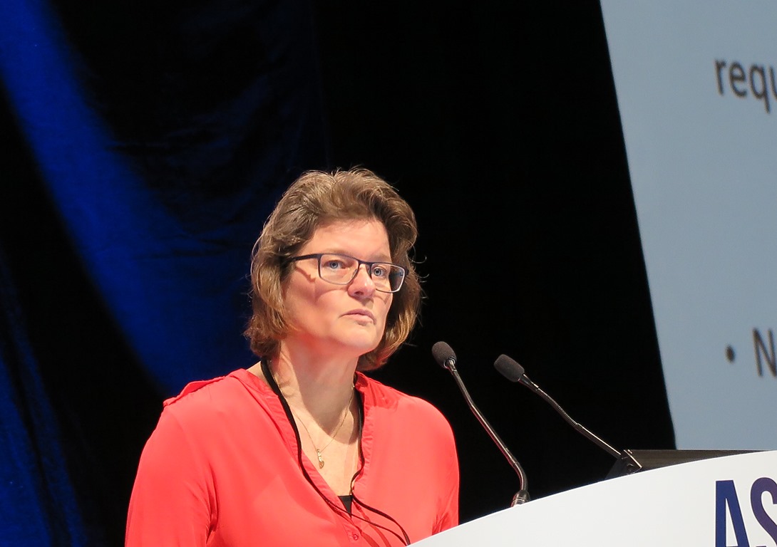
Data from this study provide “a strong rationale to conduct a randomized phase II trial in which a combination with checkpoint inhibitors might be attractive,” she added.
Combination required
Although therapeutic vaccination with HPV16-SLP has been shown to evoke T-cell–mediated shrinkage of HPV16-induced cervical neoplasia, the investigators found in a previous study that vaccination did not result in either tumor regression or prolonged progression-free survival of patients with advanced or recurrent HPV16-induced cervical cancer.
In the phase I trial reported here, the investigators explored whether combination HPV16-SLP with chemotherapy could potentiate T-cell responses.
In a second study, Welters et al. also showed that vaccination with HPV16-SLP after the second round of chemotherapy, when myeloid cells were at their nadir, resulted in robust T-cell responses in both mice and humans.
In the phase I trial reported here, the authors reported on the feasibility and efficacy of the technique in a larger cohort of patients with late-stage, HPV16-positive cervical cancer.
The investigators enrolled cohorts of 12 patients each and delivered one HPV16-SLP vaccine dose 2 weeks after the second, third, and fourth cycles of a total of six chemotherapy cycles with carboplatin and paclitaxel. The vaccine was test dosed at levels of 20, 40, 100, and 300 mcg/peptide, with or without 1 mcg/kg of pegylated interferon-alpha at each peptide dose level. The peptides covered the length of the HPV16 E6 and E7 proteins.
“Upon vaccination, we see a very strong T-cell response induced by the vaccine,” Dr. Welters said.
They also tested general immune responses to various microbial antigens and saw no significant differences in responses among the various dose cohorts.
Early response data was available for a total of 59 patients, 35 of whom received the vaccine/chemotherapy combination as first-line therapy and 24 of whom received it in the second line.
Two of the first-line patients had a complete response, as did one patient who received the combination as second-line therapy. Respective rates of partial responses were 22 and 5, stable disease was seen in 9 and 13 patients, and disease progression in 2 and 5 patients.
The overall response rate for patients treated in the first line was 69%, and the combined overall response and stable-disease rates were 94%. Among patients treated in the second line, the respective rates were 25% and 79%.
Median overall survival (OS) from the time of the first chemotherapy dose was 16.8 months among the first-line patients vs. 7.9 months among second-line patients (P = .0110)
At the data cutoff, median OS had not been reached for the two highest peptide dose levels (100 and 300 mcg).
The investigators also found that OS was independent of general immune status among the patients, suggesting that the benefit was derived specifically from induced T-cell responses.
‘Provocative and promising’
Invited discussant Heather McArthur, MD, MPH, from Cedars-Sinai Medical Center, Los Angeles, called the finding “provocative and promising.”

“So, it doesn’t matter if your immune system is suppressed overall. It’s the quality of players on the field and the focus and specificity of those players that matter, and that’s what they were able to demonstrate, which I think is incredibly powerful,” she said.
She noted, however, that although safety was listed as a study endpoint, Dr. Welters did not provide data on toxicities.
The trial was supported by the Dutch Cancer Society and ISA Pharmaceuticals BV. Dr. Welters reported no conflicts of interest. Dr. McArthur has previously disclosed participation in advisory boards for Celgene, Merck, Spectrum Pharmaceuticals, OBI Pharma, Peregrine Pharmaceuticals, and Syndax Pharmaceuticals, and research support from Bristol-Myers Squibb, MedImmune/AstraZeneca, Eli Lilly, ZIOPHARM Oncology, and Merck.
ORLANDO – In patients with advanced cervical cancer, combining chemotherapy with a vaccine against human papillomavirus (HPV) type 16 resulted in a robust, T-cell–mediated immune response and long duration survival for a large proportion of patients, reported investigators from the Netherlands.
Patients with advanced, HPV16-positive cervical cancer who were treated with standard chemotherapy and vaccinated with HPV16 synthetic long peptides (HPV16-SLP) had substantially increased T-cell responses, and these responses correlated with survival, said Marij Welters, PhD, from Leiden (the Netherlands) University Medical Center.
Data from this study provide “a strong rationale to conduct a randomized phase II trial in which a combination with checkpoint inhibitors might be attractive,” she added.
Combination required
Although therapeutic vaccination with HPV16-SLP has been shown to evoke T-cell–mediated shrinkage of HPV16-induced cervical neoplasia, the investigators found in a previous study that vaccination did not result in either tumor regression or prolonged progression-free survival of patients with advanced or recurrent HPV16-induced cervical cancer.
In the phase I trial reported here, the investigators explored whether combination HPV16-SLP with chemotherapy could potentiate T-cell responses.
In a second study, Welters et al. also showed that vaccination with HPV16-SLP after the second round of chemotherapy, when myeloid cells were at their nadir, resulted in robust T-cell responses in both mice and humans.
In the phase I trial reported here, the authors reported on the feasibility and efficacy of the technique in a larger cohort of patients with late-stage, HPV16-positive cervical cancer.
The investigators enrolled cohorts of 12 patients each and delivered one HPV16-SLP vaccine dose 2 weeks after the second, third, and fourth cycles of a total of six chemotherapy cycles with carboplatin and paclitaxel. The vaccine was test dosed at levels of 20, 40, 100, and 300 mcg/peptide, with or without 1 mcg/kg of pegylated interferon-alpha at each peptide dose level. The peptides covered the length of the HPV16 E6 and E7 proteins.
“Upon vaccination, we see a very strong T-cell response induced by the vaccine,” Dr. Welters said.
They also tested general immune responses to various microbial antigens and saw no significant differences in responses among the various dose cohorts.
Early response data was available for a total of 59 patients, 35 of whom received the vaccine/chemotherapy combination as first-line therapy and 24 of whom received it in the second line.
Two of the first-line patients had a complete response, as did one patient who received the combination as second-line therapy. Respective rates of partial responses were 22 and 5, stable disease was seen in 9 and 13 patients, and disease progression in 2 and 5 patients.
The overall response rate for patients treated in the first line was 69%, and the combined overall response and stable-disease rates were 94%. Among patients treated in the second line, the respective rates were 25% and 79%.
Median overall survival (OS) from the time of the first chemotherapy dose was 16.8 months among the first-line patients vs. 7.9 months among second-line patients (P = .0110)
At the data cutoff, median OS had not been reached for the two highest peptide dose levels (100 and 300 mcg).
The investigators also found that OS was independent of general immune status among the patients, suggesting that the benefit was derived specifically from induced T-cell responses.
‘Provocative and promising’
Invited discussant Heather McArthur, MD, MPH, from Cedars-Sinai Medical Center, Los Angeles, called the finding “provocative and promising.”
“So, it doesn’t matter if your immune system is suppressed overall. It’s the quality of players on the field and the focus and specificity of those players that matter, and that’s what they were able to demonstrate, which I think is incredibly powerful,” she said.
She noted, however, that although safety was listed as a study endpoint, Dr. Welters did not provide data on toxicities.
The trial was supported by the Dutch Cancer Society and ISA Pharmaceuticals BV. Dr. Welters reported no conflicts of interest. Dr. McArthur has previously disclosed participation in advisory boards for Celgene, Merck, Spectrum Pharmaceuticals, OBI Pharma, Peregrine Pharmaceuticals, and Syndax Pharmaceuticals, and research support from Bristol-Myers Squibb, MedImmune/AstraZeneca, Eli Lilly, ZIOPHARM Oncology, and Merck.
AT THE CLINICAL IMMUNO-ONCOLOGY SYMPOSIUM
Key clinical point: An HPV16 peptide vaccine, combined with chemotherapy, induced immune response in patients with advanced cervical cancer.
Major finding: The overall response rate for chemotherapy-naive patients treated with the combination of chemotherapy and an HPV16 synthetic long peptide vaccine was 69%.
Data source: Phase I dose-finding trial with response data on 59 patients with advanced cervical cancer.
Disclosures: The trial was supported by the Dutch Cancer Society and ISA Pharmaceuticals BV. Dr. Welters reported no conflicts of interest. Dr. McArthur has previously disclosed participation in advisory boards for Celgene, Merck, Spectrum Pharmaceuticals, OBI Pharma, Peregrine Pharmaceuticals, and Syndax Pharmaceuticals and research support from Bristol-Myers Squibb, MedImmune/AstraZeneca, Eli Lilly, ZIOPHARM Oncology, and Merck.
Staying the course after first progression yields better mRCC survival
AMSTERDAM – Patients with metastatic renal cell carcinoma (mRCC) who experience disease progression in one or more metastatic sites while on treatment with a targeted therapy may still benefit from staying on the same drug rather than switching to another following locoregional treatment, results of a retrospective study suggest.
Among 55 patients with RCC, those who continued on the same targeted therapy after locoregional treatment of a site of progression had significantly longer post–first oligoprogression overall survival (PFOPOS) than patients who had been switched to another targeted agent, reported Della De Lisi, MD, from the University of Rome and colleagues.
“Locoregional treatments represent an option for oligometastatic mRCC treated with targeted therapy. Continuing the same systemic treatment after radical locoregional treatment in one or more metastatic site[s] appear[s] to be an independent predictive factor of better outcome in this subset of patients. Bone oligoprogressive mRCC showed similar better outcome[s].” they wrote in a poster presented at an annual congress sponsored by the European Cancer Organisation.
One option for patients with mRCC with slow or limited metastatic progression is locoregional therapy with radical intent, with the goal of achieving a complete response. When a patient’s disease progresses while on a targeted agent such as sorafenib (Nexavar) or sunitinib(Sutent), he or she may be switched to a different agent, but there is a lack of data on outcomes with this strategy, the authors said.
To see whether sticking with the same therapy or switching to another could be the wiser course, they took a retrospective look at outcomes for 55 patients with mRCC who had disease progression after at least 6 months of a first-line therapy in one or more sites treated radically with locoregional therapy.
The majority of patients (52 of 55; 94.5%) had clear-cell histology tumors. Slightly more than half (31 patients, 56.4%) had good risk disease according to the Memorial Sloan Kettering Cancer Center kidney cancer risk prediction tool, and 23 (41.8%) had intermediate risk. The risk category was not calculable for the one remaining patient.
In all, 36 patients (65.5%) did not have evidence of metastasis at diagnosis. All patients had oligoprogression in a single site. The most common metastatic sites were to lung in 15 patients, bone in 10, kidney in 8, brain in 4, and liver in 4 (other sites not listed).
Forty-eight patients received sunitinib in the first line, five received pazopanib (Votrient), and two received sorafenib. Locoregional therapy at the site of progression was radiotherapy in 25 patients (45.5%), surgery in 25, and cryoablation or thermoablation in 5.
The majority of patients (48; 83.6%) remained on the same tyrosine kinase inhibitor (TKI) after locoregional therapy, while 7 were switched to another agent. Of this latter group, four patients were switched to a different TKI, and three were started on a mammalian target of rapamycin (mTOR) inhibitor.
For all patients, the median PFOPOS was 37 months. However, comparing patients who continued the same therapy after locoregional treatment with those who switched, the investigators found a significant survival advantage to sticking with the same therapy, with a median PFOPOS of 39 months, compared with 11 months for patients who were switched to another agent (P = .014)
Other factors contributing to improved survival were good vs. intermediate risk score (39 vs. 29 months; P = .036), metastases to bone vs. viscera (median PFOPOS not reached, vs. 31 months; P = .045), and Fuhrman grade 1 and 2 vs. grade 3 and 4 (57 vs. 37 months; P = .021).
Switching therapies after first progression was an independent risk factor for poor prognosis in a multivariate analysis (hazard ratio 6.280, P = .007).
An analysis of progression-free survival (PFS) after first oligoprogression showed an overall PFS of 14 months. There were no statistically significant differences in terms of post-progression PFS between patients who stayed on the same therapy or were switched, however (15 vs. 7 months, P = .207).
The study was sponsored by participating institutions. The authors reported no conflicts of interest.
AT ECCO 2017
Key clinical point: Patients with metastatic renal cell carcinoma (mRCC) who stayed on the same targeted therapy following locoregional treatment after first progression had better overall survival than those who were switched to another drug.
Major finding: Median post–first oligoprogression overall survival was 39 months for patients who stayed on the same drug, compared with 11 months for patients who were switched (P = .014).
Data source: Retrospective review of outcomes for 55 patients with mRCC treated with targeted therapy and locoregional treatment of metastases.
Disclosures: The study was sponsored by participating institutions. The authors reported no conflicts of interest.
Cabozantinib versus everolimus in advanced RCC with bone mets
AMSTERDAM – Among patients with advanced renal cell carcinoma (RCC) with metastases to bone, cabozantinib (Cabometyx) was associated with better survival compared with everolimus (Afinitor), according to a subanalysis of data from the METEOR trial.
After 2 years of follow-up, median progression-free survival (PFS), overall survival (OS), and objective response rates (ORR) were significantly better for patients with bone metastases who received cabozantinib compared with those who received everolimus, reported Sergio Bracarda, MD, of Presidio Ospedaliero San Donato, Italy, and his colleagues.
“Cabozantinib is a new treatment option for previously-treated patients with advanced RCC with benefits that are maintained in patients with bone metastases,” they wrote in a poster presented at an annual congress sponsored by the European Cancer Organisation.
Previous studies have shown that patients with advanced RCC with bone metastases have generally poor prognosis compared with patients without bone metastases, the authors noted.
As previously reported, the METEOR trial, a randomized phase III study of 658 patients with advanced RCC, showed a significant survival advantage for patients treated with cabozantinib, with a median OS of 21.4 months compared with 16.5 months for patients treated with everolimus, with a hazard ratio (HR) of 0.66 (P = .0003).
In the current sub-analysis, the investigators looked at a subgroup of 142 patients with bone metastases at baseline as seen on CT or MRI. They conducted an exploratory analysis of bone scan response among 162 patients evaluated for bone lesions at baseline by technetium bone scans, and compared the incidence of skeletal-related events (SREs) for 181 patients with a history of SREs, and 477 with no prior SREs. SREs included pathological fractures, spinal cord compression, surgery to bone, and external radiation therapy to bone.
Patients underwent CT or MRI screening every 8 weeks for the first 12 months post randomization, then every 12 weeks thereafter. All patients were screened with technetium bone scans every 18 weeks for the first years, and those patients with bone lesions at baseline were followed with additional scans every 24 weeks.
The authors also looked at serum bone biomarkers, including bone-specific alkaline phosphatase (BSAP), N-terminal propeptide of type 1 collagen (P1NP), and C-terminal cross-linked telopeptides of type I collagen.
The median PFS for patients with bone metastases treated with cabozantinib was 7.4 months, compared with 2.7 months for everolimus (HR 0.33, 95% confidence interval [CI] 0.21-0.51). For patients with both bone and visceral metastasis, median PFS was 5.6 months vs. 1.9 months, respectively (HR 0.26, 95% CI, 0.16-0.43).
Median OS for the cabozantinib group was 20.1 months compared with 12.1 months for everolimus (HR 0.54, 95% CI, 0.34-0.84) for patients with bone metastases alone. For patients with both bone and visceral metastases, median OS was 20.1 months with cabozantinib, and 10.7 months with everolimus (HR 0.45, 95% CI, 0.28-0.72).
The ORR with cabozantinib as rated by an independent radiology committee was 17% for patients with bone metastases alone, and 20% for patients with bone and visceral metastases. In contrast, there were no objective responses seen in patients treated with everolimus.
Bone scan responses, defined as a 30% or greater decrease from baseline in bone scan lesion area, were seen in 18% of patients on cabozantinib vs. 10% with everolimus (significance not shown).
Among patients with a history of SREs, 22% had an SRE on cabozantinib, compared with 31% on everolimus. Respective rates among patients without a prior history of SREs were 27% and 15%. At least one SRE occurred in 12% (cabo) and 14% (eve) of patients, including four (cabo) and eight (eve) cases of spinal cord compression. For patients with a history of SREs at randomization, the incidence of postrandomization SREs was 16% (cabo) and 34% (eve) and included zero (cabo) and five (eve) cases of spinal cord compression. Reductions in the bone markers P1NP and CTx were greater with cabo vs. eve. The most common adverse events in patients with bone metastases were consistent with those observed in the overall study population.
The investigators noted that “the safety profile of cabozantinib in the bone metastases subgroup was consistent with the safety profile in the overall population.”
Dr. Bracarda has served as a consultant to Exelixis, which supported the trial and subanalysis. Two coauthors are employees of the company.
AT ECCO2017
Key clinical point: Survival among patients with advanced renal cell carcinoma metastatic to bone was better with cabozantinib than everolimus.
Major finding: Median overall survival for the cabozantinib group was 20.1 months compared with 12.1 months for everolimus.
Data source: Subanalysis of 142 patients with bone metastases in the randomized phase III METEOR trial.
Disclosures: Dr. Bracarda has served as a consultant to Exelixis, which supported the trial and subanalysis. Two coauthors are employees of the company.
Fractures in adult osteosarcoma patients presage worse survival
AMSTERDAM – Pathological fractures are prognostic of poor outcomes in adults with osteosarcoma, but not in children with osteosarcoma, investigators have found.
A retrospective review of data on consecutive patients treated over the course of 30 years showed that among patients of all ages, both 5-year and 10-year overall survival (OS) rates were worse among patients who had pathological fractures.
But in an analysis stratified by age, the survival difference attributable to fractures was limited entirely to patients who were 18 or older at the time of an osteosarcoma diagnosis, reported Lisa Kelley, a medical student at the Ludwig-Maximilians Universität in Munich.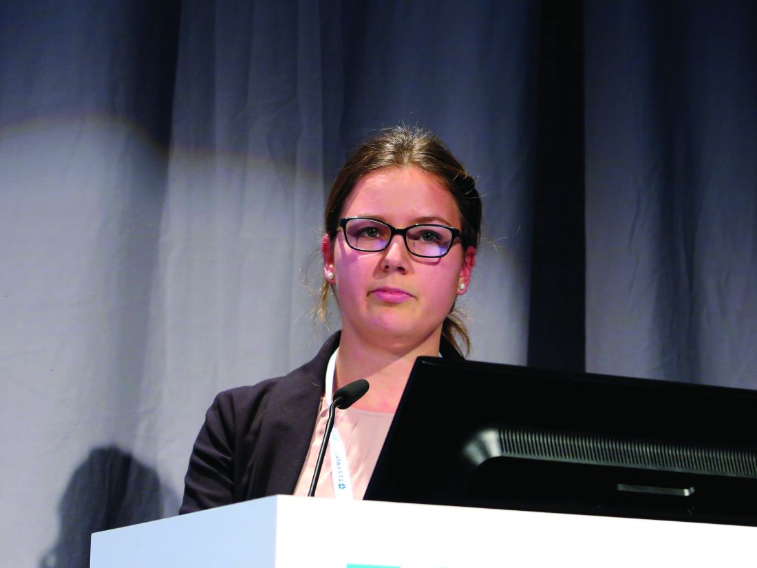
“It’s important to understand what are the prognostic factors [in osteosarcoma], and while many of the factors have been thoroughly researched, on pathological fractures the data are still fairly inconclusive due to the results of studies contradicting each other, and also having a relatively small patient number due to the fact that osteosarcoma itself is rare, and the pathological fractures occur only in approximately 10% of cases,” she said at an annual congress sponsored by the European Cancer Organisation.
To get a better handle on possible correlations between pathological fractures and prognosis in patients with central high-grade osteosarcoma, Ms. Kelley and her coauthors collected data on consecutive patients treated for localized or metastatic osteosarcoma of the extremities from 1980 through 2010 at one of the member institutions of the Cooperative Osteosarcoma Study Group (COSS).
They identified 2,847 patients, of whom 2,193 (77%) were 18 or younger at the time of diagnosis. Of the entire cohort, 321 patients (11.3%) had a pathological fracture either at presentation or soon after diagnosis.
Comparing patients with and without pathological fractures, the investigators found that factors significantly associated with fracture risk included tumor location, especially the humerus (P less than .001), tumors occurring proximally and in a diaphysis (P less than .001), telangiectatic subtype (P less than .001), the presence of primary metastases (P = .025). and tumors comprising more than one-third of the affected bone (P less than .001).
There were no significant differences in the cohort as a whole between patients with or without fractures in either age in years, sex, body-mass index, history of pain or swelling symptoms, local surgical remission at the main tumor site, total surgical remission including metastases, response to chemotherapy, use of adjuvant chemotherapy rather than neoadjuvant, or type of surgery.
Among adult patients only, however, factors associated with pathological fractures included age (P less than .001). BMI (P = .021), tumor site (P less than .001), histologic subtype (P less than .001), primary metastases (P = .011), relative tumor size (P = .047), and total surgical remission (P = .015).
Among pediatric patients, factors associated with fracture risk were (P less than .001 for all unless otherwise specified) age, BMI (P = .018), history or symptoms (P = .001). tumor site, localization within bone, histologic subtype, and relative tumor size.
In univariate analysis, 5-year OS rates were 70.6% for patients without fractures compared with 63.0% for those with fractures, and respective 10-year OS rates were 64.9% vs. 58.1% (P = .007 for both comparisons).
Among pediatric patients, the Kaplan-Meier survival curves of patients with and without pathological fractures overlapped. But for adult patients, survival of those with fractures was significantly worse, with 5-year OS of 69.3% with no fractures vs. 45.9% with fractures, and respective 10-year OS rates of 62.1% vs. 36.8% (P less than .001 for both comparisons).
Also in univariate analysis, 5-year and 10-year event-free survival (EFS) rates were significantly lower for patients who experienced pathological fractures.
Finally, in multivariable analysis of overall survival by age group, the investigators found that among adults pathological fracture was associated with a nearly twofold risk for death (hazard ratio [HR] 1.893, P = .013). Other factors were primary metastases (HR 2.486, P = .001), response to chemotherapy (P less than .001) and total surgical remission (P less than .001).
As noted before, pathological fracture among pediatric patients was not associated with worse OS. Factors significantly associated with OS in younger patients were primary metastases (HR 2.187, P less than .001), relative tumor size (HR 1.239, P = .024), response to chemotherapy (HR 2.295, P less than .001), total surgical remission (HR 4.253, P less than .001), and type of surgery (HR 1.282, P = .008).
In contrast, pathological fracture was not associated with EFS among either adult or pediatric patients. Factors significantly associated with EFS in both children and adults were primary metastases and response to chemotherapy. Among pediatric patients only, tumor site and relative tumor size were also associated with EFS.
Ms. Kelley acknowledged that the study was limited by the retrospective design.
She said that exploration of the discrepancies between adult and pediatric patients in regard to the influence of pathological fracture on survival and of the role of pathologic fracture as a negative prognostic factor for OS but not EFS in adults is warranted.
The study was funded by the Wilhelm Sander-Stiftung Foundation for cancer research. Ms. Kelley reported having no conflicts of interest.
AT ECCO2017
Key clinical point: Pathological fractures are associated with worse survival of adults but not children with osteosarcoma of the extremities.
Major finding: Among adults pathological fracture was associated with a nearly twofold risk for death (hazard ratio, 1.893, P = .013).
Data source: Retrospective review of data on 2,847 consecutive patients with osteosarcoma.
Disclosures: The study was funded by the Wilhelm Sander-Stiftung Foundation for cancer research. Ms. Kelley reported having no conflicts of interest.
PD-1, PD-L1 expression in sarcomas is subtype dependent
AMSTERDAM – Immune checkpoint inhibitors targeted against programmed death–1 (PD-1) and its ligand (PD-L1) show strong activity against several different tumor types, but unfortunately may have only limited efficacy against soft tissue and bone sarcomas, investigators reported.
“We found that the clinical expression of PD-1, PD-L1, and CD8 is sarcoma subtype dependent,” said Anke van Erp, a PhD candidate in oncology at Radboud University Medical Center in Nijmegen, the Netherlands.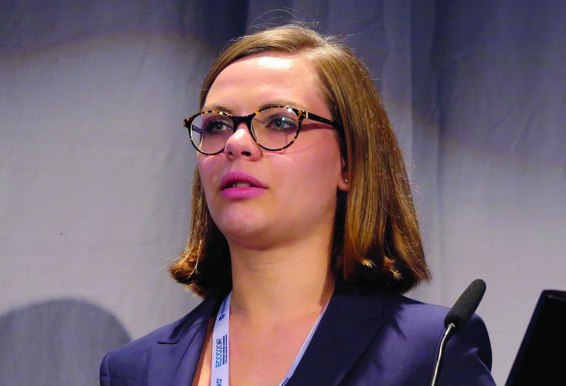
“The introduction of PD-1 antibodies has led to increased interest in the expression of PD-1, PD-L1, and CD8 in a large variety of tumor types, and has shown in several of these tumor types that PD-L1 could be associated with a worse prognosis. In addition, treatment with PD-1 antibodies has shown impressive effects in an increasing number of adult tumors. This makes the exploration of PD-1 blockade in sarcoma of interest,” she said.
Additionally, CD8-positive T cells are of interest because they are critical mediators of adaptive immunity, she noted.
Ms. van Erp and colleagues looked at expression of PD-1, PD-L1, and the presence of CD8-positive cells in formalin-fixed, paraffin-embedded tumor samples from patients with one of six sarcoma subtypes: primary osteosarcoma (48 samples), Ewing sarcoma (32), alveolar rhabdomyosarcoma (28), embryonal rhabdomyosarcoma (78), synovial sarcoma (23), and desmoplastic small round cell tumors (8).
The samples were analyzed by immunohistochemistry for the expression of PD-1, PD-L1, and CD8-positive cells, and expression levels were then correlated with clinical outcomes.
The investigators classified expression of markers on less than 10% of cells in a sample as negative, 10%-50% as positive, and more than 50% as highly positive.
PD-1 was expressed in lymphocytes in 6% of all samples, and in 19% of all tumors. PD-L1 was expressed in 10%, and CD8-positive cells in 46%.
Only 2% of samples expressed both PD-1 and PD-L1, 11% expressed both PD-1 and CD8-positive T cells, 7% had PD-L1 and CD8, and 2% expressed all three markers.
The highest percentages of PD-1 expression were in lymphocytes from synovial sarcoma samples (17%), and the highest levels of PD-1 were seen in alveolar rhabdomyosarcoma samples. However, neither PD-1 nor PD-L1 were detected at significant levels in synovial sarcoma tumor cells, and PD-1 was not detected in tumor cells from alveolar rhabdomyosarcoma.
“These data show that there is a high difference between the different subtypes, and they highlight the need to really look at PD-1 blockade per individual subtype,” Ms. van Erp said.
For all subtypes combined, the OS was significantly better for patients with PD-L1 positive vs. PD-L1–negative tumors out to 25 years of follow-up (P = .028). Similarly OS was better for patients with PD-L1–positive, CD8-positive tumors, compared with tumors negative for both markers (P = .020).
When they looked at individual sarcoma subtypes, however, they saw significant results only for alveolar rhabdomyosarcoma, with PD-L1 positivity vs. negativity associated with better OS and EFS out to more than 10.5 years (P = .007 for both). There was also a trend toward better EFS for tumors positive for both PD-L1 and CD8, but this was not significant.
For Ewing sarcoma, PD-1 was found to be expressed on tumor rather than on lymphocytes in 19% of all samples, and PD-1 expression was found to be associated with worse EFS. PD-1 was also found in tumors of all desmoplastic small round cell tumor samples, but this group was too small to determine significance, Ms. van Erp said.
She acknowledged that the study was limited by the use of tissue microarray sampling rather than examination of the whole tumor and its microenvironment. In addition, no functional assays have been performed to determine the clinical relevance of PD-1 expression for either Ewing sarcoma or desmoplastic small round cell tumor cells.
“In this study, we looked at the primary tumors for these subtypes, and as we know that there can be a difference in the pattern of expression between primary tumors and metastatic tumors in sarcoma subtypes, and as sarcoma patients are in great need for new therapeutic options, it would be very interesting to look at the expression of PD-1. PD-L1, and CD8 in these tumor types in metastatic disease,’ she said.
The study was supported by Radboud University; the Princess Maxima Center for Pediatric Oncology in Utrecht, the Netherlands; and the Institute of Cancer Research and Royal Marsden NHS Foundation Trust in London. The authors reported no conflicts of interest.
AT ECCO2017
Key clinical point: PD-1, PD-L1, and CD8 expression in tumors varies by sarcoma subtype, suggesting that success of PD-1 blockade may be subtype dependent.
Major finding: PD-L1 positivity was associated with better overall and event-free survival only in alveolar rhabdomyosarcoma.
Data source: Retrospective review of tumor samples from patients with one of six sarcoma subtypes.
Disclosures: The study was supported by Radboud University; the Princess Maxima Center for Pediatric Oncology in Utrecht, the Netherlands; and the Institute of Cancer Research and Royal Marsden NHS Foundation Trust in London. The authors reported no conflicts of interest.

Rapid deterioration of type 2 diabetes may be marker for early pancreatic cancer
AMSTERDAM – Incretin-based antidiabetic drugs do not appear to increase risk for pancreatic cancer, but the acute need for these drugs because of rapid worsening of type 2 diabetes may be a marker for early, occult pancreatic adenocarcinoma, investigators cautioned.
Results of a study of nearly 825,000 patients with type 2 diabetes in Belgium and northern Italy showed that patients who required a first-time prescription for an incretin-based antidiabetic drug had a 3.5-fold greater risk of being diagnosed with pancreatic cancer within 3 months, compared with patients who could be safely maintained on an oral noninsulin, nonincretin antidiabetic drug (NIAD), reported Alice Koechlin of the International Prevention Research Institute in Lyon, France, on behalf of coauthor Phillipe Autier, MD, also of the institute.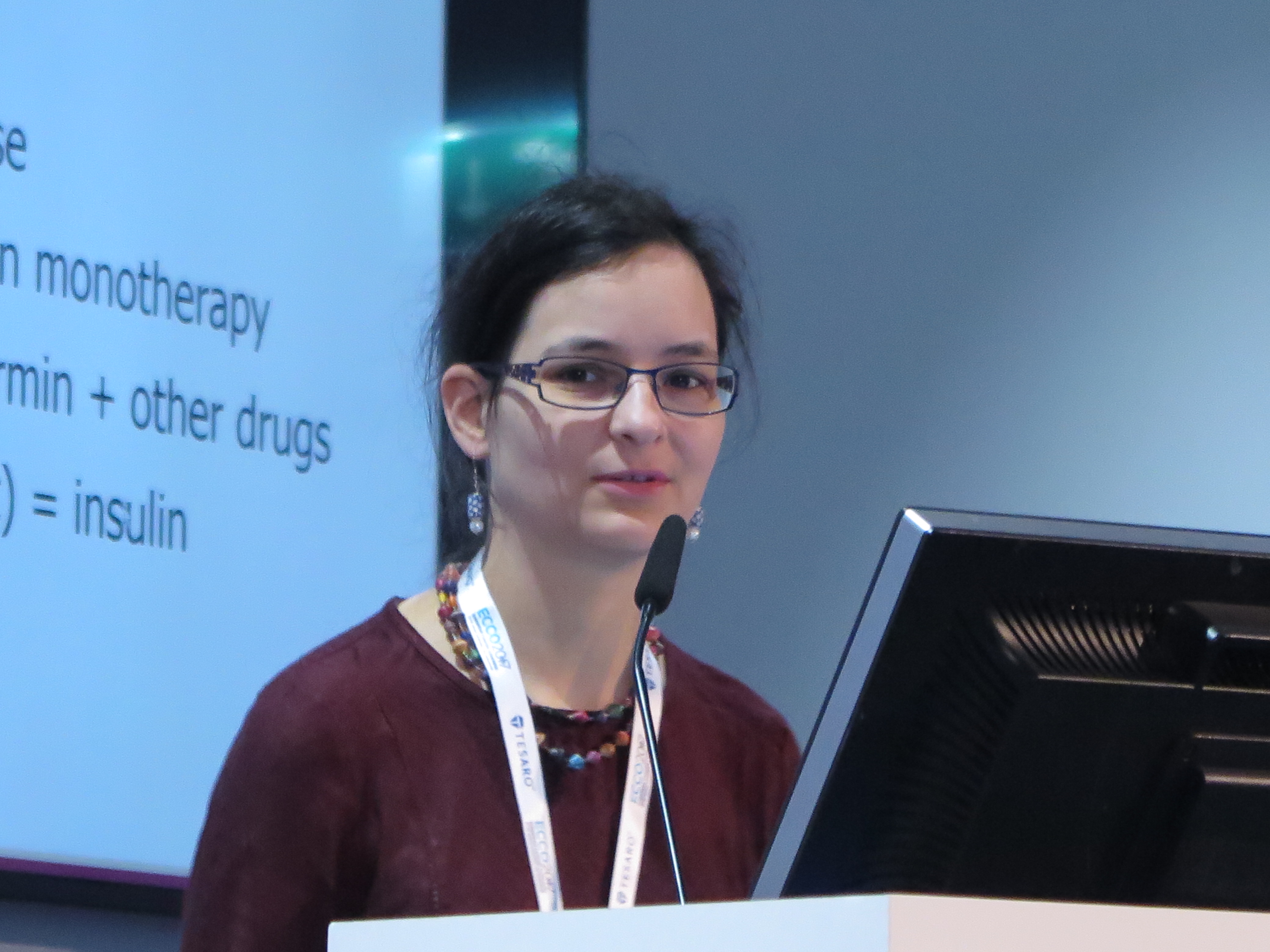
“We think that, at the beginning, there is an asymptomatic pancreatic cancer, with no clinical findings, and its first health effects are to disturb glucose metabolism. Then patients are diagnosed with type 2 diabetes, they are prescribed antidiabetic drugs, and then, as the cancer progresses but is still asymptomatic, the diabetes is less well controlled, and the patients shift to incretins and insulin more rapidly. And when the symptoms [of cancer] appear, it is too late for treatment,” Ms. Koechlin said at an annual congress sponsored by the European Cancer Organisation.
Incretin hormones stimulate the release of insulin from the pancreas. Incretin-based therapies such as dipeptidyl peptidase-4 inhibitors (DPP4 inhibitors, or gliptins) and glucagon-like peptide-1 receptor agonists (GLP-1 RAs), are generally held in reserve for patients with type 2 diabetes who have poor or inadequate glycemic control on oral agents such as metformin or the sulfonylureas.
Data from laboratory studies have suggested that incretins in general, and GLP-1 RAs in particular may increase risk of pancreatic cancer because of their direct action on the gland, Ms. Koechlin said.
As a part of postmarketing studies of the GLP-1 RAs requested by the European Medicines Agency (EMA), the investigators drew data from the Belgian Cancer Register on 11 million people in Belgium and from a registry maintained by the University of Milano-Bicocca, which covers approximately 10 million people in the Lombardy region of Italy.
They identified patients with type 2 diabetes who received a first prescription of an incretin drug or NIAD from January 2008 through the end of 2013 in Belgium, and through the end of 2012 in Italy.
They found that on first blush, incretin use did indeed appear to be associated with risk of pancreatic cancer, compared with NIAD use. Hazard ratios for cancer with incretin were 2.12 in Belgium (95% confidence interval, 1.60-2.81) and 2.17 (95% CI, 1.50-3.13) in Lombardy. The combined HR was 2.14 (95% CI, 1.71-2.67).
“In Belgium, 25% of cases were diagnosed within 90 days, and in Lombardy it was 18%. After the first year, the proportion of diagnosed pancreatic cancers dropped dramatically,” Ms. Koechlin said.
When they looked at the risk of cancer from incretin use over time, however, they found that the risk was highest at 3.5-fold, compared with NIAD use, within 3 months of starting a first prescription, but diminished to 2.3-fold during months 3-6, 2-fold for months 6-12, and 1.7-fold after the first year.
“This is not compatible with a causal relationship, because if there was a causal relationship we would observe a small risk for shorter duration of use, and higher risk for higher duration of use,” she said.
To support their findings, they looked at the relationship between cancer risk over time and first prescriptions for NIADs, and saw a similar decrease in risk as the duration of therapy increased.
They then looked at the relationship between a first prescription for insulin during follow-up, and saw significant increases in cancer risk, compared with patients who did not require insulin, with an HR in Belgium of 6.61 (95% CI, 5.63-7.77), and in Lombardy of 7.46 (95% CI, 6.00-9.35).
The perceived association between incretin drugs and cancer risk, therefore, may be due to “protopathic” or “reverse causation” bias, Ms. Koechlin said.
“Should patients diagnosed with diabetes be screened for pancreatic cancer? For now, no such test exists, but we could imagine in the future something like this: If the patient is diagnosed with diabetes, and the diabetes degrades rapidly, there could be some tests to identify markers for pancreatic cancer, and the pancreatic cancer could then be diagnosed at early stage, where there is more chance of curing the disease,” she said.
The study results raise questions about how practitioners should discuss potential risks of pancreatic cancer in patients with type 2 diabetes, commented Ian Banks, MD, president of the European Men’s Health Forum and cochair of ECCO2017.
“What is the best way of getting this message out without causing unnecessary concern, while at the same time raising the level of suspicion to go and see their GP?” he asked Ms. Koechlin, who presented a summary of the data in a briefing.
“It’s quite a difficult question, actually, because as you said, it could cause unnecessary worries,” and there are “no easy answers,” she said.
The study was sponsored by Sanofi. The investigators and Dr. Banks reported no conflicts of interest.
AT ECCO2017
Key clinical point: Rapid deterioration of type 2 diabetes may signal an underlying pancreatic lesion.
Major finding: A first incretin prescription was associated with a 3.5-fold risk for pancreatic cancer within 3 months, but the risk diminished over time, indicating no causation.
Data source: Retrospective Belgian and Italian registry data study of 824,688 patients with type 2 diabetes.
Disclosures: The study was sponsored by Sanofi. The investigators and Dr. Banks reported no conflicts of interest.
Neuropathic pain puts cancer survivors out of work
AMSTERDAM – Five years after a cancer diagnosis, patients who report having chronic neuropathic pain are twice as likely to be out of work as patients who report having no neuropathic pain, authors of a large longitudinal study said.
“For middle-term cancer survivors, suffering from chronic neuropathic pain unfortunately predicts labor-market exit,” said Marc-Karim Bendiane, from Aix-Marseille University in Marseille, France.
Pain is still frequently underdiagnosed, poorly managed, and undertreated among cancer survivors, and there is a need for alternatives to analgesics for control of chronic neuropathic pain (CNP), Mr. Bendiane said at an annual congress sponsored by the European Cancer Organisation.
Mr. Bendiane and colleagues used data from VICAN, a longitudinal survey of issues of concern to cancer survivors 2 years and 5 years after a diagnosis. The cohort consists of patients diagnosed with cancers who comprise 88% of all cancer diagnoses in France, including cancers of the breast; colon and rectum; lip, oral cavity, and pharynx; kidney; cervix; endometrium; non-Hodgkin lymphoma; melanoma; thyroid; bladder; and prostate.
To assess CNP, the researchers used data from a seven-item questionnaire designed to identify neuropathic characteristics of pain experienced by patients in the 2 weeks prior to a comprehensive patient interview.
Of the 982 patients who were working at the time of diagnosis, 36% reported pain within the previous 2 weeks, and of this group, 79% had chronic pain of neuropathic origin. CNP was more common in women than in men (P less than .01); in college-educated people, compared with less-educated people (P less than .001); those who had undergone chemotherapy, compared with no chemotherapy (P less than .001); and those who had radiotherapy vs. no radiotherapy (P less than .001).
For each cancer site, the prevalence of CNP among 5-year cancer survivors was substantially higher than the overall prevalence in France of 7%. For example, 34% of patients with cancers of the cervix and endometrium reported CNP, as did 29.9% of patients who survived cancers of the lip, oral cavity, and pharynx, 32.1% of lung cancer survivors, and 32.7% of breast cancer survivors.
Five years after diagnosis, 22.6% of patients who had been employed in 2010 were out of work in 2015.
The presence of CNP was associated with a nearly twofold greater risk of unemployment (adjusted odds ratio, 1.96; P less than .001) in a multivariate logistic regression analysis comparing employed and unemployed patients and controlling for social and demographic characteristics, job characteristics at diagnosis, and medical factors such as tumor site, prognosis, and treatment type.
The French National Cancer Institute and INSERM, the National Institute for Research in Health and Medicine, supported the study. The investigators reported no conflicts of interest.
Key clinical point: Chronic neuropathic pain is a barrier to employment for many cancer survivors.
Major finding: Cancer survivors with chronic neuropathic pain were twice as likely to be unemployed 5 years after diagnosis as patients with no pain.
Data source: Longitudinal study of French cancer survivors.
Disclosures: The French National Cancer Institute and INSERM, the National Institute for Research in Health and Medicine, supported the study. The investigators reported no conflicts of interest.
START again shows safety of hypofractionated doses in early breast cancer
AMSTERDAM – Analyses of patient-reported outcomes in the UK START A and B trials show that radiation doses greater than 2 Gy to the supraclavicular fossa or axilla are safe and are associated with acceptable near- and late-term toxicities, trial investigators reported.
The START trials compared standard adjuvant radiation doses and schedules with accelerated, hypofractionated doses in women with early breast cancer.
At the annual San Antonio Breast Cancer Symposium in 2012, lead investigator John Yarnold, MD, professor of clinical oncology at the Institute of Cancer Research in London, reported that a 40-Gy, 15-fraction regimen was associated with fewer adverse events over 10 years of follow-up compared with a 50-Gy, 25-fraction regimen that was at the time the standard in the United States and the United Kingdom.
Here at an annual congress sponsored by the European Cancer Organisation, Dr. Yarnold reported that among 479 patients in the two trials who had lymphatic radiation, there were no significant differences in arm or shoulder pain, swelling, motion problems, or shoulder stiffness between patients who received conventional 2-Gy fractions for a total of 50 Gy, or lower total doses of radiation divided into fractions larger than 2 Gy.
“A post hoc analysis of outcomes in 479 patients treated with radiotherapy fractions larger than 2 Gy to the supraclavicular fossa and/or the axilla raise no concerns in terms of safety or patient acceptability, and it was for this reason that this schedule has been the standard for locoregional radiotherapy in the UK for some years now,” he said.
START (Standardization of Breast Radiotherapy) A enrolled 2,236 patients from 35 UK centers with breast cancer stages T 1-3, nodal stage N0-1, and no distant metastases (M0) and randomly assigned them to 50 Gy in 25 fractions of 2 Gy each over 5 weeks, 39.0 Gy in 13 fractions of 3.0 Gy over 5 weeks, and 41.6 Gy in 13 fractions of 3.2 Gy over 5 weeks.
In START B, 2,215 women were randomly assigned to the 50 Gy or 40 Gy regimens.
As reported in 2012, over a median follow-up of 9.3 years in START A, the 41.6-Gy regimen was similar in its adverse events profile compared with the 50-Gy dose (hazard ratio [HR] 0.94, and the lower-dose regimen also was comparable in efficacy, as measured by the rate of locoregional tumor relapse.
There were numerically but not significantly more relapses in the 39-Gy dose, however.
In START B, the 40-Gy dose was associated with significantly fewer adverse events over a median of 9.9 years of follow-up (HR 0.77). Efficacy was similar, although there was a nonsignificant trend toward superiority for the hypofractionated regimen.
In the post hoc analysis, the investigators looked at adverse events in 479 total patients in START A and B, 365 of whom had completed patient-reported outcome measures on the European Organization for Research and Treatment of Cancer (EORTC) Breast Cancer–Specific Quality of Life Questionnaire (QLQ-BR23).
A total of 262 patients in START A, and 103 in START B completed the questionnaires at 0, 6, 12, 24, and 60 months. Of these patients. The majority in each arm had radiation to the supraclavicular fossa only, and a minority had axilla radiation alone or in addition to supraclavicular fossa radiation.
In START B, 5-year patient reported outcomes of severity of 3 or 4 on a 4-point scale were low in each treatment arm, with arm or hand swelling reported in 10% of patients on 50 Gy, and 6% of patients on 40 Gy, and shoulder stiffness reported in 12% on the 50-Gy dose, vs. 15% on the 40-Gy dose.
Among all 479 patients with clinical assessments at 10 years, there were also no significant differences in either arm edema or shoulder stiffness between patients who received 50 Gy and those who received other, hypofractionated in each of the two trials.
The findings lend support to the Danish Breast Cancer Cooperative Group’s SKAGEN Trial 1, which is evaluating moderately hypofractionated locoregional adjuvant radiation therapy of early breast cancer combined with a simultaneous integrated boost in patients with an indication for boost, Dr. Yarnold said.
The UK START trials were supported by Cancer Research UK, the Medical Research Council, and the National Cancer Research Institute. Dr. Yarnold reported having no conflicts of interest.
AT ECCO2017
Key clinical point: Additional follow-up supports continued safety of hypofractionated radiation in early breast cancer.
Major finding: There were no significant differences in either patient-reported outcomes at 5 years or clinical assessments at 10 years in patients treated at 40-Gy or 50-Gy doses.
Data source: Post hoc analysis of 479 patients treated in the UK START A and B trials.
Disclosures: The UK START trials were supported by Cancer Research UK, the Medical Research Council, and the National Cancer Research Institute. Dr. Yarnold reported having no conflicts of interest..
Survival better with breast-conserving therapy for early cancers
AMSTERDAM – In real-life practice, women with early, localized breast cancer who underwent breast conserving therapy had better breast cancer–specific and overall survival compared with women who underwent mastectomy, according to investigators in the Netherlands.
Among nearly 130,000 patients treated over two different time periods, breast-conserving surgery and radiation (BCT) was associated with superior survival for women older than 50, patients who did not receive adjuvant chemotherapy, and those with comorbidities – irrespective of either hormonal or human epidermal growth factor receptor 2 (HER2) status, reported Mirelle Lagendijk, MD, of Erasmus Medical Center Cancer Institute in Rotterdam, the Netherlands.
For patients 50 and younger, overall survival (OS), but not breast cancer–specific survival (BCSS), was superior with the more conservative approach.
“Breast conserving therapy in these identified subgroups seems to be the preferable treatment when both treatments are optional,” Dr. Lagendijk said at an annual congress sponsored by the European Cancer Organisation.
Although recent observational studies have shown survival with BCT to be at least equivalent for women with early stage disease, there is still a lack of sufficient data on BCSS, potential confounders such as systemic therapies and comorbidities, and on the relative effects of BCT or mastectomy on subgroups, she said.
The investigators drew data from the Netherlands Cancer Registry on 129,692 patients with early, primary invasive breast cancer without metastases other than to regional lymph nodes (T1-2NO-2MO).
They compared BCT to mastectomy for BCSS and OS in the population as a whole and in subgroups based on prognostic factors. They controlled for age, tumor and nodal stage, comorbidities, systemic therapy, hormone receptor and HER2 status, differentiation grade, morphology, year of treatment, axillary lymph node dissection, and contralateral breast cancer.
They divided patients into two treatment time periods. The older cohort consisted of 60,381 patients treated from 1999 through 2005, 48% of whom underwent mastectomy, with a median follow-up of 11.1 years, and 52% of whom had BCT, with a median follow-up of 12 years.
The more recent cohort consisted of 69,311 patients, 40% of whom had mastectomy with a median follow-up of 5.9 years, and 60% of whom had BCT with a median follow-up of 6.1 years.
In both time periods, deaths from all causes were lower among patients treated with BCT. In the older cohort, 13,960 of 28,968 patients (48.2%) who underwent mastectomy had died, compared with 8,915 of 31,413 patients (28.4%) who underwent BCT. In the more recent cohort, 5,504 of 27,731 (19.8%) of patients who had mastectomies had died, compared with 3,702 of 41,580 (8.9%) who underwent BCT.
“Irrespective of the time cohort and irrespective of the treatment, around 50% of the events were breast cancer related,” Dr. Lagendijk said.
BCSS was superior with BCT in each time cohort (log-rank P less than .001 for each). In the earlier cohort, BCT was significantly superior for BCSS across all disease stages; in the later cohort, it was significant for all but stages T1N1 and T1-2N2.
BCSS was superior for patients in all age categories in the early cohort, and for patients 50 and older in the later cohort.
“The final stratification performed for comorbidities present in the patients evaluated showed, surprisingly, that especially for those patients with comorbidity, there was significantly better breast cancer-specific survival when treated by breast conserving therapy as compared to a mastectomy,” Dr. Lagendijk said.
The investigators acknowledged that the study was limited by its retrospective design, potential confounding by severity, and the inability to show causal relationship between survival and treatment type.
Dutch health agencies sponsored the study. Dr. Lagendijk and Dr. Naredi reported no conflicts of interest.
AT ECCO2017
Key clinical point: Breast cancer–specific survival and overall survival were better among women who had breast-conserving therapy (BCT) compared with mastectomy.
Major finding: BCT was associated with superior survival for women older than 50, patients who did not receive adjuvant chemotherapy, and those with comorbidities.
Data source: Retrospective registry data study of 129,692 women treated for early breast cancer in the Netherlands during 1999-2005 and 2006-2012.
Disclosures: Dutch health agencies sponsored the study. Dr. Langendijk and Dr. Naredi reported no conflicts of interest.