User login
The paradox of Pompe disease
Until 2006, when a breakthrough therapy first made treatment possible, Pompe disease was a little-known metabolic myopathy fatal to infants. Those with later-onset disease experienced progressive, often severe disability into adulthood.
In this rare autosomal recessive disorder, which occurs in approximately one in 40,000 births worldwide, a deficiency or absence of the enzyme acid alpha-glucosidase causes glycogen to build up in the lysosomes of cells. While many tissues are affected, skeletal and cardiac muscle see the earliest involvement, with muscle hypotonia, cardiomyopathy, and breathing difficulties (mainly due to diaphragm weakness) comprising the hallmark symptoms of the infantile form. Muscle weakness and progressive respiratory failure are prominent in later-onset disease.

The spectrum of severity and age of onset in Pompe disease is linked to combinations of mutations on the GAA gene, some of which destroy the body’s ability to produce acid alpha-glucosidase whereas others merely hamper it. Less enzyme produced in the body generally corresponds with more severe disease activity.
The most severe end of the disease spectrum, or “classic infantile Pompe disease,” presents at birth and is recognized in early infancy. Until treatment with enzyme replacement therapy (ERT) became available, patients usually died of cardiorespiratory failure within their first year of life. With therapy, patients have survived into their 20s and beyond. Late-onset disease is a far broader category in which patients can present at any time from their first year, including into middle age.
The emergence in 2006 of alglucosidase alfa (Lumizyme, Sanofi Genzyme), an ERT used long-term to improve survival and slow progression in children and adults, resulted in a boom of research interest, a push to timelier diagnosis, and – with patients living longer – a more thorough understanding of the natural history of Pompe disease. In addition to the usual clinical picture of progressive muscle weakness, difficulty breathing, and cardiomyopathy, investigators are seeing nervous system involvement in patients with Pompe disease.
To learn more, Neurology Reviews talked to two global experts in Pompe disease: Priya Kishnani, MD, of Duke University in Durham, N.C., and Antonio Toscano, MD, of the University of Messina, in Messina, Italy.
Diagnosis: Still room to improve
“Most neurologists will encounter a patient with Pompe disease,” said Dr. Kishnani, who has been working with Pompe for her entire career as a pediatrician and medical geneticist, treating patients of all ages and disease phenotypes.
“In newborns, diagnosis is more straightforward, because you’ve got an enlarged heart,” she said. And thanks to efforts of researchers like Dr. Kishnani and Pompe advocacy groups, Pompe disease is now a part of the RUSP (Recommended Uniform Screening Panel) for newborns; currently 28 U.S. states are screening for Pompe disease.
“The challenge really is for the later-onset cases, which are 80% of all cases,” Dr. Kishnani said.
Previously, muscle biopsies were the first step toward diagnosis. Dried blot spot assays to detect enzyme deficiency have since become the standard, along with other biochemical tests. Confirmation of the diagnosis is through gene sequencing panels to detect GAA mutations.
“Now that there is a treatment for Pompe disease and the availability of blood-based testing, many previously undiagnosed patients with limb girdle weakness are evaluated and the diagnostic odyssey ends,” Dr. Kishnani said. “But there is still a diagnostic delay, and many cases remain undiagnosed.”
Routine blood tests for creatine kinase and for liver enzymes can help point to Pompe disease. But elevated liver enzymes are often misinterpreted. “It’s about the ratios,” Dr. Kishnani said. “ALT is usually much more elevated if it is coming from a liver cause, and AST is usually higher than ALT if it is coming from muscle. But patients often end up getting a liver biopsy due to so-called elevated liver enzymes. As the workup continues, it is often later recognized that the source of the elevated enzymes is muscle involvement, and a referral to the geneticist or neurologist is made. Only then is appropriate testing to confirm a diagnosis initiated.”
Dr. Toscano, a neurologist who specializes in Pompe disease and other myopathies and who has published on tools for diagnosing late-onset Pompe disease,1 said that clinicians should be vigilant when evaluating any patient with limb girdle weakness and elevated creatine kinase (CK) – “especially if the CK is under 2,000,” he said, “because it is very rare that patients with Pompe disease have a more elevated CK than that.”
“Elevated CK, myalgia, and exercise intolerance” should prompt clinicians to suspect Pompe disease in a patient of any age, Dr. Toscano said. “When you come across this, you should be very persistent and get to the end of the story.”
Dr. Toscano noted that the blood spot assay, while an important early step, is not fully diagnostic, “because you can have false positives.” The molecular GAA assay is used to confirm Pompe disease. But detecting pathogenic variants on the GAA gene – of which there are more than 500 – can be more complicated than it sounds. Whereas two mutations are required for Pompe disease, sometimes only one can be detected. Dr. Toscano said he also treated some patients for Pompe with only one known mutation but with unequivocal clinical and biochemical aspects of Pompe disease.
While delays in diagnosis for late-onset Pompe disease remain significant -- between 5 and 6 years on average for older patients, and up to 20 years for those with pediatric onset – both Dr. Kishnani and Dr. Toscano said they perceive them to be improving. With McArdle disease, another inherited glycogen storage disorder that is more common than Pompe disease but for which there is no treatment, “the delay is nearly 12 years,” Dr. Toscano said.
ERT: The sooner the better
Enzyme replacement therapy is indicated for all patients with Pompe disease. Currently two are commercially available: alglucosidase alfa (Lumizyme, Sanofi Genzyme), indicated for all forms of Pompe disease, and avalglucosidase alfa-ngpt (Nexviazyme, Sanofi Genzyme), approved in 2021 for later-onset Pompe, though its indications have yet to be fully defined.
The semimonthly infusions represent, to date, the only disease-modifying therapies commercially available. Enzyme replacement therapy can reverse cardiac damage seen in infants and allow them to meet developmental milestones previously unthinkable. In adults, it can slow progression, though many treated patients will still develop chronic disability and require a wheelchair, respiratory support, or both. “The phenotype of the patients we are seeing today is not as involved as it was prior to enzyme therapy,” said Dr. Kishnani, who was part of the research team that developed ERT and launched the first clinical trials. “This is across the disease spectrum.”
But optimal management means more than just getting a patient on therapy fast, Dr. Kishnani said.
“Very often the thinking is if the patient is on ERT, we’ve done right by the patient. Aspects we don’t look at enough include: Are we monitoring these patients well? Are patients being followed by a multidisciplinary team that includes cardiology, physical therapy, and pulmonary medicine? Are we doing appropriate musculoskeletal assessments? They might have sleep hypoventilation. The BiPap settings may not be correct. Or they have not been assessed for antibodies,” she said.
Many infants with severe phenotypes, notably those who produce no enzyme naturally, will develop immune reactions to the exogenous enzyme therapy. High antibody titers also have been seen and are associated with poor therapeutic response. While this is very clear in the infantile setting, late-onset patients also develop antibodies in response to ERT. In one study in 64 patients,2 Dr. Toscano and his colleagues saw that antibodies may affect clinical response during the first 3 years of treatment, while a small study3 by Dr. Kishnani’s group saw clinical decline associated with high antibody titers in patients with late-onset disease.
While the relationship of specific titers to therapeutic response remains unclear, it is important to consider antibodies, along with other factors, in the monitoring of patients with Pompe disease. “We need to always ask, if a patient is falling behind, what could be the reason?” Dr. Kishnani said. “These are the things we as clinicians can do to improve or enhance the impact of ERT.”
Dr. Toscano noted that a common misconception about late-onset Pompe disease is that cardiac manifestations are minimal or absent, whereas as many as about 20% of patients will have heart problems and need to be carefully monitored.
Neurological manifestations
With patients surviving longer on ERT, researchers have been able to develop a deeper understanding of the natural history of Pompe disease. Increasingly, they are seeing it as a multisystem disease that includes central nervous system involvement.
“Is Pompe an overt neurodegenerative disease? I would say no,” Dr. Kishnani said. “But there is a neurological component that we’ve got to understand and follow more.”
Glycogen accumulation, she noted, has been found in anterior horn cells, motor neurons, and other parts of the brain. “We have been doing MRIs on children with infantile Pompe, and we have seen some white matter hyperintensities. The clinical significance of this finding is still emerging. Sometimes it is present, but the child is cognitively intact. We have had college graduates who have white matter hyperintensities. So putting it in context will be important. But we know that glycogen is ubiquitous, and autopsy studies have shown that it is present in the brain.”
In recent years, Dr. Toscano’s group has investigated neurovascular complications of Pompe in late-onset patients. “This was something that really surprised us because for several years we have investigated mainly heart, muscle, or respiratory manifestations of the disease, but the central nervous system was really neglected,” he said.
“Occasionally we did some brain MRIs and we found in even young patients some ischemic areas. We thought this was related to slowed circulation – that blood vessels in these patients are weak because they are impaired by glycogen accumulation.” Dr. Toscano and his colleagues followed that observation with a study of late-onset patients,4 in which they found that more than half had cerebrovascular abnormalities. “Even in, say, patients 30 to 35 years old we saw this – it’s unusual to have a vascular disorder at that age.”
Dr. Toscano and his colleagues also reported cerebral aneurysms5 in patients with Pompe disease and have recommended that clinicians conduct MRI or cerebral angiograms on patients as part of routine follow-up. Blood pressure in Pompe patients should be carefully watched and managed with antihypertensive medication as needed, he said.
Part of the problem is that the proteins in ERT are not able to cross the blood-brain barrier, Dr. Toscano noted, adding that researchers are investigating other treatments that can.
Pompe disease as a research model
The successful development of ERT for Pompe disease marked a boom in research interest into not just Pompe – for which several experimental therapies are currently in the pipeline – but for other myopathies and glycogen storage disorders.
“I think that Pompe has served as a template both as a muscle disease and a lysosomal storage disease, and so some of our learnings from Pompe have been applied across different diseases,” Dr. Kishnani said.
Studies in spinal muscular atrophy, for example, “in some ways mirrored what was done for Pompe – treatment trials were initiated in babies at the most severe end of the disease population with infantile disease, and used similar clinical trial endpoints,” Dr. Kishnani said. “Even for the later-onset end of the spectrum, the endpoints we used in Pompe for muscle strength and function have been relevant to many other neuromuscular disorders.”
Pompe disease research also ushered in a new appreciation of immune responses in protein replacement therapies, Dr. Kishnani noted.
“In the field today, you hear the term cross-reactive immunological material, or CRIM, all the time,” she said. “But when we first started talking about it in the space of Pompe disease, there was a lot of scientific debate about what the significance of CRIM-negative status was in relationship to the risk for development of high and sustained antibody titer and a poor clinical response. To understand this involved a lot of going back to the data and digging into the small subset of nonresponders. One of the powers of rare disease research is that every patient matters, and it’s important to understand what’s going on at the patient level rather than just the group data level.”
A robust pipeline
The decade and a half since the advent of ERT has seen what Dr. Toscano described as “an explosion of interest” in Pompe disease.
“We’re seeing an extraordinary number of papers on everything from clinical, biomarkers, genetics, and rehabilitation – this disease is now considered from every point of view, and this is very important for patients,” Dr. Toscano said. Alongside this has come industry interest in this rare disease, with several companies investigating a range of treatment approaches.
The existence of a treatment, “while not perfect,” he said, “has interested the patient associations and doctors to try and improve service to patients. Patients with Pompe disease are well attended, probably more so than patients with degenerative disorders in which there is no therapy.”
Last year the second ERT, avalglucosidase alfa (Nexviazyme, Sanofi Genzyme) was approved by the U.S. Food and Drug Administration to treat late-onset Pompe disease. The drug, currently being investigated in infants as well, was designed to improve the delivery of the therapeutic enzyme to muscles and enhance glycogen clearance, and results from ongoing trials suggest some functional and clinical benefit over standard ERT.
Other drugs in development for Pompe disease include substrate reduction therapies, which aim to reduce the storage of glycogen in cells, and therapies that improve residual function of mutant GAA enzyme in the body. These and other therapies in development have the potential to modify nervous system manifestations of Pompe disease.6
Because a single gene is implicated in Pompe disease, it has long been considered a good candidate for gene therapies that prompt the body to make stable enzyme. Seven companies are now investigating gene therapies in Pompe disease.7 Some of these deliver to skeletal muscles and others aim for the liver, where proteins are synthesized and secreted and adverse immune responses might be more easily mitigated. Other gene therapies use an ex vivo approach, removing and replacing cells in bone marrow.
Dr. Kishnani’s research group at Duke University is leading a small clinical trial in late-onset patients of a GAA gene transfer to the liver using adeno-associated virus (AAV) vectors.8
“We have started AAV gene therapy trials in in adults with Pompe disease and will later evaluate children because ERT is available as a standard of care, and so from a safety perspective this makes the most sense,” Dr. Kishnani said. “We do have challenges in the field of gene therapy, but I think if we are able to overcome the immune responses, and … to treat at a lower dose, there’s a very good pathway forward.”
Dr. Toscano and Dr. Kishnani have received reimbursement from Sanofi and other manufacturers for participation on advisory boards and as speakers.
Jennie Smith is a freelance journalist and editor specializing in medicine and health.
References
1. Musumeci O, Toscano A. Diagnostic tools in late onset Pompe disease (LOPD). Ann Transl Med. 2019 Jul;7(13):286. doi: 10.21037/atm.2019.06.60.
2. Filosto M et al. Assessing the role of anti rh-GAA in modulating response to ERT in a late-onset Pompe disease cohort from the Italian GSDII Study Group. Adv Ther. 2019 May;36(5):1177-1189. doi: 10.1007/s12325-019-00926-5.
3. Patel TT et al. The impact of antibodies in late-onset Pompe disease: A case series and literature review. Mol Genet Metab. 2012 Jul;106(3):301-9. doi: 10.1016/j.ymgme.2012.04.027.
4. Montagnese F et al. Intracranial arterial abnormalities in patients with late onset Pompe disease (LOPD). J Inherit Metab Dis. 2016 May;39(3):391-398. doi: 10.1007/s10545-015-9913-x.
5. Musumeci O et al. Central nervous system involvement in late-onset Pompe disease: Clues from neuroimaging and neuropsychological analysis. Eur J Neurol. 2019 Mar;26(3):442-e35. doi: 10.1111/ene.13835.
6. Edelmann MJ, Maegawa GHB. CNS-targeting therapies for lysosomal storage diseases: Current advances and challenges. Front Mol Biosci. 2020 Nov 12;7:559804. doi: 10.3389/fmolb.2020.559804
7. Ronzitti G et al. Progress and challenges of gene therapy for Pompe disease. Ann Transl Med. 2019 Jul;7(13):287. doi: 10.21037/atm.2019.04.67.
8. Kishnani PS, Koeberl DD. Liver depot gene therapy for Pompe disease. Ann Transl Med. 2019 Jul;7(13):288. doi: 10.21037/atm.2019.05.02.
Until 2006, when a breakthrough therapy first made treatment possible, Pompe disease was a little-known metabolic myopathy fatal to infants. Those with later-onset disease experienced progressive, often severe disability into adulthood.
In this rare autosomal recessive disorder, which occurs in approximately one in 40,000 births worldwide, a deficiency or absence of the enzyme acid alpha-glucosidase causes glycogen to build up in the lysosomes of cells. While many tissues are affected, skeletal and cardiac muscle see the earliest involvement, with muscle hypotonia, cardiomyopathy, and breathing difficulties (mainly due to diaphragm weakness) comprising the hallmark symptoms of the infantile form. Muscle weakness and progressive respiratory failure are prominent in later-onset disease.
The spectrum of severity and age of onset in Pompe disease is linked to combinations of mutations on the GAA gene, some of which destroy the body’s ability to produce acid alpha-glucosidase whereas others merely hamper it. Less enzyme produced in the body generally corresponds with more severe disease activity.
The most severe end of the disease spectrum, or “classic infantile Pompe disease,” presents at birth and is recognized in early infancy. Until treatment with enzyme replacement therapy (ERT) became available, patients usually died of cardiorespiratory failure within their first year of life. With therapy, patients have survived into their 20s and beyond. Late-onset disease is a far broader category in which patients can present at any time from their first year, including into middle age.
The emergence in 2006 of alglucosidase alfa (Lumizyme, Sanofi Genzyme), an ERT used long-term to improve survival and slow progression in children and adults, resulted in a boom of research interest, a push to timelier diagnosis, and – with patients living longer – a more thorough understanding of the natural history of Pompe disease. In addition to the usual clinical picture of progressive muscle weakness, difficulty breathing, and cardiomyopathy, investigators are seeing nervous system involvement in patients with Pompe disease.
To learn more, Neurology Reviews talked to two global experts in Pompe disease: Priya Kishnani, MD, of Duke University in Durham, N.C., and Antonio Toscano, MD, of the University of Messina, in Messina, Italy.
Diagnosis: Still room to improve
“Most neurologists will encounter a patient with Pompe disease,” said Dr. Kishnani, who has been working with Pompe for her entire career as a pediatrician and medical geneticist, treating patients of all ages and disease phenotypes.
“In newborns, diagnosis is more straightforward, because you’ve got an enlarged heart,” she said. And thanks to efforts of researchers like Dr. Kishnani and Pompe advocacy groups, Pompe disease is now a part of the RUSP (Recommended Uniform Screening Panel) for newborns; currently 28 U.S. states are screening for Pompe disease.
“The challenge really is for the later-onset cases, which are 80% of all cases,” Dr. Kishnani said.
Previously, muscle biopsies were the first step toward diagnosis. Dried blot spot assays to detect enzyme deficiency have since become the standard, along with other biochemical tests. Confirmation of the diagnosis is through gene sequencing panels to detect GAA mutations.
“Now that there is a treatment for Pompe disease and the availability of blood-based testing, many previously undiagnosed patients with limb girdle weakness are evaluated and the diagnostic odyssey ends,” Dr. Kishnani said. “But there is still a diagnostic delay, and many cases remain undiagnosed.”
Routine blood tests for creatine kinase and for liver enzymes can help point to Pompe disease. But elevated liver enzymes are often misinterpreted. “It’s about the ratios,” Dr. Kishnani said. “ALT is usually much more elevated if it is coming from a liver cause, and AST is usually higher than ALT if it is coming from muscle. But patients often end up getting a liver biopsy due to so-called elevated liver enzymes. As the workup continues, it is often later recognized that the source of the elevated enzymes is muscle involvement, and a referral to the geneticist or neurologist is made. Only then is appropriate testing to confirm a diagnosis initiated.”
Dr. Toscano, a neurologist who specializes in Pompe disease and other myopathies and who has published on tools for diagnosing late-onset Pompe disease,1 said that clinicians should be vigilant when evaluating any patient with limb girdle weakness and elevated creatine kinase (CK) – “especially if the CK is under 2,000,” he said, “because it is very rare that patients with Pompe disease have a more elevated CK than that.”
“Elevated CK, myalgia, and exercise intolerance” should prompt clinicians to suspect Pompe disease in a patient of any age, Dr. Toscano said. “When you come across this, you should be very persistent and get to the end of the story.”
Dr. Toscano noted that the blood spot assay, while an important early step, is not fully diagnostic, “because you can have false positives.” The molecular GAA assay is used to confirm Pompe disease. But detecting pathogenic variants on the GAA gene – of which there are more than 500 – can be more complicated than it sounds. Whereas two mutations are required for Pompe disease, sometimes only one can be detected. Dr. Toscano said he also treated some patients for Pompe with only one known mutation but with unequivocal clinical and biochemical aspects of Pompe disease.
While delays in diagnosis for late-onset Pompe disease remain significant -- between 5 and 6 years on average for older patients, and up to 20 years for those with pediatric onset – both Dr. Kishnani and Dr. Toscano said they perceive them to be improving. With McArdle disease, another inherited glycogen storage disorder that is more common than Pompe disease but for which there is no treatment, “the delay is nearly 12 years,” Dr. Toscano said.
ERT: The sooner the better
Enzyme replacement therapy is indicated for all patients with Pompe disease. Currently two are commercially available: alglucosidase alfa (Lumizyme, Sanofi Genzyme), indicated for all forms of Pompe disease, and avalglucosidase alfa-ngpt (Nexviazyme, Sanofi Genzyme), approved in 2021 for later-onset Pompe, though its indications have yet to be fully defined.
The semimonthly infusions represent, to date, the only disease-modifying therapies commercially available. Enzyme replacement therapy can reverse cardiac damage seen in infants and allow them to meet developmental milestones previously unthinkable. In adults, it can slow progression, though many treated patients will still develop chronic disability and require a wheelchair, respiratory support, or both. “The phenotype of the patients we are seeing today is not as involved as it was prior to enzyme therapy,” said Dr. Kishnani, who was part of the research team that developed ERT and launched the first clinical trials. “This is across the disease spectrum.”
But optimal management means more than just getting a patient on therapy fast, Dr. Kishnani said.
“Very often the thinking is if the patient is on ERT, we’ve done right by the patient. Aspects we don’t look at enough include: Are we monitoring these patients well? Are patients being followed by a multidisciplinary team that includes cardiology, physical therapy, and pulmonary medicine? Are we doing appropriate musculoskeletal assessments? They might have sleep hypoventilation. The BiPap settings may not be correct. Or they have not been assessed for antibodies,” she said.
Many infants with severe phenotypes, notably those who produce no enzyme naturally, will develop immune reactions to the exogenous enzyme therapy. High antibody titers also have been seen and are associated with poor therapeutic response. While this is very clear in the infantile setting, late-onset patients also develop antibodies in response to ERT. In one study in 64 patients,2 Dr. Toscano and his colleagues saw that antibodies may affect clinical response during the first 3 years of treatment, while a small study3 by Dr. Kishnani’s group saw clinical decline associated with high antibody titers in patients with late-onset disease.
While the relationship of specific titers to therapeutic response remains unclear, it is important to consider antibodies, along with other factors, in the monitoring of patients with Pompe disease. “We need to always ask, if a patient is falling behind, what could be the reason?” Dr. Kishnani said. “These are the things we as clinicians can do to improve or enhance the impact of ERT.”
Dr. Toscano noted that a common misconception about late-onset Pompe disease is that cardiac manifestations are minimal or absent, whereas as many as about 20% of patients will have heart problems and need to be carefully monitored.
Neurological manifestations
With patients surviving longer on ERT, researchers have been able to develop a deeper understanding of the natural history of Pompe disease. Increasingly, they are seeing it as a multisystem disease that includes central nervous system involvement.
“Is Pompe an overt neurodegenerative disease? I would say no,” Dr. Kishnani said. “But there is a neurological component that we’ve got to understand and follow more.”
Glycogen accumulation, she noted, has been found in anterior horn cells, motor neurons, and other parts of the brain. “We have been doing MRIs on children with infantile Pompe, and we have seen some white matter hyperintensities. The clinical significance of this finding is still emerging. Sometimes it is present, but the child is cognitively intact. We have had college graduates who have white matter hyperintensities. So putting it in context will be important. But we know that glycogen is ubiquitous, and autopsy studies have shown that it is present in the brain.”
In recent years, Dr. Toscano’s group has investigated neurovascular complications of Pompe in late-onset patients. “This was something that really surprised us because for several years we have investigated mainly heart, muscle, or respiratory manifestations of the disease, but the central nervous system was really neglected,” he said.
“Occasionally we did some brain MRIs and we found in even young patients some ischemic areas. We thought this was related to slowed circulation – that blood vessels in these patients are weak because they are impaired by glycogen accumulation.” Dr. Toscano and his colleagues followed that observation with a study of late-onset patients,4 in which they found that more than half had cerebrovascular abnormalities. “Even in, say, patients 30 to 35 years old we saw this – it’s unusual to have a vascular disorder at that age.”
Dr. Toscano and his colleagues also reported cerebral aneurysms5 in patients with Pompe disease and have recommended that clinicians conduct MRI or cerebral angiograms on patients as part of routine follow-up. Blood pressure in Pompe patients should be carefully watched and managed with antihypertensive medication as needed, he said.
Part of the problem is that the proteins in ERT are not able to cross the blood-brain barrier, Dr. Toscano noted, adding that researchers are investigating other treatments that can.
Pompe disease as a research model
The successful development of ERT for Pompe disease marked a boom in research interest into not just Pompe – for which several experimental therapies are currently in the pipeline – but for other myopathies and glycogen storage disorders.
“I think that Pompe has served as a template both as a muscle disease and a lysosomal storage disease, and so some of our learnings from Pompe have been applied across different diseases,” Dr. Kishnani said.
Studies in spinal muscular atrophy, for example, “in some ways mirrored what was done for Pompe – treatment trials were initiated in babies at the most severe end of the disease population with infantile disease, and used similar clinical trial endpoints,” Dr. Kishnani said. “Even for the later-onset end of the spectrum, the endpoints we used in Pompe for muscle strength and function have been relevant to many other neuromuscular disorders.”
Pompe disease research also ushered in a new appreciation of immune responses in protein replacement therapies, Dr. Kishnani noted.
“In the field today, you hear the term cross-reactive immunological material, or CRIM, all the time,” she said. “But when we first started talking about it in the space of Pompe disease, there was a lot of scientific debate about what the significance of CRIM-negative status was in relationship to the risk for development of high and sustained antibody titer and a poor clinical response. To understand this involved a lot of going back to the data and digging into the small subset of nonresponders. One of the powers of rare disease research is that every patient matters, and it’s important to understand what’s going on at the patient level rather than just the group data level.”
A robust pipeline
The decade and a half since the advent of ERT has seen what Dr. Toscano described as “an explosion of interest” in Pompe disease.
“We’re seeing an extraordinary number of papers on everything from clinical, biomarkers, genetics, and rehabilitation – this disease is now considered from every point of view, and this is very important for patients,” Dr. Toscano said. Alongside this has come industry interest in this rare disease, with several companies investigating a range of treatment approaches.
The existence of a treatment, “while not perfect,” he said, “has interested the patient associations and doctors to try and improve service to patients. Patients with Pompe disease are well attended, probably more so than patients with degenerative disorders in which there is no therapy.”
Last year the second ERT, avalglucosidase alfa (Nexviazyme, Sanofi Genzyme) was approved by the U.S. Food and Drug Administration to treat late-onset Pompe disease. The drug, currently being investigated in infants as well, was designed to improve the delivery of the therapeutic enzyme to muscles and enhance glycogen clearance, and results from ongoing trials suggest some functional and clinical benefit over standard ERT.
Other drugs in development for Pompe disease include substrate reduction therapies, which aim to reduce the storage of glycogen in cells, and therapies that improve residual function of mutant GAA enzyme in the body. These and other therapies in development have the potential to modify nervous system manifestations of Pompe disease.6
Because a single gene is implicated in Pompe disease, it has long been considered a good candidate for gene therapies that prompt the body to make stable enzyme. Seven companies are now investigating gene therapies in Pompe disease.7 Some of these deliver to skeletal muscles and others aim for the liver, where proteins are synthesized and secreted and adverse immune responses might be more easily mitigated. Other gene therapies use an ex vivo approach, removing and replacing cells in bone marrow.
Dr. Kishnani’s research group at Duke University is leading a small clinical trial in late-onset patients of a GAA gene transfer to the liver using adeno-associated virus (AAV) vectors.8
“We have started AAV gene therapy trials in in adults with Pompe disease and will later evaluate children because ERT is available as a standard of care, and so from a safety perspective this makes the most sense,” Dr. Kishnani said. “We do have challenges in the field of gene therapy, but I think if we are able to overcome the immune responses, and … to treat at a lower dose, there’s a very good pathway forward.”
Dr. Toscano and Dr. Kishnani have received reimbursement from Sanofi and other manufacturers for participation on advisory boards and as speakers.
Jennie Smith is a freelance journalist and editor specializing in medicine and health.
References
1. Musumeci O, Toscano A. Diagnostic tools in late onset Pompe disease (LOPD). Ann Transl Med. 2019 Jul;7(13):286. doi: 10.21037/atm.2019.06.60.
2. Filosto M et al. Assessing the role of anti rh-GAA in modulating response to ERT in a late-onset Pompe disease cohort from the Italian GSDII Study Group. Adv Ther. 2019 May;36(5):1177-1189. doi: 10.1007/s12325-019-00926-5.
3. Patel TT et al. The impact of antibodies in late-onset Pompe disease: A case series and literature review. Mol Genet Metab. 2012 Jul;106(3):301-9. doi: 10.1016/j.ymgme.2012.04.027.
4. Montagnese F et al. Intracranial arterial abnormalities in patients with late onset Pompe disease (LOPD). J Inherit Metab Dis. 2016 May;39(3):391-398. doi: 10.1007/s10545-015-9913-x.
5. Musumeci O et al. Central nervous system involvement in late-onset Pompe disease: Clues from neuroimaging and neuropsychological analysis. Eur J Neurol. 2019 Mar;26(3):442-e35. doi: 10.1111/ene.13835.
6. Edelmann MJ, Maegawa GHB. CNS-targeting therapies for lysosomal storage diseases: Current advances and challenges. Front Mol Biosci. 2020 Nov 12;7:559804. doi: 10.3389/fmolb.2020.559804
7. Ronzitti G et al. Progress and challenges of gene therapy for Pompe disease. Ann Transl Med. 2019 Jul;7(13):287. doi: 10.21037/atm.2019.04.67.
8. Kishnani PS, Koeberl DD. Liver depot gene therapy for Pompe disease. Ann Transl Med. 2019 Jul;7(13):288. doi: 10.21037/atm.2019.05.02.
Until 2006, when a breakthrough therapy first made treatment possible, Pompe disease was a little-known metabolic myopathy fatal to infants. Those with later-onset disease experienced progressive, often severe disability into adulthood.
In this rare autosomal recessive disorder, which occurs in approximately one in 40,000 births worldwide, a deficiency or absence of the enzyme acid alpha-glucosidase causes glycogen to build up in the lysosomes of cells. While many tissues are affected, skeletal and cardiac muscle see the earliest involvement, with muscle hypotonia, cardiomyopathy, and breathing difficulties (mainly due to diaphragm weakness) comprising the hallmark symptoms of the infantile form. Muscle weakness and progressive respiratory failure are prominent in later-onset disease.
The spectrum of severity and age of onset in Pompe disease is linked to combinations of mutations on the GAA gene, some of which destroy the body’s ability to produce acid alpha-glucosidase whereas others merely hamper it. Less enzyme produced in the body generally corresponds with more severe disease activity.
The most severe end of the disease spectrum, or “classic infantile Pompe disease,” presents at birth and is recognized in early infancy. Until treatment with enzyme replacement therapy (ERT) became available, patients usually died of cardiorespiratory failure within their first year of life. With therapy, patients have survived into their 20s and beyond. Late-onset disease is a far broader category in which patients can present at any time from their first year, including into middle age.
The emergence in 2006 of alglucosidase alfa (Lumizyme, Sanofi Genzyme), an ERT used long-term to improve survival and slow progression in children and adults, resulted in a boom of research interest, a push to timelier diagnosis, and – with patients living longer – a more thorough understanding of the natural history of Pompe disease. In addition to the usual clinical picture of progressive muscle weakness, difficulty breathing, and cardiomyopathy, investigators are seeing nervous system involvement in patients with Pompe disease.
To learn more, Neurology Reviews talked to two global experts in Pompe disease: Priya Kishnani, MD, of Duke University in Durham, N.C., and Antonio Toscano, MD, of the University of Messina, in Messina, Italy.
Diagnosis: Still room to improve
“Most neurologists will encounter a patient with Pompe disease,” said Dr. Kishnani, who has been working with Pompe for her entire career as a pediatrician and medical geneticist, treating patients of all ages and disease phenotypes.
“In newborns, diagnosis is more straightforward, because you’ve got an enlarged heart,” she said. And thanks to efforts of researchers like Dr. Kishnani and Pompe advocacy groups, Pompe disease is now a part of the RUSP (Recommended Uniform Screening Panel) for newborns; currently 28 U.S. states are screening for Pompe disease.
“The challenge really is for the later-onset cases, which are 80% of all cases,” Dr. Kishnani said.
Previously, muscle biopsies were the first step toward diagnosis. Dried blot spot assays to detect enzyme deficiency have since become the standard, along with other biochemical tests. Confirmation of the diagnosis is through gene sequencing panels to detect GAA mutations.
“Now that there is a treatment for Pompe disease and the availability of blood-based testing, many previously undiagnosed patients with limb girdle weakness are evaluated and the diagnostic odyssey ends,” Dr. Kishnani said. “But there is still a diagnostic delay, and many cases remain undiagnosed.”
Routine blood tests for creatine kinase and for liver enzymes can help point to Pompe disease. But elevated liver enzymes are often misinterpreted. “It’s about the ratios,” Dr. Kishnani said. “ALT is usually much more elevated if it is coming from a liver cause, and AST is usually higher than ALT if it is coming from muscle. But patients often end up getting a liver biopsy due to so-called elevated liver enzymes. As the workup continues, it is often later recognized that the source of the elevated enzymes is muscle involvement, and a referral to the geneticist or neurologist is made. Only then is appropriate testing to confirm a diagnosis initiated.”
Dr. Toscano, a neurologist who specializes in Pompe disease and other myopathies and who has published on tools for diagnosing late-onset Pompe disease,1 said that clinicians should be vigilant when evaluating any patient with limb girdle weakness and elevated creatine kinase (CK) – “especially if the CK is under 2,000,” he said, “because it is very rare that patients with Pompe disease have a more elevated CK than that.”
“Elevated CK, myalgia, and exercise intolerance” should prompt clinicians to suspect Pompe disease in a patient of any age, Dr. Toscano said. “When you come across this, you should be very persistent and get to the end of the story.”
Dr. Toscano noted that the blood spot assay, while an important early step, is not fully diagnostic, “because you can have false positives.” The molecular GAA assay is used to confirm Pompe disease. But detecting pathogenic variants on the GAA gene – of which there are more than 500 – can be more complicated than it sounds. Whereas two mutations are required for Pompe disease, sometimes only one can be detected. Dr. Toscano said he also treated some patients for Pompe with only one known mutation but with unequivocal clinical and biochemical aspects of Pompe disease.
While delays in diagnosis for late-onset Pompe disease remain significant -- between 5 and 6 years on average for older patients, and up to 20 years for those with pediatric onset – both Dr. Kishnani and Dr. Toscano said they perceive them to be improving. With McArdle disease, another inherited glycogen storage disorder that is more common than Pompe disease but for which there is no treatment, “the delay is nearly 12 years,” Dr. Toscano said.
ERT: The sooner the better
Enzyme replacement therapy is indicated for all patients with Pompe disease. Currently two are commercially available: alglucosidase alfa (Lumizyme, Sanofi Genzyme), indicated for all forms of Pompe disease, and avalglucosidase alfa-ngpt (Nexviazyme, Sanofi Genzyme), approved in 2021 for later-onset Pompe, though its indications have yet to be fully defined.
The semimonthly infusions represent, to date, the only disease-modifying therapies commercially available. Enzyme replacement therapy can reverse cardiac damage seen in infants and allow them to meet developmental milestones previously unthinkable. In adults, it can slow progression, though many treated patients will still develop chronic disability and require a wheelchair, respiratory support, or both. “The phenotype of the patients we are seeing today is not as involved as it was prior to enzyme therapy,” said Dr. Kishnani, who was part of the research team that developed ERT and launched the first clinical trials. “This is across the disease spectrum.”
But optimal management means more than just getting a patient on therapy fast, Dr. Kishnani said.
“Very often the thinking is if the patient is on ERT, we’ve done right by the patient. Aspects we don’t look at enough include: Are we monitoring these patients well? Are patients being followed by a multidisciplinary team that includes cardiology, physical therapy, and pulmonary medicine? Are we doing appropriate musculoskeletal assessments? They might have sleep hypoventilation. The BiPap settings may not be correct. Or they have not been assessed for antibodies,” she said.
Many infants with severe phenotypes, notably those who produce no enzyme naturally, will develop immune reactions to the exogenous enzyme therapy. High antibody titers also have been seen and are associated with poor therapeutic response. While this is very clear in the infantile setting, late-onset patients also develop antibodies in response to ERT. In one study in 64 patients,2 Dr. Toscano and his colleagues saw that antibodies may affect clinical response during the first 3 years of treatment, while a small study3 by Dr. Kishnani’s group saw clinical decline associated with high antibody titers in patients with late-onset disease.
While the relationship of specific titers to therapeutic response remains unclear, it is important to consider antibodies, along with other factors, in the monitoring of patients with Pompe disease. “We need to always ask, if a patient is falling behind, what could be the reason?” Dr. Kishnani said. “These are the things we as clinicians can do to improve or enhance the impact of ERT.”
Dr. Toscano noted that a common misconception about late-onset Pompe disease is that cardiac manifestations are minimal or absent, whereas as many as about 20% of patients will have heart problems and need to be carefully monitored.
Neurological manifestations
With patients surviving longer on ERT, researchers have been able to develop a deeper understanding of the natural history of Pompe disease. Increasingly, they are seeing it as a multisystem disease that includes central nervous system involvement.
“Is Pompe an overt neurodegenerative disease? I would say no,” Dr. Kishnani said. “But there is a neurological component that we’ve got to understand and follow more.”
Glycogen accumulation, she noted, has been found in anterior horn cells, motor neurons, and other parts of the brain. “We have been doing MRIs on children with infantile Pompe, and we have seen some white matter hyperintensities. The clinical significance of this finding is still emerging. Sometimes it is present, but the child is cognitively intact. We have had college graduates who have white matter hyperintensities. So putting it in context will be important. But we know that glycogen is ubiquitous, and autopsy studies have shown that it is present in the brain.”
In recent years, Dr. Toscano’s group has investigated neurovascular complications of Pompe in late-onset patients. “This was something that really surprised us because for several years we have investigated mainly heart, muscle, or respiratory manifestations of the disease, but the central nervous system was really neglected,” he said.
“Occasionally we did some brain MRIs and we found in even young patients some ischemic areas. We thought this was related to slowed circulation – that blood vessels in these patients are weak because they are impaired by glycogen accumulation.” Dr. Toscano and his colleagues followed that observation with a study of late-onset patients,4 in which they found that more than half had cerebrovascular abnormalities. “Even in, say, patients 30 to 35 years old we saw this – it’s unusual to have a vascular disorder at that age.”
Dr. Toscano and his colleagues also reported cerebral aneurysms5 in patients with Pompe disease and have recommended that clinicians conduct MRI or cerebral angiograms on patients as part of routine follow-up. Blood pressure in Pompe patients should be carefully watched and managed with antihypertensive medication as needed, he said.
Part of the problem is that the proteins in ERT are not able to cross the blood-brain barrier, Dr. Toscano noted, adding that researchers are investigating other treatments that can.
Pompe disease as a research model
The successful development of ERT for Pompe disease marked a boom in research interest into not just Pompe – for which several experimental therapies are currently in the pipeline – but for other myopathies and glycogen storage disorders.
“I think that Pompe has served as a template both as a muscle disease and a lysosomal storage disease, and so some of our learnings from Pompe have been applied across different diseases,” Dr. Kishnani said.
Studies in spinal muscular atrophy, for example, “in some ways mirrored what was done for Pompe – treatment trials were initiated in babies at the most severe end of the disease population with infantile disease, and used similar clinical trial endpoints,” Dr. Kishnani said. “Even for the later-onset end of the spectrum, the endpoints we used in Pompe for muscle strength and function have been relevant to many other neuromuscular disorders.”
Pompe disease research also ushered in a new appreciation of immune responses in protein replacement therapies, Dr. Kishnani noted.
“In the field today, you hear the term cross-reactive immunological material, or CRIM, all the time,” she said. “But when we first started talking about it in the space of Pompe disease, there was a lot of scientific debate about what the significance of CRIM-negative status was in relationship to the risk for development of high and sustained antibody titer and a poor clinical response. To understand this involved a lot of going back to the data and digging into the small subset of nonresponders. One of the powers of rare disease research is that every patient matters, and it’s important to understand what’s going on at the patient level rather than just the group data level.”
A robust pipeline
The decade and a half since the advent of ERT has seen what Dr. Toscano described as “an explosion of interest” in Pompe disease.
“We’re seeing an extraordinary number of papers on everything from clinical, biomarkers, genetics, and rehabilitation – this disease is now considered from every point of view, and this is very important for patients,” Dr. Toscano said. Alongside this has come industry interest in this rare disease, with several companies investigating a range of treatment approaches.
The existence of a treatment, “while not perfect,” he said, “has interested the patient associations and doctors to try and improve service to patients. Patients with Pompe disease are well attended, probably more so than patients with degenerative disorders in which there is no therapy.”
Last year the second ERT, avalglucosidase alfa (Nexviazyme, Sanofi Genzyme) was approved by the U.S. Food and Drug Administration to treat late-onset Pompe disease. The drug, currently being investigated in infants as well, was designed to improve the delivery of the therapeutic enzyme to muscles and enhance glycogen clearance, and results from ongoing trials suggest some functional and clinical benefit over standard ERT.
Other drugs in development for Pompe disease include substrate reduction therapies, which aim to reduce the storage of glycogen in cells, and therapies that improve residual function of mutant GAA enzyme in the body. These and other therapies in development have the potential to modify nervous system manifestations of Pompe disease.6
Because a single gene is implicated in Pompe disease, it has long been considered a good candidate for gene therapies that prompt the body to make stable enzyme. Seven companies are now investigating gene therapies in Pompe disease.7 Some of these deliver to skeletal muscles and others aim for the liver, where proteins are synthesized and secreted and adverse immune responses might be more easily mitigated. Other gene therapies use an ex vivo approach, removing and replacing cells in bone marrow.
Dr. Kishnani’s research group at Duke University is leading a small clinical trial in late-onset patients of a GAA gene transfer to the liver using adeno-associated virus (AAV) vectors.8
“We have started AAV gene therapy trials in in adults with Pompe disease and will later evaluate children because ERT is available as a standard of care, and so from a safety perspective this makes the most sense,” Dr. Kishnani said. “We do have challenges in the field of gene therapy, but I think if we are able to overcome the immune responses, and … to treat at a lower dose, there’s a very good pathway forward.”
Dr. Toscano and Dr. Kishnani have received reimbursement from Sanofi and other manufacturers for participation on advisory boards and as speakers.
Jennie Smith is a freelance journalist and editor specializing in medicine and health.
References
1. Musumeci O, Toscano A. Diagnostic tools in late onset Pompe disease (LOPD). Ann Transl Med. 2019 Jul;7(13):286. doi: 10.21037/atm.2019.06.60.
2. Filosto M et al. Assessing the role of anti rh-GAA in modulating response to ERT in a late-onset Pompe disease cohort from the Italian GSDII Study Group. Adv Ther. 2019 May;36(5):1177-1189. doi: 10.1007/s12325-019-00926-5.
3. Patel TT et al. The impact of antibodies in late-onset Pompe disease: A case series and literature review. Mol Genet Metab. 2012 Jul;106(3):301-9. doi: 10.1016/j.ymgme.2012.04.027.
4. Montagnese F et al. Intracranial arterial abnormalities in patients with late onset Pompe disease (LOPD). J Inherit Metab Dis. 2016 May;39(3):391-398. doi: 10.1007/s10545-015-9913-x.
5. Musumeci O et al. Central nervous system involvement in late-onset Pompe disease: Clues from neuroimaging and neuropsychological analysis. Eur J Neurol. 2019 Mar;26(3):442-e35. doi: 10.1111/ene.13835.
6. Edelmann MJ, Maegawa GHB. CNS-targeting therapies for lysosomal storage diseases: Current advances and challenges. Front Mol Biosci. 2020 Nov 12;7:559804. doi: 10.3389/fmolb.2020.559804
7. Ronzitti G et al. Progress and challenges of gene therapy for Pompe disease. Ann Transl Med. 2019 Jul;7(13):287. doi: 10.21037/atm.2019.04.67.
8. Kishnani PS, Koeberl DD. Liver depot gene therapy for Pompe disease. Ann Transl Med. 2019 Jul;7(13):288. doi: 10.21037/atm.2019.05.02.
Health care providers should have higher suspicion for rare diseases
The number of cataloged rare diseases continues to grow every day. According to the National Human Genome Research Institute, more than 6,800 rare diseases have been identified and between 25 million and 30 million Americans are living with rare diseases today.

Rare diseases have collectively emerged as a unique field of medicine with an “entirely new generation of conditions,” said Marshall L. Summar, MD, chief of the division of genetics and metabolism at Children’s National Hospital in Washington, DC. He places the number of rare diseases closer to 8,000, and said it is “growing by a rate of 10 to 12 a week.”
Although the field has made significant advancements in health care providers’ ability to diagnose rare diseases, it has also highlighted what isn’t known as well, said Dr. Summar, who is also past president and a former scientific advisory board member with the National Organization for Rare Disorders (NORD).
Keeping up to date on the latest rare diseases may seem like a daunting task to the average health care professional. However, while rare diseases remain the domain of the subspecialists, the generalist “can make a tremendous impact for their patients” early in the process by having a higher suspicion for rare diseases in their practice, said Dr. Summar.
Thinking of rare diseases in categories
Many patients with undiagnosed rare diseases undergo what’s commonly referred to as a “diagnostic odyssey,” moving from one provider to another to try to find an explanation for a condition they may or may not know is rare. For some patients, this process can take many years or even decades. From the patient’s perspective, the main challenges are recognizing they have a problem that doesn’t fit a common disease model. Once they recognize they have a potential rare disease, working with a provider to find the right diagnosis among the “vast number of disease diagnoses and designations, and actually sifting through it to find the one that’s right for that patient” is the next challenge, said Dr. Summar.
However, knowledge of rare diseases among health care professionals is low, according to a 2019 paper published in the Orphanet Journal of Rare Diseases. In a survey from that paper asking general practitioners, pediatricians, specialists caring for adults, and specialists caring for children to evaluate their own knowledge of rare diseases, 42% of general practitioners said they had poor knowledge and 44% said they had a substandard understanding of rare diseases.
From a clinician’s standpoint, diagnosing rare diseases in their patients can be challenging, with the potential for overreferral or overdiagnosis. However, it is also easy to underdiagnose rare diseases by missing them, noted Dr. Summar. This issue can vary based on the experience of the provider, he said, because while general practitioners who recently began practicing may have had more exposure to rare diseases, for health care professionals who have been practicing for decades, “this is a new arrival in their field.”
During a busy day finding that extra time in an appointment to stop and question whether a patient might have a rare disease is another problem generalists face. “It is really tough for those general practitioners, because if you see 40 or 50 patients per day, how do you know which one of those [patients] were the ones that had something that wasn’t quite fitting or wasn’t quite ordinary?” he said.
When it comes to considering rare diseases in their patients, health care professionals in general practice should think in categories, rather than a particular rare disease, according to Dr. Summar. As the generalist is typically on the front lines of patient care, they don’t necessarily need to know everything about the rare disease they suspect a patient of having to help them. “You don’t need to know the specific gene and the specific mutation to make the diagnosis, or to even move the patient forward in the process,” he said.
The first steps a clinician can take include noticing when something with a patient is amiss, thinking about the disease category, and then creating a plan to move forward, such as referring the patient to a subspecialist. Learning to recognize when a cluster of symptoms doesn’t fit a pattern is important, as patients and their providers tend to gravitate toward diagnoses they are used to seeing, rather than suspecting a disease outside a usual pattern.
The framing of rare diseases as categories is a change in thinking over the last decade, said Dr. Summar. Whereas rare disease diagnoses previously focused on fitting certain criteria, the development of more refined genetic sequencing has allowed specialists to focus on categories and genes that affect rare diseases. “Getting at a diagnosis has really been turned up on its head, so that by employing both next-generation sequencing, newborn screening, and other [tools], we can actually get to diagnoses faster than we could before,” he said.
In terms of assessing for symptoms, health care professionals should be aware that “common” symptoms can be a sign of rare disease. What to look out for are the uncommon symptoms that create an “aha moment.” Having a “good filter” for sensing when something isn’t quite right with a patient is key. “It’s like any time when you’re screening things: You want high sensitivity, but you also have to have high specificity,” he said.
Another clinical pearl is that good communication between patient and provider is paramount. “We’re not always good listeners. Sometimes we hear what we expect to hear,” said Dr. Summar.
Rare disease warning signs
Within the context of rare neurological diseases, Dr. Summar noted one major category is delays in neurological development, which is typically identified in children or adolescents. As the most complex organ in the body, “the brain probably expresses more genes than any other tissue on a regular basis, both in its formation and its function,” said Dr. Summar. He said the single disease that rare disease specialists see most often is Down syndrome.
Another separate but overlapping major category is autism, identified in younger children through trouble with social interaction, lack of eye contact, and delays in speech and communication skills. A third major category is physical manifestations of neurological problems, such as in patients who have epilepsy.
A telltale sign in identifying a child with a potential rare neurological disease is when they are “not thriving in their development or not doing the things on track that you would expect, and you don’t have a really good answer for it,” said Dr. Summar. Generalists are normally on watch for developmental delays in newborns born premature or with a rough course in the nursery, but they should also be aware of delays in children born under otherwise typical circumstances. “If I have a patient who had normal pregnancy, normal labor and delivery, no real illnesses or anything like that, and yet wasn’t meeting those milestones, that’s a patient I would pay attention to,” he said.
Another clue general practitioners can use for suspecting rare diseases is when a patient is much sicker than usual during a routine illness like a cold or flu. “Those are patients we should be paying attention to because it may be there’s an underlying biochemical disorder or some disorder in how they’re responding to stress that’s just not quite right,” said Dr. Summar. How a patient responds to stressful situations can be a warning sign “because that can often unmask more severe symptoms in that rare disease and make it a little more apparent,” he said.
Learning more about rare diseases
Dr. Summar said he and his colleagues in the rare disease field have spent a lot of time working with medical schools to teach this mindset in their curricula. The concept is introduced in basic medical science courses and then reinforced in clinical rotations in the third or fourth year, he explained.
“One of the best places is during the pediatrics rotations in medical school,” he said. “Remember, kids are basically healthy. If a child has a chronic illness or a chronic disease, more often than not, it is probably a rare disease.”
For medical professionals not in pediatric practice, the concept is applied the same way for adult medicine. “You just want to make sure everyone takes a second when they have a patient and try not to assume. Don’t assume it’s exactly what it seems. Look at it carefully and make sure there’s not something else going on,” he said.
Health care professionals in general practice looking to learn more about rare diseases can increasingly find rare disease topics in their CME programs. Rare disease topics in CME programs are “one of the best places” to learn about the latest developments in the field, said Dr. Summar.
Will rare disease screening tools come to primary care?
Asking more doctors to refer out to rare disease specialists raises an issue: There simply aren’t enough rare disease specialists in the field to go around.
Dr. Summar said partnering testing – where a general practitioner contacts a specialist to begin the process of testing based on the suspected condition – is a good stopgap solution. Telemedicine, which rose in popularity during the COVID-19 pandemic, can also play an important role in connecting patients and their providers with rare disease specialists, especially for generalists in remote communities. Dr. Summar noted he continues to see approximately 30% of his patients this way today. Telemedicine appointments can take place in the patient’s home or at the provider’s office.
“It actually provides access to folks who otherwise might not be able to either take off from work for a day – particularly some of our single parent households – or have a child who just doesn’t travel well, or can’t really get there, even if it’s the patient themselves,” he explained. “We can see patients that historically would have had trouble or difficulty coming in, so for me, that’s been a good thing.”
Telemedicine also helps give access to care for more medically fragile patients, many of whom have rare diseases, he added. While some aspects of care need to occur in person, “it’s a good 80% or 90% solution for a lot of these things,” he said.
Sharing educational videos is another way for health care providers in general practice to inform patients and their families about rare diseases. Children’s National Medical Center has created a collection of these videos in a free app called GeneClips, which is available on major smartphone app stores. However, Dr. Summar emphasized that genetic counseling should still be performed by a rare disease specialist prior to testing.
“We’re still at the point where I think having genetic counseling for a family before they’re going into testing is really advisable, since a lot of the results have a probability assigned to them,” he said. “I don’t think we’re really at the level where a practitioner is going to, first of all, have the time to do those, and I don’t think there’s enough general public awareness of what these things mean.”
Although primary care providers may one day be able to perform more generalized sequencing in their own practice, that time has not yet come – but it is closer than you think. “The technology is there, and actually the cost has come down a lot,” said Dr. Summar.
One potential issue this would create is an additional discussion to manage expectations of test results with family when the results are unclear, which “actually takes more time than counseling about a yes or no, or even an outcome that is unexpected,” explained Dr. Summar.
“[W]e’re in a midlife period right now where we’re bringing forward this new technology, but we’ve got to continually prepare the field for it first,” he said. “I think in the future we’ll see that it has much greater utility in the general setting,” he said.
Jeff Craven is a freelance journalist specializing in medicine and health.
Suggested reading
Vandeborne L et al. Information needs of physicians regarding the diagnosis of rare diseases: A questionnaire-based study in Belgium. Orphanet J Rare Dis. 2019;14(1):99.
The number of cataloged rare diseases continues to grow every day. According to the National Human Genome Research Institute, more than 6,800 rare diseases have been identified and between 25 million and 30 million Americans are living with rare diseases today.
Rare diseases have collectively emerged as a unique field of medicine with an “entirely new generation of conditions,” said Marshall L. Summar, MD, chief of the division of genetics and metabolism at Children’s National Hospital in Washington, DC. He places the number of rare diseases closer to 8,000, and said it is “growing by a rate of 10 to 12 a week.”
Although the field has made significant advancements in health care providers’ ability to diagnose rare diseases, it has also highlighted what isn’t known as well, said Dr. Summar, who is also past president and a former scientific advisory board member with the National Organization for Rare Disorders (NORD).
Keeping up to date on the latest rare diseases may seem like a daunting task to the average health care professional. However, while rare diseases remain the domain of the subspecialists, the generalist “can make a tremendous impact for their patients” early in the process by having a higher suspicion for rare diseases in their practice, said Dr. Summar.
Thinking of rare diseases in categories
Many patients with undiagnosed rare diseases undergo what’s commonly referred to as a “diagnostic odyssey,” moving from one provider to another to try to find an explanation for a condition they may or may not know is rare. For some patients, this process can take many years or even decades. From the patient’s perspective, the main challenges are recognizing they have a problem that doesn’t fit a common disease model. Once they recognize they have a potential rare disease, working with a provider to find the right diagnosis among the “vast number of disease diagnoses and designations, and actually sifting through it to find the one that’s right for that patient” is the next challenge, said Dr. Summar.
However, knowledge of rare diseases among health care professionals is low, according to a 2019 paper published in the Orphanet Journal of Rare Diseases. In a survey from that paper asking general practitioners, pediatricians, specialists caring for adults, and specialists caring for children to evaluate their own knowledge of rare diseases, 42% of general practitioners said they had poor knowledge and 44% said they had a substandard understanding of rare diseases.
From a clinician’s standpoint, diagnosing rare diseases in their patients can be challenging, with the potential for overreferral or overdiagnosis. However, it is also easy to underdiagnose rare diseases by missing them, noted Dr. Summar. This issue can vary based on the experience of the provider, he said, because while general practitioners who recently began practicing may have had more exposure to rare diseases, for health care professionals who have been practicing for decades, “this is a new arrival in their field.”
During a busy day finding that extra time in an appointment to stop and question whether a patient might have a rare disease is another problem generalists face. “It is really tough for those general practitioners, because if you see 40 or 50 patients per day, how do you know which one of those [patients] were the ones that had something that wasn’t quite fitting or wasn’t quite ordinary?” he said.
When it comes to considering rare diseases in their patients, health care professionals in general practice should think in categories, rather than a particular rare disease, according to Dr. Summar. As the generalist is typically on the front lines of patient care, they don’t necessarily need to know everything about the rare disease they suspect a patient of having to help them. “You don’t need to know the specific gene and the specific mutation to make the diagnosis, or to even move the patient forward in the process,” he said.
The first steps a clinician can take include noticing when something with a patient is amiss, thinking about the disease category, and then creating a plan to move forward, such as referring the patient to a subspecialist. Learning to recognize when a cluster of symptoms doesn’t fit a pattern is important, as patients and their providers tend to gravitate toward diagnoses they are used to seeing, rather than suspecting a disease outside a usual pattern.
The framing of rare diseases as categories is a change in thinking over the last decade, said Dr. Summar. Whereas rare disease diagnoses previously focused on fitting certain criteria, the development of more refined genetic sequencing has allowed specialists to focus on categories and genes that affect rare diseases. “Getting at a diagnosis has really been turned up on its head, so that by employing both next-generation sequencing, newborn screening, and other [tools], we can actually get to diagnoses faster than we could before,” he said.
In terms of assessing for symptoms, health care professionals should be aware that “common” symptoms can be a sign of rare disease. What to look out for are the uncommon symptoms that create an “aha moment.” Having a “good filter” for sensing when something isn’t quite right with a patient is key. “It’s like any time when you’re screening things: You want high sensitivity, but you also have to have high specificity,” he said.
Another clinical pearl is that good communication between patient and provider is paramount. “We’re not always good listeners. Sometimes we hear what we expect to hear,” said Dr. Summar.
Rare disease warning signs
Within the context of rare neurological diseases, Dr. Summar noted one major category is delays in neurological development, which is typically identified in children or adolescents. As the most complex organ in the body, “the brain probably expresses more genes than any other tissue on a regular basis, both in its formation and its function,” said Dr. Summar. He said the single disease that rare disease specialists see most often is Down syndrome.
Another separate but overlapping major category is autism, identified in younger children through trouble with social interaction, lack of eye contact, and delays in speech and communication skills. A third major category is physical manifestations of neurological problems, such as in patients who have epilepsy.
A telltale sign in identifying a child with a potential rare neurological disease is when they are “not thriving in their development or not doing the things on track that you would expect, and you don’t have a really good answer for it,” said Dr. Summar. Generalists are normally on watch for developmental delays in newborns born premature or with a rough course in the nursery, but they should also be aware of delays in children born under otherwise typical circumstances. “If I have a patient who had normal pregnancy, normal labor and delivery, no real illnesses or anything like that, and yet wasn’t meeting those milestones, that’s a patient I would pay attention to,” he said.
Another clue general practitioners can use for suspecting rare diseases is when a patient is much sicker than usual during a routine illness like a cold or flu. “Those are patients we should be paying attention to because it may be there’s an underlying biochemical disorder or some disorder in how they’re responding to stress that’s just not quite right,” said Dr. Summar. How a patient responds to stressful situations can be a warning sign “because that can often unmask more severe symptoms in that rare disease and make it a little more apparent,” he said.
Learning more about rare diseases
Dr. Summar said he and his colleagues in the rare disease field have spent a lot of time working with medical schools to teach this mindset in their curricula. The concept is introduced in basic medical science courses and then reinforced in clinical rotations in the third or fourth year, he explained.
“One of the best places is during the pediatrics rotations in medical school,” he said. “Remember, kids are basically healthy. If a child has a chronic illness or a chronic disease, more often than not, it is probably a rare disease.”
For medical professionals not in pediatric practice, the concept is applied the same way for adult medicine. “You just want to make sure everyone takes a second when they have a patient and try not to assume. Don’t assume it’s exactly what it seems. Look at it carefully and make sure there’s not something else going on,” he said.
Health care professionals in general practice looking to learn more about rare diseases can increasingly find rare disease topics in their CME programs. Rare disease topics in CME programs are “one of the best places” to learn about the latest developments in the field, said Dr. Summar.
Will rare disease screening tools come to primary care?
Asking more doctors to refer out to rare disease specialists raises an issue: There simply aren’t enough rare disease specialists in the field to go around.
Dr. Summar said partnering testing – where a general practitioner contacts a specialist to begin the process of testing based on the suspected condition – is a good stopgap solution. Telemedicine, which rose in popularity during the COVID-19 pandemic, can also play an important role in connecting patients and their providers with rare disease specialists, especially for generalists in remote communities. Dr. Summar noted he continues to see approximately 30% of his patients this way today. Telemedicine appointments can take place in the patient’s home or at the provider’s office.
“It actually provides access to folks who otherwise might not be able to either take off from work for a day – particularly some of our single parent households – or have a child who just doesn’t travel well, or can’t really get there, even if it’s the patient themselves,” he explained. “We can see patients that historically would have had trouble or difficulty coming in, so for me, that’s been a good thing.”
Telemedicine also helps give access to care for more medically fragile patients, many of whom have rare diseases, he added. While some aspects of care need to occur in person, “it’s a good 80% or 90% solution for a lot of these things,” he said.
Sharing educational videos is another way for health care providers in general practice to inform patients and their families about rare diseases. Children’s National Medical Center has created a collection of these videos in a free app called GeneClips, which is available on major smartphone app stores. However, Dr. Summar emphasized that genetic counseling should still be performed by a rare disease specialist prior to testing.
“We’re still at the point where I think having genetic counseling for a family before they’re going into testing is really advisable, since a lot of the results have a probability assigned to them,” he said. “I don’t think we’re really at the level where a practitioner is going to, first of all, have the time to do those, and I don’t think there’s enough general public awareness of what these things mean.”
Although primary care providers may one day be able to perform more generalized sequencing in their own practice, that time has not yet come – but it is closer than you think. “The technology is there, and actually the cost has come down a lot,” said Dr. Summar.
One potential issue this would create is an additional discussion to manage expectations of test results with family when the results are unclear, which “actually takes more time than counseling about a yes or no, or even an outcome that is unexpected,” explained Dr. Summar.
“[W]e’re in a midlife period right now where we’re bringing forward this new technology, but we’ve got to continually prepare the field for it first,” he said. “I think in the future we’ll see that it has much greater utility in the general setting,” he said.
Jeff Craven is a freelance journalist specializing in medicine and health.
Suggested reading
Vandeborne L et al. Information needs of physicians regarding the diagnosis of rare diseases: A questionnaire-based study in Belgium. Orphanet J Rare Dis. 2019;14(1):99.
The number of cataloged rare diseases continues to grow every day. According to the National Human Genome Research Institute, more than 6,800 rare diseases have been identified and between 25 million and 30 million Americans are living with rare diseases today.
Rare diseases have collectively emerged as a unique field of medicine with an “entirely new generation of conditions,” said Marshall L. Summar, MD, chief of the division of genetics and metabolism at Children’s National Hospital in Washington, DC. He places the number of rare diseases closer to 8,000, and said it is “growing by a rate of 10 to 12 a week.”
Although the field has made significant advancements in health care providers’ ability to diagnose rare diseases, it has also highlighted what isn’t known as well, said Dr. Summar, who is also past president and a former scientific advisory board member with the National Organization for Rare Disorders (NORD).
Keeping up to date on the latest rare diseases may seem like a daunting task to the average health care professional. However, while rare diseases remain the domain of the subspecialists, the generalist “can make a tremendous impact for their patients” early in the process by having a higher suspicion for rare diseases in their practice, said Dr. Summar.
Thinking of rare diseases in categories
Many patients with undiagnosed rare diseases undergo what’s commonly referred to as a “diagnostic odyssey,” moving from one provider to another to try to find an explanation for a condition they may or may not know is rare. For some patients, this process can take many years or even decades. From the patient’s perspective, the main challenges are recognizing they have a problem that doesn’t fit a common disease model. Once they recognize they have a potential rare disease, working with a provider to find the right diagnosis among the “vast number of disease diagnoses and designations, and actually sifting through it to find the one that’s right for that patient” is the next challenge, said Dr. Summar.
However, knowledge of rare diseases among health care professionals is low, according to a 2019 paper published in the Orphanet Journal of Rare Diseases. In a survey from that paper asking general practitioners, pediatricians, specialists caring for adults, and specialists caring for children to evaluate their own knowledge of rare diseases, 42% of general practitioners said they had poor knowledge and 44% said they had a substandard understanding of rare diseases.
From a clinician’s standpoint, diagnosing rare diseases in their patients can be challenging, with the potential for overreferral or overdiagnosis. However, it is also easy to underdiagnose rare diseases by missing them, noted Dr. Summar. This issue can vary based on the experience of the provider, he said, because while general practitioners who recently began practicing may have had more exposure to rare diseases, for health care professionals who have been practicing for decades, “this is a new arrival in their field.”
During a busy day finding that extra time in an appointment to stop and question whether a patient might have a rare disease is another problem generalists face. “It is really tough for those general practitioners, because if you see 40 or 50 patients per day, how do you know which one of those [patients] were the ones that had something that wasn’t quite fitting or wasn’t quite ordinary?” he said.
When it comes to considering rare diseases in their patients, health care professionals in general practice should think in categories, rather than a particular rare disease, according to Dr. Summar. As the generalist is typically on the front lines of patient care, they don’t necessarily need to know everything about the rare disease they suspect a patient of having to help them. “You don’t need to know the specific gene and the specific mutation to make the diagnosis, or to even move the patient forward in the process,” he said.
The first steps a clinician can take include noticing when something with a patient is amiss, thinking about the disease category, and then creating a plan to move forward, such as referring the patient to a subspecialist. Learning to recognize when a cluster of symptoms doesn’t fit a pattern is important, as patients and their providers tend to gravitate toward diagnoses they are used to seeing, rather than suspecting a disease outside a usual pattern.
The framing of rare diseases as categories is a change in thinking over the last decade, said Dr. Summar. Whereas rare disease diagnoses previously focused on fitting certain criteria, the development of more refined genetic sequencing has allowed specialists to focus on categories and genes that affect rare diseases. “Getting at a diagnosis has really been turned up on its head, so that by employing both next-generation sequencing, newborn screening, and other [tools], we can actually get to diagnoses faster than we could before,” he said.
In terms of assessing for symptoms, health care professionals should be aware that “common” symptoms can be a sign of rare disease. What to look out for are the uncommon symptoms that create an “aha moment.” Having a “good filter” for sensing when something isn’t quite right with a patient is key. “It’s like any time when you’re screening things: You want high sensitivity, but you also have to have high specificity,” he said.
Another clinical pearl is that good communication between patient and provider is paramount. “We’re not always good listeners. Sometimes we hear what we expect to hear,” said Dr. Summar.
Rare disease warning signs
Within the context of rare neurological diseases, Dr. Summar noted one major category is delays in neurological development, which is typically identified in children or adolescents. As the most complex organ in the body, “the brain probably expresses more genes than any other tissue on a regular basis, both in its formation and its function,” said Dr. Summar. He said the single disease that rare disease specialists see most often is Down syndrome.
Another separate but overlapping major category is autism, identified in younger children through trouble with social interaction, lack of eye contact, and delays in speech and communication skills. A third major category is physical manifestations of neurological problems, such as in patients who have epilepsy.
A telltale sign in identifying a child with a potential rare neurological disease is when they are “not thriving in their development or not doing the things on track that you would expect, and you don’t have a really good answer for it,” said Dr. Summar. Generalists are normally on watch for developmental delays in newborns born premature or with a rough course in the nursery, but they should also be aware of delays in children born under otherwise typical circumstances. “If I have a patient who had normal pregnancy, normal labor and delivery, no real illnesses or anything like that, and yet wasn’t meeting those milestones, that’s a patient I would pay attention to,” he said.
Another clue general practitioners can use for suspecting rare diseases is when a patient is much sicker than usual during a routine illness like a cold or flu. “Those are patients we should be paying attention to because it may be there’s an underlying biochemical disorder or some disorder in how they’re responding to stress that’s just not quite right,” said Dr. Summar. How a patient responds to stressful situations can be a warning sign “because that can often unmask more severe symptoms in that rare disease and make it a little more apparent,” he said.
Learning more about rare diseases
Dr. Summar said he and his colleagues in the rare disease field have spent a lot of time working with medical schools to teach this mindset in their curricula. The concept is introduced in basic medical science courses and then reinforced in clinical rotations in the third or fourth year, he explained.
“One of the best places is during the pediatrics rotations in medical school,” he said. “Remember, kids are basically healthy. If a child has a chronic illness or a chronic disease, more often than not, it is probably a rare disease.”
For medical professionals not in pediatric practice, the concept is applied the same way for adult medicine. “You just want to make sure everyone takes a second when they have a patient and try not to assume. Don’t assume it’s exactly what it seems. Look at it carefully and make sure there’s not something else going on,” he said.
Health care professionals in general practice looking to learn more about rare diseases can increasingly find rare disease topics in their CME programs. Rare disease topics in CME programs are “one of the best places” to learn about the latest developments in the field, said Dr. Summar.
Will rare disease screening tools come to primary care?
Asking more doctors to refer out to rare disease specialists raises an issue: There simply aren’t enough rare disease specialists in the field to go around.
Dr. Summar said partnering testing – where a general practitioner contacts a specialist to begin the process of testing based on the suspected condition – is a good stopgap solution. Telemedicine, which rose in popularity during the COVID-19 pandemic, can also play an important role in connecting patients and their providers with rare disease specialists, especially for generalists in remote communities. Dr. Summar noted he continues to see approximately 30% of his patients this way today. Telemedicine appointments can take place in the patient’s home or at the provider’s office.
“It actually provides access to folks who otherwise might not be able to either take off from work for a day – particularly some of our single parent households – or have a child who just doesn’t travel well, or can’t really get there, even if it’s the patient themselves,” he explained. “We can see patients that historically would have had trouble or difficulty coming in, so for me, that’s been a good thing.”
Telemedicine also helps give access to care for more medically fragile patients, many of whom have rare diseases, he added. While some aspects of care need to occur in person, “it’s a good 80% or 90% solution for a lot of these things,” he said.
Sharing educational videos is another way for health care providers in general practice to inform patients and their families about rare diseases. Children’s National Medical Center has created a collection of these videos in a free app called GeneClips, which is available on major smartphone app stores. However, Dr. Summar emphasized that genetic counseling should still be performed by a rare disease specialist prior to testing.
“We’re still at the point where I think having genetic counseling for a family before they’re going into testing is really advisable, since a lot of the results have a probability assigned to them,” he said. “I don’t think we’re really at the level where a practitioner is going to, first of all, have the time to do those, and I don’t think there’s enough general public awareness of what these things mean.”
Although primary care providers may one day be able to perform more generalized sequencing in their own practice, that time has not yet come – but it is closer than you think. “The technology is there, and actually the cost has come down a lot,” said Dr. Summar.
One potential issue this would create is an additional discussion to manage expectations of test results with family when the results are unclear, which “actually takes more time than counseling about a yes or no, or even an outcome that is unexpected,” explained Dr. Summar.
“[W]e’re in a midlife period right now where we’re bringing forward this new technology, but we’ve got to continually prepare the field for it first,” he said. “I think in the future we’ll see that it has much greater utility in the general setting,” he said.
Jeff Craven is a freelance journalist specializing in medicine and health.
Suggested reading
Vandeborne L et al. Information needs of physicians regarding the diagnosis of rare diseases: A questionnaire-based study in Belgium. Orphanet J Rare Dis. 2019;14(1):99.
A note from NORD
The National Organization for Rare Disorders (NORD)is tremendously grateful to the dedicated healthcare professionals who, despite long days and heavy workloads, continue to seek the latest information on medical advances that might be helpful to their patients. Please know that your commitment and support are tremendously important to the patients and families whom we serve.
As you may be aware, NORD is a nonprofit organization that was established in 1983 to provide advocacy, education, patient/family services and research on behalf of all Americans affected by rare diseases and the medical professionals providing their care.

As we approach NORD’s 40th anniversary, it is astonishing to realize how far we all have come since the early 1980s, when rare disease patients and their medical providers were essentially on their own to navigate the challenging waters of rare disease diagnosis and treatment.
Today, we are living in one of the most exciting periods in medical history, with innovative new diagnostics and treatments being developed or on the horizon. You’ll find information about these medical advances, as well as resources for yourself and your patients, on the NORD website including our Rare Disease Database, Video Library, CME programs and resources, and newsletter for medical professionals.
You’ll also find information about the annual NORD Rare Diseases and Orphan Products Breakthrough Summit, the largest annual conference for professionals and patients in the rare community, along with our annual conference specifically for patients and families, the “Living Rare, Living Stronger Family Forum.”
This issue of the Rare Neurological Diseases Special Report features articles from rare disease medical experts on specific diseases, including spinal muscular atrophy, Pompe disease, and Rett syndrome, as well as more general topics such as genetic therapies for neuromuscular diseases.
Also in this issue are articles on new and exciting initiatives such as the “NORD Rare Disease Centers of Excellence.” These 31 centers, geographically dispersed across the nation, represent an attempt to provide a strong, national network of support for both patients and medical professionals to promote earlier diagnosis and optimal care, regardless of location.
An interview in this issue with one of NORD’s longtime medical advisors and a leading rare disease expert provides advice for community physicians and other HCPs related to diagnosing rare diseases and approaches that may help shorten the diagnostic odyssey for patients. In addition, you can read about how patient advocacy organizations are collecting and managing a precious asset – patient data – to advance understanding of diseases, even extremely rare ones, and support research.
We are grateful for the work you do and for your commitment to your patients, including those with extremely rare or newly identified diseases. We invite you to visit the NORD website often, sign up for our newsletter for medical professionals and contact NORD at any time if we can be helpful to you.
Peter L. Saltonstall, president and CEO
National Organization for Rare Disorders (NORD)
The National Organization for Rare Disorders (NORD)is tremendously grateful to the dedicated healthcare professionals who, despite long days and heavy workloads, continue to seek the latest information on medical advances that might be helpful to their patients. Please know that your commitment and support are tremendously important to the patients and families whom we serve.
As you may be aware, NORD is a nonprofit organization that was established in 1983 to provide advocacy, education, patient/family services and research on behalf of all Americans affected by rare diseases and the medical professionals providing their care.
As we approach NORD’s 40th anniversary, it is astonishing to realize how far we all have come since the early 1980s, when rare disease patients and their medical providers were essentially on their own to navigate the challenging waters of rare disease diagnosis and treatment.
Today, we are living in one of the most exciting periods in medical history, with innovative new diagnostics and treatments being developed or on the horizon. You’ll find information about these medical advances, as well as resources for yourself and your patients, on the NORD website including our Rare Disease Database, Video Library, CME programs and resources, and newsletter for medical professionals.
You’ll also find information about the annual NORD Rare Diseases and Orphan Products Breakthrough Summit, the largest annual conference for professionals and patients in the rare community, along with our annual conference specifically for patients and families, the “Living Rare, Living Stronger Family Forum.”
This issue of the Rare Neurological Diseases Special Report features articles from rare disease medical experts on specific diseases, including spinal muscular atrophy, Pompe disease, and Rett syndrome, as well as more general topics such as genetic therapies for neuromuscular diseases.
Also in this issue are articles on new and exciting initiatives such as the “NORD Rare Disease Centers of Excellence.” These 31 centers, geographically dispersed across the nation, represent an attempt to provide a strong, national network of support for both patients and medical professionals to promote earlier diagnosis and optimal care, regardless of location.
An interview in this issue with one of NORD’s longtime medical advisors and a leading rare disease expert provides advice for community physicians and other HCPs related to diagnosing rare diseases and approaches that may help shorten the diagnostic odyssey for patients. In addition, you can read about how patient advocacy organizations are collecting and managing a precious asset – patient data – to advance understanding of diseases, even extremely rare ones, and support research.
We are grateful for the work you do and for your commitment to your patients, including those with extremely rare or newly identified diseases. We invite you to visit the NORD website often, sign up for our newsletter for medical professionals and contact NORD at any time if we can be helpful to you.
Peter L. Saltonstall, president and CEO
National Organization for Rare Disorders (NORD)
The National Organization for Rare Disorders (NORD)is tremendously grateful to the dedicated healthcare professionals who, despite long days and heavy workloads, continue to seek the latest information on medical advances that might be helpful to their patients. Please know that your commitment and support are tremendously important to the patients and families whom we serve.
As you may be aware, NORD is a nonprofit organization that was established in 1983 to provide advocacy, education, patient/family services and research on behalf of all Americans affected by rare diseases and the medical professionals providing their care.
As we approach NORD’s 40th anniversary, it is astonishing to realize how far we all have come since the early 1980s, when rare disease patients and their medical providers were essentially on their own to navigate the challenging waters of rare disease diagnosis and treatment.
Today, we are living in one of the most exciting periods in medical history, with innovative new diagnostics and treatments being developed or on the horizon. You’ll find information about these medical advances, as well as resources for yourself and your patients, on the NORD website including our Rare Disease Database, Video Library, CME programs and resources, and newsletter for medical professionals.
You’ll also find information about the annual NORD Rare Diseases and Orphan Products Breakthrough Summit, the largest annual conference for professionals and patients in the rare community, along with our annual conference specifically for patients and families, the “Living Rare, Living Stronger Family Forum.”
This issue of the Rare Neurological Diseases Special Report features articles from rare disease medical experts on specific diseases, including spinal muscular atrophy, Pompe disease, and Rett syndrome, as well as more general topics such as genetic therapies for neuromuscular diseases.
Also in this issue are articles on new and exciting initiatives such as the “NORD Rare Disease Centers of Excellence.” These 31 centers, geographically dispersed across the nation, represent an attempt to provide a strong, national network of support for both patients and medical professionals to promote earlier diagnosis and optimal care, regardless of location.
An interview in this issue with one of NORD’s longtime medical advisors and a leading rare disease expert provides advice for community physicians and other HCPs related to diagnosing rare diseases and approaches that may help shorten the diagnostic odyssey for patients. In addition, you can read about how patient advocacy organizations are collecting and managing a precious asset – patient data – to advance understanding of diseases, even extremely rare ones, and support research.
We are grateful for the work you do and for your commitment to your patients, including those with extremely rare or newly identified diseases. We invite you to visit the NORD website often, sign up for our newsletter for medical professionals and contact NORD at any time if we can be helpful to you.
Peter L. Saltonstall, president and CEO
National Organization for Rare Disorders (NORD)
Editor’s note
Thankfully, the COVID pandemic has not killed the spirit of innovation and the relentless search for answers in the rare disease community. There were several notable FDA approvals in 2021 and early 2022, emerging genetic therapies for monogenetic disorders, and recent advances in rare disease diagnosis and testing. This 7th annual issue of the Rare Neurological Disease Special Report highlights some of these developments.
For those of you who have been following the Rare Neurological Disease Special Report over the years, it is with great pride that I report that last year’s issue won a prestigious B2B award. The 2021 issue, our 6th annual issue, won an American Society of Business Publication Editors (ASBPE) Silver Regional Award for excellence in an annual publication. It has been our honor over the years to partner with the National Organization for Rare Disorders (NORD) to serve the rare neurological disease community. That effort is rewarding enough. Winning an award is icing on the cake but much appreciated.
—Glenn Williams, VP, Group Editor; Neurology Reviews and MDedge Neurology
Thankfully, the COVID pandemic has not killed the spirit of innovation and the relentless search for answers in the rare disease community. There were several notable FDA approvals in 2021 and early 2022, emerging genetic therapies for monogenetic disorders, and recent advances in rare disease diagnosis and testing. This 7th annual issue of the Rare Neurological Disease Special Report highlights some of these developments.
For those of you who have been following the Rare Neurological Disease Special Report over the years, it is with great pride that I report that last year’s issue won a prestigious B2B award. The 2021 issue, our 6th annual issue, won an American Society of Business Publication Editors (ASBPE) Silver Regional Award for excellence in an annual publication. It has been our honor over the years to partner with the National Organization for Rare Disorders (NORD) to serve the rare neurological disease community. That effort is rewarding enough. Winning an award is icing on the cake but much appreciated.
—Glenn Williams, VP, Group Editor; Neurology Reviews and MDedge Neurology
Thankfully, the COVID pandemic has not killed the spirit of innovation and the relentless search for answers in the rare disease community. There were several notable FDA approvals in 2021 and early 2022, emerging genetic therapies for monogenetic disorders, and recent advances in rare disease diagnosis and testing. This 7th annual issue of the Rare Neurological Disease Special Report highlights some of these developments.
For those of you who have been following the Rare Neurological Disease Special Report over the years, it is with great pride that I report that last year’s issue won a prestigious B2B award. The 2021 issue, our 6th annual issue, won an American Society of Business Publication Editors (ASBPE) Silver Regional Award for excellence in an annual publication. It has been our honor over the years to partner with the National Organization for Rare Disorders (NORD) to serve the rare neurological disease community. That effort is rewarding enough. Winning an award is icing on the cake but much appreciated.
—Glenn Williams, VP, Group Editor; Neurology Reviews and MDedge Neurology

Myasthenia gravis: Finding strength in treatment options
The term myasthenia gravis (MG), from the Latin “grave muscle weakness,” denotes the rare autoimmune disorder characterized by dysfunction at the neuromuscular junction.1 The clinical presentation of the disease is variable but most often includes ocular symptoms, such as ptosis and diplopia, bulbar weakness, and muscle fatigue upon exertion.2,3 Severe symptoms can lead to myasthenic crisis, in which generalized weakness can affect respiratory muscles, leading to possible intubation or death.2,3
Onset of disease ranges from childhood to late adulthood, and largely depends on the subgroup of disease and the age of the patient.4 Although complications from MG can arise, treatment methods have considerably reduced the risk of MG-associated mortality, with the current rate estimated to be 0.06 to 0.89 deaths for every 1 million person-years (that is, approximately 5% of cases).3,5
Pathophysiology
MG is caused by binding of autoimmune antibodies to postsynaptic receptors and by molecules that prevent signal transduction at the muscle endplate.2,4,6,7 The main culprit behind the pathology (in approximately 85% of cases) is an autoimmune antibody for the acetylcholine receptor (AChR); however, other offending antibodies – against muscle-specific serine kinases (MuSK), low-density lipoprotein receptor-related protein 4 (LRP4), and the proteoglycan agrin – are known, although at a lower frequency (in approximately 15% of cases).4,8 These antibodies prevent signal transmission by blocking, destroying, or disrupting the clustering of AChR at the muscle endplate, a necessary step in formation of the neuromuscular junction.4,8,9
The activity of these antibodies is key to understanding the importance of subgrouping the types of MG on the basis of antigen-specific autoimmune interactions. Specifically, the four categories of disease following a diagnosis of MG2,7 are:
- AChR antibody-positive.
- MuSK antibody-positive.
- LRP4 antibody-positive.
- Seronegative MG.
Classifying MG into subgroups gives insight into the functional expectations and potential treatment options for a given patient, although expectations can vary.2
Regrettably, the well-understood pathophysiology, diagnosis, and prognosis of MG have limited investigation and development of new therapies. Additionally, mainstay treatments, such as thymectomy and prednisone, work to alleviate symptoms for most patients, and have also contributed to periods of slowed research and development. However, treatment of refractory MG has, in recent years, become the subject of research on new therapeutic options, aimed at treating heterogeneous disease populations.10
In this review, we discuss the diagnosis of, and treatment options for, MG, and provide an update on promising options in the therapeutic pipeline.
Diagnosis
Distinguishing MG from other neuromuscular junction disorders is a pertinent step before treatment. Although the biomarkers discussed in this section are sensitive for making a diagnosis of MG, additional research is needed to classify seronegative patients who do not have circulating autoantibodies that are pathognomonic for MG.11

Upon clinical examination of observable myasthenic weakness, next steps would require assays for anti-AChR and anti-MuSK.1 If either of those tests are inconclusive, assays for anti-LRP4 are available (although the LRP4 antibody is also a marker in other neurological disorders).12
In the MG diagnostic algorithm, next steps include an electromyography repetitive stimulation test, which, if inconclusive, is followed by single-fiber electromyography.1 If any of these tests return positive, computed tomography or magnetic resonance imaging is necessary for thymus screening.
What follows this diagnostic schema is pharmacotherapeutic or surgical intervention to reduce, or even eliminate, symptoms of MG.1
Consensus on treatment standards
A quantitative assessment of best options for treating MG was conducted by leading experts,13 who reached consensus that primary outcomes in treating MG are reached when a patient presents without symptoms or limitations on daily activities; or has only slight weakness or fatigue in some muscles.13
Pyridostigmine, an acetylcholinesterase inhibitor, is recommended as part of the initial treatment plan for MG patients. Pyridostigmine prevents normal breakdown of acetylcholine, thus increasing acetylcholine levels and allowing signal transmission at the neuromuscular junction.14 Not all patients reach the aforementioned treatment goals when taking pyridostigmine, however; some require corticosteroids or immunosuppressive agents, or both, in addition.
Steroids, such as prednisone and prednisolone, occupy the second line in MG patients because of their ability to produce a rapid response, availability, and economy.1,15 Initial dosages of these medications are gradually adjusted to a maintenance dosage and schedule, as tolerated, to maintain control of symptoms.15
In MG patients who are in respiratory crisis, it is recommended that high-dosage prednisone be given in conjunction with plasmapheresis or intravenous immunoglobulin (IVIg).15 When the response to steroids is inadequate, adverse effects cannot be tolerated, or the patient experiences symptomatic relapse, nonsteroidal immunosuppressive agents are started.
Immunosuppressives are used to weaken the immune response or block production of self-antibodies. Several agents have been identified for use in MG, including azathioprine and mycophenolate mofetil; their use is limited, however, by a lack of supporting evidence from randomized clinical trials or the potential for serious adverse effects.13
Referral and specialized treatments. Patients who are refractory to all the aforementioned treatments should be referred to a physician who is expert in the management of MG. At this point, treatment guidelines recommend chronic IVIg infusion or plasmapheresis, which removes complement, cytokines, and antibodies from the blood.14 Additionally, monoclonal antibody therapies, such as eculizumab, have been shown to have efficacy in severe, refractory AChR antibody–positive generalized MG.16
Thymectomy has been a mainstay and, sometimes, first-line treatment of MG for nearly 80 years.15 The thymus has largely been implicated in the immunopathology of AChR-positive MG. Models suggest that increased expression of inflammatory factors causes an imbalance among immune cells, resulting in lymphofollicular hyperplasia or thymoma.17
Despite the growing body of evidence implicating the thymus in the progression of MG, some patients and physicians are reluctant to proceed with surgical intervention. This could be due to a disparity in surgical treatment options offered by surgeons, and facilities, with varying experience or ability to conduct newer techniques. Minimally invasive approaches, such as video-assisted thoracoscopic surgery and robotic thymectomy, have been found to be superior to traditional open surgical techniques.18,19 Minimally invasive techniques result in significantly fewer postoperative complications, less blood loss, and shorter length of hospital stay.19
In addition to the reduced risk offered by newer operative techniques, thymectomy has also been shown to have a beneficial effect by allowing the dosage of prednisone to be reduced in MG patients. In a randomized clinical trial conducted by Wolfe and coworkers,20 thymectomy produced improvement in two endpoints after 3 years in patients with nonthymomatous MG: the Quantitative MG Score and a lower average prednisone dosage. Although thymectomy is not a necessary precursor to remission in MG patients, it is still pertinent in reducing the adverse effects of long-term steroid use – providing objective evidence to support thymectomy as a treatment option.
Emerging therapies
Although conventional treatments for MG are well-established, 10% to 20% of MG patients remain refractory to therapeutic intervention.21 These patients are more susceptible to myasthenic crisis, which can result in hospitalization, intubation, and death.21 As mentioned, rescue therapies, including plasmapheresis and IVIg, are imperative to achieve remission of refractory MG, but such remission is unsustainable. Risks associated with these therapies, including contraindications and patient comorbidity, and their limited availability have prevented plasmapheresis and IVIg from being reliable interventions.12
These shortcomings, along with promising results from randomized clinical trials of newer modes of pharmacotherapeutic intervention, have increased interest in new therapies for MG. For example, complement pathway and neonatal Fc receptor (FcRn) inhibitors have recently shown promise in removing pathogenic autoimmune antibodies.18
Efgartigimod. FcRn is of interest in treating generalized MG because of its capacity to recycle and extend the half-life of IgG.22 Efgartigimod is a high-affinity FcRn inhibitor that simultaneously reduces IgG recycling and increases its degradation.22 This therapy is unique: it is highly selective for IgG, whereas other FcRn therapies are nonspecific, causing an undesirable decrease in other immunoglobulin and albumin levels.22 In December 2021, the Food and Drug Administration approved efgartigimod for the treatment of AChR-positive generalized MG.23
Zilucoplan is a subcutaneously administered complement inhibitor that has completed phase 3 clinical trials.18,24 The drug works by inhibiting cleavage of proteins C5a and C5b in the terminal complement complex, a necessary step in forming cytotoxic pores on targeted cells.18,24 Zilucoplan also prevents tissue damage and destruction of signal transmission at the postsynaptic membrane.25 Clinical trials have already established improvement in the Quantitative MG Score and the Myasthenia Gravis Activities of Daily Living Score in patients with generalized MG.18,24
Zilucoplan is similar to eculizumab, but targets a different binding site, allowing for treatment of heterogeneous MG populations who have a mutation in the eculizumab target antigen.26 Additionally, due to specific drug-body interactions, parameters for treatment using zilucoplan are broader than for therapies such as eculizumab. In a Zilucoplan press-release, the complement inhibitor showed statistically significant improvement in the treatment group of generalized, AChR-positive MG patients compared to the placebo group. Tolerability and safety was also a favorable finding in this study. However, a similar rate of treatment-emergent adverse events were recorded between the treatment group (76.7%) and placebo group (70.5%) which could indicate that the clinical application of this treatment is still forthcoming.27 If zilucoplan is approved by the FDA, it will be used earlier in disease progression and for a larger subset of patients.26
Nipocalimab is another immunoglobulin G1, FcRn antibody that reduces IgG levels in blood.27,28 A phase 2 clinical study in patients with AChR-positive or MuSK antibody–associated MG showed that 52% of patients who received nipocalimab had a significant reduction in the Myasthenia Gravis Activities of Daily Living Score 4 weeks after infusion.28 Phase 3 studies for adults with generalized MG are underway and are expected to conclude in April 2026.29
Looking forward
Despite emerging therapies aimed at treating IgG in both refractory and nonrefractory MG, there is still a need for research into biomarkers that further differentiate disease. Developing research into new biomarkers, such as circulating microRNAs, gives insight into the promise of personalized medicine, which can shape the landscape of MG and other disorders.30 As of August 2022, only two clinical trials are slated for investigation into new biomarkers for MG.
Although the treatment of MG might have once been considered stagnant, newer expert consensus and novel research are generating optimism for innovative therapies in coming years.
Mr. van der Eb is a second-year candidate in the master’s of science in applied life sciences program, Keck Graduate Institute, Claremont, Calif.; he has an associate’s degree in natural sciences from Pasadena City College, Calif., and a bachelor’s degree in biological sciences from the University of California, Irvine. Ms. Toruno is a graduate from the master’s of science in applied life sciences program, Keck Graduate Institute; she has a bachelor’s degree in psychology, with a minor in biological sciences, from the University of California, Irvine. Dr. Laird is director of clinical education and professor of practice for the master’s of science in physician assistant studies program, Keck Graduate Institute; he practices clinically in general and thoracic surgery.
References
1. Gilhus NE et al. Myasthenia gravis. Nat Rev Dis Primers. 2019 May 2;5(1):30. doi: 10.1038/s41572-019-0079-y.
2. Gilhus NE, Verschuuren JJ. Myasthenia gravis: Subgroup classification and therapeutic strategies. Lancet Neurol. 2015 Oct;14(10):1023-36. doi: 10.1016/S1474-4422(15)00145-3.
3. Dresser L et al. Myasthenia gravis: Epidemiology, pathophysiology and clinical manifestations. J Clin Med. 2021 May;10(11):2235. doi: 10.3390/jcm10112235.
4. Iyer SR et al. The neuromuscular junction: Roles in aging and neuromuscular disease. Int J Mol Sci. 2021 Jul;22(15):8058. doi: 10.3390/ijms22158058.
5. Hehir MK, Silvestri NJ. Generalized myasthenia gravis: Classification, clinical presentation, natural history, and epidemiology. Neurol Clin. 2018 May;36(2):253-60. doi: 10.1016/j.ncl.2018.01.002.
6. Prüss H. Autoantibodies in neurological disease. Nat Rev Immunol. 2021 Dec;21(12):798-813. doi: 10.1038/s41577-021-00543-w.
7. Drachman DB et al. Myasthenic antibodies cross-link acetylcholine receptors to accelerate degradation. N Engl J Med. 1978 May 18;298(20):1116-22. doi: 10.1056/NEJM197805182982004.
8. Meriggioli MN. Myasthenia gravis with anti-acetylcholine receptor antibodies. Front Neurol Neurosci. 2009;26:94-108. doi: 10.1159/000212371.
9. Zhang HL, Peng HB. Mechanism of acetylcholine receptor cluster formation induced by DC electric field. PLoS One. 2011;6(10):e26805. doi: 10.1371/journal.pone.0026805.
10. Fichtner ML et al. Autoimmune pathology in myasthenia gravis disease subtypes is governed by divergent mechanisms of immunopathology. Front Immunol. 2020 May 27;11:776. doi: 10.3389/fimmu.2020.00776.
11. Tzartos JS et al. LRP4 antibodies in serum and CSF from amyotrophic lateral sclerosis patients. Ann Clin Transl Neurol. 2014 Feb;1(2):80-87. doi: 10.1002/acn3.26.
12. Narayanaswami P et al. International consensus guidance for management of myasthenia gravis: 2020 update. Neurology. 2021;96(3):114-22. doi: 10.1212/WNL.0000000000011124.
13. Cortés-Vicente E et al. Myasthenia gravis treatment updates. Curr Treat Options Neurol. 2020 Jul 15;22(8):24. doi: 10.1007/s11940-020-00632-6.
14. Tannemaat MR, Verschuuren JJGM. Emerging therapies for autoimmune myasthenia gravis: Towards treatment without corticosteroids. Neuromuscul Disord. 2020 Feb;30(2):111-9. doi: 10.1016/j.nmd.2019.12.003.
15. Silvestri NJ, Wolfe GI. Treatment-refractory myasthenia gravis. J Clin Neuromuscul Dis. 2014 Jun;15(4):167-78. doi: 10.1097/CND.0000000000000034.
16. Sanders DB et al. International consensus guidance for management of myasthenia gravis: Executive summary. Neurology. 2016 Jul 26;87(4):419-25. doi: 10.1212/WNL.0000000000002790.
17. Evoli A, Meacci E. An update on thymectomy in myasthenia gravis. Expert Rev Neurother. 2019 Sep;19(9):823-33. doi: 10.1080/14737175.2019.1600404.
18. Habib AA et al. Update on immune-mediated therapies for myasthenia gravis. Muscle Nerve. 2020 Nov;62(5):579-92. doi: 10.1002/mus.26919.
19. O’Sullivan KE et al. A systematic review of robotic versus open and video assisted thoracoscopic surgery (VATS) approaches for thymectomy. Ann Cardiothorac Surg. 2019 Mar;8(2):174-93. doi: 10.21037/acs.2019.02.04.
20. Wolfe GI et al; MGTX Study Group. Randomized trial of thymectomy in myasthenia gravis. N Engl J Med. 2016;375(6):511-22. doi: 10.1056/NEJMoa1602489.
21. Schneider-Gold C et al. Understanding the burden of refractory myasthenia gravis. Ther Adv Neurol Disord. 2019 Mar 1;12:1756286419832242. doi: 10.1177/1756286419832242.
22. Howard JF Jr et al; . Safety, efficacy, and tolerability of efgartigimod in patients with generalised myasthenia gravis (ADAPT): A multicentre, randomised, placebo-controlled, phase 3 trial. Lancet Neurol. 2021 Jul;20(7):526-36. doi: 10.1016/S1474-4422(21)00159-9.
23. U.S. Food and Drug Administration. FDA approves new treatment for myasthenia gravis. News release. Dec 17, 2021. Accessed Feb 21, 2022. http://www.fda.gov/news-events/press-announcements/fda-approves-new-treatment-myasthenia-gravis.
24. Ra Pharmaceuticals. A phase 3, multicenter, randomized, double blind, placebo-controlled study to confirm the safety, tolerability, and efficacy of zilucoplan in subjects with generalized myasthenia gravis. ClinicalTrials.gov Identifier: NCT04115293. Updated Jan 28, 2022. Accessed Feb 21, 2022. https://clinicaltrials.gov/ct2/show/NCT04115293.
25. Howard JF Jr et al. Zilucoplan: An investigational complement C5 inhibitor for the treatment of acetylcholine receptor autoantibody–positive generalized myasthenia gravis. Expert Opin Investig Drugs. 2021 May;30(5):483-93. doi: 10.1080/13543784.2021.1897567.
26. Albazli K et al. Complement inhibitor therapy for myasthenia gravis. Front Immunol. 2020 Jun 3;11:917. doi: 10.3389/fimmu.2020.00917.
27. UCB announces positive Phase 3 results for rozanolixizumab in generalized myasthenia gravis. UCB press release. December 10. 2021. Accessed August 15, 2022. https://www.ucb.com/stories-media/Press-Releases/article/UCB-announces-positive-Phase-3-results-for-rozanolixizumab-in-generalized-myasthenia-gravis.
28. Keller CW et al. Fc-receptor targeted therapies for the treatment of myasthenia gravis. Int J Mol Sci. 2021 May;22(11):5755. doi: 10.3390/ijms22115755.
29. Janssen Research & Development LLC. Phase 3, multicenter, randomized, double-blind, placebo-controlled study to evaluate the efficacy, safety, pharmacokinetics, and pharmacodynamics of nipocalimab administered to adults with generalized myasthenia gravis. ClinicalTrials.gov Identifier: NCT04951622. Updated Feb 17, 2022. Accessed Feb 21, 2022. https://clinicaltrials.gov/ct2/show/NCT04951622.
30. Sabre L et al. Circulating miRNAs as potential biomarkers in myasthenia gravis: Tools for personalized medicine. Front Immunol. 2020 Mar 4;11:213. doi: 10.3389/fimmu.2020.00213.
The term myasthenia gravis (MG), from the Latin “grave muscle weakness,” denotes the rare autoimmune disorder characterized by dysfunction at the neuromuscular junction.1 The clinical presentation of the disease is variable but most often includes ocular symptoms, such as ptosis and diplopia, bulbar weakness, and muscle fatigue upon exertion.2,3 Severe symptoms can lead to myasthenic crisis, in which generalized weakness can affect respiratory muscles, leading to possible intubation or death.2,3
Onset of disease ranges from childhood to late adulthood, and largely depends on the subgroup of disease and the age of the patient.4 Although complications from MG can arise, treatment methods have considerably reduced the risk of MG-associated mortality, with the current rate estimated to be 0.06 to 0.89 deaths for every 1 million person-years (that is, approximately 5% of cases).3,5
Pathophysiology
MG is caused by binding of autoimmune antibodies to postsynaptic receptors and by molecules that prevent signal transduction at the muscle endplate.2,4,6,7 The main culprit behind the pathology (in approximately 85% of cases) is an autoimmune antibody for the acetylcholine receptor (AChR); however, other offending antibodies – against muscle-specific serine kinases (MuSK), low-density lipoprotein receptor-related protein 4 (LRP4), and the proteoglycan agrin – are known, although at a lower frequency (in approximately 15% of cases).4,8 These antibodies prevent signal transmission by blocking, destroying, or disrupting the clustering of AChR at the muscle endplate, a necessary step in formation of the neuromuscular junction.4,8,9
The activity of these antibodies is key to understanding the importance of subgrouping the types of MG on the basis of antigen-specific autoimmune interactions. Specifically, the four categories of disease following a diagnosis of MG2,7 are:
- AChR antibody-positive.
- MuSK antibody-positive.
- LRP4 antibody-positive.
- Seronegative MG.
Classifying MG into subgroups gives insight into the functional expectations and potential treatment options for a given patient, although expectations can vary.2
Regrettably, the well-understood pathophysiology, diagnosis, and prognosis of MG have limited investigation and development of new therapies. Additionally, mainstay treatments, such as thymectomy and prednisone, work to alleviate symptoms for most patients, and have also contributed to periods of slowed research and development. However, treatment of refractory MG has, in recent years, become the subject of research on new therapeutic options, aimed at treating heterogeneous disease populations.10
In this review, we discuss the diagnosis of, and treatment options for, MG, and provide an update on promising options in the therapeutic pipeline.
Diagnosis
Distinguishing MG from other neuromuscular junction disorders is a pertinent step before treatment. Although the biomarkers discussed in this section are sensitive for making a diagnosis of MG, additional research is needed to classify seronegative patients who do not have circulating autoantibodies that are pathognomonic for MG.11
Upon clinical examination of observable myasthenic weakness, next steps would require assays for anti-AChR and anti-MuSK.1 If either of those tests are inconclusive, assays for anti-LRP4 are available (although the LRP4 antibody is also a marker in other neurological disorders).12
In the MG diagnostic algorithm, next steps include an electromyography repetitive stimulation test, which, if inconclusive, is followed by single-fiber electromyography.1 If any of these tests return positive, computed tomography or magnetic resonance imaging is necessary for thymus screening.
What follows this diagnostic schema is pharmacotherapeutic or surgical intervention to reduce, or even eliminate, symptoms of MG.1
Consensus on treatment standards
A quantitative assessment of best options for treating MG was conducted by leading experts,13 who reached consensus that primary outcomes in treating MG are reached when a patient presents without symptoms or limitations on daily activities; or has only slight weakness or fatigue in some muscles.13
Pyridostigmine, an acetylcholinesterase inhibitor, is recommended as part of the initial treatment plan for MG patients. Pyridostigmine prevents normal breakdown of acetylcholine, thus increasing acetylcholine levels and allowing signal transmission at the neuromuscular junction.14 Not all patients reach the aforementioned treatment goals when taking pyridostigmine, however; some require corticosteroids or immunosuppressive agents, or both, in addition.
Steroids, such as prednisone and prednisolone, occupy the second line in MG patients because of their ability to produce a rapid response, availability, and economy.1,15 Initial dosages of these medications are gradually adjusted to a maintenance dosage and schedule, as tolerated, to maintain control of symptoms.15
In MG patients who are in respiratory crisis, it is recommended that high-dosage prednisone be given in conjunction with plasmapheresis or intravenous immunoglobulin (IVIg).15 When the response to steroids is inadequate, adverse effects cannot be tolerated, or the patient experiences symptomatic relapse, nonsteroidal immunosuppressive agents are started.
Immunosuppressives are used to weaken the immune response or block production of self-antibodies. Several agents have been identified for use in MG, including azathioprine and mycophenolate mofetil; their use is limited, however, by a lack of supporting evidence from randomized clinical trials or the potential for serious adverse effects.13
Referral and specialized treatments. Patients who are refractory to all the aforementioned treatments should be referred to a physician who is expert in the management of MG. At this point, treatment guidelines recommend chronic IVIg infusion or plasmapheresis, which removes complement, cytokines, and antibodies from the blood.14 Additionally, monoclonal antibody therapies, such as eculizumab, have been shown to have efficacy in severe, refractory AChR antibody–positive generalized MG.16
Thymectomy has been a mainstay and, sometimes, first-line treatment of MG for nearly 80 years.15 The thymus has largely been implicated in the immunopathology of AChR-positive MG. Models suggest that increased expression of inflammatory factors causes an imbalance among immune cells, resulting in lymphofollicular hyperplasia or thymoma.17
Despite the growing body of evidence implicating the thymus in the progression of MG, some patients and physicians are reluctant to proceed with surgical intervention. This could be due to a disparity in surgical treatment options offered by surgeons, and facilities, with varying experience or ability to conduct newer techniques. Minimally invasive approaches, such as video-assisted thoracoscopic surgery and robotic thymectomy, have been found to be superior to traditional open surgical techniques.18,19 Minimally invasive techniques result in significantly fewer postoperative complications, less blood loss, and shorter length of hospital stay.19
In addition to the reduced risk offered by newer operative techniques, thymectomy has also been shown to have a beneficial effect by allowing the dosage of prednisone to be reduced in MG patients. In a randomized clinical trial conducted by Wolfe and coworkers,20 thymectomy produced improvement in two endpoints after 3 years in patients with nonthymomatous MG: the Quantitative MG Score and a lower average prednisone dosage. Although thymectomy is not a necessary precursor to remission in MG patients, it is still pertinent in reducing the adverse effects of long-term steroid use – providing objective evidence to support thymectomy as a treatment option.
Emerging therapies
Although conventional treatments for MG are well-established, 10% to 20% of MG patients remain refractory to therapeutic intervention.21 These patients are more susceptible to myasthenic crisis, which can result in hospitalization, intubation, and death.21 As mentioned, rescue therapies, including plasmapheresis and IVIg, are imperative to achieve remission of refractory MG, but such remission is unsustainable. Risks associated with these therapies, including contraindications and patient comorbidity, and their limited availability have prevented plasmapheresis and IVIg from being reliable interventions.12
These shortcomings, along with promising results from randomized clinical trials of newer modes of pharmacotherapeutic intervention, have increased interest in new therapies for MG. For example, complement pathway and neonatal Fc receptor (FcRn) inhibitors have recently shown promise in removing pathogenic autoimmune antibodies.18
Efgartigimod. FcRn is of interest in treating generalized MG because of its capacity to recycle and extend the half-life of IgG.22 Efgartigimod is a high-affinity FcRn inhibitor that simultaneously reduces IgG recycling and increases its degradation.22 This therapy is unique: it is highly selective for IgG, whereas other FcRn therapies are nonspecific, causing an undesirable decrease in other immunoglobulin and albumin levels.22 In December 2021, the Food and Drug Administration approved efgartigimod for the treatment of AChR-positive generalized MG.23
Zilucoplan is a subcutaneously administered complement inhibitor that has completed phase 3 clinical trials.18,24 The drug works by inhibiting cleavage of proteins C5a and C5b in the terminal complement complex, a necessary step in forming cytotoxic pores on targeted cells.18,24 Zilucoplan also prevents tissue damage and destruction of signal transmission at the postsynaptic membrane.25 Clinical trials have already established improvement in the Quantitative MG Score and the Myasthenia Gravis Activities of Daily Living Score in patients with generalized MG.18,24
Zilucoplan is similar to eculizumab, but targets a different binding site, allowing for treatment of heterogeneous MG populations who have a mutation in the eculizumab target antigen.26 Additionally, due to specific drug-body interactions, parameters for treatment using zilucoplan are broader than for therapies such as eculizumab. In a Zilucoplan press-release, the complement inhibitor showed statistically significant improvement in the treatment group of generalized, AChR-positive MG patients compared to the placebo group. Tolerability and safety was also a favorable finding in this study. However, a similar rate of treatment-emergent adverse events were recorded between the treatment group (76.7%) and placebo group (70.5%) which could indicate that the clinical application of this treatment is still forthcoming.27 If zilucoplan is approved by the FDA, it will be used earlier in disease progression and for a larger subset of patients.26
Nipocalimab is another immunoglobulin G1, FcRn antibody that reduces IgG levels in blood.27,28 A phase 2 clinical study in patients with AChR-positive or MuSK antibody–associated MG showed that 52% of patients who received nipocalimab had a significant reduction in the Myasthenia Gravis Activities of Daily Living Score 4 weeks after infusion.28 Phase 3 studies for adults with generalized MG are underway and are expected to conclude in April 2026.29
Looking forward
Despite emerging therapies aimed at treating IgG in both refractory and nonrefractory MG, there is still a need for research into biomarkers that further differentiate disease. Developing research into new biomarkers, such as circulating microRNAs, gives insight into the promise of personalized medicine, which can shape the landscape of MG and other disorders.30 As of August 2022, only two clinical trials are slated for investigation into new biomarkers for MG.
Although the treatment of MG might have once been considered stagnant, newer expert consensus and novel research are generating optimism for innovative therapies in coming years.
Mr. van der Eb is a second-year candidate in the master’s of science in applied life sciences program, Keck Graduate Institute, Claremont, Calif.; he has an associate’s degree in natural sciences from Pasadena City College, Calif., and a bachelor’s degree in biological sciences from the University of California, Irvine. Ms. Toruno is a graduate from the master’s of science in applied life sciences program, Keck Graduate Institute; she has a bachelor’s degree in psychology, with a minor in biological sciences, from the University of California, Irvine. Dr. Laird is director of clinical education and professor of practice for the master’s of science in physician assistant studies program, Keck Graduate Institute; he practices clinically in general and thoracic surgery.
References
1. Gilhus NE et al. Myasthenia gravis. Nat Rev Dis Primers. 2019 May 2;5(1):30. doi: 10.1038/s41572-019-0079-y.
2. Gilhus NE, Verschuuren JJ. Myasthenia gravis: Subgroup classification and therapeutic strategies. Lancet Neurol. 2015 Oct;14(10):1023-36. doi: 10.1016/S1474-4422(15)00145-3.
3. Dresser L et al. Myasthenia gravis: Epidemiology, pathophysiology and clinical manifestations. J Clin Med. 2021 May;10(11):2235. doi: 10.3390/jcm10112235.
4. Iyer SR et al. The neuromuscular junction: Roles in aging and neuromuscular disease. Int J Mol Sci. 2021 Jul;22(15):8058. doi: 10.3390/ijms22158058.
5. Hehir MK, Silvestri NJ. Generalized myasthenia gravis: Classification, clinical presentation, natural history, and epidemiology. Neurol Clin. 2018 May;36(2):253-60. doi: 10.1016/j.ncl.2018.01.002.
6. Prüss H. Autoantibodies in neurological disease. Nat Rev Immunol. 2021 Dec;21(12):798-813. doi: 10.1038/s41577-021-00543-w.
7. Drachman DB et al. Myasthenic antibodies cross-link acetylcholine receptors to accelerate degradation. N Engl J Med. 1978 May 18;298(20):1116-22. doi: 10.1056/NEJM197805182982004.
8. Meriggioli MN. Myasthenia gravis with anti-acetylcholine receptor antibodies. Front Neurol Neurosci. 2009;26:94-108. doi: 10.1159/000212371.
9. Zhang HL, Peng HB. Mechanism of acetylcholine receptor cluster formation induced by DC electric field. PLoS One. 2011;6(10):e26805. doi: 10.1371/journal.pone.0026805.
10. Fichtner ML et al. Autoimmune pathology in myasthenia gravis disease subtypes is governed by divergent mechanisms of immunopathology. Front Immunol. 2020 May 27;11:776. doi: 10.3389/fimmu.2020.00776.
11. Tzartos JS et al. LRP4 antibodies in serum and CSF from amyotrophic lateral sclerosis patients. Ann Clin Transl Neurol. 2014 Feb;1(2):80-87. doi: 10.1002/acn3.26.
12. Narayanaswami P et al. International consensus guidance for management of myasthenia gravis: 2020 update. Neurology. 2021;96(3):114-22. doi: 10.1212/WNL.0000000000011124.
13. Cortés-Vicente E et al. Myasthenia gravis treatment updates. Curr Treat Options Neurol. 2020 Jul 15;22(8):24. doi: 10.1007/s11940-020-00632-6.
14. Tannemaat MR, Verschuuren JJGM. Emerging therapies for autoimmune myasthenia gravis: Towards treatment without corticosteroids. Neuromuscul Disord. 2020 Feb;30(2):111-9. doi: 10.1016/j.nmd.2019.12.003.
15. Silvestri NJ, Wolfe GI. Treatment-refractory myasthenia gravis. J Clin Neuromuscul Dis. 2014 Jun;15(4):167-78. doi: 10.1097/CND.0000000000000034.
16. Sanders DB et al. International consensus guidance for management of myasthenia gravis: Executive summary. Neurology. 2016 Jul 26;87(4):419-25. doi: 10.1212/WNL.0000000000002790.
17. Evoli A, Meacci E. An update on thymectomy in myasthenia gravis. Expert Rev Neurother. 2019 Sep;19(9):823-33. doi: 10.1080/14737175.2019.1600404.
18. Habib AA et al. Update on immune-mediated therapies for myasthenia gravis. Muscle Nerve. 2020 Nov;62(5):579-92. doi: 10.1002/mus.26919.
19. O’Sullivan KE et al. A systematic review of robotic versus open and video assisted thoracoscopic surgery (VATS) approaches for thymectomy. Ann Cardiothorac Surg. 2019 Mar;8(2):174-93. doi: 10.21037/acs.2019.02.04.
20. Wolfe GI et al; MGTX Study Group. Randomized trial of thymectomy in myasthenia gravis. N Engl J Med. 2016;375(6):511-22. doi: 10.1056/NEJMoa1602489.
21. Schneider-Gold C et al. Understanding the burden of refractory myasthenia gravis. Ther Adv Neurol Disord. 2019 Mar 1;12:1756286419832242. doi: 10.1177/1756286419832242.
22. Howard JF Jr et al; . Safety, efficacy, and tolerability of efgartigimod in patients with generalised myasthenia gravis (ADAPT): A multicentre, randomised, placebo-controlled, phase 3 trial. Lancet Neurol. 2021 Jul;20(7):526-36. doi: 10.1016/S1474-4422(21)00159-9.
23. U.S. Food and Drug Administration. FDA approves new treatment for myasthenia gravis. News release. Dec 17, 2021. Accessed Feb 21, 2022. http://www.fda.gov/news-events/press-announcements/fda-approves-new-treatment-myasthenia-gravis.
24. Ra Pharmaceuticals. A phase 3, multicenter, randomized, double blind, placebo-controlled study to confirm the safety, tolerability, and efficacy of zilucoplan in subjects with generalized myasthenia gravis. ClinicalTrials.gov Identifier: NCT04115293. Updated Jan 28, 2022. Accessed Feb 21, 2022. https://clinicaltrials.gov/ct2/show/NCT04115293.
25. Howard JF Jr et al. Zilucoplan: An investigational complement C5 inhibitor for the treatment of acetylcholine receptor autoantibody–positive generalized myasthenia gravis. Expert Opin Investig Drugs. 2021 May;30(5):483-93. doi: 10.1080/13543784.2021.1897567.
26. Albazli K et al. Complement inhibitor therapy for myasthenia gravis. Front Immunol. 2020 Jun 3;11:917. doi: 10.3389/fimmu.2020.00917.
27. UCB announces positive Phase 3 results for rozanolixizumab in generalized myasthenia gravis. UCB press release. December 10. 2021. Accessed August 15, 2022. https://www.ucb.com/stories-media/Press-Releases/article/UCB-announces-positive-Phase-3-results-for-rozanolixizumab-in-generalized-myasthenia-gravis.
28. Keller CW et al. Fc-receptor targeted therapies for the treatment of myasthenia gravis. Int J Mol Sci. 2021 May;22(11):5755. doi: 10.3390/ijms22115755.
29. Janssen Research & Development LLC. Phase 3, multicenter, randomized, double-blind, placebo-controlled study to evaluate the efficacy, safety, pharmacokinetics, and pharmacodynamics of nipocalimab administered to adults with generalized myasthenia gravis. ClinicalTrials.gov Identifier: NCT04951622. Updated Feb 17, 2022. Accessed Feb 21, 2022. https://clinicaltrials.gov/ct2/show/NCT04951622.
30. Sabre L et al. Circulating miRNAs as potential biomarkers in myasthenia gravis: Tools for personalized medicine. Front Immunol. 2020 Mar 4;11:213. doi: 10.3389/fimmu.2020.00213.
The term myasthenia gravis (MG), from the Latin “grave muscle weakness,” denotes the rare autoimmune disorder characterized by dysfunction at the neuromuscular junction.1 The clinical presentation of the disease is variable but most often includes ocular symptoms, such as ptosis and diplopia, bulbar weakness, and muscle fatigue upon exertion.2,3 Severe symptoms can lead to myasthenic crisis, in which generalized weakness can affect respiratory muscles, leading to possible intubation or death.2,3
Onset of disease ranges from childhood to late adulthood, and largely depends on the subgroup of disease and the age of the patient.4 Although complications from MG can arise, treatment methods have considerably reduced the risk of MG-associated mortality, with the current rate estimated to be 0.06 to 0.89 deaths for every 1 million person-years (that is, approximately 5% of cases).3,5
Pathophysiology
MG is caused by binding of autoimmune antibodies to postsynaptic receptors and by molecules that prevent signal transduction at the muscle endplate.2,4,6,7 The main culprit behind the pathology (in approximately 85% of cases) is an autoimmune antibody for the acetylcholine receptor (AChR); however, other offending antibodies – against muscle-specific serine kinases (MuSK), low-density lipoprotein receptor-related protein 4 (LRP4), and the proteoglycan agrin – are known, although at a lower frequency (in approximately 15% of cases).4,8 These antibodies prevent signal transmission by blocking, destroying, or disrupting the clustering of AChR at the muscle endplate, a necessary step in formation of the neuromuscular junction.4,8,9
The activity of these antibodies is key to understanding the importance of subgrouping the types of MG on the basis of antigen-specific autoimmune interactions. Specifically, the four categories of disease following a diagnosis of MG2,7 are:
- AChR antibody-positive.
- MuSK antibody-positive.
- LRP4 antibody-positive.
- Seronegative MG.
Classifying MG into subgroups gives insight into the functional expectations and potential treatment options for a given patient, although expectations can vary.2
Regrettably, the well-understood pathophysiology, diagnosis, and prognosis of MG have limited investigation and development of new therapies. Additionally, mainstay treatments, such as thymectomy and prednisone, work to alleviate symptoms for most patients, and have also contributed to periods of slowed research and development. However, treatment of refractory MG has, in recent years, become the subject of research on new therapeutic options, aimed at treating heterogeneous disease populations.10
In this review, we discuss the diagnosis of, and treatment options for, MG, and provide an update on promising options in the therapeutic pipeline.
Diagnosis
Distinguishing MG from other neuromuscular junction disorders is a pertinent step before treatment. Although the biomarkers discussed in this section are sensitive for making a diagnosis of MG, additional research is needed to classify seronegative patients who do not have circulating autoantibodies that are pathognomonic for MG.11
Upon clinical examination of observable myasthenic weakness, next steps would require assays for anti-AChR and anti-MuSK.1 If either of those tests are inconclusive, assays for anti-LRP4 are available (although the LRP4 antibody is also a marker in other neurological disorders).12
In the MG diagnostic algorithm, next steps include an electromyography repetitive stimulation test, which, if inconclusive, is followed by single-fiber electromyography.1 If any of these tests return positive, computed tomography or magnetic resonance imaging is necessary for thymus screening.
What follows this diagnostic schema is pharmacotherapeutic or surgical intervention to reduce, or even eliminate, symptoms of MG.1
Consensus on treatment standards
A quantitative assessment of best options for treating MG was conducted by leading experts,13 who reached consensus that primary outcomes in treating MG are reached when a patient presents without symptoms or limitations on daily activities; or has only slight weakness or fatigue in some muscles.13
Pyridostigmine, an acetylcholinesterase inhibitor, is recommended as part of the initial treatment plan for MG patients. Pyridostigmine prevents normal breakdown of acetylcholine, thus increasing acetylcholine levels and allowing signal transmission at the neuromuscular junction.14 Not all patients reach the aforementioned treatment goals when taking pyridostigmine, however; some require corticosteroids or immunosuppressive agents, or both, in addition.
Steroids, such as prednisone and prednisolone, occupy the second line in MG patients because of their ability to produce a rapid response, availability, and economy.1,15 Initial dosages of these medications are gradually adjusted to a maintenance dosage and schedule, as tolerated, to maintain control of symptoms.15
In MG patients who are in respiratory crisis, it is recommended that high-dosage prednisone be given in conjunction with plasmapheresis or intravenous immunoglobulin (IVIg).15 When the response to steroids is inadequate, adverse effects cannot be tolerated, or the patient experiences symptomatic relapse, nonsteroidal immunosuppressive agents are started.
Immunosuppressives are used to weaken the immune response or block production of self-antibodies. Several agents have been identified for use in MG, including azathioprine and mycophenolate mofetil; their use is limited, however, by a lack of supporting evidence from randomized clinical trials or the potential for serious adverse effects.13
Referral and specialized treatments. Patients who are refractory to all the aforementioned treatments should be referred to a physician who is expert in the management of MG. At this point, treatment guidelines recommend chronic IVIg infusion or plasmapheresis, which removes complement, cytokines, and antibodies from the blood.14 Additionally, monoclonal antibody therapies, such as eculizumab, have been shown to have efficacy in severe, refractory AChR antibody–positive generalized MG.16
Thymectomy has been a mainstay and, sometimes, first-line treatment of MG for nearly 80 years.15 The thymus has largely been implicated in the immunopathology of AChR-positive MG. Models suggest that increased expression of inflammatory factors causes an imbalance among immune cells, resulting in lymphofollicular hyperplasia or thymoma.17
Despite the growing body of evidence implicating the thymus in the progression of MG, some patients and physicians are reluctant to proceed with surgical intervention. This could be due to a disparity in surgical treatment options offered by surgeons, and facilities, with varying experience or ability to conduct newer techniques. Minimally invasive approaches, such as video-assisted thoracoscopic surgery and robotic thymectomy, have been found to be superior to traditional open surgical techniques.18,19 Minimally invasive techniques result in significantly fewer postoperative complications, less blood loss, and shorter length of hospital stay.19
In addition to the reduced risk offered by newer operative techniques, thymectomy has also been shown to have a beneficial effect by allowing the dosage of prednisone to be reduced in MG patients. In a randomized clinical trial conducted by Wolfe and coworkers,20 thymectomy produced improvement in two endpoints after 3 years in patients with nonthymomatous MG: the Quantitative MG Score and a lower average prednisone dosage. Although thymectomy is not a necessary precursor to remission in MG patients, it is still pertinent in reducing the adverse effects of long-term steroid use – providing objective evidence to support thymectomy as a treatment option.
Emerging therapies
Although conventional treatments for MG are well-established, 10% to 20% of MG patients remain refractory to therapeutic intervention.21 These patients are more susceptible to myasthenic crisis, which can result in hospitalization, intubation, and death.21 As mentioned, rescue therapies, including plasmapheresis and IVIg, are imperative to achieve remission of refractory MG, but such remission is unsustainable. Risks associated with these therapies, including contraindications and patient comorbidity, and their limited availability have prevented plasmapheresis and IVIg from being reliable interventions.12
These shortcomings, along with promising results from randomized clinical trials of newer modes of pharmacotherapeutic intervention, have increased interest in new therapies for MG. For example, complement pathway and neonatal Fc receptor (FcRn) inhibitors have recently shown promise in removing pathogenic autoimmune antibodies.18
Efgartigimod. FcRn is of interest in treating generalized MG because of its capacity to recycle and extend the half-life of IgG.22 Efgartigimod is a high-affinity FcRn inhibitor that simultaneously reduces IgG recycling and increases its degradation.22 This therapy is unique: it is highly selective for IgG, whereas other FcRn therapies are nonspecific, causing an undesirable decrease in other immunoglobulin and albumin levels.22 In December 2021, the Food and Drug Administration approved efgartigimod for the treatment of AChR-positive generalized MG.23
Zilucoplan is a subcutaneously administered complement inhibitor that has completed phase 3 clinical trials.18,24 The drug works by inhibiting cleavage of proteins C5a and C5b in the terminal complement complex, a necessary step in forming cytotoxic pores on targeted cells.18,24 Zilucoplan also prevents tissue damage and destruction of signal transmission at the postsynaptic membrane.25 Clinical trials have already established improvement in the Quantitative MG Score and the Myasthenia Gravis Activities of Daily Living Score in patients with generalized MG.18,24
Zilucoplan is similar to eculizumab, but targets a different binding site, allowing for treatment of heterogeneous MG populations who have a mutation in the eculizumab target antigen.26 Additionally, due to specific drug-body interactions, parameters for treatment using zilucoplan are broader than for therapies such as eculizumab. In a Zilucoplan press-release, the complement inhibitor showed statistically significant improvement in the treatment group of generalized, AChR-positive MG patients compared to the placebo group. Tolerability and safety was also a favorable finding in this study. However, a similar rate of treatment-emergent adverse events were recorded between the treatment group (76.7%) and placebo group (70.5%) which could indicate that the clinical application of this treatment is still forthcoming.27 If zilucoplan is approved by the FDA, it will be used earlier in disease progression and for a larger subset of patients.26
Nipocalimab is another immunoglobulin G1, FcRn antibody that reduces IgG levels in blood.27,28 A phase 2 clinical study in patients with AChR-positive or MuSK antibody–associated MG showed that 52% of patients who received nipocalimab had a significant reduction in the Myasthenia Gravis Activities of Daily Living Score 4 weeks after infusion.28 Phase 3 studies for adults with generalized MG are underway and are expected to conclude in April 2026.29
Looking forward
Despite emerging therapies aimed at treating IgG in both refractory and nonrefractory MG, there is still a need for research into biomarkers that further differentiate disease. Developing research into new biomarkers, such as circulating microRNAs, gives insight into the promise of personalized medicine, which can shape the landscape of MG and other disorders.30 As of August 2022, only two clinical trials are slated for investigation into new biomarkers for MG.
Although the treatment of MG might have once been considered stagnant, newer expert consensus and novel research are generating optimism for innovative therapies in coming years.
Mr. van der Eb is a second-year candidate in the master’s of science in applied life sciences program, Keck Graduate Institute, Claremont, Calif.; he has an associate’s degree in natural sciences from Pasadena City College, Calif., and a bachelor’s degree in biological sciences from the University of California, Irvine. Ms. Toruno is a graduate from the master’s of science in applied life sciences program, Keck Graduate Institute; she has a bachelor’s degree in psychology, with a minor in biological sciences, from the University of California, Irvine. Dr. Laird is director of clinical education and professor of practice for the master’s of science in physician assistant studies program, Keck Graduate Institute; he practices clinically in general and thoracic surgery.
References
1. Gilhus NE et al. Myasthenia gravis. Nat Rev Dis Primers. 2019 May 2;5(1):30. doi: 10.1038/s41572-019-0079-y.
2. Gilhus NE, Verschuuren JJ. Myasthenia gravis: Subgroup classification and therapeutic strategies. Lancet Neurol. 2015 Oct;14(10):1023-36. doi: 10.1016/S1474-4422(15)00145-3.
3. Dresser L et al. Myasthenia gravis: Epidemiology, pathophysiology and clinical manifestations. J Clin Med. 2021 May;10(11):2235. doi: 10.3390/jcm10112235.
4. Iyer SR et al. The neuromuscular junction: Roles in aging and neuromuscular disease. Int J Mol Sci. 2021 Jul;22(15):8058. doi: 10.3390/ijms22158058.
5. Hehir MK, Silvestri NJ. Generalized myasthenia gravis: Classification, clinical presentation, natural history, and epidemiology. Neurol Clin. 2018 May;36(2):253-60. doi: 10.1016/j.ncl.2018.01.002.
6. Prüss H. Autoantibodies in neurological disease. Nat Rev Immunol. 2021 Dec;21(12):798-813. doi: 10.1038/s41577-021-00543-w.
7. Drachman DB et al. Myasthenic antibodies cross-link acetylcholine receptors to accelerate degradation. N Engl J Med. 1978 May 18;298(20):1116-22. doi: 10.1056/NEJM197805182982004.
8. Meriggioli MN. Myasthenia gravis with anti-acetylcholine receptor antibodies. Front Neurol Neurosci. 2009;26:94-108. doi: 10.1159/000212371.
9. Zhang HL, Peng HB. Mechanism of acetylcholine receptor cluster formation induced by DC electric field. PLoS One. 2011;6(10):e26805. doi: 10.1371/journal.pone.0026805.
10. Fichtner ML et al. Autoimmune pathology in myasthenia gravis disease subtypes is governed by divergent mechanisms of immunopathology. Front Immunol. 2020 May 27;11:776. doi: 10.3389/fimmu.2020.00776.
11. Tzartos JS et al. LRP4 antibodies in serum and CSF from amyotrophic lateral sclerosis patients. Ann Clin Transl Neurol. 2014 Feb;1(2):80-87. doi: 10.1002/acn3.26.
12. Narayanaswami P et al. International consensus guidance for management of myasthenia gravis: 2020 update. Neurology. 2021;96(3):114-22. doi: 10.1212/WNL.0000000000011124.
13. Cortés-Vicente E et al. Myasthenia gravis treatment updates. Curr Treat Options Neurol. 2020 Jul 15;22(8):24. doi: 10.1007/s11940-020-00632-6.
14. Tannemaat MR, Verschuuren JJGM. Emerging therapies for autoimmune myasthenia gravis: Towards treatment without corticosteroids. Neuromuscul Disord. 2020 Feb;30(2):111-9. doi: 10.1016/j.nmd.2019.12.003.
15. Silvestri NJ, Wolfe GI. Treatment-refractory myasthenia gravis. J Clin Neuromuscul Dis. 2014 Jun;15(4):167-78. doi: 10.1097/CND.0000000000000034.
16. Sanders DB et al. International consensus guidance for management of myasthenia gravis: Executive summary. Neurology. 2016 Jul 26;87(4):419-25. doi: 10.1212/WNL.0000000000002790.
17. Evoli A, Meacci E. An update on thymectomy in myasthenia gravis. Expert Rev Neurother. 2019 Sep;19(9):823-33. doi: 10.1080/14737175.2019.1600404.
18. Habib AA et al. Update on immune-mediated therapies for myasthenia gravis. Muscle Nerve. 2020 Nov;62(5):579-92. doi: 10.1002/mus.26919.
19. O’Sullivan KE et al. A systematic review of robotic versus open and video assisted thoracoscopic surgery (VATS) approaches for thymectomy. Ann Cardiothorac Surg. 2019 Mar;8(2):174-93. doi: 10.21037/acs.2019.02.04.
20. Wolfe GI et al; MGTX Study Group. Randomized trial of thymectomy in myasthenia gravis. N Engl J Med. 2016;375(6):511-22. doi: 10.1056/NEJMoa1602489.
21. Schneider-Gold C et al. Understanding the burden of refractory myasthenia gravis. Ther Adv Neurol Disord. 2019 Mar 1;12:1756286419832242. doi: 10.1177/1756286419832242.
22. Howard JF Jr et al; . Safety, efficacy, and tolerability of efgartigimod in patients with generalised myasthenia gravis (ADAPT): A multicentre, randomised, placebo-controlled, phase 3 trial. Lancet Neurol. 2021 Jul;20(7):526-36. doi: 10.1016/S1474-4422(21)00159-9.
23. U.S. Food and Drug Administration. FDA approves new treatment for myasthenia gravis. News release. Dec 17, 2021. Accessed Feb 21, 2022. http://www.fda.gov/news-events/press-announcements/fda-approves-new-treatment-myasthenia-gravis.
24. Ra Pharmaceuticals. A phase 3, multicenter, randomized, double blind, placebo-controlled study to confirm the safety, tolerability, and efficacy of zilucoplan in subjects with generalized myasthenia gravis. ClinicalTrials.gov Identifier: NCT04115293. Updated Jan 28, 2022. Accessed Feb 21, 2022. https://clinicaltrials.gov/ct2/show/NCT04115293.
25. Howard JF Jr et al. Zilucoplan: An investigational complement C5 inhibitor for the treatment of acetylcholine receptor autoantibody–positive generalized myasthenia gravis. Expert Opin Investig Drugs. 2021 May;30(5):483-93. doi: 10.1080/13543784.2021.1897567.
26. Albazli K et al. Complement inhibitor therapy for myasthenia gravis. Front Immunol. 2020 Jun 3;11:917. doi: 10.3389/fimmu.2020.00917.
27. UCB announces positive Phase 3 results for rozanolixizumab in generalized myasthenia gravis. UCB press release. December 10. 2021. Accessed August 15, 2022. https://www.ucb.com/stories-media/Press-Releases/article/UCB-announces-positive-Phase-3-results-for-rozanolixizumab-in-generalized-myasthenia-gravis.
28. Keller CW et al. Fc-receptor targeted therapies for the treatment of myasthenia gravis. Int J Mol Sci. 2021 May;22(11):5755. doi: 10.3390/ijms22115755.
29. Janssen Research & Development LLC. Phase 3, multicenter, randomized, double-blind, placebo-controlled study to evaluate the efficacy, safety, pharmacokinetics, and pharmacodynamics of nipocalimab administered to adults with generalized myasthenia gravis. ClinicalTrials.gov Identifier: NCT04951622. Updated Feb 17, 2022. Accessed Feb 21, 2022. https://clinicaltrials.gov/ct2/show/NCT04951622.
30. Sabre L et al. Circulating miRNAs as potential biomarkers in myasthenia gravis: Tools for personalized medicine. Front Immunol. 2020 Mar 4;11:213. doi: 10.3389/fimmu.2020.00213.
Cardiac biomarkers track with hormone therapy in transgender people
Cardiac biomarkers vary according to sex hormones in healthy transgender adults, just as in cisgender individuals, a new cross-sectional study suggests.
Previous research in the general population has shown that females have a lower 99th percentile upper reference limit for high-sensitivity cardiac troponin (hs-cTn) than males, whereas N-terminal prohormone brain natriuretic peptide (NT-proBNP) concentrations are higher in females than males across all ages after puberty.
“That trend is similar for people that have been on gender-affirming hormones, saying that sex hormones are playing a role in how cardiac turnover happens in a healthy state,” study author Dina M. Greene, PhD, University of Washington, Seattle, said in an interview.
Although the number of transgender people seeking gender-affirming care is increasing, studies are limited and largely retrospective cohorts, she noted. The scientific literature evaluating and defining cardiac biomarker concentrations is “currently absent.”
The American Heart Association’s recent scientific statement on the cardiovascular health of transgender and gender diverse (TGD) people says mounting evidence points to worse CV health in TGD people and that part of this excess risk is driven by significant psychosocial stressors across the lifespan. “In addition, the use of gender-affirming hormone therapy may be associated with cardiometabolic changes, but health research in this area remains limited and, at times, contradictory.”
For the present study, Dr. Greene and colleagues reached out to LGBTQ-oriented primary care and internal medicine clinics in Seattle and Iowa City to recruit 79 transgender men prescribed testosterone (mean age, 28.8 years) and 93 transgender women (mean age, 35.1 years) prescribed estradiol for at least 12 months. The mean duration of hormone therapy was 4.8 and 3.5 years, respectively.
The median estradiol concentration was 51 pg/mL in transgender men and 207 pg/mL in transgender women. Median testosterone concentrations were 4.6 ng/mL and 0.4 ng/mL, respectively.
The cardiac biomarkers were measured with the ARCHITECT STAT (Abbott Diagnostics) and ACCESS (Beckman Coulter) high-sensitivity troponin I assays, the Elecsys Troponin T Gen 5 STAT assay (Roche Diagnostics), and the Elecsys ProBNP II immunoassay (Roche Diagnostics).
As reported in JAMA Cardiology, the median hs-cTnI level on the ARCHITECT STAT assay was 0.9 ng/L (range, 0.6-1.7) in transgender men and 0.6 ng/L (range, 0.3-1.0) in transgender women. The pattern was consistent across the two other assays.
In contrast, the median NT-proBNP level was 17 ng/L (range, 13-27) in transgender men and 49 ng/L (range, 32-86) in transgender women.
“It seems that sex hormone concentration is a stronger driver of baseline cardiac troponin and NT-proBNP concentrations relative to sex assigned at birth,” Dr. Greene said.
The observed differences in hs-cTn concentrations “are likely physiological and not pathological,” given that concentrations between healthy cisgender people are also apparent and not thought to portend adverse events, the authors noted.
Teasing out the clinical implications of sex-specific hs-cTn upper reference limits for ruling in acute myocardial infarction (MI), however, is complicated by biological and social factors that contribute to poorer outcomes in women, despite lower baseline levels, they added. “Ultimately, the psychosocial benefits of gender-affirming hormones are substantial, and informed consent is likely the ideal method to balance the undetermined risks.”
Dr. Greene pointed out that the study wasn’t powered to accurately calculate gender-specific hs-cTn 99th percentiles or reference intervals for NT-proBNP and assessed the biomarkers at a single time point.
For the transgender person presenting with chest pain, she said, the clinical implications are not yet known, but the data suggest that when sex-specific 99th percentiles for hs-cTn are used, the numeric value associated with the affirmed gender, rather than the sex assigned at birth, may be the appropriate URL.
“It really depends on what the triage pathway is and if that pathway has differences for people of different sexes and how often people get serial measurements,” Dr. Greene said. “Within this population, it’s very important to look at those serial measurements because for people that are not cismen, those 99th percentiles when they’re non–sex specific, are going to favor in detection of a heart attack. So, you need to look at the second value to make sure there hasn’t been a change over time.”
The observed differences in the distribution of NT-proBNP concentrations is similar to that in the cisgender population, Dr. Greene noted. But these differences do not lead to sex-specific diagnostic thresholds because of the significant elevations present in overt heart failure and cardiovascular disease. “For NT-proBNP, it’s not as important. People don’t usually have a little bit of heart failure, they have heart failure, where people have small MIs.”
Dr. Greene said she would like to see larger trials looking at biomarker measurements and cardiac imaging before hormone therapy but that the biggest issue is the need for inclusion of transgender people in all cardiovascular trials.
“The sample sizes are never going to be as big as we get for cisgender people for a number of reasons but ensuring that it’s something that’s being asked on intake and monitored over time so we can understand how transgender people fit into the general population for cardiac disease,” Dr. Greene said. “And so, we can normalize that they exist. I keep driving this point home, but this is the biggest thing right now when it’s such a political issue.”
The study was supported in part by the department of laboratory medicine at the University of Washington, the department of pathology at the University of Iowa, and a grant from Abbott Diagnostics for in-kind high-sensitivity cardiac troponin I reagent. One coauthor reported financial relationships with Siemens Healthineers, Roche Diagnostics, Beckman Coulter, Becton, Dickinson, Abbott Diagnostics, Quidel Diagnostics, Sphingotech, and PixCell Medical. No other disclosures were reported.
A version of this article first appeared on Medscape.com.
Cardiac biomarkers vary according to sex hormones in healthy transgender adults, just as in cisgender individuals, a new cross-sectional study suggests.
Previous research in the general population has shown that females have a lower 99th percentile upper reference limit for high-sensitivity cardiac troponin (hs-cTn) than males, whereas N-terminal prohormone brain natriuretic peptide (NT-proBNP) concentrations are higher in females than males across all ages after puberty.
“That trend is similar for people that have been on gender-affirming hormones, saying that sex hormones are playing a role in how cardiac turnover happens in a healthy state,” study author Dina M. Greene, PhD, University of Washington, Seattle, said in an interview.
Although the number of transgender people seeking gender-affirming care is increasing, studies are limited and largely retrospective cohorts, she noted. The scientific literature evaluating and defining cardiac biomarker concentrations is “currently absent.”
The American Heart Association’s recent scientific statement on the cardiovascular health of transgender and gender diverse (TGD) people says mounting evidence points to worse CV health in TGD people and that part of this excess risk is driven by significant psychosocial stressors across the lifespan. “In addition, the use of gender-affirming hormone therapy may be associated with cardiometabolic changes, but health research in this area remains limited and, at times, contradictory.”
For the present study, Dr. Greene and colleagues reached out to LGBTQ-oriented primary care and internal medicine clinics in Seattle and Iowa City to recruit 79 transgender men prescribed testosterone (mean age, 28.8 years) and 93 transgender women (mean age, 35.1 years) prescribed estradiol for at least 12 months. The mean duration of hormone therapy was 4.8 and 3.5 years, respectively.
The median estradiol concentration was 51 pg/mL in transgender men and 207 pg/mL in transgender women. Median testosterone concentrations were 4.6 ng/mL and 0.4 ng/mL, respectively.
The cardiac biomarkers were measured with the ARCHITECT STAT (Abbott Diagnostics) and ACCESS (Beckman Coulter) high-sensitivity troponin I assays, the Elecsys Troponin T Gen 5 STAT assay (Roche Diagnostics), and the Elecsys ProBNP II immunoassay (Roche Diagnostics).
As reported in JAMA Cardiology, the median hs-cTnI level on the ARCHITECT STAT assay was 0.9 ng/L (range, 0.6-1.7) in transgender men and 0.6 ng/L (range, 0.3-1.0) in transgender women. The pattern was consistent across the two other assays.
In contrast, the median NT-proBNP level was 17 ng/L (range, 13-27) in transgender men and 49 ng/L (range, 32-86) in transgender women.
“It seems that sex hormone concentration is a stronger driver of baseline cardiac troponin and NT-proBNP concentrations relative to sex assigned at birth,” Dr. Greene said.
The observed differences in hs-cTn concentrations “are likely physiological and not pathological,” given that concentrations between healthy cisgender people are also apparent and not thought to portend adverse events, the authors noted.
Teasing out the clinical implications of sex-specific hs-cTn upper reference limits for ruling in acute myocardial infarction (MI), however, is complicated by biological and social factors that contribute to poorer outcomes in women, despite lower baseline levels, they added. “Ultimately, the psychosocial benefits of gender-affirming hormones are substantial, and informed consent is likely the ideal method to balance the undetermined risks.”
Dr. Greene pointed out that the study wasn’t powered to accurately calculate gender-specific hs-cTn 99th percentiles or reference intervals for NT-proBNP and assessed the biomarkers at a single time point.
For the transgender person presenting with chest pain, she said, the clinical implications are not yet known, but the data suggest that when sex-specific 99th percentiles for hs-cTn are used, the numeric value associated with the affirmed gender, rather than the sex assigned at birth, may be the appropriate URL.
“It really depends on what the triage pathway is and if that pathway has differences for people of different sexes and how often people get serial measurements,” Dr. Greene said. “Within this population, it’s very important to look at those serial measurements because for people that are not cismen, those 99th percentiles when they’re non–sex specific, are going to favor in detection of a heart attack. So, you need to look at the second value to make sure there hasn’t been a change over time.”
The observed differences in the distribution of NT-proBNP concentrations is similar to that in the cisgender population, Dr. Greene noted. But these differences do not lead to sex-specific diagnostic thresholds because of the significant elevations present in overt heart failure and cardiovascular disease. “For NT-proBNP, it’s not as important. People don’t usually have a little bit of heart failure, they have heart failure, where people have small MIs.”
Dr. Greene said she would like to see larger trials looking at biomarker measurements and cardiac imaging before hormone therapy but that the biggest issue is the need for inclusion of transgender people in all cardiovascular trials.
“The sample sizes are never going to be as big as we get for cisgender people for a number of reasons but ensuring that it’s something that’s being asked on intake and monitored over time so we can understand how transgender people fit into the general population for cardiac disease,” Dr. Greene said. “And so, we can normalize that they exist. I keep driving this point home, but this is the biggest thing right now when it’s such a political issue.”
The study was supported in part by the department of laboratory medicine at the University of Washington, the department of pathology at the University of Iowa, and a grant from Abbott Diagnostics for in-kind high-sensitivity cardiac troponin I reagent. One coauthor reported financial relationships with Siemens Healthineers, Roche Diagnostics, Beckman Coulter, Becton, Dickinson, Abbott Diagnostics, Quidel Diagnostics, Sphingotech, and PixCell Medical. No other disclosures were reported.
A version of this article first appeared on Medscape.com.
Cardiac biomarkers vary according to sex hormones in healthy transgender adults, just as in cisgender individuals, a new cross-sectional study suggests.
Previous research in the general population has shown that females have a lower 99th percentile upper reference limit for high-sensitivity cardiac troponin (hs-cTn) than males, whereas N-terminal prohormone brain natriuretic peptide (NT-proBNP) concentrations are higher in females than males across all ages after puberty.
“That trend is similar for people that have been on gender-affirming hormones, saying that sex hormones are playing a role in how cardiac turnover happens in a healthy state,” study author Dina M. Greene, PhD, University of Washington, Seattle, said in an interview.
Although the number of transgender people seeking gender-affirming care is increasing, studies are limited and largely retrospective cohorts, she noted. The scientific literature evaluating and defining cardiac biomarker concentrations is “currently absent.”
The American Heart Association’s recent scientific statement on the cardiovascular health of transgender and gender diverse (TGD) people says mounting evidence points to worse CV health in TGD people and that part of this excess risk is driven by significant psychosocial stressors across the lifespan. “In addition, the use of gender-affirming hormone therapy may be associated with cardiometabolic changes, but health research in this area remains limited and, at times, contradictory.”
For the present study, Dr. Greene and colleagues reached out to LGBTQ-oriented primary care and internal medicine clinics in Seattle and Iowa City to recruit 79 transgender men prescribed testosterone (mean age, 28.8 years) and 93 transgender women (mean age, 35.1 years) prescribed estradiol for at least 12 months. The mean duration of hormone therapy was 4.8 and 3.5 years, respectively.
The median estradiol concentration was 51 pg/mL in transgender men and 207 pg/mL in transgender women. Median testosterone concentrations were 4.6 ng/mL and 0.4 ng/mL, respectively.
The cardiac biomarkers were measured with the ARCHITECT STAT (Abbott Diagnostics) and ACCESS (Beckman Coulter) high-sensitivity troponin I assays, the Elecsys Troponin T Gen 5 STAT assay (Roche Diagnostics), and the Elecsys ProBNP II immunoassay (Roche Diagnostics).
As reported in JAMA Cardiology, the median hs-cTnI level on the ARCHITECT STAT assay was 0.9 ng/L (range, 0.6-1.7) in transgender men and 0.6 ng/L (range, 0.3-1.0) in transgender women. The pattern was consistent across the two other assays.
In contrast, the median NT-proBNP level was 17 ng/L (range, 13-27) in transgender men and 49 ng/L (range, 32-86) in transgender women.
“It seems that sex hormone concentration is a stronger driver of baseline cardiac troponin and NT-proBNP concentrations relative to sex assigned at birth,” Dr. Greene said.
The observed differences in hs-cTn concentrations “are likely physiological and not pathological,” given that concentrations between healthy cisgender people are also apparent and not thought to portend adverse events, the authors noted.
Teasing out the clinical implications of sex-specific hs-cTn upper reference limits for ruling in acute myocardial infarction (MI), however, is complicated by biological and social factors that contribute to poorer outcomes in women, despite lower baseline levels, they added. “Ultimately, the psychosocial benefits of gender-affirming hormones are substantial, and informed consent is likely the ideal method to balance the undetermined risks.”
Dr. Greene pointed out that the study wasn’t powered to accurately calculate gender-specific hs-cTn 99th percentiles or reference intervals for NT-proBNP and assessed the biomarkers at a single time point.
For the transgender person presenting with chest pain, she said, the clinical implications are not yet known, but the data suggest that when sex-specific 99th percentiles for hs-cTn are used, the numeric value associated with the affirmed gender, rather than the sex assigned at birth, may be the appropriate URL.
“It really depends on what the triage pathway is and if that pathway has differences for people of different sexes and how often people get serial measurements,” Dr. Greene said. “Within this population, it’s very important to look at those serial measurements because for people that are not cismen, those 99th percentiles when they’re non–sex specific, are going to favor in detection of a heart attack. So, you need to look at the second value to make sure there hasn’t been a change over time.”
The observed differences in the distribution of NT-proBNP concentrations is similar to that in the cisgender population, Dr. Greene noted. But these differences do not lead to sex-specific diagnostic thresholds because of the significant elevations present in overt heart failure and cardiovascular disease. “For NT-proBNP, it’s not as important. People don’t usually have a little bit of heart failure, they have heart failure, where people have small MIs.”
Dr. Greene said she would like to see larger trials looking at biomarker measurements and cardiac imaging before hormone therapy but that the biggest issue is the need for inclusion of transgender people in all cardiovascular trials.
“The sample sizes are never going to be as big as we get for cisgender people for a number of reasons but ensuring that it’s something that’s being asked on intake and monitored over time so we can understand how transgender people fit into the general population for cardiac disease,” Dr. Greene said. “And so, we can normalize that they exist. I keep driving this point home, but this is the biggest thing right now when it’s such a political issue.”
The study was supported in part by the department of laboratory medicine at the University of Washington, the department of pathology at the University of Iowa, and a grant from Abbott Diagnostics for in-kind high-sensitivity cardiac troponin I reagent. One coauthor reported financial relationships with Siemens Healthineers, Roche Diagnostics, Beckman Coulter, Becton, Dickinson, Abbott Diagnostics, Quidel Diagnostics, Sphingotech, and PixCell Medical. No other disclosures were reported.
A version of this article first appeared on Medscape.com.
FROM JAMA CARDIOLOGY
ACC calls for more career flexibility in cardiology
A new statement from the American College of Cardiology is calling for a greater degree of career flexibility in the specialty to promote cardiologists’ personal and professional well-being and preserve excellence in patient care.
The statement recommends that cardiologists, from trainees to those contemplating retirement, be granted more leeway in their careers to allow them to take time for common life events, such as child-rearing, taking care of aged parents, or reducing their workload in case of poor health or physical disabilities, without jeopardizing their careers.
The “2022 ACC Health Policy Statement on Career Flexibility in Cardiology: A Report of the American College of Cardiology Solution Set Oversight Committee” was published online in the Journal of the American College of Cardiology.
‘Hard-driving profession’
The well-being of the cardiovascular workforce is critical to the achievement of the mission of the ACC, which is to transform cardiovascular care and improve heart health, the Health Policy writing committee stated. Career flexibility is an important component of ensuring that well-being, the authors wrote.
“The ACC has critically looked at the factors that contribute to the lack of diversity and inclusion in cardiovascular practice, and one of the issues is the lack of flexibility in our profession,” writing committee chair, Mary Norine Walsh, MD, medical director of the heart failure and cardiac transplantation programs, Ascension St. Vincent Heart Center, Indianapolis, Ind., told this news organization.

The notion of work-life balance has become increasingly important but cardiology as a profession has traditionally not been open to the idea of its value, Dr. Walsh said.
“We have a very hard-driving profession. It takes many years to train to do the work we do. The need for on-call services is very significant, and we go along because we have always done it this way, but if you don’t reexamine the way that you are structuring your work, you’ll never change it,” she said.
“For example, the ‘full time, full call, come to work after you’ve been up all night’ work ethic, which is no longer allowed for trainees, is still in effect once you get into university practice or clinical practice. We have interventional cardiologists up all night doing STEMI care for patients and then having a full clinic the next day,” Dr. Walsh said. “The changes that came about for trainees have not trickled up to the faculty or clinical practice level. It’s really a patient safety issue.”
She emphasized that the new policy statement is not focused solely on women. “The need for time away or flexible time around family planning, childbirth, and parental leave is increasingly important to our younger colleagues, both men and women.”
Dr. Walsh pointed out that the writing committee was carefully composed to include representation from all stakeholders.
“We have representation from very young cardiologists, one of whom was in training at the time we began our work. We have two systems CEOs who are cardiologists, we have a chair of medicine, we have two very senior cardiologists, and someone who works in industry,” she said.
The ACC also believes that cardiologists with physically demanding roles should have pathways to transition into other opportunities in patient care, research, or education.
“Right now, there are many cardiology practices that have traditional policies, where you are either all in, or you are all out. They do not allow for what we term a ‘step down’ policy, where you perhaps stop going into the cath lab, but you still do clinic and see patients,” Dr. Walsh noted.
“One of the goals of this policy statement is to allow for such practices to look at their compensation and structure, and to realize that their most senior cardiologists may be willing to stay on for several more years and be contributing members to the practice, but they may no longer wish to stay in the cath lab or be in the night call pool,” she said.
Transparency around compensation is also very important because cardiologists contemplating a reduced work schedule need to know how this will affect the amount of money they will be earning, she added.
“Transparency about policies around compensation are crucial because if an individual cardiologist wishes to pursue a flexible scheduling at any time in their career, it’s clear that they won’t have the same compensation as someone who is a full-time employee. All of this has to be very transparent and clear on both sides, so that the person deciding toward some flexibility understands what the implications are from a financial and compensation standpoint,” Dr. Walsh said.
As an example, a senior career cardiologist who no longer wants to take night calls should know what this may cost financially.
“The practice should set a valuation of night calls, so that the individual who makes the choice to step out of the call pool understands what the impact on their compensation will be. That type of transparency is necessary for all to ensure that individuals who seek flexibility will not be blindsided by the resulting decrease in financial compensation,” she said.
A growing need
“In its new health policy statement, the American College of Cardiology addresses the growing need for career flexibility as an important component of ensuring the well-being of the cardiovascular care workforce,” Harlan M. Krumholz, MD, SM, Harold H. Hines Jr. Professor of Medicine and professor in the Institute for Social and Policy Studies at Yale University, New Haven, Conn., told this news organization.

“The writing committee reviews opportunities for offering flexibility at all career levels to combat burnout and increase retention in the field, as well as proposes system, policy, and practice solutions to allow both men and women to emphasize and embrace work-life balance,” Dr. Krumholz said.
“The document provides pathways for cardiologists looking to pursue other interests or career transitions while maintaining excellence in clinical care,” he added. “Chief among these recommendations are flexible/part-time hours, leave and reentry policies, changes in job descriptions to support overarching cultural change, and equitable compensation and opportunities. The document is intended to be used as a guide for innovation in the cardiology workforce.”
‘Thoughtful and long overdue’
“This policy statement is thoughtful and long overdue,” Steven E. Nissen, MD, Lewis and Patricia Dickey Chair in Cardiovascular Medicine and professor of medicine at Cleveland Clinic, told this news organization.
“Career flexibility will allow cardiologists to fulfill family responsibilities while continuing to advance their careers. Successfully contributing to patient care and research does not require physicians to isolate themselves from all their other responsibilities,” Dr. Nissen added.
“I am pleased that the ACC has articulated the value of a balanced approach to career and family.”
Dr. Walsh, Dr. Krumholz, and Dr. Nissen report no relevant financial relationships.
A version of this article first appeared on Medscape.com.
A new statement from the American College of Cardiology is calling for a greater degree of career flexibility in the specialty to promote cardiologists’ personal and professional well-being and preserve excellence in patient care.
The statement recommends that cardiologists, from trainees to those contemplating retirement, be granted more leeway in their careers to allow them to take time for common life events, such as child-rearing, taking care of aged parents, or reducing their workload in case of poor health or physical disabilities, without jeopardizing their careers.
The “2022 ACC Health Policy Statement on Career Flexibility in Cardiology: A Report of the American College of Cardiology Solution Set Oversight Committee” was published online in the Journal of the American College of Cardiology.
‘Hard-driving profession’
The well-being of the cardiovascular workforce is critical to the achievement of the mission of the ACC, which is to transform cardiovascular care and improve heart health, the Health Policy writing committee stated. Career flexibility is an important component of ensuring that well-being, the authors wrote.
“The ACC has critically looked at the factors that contribute to the lack of diversity and inclusion in cardiovascular practice, and one of the issues is the lack of flexibility in our profession,” writing committee chair, Mary Norine Walsh, MD, medical director of the heart failure and cardiac transplantation programs, Ascension St. Vincent Heart Center, Indianapolis, Ind., told this news organization.
The notion of work-life balance has become increasingly important but cardiology as a profession has traditionally not been open to the idea of its value, Dr. Walsh said.
“We have a very hard-driving profession. It takes many years to train to do the work we do. The need for on-call services is very significant, and we go along because we have always done it this way, but if you don’t reexamine the way that you are structuring your work, you’ll never change it,” she said.
“For example, the ‘full time, full call, come to work after you’ve been up all night’ work ethic, which is no longer allowed for trainees, is still in effect once you get into university practice or clinical practice. We have interventional cardiologists up all night doing STEMI care for patients and then having a full clinic the next day,” Dr. Walsh said. “The changes that came about for trainees have not trickled up to the faculty or clinical practice level. It’s really a patient safety issue.”
She emphasized that the new policy statement is not focused solely on women. “The need for time away or flexible time around family planning, childbirth, and parental leave is increasingly important to our younger colleagues, both men and women.”
Dr. Walsh pointed out that the writing committee was carefully composed to include representation from all stakeholders.
“We have representation from very young cardiologists, one of whom was in training at the time we began our work. We have two systems CEOs who are cardiologists, we have a chair of medicine, we have two very senior cardiologists, and someone who works in industry,” she said.
The ACC also believes that cardiologists with physically demanding roles should have pathways to transition into other opportunities in patient care, research, or education.
“Right now, there are many cardiology practices that have traditional policies, where you are either all in, or you are all out. They do not allow for what we term a ‘step down’ policy, where you perhaps stop going into the cath lab, but you still do clinic and see patients,” Dr. Walsh noted.
“One of the goals of this policy statement is to allow for such practices to look at their compensation and structure, and to realize that their most senior cardiologists may be willing to stay on for several more years and be contributing members to the practice, but they may no longer wish to stay in the cath lab or be in the night call pool,” she said.
Transparency around compensation is also very important because cardiologists contemplating a reduced work schedule need to know how this will affect the amount of money they will be earning, she added.
“Transparency about policies around compensation are crucial because if an individual cardiologist wishes to pursue a flexible scheduling at any time in their career, it’s clear that they won’t have the same compensation as someone who is a full-time employee. All of this has to be very transparent and clear on both sides, so that the person deciding toward some flexibility understands what the implications are from a financial and compensation standpoint,” Dr. Walsh said.
As an example, a senior career cardiologist who no longer wants to take night calls should know what this may cost financially.
“The practice should set a valuation of night calls, so that the individual who makes the choice to step out of the call pool understands what the impact on their compensation will be. That type of transparency is necessary for all to ensure that individuals who seek flexibility will not be blindsided by the resulting decrease in financial compensation,” she said.
A growing need
“In its new health policy statement, the American College of Cardiology addresses the growing need for career flexibility as an important component of ensuring the well-being of the cardiovascular care workforce,” Harlan M. Krumholz, MD, SM, Harold H. Hines Jr. Professor of Medicine and professor in the Institute for Social and Policy Studies at Yale University, New Haven, Conn., told this news organization.
“The writing committee reviews opportunities for offering flexibility at all career levels to combat burnout and increase retention in the field, as well as proposes system, policy, and practice solutions to allow both men and women to emphasize and embrace work-life balance,” Dr. Krumholz said.
“The document provides pathways for cardiologists looking to pursue other interests or career transitions while maintaining excellence in clinical care,” he added. “Chief among these recommendations are flexible/part-time hours, leave and reentry policies, changes in job descriptions to support overarching cultural change, and equitable compensation and opportunities. The document is intended to be used as a guide for innovation in the cardiology workforce.”
‘Thoughtful and long overdue’
“This policy statement is thoughtful and long overdue,” Steven E. Nissen, MD, Lewis and Patricia Dickey Chair in Cardiovascular Medicine and professor of medicine at Cleveland Clinic, told this news organization.
“Career flexibility will allow cardiologists to fulfill family responsibilities while continuing to advance their careers. Successfully contributing to patient care and research does not require physicians to isolate themselves from all their other responsibilities,” Dr. Nissen added.
“I am pleased that the ACC has articulated the value of a balanced approach to career and family.”
Dr. Walsh, Dr. Krumholz, and Dr. Nissen report no relevant financial relationships.
A version of this article first appeared on Medscape.com.
A new statement from the American College of Cardiology is calling for a greater degree of career flexibility in the specialty to promote cardiologists’ personal and professional well-being and preserve excellence in patient care.
The statement recommends that cardiologists, from trainees to those contemplating retirement, be granted more leeway in their careers to allow them to take time for common life events, such as child-rearing, taking care of aged parents, or reducing their workload in case of poor health or physical disabilities, without jeopardizing their careers.
The “2022 ACC Health Policy Statement on Career Flexibility in Cardiology: A Report of the American College of Cardiology Solution Set Oversight Committee” was published online in the Journal of the American College of Cardiology.
‘Hard-driving profession’
The well-being of the cardiovascular workforce is critical to the achievement of the mission of the ACC, which is to transform cardiovascular care and improve heart health, the Health Policy writing committee stated. Career flexibility is an important component of ensuring that well-being, the authors wrote.
“The ACC has critically looked at the factors that contribute to the lack of diversity and inclusion in cardiovascular practice, and one of the issues is the lack of flexibility in our profession,” writing committee chair, Mary Norine Walsh, MD, medical director of the heart failure and cardiac transplantation programs, Ascension St. Vincent Heart Center, Indianapolis, Ind., told this news organization.
The notion of work-life balance has become increasingly important but cardiology as a profession has traditionally not been open to the idea of its value, Dr. Walsh said.
“We have a very hard-driving profession. It takes many years to train to do the work we do. The need for on-call services is very significant, and we go along because we have always done it this way, but if you don’t reexamine the way that you are structuring your work, you’ll never change it,” she said.
“For example, the ‘full time, full call, come to work after you’ve been up all night’ work ethic, which is no longer allowed for trainees, is still in effect once you get into university practice or clinical practice. We have interventional cardiologists up all night doing STEMI care for patients and then having a full clinic the next day,” Dr. Walsh said. “The changes that came about for trainees have not trickled up to the faculty or clinical practice level. It’s really a patient safety issue.”
She emphasized that the new policy statement is not focused solely on women. “The need for time away or flexible time around family planning, childbirth, and parental leave is increasingly important to our younger colleagues, both men and women.”
Dr. Walsh pointed out that the writing committee was carefully composed to include representation from all stakeholders.
“We have representation from very young cardiologists, one of whom was in training at the time we began our work. We have two systems CEOs who are cardiologists, we have a chair of medicine, we have two very senior cardiologists, and someone who works in industry,” she said.
The ACC also believes that cardiologists with physically demanding roles should have pathways to transition into other opportunities in patient care, research, or education.
“Right now, there are many cardiology practices that have traditional policies, where you are either all in, or you are all out. They do not allow for what we term a ‘step down’ policy, where you perhaps stop going into the cath lab, but you still do clinic and see patients,” Dr. Walsh noted.
“One of the goals of this policy statement is to allow for such practices to look at their compensation and structure, and to realize that their most senior cardiologists may be willing to stay on for several more years and be contributing members to the practice, but they may no longer wish to stay in the cath lab or be in the night call pool,” she said.
Transparency around compensation is also very important because cardiologists contemplating a reduced work schedule need to know how this will affect the amount of money they will be earning, she added.
“Transparency about policies around compensation are crucial because if an individual cardiologist wishes to pursue a flexible scheduling at any time in their career, it’s clear that they won’t have the same compensation as someone who is a full-time employee. All of this has to be very transparent and clear on both sides, so that the person deciding toward some flexibility understands what the implications are from a financial and compensation standpoint,” Dr. Walsh said.
As an example, a senior career cardiologist who no longer wants to take night calls should know what this may cost financially.
“The practice should set a valuation of night calls, so that the individual who makes the choice to step out of the call pool understands what the impact on their compensation will be. That type of transparency is necessary for all to ensure that individuals who seek flexibility will not be blindsided by the resulting decrease in financial compensation,” she said.
A growing need
“In its new health policy statement, the American College of Cardiology addresses the growing need for career flexibility as an important component of ensuring the well-being of the cardiovascular care workforce,” Harlan M. Krumholz, MD, SM, Harold H. Hines Jr. Professor of Medicine and professor in the Institute for Social and Policy Studies at Yale University, New Haven, Conn., told this news organization.
“The writing committee reviews opportunities for offering flexibility at all career levels to combat burnout and increase retention in the field, as well as proposes system, policy, and practice solutions to allow both men and women to emphasize and embrace work-life balance,” Dr. Krumholz said.
“The document provides pathways for cardiologists looking to pursue other interests or career transitions while maintaining excellence in clinical care,” he added. “Chief among these recommendations are flexible/part-time hours, leave and reentry policies, changes in job descriptions to support overarching cultural change, and equitable compensation and opportunities. The document is intended to be used as a guide for innovation in the cardiology workforce.”
‘Thoughtful and long overdue’
“This policy statement is thoughtful and long overdue,” Steven E. Nissen, MD, Lewis and Patricia Dickey Chair in Cardiovascular Medicine and professor of medicine at Cleveland Clinic, told this news organization.
“Career flexibility will allow cardiologists to fulfill family responsibilities while continuing to advance their careers. Successfully contributing to patient care and research does not require physicians to isolate themselves from all their other responsibilities,” Dr. Nissen added.
“I am pleased that the ACC has articulated the value of a balanced approach to career and family.”
Dr. Walsh, Dr. Krumholz, and Dr. Nissen report no relevant financial relationships.
A version of this article first appeared on Medscape.com.
FROM THE JOURNAL OF THE AMERICAN COLLEGE OF CARDIOLOGY
SPRINT’s intensive therapy benefit fades once BP creeps back up
The substantial reductions in cardiovascular disease (CVD) and all-cause mortality achieved with intensive blood pressure lowering in the landmark SPRINT trial were not sustained in a newly released long-term follow-up.
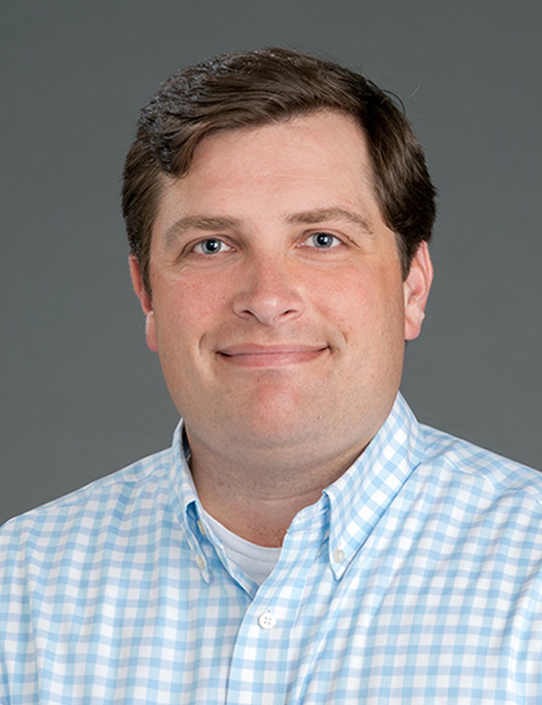
The loss of the mortality benefits corresponded with a steady climb in the average systolic blood pressures (SBP) in the intensive treatment group after the trial ended. The long-term benefit serves as a call to develop better strategies for sustained SBP control.
“We were disappointed but not surprised that the blood pressure levels in the intensive goal group were not sustained,” acknowledged William C. Cushman, MD, Medical Director, department of preventive medicine, University of Tennessee Health Science Center, Memphis. “There are many trials showing no residual or legacy effect once the intervention is stopped.”
Long-term results do not weaken SPRINT
One of the coinvestigators of this most recent analysis published in JAMA Cardiology and a member of the SPRINT writing committee at the time of its 2015 publication in the New England Journal of Medicine, Dr. Cushman pointed out that the long-term results do not weaken the main trial result. Long-term adherence was not part of the trial design.
“After the trial, we were no longer treating these participants, so it was up to them and their primary care providers to decide on blood pressure goals,” he noted in an interview. Based on the trajectory of benefit when the study was stopped, “it is possible longer intensive treatment may lead to more benefit and some long-term residual benefits.”
The senior author of this most recent analysis, Nicholas M. Pajewski, PhD, associate professor of biostatistics and data science, Wake Forest University, Winston-Salem, N.C., generally agreed. However, he pointed out that the most recent data do not rule out meaningful benefit after the study ended.
For one reason, the loss of the SBP advantage was gradual so that median SBP levels of the two groups did not meet for nearly 3 years. This likely explains why there was still an attenuation of CVD mortality for several years after the all-cause mortality benefit was lost, according to Dr. Pajewski.
“It is important to mention that we were not able to assess nonfatal cardiovascular events, so while the two groups do eventually come together, if one thinks about the distinction of healthspan versus lifespan, there was probably residual benefit in terms of delaying CVD morbidity and mortality,” Dr. Pajewski said.
In SPRINT, CVD mortality reduced 43%
In the 9,631-patient SPRINT trial, the intensive treatment group achieved a mean SBP of 121.4 mm Hg versus 136.2 mm Hg in the standard treatment group at the end of 1 year. The trial was stopped early after 3.26 years because of strength of the benefit in the intensive treatment arm. At that time, the reductions by hazard ratio were 25% (HR, 0.75; P < .001) for a composite major adverse cardiovascular event (MACE) endpoint, 43% for CVD mortality (P = .005), and 27% for all-cause mortality (P = .003).
In the new observational follow-up, mortality data were drawn from the National Death Index, and change in SBP from electronic health records in a subset of 2,944 SPRINT trial participants. Data were available and analyzed through 2020.
The newly published long-term observational analysis showed that the median SBP in the intensive treatment arm was already climbing by the end of the end of the trial. It reached 132.8 mm Hg at 5 years after randomization and then 140.4 mm Hg by 10 years.
This latter figure was essentially equivalent to the SBP among those who were initially randomized to the standard treatment arm.
Factors driving rising BP are unclear
There is limited information on what medications were taken by either group following the end of the trial, so the reason for the regression in the intensive treatment arm after leaving the trial is unknown. The authors speculated that this might have been due to therapeutic inertia among treating physicians, poor adherence among patients, the difficulty of keeping blood pressures low in patients with advancing pathology, or some combination of these.
“Perhaps the most important reason was that providers and patients were not aiming for the lower goals since guidelines did not recommend these targets until 2017,” Dr. Cushman pointed out. He noted that Healthcare Effectiveness Data and Information Set (HEDIS) “has still not adopted a performance measure goal of less than 140 mm Hg.”
In an accompanying editorial, the authors focused on what these data mean for population-based strategies to achieve sustained control of one of the most important risk factors for cardiovascular events. Led by Daniel W. Jones, MD, director of clinical and population science, University of Mississippi, Jackson, the authors of the editorial wrote that these data emphasized “the challenge of achieving sustained intensive BP reductions in the real-world setting.”

Basically, the editorial concluded that current approaches to achieving meaningful and sustained blood pressure control are not working.
This study “should be a wakeup call, but other previously published good data have also been ignored,” said Dr. Jones in an interview. Despite the compelling benefit from intensive blood pressure control the SPRINT trial, the observational follow-up emphasizes the difficulty of maintaining the rigorous reductions in blood pressure needed for sustained protection.
“Systemic change is necessary,” said Dr. Jones, reprising the major thrust of the editorial he wrote with Donald Clark III, MD, and Michael E. Hall, MD, who are both colleagues at the University of Mississippi.
“My view is that health care providers should be held responsible for motivating better compliance of their patients, just as a teacher is accountable for the outcomes of their students,” he said.
The solutions are not likely to be simple. Dr. Jones called for multiple strategies, such as employing telehealth and community health workers to monitor and reinforce blood pressure control, but he said that these and other data have convinced him that “simply trying harder at what we currently do” is not enough.
Dr. Pajewski and Dr. Jones report no potential conflicts of interest. Dr. Cushman reports a financial relationship with ReCor.
The substantial reductions in cardiovascular disease (CVD) and all-cause mortality achieved with intensive blood pressure lowering in the landmark SPRINT trial were not sustained in a newly released long-term follow-up.
The loss of the mortality benefits corresponded with a steady climb in the average systolic blood pressures (SBP) in the intensive treatment group after the trial ended. The long-term benefit serves as a call to develop better strategies for sustained SBP control.
“We were disappointed but not surprised that the blood pressure levels in the intensive goal group were not sustained,” acknowledged William C. Cushman, MD, Medical Director, department of preventive medicine, University of Tennessee Health Science Center, Memphis. “There are many trials showing no residual or legacy effect once the intervention is stopped.”
Long-term results do not weaken SPRINT
One of the coinvestigators of this most recent analysis published in JAMA Cardiology and a member of the SPRINT writing committee at the time of its 2015 publication in the New England Journal of Medicine, Dr. Cushman pointed out that the long-term results do not weaken the main trial result. Long-term adherence was not part of the trial design.
“After the trial, we were no longer treating these participants, so it was up to them and their primary care providers to decide on blood pressure goals,” he noted in an interview. Based on the trajectory of benefit when the study was stopped, “it is possible longer intensive treatment may lead to more benefit and some long-term residual benefits.”
The senior author of this most recent analysis, Nicholas M. Pajewski, PhD, associate professor of biostatistics and data science, Wake Forest University, Winston-Salem, N.C., generally agreed. However, he pointed out that the most recent data do not rule out meaningful benefit after the study ended.
For one reason, the loss of the SBP advantage was gradual so that median SBP levels of the two groups did not meet for nearly 3 years. This likely explains why there was still an attenuation of CVD mortality for several years after the all-cause mortality benefit was lost, according to Dr. Pajewski.
“It is important to mention that we were not able to assess nonfatal cardiovascular events, so while the two groups do eventually come together, if one thinks about the distinction of healthspan versus lifespan, there was probably residual benefit in terms of delaying CVD morbidity and mortality,” Dr. Pajewski said.
In SPRINT, CVD mortality reduced 43%
In the 9,631-patient SPRINT trial, the intensive treatment group achieved a mean SBP of 121.4 mm Hg versus 136.2 mm Hg in the standard treatment group at the end of 1 year. The trial was stopped early after 3.26 years because of strength of the benefit in the intensive treatment arm. At that time, the reductions by hazard ratio were 25% (HR, 0.75; P < .001) for a composite major adverse cardiovascular event (MACE) endpoint, 43% for CVD mortality (P = .005), and 27% for all-cause mortality (P = .003).
In the new observational follow-up, mortality data were drawn from the National Death Index, and change in SBP from electronic health records in a subset of 2,944 SPRINT trial participants. Data were available and analyzed through 2020.
The newly published long-term observational analysis showed that the median SBP in the intensive treatment arm was already climbing by the end of the end of the trial. It reached 132.8 mm Hg at 5 years after randomization and then 140.4 mm Hg by 10 years.
This latter figure was essentially equivalent to the SBP among those who were initially randomized to the standard treatment arm.
Factors driving rising BP are unclear
There is limited information on what medications were taken by either group following the end of the trial, so the reason for the regression in the intensive treatment arm after leaving the trial is unknown. The authors speculated that this might have been due to therapeutic inertia among treating physicians, poor adherence among patients, the difficulty of keeping blood pressures low in patients with advancing pathology, or some combination of these.
“Perhaps the most important reason was that providers and patients were not aiming for the lower goals since guidelines did not recommend these targets until 2017,” Dr. Cushman pointed out. He noted that Healthcare Effectiveness Data and Information Set (HEDIS) “has still not adopted a performance measure goal of less than 140 mm Hg.”
In an accompanying editorial, the authors focused on what these data mean for population-based strategies to achieve sustained control of one of the most important risk factors for cardiovascular events. Led by Daniel W. Jones, MD, director of clinical and population science, University of Mississippi, Jackson, the authors of the editorial wrote that these data emphasized “the challenge of achieving sustained intensive BP reductions in the real-world setting.”
Basically, the editorial concluded that current approaches to achieving meaningful and sustained blood pressure control are not working.
This study “should be a wakeup call, but other previously published good data have also been ignored,” said Dr. Jones in an interview. Despite the compelling benefit from intensive blood pressure control the SPRINT trial, the observational follow-up emphasizes the difficulty of maintaining the rigorous reductions in blood pressure needed for sustained protection.
“Systemic change is necessary,” said Dr. Jones, reprising the major thrust of the editorial he wrote with Donald Clark III, MD, and Michael E. Hall, MD, who are both colleagues at the University of Mississippi.
“My view is that health care providers should be held responsible for motivating better compliance of their patients, just as a teacher is accountable for the outcomes of their students,” he said.
The solutions are not likely to be simple. Dr. Jones called for multiple strategies, such as employing telehealth and community health workers to monitor and reinforce blood pressure control, but he said that these and other data have convinced him that “simply trying harder at what we currently do” is not enough.
Dr. Pajewski and Dr. Jones report no potential conflicts of interest. Dr. Cushman reports a financial relationship with ReCor.
The substantial reductions in cardiovascular disease (CVD) and all-cause mortality achieved with intensive blood pressure lowering in the landmark SPRINT trial were not sustained in a newly released long-term follow-up.
The loss of the mortality benefits corresponded with a steady climb in the average systolic blood pressures (SBP) in the intensive treatment group after the trial ended. The long-term benefit serves as a call to develop better strategies for sustained SBP control.
“We were disappointed but not surprised that the blood pressure levels in the intensive goal group were not sustained,” acknowledged William C. Cushman, MD, Medical Director, department of preventive medicine, University of Tennessee Health Science Center, Memphis. “There are many trials showing no residual or legacy effect once the intervention is stopped.”
Long-term results do not weaken SPRINT
One of the coinvestigators of this most recent analysis published in JAMA Cardiology and a member of the SPRINT writing committee at the time of its 2015 publication in the New England Journal of Medicine, Dr. Cushman pointed out that the long-term results do not weaken the main trial result. Long-term adherence was not part of the trial design.
“After the trial, we were no longer treating these participants, so it was up to them and their primary care providers to decide on blood pressure goals,” he noted in an interview. Based on the trajectory of benefit when the study was stopped, “it is possible longer intensive treatment may lead to more benefit and some long-term residual benefits.”
The senior author of this most recent analysis, Nicholas M. Pajewski, PhD, associate professor of biostatistics and data science, Wake Forest University, Winston-Salem, N.C., generally agreed. However, he pointed out that the most recent data do not rule out meaningful benefit after the study ended.
For one reason, the loss of the SBP advantage was gradual so that median SBP levels of the two groups did not meet for nearly 3 years. This likely explains why there was still an attenuation of CVD mortality for several years after the all-cause mortality benefit was lost, according to Dr. Pajewski.
“It is important to mention that we were not able to assess nonfatal cardiovascular events, so while the two groups do eventually come together, if one thinks about the distinction of healthspan versus lifespan, there was probably residual benefit in terms of delaying CVD morbidity and mortality,” Dr. Pajewski said.
In SPRINT, CVD mortality reduced 43%
In the 9,631-patient SPRINT trial, the intensive treatment group achieved a mean SBP of 121.4 mm Hg versus 136.2 mm Hg in the standard treatment group at the end of 1 year. The trial was stopped early after 3.26 years because of strength of the benefit in the intensive treatment arm. At that time, the reductions by hazard ratio were 25% (HR, 0.75; P < .001) for a composite major adverse cardiovascular event (MACE) endpoint, 43% for CVD mortality (P = .005), and 27% for all-cause mortality (P = .003).
In the new observational follow-up, mortality data were drawn from the National Death Index, and change in SBP from electronic health records in a subset of 2,944 SPRINT trial participants. Data were available and analyzed through 2020.
The newly published long-term observational analysis showed that the median SBP in the intensive treatment arm was already climbing by the end of the end of the trial. It reached 132.8 mm Hg at 5 years after randomization and then 140.4 mm Hg by 10 years.
This latter figure was essentially equivalent to the SBP among those who were initially randomized to the standard treatment arm.
Factors driving rising BP are unclear
There is limited information on what medications were taken by either group following the end of the trial, so the reason for the regression in the intensive treatment arm after leaving the trial is unknown. The authors speculated that this might have been due to therapeutic inertia among treating physicians, poor adherence among patients, the difficulty of keeping blood pressures low in patients with advancing pathology, or some combination of these.
“Perhaps the most important reason was that providers and patients were not aiming for the lower goals since guidelines did not recommend these targets until 2017,” Dr. Cushman pointed out. He noted that Healthcare Effectiveness Data and Information Set (HEDIS) “has still not adopted a performance measure goal of less than 140 mm Hg.”
In an accompanying editorial, the authors focused on what these data mean for population-based strategies to achieve sustained control of one of the most important risk factors for cardiovascular events. Led by Daniel W. Jones, MD, director of clinical and population science, University of Mississippi, Jackson, the authors of the editorial wrote that these data emphasized “the challenge of achieving sustained intensive BP reductions in the real-world setting.”
Basically, the editorial concluded that current approaches to achieving meaningful and sustained blood pressure control are not working.
This study “should be a wakeup call, but other previously published good data have also been ignored,” said Dr. Jones in an interview. Despite the compelling benefit from intensive blood pressure control the SPRINT trial, the observational follow-up emphasizes the difficulty of maintaining the rigorous reductions in blood pressure needed for sustained protection.
“Systemic change is necessary,” said Dr. Jones, reprising the major thrust of the editorial he wrote with Donald Clark III, MD, and Michael E. Hall, MD, who are both colleagues at the University of Mississippi.
“My view is that health care providers should be held responsible for motivating better compliance of their patients, just as a teacher is accountable for the outcomes of their students,” he said.
The solutions are not likely to be simple. Dr. Jones called for multiple strategies, such as employing telehealth and community health workers to monitor and reinforce blood pressure control, but he said that these and other data have convinced him that “simply trying harder at what we currently do” is not enough.
Dr. Pajewski and Dr. Jones report no potential conflicts of interest. Dr. Cushman reports a financial relationship with ReCor.
FROM JAMA CARDIOLOGY
CVS cuts prices of menstrual products, covers sales tax in some states
CVS is cutting the cost of its store-branded menstrual products and paying state sales taxes on them in a dozen states.
The drug store chain said that starting Thursday it was reducing prices on CVS Health and Live Better tampons, menstrual pads, liners, and cups by 25%.
“Women deserve quality when it comes to the products they may need each month,” CVS said in a statement. “We’re paying the tax on period products on behalf of our customers where and when possible, and are working to help eliminate the tax nationwide.”
The store is also trying to equalize costs between men’s and women’s hygiene products, like razors.
The chain is paying sales taxes on period products in these 12 states: Arkansas, Georgia, Hawaii, Louisiana, Missouri, South Carolina, Tennessee, Texas, Utah, Virginia, Wisconsin, and West Virginia.
It can’t pay the taxes in other states that have them because of laws that prevent third parties from paying taxes for a customer.
“This move will highlight their commitment to addressing women’s health and pave the way for reducing menstrual inequity,” Padmini Murthy, MD, the global health lead for the American Medical Women’s Association, said in an email to CNN, “and not just to promote the use of CVS products.”
Twenty-three states don’t tax feminine hygiene products, says the Alliance for Period Supplies, an advocacy group seeking to expand access to menstrual supplies.
“Too often period products are taxed as luxury items and not recognized as basic necessities,” the organization said. “Period products are taxed at a similar rate to items like decor, electronics, makeup, and toys.”
Tampon prices rose 12.2% for the year ending Oct. 2, according to market research firm IRI.
And 25% of women struggle to buy the products because of the expense, says the group.
A version of this article first appeared on WebMD.com.
CVS is cutting the cost of its store-branded menstrual products and paying state sales taxes on them in a dozen states.
The drug store chain said that starting Thursday it was reducing prices on CVS Health and Live Better tampons, menstrual pads, liners, and cups by 25%.
“Women deserve quality when it comes to the products they may need each month,” CVS said in a statement. “We’re paying the tax on period products on behalf of our customers where and when possible, and are working to help eliminate the tax nationwide.”
The store is also trying to equalize costs between men’s and women’s hygiene products, like razors.
The chain is paying sales taxes on period products in these 12 states: Arkansas, Georgia, Hawaii, Louisiana, Missouri, South Carolina, Tennessee, Texas, Utah, Virginia, Wisconsin, and West Virginia.
It can’t pay the taxes in other states that have them because of laws that prevent third parties from paying taxes for a customer.
“This move will highlight their commitment to addressing women’s health and pave the way for reducing menstrual inequity,” Padmini Murthy, MD, the global health lead for the American Medical Women’s Association, said in an email to CNN, “and not just to promote the use of CVS products.”
Twenty-three states don’t tax feminine hygiene products, says the Alliance for Period Supplies, an advocacy group seeking to expand access to menstrual supplies.
“Too often period products are taxed as luxury items and not recognized as basic necessities,” the organization said. “Period products are taxed at a similar rate to items like decor, electronics, makeup, and toys.”
Tampon prices rose 12.2% for the year ending Oct. 2, according to market research firm IRI.
And 25% of women struggle to buy the products because of the expense, says the group.
A version of this article first appeared on WebMD.com.
CVS is cutting the cost of its store-branded menstrual products and paying state sales taxes on them in a dozen states.
The drug store chain said that starting Thursday it was reducing prices on CVS Health and Live Better tampons, menstrual pads, liners, and cups by 25%.
“Women deserve quality when it comes to the products they may need each month,” CVS said in a statement. “We’re paying the tax on period products on behalf of our customers where and when possible, and are working to help eliminate the tax nationwide.”
The store is also trying to equalize costs between men’s and women’s hygiene products, like razors.
The chain is paying sales taxes on period products in these 12 states: Arkansas, Georgia, Hawaii, Louisiana, Missouri, South Carolina, Tennessee, Texas, Utah, Virginia, Wisconsin, and West Virginia.
It can’t pay the taxes in other states that have them because of laws that prevent third parties from paying taxes for a customer.
“This move will highlight their commitment to addressing women’s health and pave the way for reducing menstrual inequity,” Padmini Murthy, MD, the global health lead for the American Medical Women’s Association, said in an email to CNN, “and not just to promote the use of CVS products.”
Twenty-three states don’t tax feminine hygiene products, says the Alliance for Period Supplies, an advocacy group seeking to expand access to menstrual supplies.
“Too often period products are taxed as luxury items and not recognized as basic necessities,” the organization said. “Period products are taxed at a similar rate to items like decor, electronics, makeup, and toys.”
Tampon prices rose 12.2% for the year ending Oct. 2, according to market research firm IRI.
And 25% of women struggle to buy the products because of the expense, says the group.
A version of this article first appeared on WebMD.com.
Trial of early intensive meds at HF discharge halted for benefit: STRONG-HF
A “high-intensity-care” strategy based on early and rapid uptitration of guideline-directed meds improves postdischarge clinical outcomes for patients hospitalized with decompensated heart failure (HF), suggest topline results from a randomized trial.
The STRONG-HF study was halted early on recommendation from its data safety monitoring board after an interim analysis suggested the high-intensity-care strategy significantly cut risk of death or HF readmission, compared with a standard-of-care approach.
The trial termination was announced in a press release from one of its sponsors, The Heart Initiative, a nonprofit organization. STRONG-HF was also supported by Roche Diagnostics.
The early termination was based on interim data from the approximately 1,000 patients, out of an estimated planned enrollment of 1,800, who had been followed for at least 90 days. The study’s actual primary endpoint had been defined by death or HF readmission at 6 months.
The announcement did not include outcomes data or P values, or any other indication of the magnitude of benefit from the high-intensity-care approach.
Patients in STRONG-HF who had been assigned to a high-intensity-care strategy had been started in-hospital on a beta blocker, a renin-angiotensin system inhibitor (RASi), and a mineralocorticoid receptor blocker (MRA) with dosages uptitrated at least halfway by the time of discharge.
The meds were uptitrated fully within 2 weeks of discharge guided by clinical and biomarker assessments, especially natriuretic peptides, at frequent postdischarge visits, the press release states.
Patients conducted “safety visits 1 week after any uptitration and follow-up visits at 6 weeks and 3 months,” the announcement notes. “At each visit, patients were assessed by physical examination for congestion and blood tests, including NT-proBNP measurements.”
The “full STRONG-HF trial results” are scheduled for presentation at the American Heart Association annual scientific sessions, the announcement states.
STRONG-HF is sponsored by The Heart Initiative and Roche Diagnostics.
A version of this article first appeared on Medscape.com.
A “high-intensity-care” strategy based on early and rapid uptitration of guideline-directed meds improves postdischarge clinical outcomes for patients hospitalized with decompensated heart failure (HF), suggest topline results from a randomized trial.
The STRONG-HF study was halted early on recommendation from its data safety monitoring board after an interim analysis suggested the high-intensity-care strategy significantly cut risk of death or HF readmission, compared with a standard-of-care approach.
The trial termination was announced in a press release from one of its sponsors, The Heart Initiative, a nonprofit organization. STRONG-HF was also supported by Roche Diagnostics.
The early termination was based on interim data from the approximately 1,000 patients, out of an estimated planned enrollment of 1,800, who had been followed for at least 90 days. The study’s actual primary endpoint had been defined by death or HF readmission at 6 months.
The announcement did not include outcomes data or P values, or any other indication of the magnitude of benefit from the high-intensity-care approach.
Patients in STRONG-HF who had been assigned to a high-intensity-care strategy had been started in-hospital on a beta blocker, a renin-angiotensin system inhibitor (RASi), and a mineralocorticoid receptor blocker (MRA) with dosages uptitrated at least halfway by the time of discharge.
The meds were uptitrated fully within 2 weeks of discharge guided by clinical and biomarker assessments, especially natriuretic peptides, at frequent postdischarge visits, the press release states.
Patients conducted “safety visits 1 week after any uptitration and follow-up visits at 6 weeks and 3 months,” the announcement notes. “At each visit, patients were assessed by physical examination for congestion and blood tests, including NT-proBNP measurements.”
The “full STRONG-HF trial results” are scheduled for presentation at the American Heart Association annual scientific sessions, the announcement states.
STRONG-HF is sponsored by The Heart Initiative and Roche Diagnostics.
A version of this article first appeared on Medscape.com.
A “high-intensity-care” strategy based on early and rapid uptitration of guideline-directed meds improves postdischarge clinical outcomes for patients hospitalized with decompensated heart failure (HF), suggest topline results from a randomized trial.
The STRONG-HF study was halted early on recommendation from its data safety monitoring board after an interim analysis suggested the high-intensity-care strategy significantly cut risk of death or HF readmission, compared with a standard-of-care approach.
The trial termination was announced in a press release from one of its sponsors, The Heart Initiative, a nonprofit organization. STRONG-HF was also supported by Roche Diagnostics.
The early termination was based on interim data from the approximately 1,000 patients, out of an estimated planned enrollment of 1,800, who had been followed for at least 90 days. The study’s actual primary endpoint had been defined by death or HF readmission at 6 months.
The announcement did not include outcomes data or P values, or any other indication of the magnitude of benefit from the high-intensity-care approach.
Patients in STRONG-HF who had been assigned to a high-intensity-care strategy had been started in-hospital on a beta blocker, a renin-angiotensin system inhibitor (RASi), and a mineralocorticoid receptor blocker (MRA) with dosages uptitrated at least halfway by the time of discharge.
The meds were uptitrated fully within 2 weeks of discharge guided by clinical and biomarker assessments, especially natriuretic peptides, at frequent postdischarge visits, the press release states.
Patients conducted “safety visits 1 week after any uptitration and follow-up visits at 6 weeks and 3 months,” the announcement notes. “At each visit, patients were assessed by physical examination for congestion and blood tests, including NT-proBNP measurements.”
The “full STRONG-HF trial results” are scheduled for presentation at the American Heart Association annual scientific sessions, the announcement states.
STRONG-HF is sponsored by The Heart Initiative and Roche Diagnostics.
A version of this article first appeared on Medscape.com.