User login
TNF-alpha, oxidative stress disturbance may play role in schizophrenia pathophysiology
Disturbance of tumor necrosis factor (TNF)–alpha and oxidative stress status may be involved in the pathophysiology of schizophrenia, new study results suggest.
In a study published in Psychoneuroendocrinology, the investigators collected blood samples from 119 patients with schizophrenia and 135 controls. Along with TNF-alpha, assays for the oxidative stress markers superoxide dismutase (SOD), glutathione peroxidase (GSH-Px), catalase (CAT), and malondialdehyde (MDA) were measured. The average illness duration in patients with schizophrenia was 8.23 months, and their average total Positive and Negative Syndrome Scale score was 87.64, reported Shiguang Zhu of Nanjing (China) Medical University and associates.
Serum levels of TNF-alpha and MDA were significantly higher (P = .007 for both), and GSH-Px levels were significantly lower (P = .005), in patients with schizophrenia, compared with controls, after Bonferroni correction. The interaction between GSH-Px and TNF-alpha was negatively associated with the presence of schizophrenia (odds ratio, 0.99; 95% confidence interval, 0.98-0.99; P = .001), and the interaction between MDA and TNF-alpha was positively associated with schizophrenia risk (OR, 1.61, 95% CI, 1.16-2.24, P = .004).
“It is worth[while] to note that [the] immune-inflammatory and oxidative stress hypothesis are just one of the theories for schizophrenic development, and other neurobiological theories such as neurodevelopmental dysfunction and hypothalamus-pituitary-adrenal axis hormones disturbance should be considered,” the investigators wrote. However, their study “suggests that TNF-alpha and disturbance of oxidative stress status as well as their interaction may be involved in the pathophysiology of schizophrenia.”
The study was supported by the National Natural Science Foundation of China, Shanghai Jiao Tong University Medical Engineering Foundation, Shanghai Jiao Tong University School of Medicine, and CAS Key Laboratory of Mental Health. The investigators reported that they had no conflicts of interest.
SOURCE: Zhu S et al. Psychoneuroendocrinology. 2020 Jan 30. doi: 10.1016/j.psyneuen.2020.104595.
Disturbance of tumor necrosis factor (TNF)–alpha and oxidative stress status may be involved in the pathophysiology of schizophrenia, new study results suggest.
In a study published in Psychoneuroendocrinology, the investigators collected blood samples from 119 patients with schizophrenia and 135 controls. Along with TNF-alpha, assays for the oxidative stress markers superoxide dismutase (SOD), glutathione peroxidase (GSH-Px), catalase (CAT), and malondialdehyde (MDA) were measured. The average illness duration in patients with schizophrenia was 8.23 months, and their average total Positive and Negative Syndrome Scale score was 87.64, reported Shiguang Zhu of Nanjing (China) Medical University and associates.
Serum levels of TNF-alpha and MDA were significantly higher (P = .007 for both), and GSH-Px levels were significantly lower (P = .005), in patients with schizophrenia, compared with controls, after Bonferroni correction. The interaction between GSH-Px and TNF-alpha was negatively associated with the presence of schizophrenia (odds ratio, 0.99; 95% confidence interval, 0.98-0.99; P = .001), and the interaction between MDA and TNF-alpha was positively associated with schizophrenia risk (OR, 1.61, 95% CI, 1.16-2.24, P = .004).
“It is worth[while] to note that [the] immune-inflammatory and oxidative stress hypothesis are just one of the theories for schizophrenic development, and other neurobiological theories such as neurodevelopmental dysfunction and hypothalamus-pituitary-adrenal axis hormones disturbance should be considered,” the investigators wrote. However, their study “suggests that TNF-alpha and disturbance of oxidative stress status as well as their interaction may be involved in the pathophysiology of schizophrenia.”
The study was supported by the National Natural Science Foundation of China, Shanghai Jiao Tong University Medical Engineering Foundation, Shanghai Jiao Tong University School of Medicine, and CAS Key Laboratory of Mental Health. The investigators reported that they had no conflicts of interest.
SOURCE: Zhu S et al. Psychoneuroendocrinology. 2020 Jan 30. doi: 10.1016/j.psyneuen.2020.104595.
Disturbance of tumor necrosis factor (TNF)–alpha and oxidative stress status may be involved in the pathophysiology of schizophrenia, new study results suggest.
In a study published in Psychoneuroendocrinology, the investigators collected blood samples from 119 patients with schizophrenia and 135 controls. Along with TNF-alpha, assays for the oxidative stress markers superoxide dismutase (SOD), glutathione peroxidase (GSH-Px), catalase (CAT), and malondialdehyde (MDA) were measured. The average illness duration in patients with schizophrenia was 8.23 months, and their average total Positive and Negative Syndrome Scale score was 87.64, reported Shiguang Zhu of Nanjing (China) Medical University and associates.
Serum levels of TNF-alpha and MDA were significantly higher (P = .007 for both), and GSH-Px levels were significantly lower (P = .005), in patients with schizophrenia, compared with controls, after Bonferroni correction. The interaction between GSH-Px and TNF-alpha was negatively associated with the presence of schizophrenia (odds ratio, 0.99; 95% confidence interval, 0.98-0.99; P = .001), and the interaction between MDA and TNF-alpha was positively associated with schizophrenia risk (OR, 1.61, 95% CI, 1.16-2.24, P = .004).
“It is worth[while] to note that [the] immune-inflammatory and oxidative stress hypothesis are just one of the theories for schizophrenic development, and other neurobiological theories such as neurodevelopmental dysfunction and hypothalamus-pituitary-adrenal axis hormones disturbance should be considered,” the investigators wrote. However, their study “suggests that TNF-alpha and disturbance of oxidative stress status as well as their interaction may be involved in the pathophysiology of schizophrenia.”
The study was supported by the National Natural Science Foundation of China, Shanghai Jiao Tong University Medical Engineering Foundation, Shanghai Jiao Tong University School of Medicine, and CAS Key Laboratory of Mental Health. The investigators reported that they had no conflicts of interest.
SOURCE: Zhu S et al. Psychoneuroendocrinology. 2020 Jan 30. doi: 10.1016/j.psyneuen.2020.104595.
FROM PSYCHONEUROENDOCRINOLOGY
New lipid-lowering drug earns FDA approval
The Food and Drug Administration has approved bempedoic acid (Nexletol) for the treatment of adults with heterozygous familial hypercholesterolemia (HeFH) or established atherosclerotic cardiovascular disease (ASCVD) who require additional LDL cholesterol lowering.

The oral adenosine triphosphate–citrate lyase (ACL) inhibitor is indicated as an adjunct to diet and maximally tolerated statin therapy in these patients, and approved at the 180 mg once daily dose, the agency announced today.
The safety and efficacy of bempedoic acid were demonstrated over 52 weeks in two multicenter randomized, clinical trials involving 3,009 adults with HeFH or established ASCVD on maximally tolerated statin therapy.
The difference between bempedoic acid and placebo for the primary outcome of change in LDL cholesterol from baseline to week 12 was –18% in the first trial, CLEAR Harmony (95% confidence interval, –20% to –16%; P less than .001), and –17% in the second trial, CLEAR Wisdom (95% CI, –21% to –14%; P less than .001).
The label notes that the effect on cardiovascular morbidity and mortality has not been determined. The label also includes warnings stating that bempedoic acid may increase blood uric acid levels and is associated with an increased risk of tendon rupture or injury.
In clinical trials, 26% of bempedoic acid–treated patients with normal baseline uric acid values versus 9.5% of placebo-treated patients experienced hyperuricemia one or more times, and 3.5% of patients experienced clinically significant hyperuricemia reported as an adverse reaction versus 1.1% with placebo, according to the label. Gout was reported in 1.5% of patients treated with bempedoic acid and 0.4% of those treated with placebo.
Also in clinical trials, the risk of tendon rupture was 0.5% with bempedoic acid and 0% with placebo. Tendon rupture involved the rotator cuff, biceps tendon, or Achilles tendon, and occurred within weeks to months of starting the drug. Rupture may “occur more frequently in patients over 60 years of age, in those taking corticosteroid or fluoroquinolone drugs, in patients with renal failure, and in patients with previous tendon disorders,” the label states.
The label also advises that patients avoid concomitant use of bempedoic acid with simvastatin greater than 20 mg or pravastatin greater than 40 mg because it causes an increase in statin concentrations and may increase the risk of related myopathy.
A decision is expected shortly on a new drug application submitted by Esperion for an LDL cholesterol–lowering indication for bempedoic acid 180 mg/ezetimibe 10 mg combination tablet.
Full prescribing information is available online.
This article first appeared on Medscape.com.
The Food and Drug Administration has approved bempedoic acid (Nexletol) for the treatment of adults with heterozygous familial hypercholesterolemia (HeFH) or established atherosclerotic cardiovascular disease (ASCVD) who require additional LDL cholesterol lowering.
The oral adenosine triphosphate–citrate lyase (ACL) inhibitor is indicated as an adjunct to diet and maximally tolerated statin therapy in these patients, and approved at the 180 mg once daily dose, the agency announced today.
The safety and efficacy of bempedoic acid were demonstrated over 52 weeks in two multicenter randomized, clinical trials involving 3,009 adults with HeFH or established ASCVD on maximally tolerated statin therapy.
The difference between bempedoic acid and placebo for the primary outcome of change in LDL cholesterol from baseline to week 12 was –18% in the first trial, CLEAR Harmony (95% confidence interval, –20% to –16%; P less than .001), and –17% in the second trial, CLEAR Wisdom (95% CI, –21% to –14%; P less than .001).
The label notes that the effect on cardiovascular morbidity and mortality has not been determined. The label also includes warnings stating that bempedoic acid may increase blood uric acid levels and is associated with an increased risk of tendon rupture or injury.
In clinical trials, 26% of bempedoic acid–treated patients with normal baseline uric acid values versus 9.5% of placebo-treated patients experienced hyperuricemia one or more times, and 3.5% of patients experienced clinically significant hyperuricemia reported as an adverse reaction versus 1.1% with placebo, according to the label. Gout was reported in 1.5% of patients treated with bempedoic acid and 0.4% of those treated with placebo.
Also in clinical trials, the risk of tendon rupture was 0.5% with bempedoic acid and 0% with placebo. Tendon rupture involved the rotator cuff, biceps tendon, or Achilles tendon, and occurred within weeks to months of starting the drug. Rupture may “occur more frequently in patients over 60 years of age, in those taking corticosteroid or fluoroquinolone drugs, in patients with renal failure, and in patients with previous tendon disorders,” the label states.
The label also advises that patients avoid concomitant use of bempedoic acid with simvastatin greater than 20 mg or pravastatin greater than 40 mg because it causes an increase in statin concentrations and may increase the risk of related myopathy.
A decision is expected shortly on a new drug application submitted by Esperion for an LDL cholesterol–lowering indication for bempedoic acid 180 mg/ezetimibe 10 mg combination tablet.
Full prescribing information is available online.
This article first appeared on Medscape.com.
The Food and Drug Administration has approved bempedoic acid (Nexletol) for the treatment of adults with heterozygous familial hypercholesterolemia (HeFH) or established atherosclerotic cardiovascular disease (ASCVD) who require additional LDL cholesterol lowering.
The oral adenosine triphosphate–citrate lyase (ACL) inhibitor is indicated as an adjunct to diet and maximally tolerated statin therapy in these patients, and approved at the 180 mg once daily dose, the agency announced today.
The safety and efficacy of bempedoic acid were demonstrated over 52 weeks in two multicenter randomized, clinical trials involving 3,009 adults with HeFH or established ASCVD on maximally tolerated statin therapy.
The difference between bempedoic acid and placebo for the primary outcome of change in LDL cholesterol from baseline to week 12 was –18% in the first trial, CLEAR Harmony (95% confidence interval, –20% to –16%; P less than .001), and –17% in the second trial, CLEAR Wisdom (95% CI, –21% to –14%; P less than .001).
The label notes that the effect on cardiovascular morbidity and mortality has not been determined. The label also includes warnings stating that bempedoic acid may increase blood uric acid levels and is associated with an increased risk of tendon rupture or injury.
In clinical trials, 26% of bempedoic acid–treated patients with normal baseline uric acid values versus 9.5% of placebo-treated patients experienced hyperuricemia one or more times, and 3.5% of patients experienced clinically significant hyperuricemia reported as an adverse reaction versus 1.1% with placebo, according to the label. Gout was reported in 1.5% of patients treated with bempedoic acid and 0.4% of those treated with placebo.
Also in clinical trials, the risk of tendon rupture was 0.5% with bempedoic acid and 0% with placebo. Tendon rupture involved the rotator cuff, biceps tendon, or Achilles tendon, and occurred within weeks to months of starting the drug. Rupture may “occur more frequently in patients over 60 years of age, in those taking corticosteroid or fluoroquinolone drugs, in patients with renal failure, and in patients with previous tendon disorders,” the label states.
The label also advises that patients avoid concomitant use of bempedoic acid with simvastatin greater than 20 mg or pravastatin greater than 40 mg because it causes an increase in statin concentrations and may increase the risk of related myopathy.
A decision is expected shortly on a new drug application submitted by Esperion for an LDL cholesterol–lowering indication for bempedoic acid 180 mg/ezetimibe 10 mg combination tablet.
Full prescribing information is available online.
This article first appeared on Medscape.com.
Cigarette smoking is associated with prefrontal function in patients with schizophrenia
Patients with schizophrenia have decreased chronnectomic density in the dorsolateral prefrontal cortex, compared with healthy controls, and cigarette smoking in patients with schizophrenia may be associated with a degree of preserved function in that brain region, researchers reported. The results indicate that smoking may be associated with a preservation effect, but it “cannot restore patients’ prefrontal dysfunction to normal levels,” the researchers said.
The chronnectome depicts how brain functional connectivity patterns (i.e., the connectome) vary over time. “Therefore, the chronnectome may be an effective index to evaluate the smoking-related prefrontal functional changes in schizophrenia,” said Yun-Shuang Fan, a researcher at the Clinical Hospital of Chengdu Brain Science Institute in China, and colleagues in the report, which was published in Progress in Neuro-Psychopharmacology & Biological Psychiatry.
The investigators studied 49 patients with schizophrenia, including 22 smokers and 27 nonsmokers, and 43 healthy controls, including 22 smokers and 21 nonsmokers. Participants underwent resting-state functional magnetic resonance imaging, and the researchers analyzed chronnectomic density using a sliding-window method. The investigators examined interactions between smoking status and diagnosis.
Smoking was associated with reduced chronnectomic density in healthy controls, but increased density in patients with schizophrenia. The study provides a “framework to elaborate upon the self-medication hypothesis in schizophrenia” and sheds “some fresh light on the elevated rates of smoking in schizophrenia,” they said.
The study was relatively small, and patients’ use of antipsychotic medications, which can affect the connectome, may limit the results. In addition, the study’s cross-sectional design precludes knowing whether “smoking behavior is the cause or result of the prefrontal chronnectome alterations in schizophrenia,” the authors added.
The study was supported by the National Natural Science Foundation of China and the Sichuan Science and Technology Program. The researchers had no conflicts of interest.
SOURCE: Fan YS et al. Prog Neuropsychopharmacol Biol Psychiatry. 2020 Apr 20. doi: 10.1016/j.pnpbp.2020.109860.
Patients with schizophrenia have decreased chronnectomic density in the dorsolateral prefrontal cortex, compared with healthy controls, and cigarette smoking in patients with schizophrenia may be associated with a degree of preserved function in that brain region, researchers reported. The results indicate that smoking may be associated with a preservation effect, but it “cannot restore patients’ prefrontal dysfunction to normal levels,” the researchers said.
The chronnectome depicts how brain functional connectivity patterns (i.e., the connectome) vary over time. “Therefore, the chronnectome may be an effective index to evaluate the smoking-related prefrontal functional changes in schizophrenia,” said Yun-Shuang Fan, a researcher at the Clinical Hospital of Chengdu Brain Science Institute in China, and colleagues in the report, which was published in Progress in Neuro-Psychopharmacology & Biological Psychiatry.
The investigators studied 49 patients with schizophrenia, including 22 smokers and 27 nonsmokers, and 43 healthy controls, including 22 smokers and 21 nonsmokers. Participants underwent resting-state functional magnetic resonance imaging, and the researchers analyzed chronnectomic density using a sliding-window method. The investigators examined interactions between smoking status and diagnosis.
Smoking was associated with reduced chronnectomic density in healthy controls, but increased density in patients with schizophrenia. The study provides a “framework to elaborate upon the self-medication hypothesis in schizophrenia” and sheds “some fresh light on the elevated rates of smoking in schizophrenia,” they said.
The study was relatively small, and patients’ use of antipsychotic medications, which can affect the connectome, may limit the results. In addition, the study’s cross-sectional design precludes knowing whether “smoking behavior is the cause or result of the prefrontal chronnectome alterations in schizophrenia,” the authors added.
The study was supported by the National Natural Science Foundation of China and the Sichuan Science and Technology Program. The researchers had no conflicts of interest.
SOURCE: Fan YS et al. Prog Neuropsychopharmacol Biol Psychiatry. 2020 Apr 20. doi: 10.1016/j.pnpbp.2020.109860.
Patients with schizophrenia have decreased chronnectomic density in the dorsolateral prefrontal cortex, compared with healthy controls, and cigarette smoking in patients with schizophrenia may be associated with a degree of preserved function in that brain region, researchers reported. The results indicate that smoking may be associated with a preservation effect, but it “cannot restore patients’ prefrontal dysfunction to normal levels,” the researchers said.
The chronnectome depicts how brain functional connectivity patterns (i.e., the connectome) vary over time. “Therefore, the chronnectome may be an effective index to evaluate the smoking-related prefrontal functional changes in schizophrenia,” said Yun-Shuang Fan, a researcher at the Clinical Hospital of Chengdu Brain Science Institute in China, and colleagues in the report, which was published in Progress in Neuro-Psychopharmacology & Biological Psychiatry.
The investigators studied 49 patients with schizophrenia, including 22 smokers and 27 nonsmokers, and 43 healthy controls, including 22 smokers and 21 nonsmokers. Participants underwent resting-state functional magnetic resonance imaging, and the researchers analyzed chronnectomic density using a sliding-window method. The investigators examined interactions between smoking status and diagnosis.
Smoking was associated with reduced chronnectomic density in healthy controls, but increased density in patients with schizophrenia. The study provides a “framework to elaborate upon the self-medication hypothesis in schizophrenia” and sheds “some fresh light on the elevated rates of smoking in schizophrenia,” they said.
The study was relatively small, and patients’ use of antipsychotic medications, which can affect the connectome, may limit the results. In addition, the study’s cross-sectional design precludes knowing whether “smoking behavior is the cause or result of the prefrontal chronnectome alterations in schizophrenia,” the authors added.
The study was supported by the National Natural Science Foundation of China and the Sichuan Science and Technology Program. The researchers had no conflicts of interest.
SOURCE: Fan YS et al. Prog Neuropsychopharmacol Biol Psychiatry. 2020 Apr 20. doi: 10.1016/j.pnpbp.2020.109860.
FROM PROGRESS IN NEURO-PSYCHOPHARMACOLOGY & BIOLOGICAL PSYCHIATRY
Drop in flu activity suggests season may have peaked
Influenza activity dropped during the week ending Feb. 15, according to the Centers for Disease Control and Prevention. That decline, along with revised data from the 2 previous weeks, suggests that the 2019-2020 season has peaked for the second time. The rate of outpatient visits for influenza-like illness (ILI) came in at 6.1% for the week ending Feb. 15, after two straight weeks at 6.7%, the CDC’s influenza division reported Feb. 21.
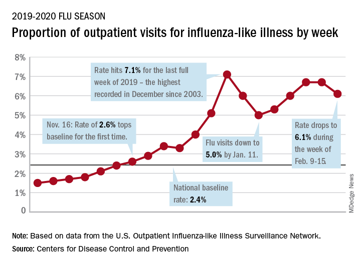
The rates for those 2 earlier weeks had previously been reported at 6.8% (Feb. 8) and 6.6% (Feb. 1), which means that there have now been 2 consecutive weeks without an increase in national ILI activity.
State-level activity was down slightly as well. For the week ending Feb. 15, there were 39 states and Puerto Rico at the highest level of activity on the CDC’s 1-10 scale, compared with 41 states and Puerto Rico the week before. The number of states in the “high” range, which includes levels 8 and 9, went from 44 to 45, however, CDC data show.
Laboratory measures also dropped a bit. For the week, 29.6% of respiratory specimens tested positive for influenza, compared with 30.3% the previous week. The predominance of influenza A continued to increase, as type A went from 59.4% to 63.5% of positive specimens and type B dropped from 40.6% to 36.5%, the influenza division said.
In a separate report, the CDC announced interim flu vaccine effectiveness estimates.For the 2019-2020 season so far, “flu vaccines are reducing doctor’s visits for flu illness by almost half (45%). This is consistent with estimates of flu vaccine effectiveness (VE) from previous flu seasons that ranged from 40% to 60% when flu vaccine viruses were similar to circulating influenza viruses,” the CDC said.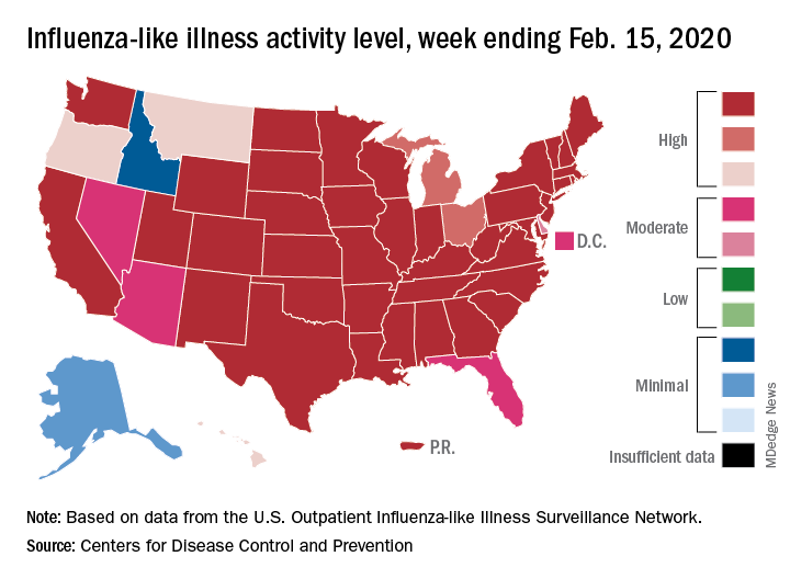
Although VE among children aged 6 months to 17 years is even higher, at 55%, this season “has been especially bad for children. Flu hospitalization rates among children are higher than at this time in other recent seasons, including the 2017-18 season,” the CDC noted.
The number of pediatric flu deaths for 2019-2020 – now up to 105 – is “higher for the same time period than in every season since reporting began in 2004-05, with the exception of the 2009 pandemic,” the CDC added.
Interim VE estimates for other age groups are 25% for adults aged 18-49 and 43% for those 50 years and older. “The lower VE point estimates observed among adults 18-49 years appear to be associated with a trend suggesting lower VE in this age group against A(H1N1)pdm09 viruses,” the CDC said.
Influenza activity dropped during the week ending Feb. 15, according to the Centers for Disease Control and Prevention. That decline, along with revised data from the 2 previous weeks, suggests that the 2019-2020 season has peaked for the second time. The rate of outpatient visits for influenza-like illness (ILI) came in at 6.1% for the week ending Feb. 15, after two straight weeks at 6.7%, the CDC’s influenza division reported Feb. 21.
The rates for those 2 earlier weeks had previously been reported at 6.8% (Feb. 8) and 6.6% (Feb. 1), which means that there have now been 2 consecutive weeks without an increase in national ILI activity.
State-level activity was down slightly as well. For the week ending Feb. 15, there were 39 states and Puerto Rico at the highest level of activity on the CDC’s 1-10 scale, compared with 41 states and Puerto Rico the week before. The number of states in the “high” range, which includes levels 8 and 9, went from 44 to 45, however, CDC data show.
Laboratory measures also dropped a bit. For the week, 29.6% of respiratory specimens tested positive for influenza, compared with 30.3% the previous week. The predominance of influenza A continued to increase, as type A went from 59.4% to 63.5% of positive specimens and type B dropped from 40.6% to 36.5%, the influenza division said.
In a separate report, the CDC announced interim flu vaccine effectiveness estimates.For the 2019-2020 season so far, “flu vaccines are reducing doctor’s visits for flu illness by almost half (45%). This is consistent with estimates of flu vaccine effectiveness (VE) from previous flu seasons that ranged from 40% to 60% when flu vaccine viruses were similar to circulating influenza viruses,” the CDC said.
Although VE among children aged 6 months to 17 years is even higher, at 55%, this season “has been especially bad for children. Flu hospitalization rates among children are higher than at this time in other recent seasons, including the 2017-18 season,” the CDC noted.
The number of pediatric flu deaths for 2019-2020 – now up to 105 – is “higher for the same time period than in every season since reporting began in 2004-05, with the exception of the 2009 pandemic,” the CDC added.
Interim VE estimates for other age groups are 25% for adults aged 18-49 and 43% for those 50 years and older. “The lower VE point estimates observed among adults 18-49 years appear to be associated with a trend suggesting lower VE in this age group against A(H1N1)pdm09 viruses,” the CDC said.
Influenza activity dropped during the week ending Feb. 15, according to the Centers for Disease Control and Prevention. That decline, along with revised data from the 2 previous weeks, suggests that the 2019-2020 season has peaked for the second time. The rate of outpatient visits for influenza-like illness (ILI) came in at 6.1% for the week ending Feb. 15, after two straight weeks at 6.7%, the CDC’s influenza division reported Feb. 21.
The rates for those 2 earlier weeks had previously been reported at 6.8% (Feb. 8) and 6.6% (Feb. 1), which means that there have now been 2 consecutive weeks without an increase in national ILI activity.
State-level activity was down slightly as well. For the week ending Feb. 15, there were 39 states and Puerto Rico at the highest level of activity on the CDC’s 1-10 scale, compared with 41 states and Puerto Rico the week before. The number of states in the “high” range, which includes levels 8 and 9, went from 44 to 45, however, CDC data show.
Laboratory measures also dropped a bit. For the week, 29.6% of respiratory specimens tested positive for influenza, compared with 30.3% the previous week. The predominance of influenza A continued to increase, as type A went from 59.4% to 63.5% of positive specimens and type B dropped from 40.6% to 36.5%, the influenza division said.
In a separate report, the CDC announced interim flu vaccine effectiveness estimates.For the 2019-2020 season so far, “flu vaccines are reducing doctor’s visits for flu illness by almost half (45%). This is consistent with estimates of flu vaccine effectiveness (VE) from previous flu seasons that ranged from 40% to 60% when flu vaccine viruses were similar to circulating influenza viruses,” the CDC said.
Although VE among children aged 6 months to 17 years is even higher, at 55%, this season “has been especially bad for children. Flu hospitalization rates among children are higher than at this time in other recent seasons, including the 2017-18 season,” the CDC noted.
The number of pediatric flu deaths for 2019-2020 – now up to 105 – is “higher for the same time period than in every season since reporting began in 2004-05, with the exception of the 2009 pandemic,” the CDC added.
Interim VE estimates for other age groups are 25% for adults aged 18-49 and 43% for those 50 years and older. “The lower VE point estimates observed among adults 18-49 years appear to be associated with a trend suggesting lower VE in this age group against A(H1N1)pdm09 viruses,” the CDC said.
FROM THE CDC
Variants in common genes linked to endometrial cancer risk

The 24 single-nucleotide polymorphisms (SNPs) were detected in genes that function in transcriptional regulation, cell survival, and estrogen metabolism.
“Understanding genetic predisposition to endometrial cancer could facilitate personalized risk assessment with a view to targeted prevention and screening interventions,” wrote Cemsel Bafligil, of the University of Manchester (England) and her coinvestigators. The group’s findings were published in the Journal of Medical Genetics.
The researchers searched major databases for primary studies that evaluated associations between endometrial cancer and SNPs. After applying the search criteria, 453 eligible records were found, and 149 of these were included in the study.
The majority of records were genome-wide association studies, case-control studies, and meta-analyses. Various data, including study type, ethnicity, and endometrial cancer type, were extracted and included in the qualitative synthesis.
After analysis, the researchers identified 24 independent genetic variants associated with a higher risk of developing endometrial cancer, and SNPs in 6 genes – CYP19A1, SOX4, HNF1B, MYC, KLF, and EIF2AK – showed a strong association.
The researchers also estimated the predictive value of the identified SNPs using a theoretical polygenic risk score model. They found that women with genome-wide significant SNPs had double the risk of developing endometrial cancer (relative risk, 2.09), and women with all 24 SNPs had a three-fold greater risk of developing the disease (RR, 3.16).
“The importance of these variants and relevance of the proximate genes in a functional or biological context is challenging to evaluate,” the researchers noted.
They also acknowledged that a key limitation of this study was the ethnic homogeneity of the cohort, with most patients being of European descent. As a result, the findings may not be fully representative of other ethnic groups.
“The multiplicative effects of these SNPs could be used in a PRS [polygenic risk score] to allow personalised risk prediction models to be developed for targeted screening and prevention interventions for women at greatest risk of endometrial cancer,” the researchers concluded.
The National Institute for Health Research Manchester Biomedical Research Centre funded the study. The authors reported having no conflicts of interest.
SOURCE: Bafligil C et al. J Med Genet. 2020 Feb 17. doi: 10.1136/jmedgenet-2019-106529.
The 24 single-nucleotide polymorphisms (SNPs) were detected in genes that function in transcriptional regulation, cell survival, and estrogen metabolism.
“Understanding genetic predisposition to endometrial cancer could facilitate personalized risk assessment with a view to targeted prevention and screening interventions,” wrote Cemsel Bafligil, of the University of Manchester (England) and her coinvestigators. The group’s findings were published in the Journal of Medical Genetics.
The researchers searched major databases for primary studies that evaluated associations between endometrial cancer and SNPs. After applying the search criteria, 453 eligible records were found, and 149 of these were included in the study.
The majority of records were genome-wide association studies, case-control studies, and meta-analyses. Various data, including study type, ethnicity, and endometrial cancer type, were extracted and included in the qualitative synthesis.
After analysis, the researchers identified 24 independent genetic variants associated with a higher risk of developing endometrial cancer, and SNPs in 6 genes – CYP19A1, SOX4, HNF1B, MYC, KLF, and EIF2AK – showed a strong association.
The researchers also estimated the predictive value of the identified SNPs using a theoretical polygenic risk score model. They found that women with genome-wide significant SNPs had double the risk of developing endometrial cancer (relative risk, 2.09), and women with all 24 SNPs had a three-fold greater risk of developing the disease (RR, 3.16).
“The importance of these variants and relevance of the proximate genes in a functional or biological context is challenging to evaluate,” the researchers noted.
They also acknowledged that a key limitation of this study was the ethnic homogeneity of the cohort, with most patients being of European descent. As a result, the findings may not be fully representative of other ethnic groups.
“The multiplicative effects of these SNPs could be used in a PRS [polygenic risk score] to allow personalised risk prediction models to be developed for targeted screening and prevention interventions for women at greatest risk of endometrial cancer,” the researchers concluded.
The National Institute for Health Research Manchester Biomedical Research Centre funded the study. The authors reported having no conflicts of interest.
SOURCE: Bafligil C et al. J Med Genet. 2020 Feb 17. doi: 10.1136/jmedgenet-2019-106529.
The 24 single-nucleotide polymorphisms (SNPs) were detected in genes that function in transcriptional regulation, cell survival, and estrogen metabolism.
“Understanding genetic predisposition to endometrial cancer could facilitate personalized risk assessment with a view to targeted prevention and screening interventions,” wrote Cemsel Bafligil, of the University of Manchester (England) and her coinvestigators. The group’s findings were published in the Journal of Medical Genetics.
The researchers searched major databases for primary studies that evaluated associations between endometrial cancer and SNPs. After applying the search criteria, 453 eligible records were found, and 149 of these were included in the study.
The majority of records were genome-wide association studies, case-control studies, and meta-analyses. Various data, including study type, ethnicity, and endometrial cancer type, were extracted and included in the qualitative synthesis.
After analysis, the researchers identified 24 independent genetic variants associated with a higher risk of developing endometrial cancer, and SNPs in 6 genes – CYP19A1, SOX4, HNF1B, MYC, KLF, and EIF2AK – showed a strong association.
The researchers also estimated the predictive value of the identified SNPs using a theoretical polygenic risk score model. They found that women with genome-wide significant SNPs had double the risk of developing endometrial cancer (relative risk, 2.09), and women with all 24 SNPs had a three-fold greater risk of developing the disease (RR, 3.16).
“The importance of these variants and relevance of the proximate genes in a functional or biological context is challenging to evaluate,” the researchers noted.
They also acknowledged that a key limitation of this study was the ethnic homogeneity of the cohort, with most patients being of European descent. As a result, the findings may not be fully representative of other ethnic groups.
“The multiplicative effects of these SNPs could be used in a PRS [polygenic risk score] to allow personalised risk prediction models to be developed for targeted screening and prevention interventions for women at greatest risk of endometrial cancer,” the researchers concluded.
The National Institute for Health Research Manchester Biomedical Research Centre funded the study. The authors reported having no conflicts of interest.
SOURCE: Bafligil C et al. J Med Genet. 2020 Feb 17. doi: 10.1136/jmedgenet-2019-106529.
FROM THE JOURNAL OF MEDICAL GENETICS
Amyloid PET Findings Correlate With Cognitive Decline in MS
Key clinical point: Lower amyloid positron imaging tomography (PET) uptake in normal-appearing white matter (NAWM) is associated with cognitive decline and an increase in white matter lesion volume.
Major finding: Cognitive decline was associated with lower standardized uptake value relative to cerebellum in NAWM (1.52 in the cognitive decline group vs. 1.67 in the cognitively stable group; Mann-Whitney U test [U] = 42.0; P = .011), lower thalamic volume (13.84 vs. 15.61; U = 55.0; P = .059), and higher white matter lesion burden (15.25 vs. 9.17; U = 49.0; P = .029).
Study details: A prospective longitudinal PET study using 18F-florbetaben included 29 patients diagnosed with MS; the mean follow-up period was 18.00 ± 3.31 months.
Disclosures: The authors declared no conflicts of interest.
Citation: Pytel V et al. Mult Scler Relat Disord. 2020 Jan 2. doi: 10.1016/j.msard.2020.101926.
Key clinical point: Lower amyloid positron imaging tomography (PET) uptake in normal-appearing white matter (NAWM) is associated with cognitive decline and an increase in white matter lesion volume.
Major finding: Cognitive decline was associated with lower standardized uptake value relative to cerebellum in NAWM (1.52 in the cognitive decline group vs. 1.67 in the cognitively stable group; Mann-Whitney U test [U] = 42.0; P = .011), lower thalamic volume (13.84 vs. 15.61; U = 55.0; P = .059), and higher white matter lesion burden (15.25 vs. 9.17; U = 49.0; P = .029).
Study details: A prospective longitudinal PET study using 18F-florbetaben included 29 patients diagnosed with MS; the mean follow-up period was 18.00 ± 3.31 months.
Disclosures: The authors declared no conflicts of interest.
Citation: Pytel V et al. Mult Scler Relat Disord. 2020 Jan 2. doi: 10.1016/j.msard.2020.101926.
Key clinical point: Lower amyloid positron imaging tomography (PET) uptake in normal-appearing white matter (NAWM) is associated with cognitive decline and an increase in white matter lesion volume.
Major finding: Cognitive decline was associated with lower standardized uptake value relative to cerebellum in NAWM (1.52 in the cognitive decline group vs. 1.67 in the cognitively stable group; Mann-Whitney U test [U] = 42.0; P = .011), lower thalamic volume (13.84 vs. 15.61; U = 55.0; P = .059), and higher white matter lesion burden (15.25 vs. 9.17; U = 49.0; P = .029).
Study details: A prospective longitudinal PET study using 18F-florbetaben included 29 patients diagnosed with MS; the mean follow-up period was 18.00 ± 3.31 months.
Disclosures: The authors declared no conflicts of interest.
Citation: Pytel V et al. Mult Scler Relat Disord. 2020 Jan 2. doi: 10.1016/j.msard.2020.101926.
Mothers of Children With MS More Likely to Use Mental Health Services
Key clinical point: Mothers of children with MS are more likely to use mental health services before and after their child’s diagnosis with multiple sclerosis (MS) than mothers of children without MS.
Major finding: The prevalence of any physical condition and mood or anxiety disorder was higher in MS-mothers vs. non-MS-mothers. The odds of having any psychiatry visit was significantly increased in MS-mothers (odds ratio, 1.60; 95% confidence interval [CI], 1.10-2.31). The annual rate of psychiatry visits did not differ between MS-mothers and non-MS-mothers (rate ratio, 0.66; 95% CI, 0.33-1.30).
Study details: A population-based retrospective matched cohort study of 156 MS-mothers and 624 non-MS mothers.
Disclosures: This study was funded by the Multiple Sclerosis Scientific Research Foundation. Dr. Marrie received research funding from CIHR, Research Manitoba, Multiple Sclerosis Society of Canada, Multiple Sclerosis Scientific Foundation, Crohn’s and Colitis Canada, National Multiple Sclerosis Society, and CMSC and was supported by the Waugh Family Chair in Multiple Sclerosis.
Citation: Marrie RA et al. Neurology. 2020 Jan 9. doi: 10.1212/WNL.0000000000008871.
Key clinical point: Mothers of children with MS are more likely to use mental health services before and after their child’s diagnosis with multiple sclerosis (MS) than mothers of children without MS.
Major finding: The prevalence of any physical condition and mood or anxiety disorder was higher in MS-mothers vs. non-MS-mothers. The odds of having any psychiatry visit was significantly increased in MS-mothers (odds ratio, 1.60; 95% confidence interval [CI], 1.10-2.31). The annual rate of psychiatry visits did not differ between MS-mothers and non-MS-mothers (rate ratio, 0.66; 95% CI, 0.33-1.30).
Study details: A population-based retrospective matched cohort study of 156 MS-mothers and 624 non-MS mothers.
Disclosures: This study was funded by the Multiple Sclerosis Scientific Research Foundation. Dr. Marrie received research funding from CIHR, Research Manitoba, Multiple Sclerosis Society of Canada, Multiple Sclerosis Scientific Foundation, Crohn’s and Colitis Canada, National Multiple Sclerosis Society, and CMSC and was supported by the Waugh Family Chair in Multiple Sclerosis.
Citation: Marrie RA et al. Neurology. 2020 Jan 9. doi: 10.1212/WNL.0000000000008871.
Key clinical point: Mothers of children with MS are more likely to use mental health services before and after their child’s diagnosis with multiple sclerosis (MS) than mothers of children without MS.
Major finding: The prevalence of any physical condition and mood or anxiety disorder was higher in MS-mothers vs. non-MS-mothers. The odds of having any psychiatry visit was significantly increased in MS-mothers (odds ratio, 1.60; 95% confidence interval [CI], 1.10-2.31). The annual rate of psychiatry visits did not differ between MS-mothers and non-MS-mothers (rate ratio, 0.66; 95% CI, 0.33-1.30).
Study details: A population-based retrospective matched cohort study of 156 MS-mothers and 624 non-MS mothers.
Disclosures: This study was funded by the Multiple Sclerosis Scientific Research Foundation. Dr. Marrie received research funding from CIHR, Research Manitoba, Multiple Sclerosis Society of Canada, Multiple Sclerosis Scientific Foundation, Crohn’s and Colitis Canada, National Multiple Sclerosis Society, and CMSC and was supported by the Waugh Family Chair in Multiple Sclerosis.
Citation: Marrie RA et al. Neurology. 2020 Jan 9. doi: 10.1212/WNL.0000000000008871.
Low Vitamin D and BMI Are Causal Factors for MS
Key clinical point: Vitamin D and body mass index (BMI) are independent causal risk factors for multiple sclerosis (MS) in adulthood and childhood.
Major finding: Genetically determined increased childhood BMI and adult BMI were associated with a 24% and 14% higher risk of MS, respectively. Each genetically determined unit increase in the natural-log-transformed vitamin D level was associated with a 43% reduction in the MS risk.
Study details: A 2-sample Mendelian randomization study estimated the effect of BMI and vitamin D status on MS risk; associations of single-nucleotide polymorphisms with both the risk factors of interest were obtained from the relevant consortia.
Disclosures: This study was funded through a grant from the Barts Charity. The authors declared no conflicts of interest.
Citation: Jacobs BM et al. Neurol Neuroimmunol Neuroinflamm. 2020 Jan 14. doi: 10.1212/NXI.0000000000000662.
Key clinical point: Vitamin D and body mass index (BMI) are independent causal risk factors for multiple sclerosis (MS) in adulthood and childhood.
Major finding: Genetically determined increased childhood BMI and adult BMI were associated with a 24% and 14% higher risk of MS, respectively. Each genetically determined unit increase in the natural-log-transformed vitamin D level was associated with a 43% reduction in the MS risk.
Study details: A 2-sample Mendelian randomization study estimated the effect of BMI and vitamin D status on MS risk; associations of single-nucleotide polymorphisms with both the risk factors of interest were obtained from the relevant consortia.
Disclosures: This study was funded through a grant from the Barts Charity. The authors declared no conflicts of interest.
Citation: Jacobs BM et al. Neurol Neuroimmunol Neuroinflamm. 2020 Jan 14. doi: 10.1212/NXI.0000000000000662.
Key clinical point: Vitamin D and body mass index (BMI) are independent causal risk factors for multiple sclerosis (MS) in adulthood and childhood.
Major finding: Genetically determined increased childhood BMI and adult BMI were associated with a 24% and 14% higher risk of MS, respectively. Each genetically determined unit increase in the natural-log-transformed vitamin D level was associated with a 43% reduction in the MS risk.
Study details: A 2-sample Mendelian randomization study estimated the effect of BMI and vitamin D status on MS risk; associations of single-nucleotide polymorphisms with both the risk factors of interest were obtained from the relevant consortia.
Disclosures: This study was funded through a grant from the Barts Charity. The authors declared no conflicts of interest.
Citation: Jacobs BM et al. Neurol Neuroimmunol Neuroinflamm. 2020 Jan 14. doi: 10.1212/NXI.0000000000000662.
Relapse Recovery and Timing of DMT Use Influence MS Progression
Key clinical point: In patients with multiple sclerosis (MS) without good recovery after the initial relapse, initiating a disease-modifying therapy (DMT) immediately increases the likelihood of a benign disease course.
Major finding: Patients with good recovery and immediate DMT initiation and those with poor recovery and delayed DMT initiation had about 65% and 20% chance, respectively, of remaining at a minimal disability level (Expanded Disability Status Scale score of less than 2.5) by age 45 years.
Study details: An analysis of data from the phase 3 CHAMPS trial in clinically isolated syndrome (n=383) and 10-year follow-up EXTENSION trial.
Disclosures: This study was funded by an unrestricted grant to Dr. Kantarci from Biogen. Dr. Kantarci and Dr. Atkinson received salary support as part of the grant from Biogen. Dr. Castrillo-Viguera was employed by Biogen.
Citation: Kantarci OH et al. Neurol Neuroimmunol Neuroinflamm. 2019 Dec 17. doi: 10.1212/NXI.0000000000000653.
Key clinical point: In patients with multiple sclerosis (MS) without good recovery after the initial relapse, initiating a disease-modifying therapy (DMT) immediately increases the likelihood of a benign disease course.
Major finding: Patients with good recovery and immediate DMT initiation and those with poor recovery and delayed DMT initiation had about 65% and 20% chance, respectively, of remaining at a minimal disability level (Expanded Disability Status Scale score of less than 2.5) by age 45 years.
Study details: An analysis of data from the phase 3 CHAMPS trial in clinically isolated syndrome (n=383) and 10-year follow-up EXTENSION trial.
Disclosures: This study was funded by an unrestricted grant to Dr. Kantarci from Biogen. Dr. Kantarci and Dr. Atkinson received salary support as part of the grant from Biogen. Dr. Castrillo-Viguera was employed by Biogen.
Citation: Kantarci OH et al. Neurol Neuroimmunol Neuroinflamm. 2019 Dec 17. doi: 10.1212/NXI.0000000000000653.
Key clinical point: In patients with multiple sclerosis (MS) without good recovery after the initial relapse, initiating a disease-modifying therapy (DMT) immediately increases the likelihood of a benign disease course.
Major finding: Patients with good recovery and immediate DMT initiation and those with poor recovery and delayed DMT initiation had about 65% and 20% chance, respectively, of remaining at a minimal disability level (Expanded Disability Status Scale score of less than 2.5) by age 45 years.
Study details: An analysis of data from the phase 3 CHAMPS trial in clinically isolated syndrome (n=383) and 10-year follow-up EXTENSION trial.
Disclosures: This study was funded by an unrestricted grant to Dr. Kantarci from Biogen. Dr. Kantarci and Dr. Atkinson received salary support as part of the grant from Biogen. Dr. Castrillo-Viguera was employed by Biogen.
Citation: Kantarci OH et al. Neurol Neuroimmunol Neuroinflamm. 2019 Dec 17. doi: 10.1212/NXI.0000000000000653.
MS: Diroximel Fumarate Shows Improved Gastrointestinal Tolerability Versus Dimethyl Fumarate
Key clinical point: Phase 3 EVOLVE-MS-2 study demonstrates that diroximel fumarate (DRF) has an improved gastrointestinal (GI) tolerability profile compared with dimethyl fumarate (DMF) in patients with relapsing-remitting multiple sclerosis (MS).
Major finding: Patients treated with DRF self-reported 46% fewer days with an Individual Gastrointestinal Symptom and Impact Scale (IGISIS) symptom intensity score of ≥2 vs those treated with DMF (rate ratio, 0.54; 95% confidence interval, 0.39-0.75). The rates of GI adverse events (AEs) were lower with DRF than DMF (34.8% vs. 49.0%). DRF-treated patients had a lower discontinuation rate because of GI AEs (0.8% vs. 4.8%) and overall AEs (1.6% vs. 5.6%).
Study details: EVOLVE-MS-2 was a 5-week randomized trial that directly compared the GI tolerability of DRF 462 mg (n = 253) with that of DMF 240 mg (n = 249); primary endpoint was the number of days with an IGISIS intensity score of 2 or greater relative to exposure.
Disclosures: This study was funded by Alkermes Inc. and Biogen. The authors reported receiving grants and personal fees from multiple pharmaceutical companies.
Citation: Naismith RT et al. CNS Drugs. 2020 Jan 17. doi: 10.1007/s40263-020-00700-0.
Key clinical point: Phase 3 EVOLVE-MS-2 study demonstrates that diroximel fumarate (DRF) has an improved gastrointestinal (GI) tolerability profile compared with dimethyl fumarate (DMF) in patients with relapsing-remitting multiple sclerosis (MS).
Major finding: Patients treated with DRF self-reported 46% fewer days with an Individual Gastrointestinal Symptom and Impact Scale (IGISIS) symptom intensity score of ≥2 vs those treated with DMF (rate ratio, 0.54; 95% confidence interval, 0.39-0.75). The rates of GI adverse events (AEs) were lower with DRF than DMF (34.8% vs. 49.0%). DRF-treated patients had a lower discontinuation rate because of GI AEs (0.8% vs. 4.8%) and overall AEs (1.6% vs. 5.6%).
Study details: EVOLVE-MS-2 was a 5-week randomized trial that directly compared the GI tolerability of DRF 462 mg (n = 253) with that of DMF 240 mg (n = 249); primary endpoint was the number of days with an IGISIS intensity score of 2 or greater relative to exposure.
Disclosures: This study was funded by Alkermes Inc. and Biogen. The authors reported receiving grants and personal fees from multiple pharmaceutical companies.
Citation: Naismith RT et al. CNS Drugs. 2020 Jan 17. doi: 10.1007/s40263-020-00700-0.
Key clinical point: Phase 3 EVOLVE-MS-2 study demonstrates that diroximel fumarate (DRF) has an improved gastrointestinal (GI) tolerability profile compared with dimethyl fumarate (DMF) in patients with relapsing-remitting multiple sclerosis (MS).
Major finding: Patients treated with DRF self-reported 46% fewer days with an Individual Gastrointestinal Symptom and Impact Scale (IGISIS) symptom intensity score of ≥2 vs those treated with DMF (rate ratio, 0.54; 95% confidence interval, 0.39-0.75). The rates of GI adverse events (AEs) were lower with DRF than DMF (34.8% vs. 49.0%). DRF-treated patients had a lower discontinuation rate because of GI AEs (0.8% vs. 4.8%) and overall AEs (1.6% vs. 5.6%).
Study details: EVOLVE-MS-2 was a 5-week randomized trial that directly compared the GI tolerability of DRF 462 mg (n = 253) with that of DMF 240 mg (n = 249); primary endpoint was the number of days with an IGISIS intensity score of 2 or greater relative to exposure.
Disclosures: This study was funded by Alkermes Inc. and Biogen. The authors reported receiving grants and personal fees from multiple pharmaceutical companies.
Citation: Naismith RT et al. CNS Drugs. 2020 Jan 17. doi: 10.1007/s40263-020-00700-0.