User login
Coccidioidomycosis a respiratory threat to construction workers in Southwest
The expansion of the solar energy industry in Coccidioides-endemic areas of the southwestern United States is exposing more workers to the infection, say the authors of a study that found an attack rate of 1.2 cases per 100 workers.
A study among 3,572 workers at two solar power–generating facilities in California identified 44 individuals with the infection between October 2011 and April 2014, 9 of whom were hospitalized, according to a paper published in the Oct. 14 edition of Emerging Infectious Diseases.

The disease is acquired through inhalation of the soil-dwelling Coccidioides fungus spores and while the majority of the patients said they had received safety training about the risk of coccidioidomycosis, only six of those who regularly performed soil-disruptive work reported regularly using respiratory protection (Emerg Infect Dis. 2015 Oct 14; doi: ).
“Large-scale construction, including solar farm construction, might involve substantial soil disturbance for months, and many employees, particularly from non–Coccidioides-endemic areas, probably lack immunity to Coccidioides,” wrote Jason A. Wilken, Ph.D., of the Centers for Disease Control and Prevention, and his coauthors.
“Medical providers should consider work-related coccidioidomycosis when evaluating construction workers with prolonged febrile respiratory illness, particularly after work in Central or Southern California or in Arizona, and medical providers should follow all statutory requirements for documenting and reporting occupational illness,” Dr. Wilken concluded.
The study was supported by the Centers for Disease Control and Prevention. No conflicts of interest were declared.
The expansion of the solar energy industry in Coccidioides-endemic areas of the southwestern United States is exposing more workers to the infection, say the authors of a study that found an attack rate of 1.2 cases per 100 workers.
A study among 3,572 workers at two solar power–generating facilities in California identified 44 individuals with the infection between October 2011 and April 2014, 9 of whom were hospitalized, according to a paper published in the Oct. 14 edition of Emerging Infectious Diseases.
The disease is acquired through inhalation of the soil-dwelling Coccidioides fungus spores and while the majority of the patients said they had received safety training about the risk of coccidioidomycosis, only six of those who regularly performed soil-disruptive work reported regularly using respiratory protection (Emerg Infect Dis. 2015 Oct 14; doi: ).
“Large-scale construction, including solar farm construction, might involve substantial soil disturbance for months, and many employees, particularly from non–Coccidioides-endemic areas, probably lack immunity to Coccidioides,” wrote Jason A. Wilken, Ph.D., of the Centers for Disease Control and Prevention, and his coauthors.
“Medical providers should consider work-related coccidioidomycosis when evaluating construction workers with prolonged febrile respiratory illness, particularly after work in Central or Southern California or in Arizona, and medical providers should follow all statutory requirements for documenting and reporting occupational illness,” Dr. Wilken concluded.
The study was supported by the Centers for Disease Control and Prevention. No conflicts of interest were declared.
The expansion of the solar energy industry in Coccidioides-endemic areas of the southwestern United States is exposing more workers to the infection, say the authors of a study that found an attack rate of 1.2 cases per 100 workers.
A study among 3,572 workers at two solar power–generating facilities in California identified 44 individuals with the infection between October 2011 and April 2014, 9 of whom were hospitalized, according to a paper published in the Oct. 14 edition of Emerging Infectious Diseases.
The disease is acquired through inhalation of the soil-dwelling Coccidioides fungus spores and while the majority of the patients said they had received safety training about the risk of coccidioidomycosis, only six of those who regularly performed soil-disruptive work reported regularly using respiratory protection (Emerg Infect Dis. 2015 Oct 14; doi: ).
“Large-scale construction, including solar farm construction, might involve substantial soil disturbance for months, and many employees, particularly from non–Coccidioides-endemic areas, probably lack immunity to Coccidioides,” wrote Jason A. Wilken, Ph.D., of the Centers for Disease Control and Prevention, and his coauthors.
“Medical providers should consider work-related coccidioidomycosis when evaluating construction workers with prolonged febrile respiratory illness, particularly after work in Central or Southern California or in Arizona, and medical providers should follow all statutory requirements for documenting and reporting occupational illness,” Dr. Wilken concluded.
The study was supported by the Centers for Disease Control and Prevention. No conflicts of interest were declared.
FROM EMERGING INFECTIOUS DISEASES
Key clinical point:Coccidioidomycosis is a significant risk in workers on solar power–generating facilities in Coccidioides-endemic areas of the Southwestern United States.
Major finding: The attack rate of Coccidioides could be as high 1.2 cases per 100 workers involved in constructing solar power–generating facilities.
Data source: A study among 3,572 workers at two solar power–generating facilities in California.
Disclosures: The study was supported by the Centers for Disease Control and Prevention. No conflicts of interest were declared.

Product News: 10 2015
Bellafill
Suneva Medical, Inc, recognized the treatment of acne scars was an unmet need, which led to research supporting a new indication for the dermal filler Bellafill for the treatment of moderate to severe, atrophic, distensible facial acne scars on the cheek in patients older than 21 years. Bellafill is a smooth, collagen-based dermal filler with polymethylmethacrylate (PMMA) microspheres. The collagen gel provides immediate volume and lift to correct the scar, and the PMMA microspheres remain in place and provide structural support for smoother-looking skin. Bellafill is not indicated for ice-pick scars. Although Bellafill can be used in all skin types, the patient’s acne cannot be active. Results have been observed to last 12 months. Patients may continue with ongoing topical treatments but should discontinue any topical treatment the night after injection. For more information, visit www.bellafill.com.
Cutanea Life Sciences
Cutanea Life Sciences renews its commitment to focusing on the unmet needs of patients to develop innovative technologies and therapeutic applications. In 2012, Maruho Co, Ltd, acquired Cutanea Life Sciences, solidifying the financial resources needed to create market-leading products to treat diseases and disorders of the skin and subcutaneous tissue. Cutaneous Life Sciences corporate headquarters are located in Wayne, Pennsylvania. Robert J. Bitterman Sr has served as president and chief executive officer since 2005, following executive leadership roles for other dermatology companies. For more information, visit www.cutanealife.com.
Humira
AbbVie Inc receives US Food and Drug Administration approval of Humira (adalimumab) for the treatment of moderate to severe hidradenitis suppurativa (HS), offering patients with HS a much-needed treatment for this chronic debilitating disease. The HS indication follows approvals for rheumatoid arthritis, plaque psoriasis, Crohn disease, ulcerative colitis, psoriatic arthritis, and ankylosing spondylitis. For more information, visit www.humira.com.
Teflaro
Actavis, Inc, announces US Food and Drug Administration approval of the supplemental new drug application to update the label for Teflaro (ceftaroline fosamil) for the treatment of adult patients with acute bacterial skin and skin structure infections (ABSSSI) and community-acquired bacterial pneumonia (CABP). With this updated label, Teflaro also is now approved to be administered by intravenous infusion over 5 minutes to 1 hour in adult patients 18 years and older, providing increased flexibility in dosing. Teflaro was first approved in 2010 for the treatment of adults with CABP and ABSSSI due to designated susceptible pathogens. For more information, visit www.teflaro.com.
If you would like your product included in Product News, please e-mail a press release to the Editorial Office at cutis@frontlinemedcom.com.
Bellafill
Suneva Medical, Inc, recognized the treatment of acne scars was an unmet need, which led to research supporting a new indication for the dermal filler Bellafill for the treatment of moderate to severe, atrophic, distensible facial acne scars on the cheek in patients older than 21 years. Bellafill is a smooth, collagen-based dermal filler with polymethylmethacrylate (PMMA) microspheres. The collagen gel provides immediate volume and lift to correct the scar, and the PMMA microspheres remain in place and provide structural support for smoother-looking skin. Bellafill is not indicated for ice-pick scars. Although Bellafill can be used in all skin types, the patient’s acne cannot be active. Results have been observed to last 12 months. Patients may continue with ongoing topical treatments but should discontinue any topical treatment the night after injection. For more information, visit www.bellafill.com.
Cutanea Life Sciences
Cutanea Life Sciences renews its commitment to focusing on the unmet needs of patients to develop innovative technologies and therapeutic applications. In 2012, Maruho Co, Ltd, acquired Cutanea Life Sciences, solidifying the financial resources needed to create market-leading products to treat diseases and disorders of the skin and subcutaneous tissue. Cutaneous Life Sciences corporate headquarters are located in Wayne, Pennsylvania. Robert J. Bitterman Sr has served as president and chief executive officer since 2005, following executive leadership roles for other dermatology companies. For more information, visit www.cutanealife.com.
Humira
AbbVie Inc receives US Food and Drug Administration approval of Humira (adalimumab) for the treatment of moderate to severe hidradenitis suppurativa (HS), offering patients with HS a much-needed treatment for this chronic debilitating disease. The HS indication follows approvals for rheumatoid arthritis, plaque psoriasis, Crohn disease, ulcerative colitis, psoriatic arthritis, and ankylosing spondylitis. For more information, visit www.humira.com.
Teflaro
Actavis, Inc, announces US Food and Drug Administration approval of the supplemental new drug application to update the label for Teflaro (ceftaroline fosamil) for the treatment of adult patients with acute bacterial skin and skin structure infections (ABSSSI) and community-acquired bacterial pneumonia (CABP). With this updated label, Teflaro also is now approved to be administered by intravenous infusion over 5 minutes to 1 hour in adult patients 18 years and older, providing increased flexibility in dosing. Teflaro was first approved in 2010 for the treatment of adults with CABP and ABSSSI due to designated susceptible pathogens. For more information, visit www.teflaro.com.
If you would like your product included in Product News, please e-mail a press release to the Editorial Office at cutis@frontlinemedcom.com.
Bellafill
Suneva Medical, Inc, recognized the treatment of acne scars was an unmet need, which led to research supporting a new indication for the dermal filler Bellafill for the treatment of moderate to severe, atrophic, distensible facial acne scars on the cheek in patients older than 21 years. Bellafill is a smooth, collagen-based dermal filler with polymethylmethacrylate (PMMA) microspheres. The collagen gel provides immediate volume and lift to correct the scar, and the PMMA microspheres remain in place and provide structural support for smoother-looking skin. Bellafill is not indicated for ice-pick scars. Although Bellafill can be used in all skin types, the patient’s acne cannot be active. Results have been observed to last 12 months. Patients may continue with ongoing topical treatments but should discontinue any topical treatment the night after injection. For more information, visit www.bellafill.com.
Cutanea Life Sciences
Cutanea Life Sciences renews its commitment to focusing on the unmet needs of patients to develop innovative technologies and therapeutic applications. In 2012, Maruho Co, Ltd, acquired Cutanea Life Sciences, solidifying the financial resources needed to create market-leading products to treat diseases and disorders of the skin and subcutaneous tissue. Cutaneous Life Sciences corporate headquarters are located in Wayne, Pennsylvania. Robert J. Bitterman Sr has served as president and chief executive officer since 2005, following executive leadership roles for other dermatology companies. For more information, visit www.cutanealife.com.
Humira
AbbVie Inc receives US Food and Drug Administration approval of Humira (adalimumab) for the treatment of moderate to severe hidradenitis suppurativa (HS), offering patients with HS a much-needed treatment for this chronic debilitating disease. The HS indication follows approvals for rheumatoid arthritis, plaque psoriasis, Crohn disease, ulcerative colitis, psoriatic arthritis, and ankylosing spondylitis. For more information, visit www.humira.com.
Teflaro
Actavis, Inc, announces US Food and Drug Administration approval of the supplemental new drug application to update the label for Teflaro (ceftaroline fosamil) for the treatment of adult patients with acute bacterial skin and skin structure infections (ABSSSI) and community-acquired bacterial pneumonia (CABP). With this updated label, Teflaro also is now approved to be administered by intravenous infusion over 5 minutes to 1 hour in adult patients 18 years and older, providing increased flexibility in dosing. Teflaro was first approved in 2010 for the treatment of adults with CABP and ABSSSI due to designated susceptible pathogens. For more information, visit www.teflaro.com.
If you would like your product included in Product News, please e-mail a press release to the Editorial Office at cutis@frontlinemedcom.com.

TCT: Routine thrombectomy with PCI raises stroke risk 66% in STEMI patients
Routine manual thrombectomy during percutaneous coronary intervention offers no significant long-term benefit over PCI alone for patients with acute ST-segment–elevation myocardial infarction.
In fact, thrombectomy was associated with a 66% increase in stroke risk in the year following a heart attack, Dr. Sanjit Jolly reported at the Transcatheter Cardiovascular Therapeutics annual meeting.

A 1-year analysis of the TOTAL trial confirmed the study’s recently published 1-month findings: Routine thrombectomy isn’t any better than PCI alone in the clinical outcomes of cardiovascular death, myocardial infarction, cardiogenic shock, or heart failure.
As it was at 1 month, however, stroke at 1 year was significantly more common; it occurred in 60 patients (1.2%) in the thrombectomy group and 36 (0.7%) in the PCI-alone group, for an increase in risk of 66% (P = .015).
The findings were simultaneously published online (Lancet 2015 Oct 13 doi: 10.1016/ S0140-6736[15]00448-1).
“Based on these endpoints, manual thrombectomy can no longer be recommended as a primary strategy in these patients,” Dr. Jolly said at the meeting, which was sponsored by the Cardiovascular Research Foundation.
Despite the unequivocal findings, it’s unclear when clinicians will completely embrace TOTAL’s results, noted Dr. Jolly of McMaster University, Hamilton, Ont.
“Unfortunately, practice has not changed with these findings. It often takes time for new evidence to get translated into the clinic, although some of my colleagues have told me that the results are giving them pause, causing them to use less thrombus aspiration. But interventional cardiologists are very visually driven. If we see something, we want to take it out. So it may take some time for these to be adopted,” he said.
The Thromwbectomy With PCI Versus PCI Alone in Patients With STEMI (TOTAL) trial randomized 10,723 patients to percutaneous coronary intervention with or without routine manual thrombectomy. The primary outcome was a composite of death from cardiovascular causes, recurrent myocardial infarction, cardiogenic shock, or New York Heart Association class IV heart failure within 180 days. The key safety outcome was stroke within 30 days.
The TOTAL results showed no difference in the primary outcome between thrombectomy and PCI patients (6.9% vs. 7%) (N Engl J Med. 2015 Apr 9;372[15]:1389-98).
Rates of cardiovascular death were similar (3% in each group) as were those for a combination of the primary outcome plus stent thrombosis or revascularization (10% in each group). Patients who had a thrombectomy were twice as likely to have a stroke within 30 days, although the absolute numbers were small (0.7% vs. 0.3%; HR, 2.06).
The 1-year follow-up study provided important perspective about the long-term risks and benefits of the two strategies.
There was no difference in the rate of the composite endpoint, which occurred in 7.8% of each group. Cardiovascular death occurred in 3.6% of the thrombectomy group and 3.8% of the PCI-alone group. There were similar rates of recurrent heart attack (2.5% vs. 2.3%), cardiogenic shock (1.9% vs. 2.1%), and class IV heart failure (2.1% vs. 1.9%).
The finding of significantly elevated stroke risk at 30 days was also present at 1 year, occurring in 1.2% of the thrombectomy patients and 0.7% of the PCI-alone patients (HR, 1.66; P = .015). The risk of a combination of stroke or transient ischemic attack was also increased significantly, by 65%, occurring in 1.4% and 0.9%, respectively (HR, 1.65; P = .008).
To further confirm the findings, Dr. Jolly also presented a meta-analysis of all-cause mortality in 20,352 patients involved in 16 studies comparing PCI plus thrombectomy with PCI alone. The meta-analysis also found that routine thrombectomy conferred no mortality benefit over PCI.
It did, however, confirm a 43% increased risk of stroke in PCI plus thrombectomy vs. PCI alone, at 0.9% and 0.6% (P = .03).
TOTAL was funded by the Canadian Institutes of Health Research, the Canadian Network and Centre for Trials Internationally, and Medtronic. Dr. Jolly received grants from Medtronic during the study.
On Twitter @Alz_Gal
Routine manual thrombectomy during percutaneous coronary intervention offers no significant long-term benefit over PCI alone for patients with acute ST-segment–elevation myocardial infarction.
In fact, thrombectomy was associated with a 66% increase in stroke risk in the year following a heart attack, Dr. Sanjit Jolly reported at the Transcatheter Cardiovascular Therapeutics annual meeting.
A 1-year analysis of the TOTAL trial confirmed the study’s recently published 1-month findings: Routine thrombectomy isn’t any better than PCI alone in the clinical outcomes of cardiovascular death, myocardial infarction, cardiogenic shock, or heart failure.
As it was at 1 month, however, stroke at 1 year was significantly more common; it occurred in 60 patients (1.2%) in the thrombectomy group and 36 (0.7%) in the PCI-alone group, for an increase in risk of 66% (P = .015).
The findings were simultaneously published online (Lancet 2015 Oct 13 doi: 10.1016/ S0140-6736[15]00448-1).
“Based on these endpoints, manual thrombectomy can no longer be recommended as a primary strategy in these patients,” Dr. Jolly said at the meeting, which was sponsored by the Cardiovascular Research Foundation.
Despite the unequivocal findings, it’s unclear when clinicians will completely embrace TOTAL’s results, noted Dr. Jolly of McMaster University, Hamilton, Ont.
“Unfortunately, practice has not changed with these findings. It often takes time for new evidence to get translated into the clinic, although some of my colleagues have told me that the results are giving them pause, causing them to use less thrombus aspiration. But interventional cardiologists are very visually driven. If we see something, we want to take it out. So it may take some time for these to be adopted,” he said.
The Thromwbectomy With PCI Versus PCI Alone in Patients With STEMI (TOTAL) trial randomized 10,723 patients to percutaneous coronary intervention with or without routine manual thrombectomy. The primary outcome was a composite of death from cardiovascular causes, recurrent myocardial infarction, cardiogenic shock, or New York Heart Association class IV heart failure within 180 days. The key safety outcome was stroke within 30 days.
The TOTAL results showed no difference in the primary outcome between thrombectomy and PCI patients (6.9% vs. 7%) (N Engl J Med. 2015 Apr 9;372[15]:1389-98).
Rates of cardiovascular death were similar (3% in each group) as were those for a combination of the primary outcome plus stent thrombosis or revascularization (10% in each group). Patients who had a thrombectomy were twice as likely to have a stroke within 30 days, although the absolute numbers were small (0.7% vs. 0.3%; HR, 2.06).
The 1-year follow-up study provided important perspective about the long-term risks and benefits of the two strategies.
There was no difference in the rate of the composite endpoint, which occurred in 7.8% of each group. Cardiovascular death occurred in 3.6% of the thrombectomy group and 3.8% of the PCI-alone group. There were similar rates of recurrent heart attack (2.5% vs. 2.3%), cardiogenic shock (1.9% vs. 2.1%), and class IV heart failure (2.1% vs. 1.9%).
The finding of significantly elevated stroke risk at 30 days was also present at 1 year, occurring in 1.2% of the thrombectomy patients and 0.7% of the PCI-alone patients (HR, 1.66; P = .015). The risk of a combination of stroke or transient ischemic attack was also increased significantly, by 65%, occurring in 1.4% and 0.9%, respectively (HR, 1.65; P = .008).
To further confirm the findings, Dr. Jolly also presented a meta-analysis of all-cause mortality in 20,352 patients involved in 16 studies comparing PCI plus thrombectomy with PCI alone. The meta-analysis also found that routine thrombectomy conferred no mortality benefit over PCI.
It did, however, confirm a 43% increased risk of stroke in PCI plus thrombectomy vs. PCI alone, at 0.9% and 0.6% (P = .03).
TOTAL was funded by the Canadian Institutes of Health Research, the Canadian Network and Centre for Trials Internationally, and Medtronic. Dr. Jolly received grants from Medtronic during the study.
On Twitter @Alz_Gal
Routine manual thrombectomy during percutaneous coronary intervention offers no significant long-term benefit over PCI alone for patients with acute ST-segment–elevation myocardial infarction.
In fact, thrombectomy was associated with a 66% increase in stroke risk in the year following a heart attack, Dr. Sanjit Jolly reported at the Transcatheter Cardiovascular Therapeutics annual meeting.
A 1-year analysis of the TOTAL trial confirmed the study’s recently published 1-month findings: Routine thrombectomy isn’t any better than PCI alone in the clinical outcomes of cardiovascular death, myocardial infarction, cardiogenic shock, or heart failure.
As it was at 1 month, however, stroke at 1 year was significantly more common; it occurred in 60 patients (1.2%) in the thrombectomy group and 36 (0.7%) in the PCI-alone group, for an increase in risk of 66% (P = .015).
The findings were simultaneously published online (Lancet 2015 Oct 13 doi: 10.1016/ S0140-6736[15]00448-1).
“Based on these endpoints, manual thrombectomy can no longer be recommended as a primary strategy in these patients,” Dr. Jolly said at the meeting, which was sponsored by the Cardiovascular Research Foundation.
Despite the unequivocal findings, it’s unclear when clinicians will completely embrace TOTAL’s results, noted Dr. Jolly of McMaster University, Hamilton, Ont.
“Unfortunately, practice has not changed with these findings. It often takes time for new evidence to get translated into the clinic, although some of my colleagues have told me that the results are giving them pause, causing them to use less thrombus aspiration. But interventional cardiologists are very visually driven. If we see something, we want to take it out. So it may take some time for these to be adopted,” he said.
The Thromwbectomy With PCI Versus PCI Alone in Patients With STEMI (TOTAL) trial randomized 10,723 patients to percutaneous coronary intervention with or without routine manual thrombectomy. The primary outcome was a composite of death from cardiovascular causes, recurrent myocardial infarction, cardiogenic shock, or New York Heart Association class IV heart failure within 180 days. The key safety outcome was stroke within 30 days.
The TOTAL results showed no difference in the primary outcome between thrombectomy and PCI patients (6.9% vs. 7%) (N Engl J Med. 2015 Apr 9;372[15]:1389-98).
Rates of cardiovascular death were similar (3% in each group) as were those for a combination of the primary outcome plus stent thrombosis or revascularization (10% in each group). Patients who had a thrombectomy were twice as likely to have a stroke within 30 days, although the absolute numbers were small (0.7% vs. 0.3%; HR, 2.06).
The 1-year follow-up study provided important perspective about the long-term risks and benefits of the two strategies.
There was no difference in the rate of the composite endpoint, which occurred in 7.8% of each group. Cardiovascular death occurred in 3.6% of the thrombectomy group and 3.8% of the PCI-alone group. There were similar rates of recurrent heart attack (2.5% vs. 2.3%), cardiogenic shock (1.9% vs. 2.1%), and class IV heart failure (2.1% vs. 1.9%).
The finding of significantly elevated stroke risk at 30 days was also present at 1 year, occurring in 1.2% of the thrombectomy patients and 0.7% of the PCI-alone patients (HR, 1.66; P = .015). The risk of a combination of stroke or transient ischemic attack was also increased significantly, by 65%, occurring in 1.4% and 0.9%, respectively (HR, 1.65; P = .008).
To further confirm the findings, Dr. Jolly also presented a meta-analysis of all-cause mortality in 20,352 patients involved in 16 studies comparing PCI plus thrombectomy with PCI alone. The meta-analysis also found that routine thrombectomy conferred no mortality benefit over PCI.
It did, however, confirm a 43% increased risk of stroke in PCI plus thrombectomy vs. PCI alone, at 0.9% and 0.6% (P = .03).
TOTAL was funded by the Canadian Institutes of Health Research, the Canadian Network and Centre for Trials Internationally, and Medtronic. Dr. Jolly received grants from Medtronic during the study.
On Twitter @Alz_Gal
AT TCT 2015
Key clinical point: Adding routine thrombectomy to PCI doesn’t improve outcomes; in fact, it increases the risk of stroke by 66% at 1 year.
Major finding: At 1 year, thrombectomy didn’t improve cardiovascular outcomes over PCI alone for patients with STEMI; in fact, it increased the risk of stroke by 66%.
Data source: The TOTAL trial randomizing 10,732 patients with STEMI to either PCI plus manual thrombectomy or PCI alone.
Disclosures: TOTAL was funded by the Canadian Institutes of Health Research, the Canadian Network and Centre for Trials Internationally, and Medtronic. Dr. Jolly received grants from Medtronic during the study.

Hemorrhage control after pelvic fracture: Methods vary widely
LAS VEGAS – Methods for controlling hemorrhage from severe pelvic fractures vary widely across institutions, according to findings from a prospective observational study.
In particular, the findings from the 2-year multicenter study of 1,339 patients show that resuscitative endovascular balloon occlusion of the aorta (REBOA) is rarely used, despite its inclusion in recent management algorithms, Dr. Todd W. Costantini reported at the annual meeting of the American Association for the Surgery of Trauma (AAST).
The most common methods used for hemorrhage control were angioembolization alone and external fixator placement alone, used in 55 (4.1%) and 78 (5.8%) patients, respectively. These methods were also used in 19 (10.7%) and 17 (9.6%) of the 178 patients of the overall study population who presented in shock, said Dr. Costantini of the University of California San Diego Health System.

Other methods included preperitoneal pelvic packing alone in 20 patients overall and 6 patients in shock, embolization plus external fixator in 11 patients overall and 6 patients in shock, embolization and pelvic packing in 6 patients overall and 2 patients in shock, external fixator plus pelvic packing in 6 patients overall and 1 patient in shock, embolization plus external fixator plus pelvic packing in 5 patients overall and 1 patient in shock.
“As most pelvic fracture algorithms suggest the use of preperitoneal packing prior to embolization in patients who present with hemodynamic instability, we were interested to find that only two patients [in shock] were treated with this method,” Dr. Costantini said.
Further, REBOA with or without any other method was used in only five patients overall (0.4%) and five patients in shock (2.8%), and all of these were from only 1 of the 11 participating centers, he noted.
Study subjects were adults with a mean age of 47 years with pelvic fracture from blunt trauma, and 57% were men. The mean Injury Severity Score was high at 19.2 on a scale of 75. Associated injury was common; 32% had an abbreviated injury scale (AIS) score of 3 or higher (out of 6) for chest injury.
The average intensive care unit length of stay was 8.2 days, and the average hospital length of stay was 10.9 days. In-hospital mortality was 9%.
“Pelvic fractures are associated with significant disability, demonstrated by the fact that only 43% of patients were discharged home from the hospital after admission for pelvic fractures. The remainder required ongoing care in either skilled nursing facilities or acute rehab facilities,” he said.
Of the patients who met criteria for shock, the mean age was 44 years, 59% were men, and the mean ISS was 28.2, with nearly half having a chest AIS of 3 or greater, nearly 39% having a head AIS of 3 or greater, and 32% having an abdominal AIS of 3 or greater. The mean ICU stay was 11.6 days, and the mean hospital stay, 19.3 days. In-hospital mortality among those presenting in shock was 32%.
Most patients underwent computed tomography, and arterial blush was noted in 10% of cases. Angiography was used in 148 patients, and half of those were noted to have contrast extravasation.
Therapeutic angioembolization was used in 79 patients (5.9%) overall, and in 60% of those undergoing angiography. The most common indication for angiography was ongoing hemorrhage, hemodynamic instability, and blush on CT scan.
The findings demonstrate significant variability in the approach to hemorrhage control across participating institutions.
“We found that there is currently limited use of REBOA in the treatment of hemorrhage associated with pelvic fracture. However, this may change as management strategies evolve with advances in training and technology,” Dr. Costantini concluded.
As a discussant for Dr. Costantini’s paper, Dr. Walter Biffl of the University of Colorado, Denver, expressed concern regarding the lack of adherence to management algorithms, saying that the data suggest a lack of standardization and orderly application of principles that have been shown to reduce mortality.
“Only 19% had pelvic binding. In our algorithm, 100% get that. And 85% of those in shock had CT scans. In our algorithm that comes after all these other interventions,” he said. “This study clearly opens the door for further research. If we could start with a pelvic binder and hemostatic resuscitation and maybe add REBOA for the severely hypertensive patients, maybe we can begin to determine the goals and efficacy of more interventions,” he said.
Dr. Costantini’s study was supported by the AAST Multi-Institutional Trials Committee. He reported having no disclosures.
LAS VEGAS – Methods for controlling hemorrhage from severe pelvic fractures vary widely across institutions, according to findings from a prospective observational study.
In particular, the findings from the 2-year multicenter study of 1,339 patients show that resuscitative endovascular balloon occlusion of the aorta (REBOA) is rarely used, despite its inclusion in recent management algorithms, Dr. Todd W. Costantini reported at the annual meeting of the American Association for the Surgery of Trauma (AAST).
The most common methods used for hemorrhage control were angioembolization alone and external fixator placement alone, used in 55 (4.1%) and 78 (5.8%) patients, respectively. These methods were also used in 19 (10.7%) and 17 (9.6%) of the 178 patients of the overall study population who presented in shock, said Dr. Costantini of the University of California San Diego Health System.
Other methods included preperitoneal pelvic packing alone in 20 patients overall and 6 patients in shock, embolization plus external fixator in 11 patients overall and 6 patients in shock, embolization and pelvic packing in 6 patients overall and 2 patients in shock, external fixator plus pelvic packing in 6 patients overall and 1 patient in shock, embolization plus external fixator plus pelvic packing in 5 patients overall and 1 patient in shock.
“As most pelvic fracture algorithms suggest the use of preperitoneal packing prior to embolization in patients who present with hemodynamic instability, we were interested to find that only two patients [in shock] were treated with this method,” Dr. Costantini said.
Further, REBOA with or without any other method was used in only five patients overall (0.4%) and five patients in shock (2.8%), and all of these were from only 1 of the 11 participating centers, he noted.
Study subjects were adults with a mean age of 47 years with pelvic fracture from blunt trauma, and 57% were men. The mean Injury Severity Score was high at 19.2 on a scale of 75. Associated injury was common; 32% had an abbreviated injury scale (AIS) score of 3 or higher (out of 6) for chest injury.
The average intensive care unit length of stay was 8.2 days, and the average hospital length of stay was 10.9 days. In-hospital mortality was 9%.
“Pelvic fractures are associated with significant disability, demonstrated by the fact that only 43% of patients were discharged home from the hospital after admission for pelvic fractures. The remainder required ongoing care in either skilled nursing facilities or acute rehab facilities,” he said.
Of the patients who met criteria for shock, the mean age was 44 years, 59% were men, and the mean ISS was 28.2, with nearly half having a chest AIS of 3 or greater, nearly 39% having a head AIS of 3 or greater, and 32% having an abdominal AIS of 3 or greater. The mean ICU stay was 11.6 days, and the mean hospital stay, 19.3 days. In-hospital mortality among those presenting in shock was 32%.
Most patients underwent computed tomography, and arterial blush was noted in 10% of cases. Angiography was used in 148 patients, and half of those were noted to have contrast extravasation.
Therapeutic angioembolization was used in 79 patients (5.9%) overall, and in 60% of those undergoing angiography. The most common indication for angiography was ongoing hemorrhage, hemodynamic instability, and blush on CT scan.
The findings demonstrate significant variability in the approach to hemorrhage control across participating institutions.
“We found that there is currently limited use of REBOA in the treatment of hemorrhage associated with pelvic fracture. However, this may change as management strategies evolve with advances in training and technology,” Dr. Costantini concluded.
As a discussant for Dr. Costantini’s paper, Dr. Walter Biffl of the University of Colorado, Denver, expressed concern regarding the lack of adherence to management algorithms, saying that the data suggest a lack of standardization and orderly application of principles that have been shown to reduce mortality.
“Only 19% had pelvic binding. In our algorithm, 100% get that. And 85% of those in shock had CT scans. In our algorithm that comes after all these other interventions,” he said. “This study clearly opens the door for further research. If we could start with a pelvic binder and hemostatic resuscitation and maybe add REBOA for the severely hypertensive patients, maybe we can begin to determine the goals and efficacy of more interventions,” he said.
Dr. Costantini’s study was supported by the AAST Multi-Institutional Trials Committee. He reported having no disclosures.
LAS VEGAS – Methods for controlling hemorrhage from severe pelvic fractures vary widely across institutions, according to findings from a prospective observational study.
In particular, the findings from the 2-year multicenter study of 1,339 patients show that resuscitative endovascular balloon occlusion of the aorta (REBOA) is rarely used, despite its inclusion in recent management algorithms, Dr. Todd W. Costantini reported at the annual meeting of the American Association for the Surgery of Trauma (AAST).
The most common methods used for hemorrhage control were angioembolization alone and external fixator placement alone, used in 55 (4.1%) and 78 (5.8%) patients, respectively. These methods were also used in 19 (10.7%) and 17 (9.6%) of the 178 patients of the overall study population who presented in shock, said Dr. Costantini of the University of California San Diego Health System.
Other methods included preperitoneal pelvic packing alone in 20 patients overall and 6 patients in shock, embolization plus external fixator in 11 patients overall and 6 patients in shock, embolization and pelvic packing in 6 patients overall and 2 patients in shock, external fixator plus pelvic packing in 6 patients overall and 1 patient in shock, embolization plus external fixator plus pelvic packing in 5 patients overall and 1 patient in shock.
“As most pelvic fracture algorithms suggest the use of preperitoneal packing prior to embolization in patients who present with hemodynamic instability, we were interested to find that only two patients [in shock] were treated with this method,” Dr. Costantini said.
Further, REBOA with or without any other method was used in only five patients overall (0.4%) and five patients in shock (2.8%), and all of these were from only 1 of the 11 participating centers, he noted.
Study subjects were adults with a mean age of 47 years with pelvic fracture from blunt trauma, and 57% were men. The mean Injury Severity Score was high at 19.2 on a scale of 75. Associated injury was common; 32% had an abbreviated injury scale (AIS) score of 3 or higher (out of 6) for chest injury.
The average intensive care unit length of stay was 8.2 days, and the average hospital length of stay was 10.9 days. In-hospital mortality was 9%.
“Pelvic fractures are associated with significant disability, demonstrated by the fact that only 43% of patients were discharged home from the hospital after admission for pelvic fractures. The remainder required ongoing care in either skilled nursing facilities or acute rehab facilities,” he said.
Of the patients who met criteria for shock, the mean age was 44 years, 59% were men, and the mean ISS was 28.2, with nearly half having a chest AIS of 3 or greater, nearly 39% having a head AIS of 3 or greater, and 32% having an abdominal AIS of 3 or greater. The mean ICU stay was 11.6 days, and the mean hospital stay, 19.3 days. In-hospital mortality among those presenting in shock was 32%.
Most patients underwent computed tomography, and arterial blush was noted in 10% of cases. Angiography was used in 148 patients, and half of those were noted to have contrast extravasation.
Therapeutic angioembolization was used in 79 patients (5.9%) overall, and in 60% of those undergoing angiography. The most common indication for angiography was ongoing hemorrhage, hemodynamic instability, and blush on CT scan.
The findings demonstrate significant variability in the approach to hemorrhage control across participating institutions.
“We found that there is currently limited use of REBOA in the treatment of hemorrhage associated with pelvic fracture. However, this may change as management strategies evolve with advances in training and technology,” Dr. Costantini concluded.
As a discussant for Dr. Costantini’s paper, Dr. Walter Biffl of the University of Colorado, Denver, expressed concern regarding the lack of adherence to management algorithms, saying that the data suggest a lack of standardization and orderly application of principles that have been shown to reduce mortality.
“Only 19% had pelvic binding. In our algorithm, 100% get that. And 85% of those in shock had CT scans. In our algorithm that comes after all these other interventions,” he said. “This study clearly opens the door for further research. If we could start with a pelvic binder and hemostatic resuscitation and maybe add REBOA for the severely hypertensive patients, maybe we can begin to determine the goals and efficacy of more interventions,” he said.
Dr. Costantini’s study was supported by the AAST Multi-Institutional Trials Committee. He reported having no disclosures.
AT THE AAST ANNUAL MEETING
Key clinical point: Methods for controlling hemorrhage from severe pelvic fractures vary widely across institutions, according to findings from a prospective observational study.
Major finding: REBOA was used in five patients overall (0.4%) and five patients in shock (2.8%), all from 1 of the 11 participating centers.
Data source: A prospective, multicenter, observational study of 1,339 patients.
Disclosures: Dr. Costantini’s study was supported by the AAST Multi-Institutional Trials Committee. He reported having no disclosures.

Nephropathic Cystinosis: Diagnosis, Management, and Challenges in Long-term Treatment
A supplement to Internal Medicine News.
Faculty
Larry A. Greenbaum, MD, PhD
Chief, Pediatric Nephrology
Children's Healthcare of Atlanta and
Emory University
Professor of Pediatrics
Department of Pediatrics
Emory University School of Medicine
A supplement supported by an educational grant from Raptor Pharmaceuticals Inc.
A supplement to Internal Medicine News.
Faculty
Larry A. Greenbaum, MD, PhD
Chief, Pediatric Nephrology
Children's Healthcare of Atlanta and
Emory University
Professor of Pediatrics
Department of Pediatrics
Emory University School of Medicine
A supplement supported by an educational grant from Raptor Pharmaceuticals Inc.
A supplement to Internal Medicine News.
Faculty
Larry A. Greenbaum, MD, PhD
Chief, Pediatric Nephrology
Children's Healthcare of Atlanta and
Emory University
Professor of Pediatrics
Department of Pediatrics
Emory University School of Medicine
A supplement supported by an educational grant from Raptor Pharmaceuticals Inc.
Advice for new rheumatology fellows
My residency program was fantastic. There were about 40 residents in my intern year and more than 100 residents in all. You were never alone. The atmosphere was congenial. You sat at the nurses’ station for hours charting away, but you interacted with co-residents, fellows, attendings, and residents from other specialties. Residency was tough, but it was easy to make friends with people sharing the experience.
I was unprepared for how different fellowship would be. I expected to be milling about in the wards, getting to know fellows in other specialties. Instead, I spent all of my time in the rheumatology office seeing patients or fulfilling research or teaching or conference obligations. I had a great relationship with my co-fellows, but there were only four of us and we each had different schedules. It felt surprisingly isolating.
The isolation led to another, more insidious change: I started forgetting internal medicine. Right out of residency, you think you know most everything there is to know. After all, you did just run an ICU by yourself and you just passed the internal medicine boards. You are eager to put that behind you, and you channel all your efforts into learning rheumatology.
But with each passing day that you are not called on to identify a murmur, feel a spleen tip, or treat a patient with diabetes, your ability to do those things diminishes. My world has shrunk significantly in ways I do not care to admit. I have never been as familiar with the nail-seeking properties of my rheumatology hammer as I am now. That’s fine until you consider that metabolic problems, infections, and malignancies can all masquerade as rheumatologic conditions.
When I realized that my IM skills were vanishing, I resolved to reverse the isolation. It helps that I belong to a fantastic community of physicians who welcomed me into their tribe. I started attending the weekly IM grand rounds and morbidity and mortality conferences. I am giddy with the excitement of being immersed in internal medicine once again and grateful to receive the collected wisdom of the brilliant people that surround me.
Dr. Chan practices rheumatology in Pawtucket, R.I.
My residency program was fantastic. There were about 40 residents in my intern year and more than 100 residents in all. You were never alone. The atmosphere was congenial. You sat at the nurses’ station for hours charting away, but you interacted with co-residents, fellows, attendings, and residents from other specialties. Residency was tough, but it was easy to make friends with people sharing the experience.
I was unprepared for how different fellowship would be. I expected to be milling about in the wards, getting to know fellows in other specialties. Instead, I spent all of my time in the rheumatology office seeing patients or fulfilling research or teaching or conference obligations. I had a great relationship with my co-fellows, but there were only four of us and we each had different schedules. It felt surprisingly isolating.
The isolation led to another, more insidious change: I started forgetting internal medicine. Right out of residency, you think you know most everything there is to know. After all, you did just run an ICU by yourself and you just passed the internal medicine boards. You are eager to put that behind you, and you channel all your efforts into learning rheumatology.
But with each passing day that you are not called on to identify a murmur, feel a spleen tip, or treat a patient with diabetes, your ability to do those things diminishes. My world has shrunk significantly in ways I do not care to admit. I have never been as familiar with the nail-seeking properties of my rheumatology hammer as I am now. That’s fine until you consider that metabolic problems, infections, and malignancies can all masquerade as rheumatologic conditions.
When I realized that my IM skills were vanishing, I resolved to reverse the isolation. It helps that I belong to a fantastic community of physicians who welcomed me into their tribe. I started attending the weekly IM grand rounds and morbidity and mortality conferences. I am giddy with the excitement of being immersed in internal medicine once again and grateful to receive the collected wisdom of the brilliant people that surround me.
Dr. Chan practices rheumatology in Pawtucket, R.I.
My residency program was fantastic. There were about 40 residents in my intern year and more than 100 residents in all. You were never alone. The atmosphere was congenial. You sat at the nurses’ station for hours charting away, but you interacted with co-residents, fellows, attendings, and residents from other specialties. Residency was tough, but it was easy to make friends with people sharing the experience.
I was unprepared for how different fellowship would be. I expected to be milling about in the wards, getting to know fellows in other specialties. Instead, I spent all of my time in the rheumatology office seeing patients or fulfilling research or teaching or conference obligations. I had a great relationship with my co-fellows, but there were only four of us and we each had different schedules. It felt surprisingly isolating.
The isolation led to another, more insidious change: I started forgetting internal medicine. Right out of residency, you think you know most everything there is to know. After all, you did just run an ICU by yourself and you just passed the internal medicine boards. You are eager to put that behind you, and you channel all your efforts into learning rheumatology.
But with each passing day that you are not called on to identify a murmur, feel a spleen tip, or treat a patient with diabetes, your ability to do those things diminishes. My world has shrunk significantly in ways I do not care to admit. I have never been as familiar with the nail-seeking properties of my rheumatology hammer as I am now. That’s fine until you consider that metabolic problems, infections, and malignancies can all masquerade as rheumatologic conditions.
When I realized that my IM skills were vanishing, I resolved to reverse the isolation. It helps that I belong to a fantastic community of physicians who welcomed me into their tribe. I started attending the weekly IM grand rounds and morbidity and mortality conferences. I am giddy with the excitement of being immersed in internal medicine once again and grateful to receive the collected wisdom of the brilliant people that surround me.
Dr. Chan practices rheumatology in Pawtucket, R.I.

Bacterium may increase risk of DLBCL, FL

Coxiella burnetii
Image courtesy of NIAID
The bacterium that causes Q fever may confer an increased risk of diffuse large B-cell lymphoma (DLBCL) and follicular lymphoma (FL), according to a study published in Blood.
Q fever is an infectious disease caused by Coxiella burnetii, a bacterium primarily transmitted through the excrements of cattle, sheep, and goats.
Because some patients with Q fever develop lymphoma, researchers believed the malignancy could be a risk factor for Q fever.
However, the experience of 1 patient prompted a group of researchers in France to consider the opposite—that the infection might cause the lymphoma.
“During a follow-up scan in a patient we had successfully treated for Q fever, we observed a tumor close to the location of the previous infection,” said study author Didier Raoult, MD, PhD, of Aix-Marseille University in Marseille, France.
“The discovery that it was a lymphoma tumor containing C burnetii encouraged us to consider that the infection might have contributed to the development of the cancer.”
To better understand the association between C burnetii and lymphoma, Dr Raoult and his colleagues screened 1468 patients treated at the French National Referral Center for Q Fever from 2004 to 2014.
The researchers imaged patient tissue samples and identified 7 people, including the initial patient, who developed lymphoma after C burnetii infection. Six patients were diagnosed with DLBCL and 1 with FL.
To determine if patients with Q fever have a higher risk of lymphoma than the general population, the researchers compared the incidence of lymphoma in the Q fever registry to the incidence reported in France’s general population.
This revealed an excess risk of DLBCL and FL in the Q fever population, with standardized incidence ratios of 25.4 for DLBCL and 6.7 for FL.
In addition, the odds of developing lymphoma were higher in patients with persistent, concentrated infections than in those with other forms of Q fever. The hazard ratio for patients with persistent, concentrated infection was 9.35.
Finally, the researchers observed interleukin-10 overproduction in Q fever patients with lymphoma. The team said this suggests that suppression of the immune system may have allowed the lymphoma cells to evade immune detection and multiply.
“As we continue to learn more about the association between C burnetii and lymphoma, these results should encourage clinicians to survey high-risk patients as early as possible for potential cancer,” Dr Raoult said.
“Ultimately, this early diagnosis and treatment would improve outcomes for Q fever patients who subsequently develop lymphoma, particularly those with B-cell non-Hodgkin lymphoma.” 
Coxiella burnetii
Image courtesy of NIAID
The bacterium that causes Q fever may confer an increased risk of diffuse large B-cell lymphoma (DLBCL) and follicular lymphoma (FL), according to a study published in Blood.
Q fever is an infectious disease caused by Coxiella burnetii, a bacterium primarily transmitted through the excrements of cattle, sheep, and goats.
Because some patients with Q fever develop lymphoma, researchers believed the malignancy could be a risk factor for Q fever.
However, the experience of 1 patient prompted a group of researchers in France to consider the opposite—that the infection might cause the lymphoma.
“During a follow-up scan in a patient we had successfully treated for Q fever, we observed a tumor close to the location of the previous infection,” said study author Didier Raoult, MD, PhD, of Aix-Marseille University in Marseille, France.
“The discovery that it was a lymphoma tumor containing C burnetii encouraged us to consider that the infection might have contributed to the development of the cancer.”
To better understand the association between C burnetii and lymphoma, Dr Raoult and his colleagues screened 1468 patients treated at the French National Referral Center for Q Fever from 2004 to 2014.
The researchers imaged patient tissue samples and identified 7 people, including the initial patient, who developed lymphoma after C burnetii infection. Six patients were diagnosed with DLBCL and 1 with FL.
To determine if patients with Q fever have a higher risk of lymphoma than the general population, the researchers compared the incidence of lymphoma in the Q fever registry to the incidence reported in France’s general population.
This revealed an excess risk of DLBCL and FL in the Q fever population, with standardized incidence ratios of 25.4 for DLBCL and 6.7 for FL.
In addition, the odds of developing lymphoma were higher in patients with persistent, concentrated infections than in those with other forms of Q fever. The hazard ratio for patients with persistent, concentrated infection was 9.35.
Finally, the researchers observed interleukin-10 overproduction in Q fever patients with lymphoma. The team said this suggests that suppression of the immune system may have allowed the lymphoma cells to evade immune detection and multiply.
“As we continue to learn more about the association between C burnetii and lymphoma, these results should encourage clinicians to survey high-risk patients as early as possible for potential cancer,” Dr Raoult said.
“Ultimately, this early diagnosis and treatment would improve outcomes for Q fever patients who subsequently develop lymphoma, particularly those with B-cell non-Hodgkin lymphoma.”
Coxiella burnetii
Image courtesy of NIAID
The bacterium that causes Q fever may confer an increased risk of diffuse large B-cell lymphoma (DLBCL) and follicular lymphoma (FL), according to a study published in Blood.
Q fever is an infectious disease caused by Coxiella burnetii, a bacterium primarily transmitted through the excrements of cattle, sheep, and goats.
Because some patients with Q fever develop lymphoma, researchers believed the malignancy could be a risk factor for Q fever.
However, the experience of 1 patient prompted a group of researchers in France to consider the opposite—that the infection might cause the lymphoma.
“During a follow-up scan in a patient we had successfully treated for Q fever, we observed a tumor close to the location of the previous infection,” said study author Didier Raoult, MD, PhD, of Aix-Marseille University in Marseille, France.
“The discovery that it was a lymphoma tumor containing C burnetii encouraged us to consider that the infection might have contributed to the development of the cancer.”
To better understand the association between C burnetii and lymphoma, Dr Raoult and his colleagues screened 1468 patients treated at the French National Referral Center for Q Fever from 2004 to 2014.
The researchers imaged patient tissue samples and identified 7 people, including the initial patient, who developed lymphoma after C burnetii infection. Six patients were diagnosed with DLBCL and 1 with FL.
To determine if patients with Q fever have a higher risk of lymphoma than the general population, the researchers compared the incidence of lymphoma in the Q fever registry to the incidence reported in France’s general population.
This revealed an excess risk of DLBCL and FL in the Q fever population, with standardized incidence ratios of 25.4 for DLBCL and 6.7 for FL.
In addition, the odds of developing lymphoma were higher in patients with persistent, concentrated infections than in those with other forms of Q fever. The hazard ratio for patients with persistent, concentrated infection was 9.35.
Finally, the researchers observed interleukin-10 overproduction in Q fever patients with lymphoma. The team said this suggests that suppression of the immune system may have allowed the lymphoma cells to evade immune detection and multiply.
“As we continue to learn more about the association between C burnetii and lymphoma, these results should encourage clinicians to survey high-risk patients as early as possible for potential cancer,” Dr Raoult said.
“Ultimately, this early diagnosis and treatment would improve outcomes for Q fever patients who subsequently develop lymphoma, particularly those with B-cell non-Hodgkin lymphoma.”
Report: Many cancer patients may be dying of VTE

chemotherapy
Photo by Rhoda Baer
Thousands of annual deaths among cancer patients in England and Wales may be caused by venous thromboembolism (VTE), according to a report from the All-Party Parliamentary Thrombosis Group (APPTG).
The report showed that, of the patients who died of cancer from 2012 through 2014, about 2.6% also had VTE listed on their death certificate as a cause of death.
This percentage corresponds to nearly 4000 deaths annually.
The report also indicated that many hospitals are not taking appropriate action to reduce the risk of VTE in cancer patients.
The APPTG conducted this research to determine awareness about VTE risk in cancer patients within the National Health Service (NHS). So the group requested data from 150 NHS hospitals in England and 7 in Wales.
The researchers received responses from 92 hospitals—28 in North England, 21 in South England, 25 in the Midlands and East England, 13 in London, and 5 in Wales. The responses included data spanning the period from 2012 through 2014.
On average, 7% of cancer patients in England and Wales were also diagnosed with VTE during the 3-year period.
The incidence of VTE in cancer patients varied by region. For example, it was 2% in the South of England and 3.6% in Wales.
The yearly incidences of cancer and VTE among all hospitals studied were as follows:
| Patients diagnosed with cancer and VTE in England and Wales | ||
| Year | Patients treated
for cancer |
Cancer patients
diagnosed with VTE |
| 2014 | 363,692 | 6301 (1.7%) |
| 2013 | 353,614 | 6506 (1.8%) |
| 2012 | 339,125 | 5716 (1.7%) |
Mortality rates among these patients were as follows:
| Mortality rates involving cancer and VTE in England and Wales | ||
| Year | Cancer deaths | Cancer deaths where VTE
was also listed as cause of death |
| 2014 | 159,187 | 4088 (2.6%) |
| 2013 | 157,848 | 4028 (2.6%) |
| 2012 | 157,293 | 3848 (2.5%) |
The researchers also found that only 41% of the hospitals studied have a dedicated policy or pathway for the management of suspected VTE in patients receiving chemotherapy.
Slightly less than half of the hospitals provide patients with both written and verbal information about the risk of developing VTE during chemotherapy, what symptoms to look out for, and what action patients should take if they suspect a VTE.
“It is a tragedy that, in today’s NHS, a patient can beat their cancer, only to then die of a clot,” said Andrew Gwynne, chair of the APPTG.
“We hope that by raising awareness of this overlooked issue, we can drive up patient safety and provide better outcomes for patients.”
chemotherapy
Photo by Rhoda Baer
Thousands of annual deaths among cancer patients in England and Wales may be caused by venous thromboembolism (VTE), according to a report from the All-Party Parliamentary Thrombosis Group (APPTG).
The report showed that, of the patients who died of cancer from 2012 through 2014, about 2.6% also had VTE listed on their death certificate as a cause of death.
This percentage corresponds to nearly 4000 deaths annually.
The report also indicated that many hospitals are not taking appropriate action to reduce the risk of VTE in cancer patients.
The APPTG conducted this research to determine awareness about VTE risk in cancer patients within the National Health Service (NHS). So the group requested data from 150 NHS hospitals in England and 7 in Wales.
The researchers received responses from 92 hospitals—28 in North England, 21 in South England, 25 in the Midlands and East England, 13 in London, and 5 in Wales. The responses included data spanning the period from 2012 through 2014.
On average, 7% of cancer patients in England and Wales were also diagnosed with VTE during the 3-year period.
The incidence of VTE in cancer patients varied by region. For example, it was 2% in the South of England and 3.6% in Wales.
The yearly incidences of cancer and VTE among all hospitals studied were as follows:
| Patients diagnosed with cancer and VTE in England and Wales | ||
| Year | Patients treated
for cancer |
Cancer patients
diagnosed with VTE |
| 2014 | 363,692 | 6301 (1.7%) |
| 2013 | 353,614 | 6506 (1.8%) |
| 2012 | 339,125 | 5716 (1.7%) |
Mortality rates among these patients were as follows:
| Mortality rates involving cancer and VTE in England and Wales | ||
| Year | Cancer deaths | Cancer deaths where VTE
was also listed as cause of death |
| 2014 | 159,187 | 4088 (2.6%) |
| 2013 | 157,848 | 4028 (2.6%) |
| 2012 | 157,293 | 3848 (2.5%) |
The researchers also found that only 41% of the hospitals studied have a dedicated policy or pathway for the management of suspected VTE in patients receiving chemotherapy.
Slightly less than half of the hospitals provide patients with both written and verbal information about the risk of developing VTE during chemotherapy, what symptoms to look out for, and what action patients should take if they suspect a VTE.
“It is a tragedy that, in today’s NHS, a patient can beat their cancer, only to then die of a clot,” said Andrew Gwynne, chair of the APPTG.
“We hope that by raising awareness of this overlooked issue, we can drive up patient safety and provide better outcomes for patients.”
chemotherapy
Photo by Rhoda Baer
Thousands of annual deaths among cancer patients in England and Wales may be caused by venous thromboembolism (VTE), according to a report from the All-Party Parliamentary Thrombosis Group (APPTG).
The report showed that, of the patients who died of cancer from 2012 through 2014, about 2.6% also had VTE listed on their death certificate as a cause of death.
This percentage corresponds to nearly 4000 deaths annually.
The report also indicated that many hospitals are not taking appropriate action to reduce the risk of VTE in cancer patients.
The APPTG conducted this research to determine awareness about VTE risk in cancer patients within the National Health Service (NHS). So the group requested data from 150 NHS hospitals in England and 7 in Wales.
The researchers received responses from 92 hospitals—28 in North England, 21 in South England, 25 in the Midlands and East England, 13 in London, and 5 in Wales. The responses included data spanning the period from 2012 through 2014.
On average, 7% of cancer patients in England and Wales were also diagnosed with VTE during the 3-year period.
The incidence of VTE in cancer patients varied by region. For example, it was 2% in the South of England and 3.6% in Wales.
The yearly incidences of cancer and VTE among all hospitals studied were as follows:
| Patients diagnosed with cancer and VTE in England and Wales | ||
| Year | Patients treated
for cancer |
Cancer patients
diagnosed with VTE |
| 2014 | 363,692 | 6301 (1.7%) |
| 2013 | 353,614 | 6506 (1.8%) |
| 2012 | 339,125 | 5716 (1.7%) |
Mortality rates among these patients were as follows:
| Mortality rates involving cancer and VTE in England and Wales | ||
| Year | Cancer deaths | Cancer deaths where VTE
was also listed as cause of death |
| 2014 | 159,187 | 4088 (2.6%) |
| 2013 | 157,848 | 4028 (2.6%) |
| 2012 | 157,293 | 3848 (2.5%) |
The researchers also found that only 41% of the hospitals studied have a dedicated policy or pathway for the management of suspected VTE in patients receiving chemotherapy.
Slightly less than half of the hospitals provide patients with both written and verbal information about the risk of developing VTE during chemotherapy, what symptoms to look out for, and what action patients should take if they suspect a VTE.
“It is a tragedy that, in today’s NHS, a patient can beat their cancer, only to then die of a clot,” said Andrew Gwynne, chair of the APPTG.
“We hope that by raising awareness of this overlooked issue, we can drive up patient safety and provide better outcomes for patients.”
Explaining treatment-related anemia
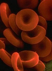
Research conducted in mice suggests that genomic screening might reveal cancer patients who are likely to develop treatment-related anemia.
The study showed that mice lacking Pten and Shp2—enzymes targeted by certain anticancer therapies—can’t produce and sustain enough red blood cells.
Investigators said this helps explain why anemia is a common side effect of anticancer drugs that target enzymes involved in tumor growth.
“Based on this unexpected finding, we might want to think about screening cancer patients’ genetic backgrounds for loss of Pten or Pten-regulated signals before prescribing anticancer drugs that might do more harm than good,” said Gen-Sheng Feng, PhD, of the University of California San Diego School of Medicine.
Dr Feng and his colleagues described their research in PNAS.
First, the team genetically engineered mice to lack Pten, Shp2, or both enzymes. The Pten-deficient mice had elevated white blood cells counts, consistent with myeloproliferative neoplasms (MPNs).
The Shp2-deficient mice experienced the opposite—lower white blood cell counts. And mice lacking both Pten and Shp2 had relatively normal white blood cell counts, suggesting that loss of Shp2 suppresses MPNs induced by Pten loss.
However, the investigators also discovered that mice lacking both enzymes had shorter lifespans than wild-type mice or mice lacking 1 of the enzymes.
This was because the combined deficiency of Shp2 and Pten induced lethal anemia. And this anemia was a result of 2 factors: red blood cells failed to develop properly and those that did form had a shortened lifespan.
To build upon these findings, the investigators treated Pten-deficient mice with the Shp2 inhibitor 11a-1 or with the MEK inhibitor trametinib. (MEK belongs to the same cellular communication network as Shp2.)
As with genetic deletion of Shp2, pharmacologic inhibition of Shp2 suppressed MPN induced by Pten loss and induced severe anemia in the mice.
Trametinib treatment had a similar effect, inducing anemia in Pten-deficient mice but not wild-type mice.
“What we’ve learned is that even if we know a lot about how individual molecules function in a cell, designing effective therapeutics that target them will require a more comprehensive understanding of the cross-talk between molecules in a particular cell type and in the context of disease,” Dr Feng concluded.
Research conducted in mice suggests that genomic screening might reveal cancer patients who are likely to develop treatment-related anemia.
The study showed that mice lacking Pten and Shp2—enzymes targeted by certain anticancer therapies—can’t produce and sustain enough red blood cells.
Investigators said this helps explain why anemia is a common side effect of anticancer drugs that target enzymes involved in tumor growth.
“Based on this unexpected finding, we might want to think about screening cancer patients’ genetic backgrounds for loss of Pten or Pten-regulated signals before prescribing anticancer drugs that might do more harm than good,” said Gen-Sheng Feng, PhD, of the University of California San Diego School of Medicine.
Dr Feng and his colleagues described their research in PNAS.
First, the team genetically engineered mice to lack Pten, Shp2, or both enzymes. The Pten-deficient mice had elevated white blood cells counts, consistent with myeloproliferative neoplasms (MPNs).
The Shp2-deficient mice experienced the opposite—lower white blood cell counts. And mice lacking both Pten and Shp2 had relatively normal white blood cell counts, suggesting that loss of Shp2 suppresses MPNs induced by Pten loss.
However, the investigators also discovered that mice lacking both enzymes had shorter lifespans than wild-type mice or mice lacking 1 of the enzymes.
This was because the combined deficiency of Shp2 and Pten induced lethal anemia. And this anemia was a result of 2 factors: red blood cells failed to develop properly and those that did form had a shortened lifespan.
To build upon these findings, the investigators treated Pten-deficient mice with the Shp2 inhibitor 11a-1 or with the MEK inhibitor trametinib. (MEK belongs to the same cellular communication network as Shp2.)
As with genetic deletion of Shp2, pharmacologic inhibition of Shp2 suppressed MPN induced by Pten loss and induced severe anemia in the mice.
Trametinib treatment had a similar effect, inducing anemia in Pten-deficient mice but not wild-type mice.
“What we’ve learned is that even if we know a lot about how individual molecules function in a cell, designing effective therapeutics that target them will require a more comprehensive understanding of the cross-talk between molecules in a particular cell type and in the context of disease,” Dr Feng concluded.
Research conducted in mice suggests that genomic screening might reveal cancer patients who are likely to develop treatment-related anemia.
The study showed that mice lacking Pten and Shp2—enzymes targeted by certain anticancer therapies—can’t produce and sustain enough red blood cells.
Investigators said this helps explain why anemia is a common side effect of anticancer drugs that target enzymes involved in tumor growth.
“Based on this unexpected finding, we might want to think about screening cancer patients’ genetic backgrounds for loss of Pten or Pten-regulated signals before prescribing anticancer drugs that might do more harm than good,” said Gen-Sheng Feng, PhD, of the University of California San Diego School of Medicine.
Dr Feng and his colleagues described their research in PNAS.
First, the team genetically engineered mice to lack Pten, Shp2, or both enzymes. The Pten-deficient mice had elevated white blood cells counts, consistent with myeloproliferative neoplasms (MPNs).
The Shp2-deficient mice experienced the opposite—lower white blood cell counts. And mice lacking both Pten and Shp2 had relatively normal white blood cell counts, suggesting that loss of Shp2 suppresses MPNs induced by Pten loss.
However, the investigators also discovered that mice lacking both enzymes had shorter lifespans than wild-type mice or mice lacking 1 of the enzymes.
This was because the combined deficiency of Shp2 and Pten induced lethal anemia. And this anemia was a result of 2 factors: red blood cells failed to develop properly and those that did form had a shortened lifespan.
To build upon these findings, the investigators treated Pten-deficient mice with the Shp2 inhibitor 11a-1 or with the MEK inhibitor trametinib. (MEK belongs to the same cellular communication network as Shp2.)
As with genetic deletion of Shp2, pharmacologic inhibition of Shp2 suppressed MPN induced by Pten loss and induced severe anemia in the mice.
Trametinib treatment had a similar effect, inducing anemia in Pten-deficient mice but not wild-type mice.
“What we’ve learned is that even if we know a lot about how individual molecules function in a cell, designing effective therapeutics that target them will require a more comprehensive understanding of the cross-talk between molecules in a particular cell type and in the context of disease,” Dr Feng concluded.
Anticoagulant granted fast track designation

Image by Kevin MacKenzie
The US Food and Drug Administration (FDA) has granted fast track designation to betrixaban as extended-duration venous thromboembolism (VTE) prophylaxis for acute medically ill patients.
This includes patients who are hospitalized for serious medical conditions such as heart failure, stroke, infection, and pulmonary disease.
Betrixaban is an investigational oral anticoagulant that directly inhibits the activity of factor Xa.
According to Portola Pharmaceuticals, the company developing betrixaban, the drug has distinct properties that may allow it to demonstrate clinical benefit without significantly increasing the risk of fatal bleeding and certain other serious side effects.
These benefits include a 19- to 25-hour half-life for once-daily dosing, a low peak-to-trough drug concentration ratio that minimizes anticoagulant variability, low renal clearance, and no significant CYP3A4 metabolism, which may reduce the risk of drug-drug interactions.
Betrixaban trials
In the phase 2 Explore-Xa trial, researchers compared betrixaban and warfarin in patients with atrial fibrillation. The team randomized 508 patients to 1 of 3 blinded doses of betrixaban (40 mg, 60 mg, or 80 mg once daily) or unblinded warfarin, adjusted to an international normalized ratio of 2.0-3.0.
The primary outcome was major or clinically relevant non-major bleeding. At a mean follow-up of 147 days, the primary outcome had been met by 1 patient in the 40 mg betrixaban arm, 5 each in the 60 mg and 80 mg betrixaban arms, and 7 in the warfarin arm.
One patient each in the 60 mg and 80 mg arms experienced an ischemic stroke. And there were 2 vascular deaths, 1 each in the 40 mg arm and the warfarin arm.
In the phase 2 EXPERT trial, researchers compared betrixaban and enoxaparin as VTE prophylaxis in patients who underwent total knee replacement.
The team enrolled 215 patients and assigned them to 1 of 3 post-operative prophylaxis regimens: betrixaban at 15 mg twice daily, betrixaban at 40 mg twice daily, or enoxaparin at 30 mg every 12 hours—all for 10 to 14 days.
The primary efficacy outcome was the incidence of VTE during the dosing period, and 175 patients were evaluable for this outcome. VTE occurred in 20% of patients in the 15 mg betrixaban arm (14/70), 15% in the 40 mg betrixaban arm (10/65), and 10% (4/40) in the enoxaparin arm.
Safety outcomes included major and clinically significant non-major bleeds through 48 hours after treatment. There were no bleeds in the 15 mg betrixaban arm, 2 (2.4%) clinically significant non-major bleeds in the 40 mg betrixaban arm, and 1 (2.3%) major bleed and 2 (4.6%) clinically significant non-major bleeds in the enoxaparin arm.
Betrixaban is currently being tested in the phase 3 APEX trial for the prevention of VTE in acute medically ill patients. Portola said it expects to complete enrollment in APEX by the end of this year and report top-line data in the first quarter of 2016.
If the trial is successful, the company plans to submit a new drug application to the FDA later in 2016 under the fast track designation.
About fast track designation
The FDA’s fast track program is designed to facilitate and expedite the development and review of new drugs intended to treat serious or life-threatening conditions and address unmet medical need.
Through the fast track program, a product may be eligible for priority review. In addition, the company developing the drug may be allowed to submit sections of the biologic license application or new drug application on a rolling basis as data become available.
Fast track designation also provides the company with opportunities for more frequent meetings with the FDA to discuss the drug’s development plan and ensure collection of the appropriate data needed to support drug approval. And the designation allows for more frequent written communication from the FDA about things such as the design of proposed clinical trials and the use of biomarkers.
Image by Kevin MacKenzie
The US Food and Drug Administration (FDA) has granted fast track designation to betrixaban as extended-duration venous thromboembolism (VTE) prophylaxis for acute medically ill patients.
This includes patients who are hospitalized for serious medical conditions such as heart failure, stroke, infection, and pulmonary disease.
Betrixaban is an investigational oral anticoagulant that directly inhibits the activity of factor Xa.
According to Portola Pharmaceuticals, the company developing betrixaban, the drug has distinct properties that may allow it to demonstrate clinical benefit without significantly increasing the risk of fatal bleeding and certain other serious side effects.
These benefits include a 19- to 25-hour half-life for once-daily dosing, a low peak-to-trough drug concentration ratio that minimizes anticoagulant variability, low renal clearance, and no significant CYP3A4 metabolism, which may reduce the risk of drug-drug interactions.
Betrixaban trials
In the phase 2 Explore-Xa trial, researchers compared betrixaban and warfarin in patients with atrial fibrillation. The team randomized 508 patients to 1 of 3 blinded doses of betrixaban (40 mg, 60 mg, or 80 mg once daily) or unblinded warfarin, adjusted to an international normalized ratio of 2.0-3.0.
The primary outcome was major or clinically relevant non-major bleeding. At a mean follow-up of 147 days, the primary outcome had been met by 1 patient in the 40 mg betrixaban arm, 5 each in the 60 mg and 80 mg betrixaban arms, and 7 in the warfarin arm.
One patient each in the 60 mg and 80 mg arms experienced an ischemic stroke. And there were 2 vascular deaths, 1 each in the 40 mg arm and the warfarin arm.
In the phase 2 EXPERT trial, researchers compared betrixaban and enoxaparin as VTE prophylaxis in patients who underwent total knee replacement.
The team enrolled 215 patients and assigned them to 1 of 3 post-operative prophylaxis regimens: betrixaban at 15 mg twice daily, betrixaban at 40 mg twice daily, or enoxaparin at 30 mg every 12 hours—all for 10 to 14 days.
The primary efficacy outcome was the incidence of VTE during the dosing period, and 175 patients were evaluable for this outcome. VTE occurred in 20% of patients in the 15 mg betrixaban arm (14/70), 15% in the 40 mg betrixaban arm (10/65), and 10% (4/40) in the enoxaparin arm.
Safety outcomes included major and clinically significant non-major bleeds through 48 hours after treatment. There were no bleeds in the 15 mg betrixaban arm, 2 (2.4%) clinically significant non-major bleeds in the 40 mg betrixaban arm, and 1 (2.3%) major bleed and 2 (4.6%) clinically significant non-major bleeds in the enoxaparin arm.
Betrixaban is currently being tested in the phase 3 APEX trial for the prevention of VTE in acute medically ill patients. Portola said it expects to complete enrollment in APEX by the end of this year and report top-line data in the first quarter of 2016.
If the trial is successful, the company plans to submit a new drug application to the FDA later in 2016 under the fast track designation.
About fast track designation
The FDA’s fast track program is designed to facilitate and expedite the development and review of new drugs intended to treat serious or life-threatening conditions and address unmet medical need.
Through the fast track program, a product may be eligible for priority review. In addition, the company developing the drug may be allowed to submit sections of the biologic license application or new drug application on a rolling basis as data become available.
Fast track designation also provides the company with opportunities for more frequent meetings with the FDA to discuss the drug’s development plan and ensure collection of the appropriate data needed to support drug approval. And the designation allows for more frequent written communication from the FDA about things such as the design of proposed clinical trials and the use of biomarkers.
Image by Kevin MacKenzie
The US Food and Drug Administration (FDA) has granted fast track designation to betrixaban as extended-duration venous thromboembolism (VTE) prophylaxis for acute medically ill patients.
This includes patients who are hospitalized for serious medical conditions such as heart failure, stroke, infection, and pulmonary disease.
Betrixaban is an investigational oral anticoagulant that directly inhibits the activity of factor Xa.
According to Portola Pharmaceuticals, the company developing betrixaban, the drug has distinct properties that may allow it to demonstrate clinical benefit without significantly increasing the risk of fatal bleeding and certain other serious side effects.
These benefits include a 19- to 25-hour half-life for once-daily dosing, a low peak-to-trough drug concentration ratio that minimizes anticoagulant variability, low renal clearance, and no significant CYP3A4 metabolism, which may reduce the risk of drug-drug interactions.
Betrixaban trials
In the phase 2 Explore-Xa trial, researchers compared betrixaban and warfarin in patients with atrial fibrillation. The team randomized 508 patients to 1 of 3 blinded doses of betrixaban (40 mg, 60 mg, or 80 mg once daily) or unblinded warfarin, adjusted to an international normalized ratio of 2.0-3.0.
The primary outcome was major or clinically relevant non-major bleeding. At a mean follow-up of 147 days, the primary outcome had been met by 1 patient in the 40 mg betrixaban arm, 5 each in the 60 mg and 80 mg betrixaban arms, and 7 in the warfarin arm.
One patient each in the 60 mg and 80 mg arms experienced an ischemic stroke. And there were 2 vascular deaths, 1 each in the 40 mg arm and the warfarin arm.
In the phase 2 EXPERT trial, researchers compared betrixaban and enoxaparin as VTE prophylaxis in patients who underwent total knee replacement.
The team enrolled 215 patients and assigned them to 1 of 3 post-operative prophylaxis regimens: betrixaban at 15 mg twice daily, betrixaban at 40 mg twice daily, or enoxaparin at 30 mg every 12 hours—all for 10 to 14 days.
The primary efficacy outcome was the incidence of VTE during the dosing period, and 175 patients were evaluable for this outcome. VTE occurred in 20% of patients in the 15 mg betrixaban arm (14/70), 15% in the 40 mg betrixaban arm (10/65), and 10% (4/40) in the enoxaparin arm.
Safety outcomes included major and clinically significant non-major bleeds through 48 hours after treatment. There were no bleeds in the 15 mg betrixaban arm, 2 (2.4%) clinically significant non-major bleeds in the 40 mg betrixaban arm, and 1 (2.3%) major bleed and 2 (4.6%) clinically significant non-major bleeds in the enoxaparin arm.
Betrixaban is currently being tested in the phase 3 APEX trial for the prevention of VTE in acute medically ill patients. Portola said it expects to complete enrollment in APEX by the end of this year and report top-line data in the first quarter of 2016.
If the trial is successful, the company plans to submit a new drug application to the FDA later in 2016 under the fast track designation.
About fast track designation
The FDA’s fast track program is designed to facilitate and expedite the development and review of new drugs intended to treat serious or life-threatening conditions and address unmet medical need.
Through the fast track program, a product may be eligible for priority review. In addition, the company developing the drug may be allowed to submit sections of the biologic license application or new drug application on a rolling basis as data become available.
Fast track designation also provides the company with opportunities for more frequent meetings with the FDA to discuss the drug’s development plan and ensure collection of the appropriate data needed to support drug approval. And the designation allows for more frequent written communication from the FDA about things such as the design of proposed clinical trials and the use of biomarkers.