User login
Exome sequencing shows potential as diagnostic tool

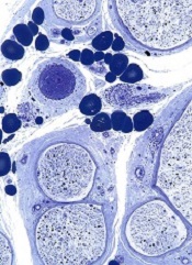
Credit: Graham Colm
In a large study, whole-exome sequencing provided 25% of patients with a diagnosis related to a known genetic disease, giving young
patients and their parents some long-sought answers.
The technology was able to detect a number of rare genetic events and new mutations contributing to disease.
Among the medically actionable findings were mutations related to Fanconi anemia, erythrocytosis, hemolytic anemia, and von Willebrand disease.
“The findings in this report, I believe, will forever change the future practice of pediatrics and medicine as a whole,” said study author James R. Lupski, MD, PhD, of the Baylor College of Medicine in Houston.
“It is just a matter of time before genomics moves up on the physician’s list of things to do and is ordered before formulating a differential diagnosis. It will be the new ‘family history’ that, better yet, gets you both the important variants inherited from each parent and the new mutations that contribute to disease susceptibility.”
This research was published in JAMA and is set to be presented on October 21 at the American Society of Human Genetics Annual Meeting in San Diego.
The researchers had previously conducted a pilot study of whole-exome sequencing that included 250 patients and revealed a 25% molecular diagnostic rate.
This current study included 2000 patients (88% pediatric) with clinical whole-exome sequencing analyzed between June 2012 and August 2014. The majority of patients—87.8%—had neurological disorders or a developmental delay, and 12.2% had non-neurological disorders.
The researchers collected peripheral blood, tissue, or extracted DNA samples from patients and their parents. The team sequenced patients’ DNA and compared those results to the normal reference. Any disease-associated mutations were then compared with the parent’s DNA to determine if the child inherited it from one or both parents.
In all, 504 patients (25.2%) received a molecular diagnosis, and 58% of the diagnostic mutations had not previously been reported. Two hundred and eighty patients had a single mutation that caused disease, 181 were autosomal recessive, 65 were X-linked, and 1 was presumed inherited through the mitochondria.
In 5 cases, the patient inherited 2 copies of the mutated gene from the same parent. Of the dominant mutations, 208 were de novo mutations not inherited from either parent, 32 were inherited, and 40 were not determined because parental samples were not available.
Among the de novo mutations, 5 demonstrated mosaicism, which suggested the mutation occurred after fertilization.
The researchers found 708 presumptive causative variant alleles in the 504 cases. Almost 30% of the diagnoses occurred in disease genes only identified by researchers in the last 3 years. In 65 cases, there was no available genetic test other than whole-exome sequencing to find the mutated gene at the time the test was ordered.
Twenty-three patients (about 5%) had mutations in 2 different genes, which could account for various aspects of the patient’s medical condition.
“Doctors generally try to find one diagnosis that explains all the issues a patient may have,” said study author Christine Eng, MD, of the Baylor College of Medicine.
“We have found that, in some cases, a patient may have a blended phenotype of 2 different conditions. That patients may have 2 different rare genetic diseases to explain their condition was an unexpected finding prior to the use of whole-exome sequencing.”
In the 2000 cases, incidental findings of medically actionable results that could result in early diagnosis, screening, or treatment were found in 92 patients. Three patients had more than 1 finding.
“Clinical exome sequencing can assist diagnosis in a wide range of disorders that are diagnostic dilemmas,” Dr Lupski said. “Rare variants and Mendelian disease are important contributors to disease populations. This is in sharp contrast to the thinking of population geneticists, who investigate how common variants contribute to disease susceptibility.”
“We find ‘rare variants’ in aggregate actually contribute to disease susceptibility in a big way. The individual diseases may be rare, but there are thousands of such diseases and many more being defined through genomics.” ![]()
Stem cells can help form blood vessels
A novel technique can jump-start the creation of blood vessels, researchers have reported in Nature Biotechnology.
The team used human embryonic stem cells (hESCs) and induced pluripotent stem cells (hiPSCs) to create endothelial colony-forming cells (ECFCs).
These lab-generated ECFCs successfully produced human blood vessels in mice and restored blood flow to damaged retinas and limbs, without posing a risk of teratoma formation.
ECFCs can lose their ability to proliferate into new blood vessels as patients age or develop diseases such as peripheral arterial disease, said Mervin C. Yoder Jr, MD, of the Indiana University School of Medicine in Indianapolis.
He and his colleagues theorized that injecting patients with “younger,” more “enthusiastic” ECFCs might jump-start the process of creating new blood vessels. Such cells are difficult to find in adults but are common in umbilical cord blood.
With that in mind, the researchers developed a novel method to mature hiPSCs or hESCs into cells with the characteristics of ECFCs found in cord blood.
Both hiPSC-ECFCs and hESC-ECFCs exhibited properties of cord blood-derived ECFCs, including a homogenous monolayer with a cobblestone appearance and high clonal proliferative potential.
In addition, hiPSC-ECFCs and hESC-ECFCs formed capillary structures when cultured on Matrigel, and they generated in vivo inosculated vessels when implanted in immune-deficient mice.
In other murine experiments, hiPSC-ECFCs contributed to vascular repair of experimentally induced ischemic limbs and injured retinas. The cells’ effects were similar to those observed with cord blood-derived ECFCs and superior to effects observed with endothelial cells isolated using other published protocols.
Furthermore, hiPSC-ECFCs did not transition to nonendothelial cells over prolonged culture, and they could expand to more than 1 trillion endothelial cells in less than 3 months.
“This is one of the first studies using induced pluripotent stem cells that has been able to produce new cells in clinically relevant numbers, enough to enable a clinical trial,” Dr Yoder said.
He added that one of the next steps for this research is reaching an agreement with a facility approved to produce cells for use in human testing. ![]()
Invention allows for precise gene control

Credit: NIGMS
A new way to control genes may provide a better understanding of cancers and aid the development of new therapies.
The key to the method is an invention called SunTag, a series of molecular hooks for hanging multiple copies of biologically active molecules onto a single protein scaffold used to target genes or other molecules.
Compared to molecules assembled without these hooks, those incorporating SunTag can greatly amplify biological activity.
SunTag was developed by researchers in the lab of Ron Vale, PhD, of the University of California, San Francisco (UCSF), and described in two papers published in Cell.
In one paper, the authors recount how they used SunTag to greatly amplify the light-emitting signal from the green fluorescent protein commonly used to label molecules within cells.
In another paper, the researchers explain how they used SunTag to supercharge a variation of a biochemical approach known as CRISPR.
CRISPR is a technique that emerged a few years ago as a way to edit DNA anywhere within the genome.
The UCSF researchers adapted CRISPR to activate genes or interfere with their activity in a reversible way without altering DNA. The team believes this capability might make previous methods for probing poorly understood cellular functions obsolete.
“With these techniques, we can fine-tune the activity of genes within cells, and this has broad implications for the reprogramming of cells,” said Jonathan Weissman, PhD, also of UCSF.
Scientists previously reported ways to activate or interfere with genes using CRISPR, but Dr Vale said these methods were inefficient, especially for activating genes.
“It depends on the gene, but this new approach appears to amplify gene-switching by as much as 50-fold,” he said. “It’s a much more robust way of activating genes.”
CRISPR with SunTag sheds light on cancers
CRISPR is a natural system that bacteria use to defend themselves against viruses. The basis for CRISPR applications in the lab is a protein called Cas9, a chassis into which scientists can insert any specific RNA partner molecule.
The selected RNA serves as an adaptor that determines the target anywhere within the genome. The researchers attached SunTag to this chassis, enabling one Cas9 to recruit many copies of a protein to a specific DNA sequence.
The adaptation of SunTag for CRISPR activation makes it possible to systematically probe the biological roles of all genes within the genome in a single experiment, the team said.
They used CRISPR activation to identify a number of tumor suppressor genes that inhibit the growth of cancer cells. In future studies, they plan to use CRISPR activation to reveal mechanisms by which cancer cells develop resistance to anticancer drugs, a process that typically involves gene activation.
Will RNA interference become obsolete?
CRISPR interference has the potential to render RNA interference obsolete, according to Dr Weissman.
Unlike conventional RNA interference techniques, CRISPR interference allows any number of individual genes to be silenced at the same time. In addition, there is little risk of turning off untargeted genes the way RNA interference techniques do.
RNA interference blocks the messenger RNA that drives protein protection based on the blueprint contained within a gene’s DNA sequence. By preventing protein production, RNA interference may be used to get around the problem of difficult-to-target proteins, a frequent challenge in drug development.
But CRISPR interference acts one step earlier in the cell’s protein-manufacturing process.
“The horse has already left the barn with RNA interference, in the sense that the RNA message already has been transcribed from DNA,” Dr Weissman said. “With CRISPR interference, we can prevent the message from being written.” ![]()
‘Nano-cocoons’ offer targeted drug delivery
Photo courtesy of the Gu lab
Biomedical engineers have developed a drug delivery system in which cancer cells are “tricked” into absorbing nanoscale “cocoons” before
they unleash anticancer drugs.
Each cocoon is made of a deoxyribonuclease (DNase)-degradable DNA nanoclew embedded with an acid-responsive DNase I nanocapsule.
A cancer cell’s acidic environment prompts the DNase to degrade the cocoon and release the drug encapsulated in the endolysosomal compartment.
The engineers described the creation of these nano-cocoons and in vitro experiments testing the delivery of doxorubicin in the Journal of the American Chemical Society.
“This drug delivery system is DNA-based, which means it is biocompatible and less toxic to patients than systems that use synthetic materials,” said study author Zhen Gu, PhD, of the University of North Carolina Chapel Hill.
“This technique also specifically targets cancer cells, can carry a large drug load, and releases the drugs very quickly once inside the cancer cell.”
“In addition, because we used self-assembling DNA techniques, it is relatively easy to manufacture,” added Wujin Sun, a PhD student in Dr Gu’s lab.
Each nano-cocoon is made of a single strand of DNA that self-assembles into a structure measuring 150 nanometers across.
The core of the nano-cocoon contains the anticancer drug doxorubicin and DNase. The DNase, an enzyme that would normally dismantle the DNA cocoon, is contained by a thin polymer coating.
The surface of the nano-cocoon is studded with folic acid ligands. When the nano-cocoon encounters a cancer cell, the ligands bind the nano-cocoon to receptors on the surface of the cell, causing the cell to engulf the nano-cocoon.
Once the cocoon is inside the cancer cell, the cell’s acidic environment destroys the polymer sheath containing the DNase. The DNase then slices through the DNA cocoon, spilling doxorubicin into the cancer cell and killing it.
“We’re preparing to launch preclinical testing now,” Dr Gu said. “We’re very excited about this system and think it holds promise for delivering a variety of drugs targeting cancer and other diseases.” ![]()
Amoeba could help fight cancers

Experiments in a soil-dwelling amoeba have provided insight that could help us treat cancers characterized by PTEN mutations, researchers have reported in PLOS ONE.
The team discovered that this amoeba has two genes that function like the human tumor suppressor PTEN.
And increasing expression of one of these genes compensated for a mutation in the other gene.
If the same method works in humans with mutated PTEN, this finding could have implications for a range of cancers.
PTEN mutations are thought to be involved in nearly half of all leukemia cases, 40% of breast cancer cases, and up to 70% of prostate cancer cases.
“If you look at tumors across the board . . . , you find that PTEN is the most generally mutated gene, and, when you mutate PTEN in mice, you cause tumors,” said study author David Soll, PhD, of the University of Iowa in Iowa City.
He and his colleagues found that the amoeba Dictyostelium discoideum has the gene ptenA, which mutates similarly to the human PTEN gene and causes behavioral defects in the cell.
They also found a close relative of ptenA in the amoeba, called lpten, that performs the same functions of ptenA but to a lesser degree.
The researchers hypothesized that ramping up the presence of lpten could compensate for the mutated ptenA.
They tested this theory by placing lpten in a plasmid behind a powerful promoter designed to overexpress the gene. They then introduced the super-charged lpten into a cell with the mutated ptenA gene.
The team found that the overexpressed lpten gene fully compensated for all of the defects in the ptenA mutant.
If this method works in human cells, it could lead to a new way to treat cancers, the researchers said. They are now aiming to identify a drug that would activate the promoter for one of PTEN’s close relatives.
Once a patient is diagnosed with cancer caused by a PTEN mutation, the patient could take the drug, overexpress the PTEN replacement gene, and potentially stop cancer in its tracks, Dr Soll said.
This research has also led Dr Soll and his colleagues to study other human genes that may be able to step in for the mutated PTEN gene and perform the same tumor-suppressing role. The team is currently studying 2 close relatives of PTEN.
“And nature might have put them there just for that; that’s the curious thing,” Dr Soll said. “Somewhere, there may be a backup system, what we call ‘redundancy,’ that might be the basis for better identifying tumors and possibly creating cancer-fighting drugs. You have another gene which might be able to step in for the broken gene to keep things normal, and that’s what we’re playing with here. It’s very sophisticated.” ![]()
Malpractice reform may not reduce ‘defensive medicine’

Credit: CDC
Making it more difficult for patients to sue physicians for medical malpractice may not reduce the amount of “defensive medicine” physicians
practice, new research suggests.
Investigators studied patient records in 3 states that raised the standard for malpractice in the emergency room to gross negligence.
And they found that strong new legal protections did not significantly reduce the use of common defensive medicine practices or the cost of care.
The team detailed these findings in NEJM.
“Our findings suggest that malpractice reform may have less effect on costs than has been projected by conventional wisdom,” said lead study author Daniel A. Waxman, MD, PhD, of RAND Health in Santa Monica, California.
“Physicians say they order unnecessary tests strictly out of fear of being sued, but our results suggest the story is more complicated.”
The investigators evaluated the results of malpractice reform in 3 states—Georgia, Texas, and South Carolina.
About a decade ago, these states changed the legal malpractice standard for emergency care to gross negligence. The higher standard means plaintiffs must prove that doctors consciously disregarded the need to use reasonable care, knowing full well that their actions were likely to cause serious injury.
“These malpractice reforms have been said to provide virtual immunity against lawsuits,” Dr Waxman noted.
He and his colleagues examined 3.8 million Medicare patient records from 1166 hospital emergency departments spanning the period from 1997 to 2011. They compared care in the 3 reform states, before and after the statutes took effect, to care in neighboring states that did not pass malpractice reform.
The team assessed whether physicians ordered an advanced imaging study (CT or MRI scan), whether the patient was hospitalized after the emergency visit, and total charges for the visit.
Advanced imaging and hospitalization are among the most costly consequences of an emergency room visit, and physicians have identified them as common defensive medicine practices.
The malpractice reform laws had no effect on the use of imaging or the rate of hospitalization following emergency visits.
For 2 of the states, Texas and South Carolina, the law did not appear to cause any reduction in charges. Relative to neighboring states, Georgia saw a drop of 3.6% in average emergency room charges following its 2005 reform.
“This study suggests that, even when the risk of being sued for malpractice decreases, the path of least resistance still may favor resource-intensive care, at least in hospital emergency departments,” Dr Waxman said. ![]()
How being a parent affects advanced cancer patients

Credit: Vera Kratochvil
BOSTON—Having dependent children may motivate advanced cancer patients to pursue more aggressive treatment, a pilot study indicates.
Parental status was an important factor in treatment decision-making for most of the 42 patients studied.
A majority of patients said being a parent motivated them to pursue life-extending treatments, although some patients mentioned the importance of staying close to their families to receive treatment and retaining the ability to function as a parent.
This research was released in a presscast in advance of ASCO’s 2014 Quality Care Symposium, which is scheduled to take place October 17-18 at the Boston Marriott Copley Place. The research is set to be presented as abstract 65.*
Researchers interviewed 42 patients with metastatic cancer who have children younger than 18 years of age. The patients had an average age of 44.
When asked how having children influenced their treatment decisions, 64% of patients said being a parent motivated them to pursue life-extending treatments, largely out of a desire to have more time with their children. However, 24% of patients said being a parent did not influence their treatment decisions.
Twenty-four percent of respondents identified preserving parental functioning as a treatment priority. They didn’t want side effects interfering with their or their children’s daily lives.
And 12% of patients mentioned the importance of receiving treatment close to their families, rather than traveling for a second opinion or pursuing treatment that might require long hospital stays.
Fifty-two percent of patients indicated an interest in using hospice services. Others were specifically interested in institutional rather than home hospice care, due to a desire to protect their children from the dying experience.
Fifty-nine percent of patients reported an interest in receiving palliative care concurrent with their cancer treatment, although several patients seemed to conflate palliative care with end-of-life care.
“Numerous psychosocial factors influence patients’ decisions about cancer treatment,” said lead study author Devon Check, a PhD student at the University of North Carolina at Chapel Hill.
“It’s important for patients with dependent children to discuss their treatment priorities with their oncologist, who may not know, for example, how important it is for a patient with children to preserve their functioning at home.”
“We hope that our study can help oncologists engage patients with children in shared decision-making and promote alignment of the treatment plan with the patients’ priorities, including family responsibilities.”
Although this study included patients with a range of physical functioning and a variety of cancer types, the findings may not generalize to other patient groups.
The researchers are planning a larger study to explore the associations between parental status, parenting concerns, and decision-making about treatment for advanced cancer. ![]()
*Some of the data presented differ from data in the abstract.
Credit: Vera Kratochvil
BOSTON—Having dependent children may motivate advanced cancer patients to pursue more aggressive treatment, a pilot study indicates.
Parental status was an important factor in treatment decision-making for most of the 42 patients studied.
A majority of patients said being a parent motivated them to pursue life-extending treatments, although some patients mentioned the importance of staying close to their families to receive treatment and retaining the ability to function as a parent.
This research was released in a presscast in advance of ASCO’s 2014 Quality Care Symposium, which is scheduled to take place October 17-18 at the Boston Marriott Copley Place. The research is set to be presented as abstract 65.*
Researchers interviewed 42 patients with metastatic cancer who have children younger than 18 years of age. The patients had an average age of 44.
When asked how having children influenced their treatment decisions, 64% of patients said being a parent motivated them to pursue life-extending treatments, largely out of a desire to have more time with their children. However, 24% of patients said being a parent did not influence their treatment decisions.
Twenty-four percent of respondents identified preserving parental functioning as a treatment priority. They didn’t want side effects interfering with their or their children’s daily lives.
And 12% of patients mentioned the importance of receiving treatment close to their families, rather than traveling for a second opinion or pursuing treatment that might require long hospital stays.
Fifty-two percent of patients indicated an interest in using hospice services. Others were specifically interested in institutional rather than home hospice care, due to a desire to protect their children from the dying experience.
Fifty-nine percent of patients reported an interest in receiving palliative care concurrent with their cancer treatment, although several patients seemed to conflate palliative care with end-of-life care.
“Numerous psychosocial factors influence patients’ decisions about cancer treatment,” said lead study author Devon Check, a PhD student at the University of North Carolina at Chapel Hill.
“It’s important for patients with dependent children to discuss their treatment priorities with their oncologist, who may not know, for example, how important it is for a patient with children to preserve their functioning at home.”
“We hope that our study can help oncologists engage patients with children in shared decision-making and promote alignment of the treatment plan with the patients’ priorities, including family responsibilities.”
Although this study included patients with a range of physical functioning and a variety of cancer types, the findings may not generalize to other patient groups.
The researchers are planning a larger study to explore the associations between parental status, parenting concerns, and decision-making about treatment for advanced cancer.
*Some of the data presented differ from data in the abstract.
Coating repels blood and bacteria

to form a blood clot
Credit: James Weaver
Researchers have developed a coating that can prevent blood and bacteria from adhering to the surface of medical devices.
The coating repelled blood from more than 20 medically relevant substrates, suppressed biofilm formation, and prevented coagulation in an animal model for at least 8 hours.
The researchers believe this technology could reduce the use of anticoagulants and help prevent thrombotic occlusion and biofouling of medical devices.
The idea for the coating evolved from a surface technology known as “slippery liquid-infused porous surfaces (SLIPS),” which was developed by Joanna Aizenberg, PhD, of Harvard University’s Wyss Institute for Biologically Inspired Engineering.
Inspired by the slippery surface of the carnivorous pitcher plant, which enables the plant to capture insects, SLIPS can repel nearly any material. The liquid layer on the surface provides a barrier to everything from ice to crude oil and blood.
“Traditional SLIPS uses porous, textured surface substrates to immobilize the liquid layer, whereas medical surfaces are mostly flat and smooth,” Dr Aizenberg said. “So we further adapted our approach by capitalizing on the natural roughness of chemically modified surfaces of medical devices.”
She and her colleagues described this work in Nature Biotechnology.
The researchers developed a super-repellent coating that can be adhered to existing, approved medical devices. In a 2-step surface-coating process, they chemically attached a monolayer of perfluorocarbon, which is similar to Teflon.
Then, they added a layer of liquid perfluorocarbon, which is widely used in medicine for applications such as liquid ventilation for infants with breathing challenges, blood substitution, and eye surgery. The team calls the tethered perfluorocarbon plus the liquid layer a “tethered-liquid perfluorocarbon surface (TLP)”.
TLP worked seamlessly when coated on more than 20 different medical surfaces, including plastics, glasses, and metals.
The researchers also implanted medical-grade tubing and catheters coated with TLP in large blood vessels in pigs, and the coating prevented blood from clotting for at least 8 hours without the use of anticoagulants.
TLP-treated medical tubing was stored for more than a year under normal temperature and humidity conditions and still prevented clot formation, repelling fibrin and platelets.
The TLP surface remained stable under the full range of clinically relevant physiological shear stresses, or rates of blood flow seen in catheters and central lines, all the way up to dialysis machines.
When the bacteria Pseudomonas aeruginosa were grown in TLP-coated medical tubing for more than 6 weeks, less than 1 in a billion bacteria were able to adhere. Central lines coated with TLP significantly reduced sepsis from central-line associated bloodstream infections.
Out of curiosity, the researchers even tested a TLP-coated surface with a gecko, whose footpads contain hair-like structures with tremendous adhesive strength. And the gecko was unable to hold on.
“We were wonderfully surprised by how well the TLP coating worked, particularly in vivo without heparin,” said Anna Waterhouse, PhD, of the Wyss Institute.
“Usually, the blood will start to clot within an hour in the extracorporeal circuit, so our experiments really demonstrate the clinical relevance of this new coating.”
System can detect sepsis early
Credit: CDC
An early warning and response system can predict a patient’s likelihood of developing sepsis, according to research published in the Journal of Hospital Medicine.
The system uses lab and vital-sign data in the electronic health record of hospital inpatients to identify those at risk for sepsis.
In a multi-hospital study, the system allowed for a marked increase in sepsis identification and care, transfer to the intensive care unit (ICU), and an indication of fewer deaths due to sepsis.
Craig A. Umscheid, MD, of Penn Medicine in Philadelphia, and his colleagues developed the system using 4575 patients admitted to the University of Pennsylvania Health System in October 2011.
The system monitored lab values and vital signs in real time. If a patient had 4 or more predefined abnormalities at any single time, an electronic communication was sent to the provider, nurse, and rapid response coordinator, who performed an immediate bedside patient evaluation.
The researchers validated the effectiveness of the system during a pre-implementation period from June to September 2012, when data on admitted patients was evaluated and alerts triggered in a database, but no notifications were sent to providers on the ground.
Outcomes in that control period were then compared to a post-implementation period from June to September 2013. The total number of patients included in both periods was 31,093.
In the pre- and post-implementation periods, 4% of patient visits triggered the alert. Analysis revealed that 90% of those patients received bedside evaluations by the care team within 30 minutes of the alert being issued.
The system resulted in a 2- to 3-fold increase in orders for tests that could help identify the presence of sepsis and a 1.5- to 2-fold increase in the administration of antibiotics and intravenous fluids.
The system prompted an increase of more than 50% in the proportion of patients quickly transferred to the ICU and a 50% increase in documentation of sepsis in the patients’ electronic health record.
There was a lower death rate from sepsis and an increase in the number of patients successfully discharged home in the post-implementation period. But these rates were not significantly different from those in the pre-implementation period.
“Our study is the first we’re aware of that was implemented throughout a multihospital health system,” Dr Umscheid said.
“Previous studies that have examined the impact of sepsis prediction tools at other institutions have only taken place on a limited number of inpatient wards. The varied patient populations, clinical staffing, practice models, and practice cultures across our health system increases the generalizability of our findings to other healthcare settings.”
Dr Umscheid also noted that the system could help triage patients for suitability of ICU transfer.
“By better identifying those with sepsis requiring advanced care,” he said, “the tool can help screen out patients not needing the inevitably limited number of ICU beds.”
Physical changes in RBCs remain a mystery

During their approximately 100-day lifespan in the bloodstream, red blood cells (RBCs) lose membrane surface area, volume, and hemoglobin content.
A study published in PLOS Computational Biology shows that, of these 3 changes, only surface-area loss can be explained by RBCs shedding small, hemoglobin-containing vesicles budding off their cells’ membrane.
Therefore, an unknown process must be primarily responsible for loss of RBC volume and hemoglobin reduction.
Roy Malka, PhD, of Massachusetts General Hospital in Boston, and his colleagues noted that variations in RBCs’ mean volume and hemoglobin content are associated with important clinical conditions, but the mechanisms controlling these physical characteristics are not well understood.
Vesicle shedding was thought to be the most important mechanism, but the researchers found evidence to suggest that a dominant role for vesicle shedding would violate empirical geometric and biophysical constraints.
So they concluded that an unknown mechanism must control loss of RBC volume and hemoglobin reduction. And this mechanism is likely responsible for 60% to 90% of volume loss and hemoglobin reduction.
The group’s work combined mathematical modeling of the mechanism that changes the physical properties of RBCs, clinical measurements of both cellular volume and hemoglobin content, and data from a new system for characterizing the non-water cellular mass of individual cells.
The researchers said the quantitative characterization of RBC loss processes will help focus future research into the molecular mechanisms of RBC maturation. And it may ultimately help in the early detection of clinical conditions where the RBC maturation pattern is altered.