User login
Efficacy of malaria vaccine varies with age, over time
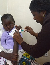
Credit: Caitlin Kleiboer
A candidate malaria vaccine is more effective in children aged 5 months to 17 months than in infants 3 months of age or younger, results of a phase 3 study suggest.
Previous research indicated the vaccine— RTS,S/AS01—may prevent malaria in young children, but its efficacy wanes over time.
Now, researchers have reported that RTS,S/AS01 can sometimes prevent clinical malaria, severe malaria, and malaria hospitalization in children aged 5 to 17 months.
In the younger age group, the vaccine offered some protection against clinical malaria but did not significantly impact the rates of severe malaria or hospitalization.
The RTS,S Clinical Trials Partnership committee reported these findings in PLOS Medicine.
The study was sponsored by GSK Biologicals SA, the developer and manufacturer of RTS,S/AS01, and funded by both GSK Biologicals SA and the PATH Malaria Vaccine Initiative.
The researchers studied 6537 infants aged 6 to 12 weeks and 8923 children aged 5 to 17 months treated at 11 sites in Africa.
Subjects were randomized to receive 3 doses of RTS,S/AS01 or a comparator vaccine—a rabies vaccine (VeroRab) for the children and a meningococcal C conjugate vaccine (Menjugate) for the infants.
The primary outcome, vaccine efficacy (VE), was the reduction in malaria incidence among subjects who received RTS,S/AS01 compared to the incidence among subjects who received a comparator vaccine.
In the 18 months following vaccination, VE was 46% in children aged 5 to 17 months and 27% in infants aged 6 to 12 weeks. In both age groups, VE was highest in the first 6 months after vaccination.
RTS,S/AS01 averted an average of 829 cases of clinical malaria per 1000 children vaccinated (range, 37 to 2365) and 449 cases per 1000 infants (range, -10 to 1402) within 18 months of vaccination.
In children aged 5 to 17 months, VE was 34% for severe malaria, 41% for malaria hospitalization, and 19% for all-cause hospitalization. In the infants, there was no significant protection against severe malaria, malaria hospitalization, or all-cause hospitalization.
Serious adverse events occurred less often in children who received RTS,S/AS01 than in those who received a comparator vaccine—18.6% and 22.7%, respectively. In infants, the rate of serious adverse events was similar between the 2 vaccination groups.
Meningitis was more common in subjects who received RTS,S/AS01 than in those who received a comparator. However, the researchers have not established a causal relationship.
There were 16 cases of meningitis among the 5949 children in the RTS,S/AS01 group, 1 case among the 2974 children in the control group, 9 cases among the 4358 infants in the RTS,S/AS01 group, and 3 cases among the 2179 infants in the control group.
Despite the adverse events associated with RTS,S/AS01 and its decreased efficacy over time, the researchers believe the vaccine shows promise. They noted that, “even at modest levels of VE, the number of malaria cases averted was substantial.”
Now, the group is evaluating whether a booster immunization given 18 months after the primary vaccination can improve the efficacy of RTS,S/AS01.
Results of this study were previously reported at the Multilateral Initiative on Malaria Pan African Conference in October 2013 and published in The New England Journal of Medicine in October 2011 and November 2012. Long-term results of a phase 2 trial of RTS,S/AS01 were published in The New England Journal of Medicine in March 2013. ![]()
Drug-resistant malaria spreading

Credit: James Gathany
Drug-resistant malaria parasites have spread to critical border regions of Southeast Asia, according to a study published in The New England Journal of Medicine.
The study confirms that resistance to the world’s most effective antimalarial drug, artemisinin, is now widespread in Southeast Asia.
This is not the first time malaria parasites have developed resistance to front-line drugs, and, each time, resistance has emerged from the same corner of Asia on the Cambodia-Thailand border.
To assess the extent of artemisinin resistance, researchers analyzed blood samples from 1241 malaria patients in 10 countries across Asia and Africa.
This revealed that artemisinin resistance in Plasmodium falciparum is now firmly established in western Cambodia, Thailand, Vietnam, eastern Myanmar, and northern Cambodia. There are also signs of emerging resistance in central Myanmar, southern Laos, and northeastern Cambodia.
There are no signs of resistance in the 3 African sites included in the study, located in Kenya, Nigeria, and the Democratic Republic of the Congo.
The study also suggested that extending the course of antimalarial treatment in areas with established resistance—for 6 days rather than the standard 3 days—could offer a temporary solution to this worsening problem.
“It may still be possible to prevent the spread of artemisinin-resistant malaria parasites across Asia and then to Africa by eliminating them, but that window of opportunity is closing fast,” said study author Nicholas White, FRS, of the University of Oxford in the UK.
“Conventional malaria control approaches won’t be enough. We will need to take more radical action and make this a global public health priority, without delay.”
He and his colleagues conducted this study by analyzing malaria-infected adults and children at 15 trial sites in 10 malaria-endemic countries between May 2011 and April 2013.
Patients received a 6-day antimalarial treatment—3 days of an artemisinin derivative and a 3-day course of artemisinin combination treatment (ACT). Then, the researchers analyzed patients’ blood to determine the rate at which the parasites were cleared.
The median parasite clearance half-life ranged from 1.8 hours in the Democratic Republic of the Congo to 7 hours at the Thailand-Cambodia border, where artemisinin resistance has been known to exist since 2005.
The proportion of patients with parasites in their blood 72 hours after treatment, a widely used test for artemisinin resistance, ranged from 0% in Kenya to 68% in Eastern Thailand.
Malaria infections that were slow to clear were strongly associated with a single point mutation in a P falciparum gene called kelch 13, an important validation of the recently discovered genetic marker (k13) in the DNA of the malaria parasite.
The researchers also found that patients who had slow-clearing infections were more likely to have parasite stages that can infect mosquitoes. This suggests artemisinin-resistant P falciparum parasites have a transmission advantage over parasites that are not resistant, which drives their spread.
“Frontline ACTs are still very effective at curing the majority of patients, but we need to be vigilant, as cure rates have fallen in areas where artemisinin resistance is established,” said study author Elizabeth Ashley, MBBS, PhD, also of the University of Oxford.
“Action is needed to prevent the spread of resistance from Myanmar into neighboring Bangladesh and India. The artemisinin drugs are arguably the best antimalarials we have ever had. We need to conserve them in areas where they are still working well.” ![]()
Malaria infection trial a game-changer, group says

Credit: Ute Frevert
and Margaret Shear
Investigators have reported success in the first clinical trial demonstrating controlled malaria infection in an African nation.
The study established that a product containing Plasmodium falciparum sporozoites can be used to safely infect volunteers with malaria in controlled lab conditions in a malaria-endemic country.
This represents a significant milestone in the search for new malaria drugs and vaccines, according to the investigators.
“We are extremely excited by the good results of this malaria challenge test, which opens up unprecedented opportunity for evaluation of new malaria drugs and vaccines in Africa,” said Salim Abdullah, PhD, of the Ifakara Health Institute Bagamoyo Research and Training Centre in Tanzania, where the study took place.
Dr Abdullah and his colleagues reported the results in the American Journal of Tropical Medicine and Hygiene. A related editorial is also available.
The researchers tested sporozoites that were grown in mosquitoes in the lab and then packaged in a purified, aseptic form acceptable for clinical trials. The product is known as PfSPZ Challenge and is owned by Sanaria, Inc., a privately held company in Rockville, Maryland.
Prior to this innovation, the ability to challenge a vaccine’s effectiveness required deliberately infecting vaccinated volunteers with malaria by exposing them to mosquito bites in an insectary.
Few such malaria insectaries exist, and due to the resources needed, these are limited to a handful in the US and Europe, far from the countries where malaria takes its toll.
This clinical trial established that injecting volunteers with cryopreserved, aseptic parasites can safely and effectively infect adult volunteers with P falciparum malaria in a malaria-endemic country.
“This innovation is a game-changer for malaria research and development in Africa,” said study author Stephen L. Hoffman, MD, of Sanaria, Inc. “This is about making available within Africa the same research tools to study malaria that we have in the USA and Europe.”
To test PfSPZ Challenge, the investigators recruited a group of 30 Tanzanian men, residents of Dar es Salaam, who had minimal exposure to malaria during the previous 5 years.
The volunteers were injected intradermally with 10,000 sporozoites (n=12), 25,000 sporozoites (n=11), or normal saline (n=6). Investigators and subjects were blinded to the intervention.
The investigators then compared the infection rate to that of volunteers who participated in a similar study in The Netherlands a few years ago.
After about 2 weeks, all but 2 of the 23 Tanzanian volunteers injected with sporozoites developed active infections, a rate similar to the Dutch volunteers.
Once active infection was established, the volunteers were immediately treated for malaria and cleared of parasites.
None of the volunteers developed serious side effects related to the study. Mild side effects included low-grade fever, headaches, and fatigue.
“This is a real step forward for developing a vaccine against malaria, which has killed more human beings throughout history than any other single cause,” said study author Christopher Plowe, MD, MPH, of the University of Maryland in Baltimore.
“The ability to safely administer malaria parasites by injection rather than by mosquito bite makes it possible to test new malaria vaccines, as well as drugs, anywhere in the world.” ![]()
Mouse model shows epigenetic changes drive cancer

Credit: Aaron Logan
Researchers say they’ve created a mouse model that provides the first in vivo evidence that epigenetic alterations alone can cause cancer.
They described the work in The Journal of Clinical Investigation.
“We knew that epigenetic changes are associated with cancer but didn’t know whether these were a cause or consequence of cancer,” said study author Lanlan Shen, MD, PhD, of Baylor College of Medicine in Houston, Texas.
“Developing this new approach for ‘epigenetic engineering’ allowed us to test whether DNA methylation changes alone can drive cancer.”
Dr Shen and her colleagues focused on p16, a gene that normally functions to prevent cancer but is commonly methylated in a range of cancers. They devised an approach to engineer DNA methylation specifically to the mouse p16 promoter.
As intended, the engineered p16 promoter acted as a “methylation magnet.” As the mice reached adulthood, gradually increasing p16 methylation led to a higher incidence of spontaneous cancers and reduced survival.
“This is not only the first in vivo evidence that epigenetic alteration alone can cause cancer,” Dr Shen said.
“This also has profound implications for future studies because epigenetic changes are potentially reversible. Our findings therefore both provide hope for new epigenetic therapies and validate a novel approach for testing them.”
Dr Shen also noted that this new approach could be widely useful because there are many other genes and diseases connected to epigenetics.
Just as genetic engineering has become a standard approach for studying how genetic mutations cause disease, epigenetic engineering will enable functional studies of epigenetics. ![]()
Resistance to malaria drug explained
Credit: Robert Boston
Researchers have uncovered a way in which the malaria parasite Plasmodium falciparum becomes resistant to an investigational drug called fosmidomycin.
The team reported this finding in Nature Communications.
The malaria parasite makes a class of molecules called isoprenoids, which play multiple roles in keeping organisms healthy.
Fosmidomycin can be used to block isoprenoid synthesis and kill the malaria parasite.
But over time, the drug often becomes less effective.
“In trials testing fosmidomycin, the malaria parasite returned in more than half the children by the end of the study,” said Audrey R. Odom, MD, PhD, of the Washington University School of Medicine in St Louis, Missouri.
“We wanted to know how the parasite is getting around the drug. How can it manage to live even though the drug is suppressing these compounds that are necessary for life?”
Using sequencing technology, she and her colleagues compared the genetics of malaria parasites that responded to the drug to the genetics of parasites that were resistant to it.
This revealed mutations in a gene called PfHAD1. With dysfunctional PfHAD1, malaria is resistant to fosmidomycin.
“The PfHAD1 protein is completely unstudied,” Dr Odom said. “It’s a member of a larger family of proteins, and there are almost no biological functions assigned to them.”
Dr Odom’s team showed that, in malaria parasites, the PfHAD1 protein normally slows down the synthesis of isoprenoids. In other words, when present, PfHAD1 is doing the same job as the drug, slowing isoprenoid manufacturing.
Since isoprenoids are necessary for life, it’s not clear why the organism would purposefully slow down isoprenoid production.
“We don’t know why the protein puts the brakes on under normal conditions; perhaps simply because it’s an energetically expensive pathway,” Dr Odom said. “But loss of PfHAD1 releases the brakes, increasing the pathway’s activity, so that even when the drug is there, it doesn’t kill the cells.”
Therefore, Dr Odom and her colleagues believe isoprenoid synthesis is an attractive drug target for malaria. ![]()
Study weakens link between nuclear facilities and cancer

Young people living near nuclear facilities in the UK since the 1990s are not at an increased risk of developing cancer, according to research published in the British Journal of Cancer.
Researchers studied cancer rates between 1963 and 2006 among individuals under age 25 who were living near Sellafield—a nuclear reprocessing site in Cumbria, England—or Dounreay, the site of 2 nuclear facilities in the highlands of Scotland—when diagnosed.
The team found no difference in cancer incidence from 1991 to 2006 between people living near the nuclear power plants and the general population.
However, the study confirmed an increased risk of cancers, particularly leukemia, already reported for earlier time periods.
“For many years, there have been concerns over the potential raised cancer risk among people—particularly children—who live near nuclear installations,” said study author Kathryn Bunch, of the University of Oxford in the UK.
“This study found that children, teenagers, and young adults living close to Sellafield and Dounreay are no longer at an increased risk of developing cancer. Furthermore, there is no evidence of any increased risk of cancer later in life for those who were born near these power plants.”
Sellafield analysis
The researchers performed a cross-sectional analysis using census data to derive age-specific estimates of cancer incidence for 3 areas:
- Seascale, the village closest to Sellafield
- The county districts of Allerdale and Copeland, which are relatively close to Sellafield; Seascale is located in Copeland, but this group excludes the Seascale ward
- The remainder of Cumbria.
Ages 0 to 14
There was a significantly increased risk of leukemia in the Seascale ward for patients aged 0 to 14 years from 1963 to 1983—standardized incidence ratio (SIR) of 9.85 (P<0.01)—and from 1963 to 2006—SIR of 6.85 (P<0.01).
There was also a significantly increased risk of all malignancies in the Seascale ward from 1963 to 1983—SIR of 4.12 (P<0.05)—and from 1963 to 2006—SIR of 3.58 (P<0.01).
There was no increased risk of leukemia or other malignancies in the Copeland and Allerdale county districts for any time period. However, there was an increased risk of leukemia from 1984 to 1990 for individuals living in the remainder of Cumbria—SIR 1.56 (P<0.05).
Ages 15 to 24
There was no increased risk in leukemia cases among 15-to-24-year-olds in the Seascale ward for any time period. However, there was an increased risk for other tumors—SIR 10.61 (P<0.05)—and all malignancies combined—SIR 9.25 (P<0.05)—from 1984 to 1990.
There was no increased risk of leukemia or other malignancies in Copeland and Allerdale county districts for any time period.
In the remainder of Cumbria, there was a decreased risk of leukemia and all malignancies combined from 1963 to 2006—SIRs of 0.58 and 0.85, respectively (P<0.05 for both).
Dounreay analysis
The researchers analyzed 2 geographical areas surrounding the Dounreay nuclear facilities. The area closest to Dounreay consists of the civil parishes of Thurso and Reay. The second area consists of the remaining civil parishes of Caithness, which is a much larger area but has a relatively sparse population.
For individuals aged 0 to 14, there was no increased incidence of leukemia or other malignancies for any time period or either geographic area.
In Thurso and Reay, there was an increased risk of leukemia among individuals aged 15 to 24, from 1984 to 1990—SIR of 9.22 (P<0.05).
In the remaining civil parishes of Caithness, the older age group had a decreased risk of all malignancies from 1963 to 2006—SIR of 0.55 (P<0.05).
The researchers said these results suggest that children, adolescents, and young adults living near Sellafield and Dounreay in recent years do not have an increased risk of leukemia or other cancers.
However, the analyses did indicate an increased incidence of leukemia and other cancers for earlier time periods.
“There has been a lot of concern that nuclear power stations could increase the risk of cancer, particularly leukemia,” said Julie Sharp, PhD, of Cancer Research UK, which funded this research.
“This study is reassuring for anyone who happens to be living near a power plant, as it shows no increased risk among children, teenagers, or young adults in recent years.” ![]()
Fasting can have beneficial effects in cancer setting

in the bone marrow
New research indicates that cycles of prolonged fasting may prevent chemotherapy-induced immunosuppressive toxicity and induce regeneration of the hematopoietic system.
Long periods of fasting reduced damage in bone marrow stem and progenitor cells and protected both mice and humans from chemotoxicity.
In mice, the fasting cycles “flipped a regenerative switch,” changing the signaling pathways for hematopoietic stem cells (HSCs).
Researchers reported these results in Cell Stem Cell.
“We could not predict that prolonged fasting would have such a remarkable effect in promoting stem cell-based regeneration of the hematopoietic system,” said study author Valter Longo, PhD, of the University of Southern California in Los Angeles.
“When you starve, the system tries to save energy, and one of the things it can do to save energy is to recycle a lot of the immune cells that are not needed, especially those that may be damaged. What we started noticing in both our human work and animal work is that the white blood cell count goes down with prolonged fasting. Then, when you re-feed, the blood cells come back. So we started thinking, well, where does it come from?”
The researchers found that prolonged fasting reduced the enzyme PKA, which regulates HSC self-renewal and pluripotency.
“PKA is the key gene that needs to shut down in order for these stem cells to switch into regenerative mode,” Dr Longo said. “It gives the ‘okay’ for stem cells to go ahead and begin proliferating and rebuild the entire system.”
Prolonged fasting also lowered levels of IGF-1, a growth-factor hormone that has been linked to aging, tumor progression, and cancer risk.
In addition to downregulating the IGF-1/PKA pathway in HSCs, prolonged fasting protected hematopoietic cells from chemotoxicity and promoted HSC self-renewal to reverse immunosuppression.
Experiments revealed that inhibiting IGF-1 or PKA signaling mimicked the effects of prolonged fasting.
The researchers also analyzed a small group of patients from a pilot study evaluating the effects of fasting before chemotherapy. Fasting for 72 hours, but not 24 hours, ensured that patients had normal lymphocyte counts and maintained a normal lineage balance in white blood cells after chemotherapy.
Dr Longo’s lab is now conducting further research on controlled dietary interventions and stem cell regeneration in both animal and clinical studies. ![]()
in the bone marrow
New research indicates that cycles of prolonged fasting may prevent chemotherapy-induced immunosuppressive toxicity and induce regeneration of the hematopoietic system.
Long periods of fasting reduced damage in bone marrow stem and progenitor cells and protected both mice and humans from chemotoxicity.
In mice, the fasting cycles “flipped a regenerative switch,” changing the signaling pathways for hematopoietic stem cells (HSCs).
Researchers reported these results in Cell Stem Cell.
“We could not predict that prolonged fasting would have such a remarkable effect in promoting stem cell-based regeneration of the hematopoietic system,” said study author Valter Longo, PhD, of the University of Southern California in Los Angeles.
“When you starve, the system tries to save energy, and one of the things it can do to save energy is to recycle a lot of the immune cells that are not needed, especially those that may be damaged. What we started noticing in both our human work and animal work is that the white blood cell count goes down with prolonged fasting. Then, when you re-feed, the blood cells come back. So we started thinking, well, where does it come from?”
The researchers found that prolonged fasting reduced the enzyme PKA, which regulates HSC self-renewal and pluripotency.
“PKA is the key gene that needs to shut down in order for these stem cells to switch into regenerative mode,” Dr Longo said. “It gives the ‘okay’ for stem cells to go ahead and begin proliferating and rebuild the entire system.”
Prolonged fasting also lowered levels of IGF-1, a growth-factor hormone that has been linked to aging, tumor progression, and cancer risk.
In addition to downregulating the IGF-1/PKA pathway in HSCs, prolonged fasting protected hematopoietic cells from chemotoxicity and promoted HSC self-renewal to reverse immunosuppression.
Experiments revealed that inhibiting IGF-1 or PKA signaling mimicked the effects of prolonged fasting.
The researchers also analyzed a small group of patients from a pilot study evaluating the effects of fasting before chemotherapy. Fasting for 72 hours, but not 24 hours, ensured that patients had normal lymphocyte counts and maintained a normal lineage balance in white blood cells after chemotherapy.
Dr Longo’s lab is now conducting further research on controlled dietary interventions and stem cell regeneration in both animal and clinical studies.
Military technology has application for malaria

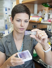
Credit: Peter H. Seeberger
Researchers have used military technology to develop a test for detecting malaria parasites in the blood.
The team used a detector known as a focal plane array (FPA), which was originally developed for heat-seeking missiles.
The FPA gives highly detailed information on a sample area in minutes. The heat-seeking detector, which is coupled to an infrared imaging microscope, could detect the malaria parasite in a single red blood cell.
The infrared signature from the fatty acids of the parasites allowed the researchers to detect the parasite at its earliest stages and determine the number of parasites in a blood smear.
The team described the technology in Analyst.
“Our test detects malaria at its very early stages, so that doctors can stop the disease in its tracks before it takes hold and kills,” said study author Bayden Wood, PhD, of Monash University in Victoria, Australia. “We believe this sets the gold standard for malaria testing.”
“There are some excellent tests that diagnose malaria. However, the sensitivity is limited, and the best methods require hours of input from skilled microscopists, and that’s a problem in developing countries where malaria is most prevalent.”
The new test, on the other hand, gives an automatic diagnosis within 4 minutes and doesn’t require a specialist technician.
Study author Leann Tilley, PhD, of the University of Melbourne in Australia, said the test could make an impact in large-scale screening of malaria parasite carriers who do not present with the classic fever-type symptoms associated with the disease.
“In many countries, only people who display signs of malaria are treated,” Dr Tilley said. “But the problem with this approach is that some people don’t have typical flu-like symptoms associated with malaria, and this means a reservoir of parasites persists that can reemerge and spread very quickly within a community.”
“Our test works because it can detect the malaria parasite at the very early stages and can reliably detect it in an automated manner in a single red blood cell. No other test can do that.”
FPA detectors were originally developed for Javelin Portable anti-tank missiles in the 1990s. The heat-seeking detector is used on shoulder-fired missiles but can also be installed on tracked, wheeled, or amphibious vehicles, providing spatial and spectral information in a matter of seconds.
The FPA detector used in this project was coupled to a synchrotron source located at the InfraRed Environmental Imaging facility at the Synchrotron Radiation Center in Wisconsin.
For the next phase of this research, Dr Wood’s team is collaborating with Patcharee Jearanaikoon, PhD, of Kohn Kaen University in Thailand, to test the technology in clinics.
Protein map may point to new cancer treatments

the endoplasmic reticulum
in green, mitochondria in red,
and chromosomes in blue
Credit: Wellcome Images
Researchers say they’ve uncovered the structure of one of the most important protein complexes involved in cell division, and their finding has implications for cancer treatment.
The team mapped the anaphase-promoting complex (APC/C), which performs a wide range of tasks associated with mitosis.
The researchers believe that seeing APC/C in unprecedented detail could transform our understanding of how cells divide and reveal binding sites for future cancer drugs.
“It’s very rewarding to finally tie down the detailed structure of this important protein, which is both one of the most important and most complicated found in all of nature,” said David Barford, DPhil, of the Medical Research Council Laboratory of Molecular Biology in Cambridge, UK.
“We hope our discovery will open up whole new avenues of research that increase our understanding of the process of mitosis and ultimately lead to the discovery of new cancer drugs.”
Dr Barford and his colleagues detailed their discovery in Nature.
The researchers reconstituted human APC/C and used a combination of electron microscopy and imaging software to visualize it at a resolution of less than a billionth of a meter.
The resolution was so fine that it allowed the team to see the secondary structure—the set of basic building blocks that combine to form every protein. Alpha-helix rods and folded beta-sheet constructions were clearly visible within the 20 subunits of the APC/C, defining the overall architecture of the complex.
Previous studies led by the same team had shown a globular structure for APC/C in much lower resolution, but the secondary structure had not been mapped.
Each of the APC/C’s subunits bond and mesh with other units at different points in the cell cycle, allowing it to control a range of mitotic processes, including the initiation of DNA replication, the segregation of chromosomes along spindles, and cytokinesis. Disrupting each of these processes could selectively kill cancer cells or prevent them from dividing.
“The fantastic insights into molecular structure provided by this study are a vivid illustration of the critical role played by fundamental cell biology in cancer research,” said Paul Workman, PhD, of The Institute of Cancer Research, London.
“The new study is a major step forward in our understanding of cell division. When this process goes awry, it is a critical difference that separates cancer cells from their healthy counterparts. Understanding exactly how cancer cells divide inappropriately is crucial to the discovery of innovative cancer treatments to improve outcomes for cancer patients.”
Two blood cancer patients cleared of HIV

Credit: CDC
MELBOURNE—Two men with hematologic malignancies who were also HIV-positive appear to be free of the virus after receiving stem cell transplants.
The patients have undetectable levels of HIV and remain free of their cancers—acute myeloid leukemia and non-Hodgkin lymphoma—more than 3 years after their transplants.
Importantly, the patients’ stem cell donors were not homozygous for CCR5-delta 32, a mutation that affords protection against HIV.
The researchers said these results herald a new direction in HIV research and provide hope for HIV-positive patients with leukemia and lymphoma.
The work was presented at the “Towards an HIV Cure Symposium,” which is part of the 20th International AIDS Conference.
Both patients were treated at St Vincent’s Hospital in partnership with the University of New South Wales’s Kirby Institute in Sydney, Australia.
One patient underwent a transplant in 2010 to treat his non-Hodgkin lymphoma, and his donor had 1 copy of CCR5-delta 32.
The second patient underwent a similar procedure for acute myeloid leukemia in 2011, and his donor did not have any copies of CCR5-delta 32.
Nevertheless, both patients were successfully cleared of HIV, although they remain on antiretroviral therapy as a protective measure.
“We’re so pleased that both patients are doing reasonably well years after the treatment for their cancers and remain free of both the original cancer and the HIV virus,” said David Cooper, MBBS, MD, DSc, of the Kirby Institute and St Vincent’s Hospital.
Until now, the only person considered to have cleared HIV is an American man, Timothy Ray Brown, who underwent 2 stem cell transplants in Berlin (in 2007 and 2008).
The cells in his second transplant included both copies of CCR5-delta 32, which affords protection against HIV and is found in less than 1% of the population. The man is no longer on antiretroviral therapy and remains free of HIV.
In Boston, 2 other patients underwent similar transplants in 2012, but the donor cells did not contain CCR5-delta 32. In both cases, HIV returned after antiretroviral treatment was stopped.
“It is very difficult to find a match for bone marrow donors and even more so to find one that affords protective immunity against HIV,” Dr Cooper said.
While his group’s results are a significant development, the researchers stressed that transplants are not a general functional “cure” for the up to 38.8 million people infected with HIV worldwide.
“This is a terrific, unexpected result for people with malignancy and HIV,” said Sam Milliken, MBBS, of St Vincent’s Hospital. “It may well give us a whole new insight into HIV, using the principles of stem cell transplantation.”
“It is important to caution that, at this stage, this form of treatment is far too dangerous for treating patients with HIV alone, but there may be potential for using transplants as an effective treatment modality for HIV down the track.”
The researchers said the 2 Sydney patients will be the subject of investigations to determine where any residual virus might be hiding and how it can be controlled. And the patients’ results point to a new direction for HIV research.
“We still don’t know why these patients have undetectable viral loads,” said Kersten Koelsch, MD, of the Kirby Institute. “One theory is that the induction therapy helps to destroy the cells in which the virus is hiding and that any remaining infected cells are destroyed by the patient’s new immune system.”
“We need more research to establish why and how bone marrow transplantation clears the virus. We also want to explore the predictors of sustained viral clearance and how this might be able to be exploited without the need for bone marrow transplantation.”
For the time being, the results mean that more patients who are eligible for transplant might be able to participate in clinical trials to determine the value of this procedure in HIV.