User login
HIV+ cancer patients more likely to go untreated

lymphocyte; Credit: CDC
Cancer patients infected with HIV are less likely than their uninfected peers to receive cancer treatment, according to research published in the Journal of Clinical Oncology.
Results showed that HIV-positive patients were roughly twice as likely to go untreated for lymphomas and other cancers.
The researchers believe a lack of clinical trial data and treatment guidelines for HIV patients with cancer may contribute to this health disparity.
To assess the role of HIV status on cancer treatment, Gita Suneja, MD, of the University of Pennsylvania in Philadelphia, and her colleagues used data from the National Cancer Institute’s HIV/AIDS Cancer Match Study.
This included 3045 HIV-infected patients and 1,087,648 uninfected patients. The patients had been diagnosed with Hodgkin lymphoma, diffuse large B-cell lymphoma, or cervical, lung, anal, prostate, colorectal, or breast cancer from 1996 through 2010.
For each cancer type, the researchers assessed the relationship between HIV status and cancer treatment, adjusted for cancer stage, sex, age at cancer diagnosis, race/ethnicity, year of cancer diagnosis, and US state.
For all but 1 cancer type, there was a significantly higher proportion of HIV-infected patients who did not receive cancer treatment when compared with uninfected patients.
The adjusted odds ratios (aORs) were 1.67 for diffuse large B-cell lymphoma, 1.77 for Hodgkin lymphoma, 1.60 for cervical cancer, 1.79 for prostate cancer, 1.81 for breast cancer, 2.18 for lung cancer, and 2.27 for colorectal cancer.
Anal cancer was the only malignancy for which HIV status did not appear to impact treatment. The aOR was 1.01.
Among HIV-infected individuals, factors independently associated with a lack of cancer treatment included low CD4 count, male sex with injection drug use as mode of HIV exposure, age 45 to 64 years, black race, and distant or unknown cancer stage.
“In my clinical experience, I have seen uncertainty surrounding treatment of HIV-infected cancer patients,” Dr Suneja said. “Patients with HIV have typically been excluded from clinical trials, and, therefore, oncologists do not know if the best available treatments are equally safe and effective in those with HIV.”
“Many oncologists rely on guidelines based on such trials for treatment decision-making, and, in the absence of guidance, they may elect not to treat HIV-infected cancer patients due to concerns about adverse side effects or poor survival. This could help explain, in part, why many HIV-positive cancer patients are not receiving appropriate cancer care.”
Therefore, Dr Suneja and her colleagues recommend that cancer trials begin enrolling HIV-infected patients and cancer management guidelines incorporate recommendations for HIV-infected patients. ![]()
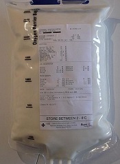
Group engineers versatile RBCs

Researchers say they’ve modified red blood cells (RBCs) to carry a range of payloads—from drugs to vaccines to imaging agents.
“We wanted to create high-value red cells that do more than simply carry oxygen,” said study author Harvey Lodish, PhD, of the Whitehead Institute in Cambridge, Massachusetts.
“Here, we’ve laid out the technology to make mouse and human red blood cells in culture that can express what we want and potentially be used for therapeutic or diagnostic purposes.”
Dr Lodish and his colleagues detailed the technology in Proceedings of the National Academy of Sciences.
The team noted that RBCs are an attractive vehicle for a variety of reasons, including their abundance and their long lifespan.
Perhaps most importantly, during RBC production, progenitor cells jettison their nuclei and all DNA therein. Without a nucleus, a mature RBC lacks any genetic material or any signs of earlier genetic manipulation that could result in tumor formation or other adverse effects.
Exploiting this characteristic, Dr Lodish and his colleagues introduced into early stage RBC progenitors genes coding for specific, slightly modified, normal red cell surface proteins.
As the RBCs approach maturity and enucleate, the proteins remain on the cell surface, where they are modified by a protein-labeling technique known as sortagging.
The technique relies on the bacterial enzyme sortase A to establish a strong chemical bond between the surface protein and a substance of choice, be it a small-molecule therapeutic or an antibody capable of binding a toxin. The modifications leave the cells and their surfaces unharmed.
“Because the modified human red blood cells can circulate in the body for up to 4 months, one could envision a scenario in which the cells are used to introduce antibodies that neutralize a toxin,” said Hidde L. Ploegh, PhD, also of the Whitehead Institute. “The result would be long-lasting reserves of antitoxin antibodies.”
The approach has captured the attention of the US military and its Defense Advanced Research Projects Agency, which is supporting the research in the interest of developing treatments or vaccines effective against biological weapons.
Dr Lodish believes the applications are potentially vast and may include RBCs modified to bind and remove bad cholesterol from the bloodstream, carry clot-busting proteins to treat ischemic strokes or deep-vein thrombosis, or deliver anti-inflammatory antibodies to alleviate chronic inflammation.
Dr Ploegh said there is evidence to suggest that modified RBCs could be used to suppress the unwanted immune response that often accompanies treatment with protein-based therapies.
He is exploring whether these RBCs could be used to prime the immune system to allow patients to better tolerate treatment with such therapies. ![]()
Study reveals new risk factors for bloodstream infections
Current guidelines to help prevent bloodstream infections during intravenous feeding may need revisions to strengthen protection for patients, a new study suggests.
These guidelines restrict how long a single bag of parenteral nutrition containing lipids can be used, due to the ability of lipids to encourage the growth of microorganisms.
But researchers found the guidelines do not account for other independent factors that can affect the growth of potentially deadly microorganisms.
Their findings appear in the Journal of Parenteral and Enteral Nutrition.
Peter David Austin, of the University of Southampton in the UK, and his colleagues looked at the growth of Escherichia coli and Enterococcus durans in parenteral nutrition to identify factors that can affect microbial growth.
They assessed the growth of E coli and E durans in quadruplicate in 12 different patients who received parenteral nutrition, with and without lipid, in varying glucose concentrations.
Results showed that glucose concentration, the proportion of glucose to lipid, and osmolarity all affected microbial growth, in addition to the presence of lipids.
The researchers therefore recommend these additional factors be considered when making clinical and policy decisions to limit the potential growth of microorganisms in parenteral nutrition. ![]()
Group finds master regulator of MYC

Credit: Juha Klefstrom
New research indicates that an unexpected partnership between the MYC oncogene and a non-coding RNA called PVT1 could be the key to understanding how MYC fuels cancers.
The researchers knew that MYC amplifications cause cancer, but MYC does not amplify alone. It often pairs with adjacent chromosomal regions.
“We wanted to know if the neighboring genes played a role,” said study author Anindya Bagchi, PhD, of the University of Minnesota in Minneapolis.
“We took a chance and were surprised to find this unexpected and counter-intuitive partnership between MYC and its neighbor, PVT1. Not only do these genes amplify together, PVT1 helps boost the MYC protein’s ability to carry out its dangerous activities in the cell.”
The researchers reported this finding in Nature.
Dr Bagchi and his team focused on a region of the genome, 8q24, which contains the MYC gene and is commonly expressed in cancer. The team separated MYC from the neighboring region containing the non-coding RNA PVT1.
Using chromosome engineering, the researchers developed mouse strains in 3 separate iterations: MYC only, the rest of the region containing PVT1 but without MYC, and the pairing of MYC with the regional genes.
The expected outcome, if MYC was the sole driver of the cancer, was tumor growth on the MYC line as well as the paired line. However, the researchers found growth only on the paired line. This suggests MYC is not acting alone and needs help from adjacent genes.
“The discovery of this partnership gives us a stronger understanding of how MYC amplification is fueled,” said David Largaespada, PhD, also of the University of Minnesota.
“When cancer promotes a cell to make more MYC, it also increases the PVT1 in the cell, which, in turn, boosts the amount of MYC. It’s a cycle, and now we’ve identified it, we can look for ways to uncouple this dangerous partnership.”
Testing this theory of uncoupling, the researchers looked closely at several breast and colorectal cancers that are driven by MYC. For example, in colorectal cancer lab models, where a mutation in the beta-catenin gene drives MYC to cancerous levels, eliminating PVT1 from these cells made the tumors nearly disappear.
“Finding the cooperation between MYC and PVT1 could be a game changer,” said Yuen-Yi Tseng, a graduate student at the University of Minnesota.
“We used to think MYC amplification is the major issue but ignored that other co-amplified genes, such as PVT1, can be significant. In this study, we show that PVT1 can be a key regulator of MYC protein, which can shift the paradigm in our understanding of MYC-amplified cancers.”
MYC has been notoriously elusive as a drug target. By uncoupling MYC and PVT1, the researchers suspect they could disable the cancer growth and limit MYC to precancerous levels. This would make PVT1 an ideal drug target to potentially control a major cancer gene.
“This is a thrilling discovery, but there are more questions that follow,” Dr Bagchi said. “Two major areas present themselves now for research. Will breaking the nexus between MYC and PVT1 perform the same in any MYC-driven cancer, even those not driven by this specific genetic location?”
“And how is PVT1 stabilizing or boosting MYC within the cells? This relationship will be a key to developing any drugs to target this mechanism.” ![]()
Review uncovers patient safety violations

Credit: CDC
Patient safety violations emerged as a common theme in a review of “first warning” letters issued to clinical trial sponsors, investigators, and internal review boards (IRBs).
The US Food and Drug Administration (FDA) issued the 84 warning letters in response to trial violations uncovered during site visits.
The review of these letters showed that 55% of investigators failed to protect subjects’ safety and report adverse events to the IRB, 24% of sponsors failed to submit adverse event data to the FDA, and 22% of IRBs failed to “address risk minimization and protect vulnerable study subjects.”
Yashashri C. Shetty and Aafreen A. Saiyed, both of King Edward Memorial Hospital in Mumbai, detailed these findings in the Journal of Medical Ethics.
The researchers reviewed the content of 84 first-warning letters issued by the FDA following site visits between 2005 and 2012.
Sponsor violations
Forty-six warning letters were issued to trial sponsors. Their most common violations were failure to follow the monitoring schedule (58.69%) and failure to obtain investigator agreement (34.78%).
The same proportion of sponsors (30.43%) failed to secure investigators’ compliance and failed to maintain records, both of study data and for shipping product to the investigator.
Other violations included failure to submit an Investigational Device Exemption or Investigational New Drug application to the FDA (28.26%) and failure to review, evaluate, and submit adverse drug event reports to the FDA (23.91%). Two sponsors did not allow FDA inspection, and 1 did not obtain IRB approval.
Investigator infractions
Twenty warning letters were issued to investigators. Ninety-five percent of the letters said investigators were guilty of deviating from the investigational plan. Fifty-five percent of letters said the researchers failed to protect subject safety and report adverse events to the IRB.
Violations regarding records—largely, the failure to maintain and produce them for inspection—were documented in 40% of the letters. And informed consent issues were highlighted in 35%.
Other violations included those related to the product under investigation (15%), failure to obtain IRB approval (10%), and failure to personally supervise the study (30%).
IRB transgressions
Eighteen warning letters were issued to IRBs. The most common violation (61.11%) was failure to follow written procedures for continuing review.
Other common violations (55.56%) were those related to membership and meetings—failure to maintain minutes, inappropriate membership, quorum issues, misuse of expedited review, and the lack of a layperson in meetings.
The remaining violations included failure to follow regulatory requirements (44.4%), failure to follow standard operating procedures and maintain documentation (44.44%), failure to address risk minimization and protect vulnerable subjects (22.22%), conflicts of interest and informed consent issues (27.78%), and failure to appoint a qualified investigator (5.55%).
Comparing past and present
The study authors compared their findings with previously published research in the same arena, dating back as far as 1997. They found that regulatory compliance generally improved over the years, but supervision worsened.
And 2 new serious violations cropped up in the interim: failure to get the green light from an IRB before pressing ahead and submitting false data to the FDA and/or sponsors.
In a bid to boost compliance with good clinical practice, the authors suggest that every regulatory agency charged with overseeing clinical trials should pay main participating centers a visit and regularly publish the details of their findings. ![]()
An easier route for cell therapy

Credit: PNAS
Laser technology can help ensure the delivery of drug and gene therapy at the cellular level without damaging surrounding tissue, according to research published in Nature Scientific Reports.
Investigators paired crystalline magnetic carbon nanoparticles and continuous wave near-infrared laser beams in what is called photothermal delivery.
And they used this delivery method to introduce impermeable dyes and small DNA molecules into human cancer cells.
This work grew out of a previous study in which the researchers used a 50 to 100 milliwatt laser and the same carbon nanoparticle, which absorbs the beam, to heat up and destroy cancer cells in the lab.
“In [the current study, we] used a lower-power, 20 to 30 milliwatt, continuous wave near-infrared laser and the nanoparticle to permeate the cell membrane without killing the cells,” said Ali Koymen, PhD, of the University of Texas at Arlington.
“This method stretches the desired cell membrane to allow for delivery and has the added bonus of creating a fluid flow that speeds the movement of what is being delivered.”
The investigators noted that, currently, the predominant practice is using viruses for delivery to cells. Unfortunately, the scope of what can be delivered with viruses is severely limited, and virus interaction can lead to inflammatory responses and other complications.
Researchers looking to create a path into the cell without employing a virus have experimented with using UV-visible light laser beams alone. But that method damages surrounding cells and has a relatively shallow level of effectiveness.
Dr Koymen and his colleagues said a significant advantage of their method is that the near-infrared light absorption of the nanoparticle can be used to selectively amplify the interaction of low-power laser with targeted tissue, and laser-induced damage to non-targeted cells can be avoided.
The magnetic properties of the nanoparticles also mean they can be localized with an external magnetic field. Therefore, a smaller concentration can be used effectively. ![]()
Team reports new method of chemo delivery

Credit: Kathy Atkinson
Researchers have devised a novel way to deliver chemotherapy drugs “on demand,” according to a paper published in Proceedings of the National Academy of Sciences.
The team loaded a biocompatible hydrogel with a chemotherapy drug and used ultrasound to trigger the gel to release the drug.
Like many other injectable gels, this one gradually releases a low level of the drug by diffusion over time. But the new hydrogel differs from others in a key way.
Researchers previously applied ultrasound to gels to temporarily increase doses of drug, but that approach was a one-shot deal, as the ultrasound was used to destroy those gels.
In the current study, the researchers used ultrasound to temporarily disrupt the gel so that it released short, high-dose bursts of the drug. But when they stopped the ultrasound, the hydrogels self-healed.
By closing back up, they were ready to go for the next “on demand” drug burst, providing a way to administer drugs with a greater level of control than was possible before.
The researchers also demonstrated in lab cultures and in mouse models of breast cancer that the pulsed, ultrasound-triggered hydrogel approach to drug delivery was more effective at stopping the growth of tumor cells than traditional, sustained-release drug therapy.
“Our approach counters the whole idea of sustained drug release and offers a double whammy,” said study author David J. Mooney, PhD, of the Harvard School of Engineering and Applied Sciences in Boston.
“We have shown that we can use the hydrogels repeatedly and turn the drug pulses on and off at will, and that the drug bursts in concert with the baseline low-level drug delivery seems to be particularly effective in killing cancer cells.”
Self-healing hydrogel
Key to the researchers’ success in designing a hydrogel that self-heals was choosing the right kind of hydrogel with the right kind of drug and applying the right intensity of ultrasound.
“We were able to trigger our system with a level of ultrasound that was much lower than high-intensity focused ultrasound that is used clinically to heat and destroy tumors,” said study author Cathal Kearney, PhD, of the Royal College of Surgeons in Ireland. “The careful selection of materials and properties make it a reversible process.”
The team carried out the majority of their work for this study with a gel made out of alginate, a natural polysaccharide from algae that is held together with calcium ions.
In a series of tests, they found that, with the right level of ultrasound, the bonds break up and enable the gel to release its drug cargo. But as long as the gel is in the presence of more calcium, the bonds reform and the gel self-heals.
Drug testing
Once the researchers knew the gel would self-heal, they tested out a drug they suspected it would hold well: the chemotherapy drug mitoxantrone.
Sure enough, the ultrasound triggered the gel to release the blue-colored drug, as indicated by the newly blue color of the surrounding medium. Just a single ultrasound dose was effective, and the gel reformed after it was disrupted, making multiple cycles possible.
Next, the team tested the treatment in mouse models of breast cancer. They injected the drug-laden gel close to the tumors.
Over the course of 6 months, the mice that received a low-level, sustained release of the drug with a daily concentrated pulse of ultrasound (2.5 minutes) fared significantly better than mice treated the same but without ultrasound.
In contrast to controls, the tumors in the ultrasound-treated mice did not grow substantially. And the mice survived for an additional 80 days.
Potential applications
The researchers believe their technique could help improve cancer treatment and other therapies requiring drugs to be delivered at the right place and the right time—from post-surgery pain medications to protein-based drugs that require daily injections.
It requires an initial injection of the hydrogel, but the approach could be a much less traumatic, minimally invasive, and more effective method of drug delivery than current methods, Dr Mooney said.
The researchers also found their hydrogel can release cargo other than drugs, including proteins and condensed plasmid DNA. This lays the groundwork for using these hydrogels for tissue regeneration and gene therapy.
Dr Mooney said he and his colleagues plan to explore these potential applications, as well as the possibility of unleashing 2 different drugs independently from the same hydrogel. ![]()
Credit: Kathy Atkinson
Researchers have devised a novel way to deliver chemotherapy drugs “on demand,” according to a paper published in Proceedings of the National Academy of Sciences.
The team loaded a biocompatible hydrogel with a chemotherapy drug and used ultrasound to trigger the gel to release the drug.
Like many other injectable gels, this one gradually releases a low level of the drug by diffusion over time. But the new hydrogel differs from others in a key way.
Researchers previously applied ultrasound to gels to temporarily increase doses of drug, but that approach was a one-shot deal, as the ultrasound was used to destroy those gels.
In the current study, the researchers used ultrasound to temporarily disrupt the gel so that it released short, high-dose bursts of the drug. But when they stopped the ultrasound, the hydrogels self-healed.
By closing back up, they were ready to go for the next “on demand” drug burst, providing a way to administer drugs with a greater level of control than was possible before.
The researchers also demonstrated in lab cultures and in mouse models of breast cancer that the pulsed, ultrasound-triggered hydrogel approach to drug delivery was more effective at stopping the growth of tumor cells than traditional, sustained-release drug therapy.
“Our approach counters the whole idea of sustained drug release and offers a double whammy,” said study author David J. Mooney, PhD, of the Harvard School of Engineering and Applied Sciences in Boston.
“We have shown that we can use the hydrogels repeatedly and turn the drug pulses on and off at will, and that the drug bursts in concert with the baseline low-level drug delivery seems to be particularly effective in killing cancer cells.”
Self-healing hydrogel
Key to the researchers’ success in designing a hydrogel that self-heals was choosing the right kind of hydrogel with the right kind of drug and applying the right intensity of ultrasound.
“We were able to trigger our system with a level of ultrasound that was much lower than high-intensity focused ultrasound that is used clinically to heat and destroy tumors,” said study author Cathal Kearney, PhD, of the Royal College of Surgeons in Ireland. “The careful selection of materials and properties make it a reversible process.”
The team carried out the majority of their work for this study with a gel made out of alginate, a natural polysaccharide from algae that is held together with calcium ions.
In a series of tests, they found that, with the right level of ultrasound, the bonds break up and enable the gel to release its drug cargo. But as long as the gel is in the presence of more calcium, the bonds reform and the gel self-heals.
Drug testing
Once the researchers knew the gel would self-heal, they tested out a drug they suspected it would hold well: the chemotherapy drug mitoxantrone.
Sure enough, the ultrasound triggered the gel to release the blue-colored drug, as indicated by the newly blue color of the surrounding medium. Just a single ultrasound dose was effective, and the gel reformed after it was disrupted, making multiple cycles possible.
Next, the team tested the treatment in mouse models of breast cancer. They injected the drug-laden gel close to the tumors.
Over the course of 6 months, the mice that received a low-level, sustained release of the drug with a daily concentrated pulse of ultrasound (2.5 minutes) fared significantly better than mice treated the same but without ultrasound.
In contrast to controls, the tumors in the ultrasound-treated mice did not grow substantially. And the mice survived for an additional 80 days.
Potential applications
The researchers believe their technique could help improve cancer treatment and other therapies requiring drugs to be delivered at the right place and the right time—from post-surgery pain medications to protein-based drugs that require daily injections.
It requires an initial injection of the hydrogel, but the approach could be a much less traumatic, minimally invasive, and more effective method of drug delivery than current methods, Dr Mooney said.
The researchers also found their hydrogel can release cargo other than drugs, including proteins and condensed plasmid DNA. This lays the groundwork for using these hydrogels for tissue regeneration and gene therapy.
Dr Mooney said he and his colleagues plan to explore these potential applications, as well as the possibility of unleashing 2 different drugs independently from the same hydrogel.
Predicting problems in families of cancer patients

Credit: Rhoda Baer
A new analysis suggests family dysfunction is the greatest predictor of emotional and behavioral problems among children who have a parent with cancer.
Other variables, such as the child’s age, did not predict the risk as accurately.
And illness-related factors, such as the parent’s prognosis, did not appear to have an impact at all.
Birgit Möller, PhD, of the University Medical Center Hamburg-Eppendorf in Germany, and her colleagues reported these findings in Cancer.
The researchers evaluated 235 families in which at least 1 parent was diagnosed with cancer. This included 402 parents and 324 children aged 11 to 21 years. Parents and children completed questionnaires that assessed emotional and behavioral health.
Responses suggested that children of cancer patients have higher-than-average levels of emotional and behavioral symptoms.
The overall mean values for emotional and behavioral problems—from both the parents’ and children’s perspectives—were significantly higher in the study population than the average values from a representative non-cancer population.
General family functioning was the strongest predictor of children’s symptom status from both the parents’ and child’s perspectives.
The effects of the child’s age and gender on behavioral and emotional symptoms varied according to the subject asked. But none of the respondents reported an association between child adjustment and illness-related factors such as poor prognoses or recurrent illness.
Dr Möller noted that screening for child mental health problems, family dysfunction, and parental depression can be easily adopted into cancer care so that families in need of support can be identified.
“Additional training of oncologists, interdisciplinary approaches, and family-based mental health liaison services are recommended to meet the needs of minor
children and their families and to minimize negative long-term effects in children,” she said.
Dr Möller and her team have developed a preventive counseling program—called the Children of Somatically Ill Parents (COSIP) program—that focuses on family communication, involvement of family members, flexible problem solving, mutual support, and parenting issues.
Tool may predict cancer patients’ risk of financial stress

patient and her father
Credit: Rhoda Baer
A new questionnaire can measure a cancer patient’s risk for financial stress, according to a paper published in Cancer.
Researchers developed the 11-item questionnaire, called the COmprehensive Score for financial Toxicity (COST), through conversations with more than 150 cancer patients.
The team used the term “financial toxicity” to describe the expense, anxiety, and loss of confidence confronting patients who face big, unpredictable costs of cancer treatment.
And the researchers said financial toxicity can be considered another side effect of cancer care.
“Few physicians discuss this increasingly significant side effect with their patients,” said study author Jonas de Souza, MD, of the University of Chicago Medicine in Illinois.
“Physicians aren’t trained to do this. It makes them, as well as patients, feel uncomfortable. [However,] we believe that a thoughtful, concise tool that could help predict a patient’s risk for financial toxicity might open the lines of communication. This gives us a way to launch that discussion.”
Development of the COST questionnaire began with a literature review and a series of extensive interviews. Dr de Souza and his colleagues spoke with 20 patients and 6 cancer professionals, as well as nurses and social workers, and this produced a list of 147 questions.
The researchers pared the list down to 58 questions. Then, they asked 35 patients to help them decide which of the remaining questions were the most important. And the patients narrowed the list down to 30.
“In the end, 155 patients led us, with some judicious editing, to a set of 11 statements,” Dr de Souza said. “This was sufficiently brief to prevent annoying those responding to the questions but thorough enough to get us the information we need.”
All 11 entries are short and easy to understand, according to the researchers. For example, item 2 states, “My out-of-pocket medical expenses are more than I thought they would be.” And item 7 states, “I am able to meet my monthly expenses.”
For each question, patients choose from 5 potential responses: “not at all”, “a little bit,” “somewhat,” “quite a bit,” or “very much.”
Learning how a patient responds may help caregivers determine who is likely to need education, financial counseling, or referral to a support network. The quiz may also predict who is likely to have problems and require interventions.
All patients who helped develop the study had been in treatment for at least 2 months and had received bills. Excluding the top 10% and the bottom 10%, patients in the study earned between $37,000 and $111,000. The median annual income for these patients was about $63,000.
The researchers expected that financial toxicity would correlate with income.
“But, in our small sample, that did not hold up,” Dr de Souza said. “People with less education seemed to have more financial distress, but variations in income did not make much difference. We need bigger studies to confirm that, but at least we now have a tool we can use to study this.”
The researchers are now conducting a larger study to validate these findings and correlate the newly developed scale with quality of life and anxiety in cancer patients.
“We need to assess outcomes that are important for patients,” Dr de Souza said. “[T]his is another important piece of information in the shared-decision-making process.”
US ranked last in healthcare report

Credit: NIH
In a study comparing healthcare in 11 industrialized countries, the US ranked last on measures of health system quality, efficiency, access to care, equity, and healthy lives.
The other countries included in this study were Australia, Canada, France, Germany, the Netherlands, New Zealand, Norway, Sweden, Switzerland, and the UK.
While the results revealed room for improvement in every country, the US stood out for having the highest costs and lowest performance.
For instance, the US spent $8508 per person on healthcare in 2011, compared with $3406 in the UK, which ranked first overall.
Details on expenditures and rankings derived from this study are available in the Commonwealth Fund report, Mirror, Mirror on the Wall: How the Performance of the U.S. Health Care System Compares Internationally, 2014 Update.
“It is disappointing, but not surprising, that, despite our significant investment in healthcare, the US has continued to lag behind other countries,” said lead report author Karen Davis, of the Johns Hopkins Bloomberg School of Public Health in Baltimore, Maryland.
The report was also produced in 2004, 2006, 2007, and 2010, with the US ranking last in each of those years. Four countries were added to this year’s report: Switzerland and Sweden, which followed the UK at the top of the rankings, and Norway and France, which were in the middle of the pack.
Australia, Germany, the Netherlands, New Zealand, and Norway also placed in the middle, and Canada ranked just above the US.
In addition to ranking last overall, the US ranked last on infant mortality and on deaths that were potentially preventable with timely access to effective healthcare. The country ranked second-to-last on healthy life expectancy at age 60.
The US also ranked last on every measure of cost-related access. More than one-third (37%) of US adults reported forgoing a recommended test, treatment, or follow-up care because of cost.
With regard to healthcare quality, the US fell somewhere in the middle. On 2 of 4 measures of quality—effective care and patient-centered care—the US ranked near the top (3rd and 4th of 11 countries, respectively). But it did not perform as well with regard to providing safe or coordinated care.
The US ranked last in efficiency, due to low marks on the time and dollars spent dealing with insurance administration, lack of communication among healthcare providers, and duplicative medical testing.
Forty percent of US adults who had visited an emergency room reported they could have been treated by a regular doctor if one had been available. This is more than double the rate of patients in the UK (16%).
The US also ranked last in healthcare equity. About 4 of 10 (39%) adults with below-average incomes in the US reported a medical problem but did not visit a doctor in the past year because of costs, compared with less than 1 of 10 in the UK, Sweden, Canada, and Norway.
There were also large discrepancies in the length of time US adults waited for specialist, emergency, and after-hours care. And wait times were associated with patient income.
The data for this research were drawn from the Commonwealth Fund 2011 International Health Policy Survey of Sicker Adults, the Commonwealth Fund 2012 International Health Policy Survey of Primary Care Physicians, and the Commonwealth Fund 2013 International Health Policy Survey.
The 2011 survey targeted a representative sample of “sicker adults,” defined as those who rated their health status as fair or poor, received medical care for a serious chronic illness, serious injury, or disability in the past year, or were hospitalized or underwent surgery in the previous 2 years.
The 2012 survey looked at the experiences of primary care physicians. The 2013 survey focused on the experiences of nationally representative samples of adults ages 18 and older.
Additional data on health outcomes were drawn from the Organization for Economic Cooperation and Development and the World Health Organization.