User login
Team unearths surprising sepsis finding

Credit: NHLBI
SAN DIEGO—Results of a retrospective study suggest sepsis may contribute to as many as half of all hospital deaths in the US.
Researchers analyzed information from 6.5 million hospital discharge records and found that, of all hospital deaths, as many as 52% occurred in patients with sepsis.
“We were surprised to find that as many as 1 in 2 patients dying in US hospitals had sepsis,” said Vincent Liu, MD, of the Kaiser Permanente Northern California Division of Research.
He and his colleagues presented this finding at the American Thoracic Society’s 2014 International Conference (abstract 50626). The results have also been published in JAMA.
The researchers analyzed data from 6.5 million hospital discharge records derived from the Healthcare Cost and Utilization Project Nationwide Inpatient Sample (NIS) in 2010. The NIS is the largest publicly available, all-payer inpatient database in the US, containing data from 100% of hospital discharges from a stratified sample of 1051 community hospitals.
The team also evaluated records for 482,828 adult patients admitted to 21 Kaiser Permanente Northern California (KPNC) hospitals.
Using diagnosis and procedure codes, the researchers identified hospital admissions and deaths of patients with sepsis and estimated the percentage of total hospital charges associated with sepsis hospitalizations.
They used 2 approaches to identify patients with sepsis from International Statistical Classification of Diseases, Ninth Revision, Clinical Modification codes. The explicit approach identified patients with codes 038 (septicemia), 995.91 (sepsis), 995.92 (severe sepsis), or 785.52 (septic shock). The team also used an implicit approach, which included patients with evidence of both infection and acute organ failure.
In the NIS cohort, there were 280,663 explicit and 717,718 implicit sepsis hospitalizations. Sepsis hospitalizations accounted for 4.3% (explicit) to 10.9% (implicit) of all hospitalizations.
There were 143,312 deaths in the NIS cohort, and 34.7% (explicit) to 52.0% (implicit) occurred among patients with sepsis.
In the KPNC cohort, there were 55,008 explicit and 80,678 implicit sepsis hospitalizations. Sepsis hospitalizations accounted for 11.4% (explicit) to 16.7% (implicit) of all hospitalizations.
There were 14,206 inpatient deaths in this cohort, and 36.9% (explicit) to 55.9% (implicit) occurred among patients with sepsis.
“[W]e found that most patients already had sepsis at the time of hospital admission,” Dr Liu said. “There was also a large number of patients with less severe sepsis, a group for whom treatment guidelines are less well-defined. The results of our study suggest that improved care for sepsis patients of all severity levels and in all hospital settings could result in many future lives saved.” ![]()
How cancer-fighting protein is held in check

Credit: A.T. Tikhonenko
A new study reveals how the protein p53 attaches to its regulatory molecule, BCL-xL.
Understanding how these molecular puzzle pieces fit together could help scientists design drugs that would unleash p53 to battle a range of cancers, according to study author Richard Kriwacki, PhD, of St Jude Children’s Research Hospital in Memphis, Tennessee.
He and his colleagues described this research in Nature Structural & Molecular Biology.
In guarding the cell against genetic damage, the p53 machinery functions both in the nucleus of the cell and in the cytosol. When this machinery detects irreparable damage to the cell, p53 is unleashed to trigger apoptosis.
In about half of all cancers, this machinery is rendered inoperable by mutation, enabling cancer cells to proliferate despite their genetic malfunctions. The protein BCL-xL is a central inhibitor of the p53 machinery, binding both p53 and BH3 proteins, which also drive apoptosis.
“The molecular details of how BCL-xl performs this dual inhibitory function were not understood,” Dr Kriwacki said. “Having those details has enabled us to determine exactly how BCL-xL can restrain or inhibit apoptosis through interactions with BH3-domain-containing proteins, as well as p53.”
He and his colleagues used a structural analysis technique called NMR spectroscopy to map the 3-D structure of p53 binding to BCL-xL.
Their experiments revealed how the DNA-binding domain of the p53 protein serves double duty in the machinery. It enables p53 to attach to DNA in the cell’s nucleus, helping the cell repair genetic damage. The same domain also acts as an attachment point for BCL-xL in the cytosol.
“The structural details that we report are novel,” Dr Kriwacki said. “And they provide the key insights for really dissecting the dual roles of BCL-xL in inhibiting apoptosis . . ., inhibiting the BH3-containing proteins on the one side and p53 on the other. Also, through these studies, we solidified the mechanistic understanding for how p53 functions in the cytosol, which complements its pro-apoptotic role in the nucleus.”
Dr Kriwacki added that these findings could help scientists design better anticancer agents. In many cancers, p53 is prevented from triggering apoptosis by its attachment to BCL-xL.
Drugs are currently being tested that bind to BCL-xL to free BH3 proteins to trigger apoptosis. However, Dr Kriwacki said new drugs could be developed that also block BCL-xL from binding p53.
“Our hypothesis is that many cancers have normal p53, but it is being tied up by BCL-xL,” he said. “If it could be released, it could play its role in triggering apoptosis. A drug that could block both of BCL-xL’s anti-apoptotic functions could potentially more profoundly induce apoptosis in cancer cells.” ![]()
Protein inhibition confers radioprotective effects

Researchers believe they may have discovered a method for treating and preventing radiation-induced gastrointestinal toxicity.
The investigators found that inhibiting prolyl hydroxylase domain (PHD) proteins in mice could help protect them from radiation-induced toxicity and prolong their life spans.
“We were very surprised by the amount of protection the animals received,” said Amato Giaccia, PhD, of the Stanford University School of Medicine in California.
“The important thing to note is that we didn’t change the amount of damage the intestinal cells sustained as a result of the radiation. We simply changed the physiology of that tissue and how it responded to that damage.”
Dr Giaccia and his colleagues described this research in Science Translational Medicine.
The study began with an interest in hypoxia-inducible factor (HIF) proteins, which are known to help cells survive stressful conditions.
“Previous studies from our group and others have suggested that the HIF proteins are important in protecting cells from many types of stress,” Dr Giaccia said. “So we wondered whether stabilizing HIF proteins, and therefore increasing their levels within the cells, could also protect the intestine from the effects of radiation.”
The researchers inhibited the degradation of HIF proteins in 2 ways. In the first experiment, they engineered mice that were unable to express PHD isoforms, a group of 3 proteins that tag HIF proteins for destruction.
In another experiment, the investigators treated unmodified mice with a small molecule called dimethyloxyallyl glycine (DMOG), which also inhibits the activity of PHD proteins.
In both cases, the levels of HIF1 and HIF2 proteins increased significantly in the manipulated mice, as compared to controls.
In addition, 70% of the genetically modified mice lived for at least 30 days after receiving a normally lethal dose of abdominal radiation, and 27% survived at least 30 days after a normally lethal dose of whole-body radiation.
Sixty-seven percent of DMOG-treated mice survived for at least 60 days after receiving a normally lethal dose of abdominal radiation, and 40% lived for at least 30 days after a normally lethal dose of whole-body radiation.
The control mice in both experiments did not survive longer than 10 days after either type of radiation exposure.
Elucidating the mechanism
Further experiments showed that HIF2, rather than HIF1, is responsible for the radioprotection the researchers observed.
To determine the cause of the treated animals’ prolonged survival, the investigators looked directly at the epithelial cells lining the intestines.
Treated mice exhibited lower levels of cell death in response to abdominal radiation exposure and improved survival of crypts, which host the rapidly dividing stem cells necessary to accommodate the intestines’ need for repeated cell turnover.
The treated animals also experienced less diarrhea and fewer imbalances in fluid and electrolyte levels than untreated animals exposed to the same dose of radiation. And they quickly gained back the weight they had lost as a result of the exposure.
Treatment after radiation exposure
“The animals that survived the abdominal radiation have a life span that is similar to unexposed animals, which was very exciting to us,” Dr Giaccia said. “However, we realized it would be impossible to pretreat humans unexpectedly exposed to large amounts of radiation like at Chernobyl or Fukushima because those exposures are, by nature, unpredictable.”
So Dr Giaccia and his colleagues experimented with treating the mice with DMOG after abdominal radiation exposure. They found that, although the protective qualities of the molecule were diminished, it did help.
When DMOG was given 4 hours after radiation exposure, 45% of the treated mice, but no untreated mice, survived at least 10 days.
After 24 hours, the effect was more subtle. DMOG treatment showed little benefit at higher doses of radiation. But at a lower dose, 75% of the treated animals lived for at least 30 days, compared to 18.2% of the untreated animals.
“We found we were still able to rescue a significant proportion of the animals,” Dr Giaccia said.
Finally, the researchers tested the effect of DMOG treatment 24 hours after total-body irradiation.
They found that 37.5% of the treated mice survived for at least 30 days, but only if the mice were also given a bone marrow transplant to restore blood and immune stem cells killed by the radiation. None of the untreated mice lived beyond 10 days.
The investigators pointed out that, although this study suggests a possible way to mitigate the effects of therapeutic radiation exposure, more work remains. But the next steps are clear.
“There are a number of drug molecules that act in a manner similar to DMOG that are already in clinical trials for unrelated conditions,” Dr Giaccia said. “Our next step will be to test some of these molecules to see if they also offer radioprotection.”
Stanford University has filed a patent application, “Use of Prolyl Hydroxylase Inhibitors as a Radioprotective Drug for the Lower Gastrointestinal Tract” (international application No. PCT/US2012/052232), based on the results of this study. ![]()
Device allows for single-cell analysis
Credit: Duke University
Using components similar to those that control electrons in microchips, engineers have designed a device that can sort, store, and retrieve individual cells for study.
The team hopes this chip-like device could be scaled up to sort and store hundreds of thousands of individual living cells in a matter of minutes.
Benjamin Yellen, PhD, of Duke University in Durham, North Carolina, and his colleagues described the device in Nature Communications.
The team created the device by printing thin electromagnetic components, like those found on microchips, onto a slide. These patterns create magnetic tracks and elements like switches, transistors, and diodes that guide magnetic beads and single cells tagged with magnetic nanoparticles through a thin, liquid film.
Like a series of small conveyer belts, localized rotating magnetic fields move the beads and cells along specific directions etched into a track, while built-in switches direct traffic to storage sites on the chip. The result is an integrated circuit that controls small magnetic objects much the way electrons are controlled on computer chips.
The engineers showed that a grid of 9 compartments—3 across by 3 down—allows the magnetic beads to enter but not leave. By tagging cells with magnetic particles and directing them to different compartments, the cells can be separated, sorted, stored, studied, and retrieved.
“You need to analyze thousands of cells to get the statistics necessary to understand which genes are being turned on and off in response to pharmaceuticals or other stimuli,” Dr Yellen said. “And if you’re looking for cells exhibiting rare behavior, which might be one cell out of a thousand, then you need arrays that can control hundreds of thousands of cells.”
As an example, Dr Yellen pointed to cells affected by cancers. Most afflicted cells are active and can be targeted by therapeutics. But a few rare cells remain dormant, biding their time and avoiding destruction before activating and bringing the disease out of remission.
With the new technology, Dr Yellen and his colleagues hope to watch millions of individual cells, pick out the few that become dormant, quickly retrieve them, and analyze their genetic activity.
“Our technology can offer new tools to improve our basic understanding of cancer metastasis at the single-cell level, how cancer cells respond to chemical and physical stimuli, and to test new concepts for gene delivery and metabolite transfer during cell division and growth,” said study author CheolGi Kim, PhD, of the Daegu Gyeongbuk Institute of Science and Technology in the Republic of Korea.
The researchers now plan to demonstrate a larger grid of 8-by-8 or 16-by-16 compartments with cells, and then to scale it up to hundreds of thousands of compartments.
“Our idea is a simple one,” Dr Kim said. “Because it is a system similar to electronics and is based on the same technology, it would be easy to fabricate. That makes the system relevant to commercialization.”
“There’s another technique paper we need to do as a follow-up before we get to actual biological applications,” Dr Yellen added. “But they’re on their way.” ![]()
Team says antioxidant has no effect on cancer risk, overall health

Contrary to previous findings, a new study suggests the antioxidant resveratrol is not associated with improvements in health, including reducing the risk of cancer.
Researchers found that Italians who consumed a diet rich in resveratrol—a compound in red wine, dark chocolate, and berries—lived no longer than and were just as likely to develop cardiovascular disease or cancer as Italians who consumed smaller amounts of the antioxidant.
However, the investigators said unknown compounds in these foods and drinks may still confer health benefits.
“The story of resveratrol turns out to be another case where you get a lot of hype about health benefits that doesn’t stand the test of time,” said study author Richard D. Semba, MD, MPH, of the Johns Hopkins University School of Medicine in Baltimore, Maryland.
“The thinking was that certain foods are good for you because they contain resveratrol. We didn’t find that at all.”
Dr Semba and his colleagues recounted their findings in JAMA Internal Medicine.
Their study included 783 subjects, all of whom were older than 65 years of age. Participants were part of the Aging in the Chianti Region study, conducted from 1998 to 2009 in 2 Italian villages where supplement use is uncommon and the consumption of red wine is the norm. The subjects were not on any prescribed diet.
The researchers wanted to determine if diet-related resveratrol levels were associated with inflammation, cancer, cardiovascular disease, and death. So they collected urine samples from study participants and used advanced mass spectrometry to analyze the samples for metabolites of resveratrol.
After accounting for such factors as age and gender, the investigators found that subjects with the highest concentration of resveratrol metabolites were no less likely to have died of any cause than subjects with the lowest levels of resveratrol in their urine.
Likewise, the concentration of resveratrol was not associated with inflammatory markers (serum CRP, IL-6, IL-1β,TNF), cardiovascular disease, or cancer rates.
During 9 years of follow-up, 268 participants (34.3%) died. From the lowest to the highest quartile of baseline total urinary resveratrol metabolites, the proportion of subjects who died from all causes was 34.4%, 31.6%, 33.5%, and 37.4%, respectively (P=0.67).
Of the 734 participants who were free of cancer at enrollment, 34 (4.6%) developed cancer during follow-up. The proportions of subjects with incident cancer from the lowest to the highest quartiles of resveratrol were 4.4%, 4.9%, 5.0%, and 4.3%, respectively (P=0.98).
Of the 639 subjects who were free of cardiovascular disease at enrollment, 174 (27.2%) developed cardiovascular disease during follow-up. The proportions of participants with incident cardiovascular disease from the lowest to the highest quartiles of resveratrol were 22.3%, 29.6%, 28.4%, and 28.0%, respectively (P=0.44).
Despite these negative results, Dr Semba noted that studies have shown the consumption of red wine, dark chocolate, and berries does reduce inflammation in some people and still appears to protect the heart.
“It’s just that the benefits, if they are there, must come from other polyphenols or substances found in those foodstuffs,” he said. “These are complex foods, and all we really know from our study is that the benefits are probably not due to resveratrol.” ![]()
90 US healthcare professionals charged with fraud

Credit: NIH
As a result of Medicare Fraud Strike Force operations in 6 US cities, 90 healthcare professionals have been charged with fraud.
These individuals—doctors, nurses, healthcare company owners, and others—are accused of participating in Medicare fraud schemes involving approximately $260 million in false billings.
They have been charged with various crimes, including conspiracy to commit healthcare fraud, violations of the anti-kickback statutes, and money laundering.
According to court documents, the defendants allegedly participated in schemes to submit claims to Medicare for treatments that were medically unnecessary and often never provided.
In many cases, court documents allege that patient recruiters, Medicare beneficiaries, and other co-conspirators were paid cash kickbacks in return for supplying beneficiary information to providers so the providers could then submit fraudulent bills to Medicare for services that were medically unnecessary or never performed.
“[T]he crimes charged represent the face of healthcare fraud today—doctors billing for services that were never rendered, supply companies providing motorized wheelchairs that were never needed, recruiters paying kickbacks to get Medicare billing numbers of patients,” said Acting Assistant Attorney General David O’Neil.
Case details
In Miami, Florida, 50 defendants were charged for their alleged participation in various fraud schemes involving approximately $65.5 million in false billings for home healthcare and mental health services, as well as pharmacy fraud.
Two of these defendants were charged in connection with a $23 million pharmacy kickback and laundering scheme. Court documents allege that the defendants solicited kickbacks from a pharmacy owner for Medicare beneficiary information, which was used to bill for drugs that were never dispensed.
The kickbacks were concealed as bi-weekly payments under a sham services contract and were laundered through shell entities owned by the defendants.
Eleven individuals were charged by the Medicare Strike Force in Houston, Texas. Five Houston-area physicians were charged with conspiring to bill Medicare for medically unnecessary home health services. According to court documents, the defendant doctors were paid by 2 co-conspirators to sign off on home healthcare services that were not necessary and often never provided.
Eight defendants were charged in Los Angeles, California, for their roles in schemes to defraud Medicare of approximately $32 million.
One doctor was charged for causing almost $24 million in losses to Medicare through his own fraudulent billing and referrals for durable medical equipment, including more than 1000 expensive power wheelchairs, and home health services that were not medically necessary and frequently not provided.
In Detroit, Michigan, 7 defendants were charged for their roles in fraud schemes involving approximately $30 million in false claims for medically unnecessary services, including home health services, psychotherapy, and infusion therapy.
Four of these individuals were charged in a $28 million fraud scheme, where a physician billed for expensive tests, physical therapy, and injections that were not necessary and not provided.
Court documents allege that when the physician’s billings raised red flags, he was put on payment review by Medicare. He was allegedly able to continue his scheme and evade detection by continuing to bill using the billing information of other Medicare providers, sometimes without their knowledge.
In Tampa, Florida, 7 individuals were charged in a variety of schemes, ranging from fraudulent physical therapy billings to a scheme involving millions of dollars in physician services and tests that never occurred.
Five of these individuals were charged for their alleged roles in a $12 million healthcare fraud and money laundering scheme that involved billing Medicare using names of beneficiaries from Miami-Dade County for services purportedly provided in Tampa-area clinics, 280 miles away. The defendants then allegedly laundered the proceeds through a number of transactions involving several shell entities.
In Brooklyn, New York, the Strike Force announced an indictment against Syed Imran Ahmed, MD, in connection with his alleged $85 million scheme involving billings for surgeries that never occurred. Dr Ahmed had been arrested last month and charged by complaint. He is now charged with healthcare fraud and making false statements.
The Brooklyn Strike Force also charged 6 other individuals, including a physician and 2 billers who allegedly concocted a $14.4 million scheme in which they recruited elderly Medicare beneficiaries and billed Medicare for medically unnecessary vitamin infusions, diagnostic tests, and physical and occupational therapy supposedly provided to these patients.
The cases are being prosecuted and investigated by Medicare Fraud Strike Force teams comprised of attorneys from the Fraud Section of the Justice Department’s Criminal Division and from the US Attorney’s Offices for the Southern District of Florida, the Eastern District of Michigan, the Eastern District of New York, the Southern District of Texas, the Central District of California, the Middle District of Louisiana, the Northern District of Illinois and the Middle District of Florida; and agents from the Federal Bureau of Investigation, Department of Health and Human Services (HHS)-Office of Inspector General (OIG), and state Medicaid Fraud Control Units.
About the Medicare Fraud Strike Force
This is the seventh national Medicare fraud takedown in Medicare Fraud Strike Force history. The Strike Force’s operations are part of the Health Care Fraud Prevention & Enforcement Action Team (HEAT), a joint initiative announced in May 2009 between the Department of Justice and the HHS to focus their efforts to prevent and deter fraud and enforce current anti-fraud laws around the country.
Since their inception in March 2007, Strike Force operations in 9 locations have charged almost 1900 defendants who collectively have falsely billed the Medicare program for almost $6 billion.
In addition, the Centers for Medicare & Medicaid Services, working in conjunction with HHS-OIG, has suspended enrollments of high-risk providers in 5 Strike Force locations and has removed more than 17,000 providers from the Medicare program since 2011.
The joint Department of Justice and HHS Medicare Fraud Strike Force is a multi-agency team of federal, state, and local investigators designed to combat Medicare fraud through the use of Medicare data analysis techniques and an increased focus on community policing.
To learn more, visit www.stopmedicarefraud.gov. ![]()
Cell phone records aid fight against malaria

Credit: James Gathany
Data that tracks cell phone activity can help us more accurately target antimalaria interventions, according to a paper published in Malaria Journal.
Researchers used anonymized cell phone records to measure population movements within Namibia, Africa, over a year.
By combining this data with information about malaria cases, topography, and climate, the group was able to identify geographical “hotspots” of the disease and design targeted plans for its elimination.
“If we are to eliminate this disease, we need to deploy the right measures in the right place, but figures on human movement patterns in endemic regions are hard to come by and often restricted to local travel surveys and census-based migration data,” said study author Andrew Tatem, PhD, of the University of Southampton in the UK.
“Our study demonstrates that the rapid global proliferation of mobile phones now provides us with an opportunity to study the movement of people, using sample sizes running in to millions. This data, combined with disease-case-based mapping, can help us plan where and how to intervene.”
Dr Tatem and his colleagues looked at anonymized Call Data Records from 2010 to 2011, provided by Mobile Telecommunications Limited. The data represented 9 billion communications from 1.19 million unique subscribers, around 52% of the population of Namibia.
The researchers analyzed aggregated movements of phone users between urban areas and urban and rural areas, in conjunction with data based on rapid diagnostic testing of malaria and information on the climate, environment, and topography of the country.
In this way, the team identified communities that were strongly connected by relatively higher levels of population movement. They quantified the net export and import of travelers and mapped malaria infection risks by region.
The researchers said these malaria risk maps can aid the design of targeted interventions to reduce the number of malaria cases exported to other regions and help manage the risk of infection in places that import the disease.
In fact, the maps have already helped the Namibia National Vector-borne Diseases Control Programme improve their targeting of malaria interventions to communities most at risk.
Specifically, the maps prompted the organization to target insecticide-treated bed net distribution in the Omusati, Kavango, and Zambezi regions in 2013.
“The importation of malaria from outside a country will always be a crucial focus of disease control programs, but movement of the disease within countries is also of huge significance,” Dr Tatem said. “Understanding the human element of this movement should be a critical component when designing elimination strategies—to help target resources most efficiently.”
“The use of mobile phone data is one example of how new technologies are overcoming past problems of quantifying and gaining a better understanding of human movement patterns in relation to disease control.” ![]()
Credit: James Gathany
Data that tracks cell phone activity can help us more accurately target antimalaria interventions, according to a paper published in Malaria Journal.
Researchers used anonymized cell phone records to measure population movements within Namibia, Africa, over a year.
By combining this data with information about malaria cases, topography, and climate, the group was able to identify geographical “hotspots” of the disease and design targeted plans for its elimination.
“If we are to eliminate this disease, we need to deploy the right measures in the right place, but figures on human movement patterns in endemic regions are hard to come by and often restricted to local travel surveys and census-based migration data,” said study author Andrew Tatem, PhD, of the University of Southampton in the UK.
“Our study demonstrates that the rapid global proliferation of mobile phones now provides us with an opportunity to study the movement of people, using sample sizes running in to millions. This data, combined with disease-case-based mapping, can help us plan where and how to intervene.”
Dr Tatem and his colleagues looked at anonymized Call Data Records from 2010 to 2011, provided by Mobile Telecommunications Limited. The data represented 9 billion communications from 1.19 million unique subscribers, around 52% of the population of Namibia.
The researchers analyzed aggregated movements of phone users between urban areas and urban and rural areas, in conjunction with data based on rapid diagnostic testing of malaria and information on the climate, environment, and topography of the country.
In this way, the team identified communities that were strongly connected by relatively higher levels of population movement. They quantified the net export and import of travelers and mapped malaria infection risks by region.
The researchers said these malaria risk maps can aid the design of targeted interventions to reduce the number of malaria cases exported to other regions and help manage the risk of infection in places that import the disease.
In fact, the maps have already helped the Namibia National Vector-borne Diseases Control Programme improve their targeting of malaria interventions to communities most at risk.
Specifically, the maps prompted the organization to target insecticide-treated bed net distribution in the Omusati, Kavango, and Zambezi regions in 2013.
“The importation of malaria from outside a country will always be a crucial focus of disease control programs, but movement of the disease within countries is also of huge significance,” Dr Tatem said. “Understanding the human element of this movement should be a critical component when designing elimination strategies—to help target resources most efficiently.”
“The use of mobile phone data is one example of how new technologies are overcoming past problems of quantifying and gaining a better understanding of human movement patterns in relation to disease control.”
Method can track circulating cancer cells

Investigators have developed a technique that allowed them to track single tumor circulating in the blood of mice.
The method, described in Chemistry & Biology, involves photoswitchable fluorescent proteins that change color in response to light.
When one laser light hits the circulating tumor cells, they appear to be fluorescent green. A second laser makes the cells appear fluorescent red.
To label cells, the investigators use a violet laser beam aimed at small blood vessels.
The fluorescence from each cell is collected, detected, and reproduced on a computer monitor as real-time signal traces, allowing the team to count and track individual cells in the bloodstream.
“This technology allows for the labeling of just one circulating pathological cell among billions of other normal blood cells by ultrafast changing color of photosensitive proteins inside the cell in response to laser light,” said study author Ekaterina Galanzha, PhD, of the University of Arkansas for Medical Sciences in Little Rock.
In tumor-bearing mice, the investigators could monitor the real-time dynamics of circulating cancer cells released from a primary tumor.
They could also image the various final destinations of individual circulating cells and observe how these cells travel through circulation and colonize healthy tissue, existing sites of metastasis, or the site of the primary tumor.
“Therefore, the approach may give oncologists knowledge on how to intervene and stop circulating cancer cell dissemination that might prevent the development of metastasis,” Dr Galanzha said.
The investigators believe the approach might also prove useful for other areas of medicine—for example, tracking bacteria during infections or immune-related cells during the development of autoimmune disease.
Paper recounts failed attempt to create STAP cells
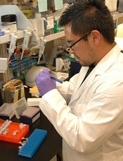
Credit: NIH
A group of researchers who tried, and failed, to replicate the STAP cell phenomenon have detailed their work in F1000Research.
Kenneth Ka Ho Lee, PhD, of the Chinese University of Hong Kong, and his colleagues attempted to create STAP (stimulus-triggered acquisition of pluripotency) cells using the method described in a recent Nature paper.
The paper’s authors had reported they could induce pluripotency in somatic cells by bathing them in acid.
However, not long after the paper and a related letter were published, members of the scientific community voiced concerns about the publications—such as suspicions of plagiarism and the possibility of doctored images—and began to question the findings.
Then, an investigation by the Japanese research institute RIKEN (where many of the researchers are employed) suggested that at least some of the paper’s authors were guilty of misconduct and/or negligence.
However, the study’s lead author, Haruko Obokata, PhD, has maintained that, despite errors in the paper, STAP cells can be created.
Dr Lee’s experiments suggest otherwise—or at least that the cells cannot be created using the methods outlined in the Nature paper.
Dr Lee and his colleagues reported that carefully replicating the original acid-treatment method does not induce pluripotency in 2 types of mouse somatic cells.
Using both white blood cells isolated from the spleen of neonatal mice—the same cells used in the original study—and lung fibroblasts, the team was unable to replicate the original findings.
They’ve published a full account of this work in F1000Research. The journal also employs open peer review by invited experts, which occurs after publication and is published in full online alongside the paper.
Dr Lee’s study will now undergo this process, and readers interested to see referees’ views as they come in can follow the paper by clicking “Track” on the published article.
RIKEN upholds misconduct allegations, calls for paper’s retraction

Credit: RIKEN
The Japanese research institute RIKEN has announced that it will not re-investigate the allegations of misconduct levelled against the researcher who claimed to have discovered a new method for inducing pluripotency in somatic cells.
In January, Haruko Obokata, PhD, and her colleagues reported the creation of stimulus-triggered acquisition of pluripotency (STAP) cells.
The team said they could induce pluripotency by exposing somatic cells to a low-pH environment.
But members of the scientific community voiced concerns about the research, so RIKEN launched an investigation.
In April, the investigative committee concluded that Dr Obokata and some of her colleagues were guilty of misconduct and/or negligence.
Dr Obokata appealed the findings, but the committee has decided another investigation is not warranted. RIKEN has also called for a retraction of the Nature paper in which the committee found evidence of misconduct.
Another RIKEN committee has already met to discuss possible disciplinary action for Dr Obokata and some of her colleagues. Potential punishments range from pay cuts to temporary suspension to disciplinary discharge.
In the meantime, a group headed by Shinichi Aizawa, PhD, special advisor to RIKEN, is attempting to verify the results of the STAP experiments and determine if the STAP phenomenon is, in fact, real.
As the events of this case unfolded, another possible case of misconduct surfaced at RIKEN.
The institute recently announced that Shunsuke Ishii, PhD, chairperson of the committee investigating misconduct in the STAP papers, had resigned from the committee amid concerns about one of his own papers.
RIKEN replaced Dr Ishii with Jun Watanabe, a lawyer already on the committee. And the institute has launched an investigation into Dr Ishii’s work.
Furthermore, RIKEN’s office for internal reform, headed by President Ryoji Noyori, PhD, has commissioned a committee of outside experts to deliberate and recommend measures to prevent research misconduct in the future.