User login
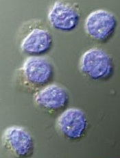
Team applies single-cell genomics to malaria

Credit: Peter H. Seeberger
Researchers have devised a way to perform genome sequencing on individual cells infected with malaria parasites.
The team found this single-cell approach could generate parasite genome sequences directly from the blood of patients infected with Plasmodium vivax or Plasmodium falciparum.
It provided new insight into the biology of the parasites, revealing their virulence and capacity for drug resistance.
The team described this work in Genome Research.
They noted that malaria infections commonly contain complex mixtures of Plasmodium parasites. These multiple-genotype infections (MGIs) can alter the impact of the infection and drive the spread of drug resistance. MGIs are extremely common in regions with high levels of malaria infection, but their biology is poorly understood.
“Up to 70% of infections in sub-Saharan Africa are MGIs, and we currently don’t know how many genotypes are present and whether parasites come from a single mosquito bite or multiple mosquito bites,” said study author Shalini Nair, of Texas Biomedical Research Institute in San Antonio.
“Current sequencing techniques really limit our understanding of malaria parasite biology,” added study author Ian Cheeseman, PhD, also of Texas Biomed.
“It’s like trying to understand human genetics by taking DNA from everyone in a village at once. The data is all jumbled up, but what we really want is information from individuals.”
To achieve a better understanding of MGIs and malaria parasites in general, the researchers developed a method for isolating an individual parasite cell and sequencing its genome. Although single-cell genomics approaches are already used in cancer research, it has been difficult to adapt the approach to other organisms.
“One of the real challenges was learning how to cope with the tiny amounts of DNA involved,” Nair said. “In a single cell, we have a thousand-million-millionth of a gram of DNA. It took a lot of effort before we developed a method where we simply didn’t lose this.”
But the researchers eventually found they could use methods of single-cell sorting and whole-genome amplification to separate out individual cells and amplify their DNA for sequencing. The team sequenced the DNA from red blood cells infected with P falciparum or P vivax.
They discovered this technique can reveal the composition of MGIs and provide information on the strength of an infection and the development of drug resistance.
“One of the major surprises we found when we started looking at individual parasites instead of whole infections was the level of variation in drug-resistance genes,” Nair said. “The patterns we saw suggested that different parasites within a single malaria infection would react very differently to drug treatment.”
Unfortunately, this technology is currently too expensive and demanding for routine use in the clinic. But the potential applications are significant, according to the researchers.
“We’re now able to look at malaria infections with incredible detail,” Dr Cheeseman said. “This will help us understand how to best design drugs and vaccines to tackle this major global killer.” ![]()
How IL-27 promotes tumor growth
Credit: Kathryn T. Iacono
Research in mice has revealed a potential approach to cancer treatment—a way to inhibit the recruitment of regulatory T cells (Tregs) to tumors.
The study showed that dendritic-cell-derived interleukin-27 (IL-27) promotes Treg recruitment in models of lymphoma, melanoma, and fibrosarcoma.
This suggests that if cancer therapies can inhibit IL-27’s immunosuppressive function, they could more effectively activate other T cells to attack and destroy tumors.
Researchers described this discovery in the Journal of Leukocyte Biology.
“Our study not only provides a new insight into the effects of interleukin-27 in regulatory T-cell biology but also greatly improves our understanding of the physiological functions of interleukin-27, especially in tumor immunology,” said study author Siyuan Xia, of Nankai University in Tianjin, China.
“We hope our study could shed new light on developing novel interventional therapies by targeting regulatory T cells in cancer patients.”
The researchers made their discovery by using mice deficient in a specific subunit of IL-27 called p28. They compared tumor-infiltrating lymphocytes between IL-27p28 knockout mice and wild-type mice.
This revealed that Tregs were significantly decreased in knockout mice transplanted with EL-4 lymphoma, B16 melanoma, and MCA-induced fibrosarcoma.
The team also found that IL-27 promotes the expression of CCL22, which is known to mediate Treg recruitment to tumors. And tumor-associated dendritic cells were the major source of CCL22.
When the researchers restored CCL22 or IL-27 in the knockout mice, they observed significant restoration of the tumor-infiltrating Tregs.
Furthermore, tumor-infiltrating CD4 T cells produced much more IFN-γ in the IL-27p28 knockout mice than in wild-type mice. According to the researchers, this reinforces the physiological importance of Tregs in suppressing an antitumor immune response.
“Suppressive and regulatory pathways in the immune system are incredibly important for normal health and preventing autoimmunity,” said John Wherry, PhD, Deputy Editor of the Journal of Leukocyte Biology.
“However, these pathways also get exploited by cancer to prevent immune responses leading to cancer progression. The current studies point to an important regulatory network centered on interleukin-27 that could be targeted to improve immunity to cancer in humans.” ![]()
A more precise method of delivering gene therapy

Jeff Fitlow/Rice University
Researchers have developed an adeno-associated virus (AAV) that releases its payload only in the presence of 2 selected proteases.
Because certain proteases are elevated at tumor sites, the virus can be designed to target and destroy cancer cells.
Junghae Suh, PhD, of Rice University in Houston, Texas, and her colleagues engineered the virus and described their work in ACS Nano.
AAVs have become the object of study as delivery vehicles for gene therapy.
Researchers often try to target AAVs to cellular receptors that may be slightly overexpressed on diseased cells, but Dr Suh’s team took a different approach.
“We were looking for other types of biomarkers beyond cellular receptors present at disease sites,” she said. “In breast cancer, for example, it’s known the tumor cells oversecrete extracellular proteases, but perhaps more important are the infiltrating immune cells that migrate into the tumor microenvironment and start dumping out a whole bunch of proteases as well.”
“So that’s what we’re going after to do targeted delivery. Our basic idea is to create viruses that, in the locked configuration, can’t do anything.”
But when the programmed AAVs encounter the right proteases at sites of disease, they unlock and bind to the cells. The AAVs then deliver payloads that will either kill the cells, in the case of cancer therapy, or deliver genes that can repair the cells.
Dr Suh and her colleagues genetically insert peptides into the self-assembling AAVs to lock the capsids, the hard shells that protect genes contained within. The target proteases recognize the peptides and “chew off the locks,” effectively unlocking the virus and allowing it to bind to the diseased cells.
“If we were just looking for 1 protease, it might be at the cancer site, but it could also be somewhere else in your body where you have inflammation,” Dr Suh said. “This could lead to undesirable side effects.”
“By requiring 2 different proteases—let’s say protease A and protease B—to open the locked virus, we may achieve higher delivery specificity since the chance of having both proteases elevated at a site becomes smaller.”
The ultimate vision of this technology is to design viruses that can carry out a combination of steps for targeting.
“To increase the specificity of virus unlocking, you can imagine creating viruses that require many more keys to open,” Dr Suh said. “For example, you may need both proteases A and B, as well as a cellular receptor, to unlock the virus. The work reported here is a good first step toward this goal.” ![]()
Wnt pathway appears key to cell reprogramming

Salk Institute
The Wnt signaling pathway plays a key role in the generation of induced pluripotent stem cells (iPSCs), according to a study published in Stem Cell Reports.
Researchers found they could increase the efficiency of the cell reprogramming process by inhibiting the Wnt pathway.
“[U]ntil now, this was a very inefficient process,” said study author Ilda Theka, a PhD student at the Centre for Genomic Regulation in Barcelona, Spain.
“There are many groups trying to understand the mechanism by which adult cells become pluripotent and what blocks that process and makes only a small percentage of cells end up being reprogrammed. We are providing information on why it happens.”
The researchers studied how the Wnt pathway behaves throughout the process of transforming mature cells into iPSCs, which usually lasts 2 weeks. It’s a dynamic process that produces oscillations from the pathway, which is not active all the time.
“We have seen that there are two phases and that, in each one of them, Wnt fulfils a different function,” Theka said. “And we have shown that, by inhibiting it at the beginning of the process and activating it at the end, we can increase the efficiency of reprogramming and obtain a larger number of pluripotent cells.”
The team also discovered that the exact moment when the Wnt pathway is activated is crucial. Activating the pathway too early makes the cells begin to differentiate, and they are not reprogrammed.
To artificially control the pathway, the researchers used a molecule called Iwp2, a Wnt-secretion inhibitor that does not permanently alter the cells. ![]()
Artificial bone marrow seems just like the real thing

Credit: James Weaver,
Harvard’s Wyss Institute
Researchers have created a device that reproduces the structure, function, and cellular make-up of bone marrow, according to a paper published
in Nature Methods.
The device, dubbed “bone marrow on a chip,” could serve as a new tool for testing the effects of radiation and other toxic agents on whole bone marrow.
The researchers believe this bone marrow on a chip could provide an alternative to animal testing, although the device itself was generated in mice.
The team also thinks that, in the future, the engineered bone marrow could be used to maintain a cancer patient’s own marrow temporarily during radiation or high-dose chemotherapy.
In an initial test, the engineered bone marrow withered in response to radiation, just as natural bone marrow does. And, as in natural marrow, granulocyte colony-stimulating factor conferred a protective effect on the engineered marrow.
“Bone marrow is an incredibly complex organ that is responsible for producing all of the blood cell types in our body, and our bone marrow chips are able to recapitulate this complexity in its entirety and maintain it in a functional form in vitro,” said study author Donald Ingber, MD, PhD, of the Wyss Institute for Biologically Inspired Engineering at Harvard University in Boston.
Dr Ingber leads an effort to develop human organs on chips—small microfluidic devices that mimic the physiology of living organs. To build these devices, researchers combine multiple types of cells from an organ on a plastic microfluidic device, while steadily supplying nutrients, removing waste, and applying mechanical forces the tissues would face in the body.
But bone marrow is so complex that Dr Ingber and his colleagues needed a new approach to mimic organ function. Rather than trying to reproduce such a complex structure cell by cell, the team used mice.
Specifically, the researchers packed dried bone powder into an open, ring-shaped mold the size of a coin battery and implanted the mold under the skin on the animal’s back.
After 8 weeks, the team surgically removed the disk-shaped bone that had formed in the mold and examined it with a specialized CAT scanner. The scan showed a honeycomb-like structure that looked identical to natural trabecular bone.
The marrow looked like the real thing as well. When the researchers stained the tissue and examined it under a microscope, the marrow was packed with blood cells, just like marrow from a living mouse.
And when the team sorted the bone marrow cells by type and tallied their numbers, the mix of different types of blood and immune cells in the engineered bone marrow was identical to that in a mouse thighbone.
To sustain the engineered bone marrow outside of a living animal, the researchers surgically removed the engineered bone from mice, then placed it in a microfluidic device that mimics the circulation the tissue would experience in the body.
Marrow in the device remained healthy for up to 1 week. This is typically long enough to test the toxicity and effectiveness of a new drug, the team said.
The device also passed an initial test of its drug-testing capabilities. Like marrow from live mice, this engineered marrow was susceptible to radiation, but granulocyte colony-stimulating factor conferred a protective effect.
The researchers believe that, in the future, they could potentially grow human bone marrow in immune-deficient mice. And their bone marrow on a chip could generate blood cells, which could circulate in an artificial circulatory system to supply a network of other organs on chips. ![]()
Portable spectrometers can detect malaria early, group says

Credit: St Jude Children’s
Research Hospital
An infrared spectroscopy technique can detect malaria parasites at early stages of development, according to preclinical research published in Analytical Chemistry.
A group of biochemists found this method could detect Plasmodium falciparum in red blood cells by picking up on a fatty acid signature.
This allowed the researchers to identify and quantify parasites at various stages of development, including the ring and gametocyte stages.
The team also pointed out that the spectrometer they used is portable, inexpensive, and does not require highly trained staff for operation. It could therefore prove useful in the field.
“Current tests for malaria suffer from serious limitations,” said study author Bayden Wood, PhD, of Monash University in Victoria, Australia.
“Many are expensive [and] require specialist instruments and highly trained staff to judge whether blood samples contain the parasite. What’s been holding us back is the lack of an accurate and inexpensive test to detect malaria early and stop it in its tracks. We believe we’ve found it.”
Dr Wood and his colleagues already knew that fatty acids were a marker for malaria from previous studies conducted at the Australian Synchrotron. The Synchrotron allowed the team to see the different life stages of the parasite and the variation in its fatty acids.
The researchers thought they might be able to use this information for diagnosis, but they needed a more portable detection method.
So they decided to test whether attenuated total reflectance Fourier transform infrared spectroscopy (ATR-FT-IR) could detect the fatty acid signature. The technique utilizes infrared light to pick up on the vibrations of molecules.
The researchers spiked red blood cells with parasites of different numbers and life stages and observed them using ATR-FT-IR.
Dr Wood said the method produced results within minutes. And it gave a clear indication of malaria at a much earlier stage of infection than current tests on the market—at the ring and gametocyte stages.
The absolute detection limit was 0.00001% parasitemia (<1 parasite/μL of blood) for cultured early ring-stage parasites in a suspension of normal red blood cells.
“Now that we can detect the early stages of a parasite’s life in the bloodstream, the disease will be much easier to test and treat,” Dr Wood said. “The big advantage of our test is that it doesn’t need scientists and expensive equipment. This has the potential to dramatically reduce the number of people dying from this disease in remote communities.”
The method also shows the potential to detect a number of other blood-borne diseases, according to the researchers. Dr Wood and his colleagues are now planning a clinical trial of ATR-FT-IR in Thailand. ![]()
Hospira issues Class I recall of infusion pumps

Credit: Daniel Gay
Hospira, Inc., has issued a Class I recall of Abbott Acclaim infusion pumps and Hospira Acclaim Encore infusion pumps, after receiving reports of broken door assemblies on these products.
If a door assembly breaks, the door may not close properly and an over-infusion or a delay of therapy may occur.
If the door cannot be closed, the pump cannot be used, and this can result in a delay of therapy.
Use of these products may cause serious adverse events, including death.
These pumps are used to deliver hydration fluids, drugs, blood and blood fractions, intravenous nutritionals, and enteral nutritionals.
The affected Abbott Acclaim Infusion Pumps, list Number 12032, were manufactured from February 1998 to November 1998 and distributed from September 1998 through February 2004.
The affected Hospira Acclaim Encore infusion pumps, list Number 12237, were manufactured from February 1997 to February 2010 and distributed from July 1999 through November 2013.
Hospira is recommending that users inspect each Hospira/Abbott Acclaim Encore infusion pump for door handle cracks prior to programming a therapy, by taking the following steps:
1. After inserting the tubing (with the roller clamp closed) and closing the door handle against the infusion pump, check that the door is fully closed.
If a pump has a door that does not close properly and a gap or separation exists between the completely closed door and the pump itself, remove the pump from clinical service, and call Hospira at 1-800-441-4100 (M-F, 8am-5pm, CT). If the door closes correctly, proceed to Step 2.
2. If the door closes correctly and a gap or separation does not exist between the completely closed door and the pump itself, check that there is no free flow activity in the drip chamber of the administration set by opening the roller clamp.
If free flow is detected, close the roller clamp, remove the pump from clinical service, and call Hospira at 1-800-441-4100 (M-F, 8am-5pm, CT).
3. If no issues are found through steps 1 and 2, the pump is acceptable for use. However, healthcare professionals should still ensure that anyone in their facility who might use these products is made aware of this safety notification and the recommended actions.
Healthcare professionals and patients can report adverse events or side effects related to the use of these products to the FDA’s MedWatch Program. ![]()
Credit: Daniel Gay
Hospira, Inc., has issued a Class I recall of Abbott Acclaim infusion pumps and Hospira Acclaim Encore infusion pumps, after receiving reports of broken door assemblies on these products.
If a door assembly breaks, the door may not close properly and an over-infusion or a delay of therapy may occur.
If the door cannot be closed, the pump cannot be used, and this can result in a delay of therapy.
Use of these products may cause serious adverse events, including death.
These pumps are used to deliver hydration fluids, drugs, blood and blood fractions, intravenous nutritionals, and enteral nutritionals.
The affected Abbott Acclaim Infusion Pumps, list Number 12032, were manufactured from February 1998 to November 1998 and distributed from September 1998 through February 2004.
The affected Hospira Acclaim Encore infusion pumps, list Number 12237, were manufactured from February 1997 to February 2010 and distributed from July 1999 through November 2013.
Hospira is recommending that users inspect each Hospira/Abbott Acclaim Encore infusion pump for door handle cracks prior to programming a therapy, by taking the following steps:
1. After inserting the tubing (with the roller clamp closed) and closing the door handle against the infusion pump, check that the door is fully closed.
If a pump has a door that does not close properly and a gap or separation exists between the completely closed door and the pump itself, remove the pump from clinical service, and call Hospira at 1-800-441-4100 (M-F, 8am-5pm, CT). If the door closes correctly, proceed to Step 2.
2. If the door closes correctly and a gap or separation does not exist between the completely closed door and the pump itself, check that there is no free flow activity in the drip chamber of the administration set by opening the roller clamp.
If free flow is detected, close the roller clamp, remove the pump from clinical service, and call Hospira at 1-800-441-4100 (M-F, 8am-5pm, CT).
3. If no issues are found through steps 1 and 2, the pump is acceptable for use. However, healthcare professionals should still ensure that anyone in their facility who might use these products is made aware of this safety notification and the recommended actions.
Healthcare professionals and patients can report adverse events or side effects related to the use of these products to the FDA’s MedWatch Program.
Liquid droplets help explain cell migration

Scientists have discovered an unexpected link between the shape of a cell and its migration efficiency, and they’ve explained its physics using a model of a liquid droplet.
Cell migration is achieved through the movement of the cell’s membrane, which is powered by the action of a protein network inside the cell.
This interaction is affected by the cell’s overall shape, but exactly how this takes place has been unclear.
Research published in Current Biology provides some insight.
The first step in cell migration occurs when the cell extends its front edge—a process called protrusion. This is driven by the growth of actin filaments, which push the cell membrane from inside. At the same time, membrane tension controls protrusion by providing resistance and protecting the cell from over-extending.
But physical laws dictate that the shape of the cell membrane must play a role in the balance between force exerted by actin and the resisting membrane tension. This was not taken into account in previous studies, which used 2D models of migrating cells.
Now, Chiara Gabella, PhD, of Ecole Polytechnique Fédérale de Lausanne in Switzerland, and her colleagues have used a 3D model to better describe the relationship between cell protrusion, shape, and membrane tension.
The scientists developed a way to evaluate the 3D shape of migrating fish epidermal keratocytes by observing the cells in a chamber filled with a fluorescent solution.
The team applied various treatments to swell, shrink, or stretch the cells. And they were able to observe the treatment’s impact on membrane tension, shape, and protrusion velocity.
The treatments only affected the cells’ shape and migration speed, not membrane tension. The results also showed that the more spherical a cell is, the faster it moves.
To interpret these unexpected findings, the scientists modeled a migrating cell as a liquid droplet spreading on a surface.
“It is well known that a droplet’s shape and, in particular, the contact angle that it makes with the surface are determined by the tension forces between the droplet, its environmental medium (eg, air or a different liquid), and the surface on which it moves,” Dr Gabella said.
Results of the modeling experiment suggested that the leading edge could be considered a triple interface between the substrate, membrane, and extracellular medium. And the contact angle between the membrane and the substrate determines the load on actin polymerization and, therefore, the protrusion rate.
“From this point of view, a more spherical cell means less load for actin filaments to overcome and, therefore, faster actin growth and migration,” said Alexander Verkhovsky, PhD, also of Ecole Polytechnique Fédérale de Lausanne.
In support of this idea, the scientists found the cells were sensitive to the surface characteristics, just as droplets would be, by slowing down or being pinned at ridges.
“The emphasis of many studies has been on discovering and characterizing individual cellular components,” Dr Verkhovsky said. “This is rooted in the common belief that a cell’s behavior is determined by intricate networks of genes and proteins.”
In contrast, this work shows that, despite their molecular complexity, cells can be described as physical objects. The findings point to a new relationship between a cell’s shape and its dynamics and may help us to understand how cell migration is guided by the cell’s 3D environment.
Hospira announces device correction for infusion pump docking station

Credit: CDC
Hospira, Inc., has announced a medical device correction for the GemStar Docking Station (list number 13075), used in conjunction with the GemStar infusion pump.
The correction follows customer reports of 2 malfunctions that may occur with the docking station.
The company is not recalling the product but is notifying US customers of the potential malfunctions and providing instructions for overriding these errors.
The errors could potentially cause delays or interruptions in therapy. And this might result in serious adverse events or death, but there have been no such events reported to date.
Potential malfunctions
The GemStar Docking Station is an accessory to the GemStar infusion pump (sold separately) and provides an alternate power source to the GemStar pump.
When the docking station is used in conjunction with a GemStar Phase 3 pump (List 13000, 13100 or 13150), there is a risk that the GemStar Phase 3 pump may fail to power up while connected to the docking station.
When a GemStar Phase 3 (List 13000, 13100 or 13150) or GemStar Phase 4 pump (List 13086, 13087 or 13088) is used in conjunction with both a docking station and an external battery pack accessory (List 13073), the GemStar pump may display error code 11/003 and give an audible alarm, indicating excessive input voltage from the external sources.
If the GemStar pump detects what is perceived to be more than 3.6 volts, as measured on the external voltage input, the pump will stop the infusion. This will trigger an audible alarm, and the device will display alarm code 11/003.
If a GemStar fails to power up or the 11/003 error code stops an infusion, a patient’s therapy might be delayed or interrupted. This could result in significant injury or death, although there have been no reports of death or serious injury associated with these malfunctions to date.
The products impacted by these issues have been in distribution since February 2002.
Responding to/preventing malfunctions
Hospira is advising that healthcare professionals weigh the risk/benefit to patients associated with the use of the docking station when administering critical therapies. Clinicians should consider the use of an alternative pump, particularly in patients for whom a delay or interruption of therapy could result in serious injury or death.
However, the company says there is no need to return the GemStar Docking Station at this time. Instead, Hospira recommends that users take the following actions.
To avoid a failure to power up, turn on the pump before connecting it with the docking station. This will prevent the failure to power up.
To mitigate the potential for an 11/003 error code, remove the external battery pack accessory (List 13073) from the docking station and pump prior to installing the pump in the docking station.
In addition, clinicians should stop using a docking station in conjunction with an external battery pack accessory (List 13073). Contact Hospira to discuss an appropriate alternative option.
Docking station users who experience a failure to power up or an 11/003 error code should report the issue to Hospira by calling 1-800-441-4100 (M-F, 8am-5pm CT) or emailing ProductComplaintsPP@hospira.com.
For additional assistance or to obtain a copy of the Urgent Medical Device Correction letter and/or a reply form, contact Stericycle at 1-866-792-5451 (M-F, 8am-5pm ET).
On May 1, 2013, Hospira announced that it would begin the process of retiring the GemStar family of infusion devices in accordance with the company’s global device strategy. As of July 31, 2015, Hospira will consider the products within the GemStar Infusion System family retired and will no longer support them.
Adverse reactions or quality problems related to the GemStar Docking Station can be reported to the US Food and Drug Administration’s MedWatch Program.
Baxter issues Class I recall of infusion pumps

of chemotherapy drugs
Credit: Bill Branson
Baxter Healthcare Corporation is recalling some of its infusion pumps after receiving more than 3500 reports of the pumps malfunctioning.
According to the US Food and Drug Administration (FDA), the malfunctioning pumps have resulted in 9 severe adverse events but no deaths.
This Class I recall includes Sigma Spectrum Infusion Pumps with Master Drug Library Model No. 35700BAX and 35700ABB.
The pumps were made between July 1, 2005, and January 15, 2014. They were distributed between February 20, 2013, and January 15, 2014.
The Sigma Spectrum infusion pumps are intended to deliver controlled amounts of medicines, blood, blood products, and other intravenous fluids.
The FDA said there have been more than 3500 reports of these pumps malfunctioning—specifically, reports of System Error 322 “Link Switch Error (low).” This error occurs when the pump detects that the door is open even though it is closed. A System Error 322 may lead to an interruption or delay in therapy.
When this error occurs, the Sigma Spectrum infusion pump stops the infusion, an alarm sounds, and a light flashes (a visual “322” alarm). This requires a clinician to reset the alarm, reprogram the pump, and confirm the infusion is running properly.
The use of affected pumps may cause serious adverse health consequences, including death; hence, the Class I recall.
Customers who encounter a System Error 322 should turn off the pump by pressing the ON/OFF key, then turn the pump back on by pressing the ON/OFF key to clear the alarm.
Clinicians will need to reprogram the infusion after the pump is turned back on. If the alarm cannot be cleared using these instructions, the device should be removed from use and sent to the facility’s biomedical engineering department.
If the System Error 322 reoccurs, the pump may need to be inspected and serviced by Baxter Healthcare. To contact Baxter, call 1-800-356-3454 (choose option 1) Monday through Friday, 7 am to 7 pm, Eastern Time.
Adverse reactions or quality problems related to these pumps can be reported to the FDA’s MedWatch Program.