User login
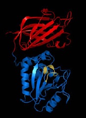
How bleomycin cuts cancer to pieces

Credit: Bill Branson
The antitumor agent bleomycin can treat a range of cancers, but its disease-fighting properties have been poorly understood.
Now, a pair of researchers have characterized bleomycin’s ability to cut through double-stranded DNA in cancerous cells.
The duo believe their research could help inform efforts to fine-tune the drug, improving its cancer-killing properties and limiting toxicity to healthy cells.
The research appears in the Journal of the American Chemical Society.
Bleomycin is part of a family of structurally related antibiotics produced by the bacterium Streptomyces verticillus. Three potent versions of the drug—labeled A2, A5, and B2—are the primary forms in clinical use against cancers.
Previous research has shown that bleomycin can cause death in aberrant cells by migrating to the cell nucleus, binding with DNA, and subsequently causing breaks in the DNA sequence. Following a binding event, a molecule of bleomycin can effectively slice through one or both strands of DNA.
Cleavage of DNA is believed to be the primary mechanism by which bleomycin kills cancer cells, particularly through double-strand cleavages, which are more challenging for the cellular machinery to repair.
“There are several mechanisms for repairing both single-strand and double-strand breaks in DNA, but double-strand breaks are a more potent form of DNA lesion,” explained study author Basab Roy, a graduate student at Arizona State University in Tempe.
For this study, Roy and Sidney Hecht, PhD, used bleomycin A5, which has similar DNA binding and cleaving properties as bleomycin A2 and B2.
Previous research revealed that bleomycin binds with highly specific regions of the DNA strand, typically G-C sites, where a guanosine base pairs with a cytidine. The strength of this binding is closely associated with the degree of double-strand DNA cleavage.
From a pool of random DNA sequences, the researchers selected a library of 10 hairpin DNAs, based on their strong binding affinity for bleomycin A5. Hairpin DNAs are looped structures that form when a segment of a DNA strand base-pairs with another portion of the same strand. These hairpin DNAs were used to investigate double-strand cleavage.
Each of the 10 DNA samples underwent double-strand cleavage at more than one site. All of the observed cleavage sites were found within or in close proximity to an 8-base-pair-variable region.
Examination of the 10 DNA samples exposed to bleomycin revealed a total of 31 double-strand cleavage sites. Earlier research had described the form of double-strand DNA cleavage bleomycin induced at 14 of these sites. But the remaining 17 cases of double-stranded cleavage occurred through a different mechanism, described for the first time by Roy and Dr Hecht.
The pair used iron (FeII) as a cofactor for bleomycin in the binding events, and they observed 2 types of bleomycin binding and cleavage activity.
In the first, bleomycin and its iron cofactor (Fe.BLM) bind with hairpin DNA at a primary site. Typically, this is a site with a particular sequence: 5´-G-Py-B-3´. (Here, 5´ refers to one end of the DNA hairpin, G refers to the base guanosine, Py refers to a pyrimidinic base—either cytidine or thymidine, B refers to any nucleobase, and 3´ refers to the other DNA end.)
The result of this binding is the abstraction of a hydrogen atom at the primary site. Two results are possible following the primary binding event, one causing a single-strand break in the primary site and the other, failing to produce full cleavage of the strand, producing instead a site lacking either a purine or pyrimidine base. This is known as an AP site.
In the first case—where bleomycin achieves single strand cleavage—the bleomycin molecule can then become reactivated, once more abstracting a hydrogen atom from the opposing DNA strand.
The opposite strand can again follow 1 of 2 pathways, (a) full cleavage of the opposing strand, yielding a double-strand cleavage or (b) formation of an AP site. The researchers noted that this AP site can lead to strand cleavage through the opposing DNA strand with the addition of a mild base like n-butylamine.
The results of this study emphasize the correlation between the strength of bleomycin binding to DNA and the frequency of double-strand cleavage. Of the 10 sample hairpin DNAs, the 2 most tightly bound to bleomycin each showed 5 double-strand cleavages, whereas the least tightly bound samples exhibited just 2 double-strand cleavages.
This suggests a plausible mechanism for DNA cleavage by bleomycin that may lead to tumor cell killing, as well as identifying the most common sequences involved in DNA site binding and subsequent strand breakage.
Roy noted, however, that more research is needed to elucidate the biochemical causes of tight binding by bleomycin. Furthermore, bleomycin’s specificity for cancer cells remains enigmatic.
“Cancer is still a black hole,” Roy said. “We’re trying to make this particular molecule better. There is still so much to learn.” ![]()
Team overcomes problem with new lab on a chip

Credit: Rhoda Baer
Chemists say they have overcome one of the main obstacles in creating effective lab-on-a-chip devices, and the device they’ve invented has many potential applications, such as screening biological molecules.
To create their device, the team developed a technique that involves printing droplets of a special solvent onto a gold-coated or glass surface.
“We use a class of ‘green’ solvents called ionic liquids, which are salts that are liquid at room temperature,” said study author Chuan Zhao, PhD, of The University of New South Wales in Sydney, Australia.
“They are non-volatile, so this overcomes one of the main problems in making useful miniaturized devices—rapid evaporation of the solvents on the chip.”
Dr Zhao and his colleagues described this research in Nature Communications.
Lab-on-a-chip devices, where chemical reactions are carried out on a miniature scale, are under intensive development because they offer the promise of faster reaction times, reduced use of materials, and high yields of product.
However, the evaporation of solvents on the chip is a problem because it can affect the concentration of substances and disrupt the reactions.
Researchers have attempted to overcome the problem by containing the solvents within tiny channels, or “walls,” and having reservoirs to store extra solvent on the chip.
Dr Zhao and his colleagues said their “wall-less” design—using non-volatile ionic liquids as solvents to fabricate a microarray of droplets chemically anchored to the chip—has several advantages.
“Ionic liquids are designer solvents and have wide application,” Dr Zhao said. “We can now carry out many reactions or analytical procedures in ionic liquids at the micro-scale on a chip with enhanced yields and efficiency.”
“These microarray chips can be easily produced in high numbers and are very stable. They can survive being turned upside down and heated to 50 degrees, and some can even survive being immersed in another liquid. These properties will be important for commercial applications, including storage and transportation of microchips.”
The droplets of ionic liquid are about 50 µm across and 10 µm high. The researchers showed these tiny droplets can act as rapid, sensitive monitors of the presence of a gas, due to their small volume.
Metal salts dissolved in the droplets could be electrically deposited as microstructures, a technique that could be of use in the fabrication of integrated circuits.
And some biological molecules added to the droplets remained stable and active, opening up the possibility of using the microarrays for diagnostic purposes.
“The versatility of our chips means they could have a wide range of prospective functions,” Dr Zhao said, “such as for use in fast and accurate hand-held sensors for environmental monitoring, medical diagnosis, and process control in manufacturing.” ![]()
FDA allows importation of more saline

In response to the ongoing shortage of normal saline (0.9% sodium chloride injection), the US Food and Drug Administration (FDA) is allowing a company to import product manufactured in Spain.
Baxter Healthcare Corp. will be allowed to temporarily distribute sodium chloride 0.9% injection solution for intravenous infusion in VIAFLO
non-polyvinyl chloride containers, which is manufactured at the company’s Bieffe Medital, Sabinanigo, Spain facility.
This is the second time in recent months that the FDA has allowed the importation of saline. Last month, the agency announced it would allow Fresenius Kabi USA to import saline products manufactured in Norway.
As with the Norway plant, the FDA has inspected Baxter’s Spain facility to ensure it meets FDA standards.
Baxter will import sodium chloride 0.9% intravenous infusion in VIAFLO containers in the following volumes and quantities: 250 mL (30 bags per carton), 500 mL (20 bags per carton), and 1 L (10 bags per carton).
The FDA is asking that healthcare professionals contact Baxter directly to obtain the product. To place an order, contact Baxter’s Center for Service at 1-888-229-0001.
For more information on the products, see Baxter’s “Dear Healthcare Professional” letter, visit the company’s website, or contact Baxter’s Medical Information Service at 1-800-933-0303.
In addition to the imported saline, US-based manufacturers—B.Braun Medical Inc., Hospira Inc., and Baxter Healthcare Corp.—are producing and releasing normal saline. (Baxter’s saline product from Spain will be distributed in addition to Baxter’s FDA-approved version that is manufactured in the US.)
The FDA noted that, although the aforementioned shipments will help reduce current disruptions, they will not resolve the shortage of 0.9% sodium chloride injection. So the agency will continue working to alleviate the shortage. ![]()
Protein is key to HSC recovery after chemo, radiation

in the bone marrow
The protein beta-catenin plays a critical role in promoting the recovery of hematopoietic stem cells (HSCs) after exposure to radiation or chemotherapy, according to preclinical research published in Genes & Development.
The study provides new insight into the impact of radiation and chemotherapy on cellular and molecular processes.
And it points to possibilities for improving HSC regeneration in cancer patients who have undergone these treatments.
Study investigators first used mouse models to show that exposure to radiation triggers activation of the Wnt signaling pathway in hematopoietic stem and progenitor cells.
“The Wnt pathway and its key mediator, beta catenin, are critical for embryonic development and establishment of the body plan,” explained Tannishtha Reya, PhD, of the University of California, San Diego.
“In addition, the Wnt pathway is activated in stem cells from many tissues and is needed for their continued maintenance.”
Dr Reya and her colleagues then found that mice deficient in beta-catenin lacked the ability to activate canonical Wnt signaling. They also suffered from impaired HSC regeneration and bone marrow recovery after radiation or chemotherapy.
Mouse HSCs without beta-catenin could not suppress the production of reactive oxygen species or resolve DNA double-strand breaks. As a result, they could not recover effectively after radiation exposure or treatment with the chemotherapeutic agent fluorouracil.
“Our work shows that Wnt signaling is important in the mammalian hematopoietic system and is critical for recovery from chemotherapy and radiation,” Dr Reya said. “There are 2 major reasons why accelerating regeneration is important clinically.”
“One is that, after cancer patients are irradiated and transplanted with stem cells, the rate and extent of recovery is often not sufficient to protect the patient from anemia or infections . . . . Identifying signals that can boost regeneration after the bone marrow is severely damaged may help improve outcomes after transplantation.”
“Second, doses of chemotherapy and radiation used to treat cancer are often limited by the collateral damage they cause to normal tissues. If we can improve and accelerate recovery, we might be able to use higher doses of radiation or chemotherapy and reduce the risk of cancer relapse.”
Dr Reya added that this research suggests HSC regeneration could be accelerated by modulating the Wnt pathway, either by delivering additional Wnt proteins directly to patients or through drugs that activate the pathway. ![]()
Vitamin D may affect outcome in cancer patients

Cancer patients with higher levels of vitamin D at diagnosis tend to have better outcomes than patients who are vitamin D-deficient, a meta-analysis suggests.
Researchers found the association between vitamin D and prognosis was most pronounced in patients with breast cancer, colorectal cancer, and lymphomas.
The team observed an association between vitamin D and prognosis in other cancers as well, but the correlations were not significant.
Hui Wang, MD, PhD, of the Chinese Academy of Sciences in Shanghai, China, and colleagues conducted this research and reported their results in the Journal of Clinical Endocrinology & Metabolism.
The team analyzed data from 25 studies including 17,332 cancer patients. In most of the studies, patients had their vitamin D levels tested before they underwent any cancer treatment.
The analysis showed that, overall, a 10 nmol/L increase in vitamin D levels was associated with a 4% increase in survival.
The strongest links between vitamin D levels and survival were seen in breast cancer, colorectal cancer, and lymphomas. There was less evidence of a connection in patients with leukemias, lung cancer, gastric cancer, prostate cancer, melanoma, or Merkel cell carcinoma, but the available data were positive.
There were 2 studies evaluating the association between survival and vitamin D levels in leukemia patients.
Vitamin D insufficiency was associated with poor overall survival in patients with newly diagnosed chronic lymphocytic leukemia, although this was not statistically significant. The hazard ratio (HR) was 0.68 (P=0.07).
For acute myeloid leukemia, patients with vitamin D levels that were considered insufficient (20-31.9 ng/mL) or deficient (<20 ng/mL) had worse overall survival (HR=0.65) and disease-free survival (HR=0.65) than patients with normal vitamin D levels.
There were also 2 studies evaluating the association between survival and vitamin D levels in lymphoma patients. The results showed that patients with the highest vitamin D levels had better overall survival than those with the lowest vitamin D levels (HR=0.48, P<0.001).
Lymphoma patients in the highest quartile of vitamin D levels also had a lower risk of cancer-specific mortality (HR=0.50, P<0.001) and better disease-free survival (HR=0.80, P=0.04).
Similarly, higher vitamin D levels were significantly associated with reduced cancer-specific mortality for patients with colorectal cancer (P=0.005) and improved disease-free survival for patients with breast cancer (P<0.001).
For overall survival, the HR for the highest vs lowest quartile of vitamin D levels was 0.55 for colorectal cancer patients and 0.63 for breast cancer patients.
Based on these results, Dr Wang concluded, “Physicians need to pay close attention to vitamin D levels in people who have been diagnosed with cancer.” ![]()
Loss of chromosome Y linked to cancer risk, survival

Credit: Rhoda Baer
New research indicates that loss of the Y chromosome in the peripheral blood may be associated with decreased survival—both higher mortality from cancer and a shorter life span in general.
Study investigators say their findings may explain why cancer incidence and mortality are both higher in men than in women, and why men have shorter average life spans than women.
Lars Forsberg, PhD, of Uppsala University in Sweden, and his colleagues recounted their findings in Nature Genetics.
The team analyzed blood samples from 1153 elderly men and found the most common genetic alteration was a loss of the Y chromosome in a proportion of leukocytes.
Further analyses revealed that loss of chromosome Y was associated with an increase in cancer diagnosis, cancer mortality, and all-cause mortality.
Specifically, the hazard ratio (HR) was 1.91 (P=0.010) for all-cause mortality and 3.29 (P=0.003) for cancer mortality. The HRs were 3.62 (P=0.003) for non-hematologic cancer mortality and 2.19 (P=0.450) for hematologic cancer mortality.
The HR for a diagnosis of any cancer was 2.47 (P=0.014). And the HR for diagnosis of a non-hematologic malignancy was 2.68 (P=0.008).
The investigators said these results suggest that chromosome Y is important in processes beyond sex determination.
“You have probably heard before that the Y chromosome is small, insignificant, and contains very little genetic information; this is not true,” said study author Jan Dumanski, PhD, of Uppsala University.
“Our results indicate that the Y chromosome has a role in tumor suppression, and they might explain why men get cancer more often than women. We believe that analyses of the Y chromosome could, in the future, become a useful general marker to predict the risk for men to develop cancer.”
![]()
Maintaining stem cell pluripotency

Salk Institute
Retrotransposons—viral elements incorporated into the human genome—may play a key role in maintaining stem cell pluripotency, a new study suggests.
Previous research indicated that an important fraction of mammalian transcriptomes consists of transcripts derived from retrotransposon elements, but the biological function of these transcripts was unknown.
Now, experiments in induced pluripotent stem cells (iPSCs) and embryonic stem cells (ESCs) have provided some insight into the transcripts’ function.
Piero Carninci, PhD, of RIKEN Center for Life Science Technologies in Yokohama, Japan, and his colleagues described these findings in Nature Genetics.
The researchers found that thousands of transcripts in stem cells that have not yet been annotated are transcribed from retrotransposons, presumably to elicit nuclear functions. These transcripts were expressed in iPSCs and ESCs but not in differentiated cells.
Furthermore, several of the transcripts were shown to be involved in the maintenance of pluripotency. Degrading them using RNA interference caused iPSCs to lose their pluripotency and differentiate.
The researchers said these transcripts appear to have been recruited, both in the human and mouse genome, where they are used to maintain the pluripotency of stem cells. But more research is needed to determine exactly how and why this occurs.
“Our work has just begun to unravel the scale of unexpected functions carried out by retrotransposons and their derived transcripts in stem cell biology,” Dr Carninci said.
“We were extremely surprised to learn from our data that what was once considered genetic junk—namely, ancient retroviruses that were thought to just parasite the genome—are, in reality, symbiotic elements that work closely with other genes to maintain [iPSCs and ESCs] in their undifferentiated state.” ![]()
Salk Institute
Retrotransposons—viral elements incorporated into the human genome—may play a key role in maintaining stem cell pluripotency, a new study suggests.
Previous research indicated that an important fraction of mammalian transcriptomes consists of transcripts derived from retrotransposon elements, but the biological function of these transcripts was unknown.
Now, experiments in induced pluripotent stem cells (iPSCs) and embryonic stem cells (ESCs) have provided some insight into the transcripts’ function.
Piero Carninci, PhD, of RIKEN Center for Life Science Technologies in Yokohama, Japan, and his colleagues described these findings in Nature Genetics.
The researchers found that thousands of transcripts in stem cells that have not yet been annotated are transcribed from retrotransposons, presumably to elicit nuclear functions. These transcripts were expressed in iPSCs and ESCs but not in differentiated cells.
Furthermore, several of the transcripts were shown to be involved in the maintenance of pluripotency. Degrading them using RNA interference caused iPSCs to lose their pluripotency and differentiate.
The researchers said these transcripts appear to have been recruited, both in the human and mouse genome, where they are used to maintain the pluripotency of stem cells. But more research is needed to determine exactly how and why this occurs.
“Our work has just begun to unravel the scale of unexpected functions carried out by retrotransposons and their derived transcripts in stem cell biology,” Dr Carninci said.
“We were extremely surprised to learn from our data that what was once considered genetic junk—namely, ancient retroviruses that were thought to just parasite the genome—are, in reality, symbiotic elements that work closely with other genes to maintain [iPSCs and ESCs] in their undifferentiated state.”
Team uncovers novel function of p53

Andrei Thomas Tikhonenko
Investigators have uncovered a novel role for the tumor suppressor p53, according to a paper published in Nature Cell Biology.
The research showed that loss of p53 function caused overproduction of the Aurora A kinase, an enzyme involved in cell division.
That overproduction led to mitotic spindle malformation and aberrant separation of duplicated chromosomes over daughter cells, a phenomenon that predicts tumor metastasis and poor patient outcomes.
“Attempts to identify which genetic defects drive chromosome reshuffling in human cancer led us to focus on cyclin B1 and B2, two key regulators of the stage in the cell cycle where duplicated chromosomes normally separate,” said principal investigator Jan van Deursen, PhD, of the Mayo Clinic in Rochester, Minnesota.
Dr van Deursen and his colleague, Hyun-Ja Nam, PhD, used mouse models to mimic the cyclin B1 and B2 gene defects observed in treatment-resistant human cancers. And the pair discovered that both cyclin B1 and B2 induce chromosome reshuffling and tumor formation.
Subsequent experiments investigating cyclin B2’s mechanism of action pinpointed Aurora A kinase hyperactivity as the main culprit and showed that damage or loss of p53 is a mimetic of cyclin B2 gene defects.
The investigators said the next step for this research will be testing whether anticancer drugs that inhibit Aurora A kinase can be effective in treating cancer patients whose tumors have defects in p53.
Tracking protein movement to improve patient monitoring, drug development

Credit: Virginia Tech
A novel technique that can detect the subcellular location of a protein may help improve the study of therapies for cancer and other diseases, according to a paper published in Chemical Science.
“Modulation of protein transport inside a cell is practiced as an important therapeutic approach for cancer treatment,” explained Chang Lu, PhD, of Virginia Tech in Blacksburg.
“The subcellular location of a target protein can also serve as a useful read-out for high-content screening of cancer drugs.”
With that in mind, Dr Lu and his colleagues set out to develop a simple and accessible protein detection method that can rapidly screen a large cell population and offers single-cell resolution.
Dr Lu noted that such techniques have been seriously lacking. For instance, fluorescence microscopy can only be used to analyze a limited number of cells.
And data collected by subcellular fractionation only reflects the average properties of the cell populations without revealing the heterogeneity that often exists among cells that seem identical.
Dr Lu and his colleagues had previously made some progress in screening cell populations using an electroporation-based technique, but it did not allow for the examination of native proteins and primary cells isolated from animals and from patients.
Their new work uses a method that “incorporates selective chemical release of cytosolic proteins with a standard procedure for fluorescent labeling of the protein,” Dr Lu said.
This simple tweak to the conventional cell-staining process allowed the researchers to pinpoint the subcellular location of the protein by measuring the amount of the residual protein after release. Using a flow cytometer, the speed of such measurement could reach 10,000 to 100,000 cells per second.
A key ingredient for the team’s process is saponin, an amphipathic glycoside. Saponin dissolves cholesterol and permeates the plasma membrane to allow protein release. And it “shows minimal effects on the state of the cell,” Dr Lu said.
New insight into PTEN’s role in cancers
Researchers say they’ve uncovered new details that help explain how the PTEN gene exerts its anticancer effects and how PTEN loss or alteration can set cells on a cancerous course.
The team’s study, published in Cell, reveals that PTEN loss and PTEN mutations are not synonymous.
This discovery provides additional insight into basic tumor biology and offers a potential new direction in the pursuit of cancer therapies, according to the researchers.
“By characterizing the ways that 2 specific PTEN mutations regulate the tumor suppressor function of the normal PTEN protein, our findings suggest that different PTEN mutations contribute to tumorigenesis by regulating different aspects of PTEN biology,” said study author Pier Paolo Pandolfi, MD, PhD, of Beth Israel Deaconess Medical Center in Boston.
“It has been suggested that cancer patients harboring mutations in PTEN had poorer outcomes than cancer patients with PTEN loss. Now, using mouse modeling, we are able to demonstrate that this is indeed the case. Because PTEN mutations are extremely frequent in various types of tumors, this discovery could help pave the way for a new level of personalized cancer treatment.”
Several of the proteins that PTEN acts upon, both lipids and proteins, are known to promote cancer when bound to a phosphate. Consequently, when PTEN removes their phosphates, it is acting as a tumor suppressor to prevent cancer. When PTEN is mutated, it loses this suppressive ability, and the cancer-promoting proteins are left intact and uninhibited.
This new study showed that the PTEN mutant protein is not only functionally impaired, it also acquires the ability to affect the function of normal PTEN proteins, thereby gaining a pro-tumorigenic function. But the researchers also wanted to determine the difference between PTEN mutation and PTEN loss.
“We wanted to know, would outcomes differ in cases when PTEN was not expressed, compared with cases when PTEN was expressed but encoded a mutation within its sequence?” said study author Antonella Papa, PhD, an investigator in the Pandolfi lab.
To find out, the team created several genetically modified strains of mice to mimic the PTEN mutations found in human cancer patients.
“All mice [and humans] have 2 copies of the PTEN gene,” Dr Papa explained. “The genetically modified mice in our study had 1 copy of the PTEN gene that contained a cancer-associated mutation [either PTENC124S or PTENG129E] and 1 normal copy of PTEN. Other mice in the study had only 1 copy of the normal PTEN gene, and the second copy was removed.”
The researchers found that the mice with a single mutated copy of PTEN were more tumor-prone than the mice with a deleted copy of PTEN. They also discovered that the mutated protein that was produced by PTENC124S or PTENG129E was binding to and inhibiting the PTEN protein made from the normal copy of the PTEN gene.
“This was very surprising, as we were expecting a reduction in tumorigenesis,” Dr Papa said. “Instead, mechanistically, we found that PTEN exists as a dimer, and, in this new conformation, the mutated protein prevents the normal protein from functioning.”
At the molecular level, this generates an increased activation of a PTEN target—the protein Akt—which is what leads to the augmented tumorigenesis in the mice. Akt is part of a signaling pathway that regulates cell growth, division and metabolism.
When PTEN is prevented from inhibiting Akt, the pathway becomes overactive. As a result, targeting Akt and its pathway may be an effective treatment strategy for patients with PTEN mutations, the researchers said, adding that inhibitors to affect this pathway are currently being tested and developed.
“This defines a new working model for the function and regulation of PTEN and tells us that PTEN mutational status can be used to determine which cancer patients might benefit from earlier and more radical therapeutic interventions and, ultimately, better prognosis,” Dr Pandolfi said.
“Our findings may help to better identify and stratify patients and their response to treatment based on the different genetic alterations found in the PTEN gene. Importantly, our study shows that cancer therapy should be tailored on the basis of the very specific type of mutations that the tumor harbors.”
“This adds a new layer of complexity but also a new opportunity for precision medicine. I would say that, based on these thorough genetic analyses, this story represents the ultimate example of why personalized cancer medicine is so urgently needed.”