User login
Researchers identify five cognitive phenotypes in MS
The lead researcher described the clinical characteristics and MRI findings unique to each phenotype during a lecture at the Joint European Committee for Treatment and Research in Multiple Sclerosis–Americas Committee for Treatment and Research in Multiple Sclerosis (ECTRIMS–ACTRIMS) 2020, this year known as MSVirtual2020.
Between 40% and 70% of patients with MS have cognitive impairment, and the current results emphasize the importance of cognitive evaluation in clinical assessment, according to the investigators. “The identification of cognitive profiles can drive tailored rehabilitative strategies and introduce a new level in the evidence of disease activity assessment,” said Ermelinda De Meo, MD, a neurologist and PhD student at San Raffaele Hospital in Milan. Physical disability has been a major influence on treatment choices to date, but neurologists should consider that patients with minimal physical disability may have cognitive impairment, she added.
Information processing speed and episodic memory are the most commonly impaired cognitive functions in patients with MS, but executive function, verbal fluency, and visuospatial abilities also can be affected. Defining the neuroanatomical basis of cognitive dysfunction and developing effective strategies for rehabilitation requires a clearer understanding of cognitive deficits on an individual level, said Dr. De Meo.
A battery of clinical and imaging tests
She and her colleagues analyzed 1,212 patients with all forms of MS who presented to eight Italian centers. They also included 196 age-, sex-, and education-matched controls in their study. Patients underwent evaluation with the Expanded Disability Status Scale (EDSS) and a neuropsychological assessment that included Rao’s Brief Repeatable Battery and Stroop Test. The investigators also administered the Fatigue Severity Scale (FSS) and Montgomery Åsberg Depression Rating Scale (MADRS).
A subset of 172 patients with MS and 50 healthy controls underwent 3-T MRI. Dr. De Meo and colleagues examined T2 hyperintense and T1 hypointense lesion volumes. In addition, they quantified normalized brain volume, white matter volume, and gray matter volume and performed deep gray matter segmentation.
The subset of patients with MS who underwent MRI was not significantly different from the full cohort of patients with MS in the study, said Dr. De Meo. Because of the relatively small number of subjects who underwent MRI, she and her colleagues used simple MRI measures that are well validated, highly reproducible, and less susceptible to measurement error. “We know that advanced MRI technique could provide additional insights about the neural bases of these phenotypes. However, we can consider our MRI results as a starting point to better address future MRI studies,” she said.
Phenotypes had specific neural bases
The mean age did not differ significantly between patients (41.1 years) and controls (40.4 years). The sex ratio also was similar in both groups. Patients’ median EDSS score was 2.0, mean disease duration was 10.5 years, mean FSS score was 14.9, and mean MADRS score was 10.1.
The five cognitive phenotypes among patients with MS were characterized by preserved cognition (19%), mild verbal memory or semantic fluency impairment (30%), mild multidomain impairment (19%), severe attention or executive impairment with mild impairment of other domains (14%), and severe multidomain impairment (18%). Compared with patients with other phenotypes, those with preserved cognition and those with mild verbal memory or semantic fluency impairment were younger and had lower clinical disability and shorter disease duration. Patients with severe multidomain impairment had greater depressive symptoms. Patients with severe attention or executive phenotypes had higher FSS scores.
On MRI, patients with preserved cognition had lower thalamic volume than healthy controls. The researchers compared all other phenotypes to these two groups. Patients with mild verbal memory or semantic fluency impairment had reduced hippocampal volume. Patients with mild multidomain impairment had reduced cortical gray matter volume. Patients with severe attention or executive impairment had higher T2 lesion load. Patients with severe multidomain phenotypes had a broader pattern of atrophy, including decreased volume in the gray matter, white matter, thalamus, hippocampus, putamen, and nucleus accumbens.
“The present findings suggest that specific neural bases can be detected for each phenotype,” said Dr. De Meo. “Advanced and multimodal MRI techniques of analysis could help individuate the neural circuits and the neurotransmitter involved, also suggesting potential targets for the pharmacological treatment of cognitive decline.”
A need for longitudinal cohort studies
The study by Dr. De Meo and colleagues continues previous investigations of cognitive phenotypes in MS, which originally considered cognition to be either intact or impaired. Further research could “inform the development of targeted treatments for cognitive dysfunction in MS, which will ultimately bring us closer to a precision medicine model,” said Victoria M. Leavitt, PhD, of Columbia University Medical Center in New York.
“Clearly, we have to acknowledge that cognitive impairment is not a one-size-fits-all problem,” she added. “If a memory problem develops as a downstream consequence of language issues, targeting the hippocampus may not be effective. Separating patients into cognitive phenotype groups may be a key to understanding and identifying neural-level differences that underlie diverse cognitive issues.”
The evolution of cognitive changes over time must be understood clearly, because patients may develop memory impairment by separate pathways (e.g., focal lesions that precipitate hippocampal atrophy versus cortical thinning in parietal regions that result in white-matter disconnections among language regions), said Dr. Leavitt. “Longitudinal cohort studies and ... testable mechanistic models that incorporate multimodal neuroimaging metrics are an essential starting point. Machine-learning methods may also be a useful tool for beginning to look at how these different neuroimaging modalities work together dynamically to yield divergent cognitive phenotypes.”
The study was not supported by external funding. Dr. De Meo reported no relevant disclosures. Dr. Leavitt also reported no relevant disclosures.
SOURCE: De Meo E et al. MSVirtual2020, Abstract YI02.03.
The lead researcher described the clinical characteristics and MRI findings unique to each phenotype during a lecture at the Joint European Committee for Treatment and Research in Multiple Sclerosis–Americas Committee for Treatment and Research in Multiple Sclerosis (ECTRIMS–ACTRIMS) 2020, this year known as MSVirtual2020.
Between 40% and 70% of patients with MS have cognitive impairment, and the current results emphasize the importance of cognitive evaluation in clinical assessment, according to the investigators. “The identification of cognitive profiles can drive tailored rehabilitative strategies and introduce a new level in the evidence of disease activity assessment,” said Ermelinda De Meo, MD, a neurologist and PhD student at San Raffaele Hospital in Milan. Physical disability has been a major influence on treatment choices to date, but neurologists should consider that patients with minimal physical disability may have cognitive impairment, she added.
Information processing speed and episodic memory are the most commonly impaired cognitive functions in patients with MS, but executive function, verbal fluency, and visuospatial abilities also can be affected. Defining the neuroanatomical basis of cognitive dysfunction and developing effective strategies for rehabilitation requires a clearer understanding of cognitive deficits on an individual level, said Dr. De Meo.
A battery of clinical and imaging tests
She and her colleagues analyzed 1,212 patients with all forms of MS who presented to eight Italian centers. They also included 196 age-, sex-, and education-matched controls in their study. Patients underwent evaluation with the Expanded Disability Status Scale (EDSS) and a neuropsychological assessment that included Rao’s Brief Repeatable Battery and Stroop Test. The investigators also administered the Fatigue Severity Scale (FSS) and Montgomery Åsberg Depression Rating Scale (MADRS).
A subset of 172 patients with MS and 50 healthy controls underwent 3-T MRI. Dr. De Meo and colleagues examined T2 hyperintense and T1 hypointense lesion volumes. In addition, they quantified normalized brain volume, white matter volume, and gray matter volume and performed deep gray matter segmentation.
The subset of patients with MS who underwent MRI was not significantly different from the full cohort of patients with MS in the study, said Dr. De Meo. Because of the relatively small number of subjects who underwent MRI, she and her colleagues used simple MRI measures that are well validated, highly reproducible, and less susceptible to measurement error. “We know that advanced MRI technique could provide additional insights about the neural bases of these phenotypes. However, we can consider our MRI results as a starting point to better address future MRI studies,” she said.
Phenotypes had specific neural bases
The mean age did not differ significantly between patients (41.1 years) and controls (40.4 years). The sex ratio also was similar in both groups. Patients’ median EDSS score was 2.0, mean disease duration was 10.5 years, mean FSS score was 14.9, and mean MADRS score was 10.1.
The five cognitive phenotypes among patients with MS were characterized by preserved cognition (19%), mild verbal memory or semantic fluency impairment (30%), mild multidomain impairment (19%), severe attention or executive impairment with mild impairment of other domains (14%), and severe multidomain impairment (18%). Compared with patients with other phenotypes, those with preserved cognition and those with mild verbal memory or semantic fluency impairment were younger and had lower clinical disability and shorter disease duration. Patients with severe multidomain impairment had greater depressive symptoms. Patients with severe attention or executive phenotypes had higher FSS scores.
On MRI, patients with preserved cognition had lower thalamic volume than healthy controls. The researchers compared all other phenotypes to these two groups. Patients with mild verbal memory or semantic fluency impairment had reduced hippocampal volume. Patients with mild multidomain impairment had reduced cortical gray matter volume. Patients with severe attention or executive impairment had higher T2 lesion load. Patients with severe multidomain phenotypes had a broader pattern of atrophy, including decreased volume in the gray matter, white matter, thalamus, hippocampus, putamen, and nucleus accumbens.
“The present findings suggest that specific neural bases can be detected for each phenotype,” said Dr. De Meo. “Advanced and multimodal MRI techniques of analysis could help individuate the neural circuits and the neurotransmitter involved, also suggesting potential targets for the pharmacological treatment of cognitive decline.”
A need for longitudinal cohort studies
The study by Dr. De Meo and colleagues continues previous investigations of cognitive phenotypes in MS, which originally considered cognition to be either intact or impaired. Further research could “inform the development of targeted treatments for cognitive dysfunction in MS, which will ultimately bring us closer to a precision medicine model,” said Victoria M. Leavitt, PhD, of Columbia University Medical Center in New York.
“Clearly, we have to acknowledge that cognitive impairment is not a one-size-fits-all problem,” she added. “If a memory problem develops as a downstream consequence of language issues, targeting the hippocampus may not be effective. Separating patients into cognitive phenotype groups may be a key to understanding and identifying neural-level differences that underlie diverse cognitive issues.”
The evolution of cognitive changes over time must be understood clearly, because patients may develop memory impairment by separate pathways (e.g., focal lesions that precipitate hippocampal atrophy versus cortical thinning in parietal regions that result in white-matter disconnections among language regions), said Dr. Leavitt. “Longitudinal cohort studies and ... testable mechanistic models that incorporate multimodal neuroimaging metrics are an essential starting point. Machine-learning methods may also be a useful tool for beginning to look at how these different neuroimaging modalities work together dynamically to yield divergent cognitive phenotypes.”
The study was not supported by external funding. Dr. De Meo reported no relevant disclosures. Dr. Leavitt also reported no relevant disclosures.
SOURCE: De Meo E et al. MSVirtual2020, Abstract YI02.03.
The lead researcher described the clinical characteristics and MRI findings unique to each phenotype during a lecture at the Joint European Committee for Treatment and Research in Multiple Sclerosis–Americas Committee for Treatment and Research in Multiple Sclerosis (ECTRIMS–ACTRIMS) 2020, this year known as MSVirtual2020.
Between 40% and 70% of patients with MS have cognitive impairment, and the current results emphasize the importance of cognitive evaluation in clinical assessment, according to the investigators. “The identification of cognitive profiles can drive tailored rehabilitative strategies and introduce a new level in the evidence of disease activity assessment,” said Ermelinda De Meo, MD, a neurologist and PhD student at San Raffaele Hospital in Milan. Physical disability has been a major influence on treatment choices to date, but neurologists should consider that patients with minimal physical disability may have cognitive impairment, she added.
Information processing speed and episodic memory are the most commonly impaired cognitive functions in patients with MS, but executive function, verbal fluency, and visuospatial abilities also can be affected. Defining the neuroanatomical basis of cognitive dysfunction and developing effective strategies for rehabilitation requires a clearer understanding of cognitive deficits on an individual level, said Dr. De Meo.
A battery of clinical and imaging tests
She and her colleagues analyzed 1,212 patients with all forms of MS who presented to eight Italian centers. They also included 196 age-, sex-, and education-matched controls in their study. Patients underwent evaluation with the Expanded Disability Status Scale (EDSS) and a neuropsychological assessment that included Rao’s Brief Repeatable Battery and Stroop Test. The investigators also administered the Fatigue Severity Scale (FSS) and Montgomery Åsberg Depression Rating Scale (MADRS).
A subset of 172 patients with MS and 50 healthy controls underwent 3-T MRI. Dr. De Meo and colleagues examined T2 hyperintense and T1 hypointense lesion volumes. In addition, they quantified normalized brain volume, white matter volume, and gray matter volume and performed deep gray matter segmentation.
The subset of patients with MS who underwent MRI was not significantly different from the full cohort of patients with MS in the study, said Dr. De Meo. Because of the relatively small number of subjects who underwent MRI, she and her colleagues used simple MRI measures that are well validated, highly reproducible, and less susceptible to measurement error. “We know that advanced MRI technique could provide additional insights about the neural bases of these phenotypes. However, we can consider our MRI results as a starting point to better address future MRI studies,” she said.
Phenotypes had specific neural bases
The mean age did not differ significantly between patients (41.1 years) and controls (40.4 years). The sex ratio also was similar in both groups. Patients’ median EDSS score was 2.0, mean disease duration was 10.5 years, mean FSS score was 14.9, and mean MADRS score was 10.1.
The five cognitive phenotypes among patients with MS were characterized by preserved cognition (19%), mild verbal memory or semantic fluency impairment (30%), mild multidomain impairment (19%), severe attention or executive impairment with mild impairment of other domains (14%), and severe multidomain impairment (18%). Compared with patients with other phenotypes, those with preserved cognition and those with mild verbal memory or semantic fluency impairment were younger and had lower clinical disability and shorter disease duration. Patients with severe multidomain impairment had greater depressive symptoms. Patients with severe attention or executive phenotypes had higher FSS scores.
On MRI, patients with preserved cognition had lower thalamic volume than healthy controls. The researchers compared all other phenotypes to these two groups. Patients with mild verbal memory or semantic fluency impairment had reduced hippocampal volume. Patients with mild multidomain impairment had reduced cortical gray matter volume. Patients with severe attention or executive impairment had higher T2 lesion load. Patients with severe multidomain phenotypes had a broader pattern of atrophy, including decreased volume in the gray matter, white matter, thalamus, hippocampus, putamen, and nucleus accumbens.
“The present findings suggest that specific neural bases can be detected for each phenotype,” said Dr. De Meo. “Advanced and multimodal MRI techniques of analysis could help individuate the neural circuits and the neurotransmitter involved, also suggesting potential targets for the pharmacological treatment of cognitive decline.”
A need for longitudinal cohort studies
The study by Dr. De Meo and colleagues continues previous investigations of cognitive phenotypes in MS, which originally considered cognition to be either intact or impaired. Further research could “inform the development of targeted treatments for cognitive dysfunction in MS, which will ultimately bring us closer to a precision medicine model,” said Victoria M. Leavitt, PhD, of Columbia University Medical Center in New York.
“Clearly, we have to acknowledge that cognitive impairment is not a one-size-fits-all problem,” she added. “If a memory problem develops as a downstream consequence of language issues, targeting the hippocampus may not be effective. Separating patients into cognitive phenotype groups may be a key to understanding and identifying neural-level differences that underlie diverse cognitive issues.”
The evolution of cognitive changes over time must be understood clearly, because patients may develop memory impairment by separate pathways (e.g., focal lesions that precipitate hippocampal atrophy versus cortical thinning in parietal regions that result in white-matter disconnections among language regions), said Dr. Leavitt. “Longitudinal cohort studies and ... testable mechanistic models that incorporate multimodal neuroimaging metrics are an essential starting point. Machine-learning methods may also be a useful tool for beginning to look at how these different neuroimaging modalities work together dynamically to yield divergent cognitive phenotypes.”
The study was not supported by external funding. Dr. De Meo reported no relevant disclosures. Dr. Leavitt also reported no relevant disclosures.
SOURCE: De Meo E et al. MSVirtual2020, Abstract YI02.03.
FROM MSVIRTUAL2020
All about puberty blockers!
While many transgender individuals develop their gender identity early on in life, medically there may not be any intervention until they hit puberty. For prepubertal children, providing a supportive environment and letting them explore gender expression with haircut, clothing, toys, name, and pronouns may be the main “interventions.” Ensure a safe bathroom and safe spaces at school (and home), and perhaps find an experienced therapist comfortable navigating gender concerns. Supporting the family supports the child and can make all the difference in the world. Often clinics specializing in gender care will see young children to provide this support and follow the child into puberty.

Once puberty starts, however, medical interventions can be discussed and puberty blockers are a great place to start, given their reversibility. Having an understanding of how puberty blockers work, the side effects, and timing of blocker use is important to the average pediatric provider as you may see some of these children and be able to intervene by sending them to a specialist early!
How do puberty blockers work?
One of the first hormonal signals of puberty is the pulsatile release of gonadotropin-releasing hormone (GnRH) from the hypothalamus. GnRH stimulates the secretion of luteinizing hormone (LH) and follicle-stimulating hormone (FSH) from the anterior pituitary. LH and FSH then stimulate sex steroidogenesis (production of estradiol or testosterone) and gametogenesis in the gonads. The most common choice for puberty blockers are GnRH agonists, such as leuprolide (a series of shots) or histrelin (an implantable rod), which have been studied extensively for the treatment of children with central precocious puberty, and more recently gender dysphoria. Interestingly, these medicines actually stimulate gonadotropin release and the overproduction makes the gonadotropin receptors less sensitive.1 Gradually the production of sex steroids decreases. allowing one to proceed with natal puberty if one so desires. Gender specialists always start with the most reversible intervention, especially at such a young age. Puberty blockers are like a pause button that gives everyone – patient, clinicians, therapists – time to process, explore, and ensure transition is the right path.
Sexual development should stop on puberty blockers. For those born with ovaries, breasts will not continue to develop and menses will not start if premenarchal or stop soon if postmenarchal. For those born with testicles, testicular and penile enlargement will not proceed, the voice will not deepen, hands will not grow in size, and an “Adam’s apple” will not develop. Preventing these changes may not only prevent future surgeries (mastectomy, tracheal shaving, etc.) but may also be lifesaving given the lack of development as secondary sex characteristics may not develop, thus avoiding telltale signs that one has transitioned physically, particularly for transwomen.
What are the side effects of puberty blockers?
Whenever an adolescent is started on puberty blockers, it is important to discuss both the main effects (i.e., cessation of puberty and sexual development) as well as the side effects. There are four main side effect areas that are important to cover: bone health and height, brain development, fertility, and surgical implications.
- Bone health & height. Adolescence is an important time for growth. During adolescence, bones grow both in length, which determines an individual’s height, and in density, which can affect risk of osteoporosis later on in life. Sex steroids are an important factor for both of these issues. Estradiol is responsible for closure of the growth plates and, in general, those born with ovaries enter puberty earlier than those born with testicles, therefore they see higher rates of estradiol earlier, which causes cessation of growth, hence why females are typically shorter than males. Delaying these high levels of estrogen may give transmales (female to male individuals) more time to grow. Conversely, decreasing release of testosterone in transfemales (male to female individuals) and then introducing estradiol at higher levels earlier than they would experience with their natal puberty may stop transfemales from growing much taller than the average cisgender woman. Bone density also is a major concern as the sex steroids are very important for bone mass accretion.1,2 Studies in transgender individuals using dual-energy x-ray absorptiometry show that, for transmale patients, z scores do decrease but they tend to catch up once gender-affirming hormones are started. For transfemale patients, the z scores don’t decrease as much but also don’t increase as much once estrogen is started.1,3 It is for these reasons that the Endocrine Society guidelines recommend monitoring bone density both before and while on puberty blockers.4,5
- Brain development. Adolescence also is an important time for brain development, particularly the areas that focus on executive function. Studies comparing transgender patients on GnRH agonists noted no detrimental effects on higher-order cognitive process associated with a specific task meant to test executive function.6 Although not performed on transgender individuals, a study examining girls with central precocious puberty on GnRH agonists found no difference with the control group on auditory and visual memory, response inhibition, spatial ability, behavioral problems, or social competence.7
- Fertility. Suspending puberty at an early Sexual Maturity Rating (such as stage 2 or 3) may make it difficult to harvest mature oocytes or spermatozoa, thus compromising long-term fertility, especially once they start on gender-affirming hormones. While some patients may choose to delay starting puberty blockers for the sake of cryopreservation, others may be in too much distress at their pubertal changes to wait. Fertility counseling is thus an important aspect of the discussion with transgender patients considering puberty blockers and/or gender-affirming hormones.
- Surgical implications. The most common “bottom surgery” performed in transfemales is called penile inversion vaginoplasty, which uses the penile and scrotal skin to create a neovagina.8 However, one has to have enough penile and scrotal development for this surgery to be successful, which may mean waiting until a patient has reached Sexual Maturity Rating stage 4 before starting blockers. There are alternative surgical options, but one must discuss the risks and benefits of waiting to start blockers with the patient and family.
When can puberty blockers be started?
Patients must meet criteria for gender dysphoria with emergence or worsening with puberty.9 Any coexisting conditions (psychological, medical, social) that could interfere with treatment have to be addressed, and both the patient and their guardian must undergo informed consent for treatment.4,5,10 Puberty blockers cannot be used until after puberty has started, so at least Sexual Maturity Rating stage 2. In the early stages of puberty, hormonally one will see LH rise followed by rise in estradiol and/or testosterone. Consideration for both the development of secondary sex characteristics and associated increased distress or dysphoria as well as surgical implications must be weighed in each individual case. The bottom line is that these medications can be life saving and are reversible, so if a patient and/or family decides to stop them, the effects will wear off and natal puberty will resume.
Dr. Lawlis is an assistant professor of pediatrics at the University of Oklahoma Health Sciences Center, Oklahoma City, and an adolescent medicine specialist at OU Children’s. She has no relevant financial disclosures. Email her at pdnews@mdedge.com.
References
1. Lancet Diabetes Endocrinol. 2017 Oct. doi: 10.1016/S2213-8587(17)30099-2.
2. Bone. 2010 Feb. doi: 10.1016/j.bone.2009.10.005.
3. J Clin Endocrinol Metab. 2015 Feb. doi: 10.1210/jc.2014-2439.
4. J Clin Endocrinol Metab. 2009 Sep. doi: 10.1210/jc.2009-0345.
5. J Clin Endocrinol Metab. 2017 Nov. doi: 10.1210/jc.2017-01658.
6. Psychoneuroendocrinology. 2015 Jun. doi: 10.1016/j.psyneuen.2015.03.007.
7. Front Psychol. 2016 Jul. doi: 10.3389/fpsyg.2016.01053.
8. Sex Med Rev. 2017 Jan. doi: 10.1016/j.sxmr.2016.08.001.
9. “Diagnostic and Statistical Manual of Mental Disorders,” 5th ed. (Arlington, Va.: American Psychiatric Association, 2013).
10. Int J Transgend. 2012. doi: 10.1080/15532739.2011.700873.
While many transgender individuals develop their gender identity early on in life, medically there may not be any intervention until they hit puberty. For prepubertal children, providing a supportive environment and letting them explore gender expression with haircut, clothing, toys, name, and pronouns may be the main “interventions.” Ensure a safe bathroom and safe spaces at school (and home), and perhaps find an experienced therapist comfortable navigating gender concerns. Supporting the family supports the child and can make all the difference in the world. Often clinics specializing in gender care will see young children to provide this support and follow the child into puberty.
Once puberty starts, however, medical interventions can be discussed and puberty blockers are a great place to start, given their reversibility. Having an understanding of how puberty blockers work, the side effects, and timing of blocker use is important to the average pediatric provider as you may see some of these children and be able to intervene by sending them to a specialist early!
How do puberty blockers work?
One of the first hormonal signals of puberty is the pulsatile release of gonadotropin-releasing hormone (GnRH) from the hypothalamus. GnRH stimulates the secretion of luteinizing hormone (LH) and follicle-stimulating hormone (FSH) from the anterior pituitary. LH and FSH then stimulate sex steroidogenesis (production of estradiol or testosterone) and gametogenesis in the gonads. The most common choice for puberty blockers are GnRH agonists, such as leuprolide (a series of shots) or histrelin (an implantable rod), which have been studied extensively for the treatment of children with central precocious puberty, and more recently gender dysphoria. Interestingly, these medicines actually stimulate gonadotropin release and the overproduction makes the gonadotropin receptors less sensitive.1 Gradually the production of sex steroids decreases. allowing one to proceed with natal puberty if one so desires. Gender specialists always start with the most reversible intervention, especially at such a young age. Puberty blockers are like a pause button that gives everyone – patient, clinicians, therapists – time to process, explore, and ensure transition is the right path.
Sexual development should stop on puberty blockers. For those born with ovaries, breasts will not continue to develop and menses will not start if premenarchal or stop soon if postmenarchal. For those born with testicles, testicular and penile enlargement will not proceed, the voice will not deepen, hands will not grow in size, and an “Adam’s apple” will not develop. Preventing these changes may not only prevent future surgeries (mastectomy, tracheal shaving, etc.) but may also be lifesaving given the lack of development as secondary sex characteristics may not develop, thus avoiding telltale signs that one has transitioned physically, particularly for transwomen.
What are the side effects of puberty blockers?
Whenever an adolescent is started on puberty blockers, it is important to discuss both the main effects (i.e., cessation of puberty and sexual development) as well as the side effects. There are four main side effect areas that are important to cover: bone health and height, brain development, fertility, and surgical implications.
- Bone health & height. Adolescence is an important time for growth. During adolescence, bones grow both in length, which determines an individual’s height, and in density, which can affect risk of osteoporosis later on in life. Sex steroids are an important factor for both of these issues. Estradiol is responsible for closure of the growth plates and, in general, those born with ovaries enter puberty earlier than those born with testicles, therefore they see higher rates of estradiol earlier, which causes cessation of growth, hence why females are typically shorter than males. Delaying these high levels of estrogen may give transmales (female to male individuals) more time to grow. Conversely, decreasing release of testosterone in transfemales (male to female individuals) and then introducing estradiol at higher levels earlier than they would experience with their natal puberty may stop transfemales from growing much taller than the average cisgender woman. Bone density also is a major concern as the sex steroids are very important for bone mass accretion.1,2 Studies in transgender individuals using dual-energy x-ray absorptiometry show that, for transmale patients, z scores do decrease but they tend to catch up once gender-affirming hormones are started. For transfemale patients, the z scores don’t decrease as much but also don’t increase as much once estrogen is started.1,3 It is for these reasons that the Endocrine Society guidelines recommend monitoring bone density both before and while on puberty blockers.4,5
- Brain development. Adolescence also is an important time for brain development, particularly the areas that focus on executive function. Studies comparing transgender patients on GnRH agonists noted no detrimental effects on higher-order cognitive process associated with a specific task meant to test executive function.6 Although not performed on transgender individuals, a study examining girls with central precocious puberty on GnRH agonists found no difference with the control group on auditory and visual memory, response inhibition, spatial ability, behavioral problems, or social competence.7
- Fertility. Suspending puberty at an early Sexual Maturity Rating (such as stage 2 or 3) may make it difficult to harvest mature oocytes or spermatozoa, thus compromising long-term fertility, especially once they start on gender-affirming hormones. While some patients may choose to delay starting puberty blockers for the sake of cryopreservation, others may be in too much distress at their pubertal changes to wait. Fertility counseling is thus an important aspect of the discussion with transgender patients considering puberty blockers and/or gender-affirming hormones.
- Surgical implications. The most common “bottom surgery” performed in transfemales is called penile inversion vaginoplasty, which uses the penile and scrotal skin to create a neovagina.8 However, one has to have enough penile and scrotal development for this surgery to be successful, which may mean waiting until a patient has reached Sexual Maturity Rating stage 4 before starting blockers. There are alternative surgical options, but one must discuss the risks and benefits of waiting to start blockers with the patient and family.
When can puberty blockers be started?
Patients must meet criteria for gender dysphoria with emergence or worsening with puberty.9 Any coexisting conditions (psychological, medical, social) that could interfere with treatment have to be addressed, and both the patient and their guardian must undergo informed consent for treatment.4,5,10 Puberty blockers cannot be used until after puberty has started, so at least Sexual Maturity Rating stage 2. In the early stages of puberty, hormonally one will see LH rise followed by rise in estradiol and/or testosterone. Consideration for both the development of secondary sex characteristics and associated increased distress or dysphoria as well as surgical implications must be weighed in each individual case. The bottom line is that these medications can be life saving and are reversible, so if a patient and/or family decides to stop them, the effects will wear off and natal puberty will resume.
Dr. Lawlis is an assistant professor of pediatrics at the University of Oklahoma Health Sciences Center, Oklahoma City, and an adolescent medicine specialist at OU Children’s. She has no relevant financial disclosures. Email her at pdnews@mdedge.com.
References
1. Lancet Diabetes Endocrinol. 2017 Oct. doi: 10.1016/S2213-8587(17)30099-2.
2. Bone. 2010 Feb. doi: 10.1016/j.bone.2009.10.005.
3. J Clin Endocrinol Metab. 2015 Feb. doi: 10.1210/jc.2014-2439.
4. J Clin Endocrinol Metab. 2009 Sep. doi: 10.1210/jc.2009-0345.
5. J Clin Endocrinol Metab. 2017 Nov. doi: 10.1210/jc.2017-01658.
6. Psychoneuroendocrinology. 2015 Jun. doi: 10.1016/j.psyneuen.2015.03.007.
7. Front Psychol. 2016 Jul. doi: 10.3389/fpsyg.2016.01053.
8. Sex Med Rev. 2017 Jan. doi: 10.1016/j.sxmr.2016.08.001.
9. “Diagnostic and Statistical Manual of Mental Disorders,” 5th ed. (Arlington, Va.: American Psychiatric Association, 2013).
10. Int J Transgend. 2012. doi: 10.1080/15532739.2011.700873.
While many transgender individuals develop their gender identity early on in life, medically there may not be any intervention until they hit puberty. For prepubertal children, providing a supportive environment and letting them explore gender expression with haircut, clothing, toys, name, and pronouns may be the main “interventions.” Ensure a safe bathroom and safe spaces at school (and home), and perhaps find an experienced therapist comfortable navigating gender concerns. Supporting the family supports the child and can make all the difference in the world. Often clinics specializing in gender care will see young children to provide this support and follow the child into puberty.
Once puberty starts, however, medical interventions can be discussed and puberty blockers are a great place to start, given their reversibility. Having an understanding of how puberty blockers work, the side effects, and timing of blocker use is important to the average pediatric provider as you may see some of these children and be able to intervene by sending them to a specialist early!
How do puberty blockers work?
One of the first hormonal signals of puberty is the pulsatile release of gonadotropin-releasing hormone (GnRH) from the hypothalamus. GnRH stimulates the secretion of luteinizing hormone (LH) and follicle-stimulating hormone (FSH) from the anterior pituitary. LH and FSH then stimulate sex steroidogenesis (production of estradiol or testosterone) and gametogenesis in the gonads. The most common choice for puberty blockers are GnRH agonists, such as leuprolide (a series of shots) or histrelin (an implantable rod), which have been studied extensively for the treatment of children with central precocious puberty, and more recently gender dysphoria. Interestingly, these medicines actually stimulate gonadotropin release and the overproduction makes the gonadotropin receptors less sensitive.1 Gradually the production of sex steroids decreases. allowing one to proceed with natal puberty if one so desires. Gender specialists always start with the most reversible intervention, especially at such a young age. Puberty blockers are like a pause button that gives everyone – patient, clinicians, therapists – time to process, explore, and ensure transition is the right path.
Sexual development should stop on puberty blockers. For those born with ovaries, breasts will not continue to develop and menses will not start if premenarchal or stop soon if postmenarchal. For those born with testicles, testicular and penile enlargement will not proceed, the voice will not deepen, hands will not grow in size, and an “Adam’s apple” will not develop. Preventing these changes may not only prevent future surgeries (mastectomy, tracheal shaving, etc.) but may also be lifesaving given the lack of development as secondary sex characteristics may not develop, thus avoiding telltale signs that one has transitioned physically, particularly for transwomen.
What are the side effects of puberty blockers?
Whenever an adolescent is started on puberty blockers, it is important to discuss both the main effects (i.e., cessation of puberty and sexual development) as well as the side effects. There are four main side effect areas that are important to cover: bone health and height, brain development, fertility, and surgical implications.
- Bone health & height. Adolescence is an important time for growth. During adolescence, bones grow both in length, which determines an individual’s height, and in density, which can affect risk of osteoporosis later on in life. Sex steroids are an important factor for both of these issues. Estradiol is responsible for closure of the growth plates and, in general, those born with ovaries enter puberty earlier than those born with testicles, therefore they see higher rates of estradiol earlier, which causes cessation of growth, hence why females are typically shorter than males. Delaying these high levels of estrogen may give transmales (female to male individuals) more time to grow. Conversely, decreasing release of testosterone in transfemales (male to female individuals) and then introducing estradiol at higher levels earlier than they would experience with their natal puberty may stop transfemales from growing much taller than the average cisgender woman. Bone density also is a major concern as the sex steroids are very important for bone mass accretion.1,2 Studies in transgender individuals using dual-energy x-ray absorptiometry show that, for transmale patients, z scores do decrease but they tend to catch up once gender-affirming hormones are started. For transfemale patients, the z scores don’t decrease as much but also don’t increase as much once estrogen is started.1,3 It is for these reasons that the Endocrine Society guidelines recommend monitoring bone density both before and while on puberty blockers.4,5
- Brain development. Adolescence also is an important time for brain development, particularly the areas that focus on executive function. Studies comparing transgender patients on GnRH agonists noted no detrimental effects on higher-order cognitive process associated with a specific task meant to test executive function.6 Although not performed on transgender individuals, a study examining girls with central precocious puberty on GnRH agonists found no difference with the control group on auditory and visual memory, response inhibition, spatial ability, behavioral problems, or social competence.7
- Fertility. Suspending puberty at an early Sexual Maturity Rating (such as stage 2 or 3) may make it difficult to harvest mature oocytes or spermatozoa, thus compromising long-term fertility, especially once they start on gender-affirming hormones. While some patients may choose to delay starting puberty blockers for the sake of cryopreservation, others may be in too much distress at their pubertal changes to wait. Fertility counseling is thus an important aspect of the discussion with transgender patients considering puberty blockers and/or gender-affirming hormones.
- Surgical implications. The most common “bottom surgery” performed in transfemales is called penile inversion vaginoplasty, which uses the penile and scrotal skin to create a neovagina.8 However, one has to have enough penile and scrotal development for this surgery to be successful, which may mean waiting until a patient has reached Sexual Maturity Rating stage 4 before starting blockers. There are alternative surgical options, but one must discuss the risks and benefits of waiting to start blockers with the patient and family.
When can puberty blockers be started?
Patients must meet criteria for gender dysphoria with emergence or worsening with puberty.9 Any coexisting conditions (psychological, medical, social) that could interfere with treatment have to be addressed, and both the patient and their guardian must undergo informed consent for treatment.4,5,10 Puberty blockers cannot be used until after puberty has started, so at least Sexual Maturity Rating stage 2. In the early stages of puberty, hormonally one will see LH rise followed by rise in estradiol and/or testosterone. Consideration for both the development of secondary sex characteristics and associated increased distress or dysphoria as well as surgical implications must be weighed in each individual case. The bottom line is that these medications can be life saving and are reversible, so if a patient and/or family decides to stop them, the effects will wear off and natal puberty will resume.
Dr. Lawlis is an assistant professor of pediatrics at the University of Oklahoma Health Sciences Center, Oklahoma City, and an adolescent medicine specialist at OU Children’s. She has no relevant financial disclosures. Email her at pdnews@mdedge.com.
References
1. Lancet Diabetes Endocrinol. 2017 Oct. doi: 10.1016/S2213-8587(17)30099-2.
2. Bone. 2010 Feb. doi: 10.1016/j.bone.2009.10.005.
3. J Clin Endocrinol Metab. 2015 Feb. doi: 10.1210/jc.2014-2439.
4. J Clin Endocrinol Metab. 2009 Sep. doi: 10.1210/jc.2009-0345.
5. J Clin Endocrinol Metab. 2017 Nov. doi: 10.1210/jc.2017-01658.
6. Psychoneuroendocrinology. 2015 Jun. doi: 10.1016/j.psyneuen.2015.03.007.
7. Front Psychol. 2016 Jul. doi: 10.3389/fpsyg.2016.01053.
8. Sex Med Rev. 2017 Jan. doi: 10.1016/j.sxmr.2016.08.001.
9. “Diagnostic and Statistical Manual of Mental Disorders,” 5th ed. (Arlington, Va.: American Psychiatric Association, 2013).
10. Int J Transgend. 2012. doi: 10.1080/15532739.2011.700873.
HPV-Mediated Head, Neck Cancers Predicted to Rise for Decades
Human papilloma virus (HPV)-mediated squamous cell carcinoma of the head and neck is on the rise, and the lack of herd immunity in young people will ensure growth for many years to come. “We’re really looking at another 30 to 40 years of HPV and oropharynx cancer growth,” said head and neck cancer surgeon Joseph Califano, MD, deputy director of the Moores Cancer Center at the University of California at San Diego, at the virtual 2020 annual meeting of the Association of VA Hematology/Oncology (AVAHO).
Califano highlighted a 2019 study that estimated the number of diagnoses of oropharynx cancer cases in the US will grow by half to 30,000 by 2030, with the wide majority (about 25,000) in men. In 2016, the annual number of oropharynx cancer cases was 20,124. “The exponential increase in oropharynx cancer incidence in young white US men has ebbed, and modest increases are occurring/anticipated in cohorts born after 1955,” the study authors wrote.
“Currently in the United States, we don't have adequate vaccine efficiency to provide herd immunity, particularly for young boys,” said Califano. He added that although HPV vaccinations may create herd immunity in 5 to 10 years, the cancers associated with HPV can take decades to develop so a dip in rates won’t come for many years.
HPV-associated head and neck squamous cell cancer (HNSCC) affects people at a younger age when compared with other head and neck cancers—a decade or 2 earlier, according to Califano. Many patients are nonsmokers and nondrinkers, he said, and tumors may be painless and asymptomatic.
It’s also becoming clear that the HPV-associated HNSCC can strike across a widespread area of the oropharynx, including the palatine and lingual tonsils, the nasal cavity, nasopharynx, and hypopharynx (the lower part of the voice box), he said. “It has an even larger footprint than we originally supposed when we realized HPV was a dominant mechanism for development of oropharyngeal cancer,” said Califano.
Describing the extent of these cancers as an “epidemic,” Califono said a turning point in the understanding of HPV’s role in oropharynx cancers came in a “definitive” 2001 study that reported that HPV-positive patients were much more likely to develop oropharynx cancer (adjusted odds ratio, 14.4). Later research found that HPV-associated oropharynx cancers were more common than HPV-associated cervical cancer. Higher lifetime numbers of vaginal sex and oral sex partners are linked to higher risk of HPV-mediated HNSCC, he said, as is prolonged daily marijuana use.
Califano emphasized the importance of counseling patients about sexual behaviors linked to the cancers, although it’s also important to consider that “the majority of patients don’t have these risk factors.”
“The diagnosis is not an indication of infidelity or promiscuity,” he added, recalling that he saw at least one marriage dissolve because of “misunderstandings” regarding how the cancer is caused.
There are multiple treatment options. Early-stage oropharynx cancers can be treated with primary excision and staging neck dissection or radiotherapy. Multimodality therapy is appropriate for late-stage cancer and can include concurrent chemotherapy and radiation, primary excision, and treatment with concurrent cisplatinum, depending on the case. Also, “patients do really benefit if they’re enrolled in clinical trials.”
The good news is that HPV-positivity is associated with improved survival in oropharynx cancer, he said. He highlighted a 2019 study that said radiotherapy and cisplatin improve survival in HPV-positive oropharynx cancer patients. “This has become the de-facto standard of care for locally advanced, low-risk HPV-positive oropharynx cancer,” he said.
Califano reported no relevant disclosures.
Human papilloma virus (HPV)-mediated squamous cell carcinoma of the head and neck is on the rise, and the lack of herd immunity in young people will ensure growth for many years to come. “We’re really looking at another 30 to 40 years of HPV and oropharynx cancer growth,” said head and neck cancer surgeon Joseph Califano, MD, deputy director of the Moores Cancer Center at the University of California at San Diego, at the virtual 2020 annual meeting of the Association of VA Hematology/Oncology (AVAHO).
Califano highlighted a 2019 study that estimated the number of diagnoses of oropharynx cancer cases in the US will grow by half to 30,000 by 2030, with the wide majority (about 25,000) in men. In 2016, the annual number of oropharynx cancer cases was 20,124. “The exponential increase in oropharynx cancer incidence in young white US men has ebbed, and modest increases are occurring/anticipated in cohorts born after 1955,” the study authors wrote.
“Currently in the United States, we don't have adequate vaccine efficiency to provide herd immunity, particularly for young boys,” said Califano. He added that although HPV vaccinations may create herd immunity in 5 to 10 years, the cancers associated with HPV can take decades to develop so a dip in rates won’t come for many years.
HPV-associated head and neck squamous cell cancer (HNSCC) affects people at a younger age when compared with other head and neck cancers—a decade or 2 earlier, according to Califano. Many patients are nonsmokers and nondrinkers, he said, and tumors may be painless and asymptomatic.
It’s also becoming clear that the HPV-associated HNSCC can strike across a widespread area of the oropharynx, including the palatine and lingual tonsils, the nasal cavity, nasopharynx, and hypopharynx (the lower part of the voice box), he said. “It has an even larger footprint than we originally supposed when we realized HPV was a dominant mechanism for development of oropharyngeal cancer,” said Califano.
Describing the extent of these cancers as an “epidemic,” Califono said a turning point in the understanding of HPV’s role in oropharynx cancers came in a “definitive” 2001 study that reported that HPV-positive patients were much more likely to develop oropharynx cancer (adjusted odds ratio, 14.4). Later research found that HPV-associated oropharynx cancers were more common than HPV-associated cervical cancer. Higher lifetime numbers of vaginal sex and oral sex partners are linked to higher risk of HPV-mediated HNSCC, he said, as is prolonged daily marijuana use.
Califano emphasized the importance of counseling patients about sexual behaviors linked to the cancers, although it’s also important to consider that “the majority of patients don’t have these risk factors.”
“The diagnosis is not an indication of infidelity or promiscuity,” he added, recalling that he saw at least one marriage dissolve because of “misunderstandings” regarding how the cancer is caused.
There are multiple treatment options. Early-stage oropharynx cancers can be treated with primary excision and staging neck dissection or radiotherapy. Multimodality therapy is appropriate for late-stage cancer and can include concurrent chemotherapy and radiation, primary excision, and treatment with concurrent cisplatinum, depending on the case. Also, “patients do really benefit if they’re enrolled in clinical trials.”
The good news is that HPV-positivity is associated with improved survival in oropharynx cancer, he said. He highlighted a 2019 study that said radiotherapy and cisplatin improve survival in HPV-positive oropharynx cancer patients. “This has become the de-facto standard of care for locally advanced, low-risk HPV-positive oropharynx cancer,” he said.
Califano reported no relevant disclosures.
Human papilloma virus (HPV)-mediated squamous cell carcinoma of the head and neck is on the rise, and the lack of herd immunity in young people will ensure growth for many years to come. “We’re really looking at another 30 to 40 years of HPV and oropharynx cancer growth,” said head and neck cancer surgeon Joseph Califano, MD, deputy director of the Moores Cancer Center at the University of California at San Diego, at the virtual 2020 annual meeting of the Association of VA Hematology/Oncology (AVAHO).
Califano highlighted a 2019 study that estimated the number of diagnoses of oropharynx cancer cases in the US will grow by half to 30,000 by 2030, with the wide majority (about 25,000) in men. In 2016, the annual number of oropharynx cancer cases was 20,124. “The exponential increase in oropharynx cancer incidence in young white US men has ebbed, and modest increases are occurring/anticipated in cohorts born after 1955,” the study authors wrote.
“Currently in the United States, we don't have adequate vaccine efficiency to provide herd immunity, particularly for young boys,” said Califano. He added that although HPV vaccinations may create herd immunity in 5 to 10 years, the cancers associated with HPV can take decades to develop so a dip in rates won’t come for many years.
HPV-associated head and neck squamous cell cancer (HNSCC) affects people at a younger age when compared with other head and neck cancers—a decade or 2 earlier, according to Califano. Many patients are nonsmokers and nondrinkers, he said, and tumors may be painless and asymptomatic.
It’s also becoming clear that the HPV-associated HNSCC can strike across a widespread area of the oropharynx, including the palatine and lingual tonsils, the nasal cavity, nasopharynx, and hypopharynx (the lower part of the voice box), he said. “It has an even larger footprint than we originally supposed when we realized HPV was a dominant mechanism for development of oropharyngeal cancer,” said Califano.
Describing the extent of these cancers as an “epidemic,” Califono said a turning point in the understanding of HPV’s role in oropharynx cancers came in a “definitive” 2001 study that reported that HPV-positive patients were much more likely to develop oropharynx cancer (adjusted odds ratio, 14.4). Later research found that HPV-associated oropharynx cancers were more common than HPV-associated cervical cancer. Higher lifetime numbers of vaginal sex and oral sex partners are linked to higher risk of HPV-mediated HNSCC, he said, as is prolonged daily marijuana use.
Califano emphasized the importance of counseling patients about sexual behaviors linked to the cancers, although it’s also important to consider that “the majority of patients don’t have these risk factors.”
“The diagnosis is not an indication of infidelity or promiscuity,” he added, recalling that he saw at least one marriage dissolve because of “misunderstandings” regarding how the cancer is caused.
There are multiple treatment options. Early-stage oropharynx cancers can be treated with primary excision and staging neck dissection or radiotherapy. Multimodality therapy is appropriate for late-stage cancer and can include concurrent chemotherapy and radiation, primary excision, and treatment with concurrent cisplatinum, depending on the case. Also, “patients do really benefit if they’re enrolled in clinical trials.”
The good news is that HPV-positivity is associated with improved survival in oropharynx cancer, he said. He highlighted a 2019 study that said radiotherapy and cisplatin improve survival in HPV-positive oropharynx cancer patients. “This has become the de-facto standard of care for locally advanced, low-risk HPV-positive oropharynx cancer,” he said.
Califano reported no relevant disclosures.
CML: New TKIs and combos show promise for resistant, intolerant disease
Most patients with chronic myeloid leukemia (CML) have a normal life expectancy thanks to dramatic improvements in treatments and outcomes over the past few decades, but new treatment approaches are needed for the subset who fail to respond or who develop resistance to existing treatments, according to Jorge Cortes, MD, director of the Georgia Cancer Center, Augusta.
Several novel tyrosine kinase inhibitors (TKIs) and combination therapies show promise in early studies, he said at the Society of Hematologic Oncology virtual meeting.
Asciminib
The allosteric inhibitor asciminib (ABL-001), for example, has completed phase 1/2 trials evaluating its use as a single agent and in combination with other therapies in the first-line setting, and a pivotal phase 3 study comparing it with bosutinib in the third-line setting is underway, Dr. Cortes said.
The rate of major cytogenetic response (MCyr) to asciminib in heavily pretreated patients in a phase 1/2 study published the New England Journal of Medicine was “very good” at 77%.
“And almost half [48%] of the patients had a major molecular response by 12 months,” he said, noting that even after excluding those who had a prior response but were enrolled because they couldn’t tolerate prior treatments, the MCyr and major molecular response (MMR) rates were 60% and 36%, respectively.
Asciminib also showed activity in patients with T315I mutations: The MCyr rate was 55% and the MMR rate at 12 months was 24%.
“Now, it is important to recognize that the doses that are required for inhibition – for getting these responses in [patients with] T315I – are higher than we need for the patients that do not have T315I, so it needs higher concentrations in vitro and it needs higher doses in vivo,” he said.
Also of note, the response rates were good both in those with two or fewer prior lines of therapy and in those with three or more (12-month MMR rates were 47% and 34%, respectively). For the latter, that’s “a very good rate, even though we’re only talking about 12 months of therapy,” Dr. Cortes said.
“And even in the patients who had been resistant or intolerant to ponatinib, 40% achieved a major molecular response, so very good results regardless of the number or type of tyrosine kinase inhibitors the patient had received, ” he added. The numbers in the group with T315I mutations are small, so further exploration is needed in subsequent studies, he noted.
The emergence of resistance is a concern with asciminib, but in a xenograft model, combining it with nilotinib appeared to prevent resistance. Therefore, the combination of asciminib and various TKIs has been explored in the clinic.
In a phase 1 study of asciminib and imatinib presented by Dr. Cortes at the European Hematology Association meeting in 2019, the complete cytogenetic response and MMR rates at 48 weeks were 50% and 42%, respectively.
“Now, this is a different type of population – perhaps a little more heavily pretreated than the ones who received single-agent asciminib, but it does show the potential for synergy, and importantly it was not associated with increased toxicity,” he said.
PF-114
Another agent in development is PF-114, a third-generation BCR-ABL inhibitor. It is a structural analogue of ponatinib that is modified to avoid inhibiting the VEGFR receptor in an effort to prevent “arterial occlusive and particularly hypertension, adverse events that we see with ponatinib,” he said.
In a phase 1 study of 51 patients with CML who failed at least two prior TKIs or had T315I mutation, the MCyr rate was 50% and the MMR rate was 36%. The drug was very well tolerated: The dose-limiting toxicity was skin toxicity involving psoriasiform lesions, which were manageable, he noted.
“Importantly ... there was no cardiovascular toxicity,” he added.
Those findings were presented at ASH 2018. The drug is now moving to a phase 2 study.
HQP1351 (GZD824)
The orally active, small-molecule BCR-ABL inhibitor HQP1351 is a third-generation TKI with activity against a broad spectrum of BCR-ABL mutations.
A phase 1 study of patients who were resistant to prior TKIs is complete, and results presented at ASH 2019 showed that most patients (67%) had only one or two prior therapies and 63% had T315I mutation. Response rates were better in the patients with T315I mutations (MCyr, 78% vs. 34%; MMR, 52% vs. 15% in 101 chronic phase patients).
The treatment was well tolerated, with grade 3 toxicity involving only hypertriglyceridemia, pyrexia, and proteinuria. No arterial occlusive events were reported.
K0706
K0706 is a selective inhibitor of BCR-ABL1 designed to inhibit enzymatic activity of BCR-ABL. The agent was efficacious and well tolerated with limited off-target activity in preclinical models. It can inhibit wild-type and mutant forms of BCR-ABL, but does not have activity against T315I.
Results of a phase 1 study presented at ASH in 2019 by Dr. Cortes showed that all the patients who received a dose of 174 mg or greater achieved or maintained a cytogenetic response at 6 months, and 50% achieved or maintained an MMR.
“This is a very good response rate in this heavily pretreated population,” he said.
Patients who received prior ponatinib had a somewhat lower response, but still, nearly 45% achieved an MCyr.
“So very good response rates, no arterial occlusive events, and phase 2 studies will be starting at the dose of 174 mg,” he said.
Additional combinations
As for combining TKIs with other agents, efforts are underway around the world to find ways to eradicate minimal residual disease. Examples include TKIs and imatinib, TKIs and azacitidine, and asciminib plus another TKI, to name a few.
One study from Germany showed that adding interferon leads to earlier achievement of MMR, but ultimately the responses were similar, Dr. Cortes said.
Adding venetoclax has shown some activity in the preclinical setting, and studies of that combination will be starting soon in the clinic, he noted.
Implications
The current survival probability in CML patients is 92% when considering CML-related deaths (68% when considering all-cause mortality), compared with 8% in the 1980s and 35%-43% in the early 1990s.
But the current benefits don’t extend to all patients, Dr. Cortes said.
“There are patients who actually end up having worse prognosis than we would expect,” he said, explaining that some CML-related deaths are attributable to lack of access to therapy and good care, but some are related to true poor prognosis, often caused by resistance or inability to tolerate treatments.
In fact, data from studies of various treatments show that almost 40% of patients on dasatinib or nilotinib change therapy by 5 years, and by 10 years, half of those randomized to nilotinib have changed therapy.
“So it is not uncommon that patients have to change therapy for one reason or another,” he said, adding that, as resistance persists through additional treatment options, the prognosis worsens significantly.
“It is important that we have new therapeutic options to be able to help these patients who are going to be in need of additional therapies,” he said.
Dr. Cortes has received grant or research support from Novartis, Pfizer, Takeda, and Sun Pharma, and he is a paid consultant for Pfizer, Novartis, and Takeda.
Most patients with chronic myeloid leukemia (CML) have a normal life expectancy thanks to dramatic improvements in treatments and outcomes over the past few decades, but new treatment approaches are needed for the subset who fail to respond or who develop resistance to existing treatments, according to Jorge Cortes, MD, director of the Georgia Cancer Center, Augusta.
Several novel tyrosine kinase inhibitors (TKIs) and combination therapies show promise in early studies, he said at the Society of Hematologic Oncology virtual meeting.
Asciminib
The allosteric inhibitor asciminib (ABL-001), for example, has completed phase 1/2 trials evaluating its use as a single agent and in combination with other therapies in the first-line setting, and a pivotal phase 3 study comparing it with bosutinib in the third-line setting is underway, Dr. Cortes said.
The rate of major cytogenetic response (MCyr) to asciminib in heavily pretreated patients in a phase 1/2 study published the New England Journal of Medicine was “very good” at 77%.
“And almost half [48%] of the patients had a major molecular response by 12 months,” he said, noting that even after excluding those who had a prior response but were enrolled because they couldn’t tolerate prior treatments, the MCyr and major molecular response (MMR) rates were 60% and 36%, respectively.
Asciminib also showed activity in patients with T315I mutations: The MCyr rate was 55% and the MMR rate at 12 months was 24%.
“Now, it is important to recognize that the doses that are required for inhibition – for getting these responses in [patients with] T315I – are higher than we need for the patients that do not have T315I, so it needs higher concentrations in vitro and it needs higher doses in vivo,” he said.
Also of note, the response rates were good both in those with two or fewer prior lines of therapy and in those with three or more (12-month MMR rates were 47% and 34%, respectively). For the latter, that’s “a very good rate, even though we’re only talking about 12 months of therapy,” Dr. Cortes said.
“And even in the patients who had been resistant or intolerant to ponatinib, 40% achieved a major molecular response, so very good results regardless of the number or type of tyrosine kinase inhibitors the patient had received, ” he added. The numbers in the group with T315I mutations are small, so further exploration is needed in subsequent studies, he noted.
The emergence of resistance is a concern with asciminib, but in a xenograft model, combining it with nilotinib appeared to prevent resistance. Therefore, the combination of asciminib and various TKIs has been explored in the clinic.
In a phase 1 study of asciminib and imatinib presented by Dr. Cortes at the European Hematology Association meeting in 2019, the complete cytogenetic response and MMR rates at 48 weeks were 50% and 42%, respectively.
“Now, this is a different type of population – perhaps a little more heavily pretreated than the ones who received single-agent asciminib, but it does show the potential for synergy, and importantly it was not associated with increased toxicity,” he said.
PF-114
Another agent in development is PF-114, a third-generation BCR-ABL inhibitor. It is a structural analogue of ponatinib that is modified to avoid inhibiting the VEGFR receptor in an effort to prevent “arterial occlusive and particularly hypertension, adverse events that we see with ponatinib,” he said.
In a phase 1 study of 51 patients with CML who failed at least two prior TKIs or had T315I mutation, the MCyr rate was 50% and the MMR rate was 36%. The drug was very well tolerated: The dose-limiting toxicity was skin toxicity involving psoriasiform lesions, which were manageable, he noted.
“Importantly ... there was no cardiovascular toxicity,” he added.
Those findings were presented at ASH 2018. The drug is now moving to a phase 2 study.
HQP1351 (GZD824)
The orally active, small-molecule BCR-ABL inhibitor HQP1351 is a third-generation TKI with activity against a broad spectrum of BCR-ABL mutations.
A phase 1 study of patients who were resistant to prior TKIs is complete, and results presented at ASH 2019 showed that most patients (67%) had only one or two prior therapies and 63% had T315I mutation. Response rates were better in the patients with T315I mutations (MCyr, 78% vs. 34%; MMR, 52% vs. 15% in 101 chronic phase patients).
The treatment was well tolerated, with grade 3 toxicity involving only hypertriglyceridemia, pyrexia, and proteinuria. No arterial occlusive events were reported.
K0706
K0706 is a selective inhibitor of BCR-ABL1 designed to inhibit enzymatic activity of BCR-ABL. The agent was efficacious and well tolerated with limited off-target activity in preclinical models. It can inhibit wild-type and mutant forms of BCR-ABL, but does not have activity against T315I.
Results of a phase 1 study presented at ASH in 2019 by Dr. Cortes showed that all the patients who received a dose of 174 mg or greater achieved or maintained a cytogenetic response at 6 months, and 50% achieved or maintained an MMR.
“This is a very good response rate in this heavily pretreated population,” he said.
Patients who received prior ponatinib had a somewhat lower response, but still, nearly 45% achieved an MCyr.
“So very good response rates, no arterial occlusive events, and phase 2 studies will be starting at the dose of 174 mg,” he said.
Additional combinations
As for combining TKIs with other agents, efforts are underway around the world to find ways to eradicate minimal residual disease. Examples include TKIs and imatinib, TKIs and azacitidine, and asciminib plus another TKI, to name a few.
One study from Germany showed that adding interferon leads to earlier achievement of MMR, but ultimately the responses were similar, Dr. Cortes said.
Adding venetoclax has shown some activity in the preclinical setting, and studies of that combination will be starting soon in the clinic, he noted.
Implications
The current survival probability in CML patients is 92% when considering CML-related deaths (68% when considering all-cause mortality), compared with 8% in the 1980s and 35%-43% in the early 1990s.
But the current benefits don’t extend to all patients, Dr. Cortes said.
“There are patients who actually end up having worse prognosis than we would expect,” he said, explaining that some CML-related deaths are attributable to lack of access to therapy and good care, but some are related to true poor prognosis, often caused by resistance or inability to tolerate treatments.
In fact, data from studies of various treatments show that almost 40% of patients on dasatinib or nilotinib change therapy by 5 years, and by 10 years, half of those randomized to nilotinib have changed therapy.
“So it is not uncommon that patients have to change therapy for one reason or another,” he said, adding that, as resistance persists through additional treatment options, the prognosis worsens significantly.
“It is important that we have new therapeutic options to be able to help these patients who are going to be in need of additional therapies,” he said.
Dr. Cortes has received grant or research support from Novartis, Pfizer, Takeda, and Sun Pharma, and he is a paid consultant for Pfizer, Novartis, and Takeda.
Most patients with chronic myeloid leukemia (CML) have a normal life expectancy thanks to dramatic improvements in treatments and outcomes over the past few decades, but new treatment approaches are needed for the subset who fail to respond or who develop resistance to existing treatments, according to Jorge Cortes, MD, director of the Georgia Cancer Center, Augusta.
Several novel tyrosine kinase inhibitors (TKIs) and combination therapies show promise in early studies, he said at the Society of Hematologic Oncology virtual meeting.
Asciminib
The allosteric inhibitor asciminib (ABL-001), for example, has completed phase 1/2 trials evaluating its use as a single agent and in combination with other therapies in the first-line setting, and a pivotal phase 3 study comparing it with bosutinib in the third-line setting is underway, Dr. Cortes said.
The rate of major cytogenetic response (MCyr) to asciminib in heavily pretreated patients in a phase 1/2 study published the New England Journal of Medicine was “very good” at 77%.
“And almost half [48%] of the patients had a major molecular response by 12 months,” he said, noting that even after excluding those who had a prior response but were enrolled because they couldn’t tolerate prior treatments, the MCyr and major molecular response (MMR) rates were 60% and 36%, respectively.
Asciminib also showed activity in patients with T315I mutations: The MCyr rate was 55% and the MMR rate at 12 months was 24%.
“Now, it is important to recognize that the doses that are required for inhibition – for getting these responses in [patients with] T315I – are higher than we need for the patients that do not have T315I, so it needs higher concentrations in vitro and it needs higher doses in vivo,” he said.
Also of note, the response rates were good both in those with two or fewer prior lines of therapy and in those with three or more (12-month MMR rates were 47% and 34%, respectively). For the latter, that’s “a very good rate, even though we’re only talking about 12 months of therapy,” Dr. Cortes said.
“And even in the patients who had been resistant or intolerant to ponatinib, 40% achieved a major molecular response, so very good results regardless of the number or type of tyrosine kinase inhibitors the patient had received, ” he added. The numbers in the group with T315I mutations are small, so further exploration is needed in subsequent studies, he noted.
The emergence of resistance is a concern with asciminib, but in a xenograft model, combining it with nilotinib appeared to prevent resistance. Therefore, the combination of asciminib and various TKIs has been explored in the clinic.
In a phase 1 study of asciminib and imatinib presented by Dr. Cortes at the European Hematology Association meeting in 2019, the complete cytogenetic response and MMR rates at 48 weeks were 50% and 42%, respectively.
“Now, this is a different type of population – perhaps a little more heavily pretreated than the ones who received single-agent asciminib, but it does show the potential for synergy, and importantly it was not associated with increased toxicity,” he said.
PF-114
Another agent in development is PF-114, a third-generation BCR-ABL inhibitor. It is a structural analogue of ponatinib that is modified to avoid inhibiting the VEGFR receptor in an effort to prevent “arterial occlusive and particularly hypertension, adverse events that we see with ponatinib,” he said.
In a phase 1 study of 51 patients with CML who failed at least two prior TKIs or had T315I mutation, the MCyr rate was 50% and the MMR rate was 36%. The drug was very well tolerated: The dose-limiting toxicity was skin toxicity involving psoriasiform lesions, which were manageable, he noted.
“Importantly ... there was no cardiovascular toxicity,” he added.
Those findings were presented at ASH 2018. The drug is now moving to a phase 2 study.
HQP1351 (GZD824)
The orally active, small-molecule BCR-ABL inhibitor HQP1351 is a third-generation TKI with activity against a broad spectrum of BCR-ABL mutations.
A phase 1 study of patients who were resistant to prior TKIs is complete, and results presented at ASH 2019 showed that most patients (67%) had only one or two prior therapies and 63% had T315I mutation. Response rates were better in the patients with T315I mutations (MCyr, 78% vs. 34%; MMR, 52% vs. 15% in 101 chronic phase patients).
The treatment was well tolerated, with grade 3 toxicity involving only hypertriglyceridemia, pyrexia, and proteinuria. No arterial occlusive events were reported.
K0706
K0706 is a selective inhibitor of BCR-ABL1 designed to inhibit enzymatic activity of BCR-ABL. The agent was efficacious and well tolerated with limited off-target activity in preclinical models. It can inhibit wild-type and mutant forms of BCR-ABL, but does not have activity against T315I.
Results of a phase 1 study presented at ASH in 2019 by Dr. Cortes showed that all the patients who received a dose of 174 mg or greater achieved or maintained a cytogenetic response at 6 months, and 50% achieved or maintained an MMR.
“This is a very good response rate in this heavily pretreated population,” he said.
Patients who received prior ponatinib had a somewhat lower response, but still, nearly 45% achieved an MCyr.
“So very good response rates, no arterial occlusive events, and phase 2 studies will be starting at the dose of 174 mg,” he said.
Additional combinations
As for combining TKIs with other agents, efforts are underway around the world to find ways to eradicate minimal residual disease. Examples include TKIs and imatinib, TKIs and azacitidine, and asciminib plus another TKI, to name a few.
One study from Germany showed that adding interferon leads to earlier achievement of MMR, but ultimately the responses were similar, Dr. Cortes said.
Adding venetoclax has shown some activity in the preclinical setting, and studies of that combination will be starting soon in the clinic, he noted.
Implications
The current survival probability in CML patients is 92% when considering CML-related deaths (68% when considering all-cause mortality), compared with 8% in the 1980s and 35%-43% in the early 1990s.
But the current benefits don’t extend to all patients, Dr. Cortes said.
“There are patients who actually end up having worse prognosis than we would expect,” he said, explaining that some CML-related deaths are attributable to lack of access to therapy and good care, but some are related to true poor prognosis, often caused by resistance or inability to tolerate treatments.
In fact, data from studies of various treatments show that almost 40% of patients on dasatinib or nilotinib change therapy by 5 years, and by 10 years, half of those randomized to nilotinib have changed therapy.
“So it is not uncommon that patients have to change therapy for one reason or another,” he said, adding that, as resistance persists through additional treatment options, the prognosis worsens significantly.
“It is important that we have new therapeutic options to be able to help these patients who are going to be in need of additional therapies,” he said.
Dr. Cortes has received grant or research support from Novartis, Pfizer, Takeda, and Sun Pharma, and he is a paid consultant for Pfizer, Novartis, and Takeda.
FROM SOHO 2020
A teen girl presents with a pinkish-red bump on her right leg
This atypical lesion might warrant a biopsy. However, upon closer examination, you can appreciate a small papule with a whitish center, at the inferior margin of the tumor (6 o’clock), and another flat-topped papule with a white center several centimeters inferior-lateral to the lesion, both consistent with molluscum lesions. Therefore, the tumor is consistent with a giant molluscum contagiosum.
Molluscum contagiosum is a cutaneous viral infection caused by the poxvirus, which commonly affects children. It can spread easily by direct physical contact, fomites, and autoinoculation.1 It usually presents with skin-colored or pink pearly dome-shaped papules with central umbilication that can occur anywhere on the face or body. The skin lesions can be asymptomatic or pruritic. When the size of the molluscum is 0.5 cm or more in diameter, it is considered a giant molluscum. Atypical size and appearance may be seen in patients with altered or impaired immunity such as those with HIV.2,3 Giant molluscum has been reported in immunocompetent patients as well.4,5
The diagnosis of molluscum contagiosum usually is made clinically. Our patient had typically appearing molluscum lesions approximate to the larger lesion of concern. She was overall healthy without any history of impaired immunity so no further work-up was pursued. However, a biopsy of the skin lesion may be considered if the diagnosis is unclear.
What’s the treatment plan?
Treatment may not be necessary for molluscum contagiosum because it is often self-limited in immunocompetent children, although it can take many months to years to resolve. Treatment may be considered to reduce autoinoculation or risk of transmission because of close contact to others, to alleviate discomfort, including itching, to reduce cosmetic concerns and to prevent secondary infection.6

The most common treatments for molluscum contagiosum are cantharidin or cryotherapy. Other treatment available include topical retinoids, immunomodulators such as cimetidine, or antivirals such as cidofovir.1 Lesions with or without treatment may exhibit the BOTE (beginning of the end) sign, which is an apparent worsening associated with the body’s immune response to the molluscum virus and generally indicates imminent resolution.
What’s the differential diagnosis?
The differential diagnosis for giant molluscum contagiosum includes epidermal inclusion cyst, skin tag, pilomatrixoma, and amelanotic melanoma.
Epidermal inclusion cyst typically presents as a firm, mobile nodule under the skin with central punctum, which can enlarge and become inflamed. It can be painful, especially when infected. Definitive treatment is surgical excision because it rarely resolves spontaneously.
Skin tags, also known as acrochordons, are benign skin-colored papules most often found in the skin folds. People with obesity and type 2 diabetes are at higher risk for skin tags. Skin tags may be treated with cryotherapy, surgical excision, or ligation.
Pilomatrixoma is a benign skin tumor derived from hair matrix cells. It is usually a nontender, firm, skin-colored or red-purple subcutaneous nodule that may have calcifications. Treatment is surgical excision.

Amelanotic melanoma is a melanoma with little or no pigment and can present as a skin- or red-colored nodule. While these are quite uncommon, recognition that many pediatric melanomas present as amelanotic lesions makes it important to consider this in the differential diagnosis of growing papules and nodules.7 Treatment and prognosis is similar to that of pigmented melanoma, but as it is often clinically challenging to diagnose because of atypical features, it may be detected in more advanced stages.
Our patient underwent cryotherapy with liquid nitrogen to the nodule given the large size of the lesion, with resolution without recurrence.
Dr. Lee is a pediatric dermatology research fellow in the division of pediatric and adolescent dermatology at the University of California, San Diego and Rady Children’s Hospital–San Diego. Dr. Eichenfield is chief of pediatric and adolescent dermatology at Rady Children’s Hospital–San Diego. He is vice chair of the department of dermatology and professor of dermatology and pediatrics at the University of California, San Diego. Neither Dr. Lee nor Dr. Eichenfield had any relevant financial disclosures. Email them at pdnews@mdedge.com.
References
1. Recent Pat Inflamm Allergy Drug Discov. 2017. doi: 10.2174/1872213X11666170518114456.
2. J Epidemiol Glob Health. 2013 Dec. doi: 10.1016/j.jegh.2013.06.002.
3. Trop Doct. 2015 Apr. doi: 10.1177/0049475514568133.
4. J Pak Med Assoc. 2013 Jun;63(6):778-9.
5. Dermatol Pract Concept. 2016 Jul. doi: 10.5826/dpc.0603a15.
6 Molluscum Contagiosum, in “Red Book: 2018 Report of the Committee on Infectious Diseases,” 31st ed. (Itasca, Ill.: American Academy of Pediatrics, 2018, pp. 565-66).
7. J Am Acad Dermatol. 2013 Jun. doi: 10.1016/j.jaad.2012.12.953.
This atypical lesion might warrant a biopsy. However, upon closer examination, you can appreciate a small papule with a whitish center, at the inferior margin of the tumor (6 o’clock), and another flat-topped papule with a white center several centimeters inferior-lateral to the lesion, both consistent with molluscum lesions. Therefore, the tumor is consistent with a giant molluscum contagiosum.
Molluscum contagiosum is a cutaneous viral infection caused by the poxvirus, which commonly affects children. It can spread easily by direct physical contact, fomites, and autoinoculation.1 It usually presents with skin-colored or pink pearly dome-shaped papules with central umbilication that can occur anywhere on the face or body. The skin lesions can be asymptomatic or pruritic. When the size of the molluscum is 0.5 cm or more in diameter, it is considered a giant molluscum. Atypical size and appearance may be seen in patients with altered or impaired immunity such as those with HIV.2,3 Giant molluscum has been reported in immunocompetent patients as well.4,5
The diagnosis of molluscum contagiosum usually is made clinically. Our patient had typically appearing molluscum lesions approximate to the larger lesion of concern. She was overall healthy without any history of impaired immunity so no further work-up was pursued. However, a biopsy of the skin lesion may be considered if the diagnosis is unclear.
What’s the treatment plan?
Treatment may not be necessary for molluscum contagiosum because it is often self-limited in immunocompetent children, although it can take many months to years to resolve. Treatment may be considered to reduce autoinoculation or risk of transmission because of close contact to others, to alleviate discomfort, including itching, to reduce cosmetic concerns and to prevent secondary infection.6
The most common treatments for molluscum contagiosum are cantharidin or cryotherapy. Other treatment available include topical retinoids, immunomodulators such as cimetidine, or antivirals such as cidofovir.1 Lesions with or without treatment may exhibit the BOTE (beginning of the end) sign, which is an apparent worsening associated with the body’s immune response to the molluscum virus and generally indicates imminent resolution.
What’s the differential diagnosis?
The differential diagnosis for giant molluscum contagiosum includes epidermal inclusion cyst, skin tag, pilomatrixoma, and amelanotic melanoma.
Epidermal inclusion cyst typically presents as a firm, mobile nodule under the skin with central punctum, which can enlarge and become inflamed. It can be painful, especially when infected. Definitive treatment is surgical excision because it rarely resolves spontaneously.
Skin tags, also known as acrochordons, are benign skin-colored papules most often found in the skin folds. People with obesity and type 2 diabetes are at higher risk for skin tags. Skin tags may be treated with cryotherapy, surgical excision, or ligation.
Pilomatrixoma is a benign skin tumor derived from hair matrix cells. It is usually a nontender, firm, skin-colored or red-purple subcutaneous nodule that may have calcifications. Treatment is surgical excision.
Amelanotic melanoma is a melanoma with little or no pigment and can present as a skin- or red-colored nodule. While these are quite uncommon, recognition that many pediatric melanomas present as amelanotic lesions makes it important to consider this in the differential diagnosis of growing papules and nodules.7 Treatment and prognosis is similar to that of pigmented melanoma, but as it is often clinically challenging to diagnose because of atypical features, it may be detected in more advanced stages.
Our patient underwent cryotherapy with liquid nitrogen to the nodule given the large size of the lesion, with resolution without recurrence.
Dr. Lee is a pediatric dermatology research fellow in the division of pediatric and adolescent dermatology at the University of California, San Diego and Rady Children’s Hospital–San Diego. Dr. Eichenfield is chief of pediatric and adolescent dermatology at Rady Children’s Hospital–San Diego. He is vice chair of the department of dermatology and professor of dermatology and pediatrics at the University of California, San Diego. Neither Dr. Lee nor Dr. Eichenfield had any relevant financial disclosures. Email them at pdnews@mdedge.com.
References
1. Recent Pat Inflamm Allergy Drug Discov. 2017. doi: 10.2174/1872213X11666170518114456.
2. J Epidemiol Glob Health. 2013 Dec. doi: 10.1016/j.jegh.2013.06.002.
3. Trop Doct. 2015 Apr. doi: 10.1177/0049475514568133.
4. J Pak Med Assoc. 2013 Jun;63(6):778-9.
5. Dermatol Pract Concept. 2016 Jul. doi: 10.5826/dpc.0603a15.
6 Molluscum Contagiosum, in “Red Book: 2018 Report of the Committee on Infectious Diseases,” 31st ed. (Itasca, Ill.: American Academy of Pediatrics, 2018, pp. 565-66).
7. J Am Acad Dermatol. 2013 Jun. doi: 10.1016/j.jaad.2012.12.953.
This atypical lesion might warrant a biopsy. However, upon closer examination, you can appreciate a small papule with a whitish center, at the inferior margin of the tumor (6 o’clock), and another flat-topped papule with a white center several centimeters inferior-lateral to the lesion, both consistent with molluscum lesions. Therefore, the tumor is consistent with a giant molluscum contagiosum.
Molluscum contagiosum is a cutaneous viral infection caused by the poxvirus, which commonly affects children. It can spread easily by direct physical contact, fomites, and autoinoculation.1 It usually presents with skin-colored or pink pearly dome-shaped papules with central umbilication that can occur anywhere on the face or body. The skin lesions can be asymptomatic or pruritic. When the size of the molluscum is 0.5 cm or more in diameter, it is considered a giant molluscum. Atypical size and appearance may be seen in patients with altered or impaired immunity such as those with HIV.2,3 Giant molluscum has been reported in immunocompetent patients as well.4,5
The diagnosis of molluscum contagiosum usually is made clinically. Our patient had typically appearing molluscum lesions approximate to the larger lesion of concern. She was overall healthy without any history of impaired immunity so no further work-up was pursued. However, a biopsy of the skin lesion may be considered if the diagnosis is unclear.
What’s the treatment plan?
Treatment may not be necessary for molluscum contagiosum because it is often self-limited in immunocompetent children, although it can take many months to years to resolve. Treatment may be considered to reduce autoinoculation or risk of transmission because of close contact to others, to alleviate discomfort, including itching, to reduce cosmetic concerns and to prevent secondary infection.6
The most common treatments for molluscum contagiosum are cantharidin or cryotherapy. Other treatment available include topical retinoids, immunomodulators such as cimetidine, or antivirals such as cidofovir.1 Lesions with or without treatment may exhibit the BOTE (beginning of the end) sign, which is an apparent worsening associated with the body’s immune response to the molluscum virus and generally indicates imminent resolution.
What’s the differential diagnosis?
The differential diagnosis for giant molluscum contagiosum includes epidermal inclusion cyst, skin tag, pilomatrixoma, and amelanotic melanoma.
Epidermal inclusion cyst typically presents as a firm, mobile nodule under the skin with central punctum, which can enlarge and become inflamed. It can be painful, especially when infected. Definitive treatment is surgical excision because it rarely resolves spontaneously.
Skin tags, also known as acrochordons, are benign skin-colored papules most often found in the skin folds. People with obesity and type 2 diabetes are at higher risk for skin tags. Skin tags may be treated with cryotherapy, surgical excision, or ligation.
Pilomatrixoma is a benign skin tumor derived from hair matrix cells. It is usually a nontender, firm, skin-colored or red-purple subcutaneous nodule that may have calcifications. Treatment is surgical excision.
Amelanotic melanoma is a melanoma with little or no pigment and can present as a skin- or red-colored nodule. While these are quite uncommon, recognition that many pediatric melanomas present as amelanotic lesions makes it important to consider this in the differential diagnosis of growing papules and nodules.7 Treatment and prognosis is similar to that of pigmented melanoma, but as it is often clinically challenging to diagnose because of atypical features, it may be detected in more advanced stages.
Our patient underwent cryotherapy with liquid nitrogen to the nodule given the large size of the lesion, with resolution without recurrence.
Dr. Lee is a pediatric dermatology research fellow in the division of pediatric and adolescent dermatology at the University of California, San Diego and Rady Children’s Hospital–San Diego. Dr. Eichenfield is chief of pediatric and adolescent dermatology at Rady Children’s Hospital–San Diego. He is vice chair of the department of dermatology and professor of dermatology and pediatrics at the University of California, San Diego. Neither Dr. Lee nor Dr. Eichenfield had any relevant financial disclosures. Email them at pdnews@mdedge.com.
References
1. Recent Pat Inflamm Allergy Drug Discov. 2017. doi: 10.2174/1872213X11666170518114456.
2. J Epidemiol Glob Health. 2013 Dec. doi: 10.1016/j.jegh.2013.06.002.
3. Trop Doct. 2015 Apr. doi: 10.1177/0049475514568133.
4. J Pak Med Assoc. 2013 Jun;63(6):778-9.
5. Dermatol Pract Concept. 2016 Jul. doi: 10.5826/dpc.0603a15.
6 Molluscum Contagiosum, in “Red Book: 2018 Report of the Committee on Infectious Diseases,” 31st ed. (Itasca, Ill.: American Academy of Pediatrics, 2018, pp. 565-66).
7. J Am Acad Dermatol. 2013 Jun. doi: 10.1016/j.jaad.2012.12.953.
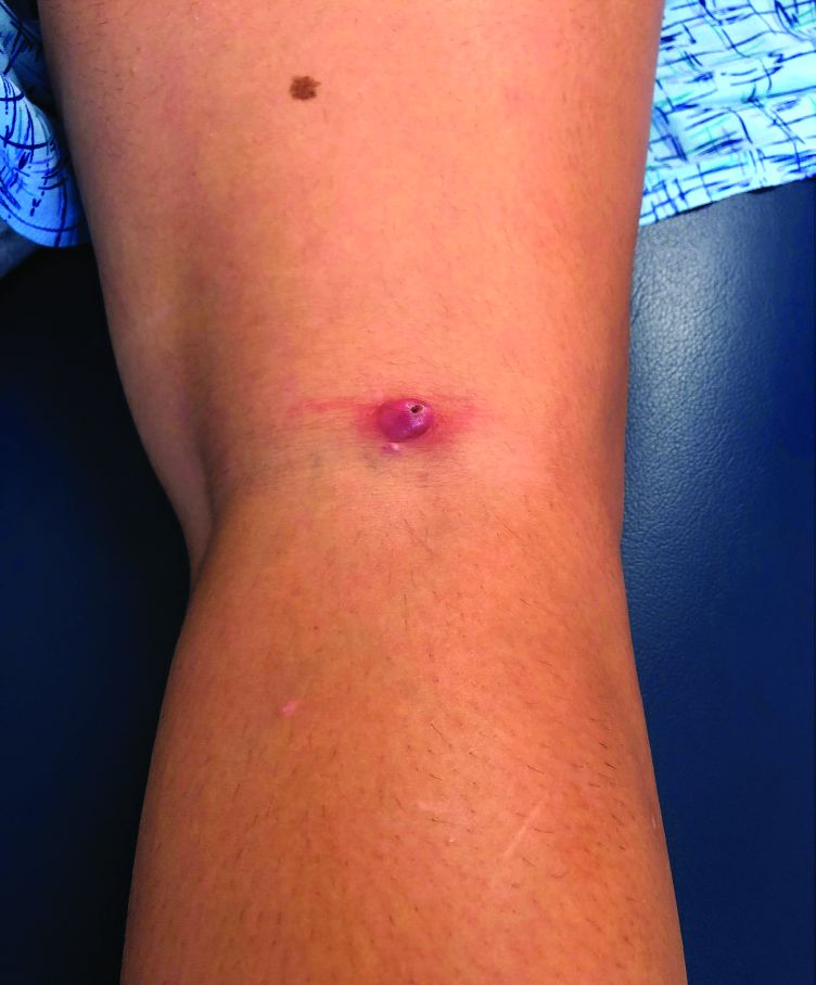
Nocturnal oxygen no help for isolated desaturation in COPD
Nocturnal oxygen therapy for patients with COPD and isolated nocturnal oxygen desaturation does not improve survival or delay disease progression, according to findings published Sept. 17 in The New England Journal of Medicine. The new report adds to evidence that the widely implemented and costly practice may be unnecessary.
Patients with COPD who do not qualify for long-term oxygen therapy (LTOT) are commonly prescribed nocturnal oxygen in the belief that it can delay disease progression, possibly by decreasing alveolar hypoventilation and ventilation-perfusion mismatch.
But investigations so far and the new study from the International Nocturnal Oxygen (INOX) Trial have not borne this out.
“There is no indication that nocturnal oxygen has a positive or negative effect on survival or progression to long-term oxygen therapy in patients with nocturnal hypoxemia in COPD. Consequently, there is no reason for physicians to screen for nocturnal hypoxemia in COPD,” study leader Yves Lacasse, MD, told Medscape Medical News.
Lacasse is from the Institut Universitaire de Cardiologie et de Pneumologie de Québec–Université Laval, Quebec, Canada.
The idea that the therapy helps is firmly entrenched.
In the early 1980s, two trials indicated that patients who had COPD and severe chronic daytime hypoxemia benefit from LTOT (15-18 hours a day or longer).
A decade later, two landmark trials (the Nocturnal Oxygen Therapy Trial and the British Medical Research Council Trial) added to evidence that LTOT may prolong life for patients with COPD and severe daytime hypoxemia.
“The good news from both trials was that oxygen saves lives. From this moment, oxygen therapy became a standard of care, and confirmatory trials would be considered unethical,” Lacasse explained.
“Oxygen therapy gained widespread acceptance by official organizations for treatment of most chronic cardiorespiratory conditions complicated by severe hypoxemia, even if proof of efficacy is lacking. New indications emerged, such as isolated nocturnal oxygen desaturation. Even in COPD, inappropriate prescriptions of home oxygen therapy are not unusual. Oxygen is everywhere,” Lacasse continued.
A meta-analysis from 2005 identified two trials that evaluated home oxygen therapy specifically for isolated nocturnal desaturation. Both found no survival benefit from nocturnal oxygen.
The study by Lacasse and colleagues assessed effects on mortality or worsening of disease (progression to LTOT) with 3-4 years of nocturnal oxygen supplementation.
Participants, whose oxygen saturation was less than 90% for at least 30% of the recording time on nocturnal oximetry, received oxygen or ambient air from a sham device as a placebo for at least 4 hours per session. The goal of treatment was nocturnal oxygen saturation exceeding 90% for at least 90% of the recorded time.
The trial protocol excluded patients with severe obesity, apnea, lung cancer, left heart failure, interstitial lung disease, or bronchiectasis.
The study was initially powered in 2010 to include 600 participants, with half to receive placebo. The study assumed mortality of 20% among control patients over 3 years; 20% of patients progressed to LTOT.
When recruiting lagged, the data safety monitoring board and steering committee extended follow-up to 4 years. In 2014, they requested an interim analysis, and recruitment ceased. Overall, 243 patients participated.
Lacasse cited several reasons for the difficulty with recruitment as well as retention: unwillingness to take the risk of receiving placebo instead of a readily available treatment, fading interest over time, and frailty that affects compliance.
Patients in the study came from 28 community or university-affiliated hospitals in Canada, Portugal, Spain, and France. At the 3-year mark, 39% of patients (48 of 123) who were assigned to nocturnal oxygen therapy and 42% (50 of 119) of those taking placebo had met criteria for LTOT or had died (difference, −3.0 percentage points; P = .64). The groups did not differ appreciably in rates of exacerbation and hospitalization.
The researchers could not analyze subgroups because the patients were very similar with regard to the severity of nocturnal oxygen desaturation, Lacasse said.
Economics enters into the picture – home oxygen therapy is second only to hospitalization as the most expensive healthcare expenditure associated with clinical care for COPD in developed countries. “The math is simple. There is enormous potential for saving money if the results of our clinical trial are applied appropriately,” said Lacasse.
William Bailey, MD, professor emeritus of pulmonary, allergy, and critical care medicine at the University of Alabama at Birmingham, agrees that the practice is overused.
“There is a built-in bias in the medical community. Most believe that anyone with lung disease benefits from oxygen. Even some of our investigators had a hard time believing the results. The study was well designed, carefully carried out, and I feel confident that the results are reliable,” he said.
Shawn P. E. Nishi, MD, director of bronchoscopy and advanced pulmonary procedures, division of pulmonary and critical care medicine, the University of Texas Medical Branch, Galveston, Texas, mentioned the study’s main limitation, which the authors readily acknowledge.
“Unfortunately, the trial had difficulty recruiting subjects, with less than half of expected enrollment achieved, and was underpowered to make any conclusions. Other studies have examined nocturnal oxygen use and have not shown a mortality benefit,” Nishi explained.
She added that the study did not evaluate use of LTOT for improving outcomes other than mortality, including quality of life, cardiovascular morbidity, depression, cognitive function, exercise capacity, and frequency of COPD exacerbations or hospitalization.
Other limitations of the study include suboptimal adherence to the therapy and interpretation of the clinical significance on the basis of a survey of Canadian pulmonologists.
This article first appeared on Medscape.com.
Nocturnal oxygen therapy for patients with COPD and isolated nocturnal oxygen desaturation does not improve survival or delay disease progression, according to findings published Sept. 17 in The New England Journal of Medicine. The new report adds to evidence that the widely implemented and costly practice may be unnecessary.
Patients with COPD who do not qualify for long-term oxygen therapy (LTOT) are commonly prescribed nocturnal oxygen in the belief that it can delay disease progression, possibly by decreasing alveolar hypoventilation and ventilation-perfusion mismatch.
But investigations so far and the new study from the International Nocturnal Oxygen (INOX) Trial have not borne this out.
“There is no indication that nocturnal oxygen has a positive or negative effect on survival or progression to long-term oxygen therapy in patients with nocturnal hypoxemia in COPD. Consequently, there is no reason for physicians to screen for nocturnal hypoxemia in COPD,” study leader Yves Lacasse, MD, told Medscape Medical News.
Lacasse is from the Institut Universitaire de Cardiologie et de Pneumologie de Québec–Université Laval, Quebec, Canada.
The idea that the therapy helps is firmly entrenched.
In the early 1980s, two trials indicated that patients who had COPD and severe chronic daytime hypoxemia benefit from LTOT (15-18 hours a day or longer).
A decade later, two landmark trials (the Nocturnal Oxygen Therapy Trial and the British Medical Research Council Trial) added to evidence that LTOT may prolong life for patients with COPD and severe daytime hypoxemia.
“The good news from both trials was that oxygen saves lives. From this moment, oxygen therapy became a standard of care, and confirmatory trials would be considered unethical,” Lacasse explained.
“Oxygen therapy gained widespread acceptance by official organizations for treatment of most chronic cardiorespiratory conditions complicated by severe hypoxemia, even if proof of efficacy is lacking. New indications emerged, such as isolated nocturnal oxygen desaturation. Even in COPD, inappropriate prescriptions of home oxygen therapy are not unusual. Oxygen is everywhere,” Lacasse continued.
A meta-analysis from 2005 identified two trials that evaluated home oxygen therapy specifically for isolated nocturnal desaturation. Both found no survival benefit from nocturnal oxygen.
The study by Lacasse and colleagues assessed effects on mortality or worsening of disease (progression to LTOT) with 3-4 years of nocturnal oxygen supplementation.
Participants, whose oxygen saturation was less than 90% for at least 30% of the recording time on nocturnal oximetry, received oxygen or ambient air from a sham device as a placebo for at least 4 hours per session. The goal of treatment was nocturnal oxygen saturation exceeding 90% for at least 90% of the recorded time.
The trial protocol excluded patients with severe obesity, apnea, lung cancer, left heart failure, interstitial lung disease, or bronchiectasis.
The study was initially powered in 2010 to include 600 participants, with half to receive placebo. The study assumed mortality of 20% among control patients over 3 years; 20% of patients progressed to LTOT.
When recruiting lagged, the data safety monitoring board and steering committee extended follow-up to 4 years. In 2014, they requested an interim analysis, and recruitment ceased. Overall, 243 patients participated.
Lacasse cited several reasons for the difficulty with recruitment as well as retention: unwillingness to take the risk of receiving placebo instead of a readily available treatment, fading interest over time, and frailty that affects compliance.
Patients in the study came from 28 community or university-affiliated hospitals in Canada, Portugal, Spain, and France. At the 3-year mark, 39% of patients (48 of 123) who were assigned to nocturnal oxygen therapy and 42% (50 of 119) of those taking placebo had met criteria for LTOT or had died (difference, −3.0 percentage points; P = .64). The groups did not differ appreciably in rates of exacerbation and hospitalization.
The researchers could not analyze subgroups because the patients were very similar with regard to the severity of nocturnal oxygen desaturation, Lacasse said.
Economics enters into the picture – home oxygen therapy is second only to hospitalization as the most expensive healthcare expenditure associated with clinical care for COPD in developed countries. “The math is simple. There is enormous potential for saving money if the results of our clinical trial are applied appropriately,” said Lacasse.
William Bailey, MD, professor emeritus of pulmonary, allergy, and critical care medicine at the University of Alabama at Birmingham, agrees that the practice is overused.
“There is a built-in bias in the medical community. Most believe that anyone with lung disease benefits from oxygen. Even some of our investigators had a hard time believing the results. The study was well designed, carefully carried out, and I feel confident that the results are reliable,” he said.
Shawn P. E. Nishi, MD, director of bronchoscopy and advanced pulmonary procedures, division of pulmonary and critical care medicine, the University of Texas Medical Branch, Galveston, Texas, mentioned the study’s main limitation, which the authors readily acknowledge.
“Unfortunately, the trial had difficulty recruiting subjects, with less than half of expected enrollment achieved, and was underpowered to make any conclusions. Other studies have examined nocturnal oxygen use and have not shown a mortality benefit,” Nishi explained.
She added that the study did not evaluate use of LTOT for improving outcomes other than mortality, including quality of life, cardiovascular morbidity, depression, cognitive function, exercise capacity, and frequency of COPD exacerbations or hospitalization.
Other limitations of the study include suboptimal adherence to the therapy and interpretation of the clinical significance on the basis of a survey of Canadian pulmonologists.
This article first appeared on Medscape.com.
Nocturnal oxygen therapy for patients with COPD and isolated nocturnal oxygen desaturation does not improve survival or delay disease progression, according to findings published Sept. 17 in The New England Journal of Medicine. The new report adds to evidence that the widely implemented and costly practice may be unnecessary.
Patients with COPD who do not qualify for long-term oxygen therapy (LTOT) are commonly prescribed nocturnal oxygen in the belief that it can delay disease progression, possibly by decreasing alveolar hypoventilation and ventilation-perfusion mismatch.
But investigations so far and the new study from the International Nocturnal Oxygen (INOX) Trial have not borne this out.
“There is no indication that nocturnal oxygen has a positive or negative effect on survival or progression to long-term oxygen therapy in patients with nocturnal hypoxemia in COPD. Consequently, there is no reason for physicians to screen for nocturnal hypoxemia in COPD,” study leader Yves Lacasse, MD, told Medscape Medical News.
Lacasse is from the Institut Universitaire de Cardiologie et de Pneumologie de Québec–Université Laval, Quebec, Canada.
The idea that the therapy helps is firmly entrenched.
In the early 1980s, two trials indicated that patients who had COPD and severe chronic daytime hypoxemia benefit from LTOT (15-18 hours a day or longer).
A decade later, two landmark trials (the Nocturnal Oxygen Therapy Trial and the British Medical Research Council Trial) added to evidence that LTOT may prolong life for patients with COPD and severe daytime hypoxemia.
“The good news from both trials was that oxygen saves lives. From this moment, oxygen therapy became a standard of care, and confirmatory trials would be considered unethical,” Lacasse explained.
“Oxygen therapy gained widespread acceptance by official organizations for treatment of most chronic cardiorespiratory conditions complicated by severe hypoxemia, even if proof of efficacy is lacking. New indications emerged, such as isolated nocturnal oxygen desaturation. Even in COPD, inappropriate prescriptions of home oxygen therapy are not unusual. Oxygen is everywhere,” Lacasse continued.
A meta-analysis from 2005 identified two trials that evaluated home oxygen therapy specifically for isolated nocturnal desaturation. Both found no survival benefit from nocturnal oxygen.
The study by Lacasse and colleagues assessed effects on mortality or worsening of disease (progression to LTOT) with 3-4 years of nocturnal oxygen supplementation.
Participants, whose oxygen saturation was less than 90% for at least 30% of the recording time on nocturnal oximetry, received oxygen or ambient air from a sham device as a placebo for at least 4 hours per session. The goal of treatment was nocturnal oxygen saturation exceeding 90% for at least 90% of the recorded time.
The trial protocol excluded patients with severe obesity, apnea, lung cancer, left heart failure, interstitial lung disease, or bronchiectasis.
The study was initially powered in 2010 to include 600 participants, with half to receive placebo. The study assumed mortality of 20% among control patients over 3 years; 20% of patients progressed to LTOT.
When recruiting lagged, the data safety monitoring board and steering committee extended follow-up to 4 years. In 2014, they requested an interim analysis, and recruitment ceased. Overall, 243 patients participated.
Lacasse cited several reasons for the difficulty with recruitment as well as retention: unwillingness to take the risk of receiving placebo instead of a readily available treatment, fading interest over time, and frailty that affects compliance.
Patients in the study came from 28 community or university-affiliated hospitals in Canada, Portugal, Spain, and France. At the 3-year mark, 39% of patients (48 of 123) who were assigned to nocturnal oxygen therapy and 42% (50 of 119) of those taking placebo had met criteria for LTOT or had died (difference, −3.0 percentage points; P = .64). The groups did not differ appreciably in rates of exacerbation and hospitalization.
The researchers could not analyze subgroups because the patients were very similar with regard to the severity of nocturnal oxygen desaturation, Lacasse said.
Economics enters into the picture – home oxygen therapy is second only to hospitalization as the most expensive healthcare expenditure associated with clinical care for COPD in developed countries. “The math is simple. There is enormous potential for saving money if the results of our clinical trial are applied appropriately,” said Lacasse.
William Bailey, MD, professor emeritus of pulmonary, allergy, and critical care medicine at the University of Alabama at Birmingham, agrees that the practice is overused.
“There is a built-in bias in the medical community. Most believe that anyone with lung disease benefits from oxygen. Even some of our investigators had a hard time believing the results. The study was well designed, carefully carried out, and I feel confident that the results are reliable,” he said.
Shawn P. E. Nishi, MD, director of bronchoscopy and advanced pulmonary procedures, division of pulmonary and critical care medicine, the University of Texas Medical Branch, Galveston, Texas, mentioned the study’s main limitation, which the authors readily acknowledge.
“Unfortunately, the trial had difficulty recruiting subjects, with less than half of expected enrollment achieved, and was underpowered to make any conclusions. Other studies have examined nocturnal oxygen use and have not shown a mortality benefit,” Nishi explained.
She added that the study did not evaluate use of LTOT for improving outcomes other than mortality, including quality of life, cardiovascular morbidity, depression, cognitive function, exercise capacity, and frequency of COPD exacerbations or hospitalization.
Other limitations of the study include suboptimal adherence to the therapy and interpretation of the clinical significance on the basis of a survey of Canadian pulmonologists.
This article first appeared on Medscape.com.
Improving interprovider communication
“Interprovider communication” is a big buzzphrase in medicine these days. Granted, it’s an important aspect of patient care. But, like many words and phrases, a lot of substance is lost in the spin of things.

I get emails, faxes, and letters all the time promising a new system that improves communication between physicians and patients. The hospital I share call at always seems to have something in its physician newsletters about a new software or app to improve communication.
The problem here isn’t that there aren’t already good ways for physicians to communicate – there are. I generally rely on the old standbys of a fax machine, with the post office as a backup for most things, and the phone for more urgent matters.
The real issue is people who don’t use the systems available, and no amount of technology will change that.
Some doctors feel they’re too busy to get a letter out, or forward tests results to another physician, or even have their office staff do it. Others just barely glance at anything that comes through, then pass it on to their staff to file it in a chart. At the hospital some doctors don’t seem to bother to read their consultants’ notes.
Granted, this isn’t entirely the doctors’ faults. As I’ve written before, many of the EMR chart systems are so full of templates and cut and paste that notes are rendered virtually meaningless. To find the impression – if it’s even in there – may need some digging. This takes time, which is always in short supply in a medical practice. The days when you could just flip through to the paragraph labeled “impression” are gone, and probably aren’t coming back. Which is good for no one on either side of the desk or bedrail.
This is sad, because that’s where the vast majority of physician communication happened. Letting people know what you’re thinking and doing, and at the same time asking specific questions you’re hoping they’ll address.
Not all doctors are poor at communication – the vast majority are not. But for those of us trying to care for a patient with one who is, there isn’t a software breakthrough now – or ever – that will make it any easier, no matter how much time, money, and glossy marketing is thrown at it.
Dr. Block has a solo neurology practice in Scottsdale, Ariz.
“Interprovider communication” is a big buzzphrase in medicine these days. Granted, it’s an important aspect of patient care. But, like many words and phrases, a lot of substance is lost in the spin of things.
I get emails, faxes, and letters all the time promising a new system that improves communication between physicians and patients. The hospital I share call at always seems to have something in its physician newsletters about a new software or app to improve communication.
The problem here isn’t that there aren’t already good ways for physicians to communicate – there are. I generally rely on the old standbys of a fax machine, with the post office as a backup for most things, and the phone for more urgent matters.
The real issue is people who don’t use the systems available, and no amount of technology will change that.
Some doctors feel they’re too busy to get a letter out, or forward tests results to another physician, or even have their office staff do it. Others just barely glance at anything that comes through, then pass it on to their staff to file it in a chart. At the hospital some doctors don’t seem to bother to read their consultants’ notes.
Granted, this isn’t entirely the doctors’ faults. As I’ve written before, many of the EMR chart systems are so full of templates and cut and paste that notes are rendered virtually meaningless. To find the impression – if it’s even in there – may need some digging. This takes time, which is always in short supply in a medical practice. The days when you could just flip through to the paragraph labeled “impression” are gone, and probably aren’t coming back. Which is good for no one on either side of the desk or bedrail.
This is sad, because that’s where the vast majority of physician communication happened. Letting people know what you’re thinking and doing, and at the same time asking specific questions you’re hoping they’ll address.
Not all doctors are poor at communication – the vast majority are not. But for those of us trying to care for a patient with one who is, there isn’t a software breakthrough now – or ever – that will make it any easier, no matter how much time, money, and glossy marketing is thrown at it.
Dr. Block has a solo neurology practice in Scottsdale, Ariz.
“Interprovider communication” is a big buzzphrase in medicine these days. Granted, it’s an important aspect of patient care. But, like many words and phrases, a lot of substance is lost in the spin of things.
I get emails, faxes, and letters all the time promising a new system that improves communication between physicians and patients. The hospital I share call at always seems to have something in its physician newsletters about a new software or app to improve communication.
The problem here isn’t that there aren’t already good ways for physicians to communicate – there are. I generally rely on the old standbys of a fax machine, with the post office as a backup for most things, and the phone for more urgent matters.
The real issue is people who don’t use the systems available, and no amount of technology will change that.
Some doctors feel they’re too busy to get a letter out, or forward tests results to another physician, or even have their office staff do it. Others just barely glance at anything that comes through, then pass it on to their staff to file it in a chart. At the hospital some doctors don’t seem to bother to read their consultants’ notes.
Granted, this isn’t entirely the doctors’ faults. As I’ve written before, many of the EMR chart systems are so full of templates and cut and paste that notes are rendered virtually meaningless. To find the impression – if it’s even in there – may need some digging. This takes time, which is always in short supply in a medical practice. The days when you could just flip through to the paragraph labeled “impression” are gone, and probably aren’t coming back. Which is good for no one on either side of the desk or bedrail.
This is sad, because that’s where the vast majority of physician communication happened. Letting people know what you’re thinking and doing, and at the same time asking specific questions you’re hoping they’ll address.
Not all doctors are poor at communication – the vast majority are not. But for those of us trying to care for a patient with one who is, there isn’t a software breakthrough now – or ever – that will make it any easier, no matter how much time, money, and glossy marketing is thrown at it.
Dr. Block has a solo neurology practice in Scottsdale, Ariz.
Pharmacologic Management of COPD
A Discussion of the new American Thoracic Society Clinical Practice Guideline
Chronic obstructive pulmonary disease (COPD) is caused by airway and alveolar abnormalities and is the third most common cause of death worldwide. COPD results in airflow obstruction that is not fully reversible. The diagnosis of COPD should be considered in patients over 40 years who have chronic cough and/or dyspnea, particularly if they have a history of tobacco use. The diagnosis is confirmed by a diminished forced expiratory volume in 1 second (FEV1) that is not fully reversible with the use of a bronchodilator and an FEV1/forced vital capacity ratio of less than or equal to 0.7.1
Recommendation 1

Patients with COPD who report dyspnea or exercise intolerance should be treated with both a long-acting muscarinic antagonist (LAMA) and a long-acting beta agonist (LABA) (dual LAMA/LABA therapy) instead of monotherapy, the guideline says.
This recommendation represents a critical change in care and is based on strong evidence. For years practitioners have been using single bronchodilator therapy, often a LAMA as the entrance to treatment for patients with symptomatic COPD. The recommendation to begin treatment with dual bronchodilator therapy is an important one. This is the only recommendation that received a “strong” grade.
The evidence comes from the compilation of 24 randomized controlled trials that altogether included 45,441 patients. Dual therapy versus monotherapy was evaluated by examining differences in dyspnea, health-related quality of life, exacerbations (which were defined as requiring antibiotics, oral steroids, or hospitalizations), and hospitalizations independently. Marked improvements were observed for exacerbations and hospitalizations in the dual LAMA/LABA group, compared with treatment with use of a single bronchodilator. In 22,733 patients across 15 RCTs, there were 88 fewer exacerbations per 1,000 patients with a rate ratio (RR) of 0.80 (P < .002), the guideline states.
The decrease in exacerbations is a critical factor in treating patients with COPD because each exacerbation can lead to a sustained decrease in airflow and increases the risk of future exacerbations.
Recommendation 2

In COPD patients who report dyspnea or exercise intolerance, with an exacerbation in the last year, the guideline recommends triple therapy with an inhaled corticosteroid (ICS) instead of just dual LAMA/LABA therapy.
In the past many clinicians have relegated triple therapy to a “last ditch resort.” This recommendation makes it clear that triple therapy is appropriate for a broad range of patients with moderate to severe COPD.
Recommendation 3
In patients with COPD who are on triple therapy, the inhaled corticosteroid component can be withdrawn if patients have not had an exacerbation within the last year, according to the guideline.
It should be noted that the committee said that the ICS can be withdrawn, not that it necessarily needs to be withdrawn. The data showed that it would be safe to withdraw the ICS, but the data is limited in time to 1 year’s follow-up.
Recommendation 4
ATS was not able to make a recommendation for or against ICS as an additive therapy to LAMA/LABA in those without an exacerbation and elevated blood eosinophilia (defined as ≥2% blood eosinophils or >149 cell/mcL). In those with at least one exacerbation and increased blood eosinophilia, the society does recommend addition of ICS to dual LAMA/LABA therapy.
An area of ongoing discussion is at what point in disease severity, before exacerbations occur, might ICS be useful in preventing a first exacerbation. This awaits further studies and evidence.
Recommendation 5
In COPD patients with frequent and severe exacerbations who are otherwise medically optimized, the ATS advises against the use of maintenance oral corticosteroid therapy.
It has been known and accepted for years that oral steroids should be avoided if at all possible because they have little benefit and can cause significant harm. The guideline reinforces this.
The Bottom Line
Dual LAMA/LABA therapy in symptomatic patients is the standard of care. If a patient has had an exacerbation within the last year, add an ICS to the LAMA/LABA, most conveniently given in the form of triple therapy in one inhaler. Finally, even in refractory COPD, maintenance oral corticosteroids bring more harm than benefit.
Dr. Skolnik is professor of family and community medicine at the Thomas Jefferson University, Philadelphia, and associate director of the Family Medicine Residency Program at Abington (Pa.) Jefferson Health. Dr. Matthews is a second-year resident in the family medicine residency program at Abington Jefferson Health.
References
1. Wells C, Joo MJ. COPD and asthma: Diagnostic accuracy requires spirometry. J Fam Pract. 2019;68(2):76-81.
2. Nici L, Mammen MJ, Charbek E, et al. Pharmacologic management of chronic obstructive pulmonary disease. An official American Thoracic Society clinical practice guideline. Am J Respir Crit Care Med. 2020;201(9):e56-69.
A Discussion of the new American Thoracic Society Clinical Practice Guideline
A Discussion of the new American Thoracic Society Clinical Practice Guideline
Chronic obstructive pulmonary disease (COPD) is caused by airway and alveolar abnormalities and is the third most common cause of death worldwide. COPD results in airflow obstruction that is not fully reversible. The diagnosis of COPD should be considered in patients over 40 years who have chronic cough and/or dyspnea, particularly if they have a history of tobacco use. The diagnosis is confirmed by a diminished forced expiratory volume in 1 second (FEV1) that is not fully reversible with the use of a bronchodilator and an FEV1/forced vital capacity ratio of less than or equal to 0.7.1
Recommendation 1
Patients with COPD who report dyspnea or exercise intolerance should be treated with both a long-acting muscarinic antagonist (LAMA) and a long-acting beta agonist (LABA) (dual LAMA/LABA therapy) instead of monotherapy, the guideline says.
This recommendation represents a critical change in care and is based on strong evidence. For years practitioners have been using single bronchodilator therapy, often a LAMA as the entrance to treatment for patients with symptomatic COPD. The recommendation to begin treatment with dual bronchodilator therapy is an important one. This is the only recommendation that received a “strong” grade.
The evidence comes from the compilation of 24 randomized controlled trials that altogether included 45,441 patients. Dual therapy versus monotherapy was evaluated by examining differences in dyspnea, health-related quality of life, exacerbations (which were defined as requiring antibiotics, oral steroids, or hospitalizations), and hospitalizations independently. Marked improvements were observed for exacerbations and hospitalizations in the dual LAMA/LABA group, compared with treatment with use of a single bronchodilator. In 22,733 patients across 15 RCTs, there were 88 fewer exacerbations per 1,000 patients with a rate ratio (RR) of 0.80 (P < .002), the guideline states.
The decrease in exacerbations is a critical factor in treating patients with COPD because each exacerbation can lead to a sustained decrease in airflow and increases the risk of future exacerbations.
Recommendation 2
In COPD patients who report dyspnea or exercise intolerance, with an exacerbation in the last year, the guideline recommends triple therapy with an inhaled corticosteroid (ICS) instead of just dual LAMA/LABA therapy.
In the past many clinicians have relegated triple therapy to a “last ditch resort.” This recommendation makes it clear that triple therapy is appropriate for a broad range of patients with moderate to severe COPD.
Recommendation 3
In patients with COPD who are on triple therapy, the inhaled corticosteroid component can be withdrawn if patients have not had an exacerbation within the last year, according to the guideline.
It should be noted that the committee said that the ICS can be withdrawn, not that it necessarily needs to be withdrawn. The data showed that it would be safe to withdraw the ICS, but the data is limited in time to 1 year’s follow-up.
Recommendation 4
ATS was not able to make a recommendation for or against ICS as an additive therapy to LAMA/LABA in those without an exacerbation and elevated blood eosinophilia (defined as ≥2% blood eosinophils or >149 cell/mcL). In those with at least one exacerbation and increased blood eosinophilia, the society does recommend addition of ICS to dual LAMA/LABA therapy.
An area of ongoing discussion is at what point in disease severity, before exacerbations occur, might ICS be useful in preventing a first exacerbation. This awaits further studies and evidence.
Recommendation 5
In COPD patients with frequent and severe exacerbations who are otherwise medically optimized, the ATS advises against the use of maintenance oral corticosteroid therapy.
It has been known and accepted for years that oral steroids should be avoided if at all possible because they have little benefit and can cause significant harm. The guideline reinforces this.
The Bottom Line
Dual LAMA/LABA therapy in symptomatic patients is the standard of care. If a patient has had an exacerbation within the last year, add an ICS to the LAMA/LABA, most conveniently given in the form of triple therapy in one inhaler. Finally, even in refractory COPD, maintenance oral corticosteroids bring more harm than benefit.
Dr. Skolnik is professor of family and community medicine at the Thomas Jefferson University, Philadelphia, and associate director of the Family Medicine Residency Program at Abington (Pa.) Jefferson Health. Dr. Matthews is a second-year resident in the family medicine residency program at Abington Jefferson Health.
References
1. Wells C, Joo MJ. COPD and asthma: Diagnostic accuracy requires spirometry. J Fam Pract. 2019;68(2):76-81.
2. Nici L, Mammen MJ, Charbek E, et al. Pharmacologic management of chronic obstructive pulmonary disease. An official American Thoracic Society clinical practice guideline. Am J Respir Crit Care Med. 2020;201(9):e56-69.
Chronic obstructive pulmonary disease (COPD) is caused by airway and alveolar abnormalities and is the third most common cause of death worldwide. COPD results in airflow obstruction that is not fully reversible. The diagnosis of COPD should be considered in patients over 40 years who have chronic cough and/or dyspnea, particularly if they have a history of tobacco use. The diagnosis is confirmed by a diminished forced expiratory volume in 1 second (FEV1) that is not fully reversible with the use of a bronchodilator and an FEV1/forced vital capacity ratio of less than or equal to 0.7.1
Recommendation 1
Patients with COPD who report dyspnea or exercise intolerance should be treated with both a long-acting muscarinic antagonist (LAMA) and a long-acting beta agonist (LABA) (dual LAMA/LABA therapy) instead of monotherapy, the guideline says.
This recommendation represents a critical change in care and is based on strong evidence. For years practitioners have been using single bronchodilator therapy, often a LAMA as the entrance to treatment for patients with symptomatic COPD. The recommendation to begin treatment with dual bronchodilator therapy is an important one. This is the only recommendation that received a “strong” grade.
The evidence comes from the compilation of 24 randomized controlled trials that altogether included 45,441 patients. Dual therapy versus monotherapy was evaluated by examining differences in dyspnea, health-related quality of life, exacerbations (which were defined as requiring antibiotics, oral steroids, or hospitalizations), and hospitalizations independently. Marked improvements were observed for exacerbations and hospitalizations in the dual LAMA/LABA group, compared with treatment with use of a single bronchodilator. In 22,733 patients across 15 RCTs, there were 88 fewer exacerbations per 1,000 patients with a rate ratio (RR) of 0.80 (P < .002), the guideline states.
The decrease in exacerbations is a critical factor in treating patients with COPD because each exacerbation can lead to a sustained decrease in airflow and increases the risk of future exacerbations.
Recommendation 2
In COPD patients who report dyspnea or exercise intolerance, with an exacerbation in the last year, the guideline recommends triple therapy with an inhaled corticosteroid (ICS) instead of just dual LAMA/LABA therapy.
In the past many clinicians have relegated triple therapy to a “last ditch resort.” This recommendation makes it clear that triple therapy is appropriate for a broad range of patients with moderate to severe COPD.
Recommendation 3
In patients with COPD who are on triple therapy, the inhaled corticosteroid component can be withdrawn if patients have not had an exacerbation within the last year, according to the guideline.
It should be noted that the committee said that the ICS can be withdrawn, not that it necessarily needs to be withdrawn. The data showed that it would be safe to withdraw the ICS, but the data is limited in time to 1 year’s follow-up.
Recommendation 4
ATS was not able to make a recommendation for or against ICS as an additive therapy to LAMA/LABA in those without an exacerbation and elevated blood eosinophilia (defined as ≥2% blood eosinophils or >149 cell/mcL). In those with at least one exacerbation and increased blood eosinophilia, the society does recommend addition of ICS to dual LAMA/LABA therapy.
An area of ongoing discussion is at what point in disease severity, before exacerbations occur, might ICS be useful in preventing a first exacerbation. This awaits further studies and evidence.
Recommendation 5
In COPD patients with frequent and severe exacerbations who are otherwise medically optimized, the ATS advises against the use of maintenance oral corticosteroid therapy.
It has been known and accepted for years that oral steroids should be avoided if at all possible because they have little benefit and can cause significant harm. The guideline reinforces this.
The Bottom Line
Dual LAMA/LABA therapy in symptomatic patients is the standard of care. If a patient has had an exacerbation within the last year, add an ICS to the LAMA/LABA, most conveniently given in the form of triple therapy in one inhaler. Finally, even in refractory COPD, maintenance oral corticosteroids bring more harm than benefit.
Dr. Skolnik is professor of family and community medicine at the Thomas Jefferson University, Philadelphia, and associate director of the Family Medicine Residency Program at Abington (Pa.) Jefferson Health. Dr. Matthews is a second-year resident in the family medicine residency program at Abington Jefferson Health.
References
1. Wells C, Joo MJ. COPD and asthma: Diagnostic accuracy requires spirometry. J Fam Pract. 2019;68(2):76-81.
2. Nici L, Mammen MJ, Charbek E, et al. Pharmacologic management of chronic obstructive pulmonary disease. An official American Thoracic Society clinical practice guideline. Am J Respir Crit Care Med. 2020;201(9):e56-69.
Many Americans still concerned about access to health care
according to the results of a survey conducted Aug. 7-26.
Nationally, 23.8% of respondents said that they were very concerned about being able to receive care during the pandemic, and another 27.4% said that they were somewhat concerned. Just under a quarter, 24.3%, said they were not very concerned, while 20.4% were not at all concerned, the COVID-19 Consortium for Understanding the Public’s Policy Preferences Across States reported after surveying 21,196 adults.
At the state level, Mississippi had the most adults (35.5%) who were very concerned about their access to care, followed by Texas (32.7%) and Nevada (32.4%). The residents of Montana were least likely (10.5%) to be very concerned, with Vermont next at 11.6% and Wyoming slightly higher at 13.8%. Montana also had the highest proportion of adults, 30.2%, who were not at all concerned, the consortium’s data show.
When asked about getting the coronavirus themselves, 67.8% of U.S. adults came down on the concerned side (33.3% somewhat and 34.5% very concerned) versus 30.8% who were not concerned (18.6% were not very concerned; 12.2% were not concerned at all.). Respondents’ concern was higher for their family members’ risk of getting coronavirus: 30.2% were somewhat concerned and 47.6% were very concerned, the consortium said.
Among many other topics, respondents were asked how closely they had followed recommended health guidelines in the last week, with the two extremes shown here:
- Avoiding contact with other people: 49.3% very closely, 4.8% not at all closely.
- Frequently washing hands: 74.7% very, 1.6% not at all.
- Disinfecting often-touched surfaces: 54.4% very, 4.3% not at all.
- Wearing a face mask in public: 75.7% very, 3.5% not at all.
The consortium is a joint project of the Network Science Institute of Northeastern University; the Shorenstein Center on Media, Politics, and Public Policy of Harvard University; Harvard Medical School; the School of Communication and Information at Rutgers University; and the department of political science at Northwestern University. The project is supported by grants from the National Science Foundation.
according to the results of a survey conducted Aug. 7-26.
Nationally, 23.8% of respondents said that they were very concerned about being able to receive care during the pandemic, and another 27.4% said that they were somewhat concerned. Just under a quarter, 24.3%, said they were not very concerned, while 20.4% were not at all concerned, the COVID-19 Consortium for Understanding the Public’s Policy Preferences Across States reported after surveying 21,196 adults.
At the state level, Mississippi had the most adults (35.5%) who were very concerned about their access to care, followed by Texas (32.7%) and Nevada (32.4%). The residents of Montana were least likely (10.5%) to be very concerned, with Vermont next at 11.6% and Wyoming slightly higher at 13.8%. Montana also had the highest proportion of adults, 30.2%, who were not at all concerned, the consortium’s data show.
When asked about getting the coronavirus themselves, 67.8% of U.S. adults came down on the concerned side (33.3% somewhat and 34.5% very concerned) versus 30.8% who were not concerned (18.6% were not very concerned; 12.2% were not concerned at all.). Respondents’ concern was higher for their family members’ risk of getting coronavirus: 30.2% were somewhat concerned and 47.6% were very concerned, the consortium said.
Among many other topics, respondents were asked how closely they had followed recommended health guidelines in the last week, with the two extremes shown here:
- Avoiding contact with other people: 49.3% very closely, 4.8% not at all closely.
- Frequently washing hands: 74.7% very, 1.6% not at all.
- Disinfecting often-touched surfaces: 54.4% very, 4.3% not at all.
- Wearing a face mask in public: 75.7% very, 3.5% not at all.
The consortium is a joint project of the Network Science Institute of Northeastern University; the Shorenstein Center on Media, Politics, and Public Policy of Harvard University; Harvard Medical School; the School of Communication and Information at Rutgers University; and the department of political science at Northwestern University. The project is supported by grants from the National Science Foundation.
according to the results of a survey conducted Aug. 7-26.
Nationally, 23.8% of respondents said that they were very concerned about being able to receive care during the pandemic, and another 27.4% said that they were somewhat concerned. Just under a quarter, 24.3%, said they were not very concerned, while 20.4% were not at all concerned, the COVID-19 Consortium for Understanding the Public’s Policy Preferences Across States reported after surveying 21,196 adults.
At the state level, Mississippi had the most adults (35.5%) who were very concerned about their access to care, followed by Texas (32.7%) and Nevada (32.4%). The residents of Montana were least likely (10.5%) to be very concerned, with Vermont next at 11.6% and Wyoming slightly higher at 13.8%. Montana also had the highest proportion of adults, 30.2%, who were not at all concerned, the consortium’s data show.
When asked about getting the coronavirus themselves, 67.8% of U.S. adults came down on the concerned side (33.3% somewhat and 34.5% very concerned) versus 30.8% who were not concerned (18.6% were not very concerned; 12.2% were not concerned at all.). Respondents’ concern was higher for their family members’ risk of getting coronavirus: 30.2% were somewhat concerned and 47.6% were very concerned, the consortium said.
Among many other topics, respondents were asked how closely they had followed recommended health guidelines in the last week, with the two extremes shown here:
- Avoiding contact with other people: 49.3% very closely, 4.8% not at all closely.
- Frequently washing hands: 74.7% very, 1.6% not at all.
- Disinfecting often-touched surfaces: 54.4% very, 4.3% not at all.
- Wearing a face mask in public: 75.7% very, 3.5% not at all.
The consortium is a joint project of the Network Science Institute of Northeastern University; the Shorenstein Center on Media, Politics, and Public Policy of Harvard University; Harvard Medical School; the School of Communication and Information at Rutgers University; and the department of political science at Northwestern University. The project is supported by grants from the National Science Foundation.