User login
Five Keys to Helping Long-COVID Patients Recover
About 7% of US adults report having or having had symptoms of long COVID such as fatigue, heart palpitations and/or dizziness. These are 3 of the 12 symptoms identified as part of the National Institute of Health’s RECOVER initiative that can be reliably used to classify someone as having long COVID.
The good news is that many people experience improvements in their symptoms over time by adopting these strategies, said Andrew Schamess, MD, an internal medicine physician at the Ohio State University Wexner Medical Center and director of its Post-COVID Recovery Program.
1. Pace yourself.
Fatigue and postexertional malaise are 2 of the 12 symptoms used to classify someone as having long COVID.
“There’s mental, or cognitive, fatigue, where people become exhausted after any span of time trying to do complicated cognitive tasks,” said Dr. Schamess. “There’s also general fatigue, or sleepiness, where after a few hours you feel like you could go right back to sleep.”
The third category, he added, is postexertional malaise, where patients are exhausted by exercise, either immediately or up to 24-48 hours later.
That’s where a technique known as “pacing” can help. Pacing is an energy-conservation technique often used among people with other disabling conditions, such as chronic fatigue syndrome, said Ravindra Ganesh, MD, an internal medicine physician at the Mayo Clinic in Minnesota who specializes in long COVID.
“I tell patients that they have to figure out what their energy envelope is, which is the fixed amount of energy that they can use every day without crashing,” he said.
You may be able to handle a daily 30-minute walk, for example, but if you pair it with something cognitively difficult, such as doing your taxes, your fatigue symptoms may flare up.
“It’s hard advice for my patients to follow, as most are real go-getters,” he said. “But I point out to them that if they aim to minimize crashes, it will help them make slow progress.”
Over time, he said, their energy levels should gradually rise so that they can engage in more and more activity.
2. Follow a plant-based, anti-inflammatory diet.
There’s no research to suggest that following a certain eating pattern will help to reverse long COVID, said Dr. Ganesh. But in general, he said his patients anecdotally report that they feel better when they limit refined sugar and follow a plant-based diet that can help to lower inflammation in the body.
“It makes sense, because it prevents dramatic blood glucose changes that can cause their body to crash,” he said. He generally recommends an anti-inflammatory diet like the Mediterranean diet, which is rich in fruits, vegetables, whole grains, and mono-unsaturated fat.
Many people with long COVID take an array of supplements, Dr. Ganesh said, although there’s little research to suggest that they may help. He does encourage patients to take about 2 g of an omega-3 supplement, such as fish oil, as it may help to reduce inflammation associated with long COVID.
He also recommends fisetin, a dietary flavonoid found in fruits such as strawberries and kiwis. Preliminary research suggests that it may help to combat some of the neurologic damage associated with long COVID.
“It appears to maintain mitochondrial function and has anti-inflammatory activities,” said Dr. Ganesh.
3. Modify exercise.
Most of the time, exercise boosts health and reduces risk for certain diseases. But this strategy may not work for people who have certain symptoms from long COVID, such as postexertional malaise or postural orthostatic tachycardia syndrome (POTS), a condition that causes symptoms such as a fast heart rate, dizziness, and fatigue when transitioning from lying down to standing up.
“With long-COVID patients, it often has to be symptom-titrated exercise,” said Dr. Schamess. This means physical activity needs to be constantly monitored and adjusted on the basis of a patient’s symptoms. “We need to figure out what they can do that doesn’t provoke their symptoms,” he explained.
Dr. Schamess often recommends that patients with long COVID, at least initially, focus on exercises in which they are sitting (such as cycling) or prone.
“The key thing is most people with long COVID can do a lot more exercise in a sitting or lying position than a standing position,” he said. “It’s baffling to them that they can’t walk two blocks but can bike 10 miles.”
For symptoms like fatigue or postexertional malaise, Dr. Schamess often refers patients to physical therapy to develop an individualized exercise program. A 2022 study published in Scandinavian Journal of Medicine & Science in Sports found that when long-COVID patients completed an 8-week program of three exercise sessions per week, they experienced significant improvements in quality of life, fatigue, muscle strength, and overall fitness compared with a control group.
“It’s important to make sure that workouts are supervised, so that they can be modified as necessary” said Dr. Schamess.
4. Take steps to improve sleep quality.
A 2023 study published in the Journal of General Internal Medicine found that about 40% of people with long COVID report sleep issues such as insomnia or not feeling refreshed in the morning.
“Sleep may become challenging, which can be frustrating for a patient with long COVID who desperately needs rest,” said Lawrence Purpura, MD, an infectious disease specialist and director of the long COVID clinic at Columbia University Medical Center in New York City.
Some of the simplest ways to improve sleep are common sense; however, these issues never affected the person pre-COVID, so they have to become new habits.
“A lot of my patients with long COVID find that they are more sensitive to caffeine, so they really can’t have it anymore later in the day,” he said. “The same goes for bright screens” such as those on cell phones, tablets, and e-book readers, he said. “They may find that it’s harder for them to fall and stay asleep if they’re on their iPhone right before bed. These are all things that may not have been issues before they were diagnosed with long COVID.”
Dr. Purpura also said that he encourages his patients to practice mindfulness or relaxation exercises before bed, such as deep breathing. One technique he recommends is called box breathing, where the patient inhales for 4 seconds, holds his or her breath for 4 seconds, exhales for 4 seconds, then holds his or her breath again for 4 seconds. Some research suggests that this paced breathing technique, when done for 20 minutes before bed, helps to improve symptoms of insomnia.
While sleep medications such as zolpidem (Ambien) are often used as short-term relief for insomnia, Dr. Schamess said he has not found them particularly helpful for sleep issues that stem from long COVID.
“They help patients fall asleep but not necessarily stay asleep, which can be an issue for people with long COVID,” he said.
5. Consider medications.
No standard drugs or treatments have yet been approved to treat long COVID (although some, such as Paxlovid, are in clinical trials). But some medications may help to relieve symptoms, said Dr. Ganesh. These include:
- Blood pressure drugs such as beta-blockers now used to treat POTS symptoms
- Nerve-pain medications such as gabapentin or pregabalin. “These can also help with sleep, since patients don’t have pain to distract them,” said Dr. Ganesh.
- Low-dose naltrexone to help with fatigue
“There’s not a one-size-fits-all approach to treat long-COVID symptoms,” said Dr. Ganesh. “You really need to work with the patient and possibly even cycle through several different medications before you find one that helps.”
A version of this article appeared on Medscape.com.
About 7% of US adults report having or having had symptoms of long COVID such as fatigue, heart palpitations and/or dizziness. These are 3 of the 12 symptoms identified as part of the National Institute of Health’s RECOVER initiative that can be reliably used to classify someone as having long COVID.
The good news is that many people experience improvements in their symptoms over time by adopting these strategies, said Andrew Schamess, MD, an internal medicine physician at the Ohio State University Wexner Medical Center and director of its Post-COVID Recovery Program.
1. Pace yourself.
Fatigue and postexertional malaise are 2 of the 12 symptoms used to classify someone as having long COVID.
“There’s mental, or cognitive, fatigue, where people become exhausted after any span of time trying to do complicated cognitive tasks,” said Dr. Schamess. “There’s also general fatigue, or sleepiness, where after a few hours you feel like you could go right back to sleep.”
The third category, he added, is postexertional malaise, where patients are exhausted by exercise, either immediately or up to 24-48 hours later.
That’s where a technique known as “pacing” can help. Pacing is an energy-conservation technique often used among people with other disabling conditions, such as chronic fatigue syndrome, said Ravindra Ganesh, MD, an internal medicine physician at the Mayo Clinic in Minnesota who specializes in long COVID.
“I tell patients that they have to figure out what their energy envelope is, which is the fixed amount of energy that they can use every day without crashing,” he said.
You may be able to handle a daily 30-minute walk, for example, but if you pair it with something cognitively difficult, such as doing your taxes, your fatigue symptoms may flare up.
“It’s hard advice for my patients to follow, as most are real go-getters,” he said. “But I point out to them that if they aim to minimize crashes, it will help them make slow progress.”
Over time, he said, their energy levels should gradually rise so that they can engage in more and more activity.
2. Follow a plant-based, anti-inflammatory diet.
There’s no research to suggest that following a certain eating pattern will help to reverse long COVID, said Dr. Ganesh. But in general, he said his patients anecdotally report that they feel better when they limit refined sugar and follow a plant-based diet that can help to lower inflammation in the body.
“It makes sense, because it prevents dramatic blood glucose changes that can cause their body to crash,” he said. He generally recommends an anti-inflammatory diet like the Mediterranean diet, which is rich in fruits, vegetables, whole grains, and mono-unsaturated fat.
Many people with long COVID take an array of supplements, Dr. Ganesh said, although there’s little research to suggest that they may help. He does encourage patients to take about 2 g of an omega-3 supplement, such as fish oil, as it may help to reduce inflammation associated with long COVID.
He also recommends fisetin, a dietary flavonoid found in fruits such as strawberries and kiwis. Preliminary research suggests that it may help to combat some of the neurologic damage associated with long COVID.
“It appears to maintain mitochondrial function and has anti-inflammatory activities,” said Dr. Ganesh.
3. Modify exercise.
Most of the time, exercise boosts health and reduces risk for certain diseases. But this strategy may not work for people who have certain symptoms from long COVID, such as postexertional malaise or postural orthostatic tachycardia syndrome (POTS), a condition that causes symptoms such as a fast heart rate, dizziness, and fatigue when transitioning from lying down to standing up.
“With long-COVID patients, it often has to be symptom-titrated exercise,” said Dr. Schamess. This means physical activity needs to be constantly monitored and adjusted on the basis of a patient’s symptoms. “We need to figure out what they can do that doesn’t provoke their symptoms,” he explained.
Dr. Schamess often recommends that patients with long COVID, at least initially, focus on exercises in which they are sitting (such as cycling) or prone.
“The key thing is most people with long COVID can do a lot more exercise in a sitting or lying position than a standing position,” he said. “It’s baffling to them that they can’t walk two blocks but can bike 10 miles.”
For symptoms like fatigue or postexertional malaise, Dr. Schamess often refers patients to physical therapy to develop an individualized exercise program. A 2022 study published in Scandinavian Journal of Medicine & Science in Sports found that when long-COVID patients completed an 8-week program of three exercise sessions per week, they experienced significant improvements in quality of life, fatigue, muscle strength, and overall fitness compared with a control group.
“It’s important to make sure that workouts are supervised, so that they can be modified as necessary” said Dr. Schamess.
4. Take steps to improve sleep quality.
A 2023 study published in the Journal of General Internal Medicine found that about 40% of people with long COVID report sleep issues such as insomnia or not feeling refreshed in the morning.
“Sleep may become challenging, which can be frustrating for a patient with long COVID who desperately needs rest,” said Lawrence Purpura, MD, an infectious disease specialist and director of the long COVID clinic at Columbia University Medical Center in New York City.
Some of the simplest ways to improve sleep are common sense; however, these issues never affected the person pre-COVID, so they have to become new habits.
“A lot of my patients with long COVID find that they are more sensitive to caffeine, so they really can’t have it anymore later in the day,” he said. “The same goes for bright screens” such as those on cell phones, tablets, and e-book readers, he said. “They may find that it’s harder for them to fall and stay asleep if they’re on their iPhone right before bed. These are all things that may not have been issues before they were diagnosed with long COVID.”
Dr. Purpura also said that he encourages his patients to practice mindfulness or relaxation exercises before bed, such as deep breathing. One technique he recommends is called box breathing, where the patient inhales for 4 seconds, holds his or her breath for 4 seconds, exhales for 4 seconds, then holds his or her breath again for 4 seconds. Some research suggests that this paced breathing technique, when done for 20 minutes before bed, helps to improve symptoms of insomnia.
While sleep medications such as zolpidem (Ambien) are often used as short-term relief for insomnia, Dr. Schamess said he has not found them particularly helpful for sleep issues that stem from long COVID.
“They help patients fall asleep but not necessarily stay asleep, which can be an issue for people with long COVID,” he said.
5. Consider medications.
No standard drugs or treatments have yet been approved to treat long COVID (although some, such as Paxlovid, are in clinical trials). But some medications may help to relieve symptoms, said Dr. Ganesh. These include:
- Blood pressure drugs such as beta-blockers now used to treat POTS symptoms
- Nerve-pain medications such as gabapentin or pregabalin. “These can also help with sleep, since patients don’t have pain to distract them,” said Dr. Ganesh.
- Low-dose naltrexone to help with fatigue
“There’s not a one-size-fits-all approach to treat long-COVID symptoms,” said Dr. Ganesh. “You really need to work with the patient and possibly even cycle through several different medications before you find one that helps.”
A version of this article appeared on Medscape.com.
About 7% of US adults report having or having had symptoms of long COVID such as fatigue, heart palpitations and/or dizziness. These are 3 of the 12 symptoms identified as part of the National Institute of Health’s RECOVER initiative that can be reliably used to classify someone as having long COVID.
The good news is that many people experience improvements in their symptoms over time by adopting these strategies, said Andrew Schamess, MD, an internal medicine physician at the Ohio State University Wexner Medical Center and director of its Post-COVID Recovery Program.
1. Pace yourself.
Fatigue and postexertional malaise are 2 of the 12 symptoms used to classify someone as having long COVID.
“There’s mental, or cognitive, fatigue, where people become exhausted after any span of time trying to do complicated cognitive tasks,” said Dr. Schamess. “There’s also general fatigue, or sleepiness, where after a few hours you feel like you could go right back to sleep.”
The third category, he added, is postexertional malaise, where patients are exhausted by exercise, either immediately or up to 24-48 hours later.
That’s where a technique known as “pacing” can help. Pacing is an energy-conservation technique often used among people with other disabling conditions, such as chronic fatigue syndrome, said Ravindra Ganesh, MD, an internal medicine physician at the Mayo Clinic in Minnesota who specializes in long COVID.
“I tell patients that they have to figure out what their energy envelope is, which is the fixed amount of energy that they can use every day without crashing,” he said.
You may be able to handle a daily 30-minute walk, for example, but if you pair it with something cognitively difficult, such as doing your taxes, your fatigue symptoms may flare up.
“It’s hard advice for my patients to follow, as most are real go-getters,” he said. “But I point out to them that if they aim to minimize crashes, it will help them make slow progress.”
Over time, he said, their energy levels should gradually rise so that they can engage in more and more activity.
2. Follow a plant-based, anti-inflammatory diet.
There’s no research to suggest that following a certain eating pattern will help to reverse long COVID, said Dr. Ganesh. But in general, he said his patients anecdotally report that they feel better when they limit refined sugar and follow a plant-based diet that can help to lower inflammation in the body.
“It makes sense, because it prevents dramatic blood glucose changes that can cause their body to crash,” he said. He generally recommends an anti-inflammatory diet like the Mediterranean diet, which is rich in fruits, vegetables, whole grains, and mono-unsaturated fat.
Many people with long COVID take an array of supplements, Dr. Ganesh said, although there’s little research to suggest that they may help. He does encourage patients to take about 2 g of an omega-3 supplement, such as fish oil, as it may help to reduce inflammation associated with long COVID.
He also recommends fisetin, a dietary flavonoid found in fruits such as strawberries and kiwis. Preliminary research suggests that it may help to combat some of the neurologic damage associated with long COVID.
“It appears to maintain mitochondrial function and has anti-inflammatory activities,” said Dr. Ganesh.
3. Modify exercise.
Most of the time, exercise boosts health and reduces risk for certain diseases. But this strategy may not work for people who have certain symptoms from long COVID, such as postexertional malaise or postural orthostatic tachycardia syndrome (POTS), a condition that causes symptoms such as a fast heart rate, dizziness, and fatigue when transitioning from lying down to standing up.
“With long-COVID patients, it often has to be symptom-titrated exercise,” said Dr. Schamess. This means physical activity needs to be constantly monitored and adjusted on the basis of a patient’s symptoms. “We need to figure out what they can do that doesn’t provoke their symptoms,” he explained.
Dr. Schamess often recommends that patients with long COVID, at least initially, focus on exercises in which they are sitting (such as cycling) or prone.
“The key thing is most people with long COVID can do a lot more exercise in a sitting or lying position than a standing position,” he said. “It’s baffling to them that they can’t walk two blocks but can bike 10 miles.”
For symptoms like fatigue or postexertional malaise, Dr. Schamess often refers patients to physical therapy to develop an individualized exercise program. A 2022 study published in Scandinavian Journal of Medicine & Science in Sports found that when long-COVID patients completed an 8-week program of three exercise sessions per week, they experienced significant improvements in quality of life, fatigue, muscle strength, and overall fitness compared with a control group.
“It’s important to make sure that workouts are supervised, so that they can be modified as necessary” said Dr. Schamess.
4. Take steps to improve sleep quality.
A 2023 study published in the Journal of General Internal Medicine found that about 40% of people with long COVID report sleep issues such as insomnia or not feeling refreshed in the morning.
“Sleep may become challenging, which can be frustrating for a patient with long COVID who desperately needs rest,” said Lawrence Purpura, MD, an infectious disease specialist and director of the long COVID clinic at Columbia University Medical Center in New York City.
Some of the simplest ways to improve sleep are common sense; however, these issues never affected the person pre-COVID, so they have to become new habits.
“A lot of my patients with long COVID find that they are more sensitive to caffeine, so they really can’t have it anymore later in the day,” he said. “The same goes for bright screens” such as those on cell phones, tablets, and e-book readers, he said. “They may find that it’s harder for them to fall and stay asleep if they’re on their iPhone right before bed. These are all things that may not have been issues before they were diagnosed with long COVID.”
Dr. Purpura also said that he encourages his patients to practice mindfulness or relaxation exercises before bed, such as deep breathing. One technique he recommends is called box breathing, where the patient inhales for 4 seconds, holds his or her breath for 4 seconds, exhales for 4 seconds, then holds his or her breath again for 4 seconds. Some research suggests that this paced breathing technique, when done for 20 minutes before bed, helps to improve symptoms of insomnia.
While sleep medications such as zolpidem (Ambien) are often used as short-term relief for insomnia, Dr. Schamess said he has not found them particularly helpful for sleep issues that stem from long COVID.
“They help patients fall asleep but not necessarily stay asleep, which can be an issue for people with long COVID,” he said.
5. Consider medications.
No standard drugs or treatments have yet been approved to treat long COVID (although some, such as Paxlovid, are in clinical trials). But some medications may help to relieve symptoms, said Dr. Ganesh. These include:
- Blood pressure drugs such as beta-blockers now used to treat POTS symptoms
- Nerve-pain medications such as gabapentin or pregabalin. “These can also help with sleep, since patients don’t have pain to distract them,” said Dr. Ganesh.
- Low-dose naltrexone to help with fatigue
“There’s not a one-size-fits-all approach to treat long-COVID symptoms,” said Dr. Ganesh. “You really need to work with the patient and possibly even cycle through several different medications before you find one that helps.”
A version of this article appeared on Medscape.com.
BTK Inhibitor Shows Promise for Hidradenitis Suppurativa
SAN DIEGO — .
“Research shows that the TNF-alpha and IL-17 signaling pathways have important roles in HS,” lead investigator Alexa B. Kimball, MD, MPH, from the Clinical Laboratory for Epidemiology and Applied Research in Skin at Beth Israel Deaconess Medical Center, Boston, said at the annual meeting of the American Academy of Dermatology. “However, several additional pathways are thought to contribute to disease pathogenesis.”
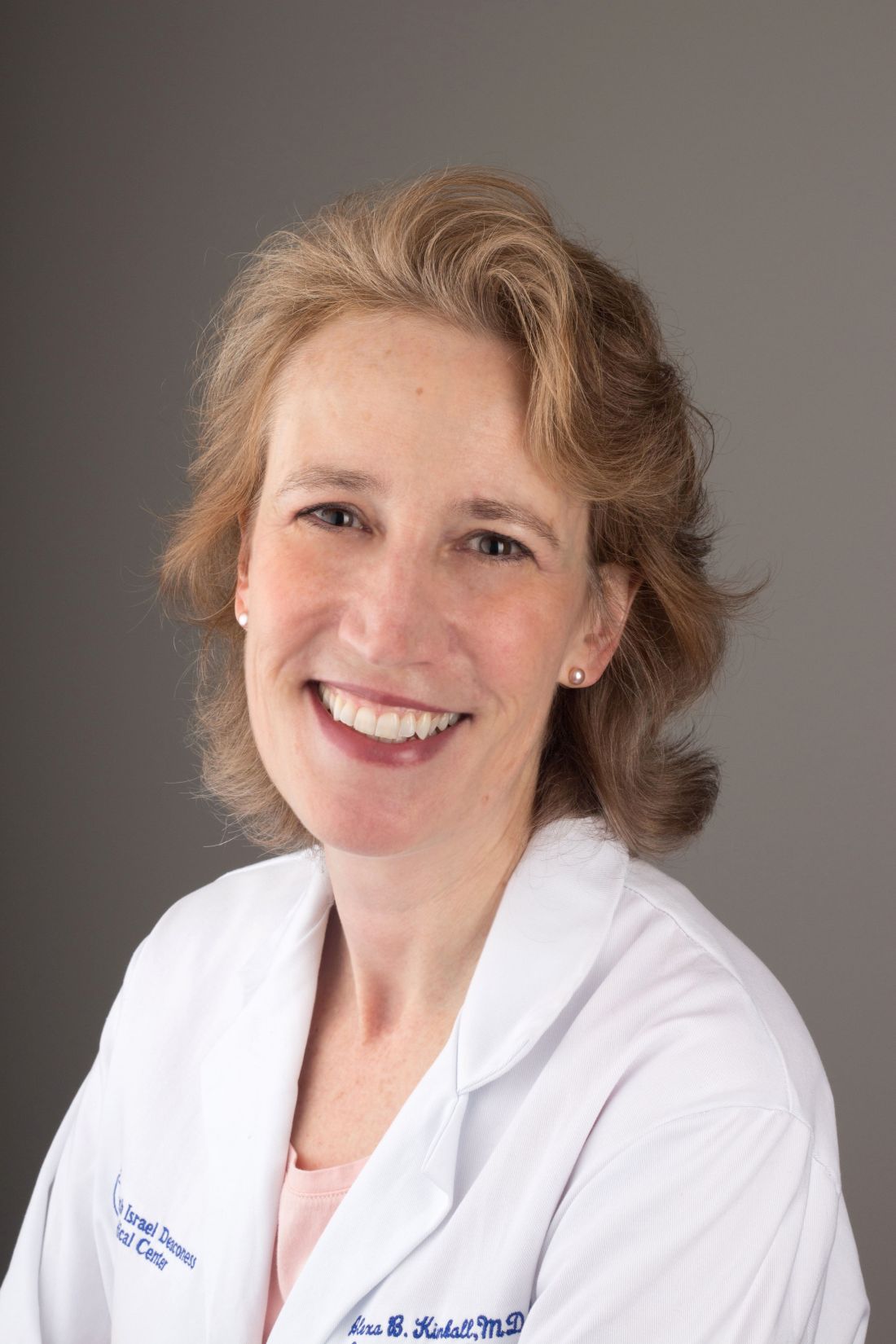
The presence of B cells and plasma cells has been reported in HS lesions, she continued, including early lesions, with BTK activation as a central signal transduction pathway. For the current study, Dr. Kimball and colleagues evaluated the safety and efficacy of remibrutinib (LOU064), an oral, highly selective BTK inhibitor, in 77 adults with moderate to severe HS for at least 12 months in 2 or more anatomical areas with 15 or fewer tunnels beneath the skin.
There were slightly more women than men and more than 90% of study participants were White. The novel drug, which is being developed by Novartis, is also under investigation in other immune-mediated inflammatory diseases, including chronic spontaneous urticaria and multiple sclerosis.
Of the 77 patients, 33 were assigned to receive 100 mg remibrutinib twice per day, 33 received a 25 mg twice-daily dose, and 11 patients received placebo twice per day. The primary endpoint was the proportion of patients who achieved a simplified Hidradenitis Suppurativa Clinical Response (HiSCR) at week 16 compared with pooled placebo. A simplified HiSCR response was defined as at least a 50% reduction in total inflammatory abscess and nodule (AN) count, with no increase in draining tunnels relative to baseline.
Dr. Kimball, professor of dermatology at Harvard University, reported that 80.2% of patients overall completed treatment: 87.9% and 78.8% in the remibrutinib 25 mg and 100 mg arms, respectively, and 76% in the pooled placebo arm. The main reason for treatment discontinuation was patient decision (60.9%). Nearly three quarters of patients in the remibrutinib 25 mg twice-daily arm (72.7%) achieved the simplified HiSCR endpoint, compared with 48.5% of those in the remibrutinib 100 mg twice-daily arm, and 34.7% of those in the placebo arm.
In other exploratory findings, HiSCR, HiSCR 75, and HiSCR 90 rates were higher at week 16 among patients in both remibrutinib treatment arms compared with placebo, and the study drug also was associated with a greater effect on reduction of the AN count and draining tunnels. Specifically, the estimated mean percentage reduction in AN count was 68% in the 25 mg twice-daily arm, compared with reductions of 57% in the 100 mg twice-daily arm and 49.7% in the placebo arm, respectively. Meanwhile, the estimated mean reductions in draining tunnels were 55.6%, 43.6%, and 10.2%, respectively, in the three arms.
The researchers also observed a greater response on the Patient’s Global Assessment of Skin Pain Numeric Rating Scale 30 (NRS30) in patients treated with remibrutinib compared with those on placebo at week 16 (57.1% in the 100 mg twice-daily arm, compared with 44.4% in the 25 mg twice-daily arm, and 30.4% in the placebo arm).
In terms of safety, adverse events (AEs) were mainly mild or moderate in severity, Dr. Kimball said, with no deaths and only one serious AE reported in each treatment arm: one case of acute pancreatitis in the 25 mg twice-daily arm, a testicular abscess in the pooled placebo arm, and a hypertensive crisis in the 100 mg twice-daily arm. Treatment discontinuations because of AEs were uncommon. Infections (primarily upper respiratory infections such as nasopharyngitis) were the most common AEs in all treatment arms.
“BTK inhibition may emerge as a promising therapeutic option in HS,” Dr. Kimball concluded. “This is wonderful news for our HS community. We are looking forward to determining what the optimal dosing will be going forward.”
Jennifer L. Hsiao, MD, associate professor of dermatology and director of the HS clinic at the University of Southern California, Los Angeles, who was asked to comment on the study, said there is “a pressing need for more treatments for patients with HS who suffer from the pain and oftentimes life-limiting nature of this condition.” She characterized the study results as “promising.”

“We will see if phase 3 trials with more balanced demographics across remibrutinib and placebo arms will reproduce these outcomes,” she continued, “It is exciting to see this potential new medication for HS under continued investigation, especially in light of the current gap in oral therapeutic options for the HS patient community.” Dr. Hsiao was not involved with the study.
Dr. Kimball disclosed numerous conflicts of interest from various pharmaceutical companies, including the receipt of research grants and consulting fees from Novartis. Dr. Hsiao disclosed that she is a member of the board of directors for the Hidradenitis Suppurativa Foundation. She has also served as a consultant for AbbVie, Aclaris, Boehringer Ingelheim, Incyte, Novartis, and UCB; as a speaker for AbbVie, Novartis, and UCB; and as an investigator for Amgen, Boehringer Ingelheim, and Incyte.
A version of this article appeared on Medscape.com.
SAN DIEGO — .
“Research shows that the TNF-alpha and IL-17 signaling pathways have important roles in HS,” lead investigator Alexa B. Kimball, MD, MPH, from the Clinical Laboratory for Epidemiology and Applied Research in Skin at Beth Israel Deaconess Medical Center, Boston, said at the annual meeting of the American Academy of Dermatology. “However, several additional pathways are thought to contribute to disease pathogenesis.”
The presence of B cells and plasma cells has been reported in HS lesions, she continued, including early lesions, with BTK activation as a central signal transduction pathway. For the current study, Dr. Kimball and colleagues evaluated the safety and efficacy of remibrutinib (LOU064), an oral, highly selective BTK inhibitor, in 77 adults with moderate to severe HS for at least 12 months in 2 or more anatomical areas with 15 or fewer tunnels beneath the skin.
There were slightly more women than men and more than 90% of study participants were White. The novel drug, which is being developed by Novartis, is also under investigation in other immune-mediated inflammatory diseases, including chronic spontaneous urticaria and multiple sclerosis.
Of the 77 patients, 33 were assigned to receive 100 mg remibrutinib twice per day, 33 received a 25 mg twice-daily dose, and 11 patients received placebo twice per day. The primary endpoint was the proportion of patients who achieved a simplified Hidradenitis Suppurativa Clinical Response (HiSCR) at week 16 compared with pooled placebo. A simplified HiSCR response was defined as at least a 50% reduction in total inflammatory abscess and nodule (AN) count, with no increase in draining tunnels relative to baseline.
Dr. Kimball, professor of dermatology at Harvard University, reported that 80.2% of patients overall completed treatment: 87.9% and 78.8% in the remibrutinib 25 mg and 100 mg arms, respectively, and 76% in the pooled placebo arm. The main reason for treatment discontinuation was patient decision (60.9%). Nearly three quarters of patients in the remibrutinib 25 mg twice-daily arm (72.7%) achieved the simplified HiSCR endpoint, compared with 48.5% of those in the remibrutinib 100 mg twice-daily arm, and 34.7% of those in the placebo arm.
In other exploratory findings, HiSCR, HiSCR 75, and HiSCR 90 rates were higher at week 16 among patients in both remibrutinib treatment arms compared with placebo, and the study drug also was associated with a greater effect on reduction of the AN count and draining tunnels. Specifically, the estimated mean percentage reduction in AN count was 68% in the 25 mg twice-daily arm, compared with reductions of 57% in the 100 mg twice-daily arm and 49.7% in the placebo arm, respectively. Meanwhile, the estimated mean reductions in draining tunnels were 55.6%, 43.6%, and 10.2%, respectively, in the three arms.
The researchers also observed a greater response on the Patient’s Global Assessment of Skin Pain Numeric Rating Scale 30 (NRS30) in patients treated with remibrutinib compared with those on placebo at week 16 (57.1% in the 100 mg twice-daily arm, compared with 44.4% in the 25 mg twice-daily arm, and 30.4% in the placebo arm).
In terms of safety, adverse events (AEs) were mainly mild or moderate in severity, Dr. Kimball said, with no deaths and only one serious AE reported in each treatment arm: one case of acute pancreatitis in the 25 mg twice-daily arm, a testicular abscess in the pooled placebo arm, and a hypertensive crisis in the 100 mg twice-daily arm. Treatment discontinuations because of AEs were uncommon. Infections (primarily upper respiratory infections such as nasopharyngitis) were the most common AEs in all treatment arms.
“BTK inhibition may emerge as a promising therapeutic option in HS,” Dr. Kimball concluded. “This is wonderful news for our HS community. We are looking forward to determining what the optimal dosing will be going forward.”
Jennifer L. Hsiao, MD, associate professor of dermatology and director of the HS clinic at the University of Southern California, Los Angeles, who was asked to comment on the study, said there is “a pressing need for more treatments for patients with HS who suffer from the pain and oftentimes life-limiting nature of this condition.” She characterized the study results as “promising.”
“We will see if phase 3 trials with more balanced demographics across remibrutinib and placebo arms will reproduce these outcomes,” she continued, “It is exciting to see this potential new medication for HS under continued investigation, especially in light of the current gap in oral therapeutic options for the HS patient community.” Dr. Hsiao was not involved with the study.
Dr. Kimball disclosed numerous conflicts of interest from various pharmaceutical companies, including the receipt of research grants and consulting fees from Novartis. Dr. Hsiao disclosed that she is a member of the board of directors for the Hidradenitis Suppurativa Foundation. She has also served as a consultant for AbbVie, Aclaris, Boehringer Ingelheim, Incyte, Novartis, and UCB; as a speaker for AbbVie, Novartis, and UCB; and as an investigator for Amgen, Boehringer Ingelheim, and Incyte.
A version of this article appeared on Medscape.com.
SAN DIEGO — .
“Research shows that the TNF-alpha and IL-17 signaling pathways have important roles in HS,” lead investigator Alexa B. Kimball, MD, MPH, from the Clinical Laboratory for Epidemiology and Applied Research in Skin at Beth Israel Deaconess Medical Center, Boston, said at the annual meeting of the American Academy of Dermatology. “However, several additional pathways are thought to contribute to disease pathogenesis.”
The presence of B cells and plasma cells has been reported in HS lesions, she continued, including early lesions, with BTK activation as a central signal transduction pathway. For the current study, Dr. Kimball and colleagues evaluated the safety and efficacy of remibrutinib (LOU064), an oral, highly selective BTK inhibitor, in 77 adults with moderate to severe HS for at least 12 months in 2 or more anatomical areas with 15 or fewer tunnels beneath the skin.
There were slightly more women than men and more than 90% of study participants were White. The novel drug, which is being developed by Novartis, is also under investigation in other immune-mediated inflammatory diseases, including chronic spontaneous urticaria and multiple sclerosis.
Of the 77 patients, 33 were assigned to receive 100 mg remibrutinib twice per day, 33 received a 25 mg twice-daily dose, and 11 patients received placebo twice per day. The primary endpoint was the proportion of patients who achieved a simplified Hidradenitis Suppurativa Clinical Response (HiSCR) at week 16 compared with pooled placebo. A simplified HiSCR response was defined as at least a 50% reduction in total inflammatory abscess and nodule (AN) count, with no increase in draining tunnels relative to baseline.
Dr. Kimball, professor of dermatology at Harvard University, reported that 80.2% of patients overall completed treatment: 87.9% and 78.8% in the remibrutinib 25 mg and 100 mg arms, respectively, and 76% in the pooled placebo arm. The main reason for treatment discontinuation was patient decision (60.9%). Nearly three quarters of patients in the remibrutinib 25 mg twice-daily arm (72.7%) achieved the simplified HiSCR endpoint, compared with 48.5% of those in the remibrutinib 100 mg twice-daily arm, and 34.7% of those in the placebo arm.
In other exploratory findings, HiSCR, HiSCR 75, and HiSCR 90 rates were higher at week 16 among patients in both remibrutinib treatment arms compared with placebo, and the study drug also was associated with a greater effect on reduction of the AN count and draining tunnels. Specifically, the estimated mean percentage reduction in AN count was 68% in the 25 mg twice-daily arm, compared with reductions of 57% in the 100 mg twice-daily arm and 49.7% in the placebo arm, respectively. Meanwhile, the estimated mean reductions in draining tunnels were 55.6%, 43.6%, and 10.2%, respectively, in the three arms.
The researchers also observed a greater response on the Patient’s Global Assessment of Skin Pain Numeric Rating Scale 30 (NRS30) in patients treated with remibrutinib compared with those on placebo at week 16 (57.1% in the 100 mg twice-daily arm, compared with 44.4% in the 25 mg twice-daily arm, and 30.4% in the placebo arm).
In terms of safety, adverse events (AEs) were mainly mild or moderate in severity, Dr. Kimball said, with no deaths and only one serious AE reported in each treatment arm: one case of acute pancreatitis in the 25 mg twice-daily arm, a testicular abscess in the pooled placebo arm, and a hypertensive crisis in the 100 mg twice-daily arm. Treatment discontinuations because of AEs were uncommon. Infections (primarily upper respiratory infections such as nasopharyngitis) were the most common AEs in all treatment arms.
“BTK inhibition may emerge as a promising therapeutic option in HS,” Dr. Kimball concluded. “This is wonderful news for our HS community. We are looking forward to determining what the optimal dosing will be going forward.”
Jennifer L. Hsiao, MD, associate professor of dermatology and director of the HS clinic at the University of Southern California, Los Angeles, who was asked to comment on the study, said there is “a pressing need for more treatments for patients with HS who suffer from the pain and oftentimes life-limiting nature of this condition.” She characterized the study results as “promising.”
“We will see if phase 3 trials with more balanced demographics across remibrutinib and placebo arms will reproduce these outcomes,” she continued, “It is exciting to see this potential new medication for HS under continued investigation, especially in light of the current gap in oral therapeutic options for the HS patient community.” Dr. Hsiao was not involved with the study.
Dr. Kimball disclosed numerous conflicts of interest from various pharmaceutical companies, including the receipt of research grants and consulting fees from Novartis. Dr. Hsiao disclosed that she is a member of the board of directors for the Hidradenitis Suppurativa Foundation. She has also served as a consultant for AbbVie, Aclaris, Boehringer Ingelheim, Incyte, Novartis, and UCB; as a speaker for AbbVie, Novartis, and UCB; and as an investigator for Amgen, Boehringer Ingelheim, and Incyte.
A version of this article appeared on Medscape.com.
FROM AAD 2024

FDA Approves First Drug for MASH
, along with diet and exercise.
Resmetirom is a once-daily, oral thyroid hormone receptor beta-selective agonist. The FDA granted the drug breakthrough therapy designation and priority review.
The approval is based on the phase 3 MAESTRO-NASH trial, in which resmetirom was superior to placebo at achieving resolution of nonalcoholic steatohepatitis (NASH) and improving liver fibrosis in both 80-mg and 100-mg doses.
The trial used the earlier nomenclature of NASH and nonalcoholic fatty liver disease (NAFLD). An international consensus group has since changed these terms to MASH and metabolic dysfunction–associated steatotic liver disease (MASLD), respectively. (Note that the terms NASH and NAFLD will be used to discuss the trial results in this article to align with the trial’s original language.)
The results were published online February 6 in The New England Journal of Medicine.
“The approval of the first medication for NASH is a true game-changer for healthcare providers, the research community and, most importantly, patients living with this serious liver condition,” lead MAESTRO-NASH investigator Stephen Harrison, MD, gastroenterologist, hepatologist, and chairman of Pinnacle Clinical Research and Summit Clinical Research, San Antonio, Texas, said in a news release.

“Based on the robust efficacy and safety data generated in two large Phase 3 MAESTRO studies, I believe Rezdiffra will become the foundational therapy for patients with NASH with moderate to advanced liver fibrosis. Importantly, we continue to study Rezdiffra to determine if the positive results observed in the MAESTRO studies will lead to reduced risk of progression to cirrhosis, liver failure, need for liver transplant and premature mortality,” Dr. Harrison added.
Addressing an Unmet Need
MASH is a progressive liver disease and the leading cause of liver-related mortality. The disease affects an estimated 1.5 million adults in the United States, of which, roughly 525,000 have MASH with significant fibrosis. Until now, there was no FDA-approved medication.
In the ongoing MAESTRO-NASH, 996 adults with biopsy-confirmed NASH and significant stage 2-3 fibrosis were randomly assigned to receive oral once-daily resmetirom (80 mg or 100 mg) or placebo.
Patients were followed for 52 weeks, at which point, they were assessed for the dual primary endpoints of NASH resolution (including a reduction in the NAFLD activity score by ≥ 2 points) with no worsening of fibrosis and an improvement (reduction) in fibrosis by at least one stage with no worsening of the NAFLD activity score.
Patients receiving resmetirom had a significant improvement across both doses and both primary endpoints.
At 52 weeks, NASH resolution with no worsening of fibrosis was achieved in 25.9% and 29.9% of the patients in the 80-mg and 100-mg groups, respectively, compared with 9.7% on placebo.
Fibrosis improved by at least one stage with no worsening of the NAFLD activity score in 24.2% and 25.9% of patients in the 80-mg and 100-mg groups, respectively, compared with 14.2% on placebo.
The trial also met multiple secondary endpoints, including statistically significant reduction from baseline in liver enzymes (alanine transaminase, aspartate aminotransferase, and gamma-glutamyl transferase) and low-density lipoprotein cholesterol with resmetirom compared with placebo.
Improvement in fibrosis biomarkers and relevant imaging tests were also observed in resmetirom treatment groups compared with placebo.
The most common adverse events included diarrhea and nausea, which typically began early in treatment and were mild to moderate in severity. Pruritus, abdominal pain, vomiting, constipation, and dizziness were also reported.
Resmetirom is expected to be available to patients in the United States in April and will be distributed through a limited specialty pharmacy network.
Full prescribing information is available online. Prescribing information does not include a liver biopsy requirement for diagnosis.
A version of this article appeared on Medscape.com.
, along with diet and exercise.
Resmetirom is a once-daily, oral thyroid hormone receptor beta-selective agonist. The FDA granted the drug breakthrough therapy designation and priority review.
The approval is based on the phase 3 MAESTRO-NASH trial, in which resmetirom was superior to placebo at achieving resolution of nonalcoholic steatohepatitis (NASH) and improving liver fibrosis in both 80-mg and 100-mg doses.
The trial used the earlier nomenclature of NASH and nonalcoholic fatty liver disease (NAFLD). An international consensus group has since changed these terms to MASH and metabolic dysfunction–associated steatotic liver disease (MASLD), respectively. (Note that the terms NASH and NAFLD will be used to discuss the trial results in this article to align with the trial’s original language.)
The results were published online February 6 in The New England Journal of Medicine.
“The approval of the first medication for NASH is a true game-changer for healthcare providers, the research community and, most importantly, patients living with this serious liver condition,” lead MAESTRO-NASH investigator Stephen Harrison, MD, gastroenterologist, hepatologist, and chairman of Pinnacle Clinical Research and Summit Clinical Research, San Antonio, Texas, said in a news release.
“Based on the robust efficacy and safety data generated in two large Phase 3 MAESTRO studies, I believe Rezdiffra will become the foundational therapy for patients with NASH with moderate to advanced liver fibrosis. Importantly, we continue to study Rezdiffra to determine if the positive results observed in the MAESTRO studies will lead to reduced risk of progression to cirrhosis, liver failure, need for liver transplant and premature mortality,” Dr. Harrison added.
Addressing an Unmet Need
MASH is a progressive liver disease and the leading cause of liver-related mortality. The disease affects an estimated 1.5 million adults in the United States, of which, roughly 525,000 have MASH with significant fibrosis. Until now, there was no FDA-approved medication.
In the ongoing MAESTRO-NASH, 996 adults with biopsy-confirmed NASH and significant stage 2-3 fibrosis were randomly assigned to receive oral once-daily resmetirom (80 mg or 100 mg) or placebo.
Patients were followed for 52 weeks, at which point, they were assessed for the dual primary endpoints of NASH resolution (including a reduction in the NAFLD activity score by ≥ 2 points) with no worsening of fibrosis and an improvement (reduction) in fibrosis by at least one stage with no worsening of the NAFLD activity score.
Patients receiving resmetirom had a significant improvement across both doses and both primary endpoints.
At 52 weeks, NASH resolution with no worsening of fibrosis was achieved in 25.9% and 29.9% of the patients in the 80-mg and 100-mg groups, respectively, compared with 9.7% on placebo.
Fibrosis improved by at least one stage with no worsening of the NAFLD activity score in 24.2% and 25.9% of patients in the 80-mg and 100-mg groups, respectively, compared with 14.2% on placebo.
The trial also met multiple secondary endpoints, including statistically significant reduction from baseline in liver enzymes (alanine transaminase, aspartate aminotransferase, and gamma-glutamyl transferase) and low-density lipoprotein cholesterol with resmetirom compared with placebo.
Improvement in fibrosis biomarkers and relevant imaging tests were also observed in resmetirom treatment groups compared with placebo.
The most common adverse events included diarrhea and nausea, which typically began early in treatment and were mild to moderate in severity. Pruritus, abdominal pain, vomiting, constipation, and dizziness were also reported.
Resmetirom is expected to be available to patients in the United States in April and will be distributed through a limited specialty pharmacy network.
Full prescribing information is available online. Prescribing information does not include a liver biopsy requirement for diagnosis.
A version of this article appeared on Medscape.com.
, along with diet and exercise.
Resmetirom is a once-daily, oral thyroid hormone receptor beta-selective agonist. The FDA granted the drug breakthrough therapy designation and priority review.
The approval is based on the phase 3 MAESTRO-NASH trial, in which resmetirom was superior to placebo at achieving resolution of nonalcoholic steatohepatitis (NASH) and improving liver fibrosis in both 80-mg and 100-mg doses.
The trial used the earlier nomenclature of NASH and nonalcoholic fatty liver disease (NAFLD). An international consensus group has since changed these terms to MASH and metabolic dysfunction–associated steatotic liver disease (MASLD), respectively. (Note that the terms NASH and NAFLD will be used to discuss the trial results in this article to align with the trial’s original language.)
The results were published online February 6 in The New England Journal of Medicine.
“The approval of the first medication for NASH is a true game-changer for healthcare providers, the research community and, most importantly, patients living with this serious liver condition,” lead MAESTRO-NASH investigator Stephen Harrison, MD, gastroenterologist, hepatologist, and chairman of Pinnacle Clinical Research and Summit Clinical Research, San Antonio, Texas, said in a news release.
“Based on the robust efficacy and safety data generated in two large Phase 3 MAESTRO studies, I believe Rezdiffra will become the foundational therapy for patients with NASH with moderate to advanced liver fibrosis. Importantly, we continue to study Rezdiffra to determine if the positive results observed in the MAESTRO studies will lead to reduced risk of progression to cirrhosis, liver failure, need for liver transplant and premature mortality,” Dr. Harrison added.
Addressing an Unmet Need
MASH is a progressive liver disease and the leading cause of liver-related mortality. The disease affects an estimated 1.5 million adults in the United States, of which, roughly 525,000 have MASH with significant fibrosis. Until now, there was no FDA-approved medication.
In the ongoing MAESTRO-NASH, 996 adults with biopsy-confirmed NASH and significant stage 2-3 fibrosis were randomly assigned to receive oral once-daily resmetirom (80 mg or 100 mg) or placebo.
Patients were followed for 52 weeks, at which point, they were assessed for the dual primary endpoints of NASH resolution (including a reduction in the NAFLD activity score by ≥ 2 points) with no worsening of fibrosis and an improvement (reduction) in fibrosis by at least one stage with no worsening of the NAFLD activity score.
Patients receiving resmetirom had a significant improvement across both doses and both primary endpoints.
At 52 weeks, NASH resolution with no worsening of fibrosis was achieved in 25.9% and 29.9% of the patients in the 80-mg and 100-mg groups, respectively, compared with 9.7% on placebo.
Fibrosis improved by at least one stage with no worsening of the NAFLD activity score in 24.2% and 25.9% of patients in the 80-mg and 100-mg groups, respectively, compared with 14.2% on placebo.
The trial also met multiple secondary endpoints, including statistically significant reduction from baseline in liver enzymes (alanine transaminase, aspartate aminotransferase, and gamma-glutamyl transferase) and low-density lipoprotein cholesterol with resmetirom compared with placebo.
Improvement in fibrosis biomarkers and relevant imaging tests were also observed in resmetirom treatment groups compared with placebo.
The most common adverse events included diarrhea and nausea, which typically began early in treatment and were mild to moderate in severity. Pruritus, abdominal pain, vomiting, constipation, and dizziness were also reported.
Resmetirom is expected to be available to patients in the United States in April and will be distributed through a limited specialty pharmacy network.
Full prescribing information is available online. Prescribing information does not include a liver biopsy requirement for diagnosis.
A version of this article appeared on Medscape.com.

Immunomodulators Do Not Affect COVID-19 Vaccine Efficacy
TOPLINE:
The results of a recent study suggest that biologics and small molecule inhibitors (SMIs) do not impair the protective effect of COVID-19 vaccine against hospitalization in patients with psoriasis and hidradenitis suppurativa (HS).
METHODOLOGY:
- It remains unknown whether immunomodulatory therapies impair COVID-19 vaccine efficacy and increase hospitalization rates linked to COVID-19 in patients with inflammatory skin conditions such as psoriasis or HS.
- Researchers conducted a cross-sectional study using data from the Epic Cosmos database from January 2020 to October 2023, identifying 30,845 patients with psoriasis or HS.
- Overall, 22,293 patients with documented completion of their primary COVID-19 vaccine series were included in the analysis.
- Of the vaccinated patients, they compared 7046 patients with psoriasis on SMIs and 2033 with psoriasis or HS on biologics with 13,214 patients who did not receive biologics or SMIs.
- The primary outcome was the COVID-19 hospitalization rate.
- Treatment with biologics did not increase COVID-19-related hospitalization rates in vaccinated patients with psoriasis or HS (hospitalization rate, 6.0% for both those taking and those not taking a biologic; P > .99).
- Similarly, hospitalization rates did not significantly differ between vaccinated patients who received SMIs vs those who did not (7.1% vs 6.0%; P = .0596).
IN PRACTICE:
These findings “encourage dermatologists to continue treating [psoriasis]/HS confidently despite the ongoing COVID-19 pandemic,” the authors concluded.
SOURCE:
The study led by Bella R. Lee from Ohio State University Wexner Medical Center, Columbus, was published online on March 13, 2024, in the Journal of the American Academy of Dermatology.
LIMITATIONS:
Multivariable adjustments could not be performed in this study due to unavailability of individual-level data, and hospital admissions that occurred outside the Epic system were not captured.
DISCLOSURES:
The study did not receive any funding. All authors declared no conflicts of interest.
A version of this article appeared on Medscape.com.
TOPLINE:
The results of a recent study suggest that biologics and small molecule inhibitors (SMIs) do not impair the protective effect of COVID-19 vaccine against hospitalization in patients with psoriasis and hidradenitis suppurativa (HS).
METHODOLOGY:
- It remains unknown whether immunomodulatory therapies impair COVID-19 vaccine efficacy and increase hospitalization rates linked to COVID-19 in patients with inflammatory skin conditions such as psoriasis or HS.
- Researchers conducted a cross-sectional study using data from the Epic Cosmos database from January 2020 to October 2023, identifying 30,845 patients with psoriasis or HS.
- Overall, 22,293 patients with documented completion of their primary COVID-19 vaccine series were included in the analysis.
- Of the vaccinated patients, they compared 7046 patients with psoriasis on SMIs and 2033 with psoriasis or HS on biologics with 13,214 patients who did not receive biologics or SMIs.
- The primary outcome was the COVID-19 hospitalization rate.
- Treatment with biologics did not increase COVID-19-related hospitalization rates in vaccinated patients with psoriasis or HS (hospitalization rate, 6.0% for both those taking and those not taking a biologic; P > .99).
- Similarly, hospitalization rates did not significantly differ between vaccinated patients who received SMIs vs those who did not (7.1% vs 6.0%; P = .0596).
IN PRACTICE:
These findings “encourage dermatologists to continue treating [psoriasis]/HS confidently despite the ongoing COVID-19 pandemic,” the authors concluded.
SOURCE:
The study led by Bella R. Lee from Ohio State University Wexner Medical Center, Columbus, was published online on March 13, 2024, in the Journal of the American Academy of Dermatology.
LIMITATIONS:
Multivariable adjustments could not be performed in this study due to unavailability of individual-level data, and hospital admissions that occurred outside the Epic system were not captured.
DISCLOSURES:
The study did not receive any funding. All authors declared no conflicts of interest.
A version of this article appeared on Medscape.com.
TOPLINE:
The results of a recent study suggest that biologics and small molecule inhibitors (SMIs) do not impair the protective effect of COVID-19 vaccine against hospitalization in patients with psoriasis and hidradenitis suppurativa (HS).
METHODOLOGY:
- It remains unknown whether immunomodulatory therapies impair COVID-19 vaccine efficacy and increase hospitalization rates linked to COVID-19 in patients with inflammatory skin conditions such as psoriasis or HS.
- Researchers conducted a cross-sectional study using data from the Epic Cosmos database from January 2020 to October 2023, identifying 30,845 patients with psoriasis or HS.
- Overall, 22,293 patients with documented completion of their primary COVID-19 vaccine series were included in the analysis.
- Of the vaccinated patients, they compared 7046 patients with psoriasis on SMIs and 2033 with psoriasis or HS on biologics with 13,214 patients who did not receive biologics or SMIs.
- The primary outcome was the COVID-19 hospitalization rate.
- Treatment with biologics did not increase COVID-19-related hospitalization rates in vaccinated patients with psoriasis or HS (hospitalization rate, 6.0% for both those taking and those not taking a biologic; P > .99).
- Similarly, hospitalization rates did not significantly differ between vaccinated patients who received SMIs vs those who did not (7.1% vs 6.0%; P = .0596).
IN PRACTICE:
These findings “encourage dermatologists to continue treating [psoriasis]/HS confidently despite the ongoing COVID-19 pandemic,” the authors concluded.
SOURCE:
The study led by Bella R. Lee from Ohio State University Wexner Medical Center, Columbus, was published online on March 13, 2024, in the Journal of the American Academy of Dermatology.
LIMITATIONS:
Multivariable adjustments could not be performed in this study due to unavailability of individual-level data, and hospital admissions that occurred outside the Epic system were not captured.
DISCLOSURES:
The study did not receive any funding. All authors declared no conflicts of interest.
A version of this article appeared on Medscape.com.
Study Links Maternal Hidradenitis Suppurativa to Risk for Childhood Morbidity
SAN DIEGO — , and other conditions.
Those are key findings from a longitudinal cohort study that was presented during a poster session at the annual meeting of the American Academy of Dermatology.
“HS is associated with morbidity in women of reproductive age and adverse pregnancy outcomes, [but] its effect on offspring outcomes remains unclear,” corresponding author Kaiyang Li, a third-year medical student at McGill University, Quebec, Canada, and coauthors wrote in their abstract.
To investigate the association between maternal HS and offspring outcomes at birth and with up to 16 years of follow-up, the researchers drew from a longitudinal cohort of 1,275,593 children born in Quebec between April 1, 2006 and March 31, 2022. They matched children with their mothers and used identification numbers to follow the children to note morbidities that led to hospital admissions before age 16 years. The exposure of interest was HS, and the main outcome measure was childhood hospitalizations for respiratory, cardiovascular, metabolic, and other morbidities prior to age 16 years.
Next, they estimated hazard ratios (HR) and 95% CIs for the association of maternal HS with childhood morbidity in adjusted Cox proportional hazards regression models. “As prenatal exposure to hyperandrogenism may influence boys and girls differently, we carried out subgroup analyses stratified by child sex,” they wrote.
The study population included 1283 children whose mothers had HS and 1,274,310 unexposed children. As for infant outcomes, compared with no exposure, maternal HS was associated with an increased risk for preterm birth (relative risk [RR], 1.29; 95% CI, 1,08-1.55), neonatal death (RR, 2.07; 95% CI, 1.03-14.13), birth defects (RR, 1.29; 95% CI, 1.07-1.56), congenital heart defects (RR, 1.57; 95% CI, 1.02-2.44), and orofacial defects (RR 4.29; 95% CI, 1.85-9.97).
As for long-term outcomes in the children, compared with those whose mothers did not have HS, maternal HS was associated with an increased risk for any childhood hospitalization (HR, 1.31; 95% CI, 1.19-1.44), respiratory hospitalization (HR, 1.21; 95% CI, 1.05-1.40), metabolic hospitalization (HR, 2.64; 95% CI, 1.67-4.20), gastrointestinal hospitalization (HR, 1.34; 95% CI, 1.03-1.74), and developmental hospitalization (HR, 1.92; 95% CI, 1.43-2.58).
Commenting on the results after the meeting, Ms. Li said that the findings support the need for timely management of HS in expectant mothers and people planning to conceive, and for “interdisciplinary care and follow up for both the mother and the baby, involving the dermatologist, the obstetrician, and the neonatologist or pediatrician if needed.”
“HS is a multidisciplinary disease, plain and simple,” Adam Friedman, MD, professor and chair of dermatology, George Washington University, Washington, who was asked to comment on the study, said in an interview. “This study highlights the importance of collaboration between dermatology and obstetrician-gynecologist given the potential negative pregnancy outcomes, but to me raising alarm bells given the known gaps and delays in diagnosis matched to disease onset,” said Dr. Friedman, who was not involved with the study. “We need to do better to ensure the safety of both patient and patient-to-be.”
The researchers reported having no financial disclosures. The abstract was selected as the second-place winner in the AAD’s poster competition. Dr. Friedman has no relevant disclosures.
A version of this article appeared on Medscape.com.
SAN DIEGO — , and other conditions.
Those are key findings from a longitudinal cohort study that was presented during a poster session at the annual meeting of the American Academy of Dermatology.
“HS is associated with morbidity in women of reproductive age and adverse pregnancy outcomes, [but] its effect on offspring outcomes remains unclear,” corresponding author Kaiyang Li, a third-year medical student at McGill University, Quebec, Canada, and coauthors wrote in their abstract.
To investigate the association between maternal HS and offspring outcomes at birth and with up to 16 years of follow-up, the researchers drew from a longitudinal cohort of 1,275,593 children born in Quebec between April 1, 2006 and March 31, 2022. They matched children with their mothers and used identification numbers to follow the children to note morbidities that led to hospital admissions before age 16 years. The exposure of interest was HS, and the main outcome measure was childhood hospitalizations for respiratory, cardiovascular, metabolic, and other morbidities prior to age 16 years.
Next, they estimated hazard ratios (HR) and 95% CIs for the association of maternal HS with childhood morbidity in adjusted Cox proportional hazards regression models. “As prenatal exposure to hyperandrogenism may influence boys and girls differently, we carried out subgroup analyses stratified by child sex,” they wrote.
The study population included 1283 children whose mothers had HS and 1,274,310 unexposed children. As for infant outcomes, compared with no exposure, maternal HS was associated with an increased risk for preterm birth (relative risk [RR], 1.29; 95% CI, 1,08-1.55), neonatal death (RR, 2.07; 95% CI, 1.03-14.13), birth defects (RR, 1.29; 95% CI, 1.07-1.56), congenital heart defects (RR, 1.57; 95% CI, 1.02-2.44), and orofacial defects (RR 4.29; 95% CI, 1.85-9.97).
As for long-term outcomes in the children, compared with those whose mothers did not have HS, maternal HS was associated with an increased risk for any childhood hospitalization (HR, 1.31; 95% CI, 1.19-1.44), respiratory hospitalization (HR, 1.21; 95% CI, 1.05-1.40), metabolic hospitalization (HR, 2.64; 95% CI, 1.67-4.20), gastrointestinal hospitalization (HR, 1.34; 95% CI, 1.03-1.74), and developmental hospitalization (HR, 1.92; 95% CI, 1.43-2.58).
Commenting on the results after the meeting, Ms. Li said that the findings support the need for timely management of HS in expectant mothers and people planning to conceive, and for “interdisciplinary care and follow up for both the mother and the baby, involving the dermatologist, the obstetrician, and the neonatologist or pediatrician if needed.”
“HS is a multidisciplinary disease, plain and simple,” Adam Friedman, MD, professor and chair of dermatology, George Washington University, Washington, who was asked to comment on the study, said in an interview. “This study highlights the importance of collaboration between dermatology and obstetrician-gynecologist given the potential negative pregnancy outcomes, but to me raising alarm bells given the known gaps and delays in diagnosis matched to disease onset,” said Dr. Friedman, who was not involved with the study. “We need to do better to ensure the safety of both patient and patient-to-be.”
The researchers reported having no financial disclosures. The abstract was selected as the second-place winner in the AAD’s poster competition. Dr. Friedman has no relevant disclosures.
A version of this article appeared on Medscape.com.
SAN DIEGO — , and other conditions.
Those are key findings from a longitudinal cohort study that was presented during a poster session at the annual meeting of the American Academy of Dermatology.
“HS is associated with morbidity in women of reproductive age and adverse pregnancy outcomes, [but] its effect on offspring outcomes remains unclear,” corresponding author Kaiyang Li, a third-year medical student at McGill University, Quebec, Canada, and coauthors wrote in their abstract.
To investigate the association between maternal HS and offspring outcomes at birth and with up to 16 years of follow-up, the researchers drew from a longitudinal cohort of 1,275,593 children born in Quebec between April 1, 2006 and March 31, 2022. They matched children with their mothers and used identification numbers to follow the children to note morbidities that led to hospital admissions before age 16 years. The exposure of interest was HS, and the main outcome measure was childhood hospitalizations for respiratory, cardiovascular, metabolic, and other morbidities prior to age 16 years.
Next, they estimated hazard ratios (HR) and 95% CIs for the association of maternal HS with childhood morbidity in adjusted Cox proportional hazards regression models. “As prenatal exposure to hyperandrogenism may influence boys and girls differently, we carried out subgroup analyses stratified by child sex,” they wrote.
The study population included 1283 children whose mothers had HS and 1,274,310 unexposed children. As for infant outcomes, compared with no exposure, maternal HS was associated with an increased risk for preterm birth (relative risk [RR], 1.29; 95% CI, 1,08-1.55), neonatal death (RR, 2.07; 95% CI, 1.03-14.13), birth defects (RR, 1.29; 95% CI, 1.07-1.56), congenital heart defects (RR, 1.57; 95% CI, 1.02-2.44), and orofacial defects (RR 4.29; 95% CI, 1.85-9.97).
As for long-term outcomes in the children, compared with those whose mothers did not have HS, maternal HS was associated with an increased risk for any childhood hospitalization (HR, 1.31; 95% CI, 1.19-1.44), respiratory hospitalization (HR, 1.21; 95% CI, 1.05-1.40), metabolic hospitalization (HR, 2.64; 95% CI, 1.67-4.20), gastrointestinal hospitalization (HR, 1.34; 95% CI, 1.03-1.74), and developmental hospitalization (HR, 1.92; 95% CI, 1.43-2.58).
Commenting on the results after the meeting, Ms. Li said that the findings support the need for timely management of HS in expectant mothers and people planning to conceive, and for “interdisciplinary care and follow up for both the mother and the baby, involving the dermatologist, the obstetrician, and the neonatologist or pediatrician if needed.”
“HS is a multidisciplinary disease, plain and simple,” Adam Friedman, MD, professor and chair of dermatology, George Washington University, Washington, who was asked to comment on the study, said in an interview. “This study highlights the importance of collaboration between dermatology and obstetrician-gynecologist given the potential negative pregnancy outcomes, but to me raising alarm bells given the known gaps and delays in diagnosis matched to disease onset,” said Dr. Friedman, who was not involved with the study. “We need to do better to ensure the safety of both patient and patient-to-be.”
The researchers reported having no financial disclosures. The abstract was selected as the second-place winner in the AAD’s poster competition. Dr. Friedman has no relevant disclosures.
A version of this article appeared on Medscape.com.
FROM AAD 2024
Most Cancer Trial Centers Located Closer to White, Affluent Populations
This inequity may be potentiating the underrepresentation of racially minoritized and socioeconomically disadvantaged populations in clinical trials, suggesting that employment of satellite hospitals is needed to expand access to investigational therapies, reported lead author Hassal Lee, MD, PhD, of Cold Spring Harbor Laboratory, Cold Spring Harbor, New York, and colleagues.
“Minoritized and socioeconomically disadvantaged populations are underrepresented in clinical trials,” the investigators wrote in JAMA Oncology. “This may reduce the generalizability of trial results and propagate health disparities. Contributors to inequitable trial participation include individual-level factors and structural factors.”
Specifically, travel time to trial centers, as well as socioeconomic deprivation, can reduce likelihood of trial participation.
“Data on these parameters and population data on self-identified race exist, but their interrelation with clinical research facilities has not been systematically analyzed,” they wrote.
To try to draw comparisons between the distribution of patients of different races and socioeconomic statuses and the locations of clinical research facilities, Dr. Lee and colleagues aggregated data from the US Census, National Trial registry, Nature Index of Cancer Research Health Institutions, OpenStreetMap, National Cancer Institute–designated Cancer Centers list, and National Homeland Infrastructure Foundation. They then characterized catchment population demographics within 30-, 60-, and 120-minute driving commute times of all US hospitals, along with a more focused look at centers capable of conducting phase 1, phase 2, and phase 3 trials.
These efforts revealed broad geographic inequity.The 78 major centers that conduct 94% of all US cancer trials are located within 30 minutes of populations that have a 10.1% higher proportion of self-identified White individuals than the average US county, and a median income $18,900 higher than average (unpaired mean differences).
The publication also includes several maps characterizing racial and socioeconomic demographics within various catchment areas. For example, centers in New York City, Houston, and Chicago have the most diverse catchment populations within a 30-minute commute. Maps of all cities in the United States with populations greater than 500,000 are available in a supplementary index.
“This study indicates that geographical population distributions may present barriers to equitable clinical trial access and that data are available to proactively strategize about reduction of such barriers,” Dr. Lee and colleagues wrote.
The findings call attention to modifiable socioeconomic factors associated with trial participation, they added, like financial toxicity and affordable transportation, noting that ethnic and racial groups consent to trials at similar rates after controlling for income.
In addition, Dr. Lee and colleagues advised clinical trial designers to enlist satellite hospitals to increase participant diversity, since long commutes exacerbate “socioeconomic burdens associated with clinical trial participation,” with trial participation decreasing as commute time increases.
“Existing clinical trial centers may build collaborative efforts with nearby hospitals closer to underrepresented populations or set up community centers to support new collaborative networks to improve geographical access equity,” they wrote. “Methodologically, our approach is transferable to any country, region, or global effort with sufficient source data and can inform decision-making along the continuum of cancer care, from screening to implementing specialist care.”
A coauthor disclosed relationships with Flagship Therapeutics, Leidos Holding Ltd, Pershing Square Foundation, and others.
This inequity may be potentiating the underrepresentation of racially minoritized and socioeconomically disadvantaged populations in clinical trials, suggesting that employment of satellite hospitals is needed to expand access to investigational therapies, reported lead author Hassal Lee, MD, PhD, of Cold Spring Harbor Laboratory, Cold Spring Harbor, New York, and colleagues.
“Minoritized and socioeconomically disadvantaged populations are underrepresented in clinical trials,” the investigators wrote in JAMA Oncology. “This may reduce the generalizability of trial results and propagate health disparities. Contributors to inequitable trial participation include individual-level factors and structural factors.”
Specifically, travel time to trial centers, as well as socioeconomic deprivation, can reduce likelihood of trial participation.
“Data on these parameters and population data on self-identified race exist, but their interrelation with clinical research facilities has not been systematically analyzed,” they wrote.
To try to draw comparisons between the distribution of patients of different races and socioeconomic statuses and the locations of clinical research facilities, Dr. Lee and colleagues aggregated data from the US Census, National Trial registry, Nature Index of Cancer Research Health Institutions, OpenStreetMap, National Cancer Institute–designated Cancer Centers list, and National Homeland Infrastructure Foundation. They then characterized catchment population demographics within 30-, 60-, and 120-minute driving commute times of all US hospitals, along with a more focused look at centers capable of conducting phase 1, phase 2, and phase 3 trials.
These efforts revealed broad geographic inequity.The 78 major centers that conduct 94% of all US cancer trials are located within 30 minutes of populations that have a 10.1% higher proportion of self-identified White individuals than the average US county, and a median income $18,900 higher than average (unpaired mean differences).
The publication also includes several maps characterizing racial and socioeconomic demographics within various catchment areas. For example, centers in New York City, Houston, and Chicago have the most diverse catchment populations within a 30-minute commute. Maps of all cities in the United States with populations greater than 500,000 are available in a supplementary index.
“This study indicates that geographical population distributions may present barriers to equitable clinical trial access and that data are available to proactively strategize about reduction of such barriers,” Dr. Lee and colleagues wrote.
The findings call attention to modifiable socioeconomic factors associated with trial participation, they added, like financial toxicity and affordable transportation, noting that ethnic and racial groups consent to trials at similar rates after controlling for income.
In addition, Dr. Lee and colleagues advised clinical trial designers to enlist satellite hospitals to increase participant diversity, since long commutes exacerbate “socioeconomic burdens associated with clinical trial participation,” with trial participation decreasing as commute time increases.
“Existing clinical trial centers may build collaborative efforts with nearby hospitals closer to underrepresented populations or set up community centers to support new collaborative networks to improve geographical access equity,” they wrote. “Methodologically, our approach is transferable to any country, region, or global effort with sufficient source data and can inform decision-making along the continuum of cancer care, from screening to implementing specialist care.”
A coauthor disclosed relationships with Flagship Therapeutics, Leidos Holding Ltd, Pershing Square Foundation, and others.
This inequity may be potentiating the underrepresentation of racially minoritized and socioeconomically disadvantaged populations in clinical trials, suggesting that employment of satellite hospitals is needed to expand access to investigational therapies, reported lead author Hassal Lee, MD, PhD, of Cold Spring Harbor Laboratory, Cold Spring Harbor, New York, and colleagues.
“Minoritized and socioeconomically disadvantaged populations are underrepresented in clinical trials,” the investigators wrote in JAMA Oncology. “This may reduce the generalizability of trial results and propagate health disparities. Contributors to inequitable trial participation include individual-level factors and structural factors.”
Specifically, travel time to trial centers, as well as socioeconomic deprivation, can reduce likelihood of trial participation.
“Data on these parameters and population data on self-identified race exist, but their interrelation with clinical research facilities has not been systematically analyzed,” they wrote.
To try to draw comparisons between the distribution of patients of different races and socioeconomic statuses and the locations of clinical research facilities, Dr. Lee and colleagues aggregated data from the US Census, National Trial registry, Nature Index of Cancer Research Health Institutions, OpenStreetMap, National Cancer Institute–designated Cancer Centers list, and National Homeland Infrastructure Foundation. They then characterized catchment population demographics within 30-, 60-, and 120-minute driving commute times of all US hospitals, along with a more focused look at centers capable of conducting phase 1, phase 2, and phase 3 trials.
These efforts revealed broad geographic inequity.The 78 major centers that conduct 94% of all US cancer trials are located within 30 minutes of populations that have a 10.1% higher proportion of self-identified White individuals than the average US county, and a median income $18,900 higher than average (unpaired mean differences).
The publication also includes several maps characterizing racial and socioeconomic demographics within various catchment areas. For example, centers in New York City, Houston, and Chicago have the most diverse catchment populations within a 30-minute commute. Maps of all cities in the United States with populations greater than 500,000 are available in a supplementary index.
“This study indicates that geographical population distributions may present barriers to equitable clinical trial access and that data are available to proactively strategize about reduction of such barriers,” Dr. Lee and colleagues wrote.
The findings call attention to modifiable socioeconomic factors associated with trial participation, they added, like financial toxicity and affordable transportation, noting that ethnic and racial groups consent to trials at similar rates after controlling for income.
In addition, Dr. Lee and colleagues advised clinical trial designers to enlist satellite hospitals to increase participant diversity, since long commutes exacerbate “socioeconomic burdens associated with clinical trial participation,” with trial participation decreasing as commute time increases.
“Existing clinical trial centers may build collaborative efforts with nearby hospitals closer to underrepresented populations or set up community centers to support new collaborative networks to improve geographical access equity,” they wrote. “Methodologically, our approach is transferable to any country, region, or global effort with sufficient source data and can inform decision-making along the continuum of cancer care, from screening to implementing specialist care.”
A coauthor disclosed relationships with Flagship Therapeutics, Leidos Holding Ltd, Pershing Square Foundation, and others.
FROM JAMA ONCOLOGY
Should All Diabetic Ketoacidosis Be Treated the Same?
TOPLINE:
Sodium-glucose cotransporter 2 inhibitor (SGLT2i)-associated diabetes ketoacidosis (DKA) in people with type 2 diabetes (T2D) is associated with lower degrees of hyperglycemia over the first 24 hours of treatment than seen in type 1 diabetes (T1D)-associated DKA, potentially leading to hypo- or hyperglycemia if the same insulin infusion protocols are used.
METHODOLOGY:
- Retrospective cohort study comparing natural history and response to treatment for 37 episodes of SGLT2i-associated DKA (n = 27) or ketosis (n = 10) in people with T2D vs 19 episodes of T1D-associated DKA in people of the same age range as the T2D group, identified from endocrine consultation requests in two South Australian tertiary hospitals.
TAKEAWAY:
- Patients with T2D and SGLT2i-associated DKA had milder DKA than T1D-associated DKA (median ketone peak, 5.3 vs 6.5 mmol/L; P = .02).
- The SGLT2i group had delayed resolution compared with the T1D group (median time, 36 vs 18 h; P = .002).
- Weight was higher in the T2D SGLT2i group than the T1D group (81.8 vs 67.7 kg; P = .04) yet patients with SGLT2i DKA received significantly less insulin (intravenous and subcutaneous) in the first 24 hours of treatment compared with the T1D DKA group (median dose, 44.0 vs 87.0 units; P = .01).
- In SGLT2i DKA, changes in ketone levels over the first 24 hours were significantly associated with baseline insulin therapy (P = .002), lower bicarbonate nadir (P = .02), and higher admission plasma glucose (P = .24).
IN PRACTICE:
“T1D DKA is driven by absolute insulin deficiency, leading to ketosis and hyperglycemia. In contrast, SGLT2i DKA occurs due to a reduction in plasma glucose from urinary glucose losses, which reduces insulin secretion and stimulates glucagon secretion, leading to ketosis. Accordingly, plasma glucose levels in SGLT2i DKA are often normal or mildly elevated.” “Despite these differences, the American Association of Clinical Endocrinologists and American College of Endocrinology recommend treatment with the same protocols for both types. This may result in hypoglycemia when patients receive fixed-dose insulin infusion or inadequate insulin dosing and reduced ketone clearance when patients receive dynamic insulin infusions.” “It would be reasonable, based on the evidence and the safety profile of intravenous dextrose, to increase dextrose infusion rates and concentration to allow increased insulin administration and suppression of ketosis.”
SOURCE:
Conducted by Mahesh M. Umapathysivam, DPhil, of Southern Adelaide Diabetes and Endocrine Services, Flinders Medical Centre, Adelaide, South Australia, and colleagues. The study was published online in JAMA Network Open.
LIMITATIONS:
The study was retrospective, and the sample size was small.
DISCLOSURES:
This work has been supported by a Diabetes South Australia (SA) investigator grant and support from the Hospital Research Foundation. Umapathysivam reported receiving grants from Diabetes SA during the conduct of the study and grants from the Australian Diabetes Society funded by AstraZeneca outside the submitted work.
A version of this article first appeared on Medscape.com.
TOPLINE:
Sodium-glucose cotransporter 2 inhibitor (SGLT2i)-associated diabetes ketoacidosis (DKA) in people with type 2 diabetes (T2D) is associated with lower degrees of hyperglycemia over the first 24 hours of treatment than seen in type 1 diabetes (T1D)-associated DKA, potentially leading to hypo- or hyperglycemia if the same insulin infusion protocols are used.
METHODOLOGY:
- Retrospective cohort study comparing natural history and response to treatment for 37 episodes of SGLT2i-associated DKA (n = 27) or ketosis (n = 10) in people with T2D vs 19 episodes of T1D-associated DKA in people of the same age range as the T2D group, identified from endocrine consultation requests in two South Australian tertiary hospitals.
TAKEAWAY:
- Patients with T2D and SGLT2i-associated DKA had milder DKA than T1D-associated DKA (median ketone peak, 5.3 vs 6.5 mmol/L; P = .02).
- The SGLT2i group had delayed resolution compared with the T1D group (median time, 36 vs 18 h; P = .002).
- Weight was higher in the T2D SGLT2i group than the T1D group (81.8 vs 67.7 kg; P = .04) yet patients with SGLT2i DKA received significantly less insulin (intravenous and subcutaneous) in the first 24 hours of treatment compared with the T1D DKA group (median dose, 44.0 vs 87.0 units; P = .01).
- In SGLT2i DKA, changes in ketone levels over the first 24 hours were significantly associated with baseline insulin therapy (P = .002), lower bicarbonate nadir (P = .02), and higher admission plasma glucose (P = .24).
IN PRACTICE:
“T1D DKA is driven by absolute insulin deficiency, leading to ketosis and hyperglycemia. In contrast, SGLT2i DKA occurs due to a reduction in plasma glucose from urinary glucose losses, which reduces insulin secretion and stimulates glucagon secretion, leading to ketosis. Accordingly, plasma glucose levels in SGLT2i DKA are often normal or mildly elevated.” “Despite these differences, the American Association of Clinical Endocrinologists and American College of Endocrinology recommend treatment with the same protocols for both types. This may result in hypoglycemia when patients receive fixed-dose insulin infusion or inadequate insulin dosing and reduced ketone clearance when patients receive dynamic insulin infusions.” “It would be reasonable, based on the evidence and the safety profile of intravenous dextrose, to increase dextrose infusion rates and concentration to allow increased insulin administration and suppression of ketosis.”
SOURCE:
Conducted by Mahesh M. Umapathysivam, DPhil, of Southern Adelaide Diabetes and Endocrine Services, Flinders Medical Centre, Adelaide, South Australia, and colleagues. The study was published online in JAMA Network Open.
LIMITATIONS:
The study was retrospective, and the sample size was small.
DISCLOSURES:
This work has been supported by a Diabetes South Australia (SA) investigator grant and support from the Hospital Research Foundation. Umapathysivam reported receiving grants from Diabetes SA during the conduct of the study and grants from the Australian Diabetes Society funded by AstraZeneca outside the submitted work.
A version of this article first appeared on Medscape.com.
TOPLINE:
Sodium-glucose cotransporter 2 inhibitor (SGLT2i)-associated diabetes ketoacidosis (DKA) in people with type 2 diabetes (T2D) is associated with lower degrees of hyperglycemia over the first 24 hours of treatment than seen in type 1 diabetes (T1D)-associated DKA, potentially leading to hypo- or hyperglycemia if the same insulin infusion protocols are used.
METHODOLOGY:
- Retrospective cohort study comparing natural history and response to treatment for 37 episodes of SGLT2i-associated DKA (n = 27) or ketosis (n = 10) in people with T2D vs 19 episodes of T1D-associated DKA in people of the same age range as the T2D group, identified from endocrine consultation requests in two South Australian tertiary hospitals.
TAKEAWAY:
- Patients with T2D and SGLT2i-associated DKA had milder DKA than T1D-associated DKA (median ketone peak, 5.3 vs 6.5 mmol/L; P = .02).
- The SGLT2i group had delayed resolution compared with the T1D group (median time, 36 vs 18 h; P = .002).
- Weight was higher in the T2D SGLT2i group than the T1D group (81.8 vs 67.7 kg; P = .04) yet patients with SGLT2i DKA received significantly less insulin (intravenous and subcutaneous) in the first 24 hours of treatment compared with the T1D DKA group (median dose, 44.0 vs 87.0 units; P = .01).
- In SGLT2i DKA, changes in ketone levels over the first 24 hours were significantly associated with baseline insulin therapy (P = .002), lower bicarbonate nadir (P = .02), and higher admission plasma glucose (P = .24).
IN PRACTICE:
“T1D DKA is driven by absolute insulin deficiency, leading to ketosis and hyperglycemia. In contrast, SGLT2i DKA occurs due to a reduction in plasma glucose from urinary glucose losses, which reduces insulin secretion and stimulates glucagon secretion, leading to ketosis. Accordingly, plasma glucose levels in SGLT2i DKA are often normal or mildly elevated.” “Despite these differences, the American Association of Clinical Endocrinologists and American College of Endocrinology recommend treatment with the same protocols for both types. This may result in hypoglycemia when patients receive fixed-dose insulin infusion or inadequate insulin dosing and reduced ketone clearance when patients receive dynamic insulin infusions.” “It would be reasonable, based on the evidence and the safety profile of intravenous dextrose, to increase dextrose infusion rates and concentration to allow increased insulin administration and suppression of ketosis.”
SOURCE:
Conducted by Mahesh M. Umapathysivam, DPhil, of Southern Adelaide Diabetes and Endocrine Services, Flinders Medical Centre, Adelaide, South Australia, and colleagues. The study was published online in JAMA Network Open.
LIMITATIONS:
The study was retrospective, and the sample size was small.
DISCLOSURES:
This work has been supported by a Diabetes South Australia (SA) investigator grant and support from the Hospital Research Foundation. Umapathysivam reported receiving grants from Diabetes SA during the conduct of the study and grants from the Australian Diabetes Society funded by AstraZeneca outside the submitted work.
A version of this article first appeared on Medscape.com.
Acne Risk With Progestin-Only Long-Acting Reversible Contraceptives Evaluated
TOPLINE:
Despite the .
METHODOLOGY:
- Progestin-only LARC may increase the risk for acne, but this has not been well studied in adolescents and young adults.
- In the study, researchers evaluated the incidence of acne, acne as a reason for removal, and strategies used to manage acne after insertion of a progestin-only intrauterine device (IUD) or contraceptive implant in 1319 adolescents and young adults across four Adolescent Medicine LARC Collaborative study sites from January 2017 to June 2021.The mean age at insertion was 18.6 years.
- Overall, 24% of participants had acne at the time of LARC insertion.
- Worsening acne was defined as new patient reports of concern about acne, observations of acne, or addition of an acne medication after insertion; increased severity noted on an exam during follow-up or at the time of LARC removal; or acne reported as a side effect and/or reason for LARC removal.
TAKEAWAY:
- During the study period, 376 participants (28.5%) experienced worsening acne after LARC insertion, and 17% reported acne as a new concern, with no differences between those who received an IUD or an implant.
- Only 44 of the 376 participants (11.7%) who reported worsening acne were being treated with an oral agent at follow-up.
- Of the 542 individuals (41% of the total) who had the LARC device removed, 40 (7.4%) cited concerns about acne for removing the device, although just 5 (0.92%) said that acne was the only reason for removal. Of the 40 with concerns about acne when the device was removed, 18 (45%) had documented acne at the time of insertion.
IN PRACTICE:
The authors recommend that clinicians prescribing progestin-only LARC should counsel patients that acne may be a side effect, reassuring them that if they develop acne, “it typically is not problematic enough to warrant discontinuation,” and concluded that “concerns about the development or worsening of acne should not be cause to avoid these forms of contraception.”
SOURCE:
The study, led by Markus D. Boos, MD, PhD, of the division of dermatology in the Department of Pediatrics, University of Washington in Seattle and Seattle Children’s Hospital, was published in Pediatric Dermatology.
LIMITATIONS:
Individuals without documented acne were assumed to be acne-free, creating potential bias. Acne evaluation and treatment were not standardized and were not performed by dermatologists; acne severity was not recorded for many participants, possibly underestimating severity, and excluding LARC insertions without follow-up or with removal within 8 weeks may have underestimated the percentage of participants who developed new or worsening acne.
DISCLOSURES:
The study was supported by Investigator-Initiated Studies Program of Organon and by the Health Resources and Services Administration of the US Department of Health and Human Services. Many authors received grants for this work. The authors did not disclose any other competing interests.
A version of this article appeared on Medscape.com.
TOPLINE:
Despite the .
METHODOLOGY:
- Progestin-only LARC may increase the risk for acne, but this has not been well studied in adolescents and young adults.
- In the study, researchers evaluated the incidence of acne, acne as a reason for removal, and strategies used to manage acne after insertion of a progestin-only intrauterine device (IUD) or contraceptive implant in 1319 adolescents and young adults across four Adolescent Medicine LARC Collaborative study sites from January 2017 to June 2021.The mean age at insertion was 18.6 years.
- Overall, 24% of participants had acne at the time of LARC insertion.
- Worsening acne was defined as new patient reports of concern about acne, observations of acne, or addition of an acne medication after insertion; increased severity noted on an exam during follow-up or at the time of LARC removal; or acne reported as a side effect and/or reason for LARC removal.
TAKEAWAY:
- During the study period, 376 participants (28.5%) experienced worsening acne after LARC insertion, and 17% reported acne as a new concern, with no differences between those who received an IUD or an implant.
- Only 44 of the 376 participants (11.7%) who reported worsening acne were being treated with an oral agent at follow-up.
- Of the 542 individuals (41% of the total) who had the LARC device removed, 40 (7.4%) cited concerns about acne for removing the device, although just 5 (0.92%) said that acne was the only reason for removal. Of the 40 with concerns about acne when the device was removed, 18 (45%) had documented acne at the time of insertion.
IN PRACTICE:
The authors recommend that clinicians prescribing progestin-only LARC should counsel patients that acne may be a side effect, reassuring them that if they develop acne, “it typically is not problematic enough to warrant discontinuation,” and concluded that “concerns about the development or worsening of acne should not be cause to avoid these forms of contraception.”
SOURCE:
The study, led by Markus D. Boos, MD, PhD, of the division of dermatology in the Department of Pediatrics, University of Washington in Seattle and Seattle Children’s Hospital, was published in Pediatric Dermatology.
LIMITATIONS:
Individuals without documented acne were assumed to be acne-free, creating potential bias. Acne evaluation and treatment were not standardized and were not performed by dermatologists; acne severity was not recorded for many participants, possibly underestimating severity, and excluding LARC insertions without follow-up or with removal within 8 weeks may have underestimated the percentage of participants who developed new or worsening acne.
DISCLOSURES:
The study was supported by Investigator-Initiated Studies Program of Organon and by the Health Resources and Services Administration of the US Department of Health and Human Services. Many authors received grants for this work. The authors did not disclose any other competing interests.
A version of this article appeared on Medscape.com.
TOPLINE:
Despite the .
METHODOLOGY:
- Progestin-only LARC may increase the risk for acne, but this has not been well studied in adolescents and young adults.
- In the study, researchers evaluated the incidence of acne, acne as a reason for removal, and strategies used to manage acne after insertion of a progestin-only intrauterine device (IUD) or contraceptive implant in 1319 adolescents and young adults across four Adolescent Medicine LARC Collaborative study sites from January 2017 to June 2021.The mean age at insertion was 18.6 years.
- Overall, 24% of participants had acne at the time of LARC insertion.
- Worsening acne was defined as new patient reports of concern about acne, observations of acne, or addition of an acne medication after insertion; increased severity noted on an exam during follow-up or at the time of LARC removal; or acne reported as a side effect and/or reason for LARC removal.
TAKEAWAY:
- During the study period, 376 participants (28.5%) experienced worsening acne after LARC insertion, and 17% reported acne as a new concern, with no differences between those who received an IUD or an implant.
- Only 44 of the 376 participants (11.7%) who reported worsening acne were being treated with an oral agent at follow-up.
- Of the 542 individuals (41% of the total) who had the LARC device removed, 40 (7.4%) cited concerns about acne for removing the device, although just 5 (0.92%) said that acne was the only reason for removal. Of the 40 with concerns about acne when the device was removed, 18 (45%) had documented acne at the time of insertion.
IN PRACTICE:
The authors recommend that clinicians prescribing progestin-only LARC should counsel patients that acne may be a side effect, reassuring them that if they develop acne, “it typically is not problematic enough to warrant discontinuation,” and concluded that “concerns about the development or worsening of acne should not be cause to avoid these forms of contraception.”
SOURCE:
The study, led by Markus D. Boos, MD, PhD, of the division of dermatology in the Department of Pediatrics, University of Washington in Seattle and Seattle Children’s Hospital, was published in Pediatric Dermatology.
LIMITATIONS:
Individuals without documented acne were assumed to be acne-free, creating potential bias. Acne evaluation and treatment were not standardized and were not performed by dermatologists; acne severity was not recorded for many participants, possibly underestimating severity, and excluding LARC insertions without follow-up or with removal within 8 weeks may have underestimated the percentage of participants who developed new or worsening acne.
DISCLOSURES:
The study was supported by Investigator-Initiated Studies Program of Organon and by the Health Resources and Services Administration of the US Department of Health and Human Services. Many authors received grants for this work. The authors did not disclose any other competing interests.
A version of this article appeared on Medscape.com.
Pro-Inflammatory Diet, Salt Intake Increases T2D Risk
TOPLINE:
The risk for type 2 diabetes (T2D) was higher in individuals who followed a pro-inflammatory diet and had a high habitual salt intake than in those who followed an anti-inflammatory diet and used less salt.
METHODOLOGY:
- High scores on the dietary inflammatory index (DII) — a scoring system that measures the inflammatory potential of an individual’s diet — and high salt intake are associated with increased cardiovascular disease risk; however, studies investigating the association between DII and salt intake with incident T2D risk are scarce.
- Researchers investigated the association between a pro-inflammatory diet, habitual salt intake, and the risk for T2D among 171,094 participants from the UK Biobank (mean age, 55.98 years; 40.7% men).
- Participants were free of diabetes at baseline, had completed at least one dietary recall questionnaire, and were followed up for a median period of 13.5 years.
- The energy-adjusted DII was calculated on the basis of 28 food and nutrient parameter-specific scores, while habitual salt intake was assessed through self-reported frequency of adding salt to foods.
- Any newly diagnosed cases of T2D were considered the first occurrences of health outcomes.
TAKEAWAY:
- Incident cases of T2D were reported in 6216 individuals over the median follow-up period.
- The risk of developing T2D was 18% higher in individuals who followed a pro-inflammatory vs anti-inflammatory diet (adjusted hazard ratio [HR], 1.18; 95% CI, 1.11-1.25); the risk for T2D was elevated by 4% for each one-point increment in the energy-adjusted DII.
- Compared with participants who never or rarely added salt to foods, the risk for T2D increased gradually in those who sometimes (HR, 1.10; 95% CI, 1.04-1.16), usually (HR, 1.14; 95% CI, 1.05-1.24), and always (HR, 1.30; 95% CI, 1.15-1.47) added salt to foods.
- The risk for T2D was the highest in participants who followed a pro-inflammatory diet and always added salt to foods (HR, 1.60; 95% CI, 1.32-1.90) compared with those who followed an anti-inflammatory diet and never or rarely added salt to foods.
IN PRACTICE:
“Our findings indicate that a pro-inflammatory diet and higher habitual salt intake were associated with an increased risk of type 2 diabetes. These results support the public health promotion of an anti-inflammatory diet and reducing salt intake to prevent the onset of type 2 diabetes,” the authors wrote.
SOURCE:
This study was led by Wenqui Shen, MD, from the Department of Endocrinology and Metabolism, Shanghai Ninth People’s Hospital, Shanghai Jiao Tong University School of Medicine, Shanghai, China, and published online in Diabetes, Obesity and Metabolism.
LIMITATIONS:
Data from a 24-hour dietary recall questionnaire were used to calculate the energy-adjusted DII, which might have led to incidences of incorrect reporting. This study could not measure all components of the DII score. Unmeasured variables and residual confounders might also be present, which were not considered in this analysis.
DISCLOSURES:
This study was supported by grants from the National Natural Science Foundation of China, Science and Technology Commission of Shanghai Municipality, Project of Biobank from the Shanghai Ninth People’s Hospital, Shanghai Jiao Tong University School of Medicine, and other sources. The authors declared no conflicts of interest.
A version of this article first appeared on Medscape.com.
TOPLINE:
The risk for type 2 diabetes (T2D) was higher in individuals who followed a pro-inflammatory diet and had a high habitual salt intake than in those who followed an anti-inflammatory diet and used less salt.
METHODOLOGY:
- High scores on the dietary inflammatory index (DII) — a scoring system that measures the inflammatory potential of an individual’s diet — and high salt intake are associated with increased cardiovascular disease risk; however, studies investigating the association between DII and salt intake with incident T2D risk are scarce.
- Researchers investigated the association between a pro-inflammatory diet, habitual salt intake, and the risk for T2D among 171,094 participants from the UK Biobank (mean age, 55.98 years; 40.7% men).
- Participants were free of diabetes at baseline, had completed at least one dietary recall questionnaire, and were followed up for a median period of 13.5 years.
- The energy-adjusted DII was calculated on the basis of 28 food and nutrient parameter-specific scores, while habitual salt intake was assessed through self-reported frequency of adding salt to foods.
- Any newly diagnosed cases of T2D were considered the first occurrences of health outcomes.
TAKEAWAY:
- Incident cases of T2D were reported in 6216 individuals over the median follow-up period.
- The risk of developing T2D was 18% higher in individuals who followed a pro-inflammatory vs anti-inflammatory diet (adjusted hazard ratio [HR], 1.18; 95% CI, 1.11-1.25); the risk for T2D was elevated by 4% for each one-point increment in the energy-adjusted DII.
- Compared with participants who never or rarely added salt to foods, the risk for T2D increased gradually in those who sometimes (HR, 1.10; 95% CI, 1.04-1.16), usually (HR, 1.14; 95% CI, 1.05-1.24), and always (HR, 1.30; 95% CI, 1.15-1.47) added salt to foods.
- The risk for T2D was the highest in participants who followed a pro-inflammatory diet and always added salt to foods (HR, 1.60; 95% CI, 1.32-1.90) compared with those who followed an anti-inflammatory diet and never or rarely added salt to foods.
IN PRACTICE:
“Our findings indicate that a pro-inflammatory diet and higher habitual salt intake were associated with an increased risk of type 2 diabetes. These results support the public health promotion of an anti-inflammatory diet and reducing salt intake to prevent the onset of type 2 diabetes,” the authors wrote.
SOURCE:
This study was led by Wenqui Shen, MD, from the Department of Endocrinology and Metabolism, Shanghai Ninth People’s Hospital, Shanghai Jiao Tong University School of Medicine, Shanghai, China, and published online in Diabetes, Obesity and Metabolism.
LIMITATIONS:
Data from a 24-hour dietary recall questionnaire were used to calculate the energy-adjusted DII, which might have led to incidences of incorrect reporting. This study could not measure all components of the DII score. Unmeasured variables and residual confounders might also be present, which were not considered in this analysis.
DISCLOSURES:
This study was supported by grants from the National Natural Science Foundation of China, Science and Technology Commission of Shanghai Municipality, Project of Biobank from the Shanghai Ninth People’s Hospital, Shanghai Jiao Tong University School of Medicine, and other sources. The authors declared no conflicts of interest.
A version of this article first appeared on Medscape.com.
TOPLINE:
The risk for type 2 diabetes (T2D) was higher in individuals who followed a pro-inflammatory diet and had a high habitual salt intake than in those who followed an anti-inflammatory diet and used less salt.
METHODOLOGY:
- High scores on the dietary inflammatory index (DII) — a scoring system that measures the inflammatory potential of an individual’s diet — and high salt intake are associated with increased cardiovascular disease risk; however, studies investigating the association between DII and salt intake with incident T2D risk are scarce.
- Researchers investigated the association between a pro-inflammatory diet, habitual salt intake, and the risk for T2D among 171,094 participants from the UK Biobank (mean age, 55.98 years; 40.7% men).
- Participants were free of diabetes at baseline, had completed at least one dietary recall questionnaire, and were followed up for a median period of 13.5 years.
- The energy-adjusted DII was calculated on the basis of 28 food and nutrient parameter-specific scores, while habitual salt intake was assessed through self-reported frequency of adding salt to foods.
- Any newly diagnosed cases of T2D were considered the first occurrences of health outcomes.
TAKEAWAY:
- Incident cases of T2D were reported in 6216 individuals over the median follow-up period.
- The risk of developing T2D was 18% higher in individuals who followed a pro-inflammatory vs anti-inflammatory diet (adjusted hazard ratio [HR], 1.18; 95% CI, 1.11-1.25); the risk for T2D was elevated by 4% for each one-point increment in the energy-adjusted DII.
- Compared with participants who never or rarely added salt to foods, the risk for T2D increased gradually in those who sometimes (HR, 1.10; 95% CI, 1.04-1.16), usually (HR, 1.14; 95% CI, 1.05-1.24), and always (HR, 1.30; 95% CI, 1.15-1.47) added salt to foods.
- The risk for T2D was the highest in participants who followed a pro-inflammatory diet and always added salt to foods (HR, 1.60; 95% CI, 1.32-1.90) compared with those who followed an anti-inflammatory diet and never or rarely added salt to foods.
IN PRACTICE:
“Our findings indicate that a pro-inflammatory diet and higher habitual salt intake were associated with an increased risk of type 2 diabetes. These results support the public health promotion of an anti-inflammatory diet and reducing salt intake to prevent the onset of type 2 diabetes,” the authors wrote.
SOURCE:
This study was led by Wenqui Shen, MD, from the Department of Endocrinology and Metabolism, Shanghai Ninth People’s Hospital, Shanghai Jiao Tong University School of Medicine, Shanghai, China, and published online in Diabetes, Obesity and Metabolism.
LIMITATIONS:
Data from a 24-hour dietary recall questionnaire were used to calculate the energy-adjusted DII, which might have led to incidences of incorrect reporting. This study could not measure all components of the DII score. Unmeasured variables and residual confounders might also be present, which were not considered in this analysis.
DISCLOSURES:
This study was supported by grants from the National Natural Science Foundation of China, Science and Technology Commission of Shanghai Municipality, Project of Biobank from the Shanghai Ninth People’s Hospital, Shanghai Jiao Tong University School of Medicine, and other sources. The authors declared no conflicts of interest.
A version of this article first appeared on Medscape.com.
Diagnosis Denial: How Doctors Help Patients Accept Their Condition
Informing patients of a dire diagnosis — or even one that will require significant lifestyle changes — is never easy. But what’s even more challenging is when patients don’t accept their medical condition or a future that might include a difficult treatment protocol or even new medications or surgery.
“This is a challenging space to be in because this isn’t an exact science,” said Jack Jacoub, MD, medical director of MemorialCare Cancer Institute at Orange Coast Memorial in Fountain Valley, California. “There’s no formal training to deal with this — experience is your best teacher.”
Ultimately, helping a person reconceptualize what their future looks like is at the heart of every one of these conversations, said Sourav Sengupta, MD, MPH, associate professor of psychiatry and pediatrics at the Jacobs School of Medicine and Biomedical Sciences at the University at Buffalo, Buffalo, New York. “As physicians, we’re charged with helping our patients navigate a difficult and challenging time in their life,” he told this news organization.
“It’s not infrequent that patients are struggling to rethink what it will be like to be a person with an illness that might be chronic and how this will change their life,” he said.
And because denial is commonly the initial way a patient might cope with absorbing news that’s hard to hear, you’ll need to be extremely patient and empathetic.
“The goal is to build trust with this person, including trust in you, the hospital itself, and the entire team treating the patient,” Dr. Jacoub said.
“A diagnosis, especially in my field of oncology, can be scary. Spending time explaining their prognosis is very important. This can’t be a rushed scenario.”
More advice on helping patients who are in denial about their medical condition:
Make Sure They Understand What’s Going on
In cardiology, it’s common for patients to be hospitalized when they first learn that they have a disease they must manage for the rest of their life, said Stephanie Saucier, MD, a noninvasive cardiologist and codirector of the Women’s Heart Wellness Program at Hartford Healthcare’s Heart and Vascular Institute.
“Especially after someone has had a heart attack, a stroke, or they had bypass or stents placed, I like to see what their understanding of the disease is,” Dr. Saucier said. “I ask them, ‘What do you understand about what happened to you’. It can get confusing when you’re in the hospital and are told a lot of information in a short period of time.”
Share the Data
If a patient remains resistant to the news of a diagnosis, sharing test results can be beneficial. “I’ll often say, ‘here are the scans; this is the path report; this is the bloodwork; this is your biopsy report; these are the things we have’,” Dr. Jacoub said.
“Yes, this is clinical, but it helps to communicate the information you have and do it with data. For example, I might add, ‘Would you like to see some of the things [results, scans, tests] we’re talking about today?’ This also helps establish trust.”
Help Them Wrap Their Mind Around a Lifelong Condition
It’s often challenging for patients to accept that what they think is a one-time health issue will affect them for a lifetime. “I use juvenile diabetes as a way to explain this,” Dr. Saucier said. “I ask them what they would do if, say, their child was diagnosed with juvenile diabetes.”
Of course, patients agree that they wouldn’t give a child insulin for only a brief period. They understand that the condition must be treated in the long term. This kind of analogy can help patients understand that they, too, have a disorder requiring lifelong treatment.
Be Ready to Respond
Dr. Sengupta says that it’s important to be prepared with an answer if your patient is challenging or suggests that the diagnosis is fake or that you don’t have their best interests in mind.
“It’s understandable that patients might feel frustrated and upset,” he said. “It’s challenging when somehow a patient doesn’t assume my best intent.”
They might say something like, “You’re trying to make more money” or “you’re a shill for a pharma company.” In that case, you must listen. Patiently explain, “I’m your doctor; I work for you; I’m most interested in you feeling healthy and well.”
Occasionally, you’ll need a thick skin when it comes to inaccurate, controversial, or conspiratorial conversations with patients.
Acknowledge Differences
News of an illness may clash with a person’s take on the world. “A cancer diagnosis, for example, may clash with religious beliefs or faith-based ideology about the healthcare system,” said Aaron Fletcher, MD, a board-certified otolaryngologist specializing in head and neck surgery at the Georgia Center for Ear, Nose, Throat, and Facial Plastic Surgery in Atlanta, Georgia.
“If you have a patient who is coming to you with these beliefs, you need to have a lot of empathy, patience, and good communication skills. It’s up to you to break through the initial doubt and do your best to explain things in layman’s terms.”
Find Mutual Ground
If your patient still denies their health issues, try to find one thing you can agree on regarding a long-term game plan. “I’ll say, ‘Can we at least agree to discuss this with other family members or people who care about you’?” Dr. Jacoub said.
“I always tell patients that loved ones are welcome to call me so long as they [the patient] give permission. Sometimes, this is all that it takes to get them to accept their health situation.”
Seven Ways to Cope With Diagnosis Denial
This news organization asked David Cutler, MD, a board-certified family medicine physician at Providence Saint John›s Health Center in Santa Monica, California, for tips in helping patients who are having a challenging time accepting their condition:
- Listen Actively. Allow the patient to express their feelings and concerns without judgment. Active listening can help them feel heard and understood, which may open the door to discussing their condition more openly.
- Provide Information. Offer factual information about their medical condition, treatment options, and the potential consequences of denial. Provide resources such as pamphlets, websites, or books that they can review at their own pace.
- Encourage Professional Help. You may want to suggest that your patient seek professional help from a therapist, counselor, or support group. A mental health professional can assist patients in processing their emotions and addressing their denial constructively.
- Involve Trusted Individuals. Enlist the support of trusted friends, family members, or healthcare professionals who can help reinforce the importance of facing their medical condition.
- Respect Autonomy. While it’s essential to encourage the person to accept their diagnosis, ultimately, the decision to get treatment lies with them. Respect their autonomy and avoid pushing them too hard, which could lead to resistance or further denial.
- Be Patient and Persistent. Overcoming denial is often a gradual process. Be patient and persistent in supporting the person, even if progress seems slow.
- Set Boundaries. It’s essential to set boundaries to protect your well-being. While you can offer support and encouragement, you cannot force someone to accept their medical condition. Recognize when your efforts are not being productive and take care of yourself in the process.
A version of this article first appeared on Medscape.com.
Informing patients of a dire diagnosis — or even one that will require significant lifestyle changes — is never easy. But what’s even more challenging is when patients don’t accept their medical condition or a future that might include a difficult treatment protocol or even new medications or surgery.
“This is a challenging space to be in because this isn’t an exact science,” said Jack Jacoub, MD, medical director of MemorialCare Cancer Institute at Orange Coast Memorial in Fountain Valley, California. “There’s no formal training to deal with this — experience is your best teacher.”
Ultimately, helping a person reconceptualize what their future looks like is at the heart of every one of these conversations, said Sourav Sengupta, MD, MPH, associate professor of psychiatry and pediatrics at the Jacobs School of Medicine and Biomedical Sciences at the University at Buffalo, Buffalo, New York. “As physicians, we’re charged with helping our patients navigate a difficult and challenging time in their life,” he told this news organization.
“It’s not infrequent that patients are struggling to rethink what it will be like to be a person with an illness that might be chronic and how this will change their life,” he said.
And because denial is commonly the initial way a patient might cope with absorbing news that’s hard to hear, you’ll need to be extremely patient and empathetic.
“The goal is to build trust with this person, including trust in you, the hospital itself, and the entire team treating the patient,” Dr. Jacoub said.
“A diagnosis, especially in my field of oncology, can be scary. Spending time explaining their prognosis is very important. This can’t be a rushed scenario.”
More advice on helping patients who are in denial about their medical condition:
Make Sure They Understand What’s Going on
In cardiology, it’s common for patients to be hospitalized when they first learn that they have a disease they must manage for the rest of their life, said Stephanie Saucier, MD, a noninvasive cardiologist and codirector of the Women’s Heart Wellness Program at Hartford Healthcare’s Heart and Vascular Institute.
“Especially after someone has had a heart attack, a stroke, or they had bypass or stents placed, I like to see what their understanding of the disease is,” Dr. Saucier said. “I ask them, ‘What do you understand about what happened to you’. It can get confusing when you’re in the hospital and are told a lot of information in a short period of time.”
Share the Data
If a patient remains resistant to the news of a diagnosis, sharing test results can be beneficial. “I’ll often say, ‘here are the scans; this is the path report; this is the bloodwork; this is your biopsy report; these are the things we have’,” Dr. Jacoub said.
“Yes, this is clinical, but it helps to communicate the information you have and do it with data. For example, I might add, ‘Would you like to see some of the things [results, scans, tests] we’re talking about today?’ This also helps establish trust.”
Help Them Wrap Their Mind Around a Lifelong Condition
It’s often challenging for patients to accept that what they think is a one-time health issue will affect them for a lifetime. “I use juvenile diabetes as a way to explain this,” Dr. Saucier said. “I ask them what they would do if, say, their child was diagnosed with juvenile diabetes.”
Of course, patients agree that they wouldn’t give a child insulin for only a brief period. They understand that the condition must be treated in the long term. This kind of analogy can help patients understand that they, too, have a disorder requiring lifelong treatment.
Be Ready to Respond
Dr. Sengupta says that it’s important to be prepared with an answer if your patient is challenging or suggests that the diagnosis is fake or that you don’t have their best interests in mind.
“It’s understandable that patients might feel frustrated and upset,” he said. “It’s challenging when somehow a patient doesn’t assume my best intent.”
They might say something like, “You’re trying to make more money” or “you’re a shill for a pharma company.” In that case, you must listen. Patiently explain, “I’m your doctor; I work for you; I’m most interested in you feeling healthy and well.”
Occasionally, you’ll need a thick skin when it comes to inaccurate, controversial, or conspiratorial conversations with patients.
Acknowledge Differences
News of an illness may clash with a person’s take on the world. “A cancer diagnosis, for example, may clash with religious beliefs or faith-based ideology about the healthcare system,” said Aaron Fletcher, MD, a board-certified otolaryngologist specializing in head and neck surgery at the Georgia Center for Ear, Nose, Throat, and Facial Plastic Surgery in Atlanta, Georgia.
“If you have a patient who is coming to you with these beliefs, you need to have a lot of empathy, patience, and good communication skills. It’s up to you to break through the initial doubt and do your best to explain things in layman’s terms.”
Find Mutual Ground
If your patient still denies their health issues, try to find one thing you can agree on regarding a long-term game plan. “I’ll say, ‘Can we at least agree to discuss this with other family members or people who care about you’?” Dr. Jacoub said.
“I always tell patients that loved ones are welcome to call me so long as they [the patient] give permission. Sometimes, this is all that it takes to get them to accept their health situation.”
Seven Ways to Cope With Diagnosis Denial
This news organization asked David Cutler, MD, a board-certified family medicine physician at Providence Saint John›s Health Center in Santa Monica, California, for tips in helping patients who are having a challenging time accepting their condition:
- Listen Actively. Allow the patient to express their feelings and concerns without judgment. Active listening can help them feel heard and understood, which may open the door to discussing their condition more openly.
- Provide Information. Offer factual information about their medical condition, treatment options, and the potential consequences of denial. Provide resources such as pamphlets, websites, or books that they can review at their own pace.
- Encourage Professional Help. You may want to suggest that your patient seek professional help from a therapist, counselor, or support group. A mental health professional can assist patients in processing their emotions and addressing their denial constructively.
- Involve Trusted Individuals. Enlist the support of trusted friends, family members, or healthcare professionals who can help reinforce the importance of facing their medical condition.
- Respect Autonomy. While it’s essential to encourage the person to accept their diagnosis, ultimately, the decision to get treatment lies with them. Respect their autonomy and avoid pushing them too hard, which could lead to resistance or further denial.
- Be Patient and Persistent. Overcoming denial is often a gradual process. Be patient and persistent in supporting the person, even if progress seems slow.
- Set Boundaries. It’s essential to set boundaries to protect your well-being. While you can offer support and encouragement, you cannot force someone to accept their medical condition. Recognize when your efforts are not being productive and take care of yourself in the process.
A version of this article first appeared on Medscape.com.
Informing patients of a dire diagnosis — or even one that will require significant lifestyle changes — is never easy. But what’s even more challenging is when patients don’t accept their medical condition or a future that might include a difficult treatment protocol or even new medications or surgery.
“This is a challenging space to be in because this isn’t an exact science,” said Jack Jacoub, MD, medical director of MemorialCare Cancer Institute at Orange Coast Memorial in Fountain Valley, California. “There’s no formal training to deal with this — experience is your best teacher.”
Ultimately, helping a person reconceptualize what their future looks like is at the heart of every one of these conversations, said Sourav Sengupta, MD, MPH, associate professor of psychiatry and pediatrics at the Jacobs School of Medicine and Biomedical Sciences at the University at Buffalo, Buffalo, New York. “As physicians, we’re charged with helping our patients navigate a difficult and challenging time in their life,” he told this news organization.
“It’s not infrequent that patients are struggling to rethink what it will be like to be a person with an illness that might be chronic and how this will change their life,” he said.
And because denial is commonly the initial way a patient might cope with absorbing news that’s hard to hear, you’ll need to be extremely patient and empathetic.
“The goal is to build trust with this person, including trust in you, the hospital itself, and the entire team treating the patient,” Dr. Jacoub said.
“A diagnosis, especially in my field of oncology, can be scary. Spending time explaining their prognosis is very important. This can’t be a rushed scenario.”
More advice on helping patients who are in denial about their medical condition:
Make Sure They Understand What’s Going on
In cardiology, it’s common for patients to be hospitalized when they first learn that they have a disease they must manage for the rest of their life, said Stephanie Saucier, MD, a noninvasive cardiologist and codirector of the Women’s Heart Wellness Program at Hartford Healthcare’s Heart and Vascular Institute.
“Especially after someone has had a heart attack, a stroke, or they had bypass or stents placed, I like to see what their understanding of the disease is,” Dr. Saucier said. “I ask them, ‘What do you understand about what happened to you’. It can get confusing when you’re in the hospital and are told a lot of information in a short period of time.”
Share the Data
If a patient remains resistant to the news of a diagnosis, sharing test results can be beneficial. “I’ll often say, ‘here are the scans; this is the path report; this is the bloodwork; this is your biopsy report; these are the things we have’,” Dr. Jacoub said.
“Yes, this is clinical, but it helps to communicate the information you have and do it with data. For example, I might add, ‘Would you like to see some of the things [results, scans, tests] we’re talking about today?’ This also helps establish trust.”
Help Them Wrap Their Mind Around a Lifelong Condition
It’s often challenging for patients to accept that what they think is a one-time health issue will affect them for a lifetime. “I use juvenile diabetes as a way to explain this,” Dr. Saucier said. “I ask them what they would do if, say, their child was diagnosed with juvenile diabetes.”
Of course, patients agree that they wouldn’t give a child insulin for only a brief period. They understand that the condition must be treated in the long term. This kind of analogy can help patients understand that they, too, have a disorder requiring lifelong treatment.
Be Ready to Respond
Dr. Sengupta says that it’s important to be prepared with an answer if your patient is challenging or suggests that the diagnosis is fake or that you don’t have their best interests in mind.
“It’s understandable that patients might feel frustrated and upset,” he said. “It’s challenging when somehow a patient doesn’t assume my best intent.”
They might say something like, “You’re trying to make more money” or “you’re a shill for a pharma company.” In that case, you must listen. Patiently explain, “I’m your doctor; I work for you; I’m most interested in you feeling healthy and well.”
Occasionally, you’ll need a thick skin when it comes to inaccurate, controversial, or conspiratorial conversations with patients.
Acknowledge Differences
News of an illness may clash with a person’s take on the world. “A cancer diagnosis, for example, may clash with religious beliefs or faith-based ideology about the healthcare system,” said Aaron Fletcher, MD, a board-certified otolaryngologist specializing in head and neck surgery at the Georgia Center for Ear, Nose, Throat, and Facial Plastic Surgery in Atlanta, Georgia.
“If you have a patient who is coming to you with these beliefs, you need to have a lot of empathy, patience, and good communication skills. It’s up to you to break through the initial doubt and do your best to explain things in layman’s terms.”
Find Mutual Ground
If your patient still denies their health issues, try to find one thing you can agree on regarding a long-term game plan. “I’ll say, ‘Can we at least agree to discuss this with other family members or people who care about you’?” Dr. Jacoub said.
“I always tell patients that loved ones are welcome to call me so long as they [the patient] give permission. Sometimes, this is all that it takes to get them to accept their health situation.”
Seven Ways to Cope With Diagnosis Denial
This news organization asked David Cutler, MD, a board-certified family medicine physician at Providence Saint John›s Health Center in Santa Monica, California, for tips in helping patients who are having a challenging time accepting their condition:
- Listen Actively. Allow the patient to express their feelings and concerns without judgment. Active listening can help them feel heard and understood, which may open the door to discussing their condition more openly.
- Provide Information. Offer factual information about their medical condition, treatment options, and the potential consequences of denial. Provide resources such as pamphlets, websites, or books that they can review at their own pace.
- Encourage Professional Help. You may want to suggest that your patient seek professional help from a therapist, counselor, or support group. A mental health professional can assist patients in processing their emotions and addressing their denial constructively.
- Involve Trusted Individuals. Enlist the support of trusted friends, family members, or healthcare professionals who can help reinforce the importance of facing their medical condition.
- Respect Autonomy. While it’s essential to encourage the person to accept their diagnosis, ultimately, the decision to get treatment lies with them. Respect their autonomy and avoid pushing them too hard, which could lead to resistance or further denial.
- Be Patient and Persistent. Overcoming denial is often a gradual process. Be patient and persistent in supporting the person, even if progress seems slow.
- Set Boundaries. It’s essential to set boundaries to protect your well-being. While you can offer support and encouragement, you cannot force someone to accept their medical condition. Recognize when your efforts are not being productive and take care of yourself in the process.
A version of this article first appeared on Medscape.com.