User login
Novel Agent Shows Promise for Hidradenitis Suppurativa
SAN DIEGO — After 24 weeks of treatment with subcutaneously administered relative to baseline, results from a randomized clinical trial showed.
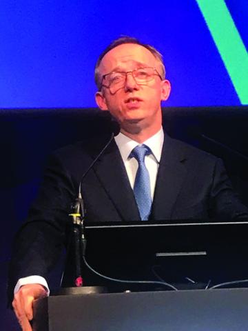
Sonelokimab is a novel humanized nanobody that selectively binds to interleukin (IL)-17A and IL-17F, presenting author Brian Kirby, MD, a dermatologist at St. Vincent’s Private Hospital, Dublin, Ireland, said during a late-breaking abstract session at the annual meeting of the American Academy of Dermatology. Sonelokimab is smaller than traditional monoclonal antibodies, he said, “which means it may be able to penetrate tissues better and stay there longer.” It is being developed by MoonLake Immunotherapeutics, based in Zug, Switzerland

According to a press release from the company, nanobodies represent a new generation of targeted therapies derived from antibodies. They consist of one or more domains based on the small antigen-binding variable regions of heavy-chain-only antibodies.
In a phase 2 study known as the MIRA trial, researchers recruited 234 patients with moderate to severe HS to evaluate two different doses of sonelokimab (120 mg and 240 mg every 2 weeks) with a placebo control arm and adalimumab as an active reference arm. The primary endpoint of was the percentage of participants who achieved a HiSCR75. The study population included adults with Hurley Stage II or III HS who had HS lesions in 2 or more anatomical areas and a total abscess and inflammatory nodule (AN) count of 5 or more lesions; and had been treated with 2 or fewer biologics.
Dr. Kirby reported results from 67 patients in the sonelokimab 120 mg arm, 66 in the sonelokimab 240 mg arm, and 39 in the placebo arm. “It’s worth noting that the baseline AN count ranged between 12 and nearly 15, the mean draining tunnel count ranged between 2.9 and 3.7, and between 7% and 13% of patients were on concomitant antibiotics,” he said.
At 24 weeks, 43.3% of patients in the sonelokimab 120 mg arm achieved a HiSCR 75, compared with 34.8% of those in the sonelokimab 240 mg arm and 14.7% of those in the placebo arm, he reported. Meanwhile, 65.7% of patients in the sonelokimab 120 mg arm achieved an HiSCR 75, compared with 53% of those in the sonelokimab 240 mg arm and 27.9% of those in the placebo arm. Discontinuation rates were low and similar between treatment arms, with fewer than 10% of patients failing to complete week 24 of treatment.
In other findings, 69% of patients in the 120 mg arm achieved a HiSCR 50 at week 24 compared with 60.3% in the 240 mg arm; 56.9% in the 120 mg arm achieved a HiSCR 75 compared with 37.9% in the 240 mg arm; and 37.9% in the 120 mg arm achieved a HiSCR 75 compared with 27.6% in the 240 mg arm.
In addition, complete inflammatory remission as defined by the International HS Severity Score System (IHS4-100) continued to increase to week 24, with 24.1% of patients in the 120 mg arm achieving complete remission, compared with 15.5% of those in the 240 mg arm. Meaningful improvements in quality of life, skin pain, and HS symptoms reported by patients treated with sonelokimab were maintained or increased to week 24. Specifically, more than 60% of patients had a meaningful clinical improvement in their Dermatology Life Quality Index, over 45% had a minimum of a 30% increase in the Numerical Rating Scale **30, and more than 41% of patients reported absent or minimal symptoms on the Patient’s Global Impression of their Disease Severity, “which is a high bar to achieve in HS,” Dr. Kirby said.
No Serious Safety Signals Noted
There were no unexpected safety signals to week 24. The incidence of treatment-related adverse events was low, and there were no cases of inflammatory bowel disease. There were no serious infections, no major adverse cardiovascular event (MACE) reports, and no significant abnormalities on liver function tests. “There were also no safety signals on suicidal behavior, attempted suicides, or completed suicides,” he said.
“As you would expect with in IL-17 inhibitor, there was a signal for candidiasis, but all cases were judged to be mild or moderate, and no cases led to discontinuation of treatment from the trial because of candidal infection.”
Based on these data, Dr. Kirby said that larger and longer-term phase 3 trials are planned to further examine the safety and efficacy of sonelokimab at the 120 mg dose for the treatment of moderate-to-severe HS.
One of the session moderators, Joel M. Gelfand, MD, professor of dermatology and epidemiology at the University of Pennsylvania, Philadelphia, asked Dr. Kirby why he thought the lower dose resulted in generally better outcomes compared with the higher dose.
“There were no statistically significant differences between the two groups,” Dr. Kirby responded. “The 120 mg dose we know is highly effective in psoriasis, so there may be a ceiling effect. This may also be due to population variability, but the bottom line is that the 120 mg dose performs extremely well.”
Dr. Kirby disclosed that he has received research support from/has been a principal investigator for several pharmaceutical companies, including MoonLake Immunotherapeutics. Dr. Gelfand reported that he has been a consultant to and/or a member of the data safety monitoring board member for several pharmaceutical companies, including MoonLake.
SAN DIEGO — After 24 weeks of treatment with subcutaneously administered relative to baseline, results from a randomized clinical trial showed.
Sonelokimab is a novel humanized nanobody that selectively binds to interleukin (IL)-17A and IL-17F, presenting author Brian Kirby, MD, a dermatologist at St. Vincent’s Private Hospital, Dublin, Ireland, said during a late-breaking abstract session at the annual meeting of the American Academy of Dermatology. Sonelokimab is smaller than traditional monoclonal antibodies, he said, “which means it may be able to penetrate tissues better and stay there longer.” It is being developed by MoonLake Immunotherapeutics, based in Zug, Switzerland
According to a press release from the company, nanobodies represent a new generation of targeted therapies derived from antibodies. They consist of one or more domains based on the small antigen-binding variable regions of heavy-chain-only antibodies.
In a phase 2 study known as the MIRA trial, researchers recruited 234 patients with moderate to severe HS to evaluate two different doses of sonelokimab (120 mg and 240 mg every 2 weeks) with a placebo control arm and adalimumab as an active reference arm. The primary endpoint of was the percentage of participants who achieved a HiSCR75. The study population included adults with Hurley Stage II or III HS who had HS lesions in 2 or more anatomical areas and a total abscess and inflammatory nodule (AN) count of 5 or more lesions; and had been treated with 2 or fewer biologics.
Dr. Kirby reported results from 67 patients in the sonelokimab 120 mg arm, 66 in the sonelokimab 240 mg arm, and 39 in the placebo arm. “It’s worth noting that the baseline AN count ranged between 12 and nearly 15, the mean draining tunnel count ranged between 2.9 and 3.7, and between 7% and 13% of patients were on concomitant antibiotics,” he said.
At 24 weeks, 43.3% of patients in the sonelokimab 120 mg arm achieved a HiSCR 75, compared with 34.8% of those in the sonelokimab 240 mg arm and 14.7% of those in the placebo arm, he reported. Meanwhile, 65.7% of patients in the sonelokimab 120 mg arm achieved an HiSCR 75, compared with 53% of those in the sonelokimab 240 mg arm and 27.9% of those in the placebo arm. Discontinuation rates were low and similar between treatment arms, with fewer than 10% of patients failing to complete week 24 of treatment.
In other findings, 69% of patients in the 120 mg arm achieved a HiSCR 50 at week 24 compared with 60.3% in the 240 mg arm; 56.9% in the 120 mg arm achieved a HiSCR 75 compared with 37.9% in the 240 mg arm; and 37.9% in the 120 mg arm achieved a HiSCR 75 compared with 27.6% in the 240 mg arm.
In addition, complete inflammatory remission as defined by the International HS Severity Score System (IHS4-100) continued to increase to week 24, with 24.1% of patients in the 120 mg arm achieving complete remission, compared with 15.5% of those in the 240 mg arm. Meaningful improvements in quality of life, skin pain, and HS symptoms reported by patients treated with sonelokimab were maintained or increased to week 24. Specifically, more than 60% of patients had a meaningful clinical improvement in their Dermatology Life Quality Index, over 45% had a minimum of a 30% increase in the Numerical Rating Scale **30, and more than 41% of patients reported absent or minimal symptoms on the Patient’s Global Impression of their Disease Severity, “which is a high bar to achieve in HS,” Dr. Kirby said.
No Serious Safety Signals Noted
There were no unexpected safety signals to week 24. The incidence of treatment-related adverse events was low, and there were no cases of inflammatory bowel disease. There were no serious infections, no major adverse cardiovascular event (MACE) reports, and no significant abnormalities on liver function tests. “There were also no safety signals on suicidal behavior, attempted suicides, or completed suicides,” he said.
“As you would expect with in IL-17 inhibitor, there was a signal for candidiasis, but all cases were judged to be mild or moderate, and no cases led to discontinuation of treatment from the trial because of candidal infection.”
Based on these data, Dr. Kirby said that larger and longer-term phase 3 trials are planned to further examine the safety and efficacy of sonelokimab at the 120 mg dose for the treatment of moderate-to-severe HS.
One of the session moderators, Joel M. Gelfand, MD, professor of dermatology and epidemiology at the University of Pennsylvania, Philadelphia, asked Dr. Kirby why he thought the lower dose resulted in generally better outcomes compared with the higher dose.
“There were no statistically significant differences between the two groups,” Dr. Kirby responded. “The 120 mg dose we know is highly effective in psoriasis, so there may be a ceiling effect. This may also be due to population variability, but the bottom line is that the 120 mg dose performs extremely well.”
Dr. Kirby disclosed that he has received research support from/has been a principal investigator for several pharmaceutical companies, including MoonLake Immunotherapeutics. Dr. Gelfand reported that he has been a consultant to and/or a member of the data safety monitoring board member for several pharmaceutical companies, including MoonLake.
SAN DIEGO — After 24 weeks of treatment with subcutaneously administered relative to baseline, results from a randomized clinical trial showed.
Sonelokimab is a novel humanized nanobody that selectively binds to interleukin (IL)-17A and IL-17F, presenting author Brian Kirby, MD, a dermatologist at St. Vincent’s Private Hospital, Dublin, Ireland, said during a late-breaking abstract session at the annual meeting of the American Academy of Dermatology. Sonelokimab is smaller than traditional monoclonal antibodies, he said, “which means it may be able to penetrate tissues better and stay there longer.” It is being developed by MoonLake Immunotherapeutics, based in Zug, Switzerland
According to a press release from the company, nanobodies represent a new generation of targeted therapies derived from antibodies. They consist of one or more domains based on the small antigen-binding variable regions of heavy-chain-only antibodies.
In a phase 2 study known as the MIRA trial, researchers recruited 234 patients with moderate to severe HS to evaluate two different doses of sonelokimab (120 mg and 240 mg every 2 weeks) with a placebo control arm and adalimumab as an active reference arm. The primary endpoint of was the percentage of participants who achieved a HiSCR75. The study population included adults with Hurley Stage II or III HS who had HS lesions in 2 or more anatomical areas and a total abscess and inflammatory nodule (AN) count of 5 or more lesions; and had been treated with 2 or fewer biologics.
Dr. Kirby reported results from 67 patients in the sonelokimab 120 mg arm, 66 in the sonelokimab 240 mg arm, and 39 in the placebo arm. “It’s worth noting that the baseline AN count ranged between 12 and nearly 15, the mean draining tunnel count ranged between 2.9 and 3.7, and between 7% and 13% of patients were on concomitant antibiotics,” he said.
At 24 weeks, 43.3% of patients in the sonelokimab 120 mg arm achieved a HiSCR 75, compared with 34.8% of those in the sonelokimab 240 mg arm and 14.7% of those in the placebo arm, he reported. Meanwhile, 65.7% of patients in the sonelokimab 120 mg arm achieved an HiSCR 75, compared with 53% of those in the sonelokimab 240 mg arm and 27.9% of those in the placebo arm. Discontinuation rates were low and similar between treatment arms, with fewer than 10% of patients failing to complete week 24 of treatment.
In other findings, 69% of patients in the 120 mg arm achieved a HiSCR 50 at week 24 compared with 60.3% in the 240 mg arm; 56.9% in the 120 mg arm achieved a HiSCR 75 compared with 37.9% in the 240 mg arm; and 37.9% in the 120 mg arm achieved a HiSCR 75 compared with 27.6% in the 240 mg arm.
In addition, complete inflammatory remission as defined by the International HS Severity Score System (IHS4-100) continued to increase to week 24, with 24.1% of patients in the 120 mg arm achieving complete remission, compared with 15.5% of those in the 240 mg arm. Meaningful improvements in quality of life, skin pain, and HS symptoms reported by patients treated with sonelokimab were maintained or increased to week 24. Specifically, more than 60% of patients had a meaningful clinical improvement in their Dermatology Life Quality Index, over 45% had a minimum of a 30% increase in the Numerical Rating Scale **30, and more than 41% of patients reported absent or minimal symptoms on the Patient’s Global Impression of their Disease Severity, “which is a high bar to achieve in HS,” Dr. Kirby said.
No Serious Safety Signals Noted
There were no unexpected safety signals to week 24. The incidence of treatment-related adverse events was low, and there were no cases of inflammatory bowel disease. There were no serious infections, no major adverse cardiovascular event (MACE) reports, and no significant abnormalities on liver function tests. “There were also no safety signals on suicidal behavior, attempted suicides, or completed suicides,” he said.
“As you would expect with in IL-17 inhibitor, there was a signal for candidiasis, but all cases were judged to be mild or moderate, and no cases led to discontinuation of treatment from the trial because of candidal infection.”
Based on these data, Dr. Kirby said that larger and longer-term phase 3 trials are planned to further examine the safety and efficacy of sonelokimab at the 120 mg dose for the treatment of moderate-to-severe HS.
One of the session moderators, Joel M. Gelfand, MD, professor of dermatology and epidemiology at the University of Pennsylvania, Philadelphia, asked Dr. Kirby why he thought the lower dose resulted in generally better outcomes compared with the higher dose.
“There were no statistically significant differences between the two groups,” Dr. Kirby responded. “The 120 mg dose we know is highly effective in psoriasis, so there may be a ceiling effect. This may also be due to population variability, but the bottom line is that the 120 mg dose performs extremely well.”
Dr. Kirby disclosed that he has received research support from/has been a principal investigator for several pharmaceutical companies, including MoonLake Immunotherapeutics. Dr. Gelfand reported that he has been a consultant to and/or a member of the data safety monitoring board member for several pharmaceutical companies, including MoonLake.
FROM AAD 2024
New Analysis Defines Three Distinct Sjögren Syndrome Subgroups
Researchers have identified three distinct subgroups of Sjögren syndrome (SS) with different prognoses. While previous efforts to stratify these patients were based on reported symptoms, this new analysis is the first to add clinical and biological manifestations into the equation.
The three distinct patient clusters are those with B-cell active disease with low symptom burden (BALS), those with high systemic disease activity (HSA), and those with low systemic disease activity and high symptom burden (LSAHS). Each group had distinct long-term outcomes, including disease evolution and lymphoma incidence.
The findings were published in The Lancet Rheumatology.
Derived and Validated in Separate Cohorts
Researchers led by Yann Nguyen, MD, PhD, of Bicêtre Hospital and Paris-Saclay University, Paris, France, identified distinct subgroups with data from the French Paris-Saclay cohort, a group of patients suspected of having SS who participated in a multidisciplinary diagnostic session at a French National Referral Center for Rare Systemic Autoimmune Diseases and were recruited between 1999 and 2022.

The study included only patients who met the 2002 American-European Consensus Group criteria for SS and had European Alliance of Associations for Rheumatology (EULAR) Sjögren’s Syndrome Disease Activity Index (ESSDAI) and EULAR Sjögren’s Syndrome Patient Reported Index (ESSPRI) scores at inclusion. Researchers excluded patients with other autoimmune diseases associated with SS.
Researchers identified distinct subgroups using 26 variables, including patient-reported symptoms, clinical parameters, and biological data.
Researchers then validated the subgroups using the same methodology in the Assessment of Systemic Signs and Evolution of Sjögren’s syndrome (ASSESS) cohort, a national French multicenter prospective cohort formed in 2006. They then compared changes in disease activity (measured by ESSDAI) and patient-acceptable symptom state (measured by ESSPRI) between the groups over 5 years of follow-up, as well as differences in lymphoma incidence over 15 years of follow-up.
Patients With High Systemic Disease, Low Symptoms Fare Worse
The study ultimately included 534 patients from the Paris-Saclay cohort and 395 patients from the ASSESS cohort. For both groups, 94% of patients were women, and the median age was between 53 and 54 years.
In the Paris-Saclay group, 205 patients were classified as BALS, 160 were HSA, and 169 were LSAHS. In the ASSESS cohort, BALS remained the largest group (186 patients), followed by HSA (158 patients). Only 51 patients in the second cohort were classified as LSAHS.
During 5 years of follow-up in the ASSESS cohort, systemic disease activity improved in the HSA cluster and worsened in the BALS cluster, while there were no significant changes in the LSAHS cluster. The BALS cluster was the only group to experience significant changes in patient-acceptable symptom states during follow-up: 49% of patients with BALS had an ESSPRI score of less than 5 at inclusion, but this percentage dipped to 36% at month 60.
The findings “highlight the fact that even in patients who present with predominantly systemic manifestations, the symptom burden is high and should not be neglected,” the authors wrote.
The three categories established in this study did not correlate well with previous symptom-based stratification of patients with SS, the authors noted, which looked at the five most common symptoms associated with the condition: Pain, fatigue, dryness, anxiety, and depression.
“Patients from the high symptom burden, dryness dominant with fatigue, and pain dominant with fatigue subgroups were present in each of our three clusters,” they wrote. “This finding is consistent with the poor correlation between patient-reported outcomes and systemic disease activity.”
Lymphoma was diagnosed in 5 of 186 patients (3%) in the BALS cluster, diagnosed after a median of 70 months of follow-up, and 6 of 158 patients (4%) in the HSA cluster, diagnosed after a median of 23 months follow-up. There were no cases of lymphoma in the LSAHS group.
“Notably, in the BALS cluster, lymphoma occurred later than in the HSA cluster, and after 5 years, systemic manifestations in this cluster tended to be similar to those in the HSA cluster at inclusion,” the authors added. “The BALS cluster could therefore represent an earlier stage of the disease and carry the risk of progressing toward a more systemic phenotype.”
A ‘First Step’ to Subgrouping Patients
Alan Baer, MD, director of the Jerome Greene Sjogren’s Syndrome Center at John Hopkins Medical Center in Baltimore, Maryland, who commented on the study, noted that these three subgroups did “resonate” with what clinicians see in practice. Certain patients may have lab results that are “quite striking” even though they report minimal symptoms. The reverse is also true: Patients who have a high symptom burden without signs of systemic disease activity. Whether these patients should be managed differently “remains the key question,” he said.

“The hope is that when you have a group of patients that’s relatively homogenous in terms of their clinical features, that also translates to similarities into the underlying pathogenesis that can then lead to treatments that are targeted to specific subgroups of patients,” he added.
Another approach could be looking directly at molecular alterations across patients with SS and seeing how they correlate to clinical features, Dr. Baer noted.
This study “is a first step” in figuring out how to best define SS subgroups, he said, adding that “there’s more work to be done.”
This research was funded by the Fondation pour la Recherche Medicale, French Ministry of Health, French Society of Rheumatology, Innovative Medicines Initiative 2 Joint Undertaking, Medical Research Council UK, and Foundation for Research in Rheumatology. The study authors disclosed financial relationships with various pharmaceutical companies. Dr. Baer had no relevant disclosures.
A version of this article appeared on Medscape.com .
Researchers have identified three distinct subgroups of Sjögren syndrome (SS) with different prognoses. While previous efforts to stratify these patients were based on reported symptoms, this new analysis is the first to add clinical and biological manifestations into the equation.
The three distinct patient clusters are those with B-cell active disease with low symptom burden (BALS), those with high systemic disease activity (HSA), and those with low systemic disease activity and high symptom burden (LSAHS). Each group had distinct long-term outcomes, including disease evolution and lymphoma incidence.
The findings were published in The Lancet Rheumatology.
Derived and Validated in Separate Cohorts
Researchers led by Yann Nguyen, MD, PhD, of Bicêtre Hospital and Paris-Saclay University, Paris, France, identified distinct subgroups with data from the French Paris-Saclay cohort, a group of patients suspected of having SS who participated in a multidisciplinary diagnostic session at a French National Referral Center for Rare Systemic Autoimmune Diseases and were recruited between 1999 and 2022.
The study included only patients who met the 2002 American-European Consensus Group criteria for SS and had European Alliance of Associations for Rheumatology (EULAR) Sjögren’s Syndrome Disease Activity Index (ESSDAI) and EULAR Sjögren’s Syndrome Patient Reported Index (ESSPRI) scores at inclusion. Researchers excluded patients with other autoimmune diseases associated with SS.
Researchers identified distinct subgroups using 26 variables, including patient-reported symptoms, clinical parameters, and biological data.
Researchers then validated the subgroups using the same methodology in the Assessment of Systemic Signs and Evolution of Sjögren’s syndrome (ASSESS) cohort, a national French multicenter prospective cohort formed in 2006. They then compared changes in disease activity (measured by ESSDAI) and patient-acceptable symptom state (measured by ESSPRI) between the groups over 5 years of follow-up, as well as differences in lymphoma incidence over 15 years of follow-up.
Patients With High Systemic Disease, Low Symptoms Fare Worse
The study ultimately included 534 patients from the Paris-Saclay cohort and 395 patients from the ASSESS cohort. For both groups, 94% of patients were women, and the median age was between 53 and 54 years.
In the Paris-Saclay group, 205 patients were classified as BALS, 160 were HSA, and 169 were LSAHS. In the ASSESS cohort, BALS remained the largest group (186 patients), followed by HSA (158 patients). Only 51 patients in the second cohort were classified as LSAHS.
During 5 years of follow-up in the ASSESS cohort, systemic disease activity improved in the HSA cluster and worsened in the BALS cluster, while there were no significant changes in the LSAHS cluster. The BALS cluster was the only group to experience significant changes in patient-acceptable symptom states during follow-up: 49% of patients with BALS had an ESSPRI score of less than 5 at inclusion, but this percentage dipped to 36% at month 60.
The findings “highlight the fact that even in patients who present with predominantly systemic manifestations, the symptom burden is high and should not be neglected,” the authors wrote.
The three categories established in this study did not correlate well with previous symptom-based stratification of patients with SS, the authors noted, which looked at the five most common symptoms associated with the condition: Pain, fatigue, dryness, anxiety, and depression.
“Patients from the high symptom burden, dryness dominant with fatigue, and pain dominant with fatigue subgroups were present in each of our three clusters,” they wrote. “This finding is consistent with the poor correlation between patient-reported outcomes and systemic disease activity.”
Lymphoma was diagnosed in 5 of 186 patients (3%) in the BALS cluster, diagnosed after a median of 70 months of follow-up, and 6 of 158 patients (4%) in the HSA cluster, diagnosed after a median of 23 months follow-up. There were no cases of lymphoma in the LSAHS group.
“Notably, in the BALS cluster, lymphoma occurred later than in the HSA cluster, and after 5 years, systemic manifestations in this cluster tended to be similar to those in the HSA cluster at inclusion,” the authors added. “The BALS cluster could therefore represent an earlier stage of the disease and carry the risk of progressing toward a more systemic phenotype.”
A ‘First Step’ to Subgrouping Patients
Alan Baer, MD, director of the Jerome Greene Sjogren’s Syndrome Center at John Hopkins Medical Center in Baltimore, Maryland, who commented on the study, noted that these three subgroups did “resonate” with what clinicians see in practice. Certain patients may have lab results that are “quite striking” even though they report minimal symptoms. The reverse is also true: Patients who have a high symptom burden without signs of systemic disease activity. Whether these patients should be managed differently “remains the key question,” he said.
“The hope is that when you have a group of patients that’s relatively homogenous in terms of their clinical features, that also translates to similarities into the underlying pathogenesis that can then lead to treatments that are targeted to specific subgroups of patients,” he added.
Another approach could be looking directly at molecular alterations across patients with SS and seeing how they correlate to clinical features, Dr. Baer noted.
This study “is a first step” in figuring out how to best define SS subgroups, he said, adding that “there’s more work to be done.”
This research was funded by the Fondation pour la Recherche Medicale, French Ministry of Health, French Society of Rheumatology, Innovative Medicines Initiative 2 Joint Undertaking, Medical Research Council UK, and Foundation for Research in Rheumatology. The study authors disclosed financial relationships with various pharmaceutical companies. Dr. Baer had no relevant disclosures.
A version of this article appeared on Medscape.com .
Researchers have identified three distinct subgroups of Sjögren syndrome (SS) with different prognoses. While previous efforts to stratify these patients were based on reported symptoms, this new analysis is the first to add clinical and biological manifestations into the equation.
The three distinct patient clusters are those with B-cell active disease with low symptom burden (BALS), those with high systemic disease activity (HSA), and those with low systemic disease activity and high symptom burden (LSAHS). Each group had distinct long-term outcomes, including disease evolution and lymphoma incidence.
The findings were published in The Lancet Rheumatology.
Derived and Validated in Separate Cohorts
Researchers led by Yann Nguyen, MD, PhD, of Bicêtre Hospital and Paris-Saclay University, Paris, France, identified distinct subgroups with data from the French Paris-Saclay cohort, a group of patients suspected of having SS who participated in a multidisciplinary diagnostic session at a French National Referral Center for Rare Systemic Autoimmune Diseases and were recruited between 1999 and 2022.
The study included only patients who met the 2002 American-European Consensus Group criteria for SS and had European Alliance of Associations for Rheumatology (EULAR) Sjögren’s Syndrome Disease Activity Index (ESSDAI) and EULAR Sjögren’s Syndrome Patient Reported Index (ESSPRI) scores at inclusion. Researchers excluded patients with other autoimmune diseases associated with SS.
Researchers identified distinct subgroups using 26 variables, including patient-reported symptoms, clinical parameters, and biological data.
Researchers then validated the subgroups using the same methodology in the Assessment of Systemic Signs and Evolution of Sjögren’s syndrome (ASSESS) cohort, a national French multicenter prospective cohort formed in 2006. They then compared changes in disease activity (measured by ESSDAI) and patient-acceptable symptom state (measured by ESSPRI) between the groups over 5 years of follow-up, as well as differences in lymphoma incidence over 15 years of follow-up.
Patients With High Systemic Disease, Low Symptoms Fare Worse
The study ultimately included 534 patients from the Paris-Saclay cohort and 395 patients from the ASSESS cohort. For both groups, 94% of patients were women, and the median age was between 53 and 54 years.
In the Paris-Saclay group, 205 patients were classified as BALS, 160 were HSA, and 169 were LSAHS. In the ASSESS cohort, BALS remained the largest group (186 patients), followed by HSA (158 patients). Only 51 patients in the second cohort were classified as LSAHS.
During 5 years of follow-up in the ASSESS cohort, systemic disease activity improved in the HSA cluster and worsened in the BALS cluster, while there were no significant changes in the LSAHS cluster. The BALS cluster was the only group to experience significant changes in patient-acceptable symptom states during follow-up: 49% of patients with BALS had an ESSPRI score of less than 5 at inclusion, but this percentage dipped to 36% at month 60.
The findings “highlight the fact that even in patients who present with predominantly systemic manifestations, the symptom burden is high and should not be neglected,” the authors wrote.
The three categories established in this study did not correlate well with previous symptom-based stratification of patients with SS, the authors noted, which looked at the five most common symptoms associated with the condition: Pain, fatigue, dryness, anxiety, and depression.
“Patients from the high symptom burden, dryness dominant with fatigue, and pain dominant with fatigue subgroups were present in each of our three clusters,” they wrote. “This finding is consistent with the poor correlation between patient-reported outcomes and systemic disease activity.”
Lymphoma was diagnosed in 5 of 186 patients (3%) in the BALS cluster, diagnosed after a median of 70 months of follow-up, and 6 of 158 patients (4%) in the HSA cluster, diagnosed after a median of 23 months follow-up. There were no cases of lymphoma in the LSAHS group.
“Notably, in the BALS cluster, lymphoma occurred later than in the HSA cluster, and after 5 years, systemic manifestations in this cluster tended to be similar to those in the HSA cluster at inclusion,” the authors added. “The BALS cluster could therefore represent an earlier stage of the disease and carry the risk of progressing toward a more systemic phenotype.”
A ‘First Step’ to Subgrouping Patients
Alan Baer, MD, director of the Jerome Greene Sjogren’s Syndrome Center at John Hopkins Medical Center in Baltimore, Maryland, who commented on the study, noted that these three subgroups did “resonate” with what clinicians see in practice. Certain patients may have lab results that are “quite striking” even though they report minimal symptoms. The reverse is also true: Patients who have a high symptom burden without signs of systemic disease activity. Whether these patients should be managed differently “remains the key question,” he said.
“The hope is that when you have a group of patients that’s relatively homogenous in terms of their clinical features, that also translates to similarities into the underlying pathogenesis that can then lead to treatments that are targeted to specific subgroups of patients,” he added.
Another approach could be looking directly at molecular alterations across patients with SS and seeing how they correlate to clinical features, Dr. Baer noted.
This study “is a first step” in figuring out how to best define SS subgroups, he said, adding that “there’s more work to be done.”
This research was funded by the Fondation pour la Recherche Medicale, French Ministry of Health, French Society of Rheumatology, Innovative Medicines Initiative 2 Joint Undertaking, Medical Research Council UK, and Foundation for Research in Rheumatology. The study authors disclosed financial relationships with various pharmaceutical companies. Dr. Baer had no relevant disclosures.
A version of this article appeared on Medscape.com .
FROM THE LANCET RHEUMATOLOGY

Non-Radical Surgery a Win-Win for Early Cervical Cancer
according to results of the GOG-278 trial.
In fact, patients’ quality of life was improved after surgery in both groups, and their concerns about cancer recurrence decreased, especially for those undergoing simple hysterectomy, said Allan Covens, MD, in his late-breaking abstract presentation at the Society of Gynecologic Oncology (SGO)’s Annual Meeting on Women’s Cancer.
“Cone biopsy patients reported less concerns about reproductive fertility after surgery and over time compared to preop assessments,” he added.
Due to screening in developed countries, a large proportion of cervical cancers are discovered at an early stage. Treatment of these cancers with radical surgery is associated with high cure rates but significant adverse effects on quality of life, said Dr. Covens, who is with the University of Toronto, Toronto, Ontario, Canada.
He and his colleagues wanted to see if non-radical surgery could be safely used instead. “Multiple case series have indicated that non-radical surgery is associated with less morbidity and improved quality of life,” he explained. “If this can be proven in a prospective evaluation, it will change future practice.”
GOG-278 was a prospective cohort study of women with stage IA1 (lymph-vascular space invasion+) and IA2-IB1 (≤ 2 cm) carcinoma of the cervix who underwent non-radical surgery (simple hysterectomy or fertility-preserving cone biopsy) and pelvic lymphadenectomy. Criteria included ≤ 10 mm stromal invasion and negative margins on the final cone biopsy.
The primary objectives were to assess changes in functional outcomes of quality of life (bladder/bowel function, sexual function, cancer worry, and reproductive concerns), using validated instruments. Findings were based on 55 patients who underwent cone biopsy and 113 who underwent simple hysterectomy.
Both simple hysterectomy and cone biopsy were associated with “small” declines in sexual function and bladder/bowel function at 4-6 weeks after surgery, but function “quickly” recovered to baseline by 6 months, Dr. Covens reported.
Twelve patients reported a diagnosis of lymphedema, with a Gynecologic Cancer Lymphedema Questionnaire score change of 4 or higher on at least two consecutive evaluations from baseline. This occurred in six cone biopsy and six simple hysterectomy patients.
In a separate presentation, Dr. Covens reported secondary oncologic outcomes from GOG-278, which suggest that non-radical surgery for early-stage cervical cancer is safe, with low perioperative morbidity, although longer follow-up is needed.
He also reported 16 pregnancies in 15 patients who had undergone cone biopsies; 12 of these were successful, and there were four early pregnancy losses.
‘Impressive’ Data
Study discussant Kristin Bixel, MD, with The Ohio State University, Columbus, Ohio, said the data are “impressive” and clearly show that non-radical surgery has “minimal impact on bladder/bowel function, with no long-term differences from baseline.”
She added that the incidence of lymphedema was “honestly significantly lower than what I typically counsel patients about” and wondered if the percentage of patients with lymphedema would increase over time.
Dr. Bixel particularly noted the decrease in cancer worry scores after surgery, as sometimes patients who have less radical procedures fear that this comes with an increased risk for recurrence.
The “growing body of data suggests that less radical surgery is safe and effective for early-stage low-risk cervical cancer and highlights the potential reproductive success,” she concluded.
Funding for the study was provided by grants from NRG Oncology. Dr. Covens had no disclosures. Dr. Bixel has received research funding from the Intuitive Foundation.
A version of this article appeared on Medscape.com.
according to results of the GOG-278 trial.
In fact, patients’ quality of life was improved after surgery in both groups, and their concerns about cancer recurrence decreased, especially for those undergoing simple hysterectomy, said Allan Covens, MD, in his late-breaking abstract presentation at the Society of Gynecologic Oncology (SGO)’s Annual Meeting on Women’s Cancer.
“Cone biopsy patients reported less concerns about reproductive fertility after surgery and over time compared to preop assessments,” he added.
Due to screening in developed countries, a large proportion of cervical cancers are discovered at an early stage. Treatment of these cancers with radical surgery is associated with high cure rates but significant adverse effects on quality of life, said Dr. Covens, who is with the University of Toronto, Toronto, Ontario, Canada.
He and his colleagues wanted to see if non-radical surgery could be safely used instead. “Multiple case series have indicated that non-radical surgery is associated with less morbidity and improved quality of life,” he explained. “If this can be proven in a prospective evaluation, it will change future practice.”
GOG-278 was a prospective cohort study of women with stage IA1 (lymph-vascular space invasion+) and IA2-IB1 (≤ 2 cm) carcinoma of the cervix who underwent non-radical surgery (simple hysterectomy or fertility-preserving cone biopsy) and pelvic lymphadenectomy. Criteria included ≤ 10 mm stromal invasion and negative margins on the final cone biopsy.
The primary objectives were to assess changes in functional outcomes of quality of life (bladder/bowel function, sexual function, cancer worry, and reproductive concerns), using validated instruments. Findings were based on 55 patients who underwent cone biopsy and 113 who underwent simple hysterectomy.
Both simple hysterectomy and cone biopsy were associated with “small” declines in sexual function and bladder/bowel function at 4-6 weeks after surgery, but function “quickly” recovered to baseline by 6 months, Dr. Covens reported.
Twelve patients reported a diagnosis of lymphedema, with a Gynecologic Cancer Lymphedema Questionnaire score change of 4 or higher on at least two consecutive evaluations from baseline. This occurred in six cone biopsy and six simple hysterectomy patients.
In a separate presentation, Dr. Covens reported secondary oncologic outcomes from GOG-278, which suggest that non-radical surgery for early-stage cervical cancer is safe, with low perioperative morbidity, although longer follow-up is needed.
He also reported 16 pregnancies in 15 patients who had undergone cone biopsies; 12 of these were successful, and there were four early pregnancy losses.
‘Impressive’ Data
Study discussant Kristin Bixel, MD, with The Ohio State University, Columbus, Ohio, said the data are “impressive” and clearly show that non-radical surgery has “minimal impact on bladder/bowel function, with no long-term differences from baseline.”
She added that the incidence of lymphedema was “honestly significantly lower than what I typically counsel patients about” and wondered if the percentage of patients with lymphedema would increase over time.
Dr. Bixel particularly noted the decrease in cancer worry scores after surgery, as sometimes patients who have less radical procedures fear that this comes with an increased risk for recurrence.
The “growing body of data suggests that less radical surgery is safe and effective for early-stage low-risk cervical cancer and highlights the potential reproductive success,” she concluded.
Funding for the study was provided by grants from NRG Oncology. Dr. Covens had no disclosures. Dr. Bixel has received research funding from the Intuitive Foundation.
A version of this article appeared on Medscape.com.
according to results of the GOG-278 trial.
In fact, patients’ quality of life was improved after surgery in both groups, and their concerns about cancer recurrence decreased, especially for those undergoing simple hysterectomy, said Allan Covens, MD, in his late-breaking abstract presentation at the Society of Gynecologic Oncology (SGO)’s Annual Meeting on Women’s Cancer.
“Cone biopsy patients reported less concerns about reproductive fertility after surgery and over time compared to preop assessments,” he added.
Due to screening in developed countries, a large proportion of cervical cancers are discovered at an early stage. Treatment of these cancers with radical surgery is associated with high cure rates but significant adverse effects on quality of life, said Dr. Covens, who is with the University of Toronto, Toronto, Ontario, Canada.
He and his colleagues wanted to see if non-radical surgery could be safely used instead. “Multiple case series have indicated that non-radical surgery is associated with less morbidity and improved quality of life,” he explained. “If this can be proven in a prospective evaluation, it will change future practice.”
GOG-278 was a prospective cohort study of women with stage IA1 (lymph-vascular space invasion+) and IA2-IB1 (≤ 2 cm) carcinoma of the cervix who underwent non-radical surgery (simple hysterectomy or fertility-preserving cone biopsy) and pelvic lymphadenectomy. Criteria included ≤ 10 mm stromal invasion and negative margins on the final cone biopsy.
The primary objectives were to assess changes in functional outcomes of quality of life (bladder/bowel function, sexual function, cancer worry, and reproductive concerns), using validated instruments. Findings were based on 55 patients who underwent cone biopsy and 113 who underwent simple hysterectomy.
Both simple hysterectomy and cone biopsy were associated with “small” declines in sexual function and bladder/bowel function at 4-6 weeks after surgery, but function “quickly” recovered to baseline by 6 months, Dr. Covens reported.
Twelve patients reported a diagnosis of lymphedema, with a Gynecologic Cancer Lymphedema Questionnaire score change of 4 or higher on at least two consecutive evaluations from baseline. This occurred in six cone biopsy and six simple hysterectomy patients.
In a separate presentation, Dr. Covens reported secondary oncologic outcomes from GOG-278, which suggest that non-radical surgery for early-stage cervical cancer is safe, with low perioperative morbidity, although longer follow-up is needed.
He also reported 16 pregnancies in 15 patients who had undergone cone biopsies; 12 of these were successful, and there were four early pregnancy losses.
‘Impressive’ Data
Study discussant Kristin Bixel, MD, with The Ohio State University, Columbus, Ohio, said the data are “impressive” and clearly show that non-radical surgery has “minimal impact on bladder/bowel function, with no long-term differences from baseline.”
She added that the incidence of lymphedema was “honestly significantly lower than what I typically counsel patients about” and wondered if the percentage of patients with lymphedema would increase over time.
Dr. Bixel particularly noted the decrease in cancer worry scores after surgery, as sometimes patients who have less radical procedures fear that this comes with an increased risk for recurrence.
The “growing body of data suggests that less radical surgery is safe and effective for early-stage low-risk cervical cancer and highlights the potential reproductive success,” she concluded.
Funding for the study was provided by grants from NRG Oncology. Dr. Covens had no disclosures. Dr. Bixel has received research funding from the Intuitive Foundation.
A version of this article appeared on Medscape.com.
FROM SGO 2024
Very Low-Energy Diet Safe, Acceptable for Adolescents
More research is needed to understand which patients are best suited for the diet; “however, given the associated rapid weight loss, the use of [very low-energy diets] should be emphasized in clinical practice guidelines for the treatment of severe obesity and obesity-related complications in adolescents, especially before pharmacological or surgical intervention,” first author Megan Gow, PhD, of Children’s Hospital Westmead Clinical School, The University of Sydney, Westmead, Australia, said in a press statement.
The study will be presented in May at the upcoming European Congress on Obesity, in Venice, Italy.
While very low-calorie diets have been shown to promote rapid weight loss in adolescents, research is lacking on general side effects and acceptability of the regimens. Data is also lacking on important issues including the diet’s effect on growth, heart health, and psychological wellbeing.
To investigate, Dr. Gow and colleagues conducted a subanalysis of the 52-week Fast Track to Health study evaluating the acceptability of different dietary plans for adolescents with obesity.
The analysis included 141 adolescents between the ages of 13 and 17 years with moderate to severe obesity (average body mass index, 35 kg/m2) and at least one obesity-related complication, such as high blood pressure or insulin resistance.
The participants were placed on a nutritionally balanced very low-energy diet consisting of 800 calories per day.
The diet involved one of two regimens — either four Optifast-formulated meal replacement products per day, including shakes, soups, bars, and/or dessert, along with low carbohydrate vegetables, such as broccoli, celery, capsicum, mushrooms, and tomatoes, with one teaspoon of vegetable oil, or a regimen of three Optifast-formulated meal replacements and one meal consisting of 100-150 g lean cooked meat, low-carbohydrate vegetables, and one teaspoon of vegetable oil.
Participants, about half of whom were women, also received dietitian support at least weekly.
After 4 weeks, most of the adolescents, ie, 134 of the 141, with an average age of 14.9 years, completed the diet, with an average weight loss of 5.5 kg or 12 pounds (P < .001).
Most patients (95%) experienced at least one side effect, and 70% reported at least 3 of the side effects, with the most common side effects including hunger, fatigue, headache, irritability, loose stools, constipation, nausea, and a lack of concentration.
Viral infections occurred in seven participants.
While most side effects occurred at the end of week 1, the development of side effects earlier, at day 3-4, was associated with higher levels of weight loss at the 4-week cut-off, which the authors noted could suggest a greater adherence to the diet at that stage.
One adverse event occurred, consisting of a single fainting episode determined to be potentially related to the dietary intervention.
In surveys, the adolescents gave the intervention an acceptability rating of 61 on a scale of 100, the score was 53 of 100 in terms of being “enjoyable to follow.”
The most-liked aspects of the intervention were losing weight (described by 34% of participants) and the prescriptive structure (listed by 28% of participants).
The least-liked aspects included the diet’s restrictive nature, described by 45% of participants, and the taste of meal replacement products, listed by 20% of participants.
Alternative to Weight Loss Drugs?
While weight loss drugs are transforming the obesity treatment and semaglutide is now approved for adolescents as young as age 12 years, “access to these medications is limited, and not all families want to commence on medication for their child›s obesity,” Dr. Gow said.
As an alternative, a very low-energy diet, with the interaction of a dietitian, can enable adolescents “to develop a healthier relationship with food, including encouraging the consumption of more fruits and vegetables in their diet, not only to assist in weight loss but for good health,” she said.
Very Low-Calorie Diet Concerns for Adolescents Addressed
Early studies suggested concerns of health effects from very low-calorie diets in adolescents, including potential cardiac effects; however, subsequent studies, including a systematic review published by Dr. Gow and her team, have shown that such results were likely the result of nutritional deficiencies in the diets, which can be overcome with careful food selection and dietary counseling.
Another key concern has been a potential effect on growth, but Dr. Gow noted that “in our short-term study we saw small increase in height (0.1 cm), and other more recent studies suggest that a short-term very low-energy diet does not impact growth.”
And in an earlier pilot study, the authors also found an association between the very low-calorie diet and an improvement in the quality of life for youth with type 2 diabetes.
A key caveat with the findings is that participants in the study all received supervision and monitoring from a trained dietitian, and Gow noted that that element is essential.
“We therefore do not recommend adolescents in the community undertake this type of diet without appropriate support,” she said.
“Close monitoring of adolescents by a health professional following a very low-energy diet is essential to ensure that the very low-energy diet is leading to holistic health benefits for the individual.”
Following the 4-week regimen, participants were randomized to transition to interventions of either continuous energy restriction or intermittent energy restriction over the 52 weeks, and further findings from the study will be presented at the obesity meeting in May.
The authors had no disclosures to report.
A version of this article appeared on Medscape.com.
More research is needed to understand which patients are best suited for the diet; “however, given the associated rapid weight loss, the use of [very low-energy diets] should be emphasized in clinical practice guidelines for the treatment of severe obesity and obesity-related complications in adolescents, especially before pharmacological or surgical intervention,” first author Megan Gow, PhD, of Children’s Hospital Westmead Clinical School, The University of Sydney, Westmead, Australia, said in a press statement.
The study will be presented in May at the upcoming European Congress on Obesity, in Venice, Italy.
While very low-calorie diets have been shown to promote rapid weight loss in adolescents, research is lacking on general side effects and acceptability of the regimens. Data is also lacking on important issues including the diet’s effect on growth, heart health, and psychological wellbeing.
To investigate, Dr. Gow and colleagues conducted a subanalysis of the 52-week Fast Track to Health study evaluating the acceptability of different dietary plans for adolescents with obesity.
The analysis included 141 adolescents between the ages of 13 and 17 years with moderate to severe obesity (average body mass index, 35 kg/m2) and at least one obesity-related complication, such as high blood pressure or insulin resistance.
The participants were placed on a nutritionally balanced very low-energy diet consisting of 800 calories per day.
The diet involved one of two regimens — either four Optifast-formulated meal replacement products per day, including shakes, soups, bars, and/or dessert, along with low carbohydrate vegetables, such as broccoli, celery, capsicum, mushrooms, and tomatoes, with one teaspoon of vegetable oil, or a regimen of three Optifast-formulated meal replacements and one meal consisting of 100-150 g lean cooked meat, low-carbohydrate vegetables, and one teaspoon of vegetable oil.
Participants, about half of whom were women, also received dietitian support at least weekly.
After 4 weeks, most of the adolescents, ie, 134 of the 141, with an average age of 14.9 years, completed the diet, with an average weight loss of 5.5 kg or 12 pounds (P < .001).
Most patients (95%) experienced at least one side effect, and 70% reported at least 3 of the side effects, with the most common side effects including hunger, fatigue, headache, irritability, loose stools, constipation, nausea, and a lack of concentration.
Viral infections occurred in seven participants.
While most side effects occurred at the end of week 1, the development of side effects earlier, at day 3-4, was associated with higher levels of weight loss at the 4-week cut-off, which the authors noted could suggest a greater adherence to the diet at that stage.
One adverse event occurred, consisting of a single fainting episode determined to be potentially related to the dietary intervention.
In surveys, the adolescents gave the intervention an acceptability rating of 61 on a scale of 100, the score was 53 of 100 in terms of being “enjoyable to follow.”
The most-liked aspects of the intervention were losing weight (described by 34% of participants) and the prescriptive structure (listed by 28% of participants).
The least-liked aspects included the diet’s restrictive nature, described by 45% of participants, and the taste of meal replacement products, listed by 20% of participants.
Alternative to Weight Loss Drugs?
While weight loss drugs are transforming the obesity treatment and semaglutide is now approved for adolescents as young as age 12 years, “access to these medications is limited, and not all families want to commence on medication for their child›s obesity,” Dr. Gow said.
As an alternative, a very low-energy diet, with the interaction of a dietitian, can enable adolescents “to develop a healthier relationship with food, including encouraging the consumption of more fruits and vegetables in their diet, not only to assist in weight loss but for good health,” she said.
Very Low-Calorie Diet Concerns for Adolescents Addressed
Early studies suggested concerns of health effects from very low-calorie diets in adolescents, including potential cardiac effects; however, subsequent studies, including a systematic review published by Dr. Gow and her team, have shown that such results were likely the result of nutritional deficiencies in the diets, which can be overcome with careful food selection and dietary counseling.
Another key concern has been a potential effect on growth, but Dr. Gow noted that “in our short-term study we saw small increase in height (0.1 cm), and other more recent studies suggest that a short-term very low-energy diet does not impact growth.”
And in an earlier pilot study, the authors also found an association between the very low-calorie diet and an improvement in the quality of life for youth with type 2 diabetes.
A key caveat with the findings is that participants in the study all received supervision and monitoring from a trained dietitian, and Gow noted that that element is essential.
“We therefore do not recommend adolescents in the community undertake this type of diet without appropriate support,” she said.
“Close monitoring of adolescents by a health professional following a very low-energy diet is essential to ensure that the very low-energy diet is leading to holistic health benefits for the individual.”
Following the 4-week regimen, participants were randomized to transition to interventions of either continuous energy restriction or intermittent energy restriction over the 52 weeks, and further findings from the study will be presented at the obesity meeting in May.
The authors had no disclosures to report.
A version of this article appeared on Medscape.com.
More research is needed to understand which patients are best suited for the diet; “however, given the associated rapid weight loss, the use of [very low-energy diets] should be emphasized in clinical practice guidelines for the treatment of severe obesity and obesity-related complications in adolescents, especially before pharmacological or surgical intervention,” first author Megan Gow, PhD, of Children’s Hospital Westmead Clinical School, The University of Sydney, Westmead, Australia, said in a press statement.
The study will be presented in May at the upcoming European Congress on Obesity, in Venice, Italy.
While very low-calorie diets have been shown to promote rapid weight loss in adolescents, research is lacking on general side effects and acceptability of the regimens. Data is also lacking on important issues including the diet’s effect on growth, heart health, and psychological wellbeing.
To investigate, Dr. Gow and colleagues conducted a subanalysis of the 52-week Fast Track to Health study evaluating the acceptability of different dietary plans for adolescents with obesity.
The analysis included 141 adolescents between the ages of 13 and 17 years with moderate to severe obesity (average body mass index, 35 kg/m2) and at least one obesity-related complication, such as high blood pressure or insulin resistance.
The participants were placed on a nutritionally balanced very low-energy diet consisting of 800 calories per day.
The diet involved one of two regimens — either four Optifast-formulated meal replacement products per day, including shakes, soups, bars, and/or dessert, along with low carbohydrate vegetables, such as broccoli, celery, capsicum, mushrooms, and tomatoes, with one teaspoon of vegetable oil, or a regimen of three Optifast-formulated meal replacements and one meal consisting of 100-150 g lean cooked meat, low-carbohydrate vegetables, and one teaspoon of vegetable oil.
Participants, about half of whom were women, also received dietitian support at least weekly.
After 4 weeks, most of the adolescents, ie, 134 of the 141, with an average age of 14.9 years, completed the diet, with an average weight loss of 5.5 kg or 12 pounds (P < .001).
Most patients (95%) experienced at least one side effect, and 70% reported at least 3 of the side effects, with the most common side effects including hunger, fatigue, headache, irritability, loose stools, constipation, nausea, and a lack of concentration.
Viral infections occurred in seven participants.
While most side effects occurred at the end of week 1, the development of side effects earlier, at day 3-4, was associated with higher levels of weight loss at the 4-week cut-off, which the authors noted could suggest a greater adherence to the diet at that stage.
One adverse event occurred, consisting of a single fainting episode determined to be potentially related to the dietary intervention.
In surveys, the adolescents gave the intervention an acceptability rating of 61 on a scale of 100, the score was 53 of 100 in terms of being “enjoyable to follow.”
The most-liked aspects of the intervention were losing weight (described by 34% of participants) and the prescriptive structure (listed by 28% of participants).
The least-liked aspects included the diet’s restrictive nature, described by 45% of participants, and the taste of meal replacement products, listed by 20% of participants.
Alternative to Weight Loss Drugs?
While weight loss drugs are transforming the obesity treatment and semaglutide is now approved for adolescents as young as age 12 years, “access to these medications is limited, and not all families want to commence on medication for their child›s obesity,” Dr. Gow said.
As an alternative, a very low-energy diet, with the interaction of a dietitian, can enable adolescents “to develop a healthier relationship with food, including encouraging the consumption of more fruits and vegetables in their diet, not only to assist in weight loss but for good health,” she said.
Very Low-Calorie Diet Concerns for Adolescents Addressed
Early studies suggested concerns of health effects from very low-calorie diets in adolescents, including potential cardiac effects; however, subsequent studies, including a systematic review published by Dr. Gow and her team, have shown that such results were likely the result of nutritional deficiencies in the diets, which can be overcome with careful food selection and dietary counseling.
Another key concern has been a potential effect on growth, but Dr. Gow noted that “in our short-term study we saw small increase in height (0.1 cm), and other more recent studies suggest that a short-term very low-energy diet does not impact growth.”
And in an earlier pilot study, the authors also found an association between the very low-calorie diet and an improvement in the quality of life for youth with type 2 diabetes.
A key caveat with the findings is that participants in the study all received supervision and monitoring from a trained dietitian, and Gow noted that that element is essential.
“We therefore do not recommend adolescents in the community undertake this type of diet without appropriate support,” she said.
“Close monitoring of adolescents by a health professional following a very low-energy diet is essential to ensure that the very low-energy diet is leading to holistic health benefits for the individual.”
Following the 4-week regimen, participants were randomized to transition to interventions of either continuous energy restriction or intermittent energy restriction over the 52 weeks, and further findings from the study will be presented at the obesity meeting in May.
The authors had no disclosures to report.
A version of this article appeared on Medscape.com.
FROM THE EUROPEAN CONGRESS ON OBESITY
PT Delivered Via Telemedicine Proves Noninferior to In-Person Care for Chronic Knee Pain
Physiotherapy conducted via video conference is noninferior to in-person sessions for the treatment of chronic knee pain, according to new research.
In the trial, participants assigned to in-person or telehealth sessions had similar improvements in knee pain and physical function over 3 months, while the online group had better session attendance and reported higher convenience.
While the COVID-19 pandemic increased the use of telerehabilitation physiotherapy services, it is not clear how these teleservices will be utilized moving forward, the study authors wrote. There is some research suggesting that both in-person and online physiotherapy are equally effective, but surveys suggest that both providers and patients remain unconvinced.
“Based on pandemic telerehabilitation experiences, less than half of allied health clinicians believe telerehabilitation is as effective as in-person care and almost half of patients think video conferencing with a physiotherapist provides lower quality care,” study first author Rana Hinman, PhD, professor of physiotherapy at the University of Melbourne, Australia, and colleagues wrote in their report published online in The Lancet.
‘A Game Changer’ for Physical Therapy
Commenting on the study for this news organization, Daniel White, ScD, an associate professor in the department of physical therapy at the University of Delaware in Newark, Delaware, called the research “a game changer” for physical therapy.
“It’s showing that in-person care can be replicated in terms of efficacy,” in telehealth settings, he said. “From a telehealth perspective, it really opens the doors to access to people who have difficulty reaching physical therapists,” he added, “and puts us on stage with other modes of telehealth that are given as part of modern medicine.”
Dr. White noted that physical therapy treatment for knee osteoarthritis is underused, with just 10% of patients seeing a physical therapist prior to undergoing knee replacement. While knee replacements are effective interventions, he said, access to physical therapy could allow many patients to put off having surgery.
The findings not only provide solutions for access issues but also assuage concerns “that you’re going to get ‘physical therapy lite’” with telehealth, Dr. White added.
“You can deliver physical therapy to this group that typically is not getting enough of it,” he said, “and it is just as effective when delivered online than if it were to be delivered in person.”
Noninferiority Maintained at 9 Months’ Follow-up
To understand how video conferencing physiotherapy consultations compared to in-person care, the researchers designed a non-inferiority randomized controlled trial.
For the trial, researchers enrolled 394 adults with chronic knee pain who were aged ≥ 45 years, had activity-related joint pain, and either had no morning stiffness or morning stiffness lasting < 30 minutes. Other inclusion criteria were history of knee pain of ≥ 3 months, knee pain most days of the previous month, average walking pain score of four or more on the 11-point numeric rating scale (NRS) over the previous week, and difficulty walking and climbing stairs.
Participants also needed access to a computer device with internet as well as the ability to travel to the nearest trial physiotherapist.
The study recruited 15 physiotherapists across 27 practices in metropolitan Queensland and Victoria, Australia, of which 60% had no previous telerehabilitation experience. Physiotherapists were trained to conduct video sessions via e-learning, practice video consultations, and a competency video conferencing evaluation.
Participants were randomly assigned to in-person or video physiotherapist consultations, with both groups receiving five consultations over 3 months. All clients were prescribed a home-based strength training program and physical activity plan.
The primary outcomes were changes at 3 months in patient-reported knee pain (on a scale of 0-10), with an inferiority margin of 0.95, and physical function — assessed using the Western Ontario and McMaster Universities Arthritis Index (WOMAC) — with an inferiority margin of −5.44.
From December 10, 2019, to June 17, 2022, 204 participants were assigned to in-person sessions and 190 were assigned to telerehabilitation. At 3 months, both groups reported improved pain and physical function, with no significant differences between the two groups with either measure. The mean between-group difference was 0.16 (95% CI, −0.26 to 0.57) for knee pain and 1.65 (−0.23 to 3.53) for physical function. Noninferiority was also maintained at 9 months’ follow-up.
The trial took place over the COVID-19 pandemic, which limited participant ability to attend in-person consultations. In total, 84% of participants assigned to in-person rehabilitation attended at least three or more consultations compared with 96% of those assigned to telerehabilitation. In an additional analysis including only participants attending three or more sessions, improvement in knee pain and physical function was similar between the in-person or tele-rehabilitation groups “showing that the findings are robust,” the authors noted.
At 3 months, the telerehabilitation group ranked their sessions as more convenient that their in-person counterparts and reported greater adherence to their strengthening program. At 9 months’ follow-up, the telerehabilitation group had higher physical activity scores than the in-person group.
This research was funded by the Australian National Health and Medical Research Council. Two authors reported grant funding paid to the University of Melbourne from the National Health & Medical Research Council, Australian Research Council, Medical Research Future Fund, and Medibank for research. Dr. White has been a paid speaker for Viatris.
A version of this article appeared on Medscape.com.
Physiotherapy conducted via video conference is noninferior to in-person sessions for the treatment of chronic knee pain, according to new research.
In the trial, participants assigned to in-person or telehealth sessions had similar improvements in knee pain and physical function over 3 months, while the online group had better session attendance and reported higher convenience.
While the COVID-19 pandemic increased the use of telerehabilitation physiotherapy services, it is not clear how these teleservices will be utilized moving forward, the study authors wrote. There is some research suggesting that both in-person and online physiotherapy are equally effective, but surveys suggest that both providers and patients remain unconvinced.
“Based on pandemic telerehabilitation experiences, less than half of allied health clinicians believe telerehabilitation is as effective as in-person care and almost half of patients think video conferencing with a physiotherapist provides lower quality care,” study first author Rana Hinman, PhD, professor of physiotherapy at the University of Melbourne, Australia, and colleagues wrote in their report published online in The Lancet.
‘A Game Changer’ for Physical Therapy
Commenting on the study for this news organization, Daniel White, ScD, an associate professor in the department of physical therapy at the University of Delaware in Newark, Delaware, called the research “a game changer” for physical therapy.
“It’s showing that in-person care can be replicated in terms of efficacy,” in telehealth settings, he said. “From a telehealth perspective, it really opens the doors to access to people who have difficulty reaching physical therapists,” he added, “and puts us on stage with other modes of telehealth that are given as part of modern medicine.”
Dr. White noted that physical therapy treatment for knee osteoarthritis is underused, with just 10% of patients seeing a physical therapist prior to undergoing knee replacement. While knee replacements are effective interventions, he said, access to physical therapy could allow many patients to put off having surgery.
The findings not only provide solutions for access issues but also assuage concerns “that you’re going to get ‘physical therapy lite’” with telehealth, Dr. White added.
“You can deliver physical therapy to this group that typically is not getting enough of it,” he said, “and it is just as effective when delivered online than if it were to be delivered in person.”
Noninferiority Maintained at 9 Months’ Follow-up
To understand how video conferencing physiotherapy consultations compared to in-person care, the researchers designed a non-inferiority randomized controlled trial.
For the trial, researchers enrolled 394 adults with chronic knee pain who were aged ≥ 45 years, had activity-related joint pain, and either had no morning stiffness or morning stiffness lasting < 30 minutes. Other inclusion criteria were history of knee pain of ≥ 3 months, knee pain most days of the previous month, average walking pain score of four or more on the 11-point numeric rating scale (NRS) over the previous week, and difficulty walking and climbing stairs.
Participants also needed access to a computer device with internet as well as the ability to travel to the nearest trial physiotherapist.
The study recruited 15 physiotherapists across 27 practices in metropolitan Queensland and Victoria, Australia, of which 60% had no previous telerehabilitation experience. Physiotherapists were trained to conduct video sessions via e-learning, practice video consultations, and a competency video conferencing evaluation.
Participants were randomly assigned to in-person or video physiotherapist consultations, with both groups receiving five consultations over 3 months. All clients were prescribed a home-based strength training program and physical activity plan.
The primary outcomes were changes at 3 months in patient-reported knee pain (on a scale of 0-10), with an inferiority margin of 0.95, and physical function — assessed using the Western Ontario and McMaster Universities Arthritis Index (WOMAC) — with an inferiority margin of −5.44.
From December 10, 2019, to June 17, 2022, 204 participants were assigned to in-person sessions and 190 were assigned to telerehabilitation. At 3 months, both groups reported improved pain and physical function, with no significant differences between the two groups with either measure. The mean between-group difference was 0.16 (95% CI, −0.26 to 0.57) for knee pain and 1.65 (−0.23 to 3.53) for physical function. Noninferiority was also maintained at 9 months’ follow-up.
The trial took place over the COVID-19 pandemic, which limited participant ability to attend in-person consultations. In total, 84% of participants assigned to in-person rehabilitation attended at least three or more consultations compared with 96% of those assigned to telerehabilitation. In an additional analysis including only participants attending three or more sessions, improvement in knee pain and physical function was similar between the in-person or tele-rehabilitation groups “showing that the findings are robust,” the authors noted.
At 3 months, the telerehabilitation group ranked their sessions as more convenient that their in-person counterparts and reported greater adherence to their strengthening program. At 9 months’ follow-up, the telerehabilitation group had higher physical activity scores than the in-person group.
This research was funded by the Australian National Health and Medical Research Council. Two authors reported grant funding paid to the University of Melbourne from the National Health & Medical Research Council, Australian Research Council, Medical Research Future Fund, and Medibank for research. Dr. White has been a paid speaker for Viatris.
A version of this article appeared on Medscape.com.
Physiotherapy conducted via video conference is noninferior to in-person sessions for the treatment of chronic knee pain, according to new research.
In the trial, participants assigned to in-person or telehealth sessions had similar improvements in knee pain and physical function over 3 months, while the online group had better session attendance and reported higher convenience.
While the COVID-19 pandemic increased the use of telerehabilitation physiotherapy services, it is not clear how these teleservices will be utilized moving forward, the study authors wrote. There is some research suggesting that both in-person and online physiotherapy are equally effective, but surveys suggest that both providers and patients remain unconvinced.
“Based on pandemic telerehabilitation experiences, less than half of allied health clinicians believe telerehabilitation is as effective as in-person care and almost half of patients think video conferencing with a physiotherapist provides lower quality care,” study first author Rana Hinman, PhD, professor of physiotherapy at the University of Melbourne, Australia, and colleagues wrote in their report published online in The Lancet.
‘A Game Changer’ for Physical Therapy
Commenting on the study for this news organization, Daniel White, ScD, an associate professor in the department of physical therapy at the University of Delaware in Newark, Delaware, called the research “a game changer” for physical therapy.
“It’s showing that in-person care can be replicated in terms of efficacy,” in telehealth settings, he said. “From a telehealth perspective, it really opens the doors to access to people who have difficulty reaching physical therapists,” he added, “and puts us on stage with other modes of telehealth that are given as part of modern medicine.”
Dr. White noted that physical therapy treatment for knee osteoarthritis is underused, with just 10% of patients seeing a physical therapist prior to undergoing knee replacement. While knee replacements are effective interventions, he said, access to physical therapy could allow many patients to put off having surgery.
The findings not only provide solutions for access issues but also assuage concerns “that you’re going to get ‘physical therapy lite’” with telehealth, Dr. White added.
“You can deliver physical therapy to this group that typically is not getting enough of it,” he said, “and it is just as effective when delivered online than if it were to be delivered in person.”
Noninferiority Maintained at 9 Months’ Follow-up
To understand how video conferencing physiotherapy consultations compared to in-person care, the researchers designed a non-inferiority randomized controlled trial.
For the trial, researchers enrolled 394 adults with chronic knee pain who were aged ≥ 45 years, had activity-related joint pain, and either had no morning stiffness or morning stiffness lasting < 30 minutes. Other inclusion criteria were history of knee pain of ≥ 3 months, knee pain most days of the previous month, average walking pain score of four or more on the 11-point numeric rating scale (NRS) over the previous week, and difficulty walking and climbing stairs.
Participants also needed access to a computer device with internet as well as the ability to travel to the nearest trial physiotherapist.
The study recruited 15 physiotherapists across 27 practices in metropolitan Queensland and Victoria, Australia, of which 60% had no previous telerehabilitation experience. Physiotherapists were trained to conduct video sessions via e-learning, practice video consultations, and a competency video conferencing evaluation.
Participants were randomly assigned to in-person or video physiotherapist consultations, with both groups receiving five consultations over 3 months. All clients were prescribed a home-based strength training program and physical activity plan.
The primary outcomes were changes at 3 months in patient-reported knee pain (on a scale of 0-10), with an inferiority margin of 0.95, and physical function — assessed using the Western Ontario and McMaster Universities Arthritis Index (WOMAC) — with an inferiority margin of −5.44.
From December 10, 2019, to June 17, 2022, 204 participants were assigned to in-person sessions and 190 were assigned to telerehabilitation. At 3 months, both groups reported improved pain and physical function, with no significant differences between the two groups with either measure. The mean between-group difference was 0.16 (95% CI, −0.26 to 0.57) for knee pain and 1.65 (−0.23 to 3.53) for physical function. Noninferiority was also maintained at 9 months’ follow-up.
The trial took place over the COVID-19 pandemic, which limited participant ability to attend in-person consultations. In total, 84% of participants assigned to in-person rehabilitation attended at least three or more consultations compared with 96% of those assigned to telerehabilitation. In an additional analysis including only participants attending three or more sessions, improvement in knee pain and physical function was similar between the in-person or tele-rehabilitation groups “showing that the findings are robust,” the authors noted.
At 3 months, the telerehabilitation group ranked their sessions as more convenient that their in-person counterparts and reported greater adherence to their strengthening program. At 9 months’ follow-up, the telerehabilitation group had higher physical activity scores than the in-person group.
This research was funded by the Australian National Health and Medical Research Council. Two authors reported grant funding paid to the University of Melbourne from the National Health & Medical Research Council, Australian Research Council, Medical Research Future Fund, and Medibank for research. Dr. White has been a paid speaker for Viatris.
A version of this article appeared on Medscape.com.
FROM THE LANCET
Debate Arises Over Ovarian Tissue Transplants to Delay Menopause
The transplantation of ovarian tissue is often performed to extend fertility among women and adolescents with cancer. But some reproductive specialists believe the procedure may have another role to play with much wider application: delaying, or even preventing, menopause in healthy women.
Kutluk Oktay, MD, director of the Laboratory of Molecular Reproduction and Fertility Preservation at the Yale School of Medicine in New Haven, Connecticut, has used ovarian tissue transplantation (OTT) in his own practice — Innovation Fertility Preservation & IVF — for several years. He said the approach can reduce health risks associated with menopause, such as the loss of bone density and cardiovascular disease.
“We have started offering [ovarian tissue transplantation] in carefully selected candidates, but the pace will accelerate now that we have a way to better inform the candidates on the potential of the procedure,” Dr. Oktay said. To date, he said he has performed the procedure on approximately 20 patients.
But Dr. Oktay’s vision of the future for OTT remains on the fringe of reproductive medicine.
“I think there are ethical considerations to take into account here,” said Stephanie Faubion, MD, Medical Director for the North American Menopause Society. “You’re taking a perfectly healthy 25- to 30-year-old woman and putting her through surgery to take out a healthy organ. Let’s just think about that.”
The Promise and Risks of OTT
OTT involves removing part of the ovarian tissue, cryopreservation, and then transplanting it back into the body. The procedure has reversed early menopause in women who underwent cancer treatment and resulted in over 140 live births worldwide.
Dr. Oktay recently published a nonclinical study in the American Journal of Obstetrics & Gynecology using a mathematical model based on decades of clinical research on cancer patients and ovarian follicle counts in cadaver to forecast how OTT can delay the onset of menopause through restored ovarian function and hormonal shifts.
The model forecasts a delay in menopause of up to 47 years, depending on factors such as the age of tissue removal, a woman’s ovarian reserve, and an estimated number of primordial follicles — where tens to hundreds of thousands of undeveloped eggs can live — that survive the process of removal, freezing, and reimplantation.
OTT is currently associated with a survival rate of 40% for follicles, Dr. Oktay said. But technological advancements, including revascularization drugs and robotic surgery, are likely to extend the survival rate to 80% by the time reimplantation occurs, potentially 15-20 years after tissue removal, he said.
Prospective patients at Dr. Oktay’s practice can use an interactive tool to receive an estimate of their potential menopausal delay. Patients receive a clinical assessment, including tests for ovarian reserve markers, to determine their potential for the procedure.
The model predicted that harvesting tissue before age 30 could delay menopause significantly. A 25-year-old woman with an average ovarian reserve who preserved a quarter of one ovary would have a delay in menopause of 11.8 years if 40% of the follicles survived. Women around age 40, and especially those with a low ovarian reserve, would need a follicle survival rate of close to 100% to result in a delay significant enough to justify the procedure.
The procedure also comes with risks. Removing ovarian tissue can bring on early menopause, Dr. Oktay said. Removing part or all of the ovarian cortex — the outer part of the ovary that contains the follicles — can start menopause about 1.5 years earlier. But as long as the tissue is transplanted, a woman would gain many more years of fertility before menopause.
While potentially promising, some obstetrics and gynecology experts question the procedure, with no proven benefits.
“While theoretically possible, my biggest question is, how is this better than egg freezing in your 20s or 30s combined with hormone replacement for the aging benefits, given the risks associated with potentially multiple surgeries?” said Paula Amato, MD, professor of obstetrics and gynecology at Oregon Health & Science University in Portland, Oregon.
Any risks associated with receiving hormone therapy through OTT rather than traditional hormone replacement therapy are also unknown, Dr. Amato said.
A UK clinic, ProFam, based in Birmingham, also offered the procedure but faced criticism in 2020 for being unnecessary and experimental. This news organization could not confirm if the clinic is still in operation.
Why Delay Menopause?
While the procedure may extend fertility, the goal of the procedure is not to enable patients to become pregnant at ages that are not safe, Dr. Oktay said. Rather, he said postponing menopause is medically beneficial.
Some research shows that women who have late menopause have a lower risk for all-cause mortality and cardiovascular disease but a higher risk for breast, endometrial, and ovarian cancers.
Dr. Oktay said that delaying menopause could improve the quality of life for women by reducing menopausal symptoms like anxiety and depression. Clinicians could also use the procedure as preventive care for those who are at high risk for conditions associated with menopause, such as osteoporosis and dementia.
But Dr. Faubion is unconvinced that delaying menopause through OTT carries health benefits.
“Just because we can do this, should we?” she said. “And will it do the things that we think it will? Does preventing or delaying menopause delay the aging process? I think that’s what they’re trying to imply, and we don’t have evidence that that’s true.”
The study was funded by the National Science Foundation, U-Anschutz Department of Obstetrics and Gynecology Research Funds, SF Faculty Early Career Development Program, and the National Institutes of Health awards. The authors reported no disclosures.
A version of this article appeared on Medscape.com.
The transplantation of ovarian tissue is often performed to extend fertility among women and adolescents with cancer. But some reproductive specialists believe the procedure may have another role to play with much wider application: delaying, or even preventing, menopause in healthy women.
Kutluk Oktay, MD, director of the Laboratory of Molecular Reproduction and Fertility Preservation at the Yale School of Medicine in New Haven, Connecticut, has used ovarian tissue transplantation (OTT) in his own practice — Innovation Fertility Preservation & IVF — for several years. He said the approach can reduce health risks associated with menopause, such as the loss of bone density and cardiovascular disease.
“We have started offering [ovarian tissue transplantation] in carefully selected candidates, but the pace will accelerate now that we have a way to better inform the candidates on the potential of the procedure,” Dr. Oktay said. To date, he said he has performed the procedure on approximately 20 patients.
But Dr. Oktay’s vision of the future for OTT remains on the fringe of reproductive medicine.
“I think there are ethical considerations to take into account here,” said Stephanie Faubion, MD, Medical Director for the North American Menopause Society. “You’re taking a perfectly healthy 25- to 30-year-old woman and putting her through surgery to take out a healthy organ. Let’s just think about that.”
The Promise and Risks of OTT
OTT involves removing part of the ovarian tissue, cryopreservation, and then transplanting it back into the body. The procedure has reversed early menopause in women who underwent cancer treatment and resulted in over 140 live births worldwide.
Dr. Oktay recently published a nonclinical study in the American Journal of Obstetrics & Gynecology using a mathematical model based on decades of clinical research on cancer patients and ovarian follicle counts in cadaver to forecast how OTT can delay the onset of menopause through restored ovarian function and hormonal shifts.
The model forecasts a delay in menopause of up to 47 years, depending on factors such as the age of tissue removal, a woman’s ovarian reserve, and an estimated number of primordial follicles — where tens to hundreds of thousands of undeveloped eggs can live — that survive the process of removal, freezing, and reimplantation.
OTT is currently associated with a survival rate of 40% for follicles, Dr. Oktay said. But technological advancements, including revascularization drugs and robotic surgery, are likely to extend the survival rate to 80% by the time reimplantation occurs, potentially 15-20 years after tissue removal, he said.
Prospective patients at Dr. Oktay’s practice can use an interactive tool to receive an estimate of their potential menopausal delay. Patients receive a clinical assessment, including tests for ovarian reserve markers, to determine their potential for the procedure.
The model predicted that harvesting tissue before age 30 could delay menopause significantly. A 25-year-old woman with an average ovarian reserve who preserved a quarter of one ovary would have a delay in menopause of 11.8 years if 40% of the follicles survived. Women around age 40, and especially those with a low ovarian reserve, would need a follicle survival rate of close to 100% to result in a delay significant enough to justify the procedure.
The procedure also comes with risks. Removing ovarian tissue can bring on early menopause, Dr. Oktay said. Removing part or all of the ovarian cortex — the outer part of the ovary that contains the follicles — can start menopause about 1.5 years earlier. But as long as the tissue is transplanted, a woman would gain many more years of fertility before menopause.
While potentially promising, some obstetrics and gynecology experts question the procedure, with no proven benefits.
“While theoretically possible, my biggest question is, how is this better than egg freezing in your 20s or 30s combined with hormone replacement for the aging benefits, given the risks associated with potentially multiple surgeries?” said Paula Amato, MD, professor of obstetrics and gynecology at Oregon Health & Science University in Portland, Oregon.
Any risks associated with receiving hormone therapy through OTT rather than traditional hormone replacement therapy are also unknown, Dr. Amato said.
A UK clinic, ProFam, based in Birmingham, also offered the procedure but faced criticism in 2020 for being unnecessary and experimental. This news organization could not confirm if the clinic is still in operation.
Why Delay Menopause?
While the procedure may extend fertility, the goal of the procedure is not to enable patients to become pregnant at ages that are not safe, Dr. Oktay said. Rather, he said postponing menopause is medically beneficial.
Some research shows that women who have late menopause have a lower risk for all-cause mortality and cardiovascular disease but a higher risk for breast, endometrial, and ovarian cancers.
Dr. Oktay said that delaying menopause could improve the quality of life for women by reducing menopausal symptoms like anxiety and depression. Clinicians could also use the procedure as preventive care for those who are at high risk for conditions associated with menopause, such as osteoporosis and dementia.
But Dr. Faubion is unconvinced that delaying menopause through OTT carries health benefits.
“Just because we can do this, should we?” she said. “And will it do the things that we think it will? Does preventing or delaying menopause delay the aging process? I think that’s what they’re trying to imply, and we don’t have evidence that that’s true.”
The study was funded by the National Science Foundation, U-Anschutz Department of Obstetrics and Gynecology Research Funds, SF Faculty Early Career Development Program, and the National Institutes of Health awards. The authors reported no disclosures.
A version of this article appeared on Medscape.com.
The transplantation of ovarian tissue is often performed to extend fertility among women and adolescents with cancer. But some reproductive specialists believe the procedure may have another role to play with much wider application: delaying, or even preventing, menopause in healthy women.
Kutluk Oktay, MD, director of the Laboratory of Molecular Reproduction and Fertility Preservation at the Yale School of Medicine in New Haven, Connecticut, has used ovarian tissue transplantation (OTT) in his own practice — Innovation Fertility Preservation & IVF — for several years. He said the approach can reduce health risks associated with menopause, such as the loss of bone density and cardiovascular disease.
“We have started offering [ovarian tissue transplantation] in carefully selected candidates, but the pace will accelerate now that we have a way to better inform the candidates on the potential of the procedure,” Dr. Oktay said. To date, he said he has performed the procedure on approximately 20 patients.
But Dr. Oktay’s vision of the future for OTT remains on the fringe of reproductive medicine.
“I think there are ethical considerations to take into account here,” said Stephanie Faubion, MD, Medical Director for the North American Menopause Society. “You’re taking a perfectly healthy 25- to 30-year-old woman and putting her through surgery to take out a healthy organ. Let’s just think about that.”
The Promise and Risks of OTT
OTT involves removing part of the ovarian tissue, cryopreservation, and then transplanting it back into the body. The procedure has reversed early menopause in women who underwent cancer treatment and resulted in over 140 live births worldwide.
Dr. Oktay recently published a nonclinical study in the American Journal of Obstetrics & Gynecology using a mathematical model based on decades of clinical research on cancer patients and ovarian follicle counts in cadaver to forecast how OTT can delay the onset of menopause through restored ovarian function and hormonal shifts.
The model forecasts a delay in menopause of up to 47 years, depending on factors such as the age of tissue removal, a woman’s ovarian reserve, and an estimated number of primordial follicles — where tens to hundreds of thousands of undeveloped eggs can live — that survive the process of removal, freezing, and reimplantation.
OTT is currently associated with a survival rate of 40% for follicles, Dr. Oktay said. But technological advancements, including revascularization drugs and robotic surgery, are likely to extend the survival rate to 80% by the time reimplantation occurs, potentially 15-20 years after tissue removal, he said.
Prospective patients at Dr. Oktay’s practice can use an interactive tool to receive an estimate of their potential menopausal delay. Patients receive a clinical assessment, including tests for ovarian reserve markers, to determine their potential for the procedure.
The model predicted that harvesting tissue before age 30 could delay menopause significantly. A 25-year-old woman with an average ovarian reserve who preserved a quarter of one ovary would have a delay in menopause of 11.8 years if 40% of the follicles survived. Women around age 40, and especially those with a low ovarian reserve, would need a follicle survival rate of close to 100% to result in a delay significant enough to justify the procedure.
The procedure also comes with risks. Removing ovarian tissue can bring on early menopause, Dr. Oktay said. Removing part or all of the ovarian cortex — the outer part of the ovary that contains the follicles — can start menopause about 1.5 years earlier. But as long as the tissue is transplanted, a woman would gain many more years of fertility before menopause.
While potentially promising, some obstetrics and gynecology experts question the procedure, with no proven benefits.
“While theoretically possible, my biggest question is, how is this better than egg freezing in your 20s or 30s combined with hormone replacement for the aging benefits, given the risks associated with potentially multiple surgeries?” said Paula Amato, MD, professor of obstetrics and gynecology at Oregon Health & Science University in Portland, Oregon.
Any risks associated with receiving hormone therapy through OTT rather than traditional hormone replacement therapy are also unknown, Dr. Amato said.
A UK clinic, ProFam, based in Birmingham, also offered the procedure but faced criticism in 2020 for being unnecessary and experimental. This news organization could not confirm if the clinic is still in operation.
Why Delay Menopause?
While the procedure may extend fertility, the goal of the procedure is not to enable patients to become pregnant at ages that are not safe, Dr. Oktay said. Rather, he said postponing menopause is medically beneficial.
Some research shows that women who have late menopause have a lower risk for all-cause mortality and cardiovascular disease but a higher risk for breast, endometrial, and ovarian cancers.
Dr. Oktay said that delaying menopause could improve the quality of life for women by reducing menopausal symptoms like anxiety and depression. Clinicians could also use the procedure as preventive care for those who are at high risk for conditions associated with menopause, such as osteoporosis and dementia.
But Dr. Faubion is unconvinced that delaying menopause through OTT carries health benefits.
“Just because we can do this, should we?” she said. “And will it do the things that we think it will? Does preventing or delaying menopause delay the aging process? I think that’s what they’re trying to imply, and we don’t have evidence that that’s true.”
The study was funded by the National Science Foundation, U-Anschutz Department of Obstetrics and Gynecology Research Funds, SF Faculty Early Career Development Program, and the National Institutes of Health awards. The authors reported no disclosures.
A version of this article appeared on Medscape.com.
Flexibility Recommended for Patients With Personality Disorders
SAN DIEGO — .
“You want to recognize the personality disorder, understand that there are underlying conflicts and needs, and adjust accordingly,” Dr. Nakamura, clinical assistant professor of dermatology at the University of Michigan, Ann Arbor, said at the annual meeting of the American Academy of Dermatology.
Personality disorders, which she defined as enduring patterns of maladaptive thinking and behavior that deviate from the cultural norm, affect up to 15% of the general population and can be difficult “if not impossible to treat, which can be frustrating.” She shared her approach to providing dermatologic care for individuals with these three conditions:
Borderline personality disorder (BPD). This condition is marked by instability in interpersonal relationships, self-image, and emotions. Affected individuals are usually impulsive and often demonstrate self-injurious conduct such as risky sexual behaviors, cutting, or suicide attempts. “They often express feelings of emptiness, a fear of abandonment, and they are labile and sensitive to environmental circumstances,” Dr. Nakamura said. “They can be needy and display inappropriate, intense anger.”
In her clinical experience, a patient’s presenting dermatologic complaint is often a “screen” to hide a real, inner psychological problem, “a need to fill the emptiness,” she explained. “They’re kind of lonely, and there is a fear of abandonment. Rejection is frequently perceived as abandonment, creating intense anger and other negative emotions such as splitting.”
She advises against providing tests, treatments, or procedures for individuals with BPD that are not clinically indicated. “If the test is negative, such patients may ask for further testing,” she said. “Especially for cosmetic procedures, the patient may be more dissatisfied with the outcome of a procedure compared to before. Don’t let the patient’s emotions cloud your judgment. Trying to reason with the patient is often ineffective.”
To avoid saying “no” to such patients, Dr. Nakamura recommended discussing other treatment options so that they don’t feel abandoned. “Show that you care,” she said. “Meet the patient’s emotional needs, which may be the real agenda, and schedule regular follow-ups.”
Obsessive-compulsive personality disorder (OCPD). This condition is characterized by a preoccupation with orderliness, perfectionism, and control. “OCPD individuals are excessively concerned with details, rules, and organization to the extent that the major point of the activity is often lost,” Dr. Nakamura said. “They can be over-conscientious with excessive regard for morality and ethics.”
Such patients often fear losing control, she continued, which can lead to anxiety, depression, and sometimes anger. During office visits with patients with OCPD, she recommends that dermatologists “focus on facts and knowledge to replace or subdue emotions. Knowledge and information give a sense of control over illness.” Her approach involves professional, structured encounters that include detailed explanations and plans. “Provide step-by-step written instructions and give specific reasons for the prescribed treatment,” Dr. Nakamura advised. “Schedule regular follow-up appointments.”
Narcissistic personality disorder (NPD). This condition is characterized by a grandiose sense of self-importance, in which the person believes that they are special, unique, and superior to others. These individuals have a sense of entitlement, fantasize about unlimited success or power, display a lack of empathy toward others, and show a constant need for admiration. “The patient’s personality traits are often a ‘screen’ to hide a real, inner psychological problem such as unrecognized low self-esteem or insecurity,” Dr. Nakamura said. “These patients need praise and a sense of power.”
To provide patients with NPD with “a sense of uniqueness,” she recommended engaging with them at a medical level as one might with a work colleague. “Such patients often respond better to respect and concern rather than warmth and caring,” she said. Asking them to make decisions about their care can also give them a sense of power: asking them, for example, about which type of topical steroid they might prefer from those in the same class, whether they prefer creams or ointments, and that they can choose to follow up in 4 weeks or 6 weeks.
“Do not let the patient dictate the encounter [or] get under your skin,” Dr. Nakamura emphasized. “Be careful about rejecting the patient from your practice. Even though that is perfectly within your rights, it could lead to ‘narcissistic injury’ where the patient becomes very angry and wants to get back at you.”
Dr. Nakamura disclosed that she is an investigator for Amgen, argenx, Boehringer Ingelheim, Bristol-Myers Squibb, Pfizer, and Regeneron. She is also a member of the advisory board for argenx, Boehringer Ingelheim, and Bristol-Myers Squibb.
A version of this article appeared on Medscape.com.
SAN DIEGO — .
“You want to recognize the personality disorder, understand that there are underlying conflicts and needs, and adjust accordingly,” Dr. Nakamura, clinical assistant professor of dermatology at the University of Michigan, Ann Arbor, said at the annual meeting of the American Academy of Dermatology.
Personality disorders, which she defined as enduring patterns of maladaptive thinking and behavior that deviate from the cultural norm, affect up to 15% of the general population and can be difficult “if not impossible to treat, which can be frustrating.” She shared her approach to providing dermatologic care for individuals with these three conditions:
Borderline personality disorder (BPD). This condition is marked by instability in interpersonal relationships, self-image, and emotions. Affected individuals are usually impulsive and often demonstrate self-injurious conduct such as risky sexual behaviors, cutting, or suicide attempts. “They often express feelings of emptiness, a fear of abandonment, and they are labile and sensitive to environmental circumstances,” Dr. Nakamura said. “They can be needy and display inappropriate, intense anger.”
In her clinical experience, a patient’s presenting dermatologic complaint is often a “screen” to hide a real, inner psychological problem, “a need to fill the emptiness,” she explained. “They’re kind of lonely, and there is a fear of abandonment. Rejection is frequently perceived as abandonment, creating intense anger and other negative emotions such as splitting.”
She advises against providing tests, treatments, or procedures for individuals with BPD that are not clinically indicated. “If the test is negative, such patients may ask for further testing,” she said. “Especially for cosmetic procedures, the patient may be more dissatisfied with the outcome of a procedure compared to before. Don’t let the patient’s emotions cloud your judgment. Trying to reason with the patient is often ineffective.”
To avoid saying “no” to such patients, Dr. Nakamura recommended discussing other treatment options so that they don’t feel abandoned. “Show that you care,” she said. “Meet the patient’s emotional needs, which may be the real agenda, and schedule regular follow-ups.”
Obsessive-compulsive personality disorder (OCPD). This condition is characterized by a preoccupation with orderliness, perfectionism, and control. “OCPD individuals are excessively concerned with details, rules, and organization to the extent that the major point of the activity is often lost,” Dr. Nakamura said. “They can be over-conscientious with excessive regard for morality and ethics.”
Such patients often fear losing control, she continued, which can lead to anxiety, depression, and sometimes anger. During office visits with patients with OCPD, she recommends that dermatologists “focus on facts and knowledge to replace or subdue emotions. Knowledge and information give a sense of control over illness.” Her approach involves professional, structured encounters that include detailed explanations and plans. “Provide step-by-step written instructions and give specific reasons for the prescribed treatment,” Dr. Nakamura advised. “Schedule regular follow-up appointments.”
Narcissistic personality disorder (NPD). This condition is characterized by a grandiose sense of self-importance, in which the person believes that they are special, unique, and superior to others. These individuals have a sense of entitlement, fantasize about unlimited success or power, display a lack of empathy toward others, and show a constant need for admiration. “The patient’s personality traits are often a ‘screen’ to hide a real, inner psychological problem such as unrecognized low self-esteem or insecurity,” Dr. Nakamura said. “These patients need praise and a sense of power.”
To provide patients with NPD with “a sense of uniqueness,” she recommended engaging with them at a medical level as one might with a work colleague. “Such patients often respond better to respect and concern rather than warmth and caring,” she said. Asking them to make decisions about their care can also give them a sense of power: asking them, for example, about which type of topical steroid they might prefer from those in the same class, whether they prefer creams or ointments, and that they can choose to follow up in 4 weeks or 6 weeks.
“Do not let the patient dictate the encounter [or] get under your skin,” Dr. Nakamura emphasized. “Be careful about rejecting the patient from your practice. Even though that is perfectly within your rights, it could lead to ‘narcissistic injury’ where the patient becomes very angry and wants to get back at you.”
Dr. Nakamura disclosed that she is an investigator for Amgen, argenx, Boehringer Ingelheim, Bristol-Myers Squibb, Pfizer, and Regeneron. She is also a member of the advisory board for argenx, Boehringer Ingelheim, and Bristol-Myers Squibb.
A version of this article appeared on Medscape.com.
SAN DIEGO — .
“You want to recognize the personality disorder, understand that there are underlying conflicts and needs, and adjust accordingly,” Dr. Nakamura, clinical assistant professor of dermatology at the University of Michigan, Ann Arbor, said at the annual meeting of the American Academy of Dermatology.
Personality disorders, which she defined as enduring patterns of maladaptive thinking and behavior that deviate from the cultural norm, affect up to 15% of the general population and can be difficult “if not impossible to treat, which can be frustrating.” She shared her approach to providing dermatologic care for individuals with these three conditions:
Borderline personality disorder (BPD). This condition is marked by instability in interpersonal relationships, self-image, and emotions. Affected individuals are usually impulsive and often demonstrate self-injurious conduct such as risky sexual behaviors, cutting, or suicide attempts. “They often express feelings of emptiness, a fear of abandonment, and they are labile and sensitive to environmental circumstances,” Dr. Nakamura said. “They can be needy and display inappropriate, intense anger.”
In her clinical experience, a patient’s presenting dermatologic complaint is often a “screen” to hide a real, inner psychological problem, “a need to fill the emptiness,” she explained. “They’re kind of lonely, and there is a fear of abandonment. Rejection is frequently perceived as abandonment, creating intense anger and other negative emotions such as splitting.”
She advises against providing tests, treatments, or procedures for individuals with BPD that are not clinically indicated. “If the test is negative, such patients may ask for further testing,” she said. “Especially for cosmetic procedures, the patient may be more dissatisfied with the outcome of a procedure compared to before. Don’t let the patient’s emotions cloud your judgment. Trying to reason with the patient is often ineffective.”
To avoid saying “no” to such patients, Dr. Nakamura recommended discussing other treatment options so that they don’t feel abandoned. “Show that you care,” she said. “Meet the patient’s emotional needs, which may be the real agenda, and schedule regular follow-ups.”
Obsessive-compulsive personality disorder (OCPD). This condition is characterized by a preoccupation with orderliness, perfectionism, and control. “OCPD individuals are excessively concerned with details, rules, and organization to the extent that the major point of the activity is often lost,” Dr. Nakamura said. “They can be over-conscientious with excessive regard for morality and ethics.”
Such patients often fear losing control, she continued, which can lead to anxiety, depression, and sometimes anger. During office visits with patients with OCPD, she recommends that dermatologists “focus on facts and knowledge to replace or subdue emotions. Knowledge and information give a sense of control over illness.” Her approach involves professional, structured encounters that include detailed explanations and plans. “Provide step-by-step written instructions and give specific reasons for the prescribed treatment,” Dr. Nakamura advised. “Schedule regular follow-up appointments.”
Narcissistic personality disorder (NPD). This condition is characterized by a grandiose sense of self-importance, in which the person believes that they are special, unique, and superior to others. These individuals have a sense of entitlement, fantasize about unlimited success or power, display a lack of empathy toward others, and show a constant need for admiration. “The patient’s personality traits are often a ‘screen’ to hide a real, inner psychological problem such as unrecognized low self-esteem or insecurity,” Dr. Nakamura said. “These patients need praise and a sense of power.”
To provide patients with NPD with “a sense of uniqueness,” she recommended engaging with them at a medical level as one might with a work colleague. “Such patients often respond better to respect and concern rather than warmth and caring,” she said. Asking them to make decisions about their care can also give them a sense of power: asking them, for example, about which type of topical steroid they might prefer from those in the same class, whether they prefer creams or ointments, and that they can choose to follow up in 4 weeks or 6 weeks.
“Do not let the patient dictate the encounter [or] get under your skin,” Dr. Nakamura emphasized. “Be careful about rejecting the patient from your practice. Even though that is perfectly within your rights, it could lead to ‘narcissistic injury’ where the patient becomes very angry and wants to get back at you.”
Dr. Nakamura disclosed that she is an investigator for Amgen, argenx, Boehringer Ingelheim, Bristol-Myers Squibb, Pfizer, and Regeneron. She is also a member of the advisory board for argenx, Boehringer Ingelheim, and Bristol-Myers Squibb.
A version of this article appeared on Medscape.com.
FROM AAD 2024
We Must Learn About Abortion as Primary Care Doctors
“No greater opportunity, responsibility, or obligation can fall to the lot of a human being than to become a physician. In the care of the suffering, [the physician] needs technical skill, scientific knowledge, and human understanding.”1 Internal medicine physicians have risen to this challenge for centuries. Today, it is time for us to use these skills to care for patients who need access to reproductive care — particularly medication abortion. Nationally accredited internal medicine training programs have not been required to provide abortion education, and this may evolve in the future.
However, considering the difficulty in people receiving contraception, the failure rate of contraception, the known risks from pregnancy, the increasing difficulty in accessing abortion, and the recent advocating to protect access to reproductive care by leadership of internal medicine and internal medicine subspecialty societies, we advocate that abortion must become a part of our education and practice.2
Most abortions are performed during the first trimester and can be managed with medications that are very safe.3 In fact, legal medication abortion is so safe that pregnancy in the United States has fourteen times the mortality risk as does legal medication abortion.4 Inability to access an abortion has widely documented negative health effects for women and their children.5,6
Within this context, it is important for internal medicine physicians to understand that the ability to access an abortion is the ability to access a life-saving procedure and there is no medical justification for restricting such a prescription any more than restricting any other standard medical therapy. Furthermore, the recent widespread criminalization of abortion gives new urgency to expanding the pool of physicians who understand this and are trained, able, and willing to prescribe medication abortion.
We understand that reproductive health care may not now be a component of clinical practice for some, but given the heterogeneity of internal medicine, we believe that some knowledge about medical abortion is an essential competency of foundational medical knowledge.7 The heterogeneity of practice in internal medicine lends itself to different levels of knowledge that should be embraced. Because of poor access to abortion, both ambulatory and hospital-based physicians will increasingly be required to care for patients who need abortion for medical or other reasons.
We advocate that all physicians — including those with internal medicine training — should understand counseling about choices and options (including an unbiased discussion of the options to continue or terminate the pregnancy), the safety of medication abortion in contrast to the risks from pregnancy, and where to refer someone seeking an abortion. In addition to this information, primary care physicians with a special interest in women’s health must have basic knowledge about mifepristone and misoprostol and how they work, the benefits and risks of these, and what the pregnant person seeking an abortion will experience.8
Lastly, physicians who wish to provide medication abortion — including in primary care, hospital medicine, and subspecialty care — should receive training and ongoing professional development. Such professional development should include counseling, indications, contraindications, medication regimens, navigating required documentation and reporting, and anticipating possible side effects and complications.
A major challenge to internal medicine and other primary care physicians, subspecialists, and hospitalists addressing abortion is the inadequate training in and knowledge about providing this care. However, the entire spectrum of medical education (undergraduate, graduate, and continuing education) should evolve to address this lack.
Integrating this education into medical conferences and journals is a meaningful start, possibly in partnership with medical societies that have been teaching these skills for decades. Partnering with other specialties can also help us stay current on the local legal landscape and engage in collaborative advocacy.
Specifically, some resources for training can be found at:
- www.acog.org/clinical/clinical-guidance/committee-opinion/articles/2014/11/abortion-training-and-education
- https://prochoice.org/providers/continuing-medical-education/
- www.reproductiveaccess.org/medicationabortion/
Some may have concerns that managing the possible complications of medication abortion is a reason for internal medicine to not be involved in abortion care. However, medication abortions are safe and effective for pregnancy termination and internal medicine physicians can refer patients with complications to peers in gynecology, family medicine, and emergency medicine should complications arise.8 We have managed countless other conditions this way, including most recently during the pandemic.
We live in a country with increasing barriers to care – now with laws in many states that prevent basic health care for women. Internal medicine doctors increasingly may see patients who need care urgently, particularly those who practice in states that neighbor those that prevent this access. We are calling for all who practice internal medicine to educate themselves, optimizing their skills within the full scope of medical practice to provide possibly lifesaving care and thereby address increased needs for medical services.
We must continue to advocate for our patients. The COVID-19 pandemic has reinforced the fact that internal medicine–trained physicians are able to care for conditions that are new and, as a profession, we are capable of rapidly switching practices and learning new modalities of care. It is time for us to extend this competency to care for patients who constitute half the population and are at risk: women.
Dr. Barrett is an internal medicine hospitalist based in Albuquerque, New Mexico; she completed a medical justice in advocacy fellowship in 2022. Dr. Radhakrishnan is an internal medicine physician educator who completed an equity matters fellowship in 2022 and is based in Scottsdale, Arizona. Neither reports conflicts of interest.
References
1. Harrison’s Principles of Internal Medicine, 20e. Jameson J et al., eds. McGraw Hill; 2018. Accessed Sept. 27, 2023.
2. Serchen J et al. Reproductive Health Policy in the United States: An American College of Physicians Policy Brief. Ann Intern Med.2023;176:364-6. epub 28 Feb. 2023.
3. Jatlaoui TC et al. Abortion Surveillance — United States, 2016. MMWR Surveill Summ 2019;68(No. SS-11):1-41.
4. Raymond EG and Grimes DA. The comparative safety of legal induced abortion and childbirth in the United States. Obstet Gynecol. 2012;119(2 Pt 1):215-9.
5. Ralph LJ et al. Self-reported Physical Health of Women Who Did and Did Not Terminate Pregnancy After Seeking Abortion Services: A Cohort Study. Ann Intern Med.2019;171:238-47. epub 11 June 2019.
6. Gerdts C et al. Side effects, physical health consequences, and mortality associated with abortion and birth after an unwanted pregnancy. Women’s Health Issues 2016;26:55-59.
7. Nobel K et al. Patient-reported experience with discussion of all options during pregnancy options counseling in the US south. Contraception. 2022;106:68-74.
8. Liu N and Ray JG. Short-Term Adverse Outcomes After Mifepristone–Misoprostol Versus Procedural Induced Abortion: A Population-Based Propensity-Weighted Study. Ann Intern Med.2023;176:145-53. epub 3 January 2023.
“No greater opportunity, responsibility, or obligation can fall to the lot of a human being than to become a physician. In the care of the suffering, [the physician] needs technical skill, scientific knowledge, and human understanding.”1 Internal medicine physicians have risen to this challenge for centuries. Today, it is time for us to use these skills to care for patients who need access to reproductive care — particularly medication abortion. Nationally accredited internal medicine training programs have not been required to provide abortion education, and this may evolve in the future.
However, considering the difficulty in people receiving contraception, the failure rate of contraception, the known risks from pregnancy, the increasing difficulty in accessing abortion, and the recent advocating to protect access to reproductive care by leadership of internal medicine and internal medicine subspecialty societies, we advocate that abortion must become a part of our education and practice.2
Most abortions are performed during the first trimester and can be managed with medications that are very safe.3 In fact, legal medication abortion is so safe that pregnancy in the United States has fourteen times the mortality risk as does legal medication abortion.4 Inability to access an abortion has widely documented negative health effects for women and their children.5,6
Within this context, it is important for internal medicine physicians to understand that the ability to access an abortion is the ability to access a life-saving procedure and there is no medical justification for restricting such a prescription any more than restricting any other standard medical therapy. Furthermore, the recent widespread criminalization of abortion gives new urgency to expanding the pool of physicians who understand this and are trained, able, and willing to prescribe medication abortion.
We understand that reproductive health care may not now be a component of clinical practice for some, but given the heterogeneity of internal medicine, we believe that some knowledge about medical abortion is an essential competency of foundational medical knowledge.7 The heterogeneity of practice in internal medicine lends itself to different levels of knowledge that should be embraced. Because of poor access to abortion, both ambulatory and hospital-based physicians will increasingly be required to care for patients who need abortion for medical or other reasons.
We advocate that all physicians — including those with internal medicine training — should understand counseling about choices and options (including an unbiased discussion of the options to continue or terminate the pregnancy), the safety of medication abortion in contrast to the risks from pregnancy, and where to refer someone seeking an abortion. In addition to this information, primary care physicians with a special interest in women’s health must have basic knowledge about mifepristone and misoprostol and how they work, the benefits and risks of these, and what the pregnant person seeking an abortion will experience.8
Lastly, physicians who wish to provide medication abortion — including in primary care, hospital medicine, and subspecialty care — should receive training and ongoing professional development. Such professional development should include counseling, indications, contraindications, medication regimens, navigating required documentation and reporting, and anticipating possible side effects and complications.
A major challenge to internal medicine and other primary care physicians, subspecialists, and hospitalists addressing abortion is the inadequate training in and knowledge about providing this care. However, the entire spectrum of medical education (undergraduate, graduate, and continuing education) should evolve to address this lack.
Integrating this education into medical conferences and journals is a meaningful start, possibly in partnership with medical societies that have been teaching these skills for decades. Partnering with other specialties can also help us stay current on the local legal landscape and engage in collaborative advocacy.
Specifically, some resources for training can be found at:
- www.acog.org/clinical/clinical-guidance/committee-opinion/articles/2014/11/abortion-training-and-education
- https://prochoice.org/providers/continuing-medical-education/
- www.reproductiveaccess.org/medicationabortion/
Some may have concerns that managing the possible complications of medication abortion is a reason for internal medicine to not be involved in abortion care. However, medication abortions are safe and effective for pregnancy termination and internal medicine physicians can refer patients with complications to peers in gynecology, family medicine, and emergency medicine should complications arise.8 We have managed countless other conditions this way, including most recently during the pandemic.
We live in a country with increasing barriers to care – now with laws in many states that prevent basic health care for women. Internal medicine doctors increasingly may see patients who need care urgently, particularly those who practice in states that neighbor those that prevent this access. We are calling for all who practice internal medicine to educate themselves, optimizing their skills within the full scope of medical practice to provide possibly lifesaving care and thereby address increased needs for medical services.
We must continue to advocate for our patients. The COVID-19 pandemic has reinforced the fact that internal medicine–trained physicians are able to care for conditions that are new and, as a profession, we are capable of rapidly switching practices and learning new modalities of care. It is time for us to extend this competency to care for patients who constitute half the population and are at risk: women.
Dr. Barrett is an internal medicine hospitalist based in Albuquerque, New Mexico; she completed a medical justice in advocacy fellowship in 2022. Dr. Radhakrishnan is an internal medicine physician educator who completed an equity matters fellowship in 2022 and is based in Scottsdale, Arizona. Neither reports conflicts of interest.
References
1. Harrison’s Principles of Internal Medicine, 20e. Jameson J et al., eds. McGraw Hill; 2018. Accessed Sept. 27, 2023.
2. Serchen J et al. Reproductive Health Policy in the United States: An American College of Physicians Policy Brief. Ann Intern Med.2023;176:364-6. epub 28 Feb. 2023.
3. Jatlaoui TC et al. Abortion Surveillance — United States, 2016. MMWR Surveill Summ 2019;68(No. SS-11):1-41.
4. Raymond EG and Grimes DA. The comparative safety of legal induced abortion and childbirth in the United States. Obstet Gynecol. 2012;119(2 Pt 1):215-9.
5. Ralph LJ et al. Self-reported Physical Health of Women Who Did and Did Not Terminate Pregnancy After Seeking Abortion Services: A Cohort Study. Ann Intern Med.2019;171:238-47. epub 11 June 2019.
6. Gerdts C et al. Side effects, physical health consequences, and mortality associated with abortion and birth after an unwanted pregnancy. Women’s Health Issues 2016;26:55-59.
7. Nobel K et al. Patient-reported experience with discussion of all options during pregnancy options counseling in the US south. Contraception. 2022;106:68-74.
8. Liu N and Ray JG. Short-Term Adverse Outcomes After Mifepristone–Misoprostol Versus Procedural Induced Abortion: A Population-Based Propensity-Weighted Study. Ann Intern Med.2023;176:145-53. epub 3 January 2023.
“No greater opportunity, responsibility, or obligation can fall to the lot of a human being than to become a physician. In the care of the suffering, [the physician] needs technical skill, scientific knowledge, and human understanding.”1 Internal medicine physicians have risen to this challenge for centuries. Today, it is time for us to use these skills to care for patients who need access to reproductive care — particularly medication abortion. Nationally accredited internal medicine training programs have not been required to provide abortion education, and this may evolve in the future.
However, considering the difficulty in people receiving contraception, the failure rate of contraception, the known risks from pregnancy, the increasing difficulty in accessing abortion, and the recent advocating to protect access to reproductive care by leadership of internal medicine and internal medicine subspecialty societies, we advocate that abortion must become a part of our education and practice.2
Most abortions are performed during the first trimester and can be managed with medications that are very safe.3 In fact, legal medication abortion is so safe that pregnancy in the United States has fourteen times the mortality risk as does legal medication abortion.4 Inability to access an abortion has widely documented negative health effects for women and their children.5,6
Within this context, it is important for internal medicine physicians to understand that the ability to access an abortion is the ability to access a life-saving procedure and there is no medical justification for restricting such a prescription any more than restricting any other standard medical therapy. Furthermore, the recent widespread criminalization of abortion gives new urgency to expanding the pool of physicians who understand this and are trained, able, and willing to prescribe medication abortion.
We understand that reproductive health care may not now be a component of clinical practice for some, but given the heterogeneity of internal medicine, we believe that some knowledge about medical abortion is an essential competency of foundational medical knowledge.7 The heterogeneity of practice in internal medicine lends itself to different levels of knowledge that should be embraced. Because of poor access to abortion, both ambulatory and hospital-based physicians will increasingly be required to care for patients who need abortion for medical or other reasons.
We advocate that all physicians — including those with internal medicine training — should understand counseling about choices and options (including an unbiased discussion of the options to continue or terminate the pregnancy), the safety of medication abortion in contrast to the risks from pregnancy, and where to refer someone seeking an abortion. In addition to this information, primary care physicians with a special interest in women’s health must have basic knowledge about mifepristone and misoprostol and how they work, the benefits and risks of these, and what the pregnant person seeking an abortion will experience.8
Lastly, physicians who wish to provide medication abortion — including in primary care, hospital medicine, and subspecialty care — should receive training and ongoing professional development. Such professional development should include counseling, indications, contraindications, medication regimens, navigating required documentation and reporting, and anticipating possible side effects and complications.
A major challenge to internal medicine and other primary care physicians, subspecialists, and hospitalists addressing abortion is the inadequate training in and knowledge about providing this care. However, the entire spectrum of medical education (undergraduate, graduate, and continuing education) should evolve to address this lack.
Integrating this education into medical conferences and journals is a meaningful start, possibly in partnership with medical societies that have been teaching these skills for decades. Partnering with other specialties can also help us stay current on the local legal landscape and engage in collaborative advocacy.
Specifically, some resources for training can be found at:
- www.acog.org/clinical/clinical-guidance/committee-opinion/articles/2014/11/abortion-training-and-education
- https://prochoice.org/providers/continuing-medical-education/
- www.reproductiveaccess.org/medicationabortion/
Some may have concerns that managing the possible complications of medication abortion is a reason for internal medicine to not be involved in abortion care. However, medication abortions are safe and effective for pregnancy termination and internal medicine physicians can refer patients with complications to peers in gynecology, family medicine, and emergency medicine should complications arise.8 We have managed countless other conditions this way, including most recently during the pandemic.
We live in a country with increasing barriers to care – now with laws in many states that prevent basic health care for women. Internal medicine doctors increasingly may see patients who need care urgently, particularly those who practice in states that neighbor those that prevent this access. We are calling for all who practice internal medicine to educate themselves, optimizing their skills within the full scope of medical practice to provide possibly lifesaving care and thereby address increased needs for medical services.
We must continue to advocate for our patients. The COVID-19 pandemic has reinforced the fact that internal medicine–trained physicians are able to care for conditions that are new and, as a profession, we are capable of rapidly switching practices and learning new modalities of care. It is time for us to extend this competency to care for patients who constitute half the population and are at risk: women.
Dr. Barrett is an internal medicine hospitalist based in Albuquerque, New Mexico; she completed a medical justice in advocacy fellowship in 2022. Dr. Radhakrishnan is an internal medicine physician educator who completed an equity matters fellowship in 2022 and is based in Scottsdale, Arizona. Neither reports conflicts of interest.
References
1. Harrison’s Principles of Internal Medicine, 20e. Jameson J et al., eds. McGraw Hill; 2018. Accessed Sept. 27, 2023.
2. Serchen J et al. Reproductive Health Policy in the United States: An American College of Physicians Policy Brief. Ann Intern Med.2023;176:364-6. epub 28 Feb. 2023.
3. Jatlaoui TC et al. Abortion Surveillance — United States, 2016. MMWR Surveill Summ 2019;68(No. SS-11):1-41.
4. Raymond EG and Grimes DA. The comparative safety of legal induced abortion and childbirth in the United States. Obstet Gynecol. 2012;119(2 Pt 1):215-9.
5. Ralph LJ et al. Self-reported Physical Health of Women Who Did and Did Not Terminate Pregnancy After Seeking Abortion Services: A Cohort Study. Ann Intern Med.2019;171:238-47. epub 11 June 2019.
6. Gerdts C et al. Side effects, physical health consequences, and mortality associated with abortion and birth after an unwanted pregnancy. Women’s Health Issues 2016;26:55-59.
7. Nobel K et al. Patient-reported experience with discussion of all options during pregnancy options counseling in the US south. Contraception. 2022;106:68-74.
8. Liu N and Ray JG. Short-Term Adverse Outcomes After Mifepristone–Misoprostol Versus Procedural Induced Abortion: A Population-Based Propensity-Weighted Study. Ann Intern Med.2023;176:145-53. epub 3 January 2023.
Mental Health and Slow Concussion Recovery
Those of you who are regular readers of Letters from Maine have probably noticed that concussion is one of my favorite topics. The explanation for this perseveration is personal and may lie in the fact that I played two contact sports in college. In high school we still wore leather helmets and in college the lacrosse helmets were constructed of plastic-coated cardboard. I can recall just a few of what might be now labeled as sports-related concussions. Ironically, my only loss of consciousness came on the first dinner date with the woman who would eventually become my wife. A hypotensive episode resulting from the combination of sweat loss (2 hours of basketball) and blood loss from selling some platelets earlier in the day (to pay for the dinner) led to the unfortunate meeting of my head and the beautifully tiled floor at the restaurant.

Postconcussion Recovery
The phenomenon of delayed symptomatic recovery has been a particular interest of mine. Within the last 12 months I have written about an excellent companion commentary in Pediatricsby Talin Babikian PhD, a psychologist at University of California, Los Angeles, in which he urges us to “Consider the comorbidities or premorbidities,” including, among others, anxiety and/or depression, post-traumatic stress, and poor sleep when we are faced with a patient who is slow in shedding his postconcussion symptoms. A short 6 months after reading Dr. Babikian’s prescient commentary, I have encountered some evidence supporting his advice.
Investigators at the Sports Medicine and Performance Center at the Children’s Hospital of Philadelphia have recently published a study in which they have found “Preexisting mental health diagnoses are associated with greater postinjury emotional symptom burden and longer concussion recovery in a dose-response fashion.” In their prospective study of over 3000 children and adolescents, they found that, although patients with more mental health diagnoses were at greater risk of increased emotional symptoms after concussion, “Children and adolescents with any preexisting mental health diagnosis took longer to recover.”
Female patients and those with abnormal visio-vestibular test results at the initial postinjury evaluation took longer to recover, although boys with prolonged recovery had more emotional symptoms. In general, patients with preexisting mental health diagnoses returned to exercise later, a known factor in delayed concussion recovery.
Making Sense of It All
There are a couple of ways to look at this paper’s findings. The first is through the lens that focuses on the population of children and adolescents who have known mental health conditions. If our patient has a mental health diagnosis, we shouldn’t be surprised that he/she is taking longer to recover from his/her concussion and is experiencing an increase in symptoms. Most of us probably suspected this already. However, we should be particularly aware of this phenomenon if the patient is male.
The other perspective is probably more valuable to us as primary care physicians. Even one diagnosis so subtle that we may have overlooked it is likely to slow his/her progress toward recovery. And, of course, it may be that injury has triggered an underlying condition that none — not even the most astute diagnostician — would have found without a focused investigation.
I can’t leave this subject without wondering whether the findings in this paper should be extrapolated to other conditions of delayed recovery, including Lyme disease and COVID 19. Patients with these conditions are understandably resistant to the suggestion that their mental health may be contributing to the situation. Too many have been told too often it is “all in their head.” However, I think we as clinicians should keep open minds when symptoms are resolving more slowly than we would expect.
Finally, in their conclusion the authors of this paper reinforce a principle that has unfortunately taken some of us a while to accept. Early introduction of symptom-limited exercise should be a standard of postconcussion management, especially for patients with a mental health diagnosis.
Dr. Wilkoff practiced primary care pediatrics in Brunswick, Maine, for nearly 40 years. He has authored several books on behavioral pediatrics, including “How to Say No to Your Toddler.” Other than a Littman stethoscope he accepted as a first-year medical student in 1966, Dr. Wilkoff reports having nothing to disclose. Email him at pdnews@mdedge.com.
Those of you who are regular readers of Letters from Maine have probably noticed that concussion is one of my favorite topics. The explanation for this perseveration is personal and may lie in the fact that I played two contact sports in college. In high school we still wore leather helmets and in college the lacrosse helmets were constructed of plastic-coated cardboard. I can recall just a few of what might be now labeled as sports-related concussions. Ironically, my only loss of consciousness came on the first dinner date with the woman who would eventually become my wife. A hypotensive episode resulting from the combination of sweat loss (2 hours of basketball) and blood loss from selling some platelets earlier in the day (to pay for the dinner) led to the unfortunate meeting of my head and the beautifully tiled floor at the restaurant.
Postconcussion Recovery
The phenomenon of delayed symptomatic recovery has been a particular interest of mine. Within the last 12 months I have written about an excellent companion commentary in Pediatricsby Talin Babikian PhD, a psychologist at University of California, Los Angeles, in which he urges us to “Consider the comorbidities or premorbidities,” including, among others, anxiety and/or depression, post-traumatic stress, and poor sleep when we are faced with a patient who is slow in shedding his postconcussion symptoms. A short 6 months after reading Dr. Babikian’s prescient commentary, I have encountered some evidence supporting his advice.
Investigators at the Sports Medicine and Performance Center at the Children’s Hospital of Philadelphia have recently published a study in which they have found “Preexisting mental health diagnoses are associated with greater postinjury emotional symptom burden and longer concussion recovery in a dose-response fashion.” In their prospective study of over 3000 children and adolescents, they found that, although patients with more mental health diagnoses were at greater risk of increased emotional symptoms after concussion, “Children and adolescents with any preexisting mental health diagnosis took longer to recover.”
Female patients and those with abnormal visio-vestibular test results at the initial postinjury evaluation took longer to recover, although boys with prolonged recovery had more emotional symptoms. In general, patients with preexisting mental health diagnoses returned to exercise later, a known factor in delayed concussion recovery.
Making Sense of It All
There are a couple of ways to look at this paper’s findings. The first is through the lens that focuses on the population of children and adolescents who have known mental health conditions. If our patient has a mental health diagnosis, we shouldn’t be surprised that he/she is taking longer to recover from his/her concussion and is experiencing an increase in symptoms. Most of us probably suspected this already. However, we should be particularly aware of this phenomenon if the patient is male.
The other perspective is probably more valuable to us as primary care physicians. Even one diagnosis so subtle that we may have overlooked it is likely to slow his/her progress toward recovery. And, of course, it may be that injury has triggered an underlying condition that none — not even the most astute diagnostician — would have found without a focused investigation.
I can’t leave this subject without wondering whether the findings in this paper should be extrapolated to other conditions of delayed recovery, including Lyme disease and COVID 19. Patients with these conditions are understandably resistant to the suggestion that their mental health may be contributing to the situation. Too many have been told too often it is “all in their head.” However, I think we as clinicians should keep open minds when symptoms are resolving more slowly than we would expect.
Finally, in their conclusion the authors of this paper reinforce a principle that has unfortunately taken some of us a while to accept. Early introduction of symptom-limited exercise should be a standard of postconcussion management, especially for patients with a mental health diagnosis.
Dr. Wilkoff practiced primary care pediatrics in Brunswick, Maine, for nearly 40 years. He has authored several books on behavioral pediatrics, including “How to Say No to Your Toddler.” Other than a Littman stethoscope he accepted as a first-year medical student in 1966, Dr. Wilkoff reports having nothing to disclose. Email him at pdnews@mdedge.com.
Those of you who are regular readers of Letters from Maine have probably noticed that concussion is one of my favorite topics. The explanation for this perseveration is personal and may lie in the fact that I played two contact sports in college. In high school we still wore leather helmets and in college the lacrosse helmets were constructed of plastic-coated cardboard. I can recall just a few of what might be now labeled as sports-related concussions. Ironically, my only loss of consciousness came on the first dinner date with the woman who would eventually become my wife. A hypotensive episode resulting from the combination of sweat loss (2 hours of basketball) and blood loss from selling some platelets earlier in the day (to pay for the dinner) led to the unfortunate meeting of my head and the beautifully tiled floor at the restaurant.
Postconcussion Recovery
The phenomenon of delayed symptomatic recovery has been a particular interest of mine. Within the last 12 months I have written about an excellent companion commentary in Pediatricsby Talin Babikian PhD, a psychologist at University of California, Los Angeles, in which he urges us to “Consider the comorbidities or premorbidities,” including, among others, anxiety and/or depression, post-traumatic stress, and poor sleep when we are faced with a patient who is slow in shedding his postconcussion symptoms. A short 6 months after reading Dr. Babikian’s prescient commentary, I have encountered some evidence supporting his advice.
Investigators at the Sports Medicine and Performance Center at the Children’s Hospital of Philadelphia have recently published a study in which they have found “Preexisting mental health diagnoses are associated with greater postinjury emotional symptom burden and longer concussion recovery in a dose-response fashion.” In their prospective study of over 3000 children and adolescents, they found that, although patients with more mental health diagnoses were at greater risk of increased emotional symptoms after concussion, “Children and adolescents with any preexisting mental health diagnosis took longer to recover.”
Female patients and those with abnormal visio-vestibular test results at the initial postinjury evaluation took longer to recover, although boys with prolonged recovery had more emotional symptoms. In general, patients with preexisting mental health diagnoses returned to exercise later, a known factor in delayed concussion recovery.
Making Sense of It All
There are a couple of ways to look at this paper’s findings. The first is through the lens that focuses on the population of children and adolescents who have known mental health conditions. If our patient has a mental health diagnosis, we shouldn’t be surprised that he/she is taking longer to recover from his/her concussion and is experiencing an increase in symptoms. Most of us probably suspected this already. However, we should be particularly aware of this phenomenon if the patient is male.
The other perspective is probably more valuable to us as primary care physicians. Even one diagnosis so subtle that we may have overlooked it is likely to slow his/her progress toward recovery. And, of course, it may be that injury has triggered an underlying condition that none — not even the most astute diagnostician — would have found without a focused investigation.
I can’t leave this subject without wondering whether the findings in this paper should be extrapolated to other conditions of delayed recovery, including Lyme disease and COVID 19. Patients with these conditions are understandably resistant to the suggestion that their mental health may be contributing to the situation. Too many have been told too often it is “all in their head.” However, I think we as clinicians should keep open minds when symptoms are resolving more slowly than we would expect.
Finally, in their conclusion the authors of this paper reinforce a principle that has unfortunately taken some of us a while to accept. Early introduction of symptom-limited exercise should be a standard of postconcussion management, especially for patients with a mental health diagnosis.
Dr. Wilkoff practiced primary care pediatrics in Brunswick, Maine, for nearly 40 years. He has authored several books on behavioral pediatrics, including “How to Say No to Your Toddler.” Other than a Littman stethoscope he accepted as a first-year medical student in 1966, Dr. Wilkoff reports having nothing to disclose. Email him at pdnews@mdedge.com.

Clock Watchers
The following scenario was discussed during a forum at a meeting recently:
Two employees managing the front desk are clock watchers, always the first to leave at 11:59 a.m. for lunch and at 4:59 p.m. for the end of the day no matter what is happening. This leaves the other employees stuck with their work.
I have seen clock watching often enough to know that it is widely practiced, and widely reviled by coworkers and managers alike. Generally, clock watchers — sometimes referred to in modern parlance as “quiet quitters” — radiate a palpable sense of “I don’t want to be here.”
; if that involves working past the usual “quitting time,” so be it. So your first task in dealing with this problem is to determine its cause. The clock watcher label may be unfair. There may be legitimate reasons for certain employees to leave work at precisely 4:59 every day. Perhaps they must pick up children, or they have a second job to get to. The label usually comes from a pattern of consistent, repeated behavior. And if more than one employee is exhibiting the same behavior in the same office, the likelihood of a valid explanation decreases proportionally.
A common cause of clock watching is a lack of employees’ commitment to their jobs. They don’t see the point in putting in extra effort, so they run out the door as soon as possible. There are many reasons why this might be the case. For example, the workload in your office may be too large to be accomplished in the time available by the number of people you employ. The solution might be to simply hire additional personnel.
Another common cause is a lack of communication between physicians, managers, and lower-level employees. If staffers are raising concerns or potential solutions, and management is not listening to their opinions or ideas, they will stop offering them. Alternatively, other staff members may not be pulling their weight. When there is a large imbalance in the contribution of team members, the higher performers will stop trying.

Over my 40 plus years in practice, I have had my share of clock watchers. I try the best I can not to let employees’ time commitment practices impact my valuation of their work. I always attempt to focus on quality and productivity. It isn’t easy, but I always try to address the issues behind clock watching behavior. As such, I can’t recall ever having to fire anyone for clock watching. Here are some of the strategies that have worked for me over the years:
1. Set clear expectations. Clearly communicate job responsibilities and expectations regarding time management and patient care. Ensure that all staff understand the importance of dedicating the necessary time to each patient, regardless of the time of day.
2. Foster a patient-centered culture. Cultivate a work environment that prioritizes patient care above all. This can help shift the focus from watching the clock to ensuring high-quality patient care.
3. Provide adequate breaks. Ensure that staff schedules include sufficient breaks. Overworked staff are more likely to watch the clock. Adequate rest periods can help alleviate this issue.
4. Offer flexibility where possible. If feasible, offer some degree of scheduling flexibility. This can help staff manage their personal time more effectively, potentially reducing the tendency to watch the clock.
5. Implement time management training. Offer training sessions focused on time management and efficiency. This can help staff manage their duties more effectively, reducing the need to constantly check the time.
6. Encourage open communication. Create an environment where staff feel comfortable discussing their concerns, including issues related to workload and time management. This can help identify and address specific factors contributing to clock watching.
7. Monitor and provide feedback. Regularly monitor staff performance and provide constructive feedback. If clock watching is observed, discuss it directly with the employee, focusing on the impact on patient care and the work environment.
8. Recognize and reward. Acknowledge and reward staff who consistently provide high-quality care and demonstrate effective time management. Recognition can motivate others to adjust their behavior.
9. Evaluate workloads. Regularly assess staff workloads to ensure they are manageable. Overburdened employees are more likely to engage in clock watching.
10. Lead by example. Management should model the behavior they wish to see in their staff. Demonstrating a commitment to patient care and effective time management can set a positive example.
Dr. Eastern practices dermatology and dermatologic surgery in Belleville, N.J. He is the author of numerous articles and textbook chapters, and is a longtime monthly columnist for Dermatology News. Write to him at dermnews@mdedge.com.
The following scenario was discussed during a forum at a meeting recently:
Two employees managing the front desk are clock watchers, always the first to leave at 11:59 a.m. for lunch and at 4:59 p.m. for the end of the day no matter what is happening. This leaves the other employees stuck with their work.
I have seen clock watching often enough to know that it is widely practiced, and widely reviled by coworkers and managers alike. Generally, clock watchers — sometimes referred to in modern parlance as “quiet quitters” — radiate a palpable sense of “I don’t want to be here.”
; if that involves working past the usual “quitting time,” so be it. So your first task in dealing with this problem is to determine its cause. The clock watcher label may be unfair. There may be legitimate reasons for certain employees to leave work at precisely 4:59 every day. Perhaps they must pick up children, or they have a second job to get to. The label usually comes from a pattern of consistent, repeated behavior. And if more than one employee is exhibiting the same behavior in the same office, the likelihood of a valid explanation decreases proportionally.
A common cause of clock watching is a lack of employees’ commitment to their jobs. They don’t see the point in putting in extra effort, so they run out the door as soon as possible. There are many reasons why this might be the case. For example, the workload in your office may be too large to be accomplished in the time available by the number of people you employ. The solution might be to simply hire additional personnel.
Another common cause is a lack of communication between physicians, managers, and lower-level employees. If staffers are raising concerns or potential solutions, and management is not listening to their opinions or ideas, they will stop offering them. Alternatively, other staff members may not be pulling their weight. When there is a large imbalance in the contribution of team members, the higher performers will stop trying.
Over my 40 plus years in practice, I have had my share of clock watchers. I try the best I can not to let employees’ time commitment practices impact my valuation of their work. I always attempt to focus on quality and productivity. It isn’t easy, but I always try to address the issues behind clock watching behavior. As such, I can’t recall ever having to fire anyone for clock watching. Here are some of the strategies that have worked for me over the years:
1. Set clear expectations. Clearly communicate job responsibilities and expectations regarding time management and patient care. Ensure that all staff understand the importance of dedicating the necessary time to each patient, regardless of the time of day.
2. Foster a patient-centered culture. Cultivate a work environment that prioritizes patient care above all. This can help shift the focus from watching the clock to ensuring high-quality patient care.
3. Provide adequate breaks. Ensure that staff schedules include sufficient breaks. Overworked staff are more likely to watch the clock. Adequate rest periods can help alleviate this issue.
4. Offer flexibility where possible. If feasible, offer some degree of scheduling flexibility. This can help staff manage their personal time more effectively, potentially reducing the tendency to watch the clock.
5. Implement time management training. Offer training sessions focused on time management and efficiency. This can help staff manage their duties more effectively, reducing the need to constantly check the time.
6. Encourage open communication. Create an environment where staff feel comfortable discussing their concerns, including issues related to workload and time management. This can help identify and address specific factors contributing to clock watching.
7. Monitor and provide feedback. Regularly monitor staff performance and provide constructive feedback. If clock watching is observed, discuss it directly with the employee, focusing on the impact on patient care and the work environment.
8. Recognize and reward. Acknowledge and reward staff who consistently provide high-quality care and demonstrate effective time management. Recognition can motivate others to adjust their behavior.
9. Evaluate workloads. Regularly assess staff workloads to ensure they are manageable. Overburdened employees are more likely to engage in clock watching.
10. Lead by example. Management should model the behavior they wish to see in their staff. Demonstrating a commitment to patient care and effective time management can set a positive example.
Dr. Eastern practices dermatology and dermatologic surgery in Belleville, N.J. He is the author of numerous articles and textbook chapters, and is a longtime monthly columnist for Dermatology News. Write to him at dermnews@mdedge.com.
The following scenario was discussed during a forum at a meeting recently:
Two employees managing the front desk are clock watchers, always the first to leave at 11:59 a.m. for lunch and at 4:59 p.m. for the end of the day no matter what is happening. This leaves the other employees stuck with their work.
I have seen clock watching often enough to know that it is widely practiced, and widely reviled by coworkers and managers alike. Generally, clock watchers — sometimes referred to in modern parlance as “quiet quitters” — radiate a palpable sense of “I don’t want to be here.”
; if that involves working past the usual “quitting time,” so be it. So your first task in dealing with this problem is to determine its cause. The clock watcher label may be unfair. There may be legitimate reasons for certain employees to leave work at precisely 4:59 every day. Perhaps they must pick up children, or they have a second job to get to. The label usually comes from a pattern of consistent, repeated behavior. And if more than one employee is exhibiting the same behavior in the same office, the likelihood of a valid explanation decreases proportionally.
A common cause of clock watching is a lack of employees’ commitment to their jobs. They don’t see the point in putting in extra effort, so they run out the door as soon as possible. There are many reasons why this might be the case. For example, the workload in your office may be too large to be accomplished in the time available by the number of people you employ. The solution might be to simply hire additional personnel.
Another common cause is a lack of communication between physicians, managers, and lower-level employees. If staffers are raising concerns or potential solutions, and management is not listening to their opinions or ideas, they will stop offering them. Alternatively, other staff members may not be pulling their weight. When there is a large imbalance in the contribution of team members, the higher performers will stop trying.
Over my 40 plus years in practice, I have had my share of clock watchers. I try the best I can not to let employees’ time commitment practices impact my valuation of their work. I always attempt to focus on quality and productivity. It isn’t easy, but I always try to address the issues behind clock watching behavior. As such, I can’t recall ever having to fire anyone for clock watching. Here are some of the strategies that have worked for me over the years:
1. Set clear expectations. Clearly communicate job responsibilities and expectations regarding time management and patient care. Ensure that all staff understand the importance of dedicating the necessary time to each patient, regardless of the time of day.
2. Foster a patient-centered culture. Cultivate a work environment that prioritizes patient care above all. This can help shift the focus from watching the clock to ensuring high-quality patient care.
3. Provide adequate breaks. Ensure that staff schedules include sufficient breaks. Overworked staff are more likely to watch the clock. Adequate rest periods can help alleviate this issue.
4. Offer flexibility where possible. If feasible, offer some degree of scheduling flexibility. This can help staff manage their personal time more effectively, potentially reducing the tendency to watch the clock.
5. Implement time management training. Offer training sessions focused on time management and efficiency. This can help staff manage their duties more effectively, reducing the need to constantly check the time.
6. Encourage open communication. Create an environment where staff feel comfortable discussing their concerns, including issues related to workload and time management. This can help identify and address specific factors contributing to clock watching.
7. Monitor and provide feedback. Regularly monitor staff performance and provide constructive feedback. If clock watching is observed, discuss it directly with the employee, focusing on the impact on patient care and the work environment.
8. Recognize and reward. Acknowledge and reward staff who consistently provide high-quality care and demonstrate effective time management. Recognition can motivate others to adjust their behavior.
9. Evaluate workloads. Regularly assess staff workloads to ensure they are manageable. Overburdened employees are more likely to engage in clock watching.
10. Lead by example. Management should model the behavior they wish to see in their staff. Demonstrating a commitment to patient care and effective time management can set a positive example.
Dr. Eastern practices dermatology and dermatologic surgery in Belleville, N.J. He is the author of numerous articles and textbook chapters, and is a longtime monthly columnist for Dermatology News. Write to him at dermnews@mdedge.com.