User login
Restoring dignity to sex trafficking survivors, one tattoo removal at a time
SAN DIEGO – , according to the results of an online survey evaluating the need for and impact of tattoo removal in this population.
Sex trafficking involves the use of force, fraud, or coercion to compel another person to engage in commercial sex acts, and traffickers often brand their victims with tattoos that convey ownership, including tattoos of names, symbols, and barcodes. According to data from Polaris, a nonprofit organization that works to combat and prevent sex and labor trafficking in the United States, 16,658 sex trafficking victims were identified in the country in 2020, but tens of thousands go unreported.
“Given the inherently covert nature of this crime, it is difficult to determine exact statistics,” Emily L. Guo, MD, a cosmetic dermatologic surgery fellow at the Dermatology and Laser Surgery Center in Houston, said during a clinical abstract session at the annual meeting of the American Society for Laser Medicine and Surgery. “We have been working with sex trafficking survivors local to our practice in Houston providing pro bono tattoo removal, and we’ve observed how impactful that is in their recovery. We wanted to see if there was a national need for support of these survivors, allowing them to reclaim their lives.”
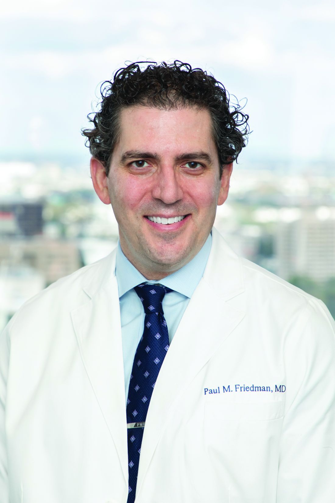
In collaboration with Elizabeth Kream, MD, a dermatology resident at the University of Illinois at Chicago, and Paul M. Friedman, MD, director of the Dermatology and Laser Surgery Center and the current ASLMS president, Dr. Guo conducted an online needs and impact survey regarding laser removal of branding tattoos. With assistance from the National Trafficking Sheltered Alliance, the researchers distributed the survey to U.S. organizations that support sex trafficking survivors. Representatives from 40 organizations responded to the survey. Most were based in the South (45%), followed by the West (20%) and Midwest (20%), and the Northeast (15%).

“On average, these programs support 81 survivors per year, which translates into 3,240 victims per year,” Dr. Guo said. Survey respondents estimated that 47% of sex trafficking survivors had branding tattoos. Of those, 67% were in a stable situation that would make it possible to undergo tattoo removal.
On a scale of 1 to 10 with 10 being the highest, “pro bono removal of branding tattoos received a survivor impact recovery score of 9.2 by these respondents,” Dr. Guo said. “Breaking down these numbers, there are at least 1,200 survivors per year who would benefit from tattoo removal during recovery. Qualitative responses to our survey echoed the same messages: There is a great need and a large impact for pro bono tattoo removal.”
For example, one survey respondent wrote, “Thank you for being willing to remove tattoos, allowing them to feel as though they are no longer owned by their trafficker.” Another wrote, “Erasing or revising the mark of her trafficker is a critical part of every survivor’s recovery journey.”
Sometimes branding tattoos are placed in highly visible locations. One sex trafficking survivor presented to Dr. Guo with a large dark blue tattoo above an eyebrow. “She shared with me that because the tattoo was so highly visible, nobody would offer her a job,” Dr. Guo said. Another survivor had her trafficker’s initial tattooed on her left ring finger. Yet another had a large tattoo on her forearm branded with her trafficker’s name as well as the word cash, “indicating that she is source of money for him,” she said, noting that on average, one sex trafficking victim generates about $100,000 per year for their trafficker.
Although there has been work published on recognition of branding tattoos in the medical community, including the difficulty in differentiating branding tattoos from voluntary tattoos, Dr. Friedman said that there have not been any studies evaluating the need and impact of laser branding tattoo removal in the recovery of sex trafficking survivors. Findings from the current survey “illuminate that the removal of branding tattoos is highly impactful on recovery and may be preferred over tattoo cover-ups,” Dr. Friedman told this news organization.
“Furthermore, survivors frequently move during their recovery process, so a national partnership is essential to allowing survivors to continue the removal process wherever they may be.”
The findings support a proposed ASLMS campaign that intends to connect sex trafficking survivors with board-certified physicians for pro bono removal of branding tattoos. “This will not only aid in survivors’ recovery, but this work will also be beneficial to allow for an avenue to create a repository of sex trafficking tattoo images to improve branding tattoo identification competency among health care providers,” Dr. Friedman said.
He acknowledged certain limitations of the survey, including the fact that “thorough and exact data collection regarding human trafficking is challenging given the inherently covert and underground nature of this crime.” In addition, the study involved surveying organizations supporting sex trafficking survivors rather than the survivors themselves. However, he noted, “we felt for this initial study we wanted to be sensitive to the survivors.”
In an interview at the meeting, one of the session moderators, Oge Onwudiwe, MD, a dermatologist who practices at AllPhases Dermatology in Alexandria, Va., said that pro bono laser removal of branding tattoos “is something that a lot of us can work on and do, and have an impact on. There’s no reason why we shouldn’t help. I can only imagine the psychological impact of having a daily reminder of that [in the form of a branding tattoo]. That’s like PTSD every day almost. You have a trigger there.”
Another session moderator, Eliot Battle, MD, CEO of Cultura Dermatology and Laser Center in Washington, is a board member of Innocents at Risk, a nonprofit that works to fight child exploitation and human trafficking. With pro bono laser removal of a branded tattoo, “this is not just a cosmetic correction you’re making,” Dr. Battle said. “It’s so much deeper than that. It changes people’s lives.”
The researchers and Dr. Onwudiwe reported having no financial disclosures. Dr. Battle disclosed that he conducts research for Cynosure, and has received discounts from Cynosure, Cutera, Solta Medical, Lumenis, Be Inc., and Sciton.
SAN DIEGO – , according to the results of an online survey evaluating the need for and impact of tattoo removal in this population.
Sex trafficking involves the use of force, fraud, or coercion to compel another person to engage in commercial sex acts, and traffickers often brand their victims with tattoos that convey ownership, including tattoos of names, symbols, and barcodes. According to data from Polaris, a nonprofit organization that works to combat and prevent sex and labor trafficking in the United States, 16,658 sex trafficking victims were identified in the country in 2020, but tens of thousands go unreported.
“Given the inherently covert nature of this crime, it is difficult to determine exact statistics,” Emily L. Guo, MD, a cosmetic dermatologic surgery fellow at the Dermatology and Laser Surgery Center in Houston, said during a clinical abstract session at the annual meeting of the American Society for Laser Medicine and Surgery. “We have been working with sex trafficking survivors local to our practice in Houston providing pro bono tattoo removal, and we’ve observed how impactful that is in their recovery. We wanted to see if there was a national need for support of these survivors, allowing them to reclaim their lives.”
In collaboration with Elizabeth Kream, MD, a dermatology resident at the University of Illinois at Chicago, and Paul M. Friedman, MD, director of the Dermatology and Laser Surgery Center and the current ASLMS president, Dr. Guo conducted an online needs and impact survey regarding laser removal of branding tattoos. With assistance from the National Trafficking Sheltered Alliance, the researchers distributed the survey to U.S. organizations that support sex trafficking survivors. Representatives from 40 organizations responded to the survey. Most were based in the South (45%), followed by the West (20%) and Midwest (20%), and the Northeast (15%).
“On average, these programs support 81 survivors per year, which translates into 3,240 victims per year,” Dr. Guo said. Survey respondents estimated that 47% of sex trafficking survivors had branding tattoos. Of those, 67% were in a stable situation that would make it possible to undergo tattoo removal.
On a scale of 1 to 10 with 10 being the highest, “pro bono removal of branding tattoos received a survivor impact recovery score of 9.2 by these respondents,” Dr. Guo said. “Breaking down these numbers, there are at least 1,200 survivors per year who would benefit from tattoo removal during recovery. Qualitative responses to our survey echoed the same messages: There is a great need and a large impact for pro bono tattoo removal.”
For example, one survey respondent wrote, “Thank you for being willing to remove tattoos, allowing them to feel as though they are no longer owned by their trafficker.” Another wrote, “Erasing or revising the mark of her trafficker is a critical part of every survivor’s recovery journey.”
Sometimes branding tattoos are placed in highly visible locations. One sex trafficking survivor presented to Dr. Guo with a large dark blue tattoo above an eyebrow. “She shared with me that because the tattoo was so highly visible, nobody would offer her a job,” Dr. Guo said. Another survivor had her trafficker’s initial tattooed on her left ring finger. Yet another had a large tattoo on her forearm branded with her trafficker’s name as well as the word cash, “indicating that she is source of money for him,” she said, noting that on average, one sex trafficking victim generates about $100,000 per year for their trafficker.
Although there has been work published on recognition of branding tattoos in the medical community, including the difficulty in differentiating branding tattoos from voluntary tattoos, Dr. Friedman said that there have not been any studies evaluating the need and impact of laser branding tattoo removal in the recovery of sex trafficking survivors. Findings from the current survey “illuminate that the removal of branding tattoos is highly impactful on recovery and may be preferred over tattoo cover-ups,” Dr. Friedman told this news organization.
“Furthermore, survivors frequently move during their recovery process, so a national partnership is essential to allowing survivors to continue the removal process wherever they may be.”
The findings support a proposed ASLMS campaign that intends to connect sex trafficking survivors with board-certified physicians for pro bono removal of branding tattoos. “This will not only aid in survivors’ recovery, but this work will also be beneficial to allow for an avenue to create a repository of sex trafficking tattoo images to improve branding tattoo identification competency among health care providers,” Dr. Friedman said.
He acknowledged certain limitations of the survey, including the fact that “thorough and exact data collection regarding human trafficking is challenging given the inherently covert and underground nature of this crime.” In addition, the study involved surveying organizations supporting sex trafficking survivors rather than the survivors themselves. However, he noted, “we felt for this initial study we wanted to be sensitive to the survivors.”
In an interview at the meeting, one of the session moderators, Oge Onwudiwe, MD, a dermatologist who practices at AllPhases Dermatology in Alexandria, Va., said that pro bono laser removal of branding tattoos “is something that a lot of us can work on and do, and have an impact on. There’s no reason why we shouldn’t help. I can only imagine the psychological impact of having a daily reminder of that [in the form of a branding tattoo]. That’s like PTSD every day almost. You have a trigger there.”
Another session moderator, Eliot Battle, MD, CEO of Cultura Dermatology and Laser Center in Washington, is a board member of Innocents at Risk, a nonprofit that works to fight child exploitation and human trafficking. With pro bono laser removal of a branded tattoo, “this is not just a cosmetic correction you’re making,” Dr. Battle said. “It’s so much deeper than that. It changes people’s lives.”
The researchers and Dr. Onwudiwe reported having no financial disclosures. Dr. Battle disclosed that he conducts research for Cynosure, and has received discounts from Cynosure, Cutera, Solta Medical, Lumenis, Be Inc., and Sciton.
AT ASLMS 2022

Removing eyebrow and eyelid tattoos possible with laser, case series finds
SAN DIEGO – , results from a single-center retrospective study showed.
There is a market for these types of cosmetic tattoos today, “and a need for removal,” David Orbuch, MD, MBA, said during a clinical abstract session at the annual meeting of the American Society for Laser Medicine and Surgery.
Dr. Orbuch, a fellow at the Laser & Skin Surgery Center of New York, and his colleagues retrospectively reviewed the charts of 57 adults who underwent laser tattoo removal of eyebrow and eyelid tattoos at the center from January 2018 to December 2021. Data recorded included demographics, site location, initial parameters, colors treated, and clinical safety and efficacy. The mean age of the patients was 46 years, 98.8% were female, 50.9% were Fitzpatrick skin type I-II, and the remainder were types III-V.
Among the most common sites treated were the bilateral eyebrows (35%). Other common sites were the upper eyelids (21.1%), the lower eyelids (10.5%), and both the upper and lower eyelids (12%). Each patient underwent an average of 2.5 treatments (range, 1-11). The most common lasers used were a 755-nm picosecond laser (79%), a high‐power 1,064-nm picosecond laser (12.3%), a high‐power 532-nm picosecond laser (3.5%), and a 10,600-nm carbon dioxide laser (1.7%). The most common tattoo colors were black (94.7%), the far most common, followed by red (3.5%), and yellow (1.7%).
For removal of black tattoos, the most common treatment parameters for the 755 picosecond laser were a 2.5-mm spot size and a fluence of 3.36 J/cm2. For the 1,064-nm picosecond laser, the most common treatment parameters were a 2-mm spot size and a fluence of 4 J/cm2.
For removal of red tattoos, the most common treatment parameters for the 532-nm picosecond laser were a 3.3-mm spot size and a fluence of 2 J/cm2. For the 10,600-nm CO2 laser, the most common treatment parameters were a spot size of 7 mm and a fluence of 28.2 J/cm2.
As for removal of yellow tattoos, the most common treatment parameters with the 532-nm picosecond laser were a 3.3-mm spot size and a fluence of 0.5 J/cm2.
There were no documented cases of scarring, eyelash/eyebrow loss, necrosis, burns, prolonged erythema, prolonged swelling, or prolonged dyspigmentation noted.
“With all of these treatments, you can get a great effect, but you have to do it safely,” Dr. Orbuch said. “With all of these wavelengths, the 1,064 nm especially, there can be serious eye damage if done improperly,” he added. “As such, placement of the metallic eye shields is important. If they’re not properly placed, they can fall out. Make sure you are comfortable using these shields before doing these treatments.”
Pooja Sodha, MD, director of the Center for Laser and Cosmetic Dermatology at George Washington University, Washington, who was asked to comment on the study, said that cosmetic tattoos pose treatment challenges for several reasons. First, “there can be variability in the composition of the pigments since they are often tailored to fit the location and complexion of the patient,” she said. “Second, there can be placement of multiple layers of tattoo pigment to provide the final effect. Third, the pigment may contain two metal oxides (titanium dioxide and ferric oxide), which are often used to calibrate skin tone colors.”

Unfortunately, she noted, “these metal oxides are prone to reduction reactions with laser exposure, causing paradoxical darkening of tattoo pigment. In the past, these darker colors were treated with continued laser therapy and even fractional or fully ablative CO2/Er:YAG resurfacing.”
Dr. Sodha noted that prior studies have shown picosecond lasers to be effective cosmetic lasers, “and this study further supports this with a larger cohort of patients who were treated with the array of picosecond wavelengths (532, 755, and 1,064 nm) without long-term sequelae. Interestingly, there did not appear to be long-term sequelae with dyspigmentation or paradoxical darkening, with fewer than 2% necessitating treatment with a carbon dioxide laser.”
Neither Dr. Orbuch nor Dr. Sodha reported having financial disclosures.
SAN DIEGO – , results from a single-center retrospective study showed.
There is a market for these types of cosmetic tattoos today, “and a need for removal,” David Orbuch, MD, MBA, said during a clinical abstract session at the annual meeting of the American Society for Laser Medicine and Surgery.
Dr. Orbuch, a fellow at the Laser & Skin Surgery Center of New York, and his colleagues retrospectively reviewed the charts of 57 adults who underwent laser tattoo removal of eyebrow and eyelid tattoos at the center from January 2018 to December 2021. Data recorded included demographics, site location, initial parameters, colors treated, and clinical safety and efficacy. The mean age of the patients was 46 years, 98.8% were female, 50.9% were Fitzpatrick skin type I-II, and the remainder were types III-V.
Among the most common sites treated were the bilateral eyebrows (35%). Other common sites were the upper eyelids (21.1%), the lower eyelids (10.5%), and both the upper and lower eyelids (12%). Each patient underwent an average of 2.5 treatments (range, 1-11). The most common lasers used were a 755-nm picosecond laser (79%), a high‐power 1,064-nm picosecond laser (12.3%), a high‐power 532-nm picosecond laser (3.5%), and a 10,600-nm carbon dioxide laser (1.7%). The most common tattoo colors were black (94.7%), the far most common, followed by red (3.5%), and yellow (1.7%).
For removal of black tattoos, the most common treatment parameters for the 755 picosecond laser were a 2.5-mm spot size and a fluence of 3.36 J/cm2. For the 1,064-nm picosecond laser, the most common treatment parameters were a 2-mm spot size and a fluence of 4 J/cm2.
For removal of red tattoos, the most common treatment parameters for the 532-nm picosecond laser were a 3.3-mm spot size and a fluence of 2 J/cm2. For the 10,600-nm CO2 laser, the most common treatment parameters were a spot size of 7 mm and a fluence of 28.2 J/cm2.
As for removal of yellow tattoos, the most common treatment parameters with the 532-nm picosecond laser were a 3.3-mm spot size and a fluence of 0.5 J/cm2.
There were no documented cases of scarring, eyelash/eyebrow loss, necrosis, burns, prolonged erythema, prolonged swelling, or prolonged dyspigmentation noted.
“With all of these treatments, you can get a great effect, but you have to do it safely,” Dr. Orbuch said. “With all of these wavelengths, the 1,064 nm especially, there can be serious eye damage if done improperly,” he added. “As such, placement of the metallic eye shields is important. If they’re not properly placed, they can fall out. Make sure you are comfortable using these shields before doing these treatments.”
Pooja Sodha, MD, director of the Center for Laser and Cosmetic Dermatology at George Washington University, Washington, who was asked to comment on the study, said that cosmetic tattoos pose treatment challenges for several reasons. First, “there can be variability in the composition of the pigments since they are often tailored to fit the location and complexion of the patient,” she said. “Second, there can be placement of multiple layers of tattoo pigment to provide the final effect. Third, the pigment may contain two metal oxides (titanium dioxide and ferric oxide), which are often used to calibrate skin tone colors.”
Unfortunately, she noted, “these metal oxides are prone to reduction reactions with laser exposure, causing paradoxical darkening of tattoo pigment. In the past, these darker colors were treated with continued laser therapy and even fractional or fully ablative CO2/Er:YAG resurfacing.”
Dr. Sodha noted that prior studies have shown picosecond lasers to be effective cosmetic lasers, “and this study further supports this with a larger cohort of patients who were treated with the array of picosecond wavelengths (532, 755, and 1,064 nm) without long-term sequelae. Interestingly, there did not appear to be long-term sequelae with dyspigmentation or paradoxical darkening, with fewer than 2% necessitating treatment with a carbon dioxide laser.”
Neither Dr. Orbuch nor Dr. Sodha reported having financial disclosures.
AT ASLMS 2022
PIH in patients with dark skin responds to laser treatment: Small case series
SAN DIEGO – , results from a small retrospective case series suggest.
“Postinflammatory hyperpigmentation is a leading chief of complaint of many skin of color persons seeking a dermatologist,” Elizabeth J. Kream, MD, told this news organization in advance of the annual conference of American Society for Laser Medicine and Surgery. “I describe PIH to patients as the ‘ashes after a fire is extinguished.’ It’s the stubborn brown to gray/black spots that persist after conditions like acne and folliculitis, but it can be caused by any insult to the skin including external injury. In fact, there’s a risk of inciting PIH with lasers and energy-based devices and this risk is greater in skin of color given the greater melanin content. Unfortunately, we see patients present after visiting a med spa who were treated with the wrong devices and/or the wrong settings and they have disfiguring scarring and/or dyspigmentation.”

During an abstract session at the meeting, Dr. Kream, a dermatology resident at the University of Illinois at Chicago, discussed three patients with recalcitrant PIH and Fitzpatrick skin phototype V and VI who were treated in San Diego with a combination of topical and laser therapies. She presented the case series on behalf of coauthors Monica Boen, MD and Douglas C. Wu, MD, dermatologists who practice in San Diego.
The first patient was a 37-year-old Black female who presented for evaluation of longstanding hyperpigmentation on the face and neck determined to be PIH secondary to folliculitis on the chin and neck. She was started on 8% hydroquinone with kojic acid daily and received four treatments spaced 4-8 weeks apart with the 1,927-nm fractional nonablative diode laser. Laser settings were 5 mJ pulse energy and 5% coverage after eight passes. Triamcinolone 0.1% ointment was applied immediately after treatment and for 3 days following treatment, and the “patient experienced near complete resolution of PIH with no unexpected adverse events,” Dr. Kream said.
The second patient was a 20-year-old Black male who presented with a 3-month history of facial hyperpigmentation after suffering a laser-induced injury. He was started on a non-hydroquinone topical lightening agent and received five treatments spaced 2 weeks apart with a 1,927-nm fractional nonablative diode laser. The laser settings were 5 mJ pulse energy and 5% coverage after eight passes. The patient experienced 80%-90% resolution of his PIH with no unexpected adverse reactions.
The third patient in the series was a 39-year-old Black male who presented with a 6-month history of hyperpigmentation on his right shin and calf, secondary to minor occupational-related trauma. Treatment was initiated with a fractional 1,064-nm picosecond laser. The laser settings were 2.1 mJ per microbeam microwave pulse energy and a 450 picosecond pulse duration delivered at 2 Hz through a holographic beam splitter with a 6 x 6–mm spot size containing 101 microbeams, for an estimated coverage of 4% per pulse. Four passes were performed for each area. The endpoint was a mild erythema to several treated areas a few minutes following laser treatment. Postoperative care consisted of applying a non-hydroquinone topical lightening agent twice daily to the affected area for 1 month. Near-complete resolution of the PIH was achieved, with no unexpected adverse reactions.
“In our clinical experience, PIH can be treated with the combination of topical skin lighteners and low density, low fluence laser therapy in almost all skin types,” Dr. Kream said. “The rationale behind this combination is to treat and remove existing pigment with the laser therapy while minimizing and preventing any pigmentary recurrence with diligent topical therapy and photoprotection.”
It is important to identify the cause of PIH “because some cases are trickier than others,” such as a lichenoid process that deposits pigment “a little bit deeper into the dermis,” she said. “When selecting an appropriate laser modality for the treatment of PIH in skin types V and VI, it’s especially important to consider the mechanism of action, depth of penetration, degree of tissue damage, and the extent of disruption to the dermal-epidermal junction.”
Following the presentation, one of the session moderators, Albert Wolkerstorfer, MD, PhD, a dermatologist at Amsterdam University Medical Center, the Netherlands, emphasized the importance of proper patient selection for laser treatment of PIH. “Not every patient with PIH is adapted to treatment with the laser,” Dr. Wolkerstorfer said. “I think it’s also important to choose stable PIH, meaning you often see patients with an underlying disorder who want to get rid of the pigment. They often believe that the laser is the solution, but it often isn’t.”
During a question-and-answer session, a meeting attendee pointed out that the study lacked a control area to compare the treatment results to. “This was a retrospective case series,” Dr. Kream replied. “I’d like to see more elegant studies in the future, with a control [area],” she said.
Dr. Kream reported having no financial disclosures, Dr. Boen has no disclosures, and Dr. Wu has conducted research for many pharmaceutical and device companies. Dr. Wolkerstorfer disclosed that he has received grant or research funding from Lumenis, Novartis, and Avita Medical, and is an advisory board member for Incyte.
SAN DIEGO – , results from a small retrospective case series suggest.
“Postinflammatory hyperpigmentation is a leading chief of complaint of many skin of color persons seeking a dermatologist,” Elizabeth J. Kream, MD, told this news organization in advance of the annual conference of American Society for Laser Medicine and Surgery. “I describe PIH to patients as the ‘ashes after a fire is extinguished.’ It’s the stubborn brown to gray/black spots that persist after conditions like acne and folliculitis, but it can be caused by any insult to the skin including external injury. In fact, there’s a risk of inciting PIH with lasers and energy-based devices and this risk is greater in skin of color given the greater melanin content. Unfortunately, we see patients present after visiting a med spa who were treated with the wrong devices and/or the wrong settings and they have disfiguring scarring and/or dyspigmentation.”
During an abstract session at the meeting, Dr. Kream, a dermatology resident at the University of Illinois at Chicago, discussed three patients with recalcitrant PIH and Fitzpatrick skin phototype V and VI who were treated in San Diego with a combination of topical and laser therapies. She presented the case series on behalf of coauthors Monica Boen, MD and Douglas C. Wu, MD, dermatologists who practice in San Diego.
The first patient was a 37-year-old Black female who presented for evaluation of longstanding hyperpigmentation on the face and neck determined to be PIH secondary to folliculitis on the chin and neck. She was started on 8% hydroquinone with kojic acid daily and received four treatments spaced 4-8 weeks apart with the 1,927-nm fractional nonablative diode laser. Laser settings were 5 mJ pulse energy and 5% coverage after eight passes. Triamcinolone 0.1% ointment was applied immediately after treatment and for 3 days following treatment, and the “patient experienced near complete resolution of PIH with no unexpected adverse events,” Dr. Kream said.
The second patient was a 20-year-old Black male who presented with a 3-month history of facial hyperpigmentation after suffering a laser-induced injury. He was started on a non-hydroquinone topical lightening agent and received five treatments spaced 2 weeks apart with a 1,927-nm fractional nonablative diode laser. The laser settings were 5 mJ pulse energy and 5% coverage after eight passes. The patient experienced 80%-90% resolution of his PIH with no unexpected adverse reactions.
The third patient in the series was a 39-year-old Black male who presented with a 6-month history of hyperpigmentation on his right shin and calf, secondary to minor occupational-related trauma. Treatment was initiated with a fractional 1,064-nm picosecond laser. The laser settings were 2.1 mJ per microbeam microwave pulse energy and a 450 picosecond pulse duration delivered at 2 Hz through a holographic beam splitter with a 6 x 6–mm spot size containing 101 microbeams, for an estimated coverage of 4% per pulse. Four passes were performed for each area. The endpoint was a mild erythema to several treated areas a few minutes following laser treatment. Postoperative care consisted of applying a non-hydroquinone topical lightening agent twice daily to the affected area for 1 month. Near-complete resolution of the PIH was achieved, with no unexpected adverse reactions.
“In our clinical experience, PIH can be treated with the combination of topical skin lighteners and low density, low fluence laser therapy in almost all skin types,” Dr. Kream said. “The rationale behind this combination is to treat and remove existing pigment with the laser therapy while minimizing and preventing any pigmentary recurrence with diligent topical therapy and photoprotection.”
It is important to identify the cause of PIH “because some cases are trickier than others,” such as a lichenoid process that deposits pigment “a little bit deeper into the dermis,” she said. “When selecting an appropriate laser modality for the treatment of PIH in skin types V and VI, it’s especially important to consider the mechanism of action, depth of penetration, degree of tissue damage, and the extent of disruption to the dermal-epidermal junction.”
Following the presentation, one of the session moderators, Albert Wolkerstorfer, MD, PhD, a dermatologist at Amsterdam University Medical Center, the Netherlands, emphasized the importance of proper patient selection for laser treatment of PIH. “Not every patient with PIH is adapted to treatment with the laser,” Dr. Wolkerstorfer said. “I think it’s also important to choose stable PIH, meaning you often see patients with an underlying disorder who want to get rid of the pigment. They often believe that the laser is the solution, but it often isn’t.”
During a question-and-answer session, a meeting attendee pointed out that the study lacked a control area to compare the treatment results to. “This was a retrospective case series,” Dr. Kream replied. “I’d like to see more elegant studies in the future, with a control [area],” she said.
Dr. Kream reported having no financial disclosures, Dr. Boen has no disclosures, and Dr. Wu has conducted research for many pharmaceutical and device companies. Dr. Wolkerstorfer disclosed that he has received grant or research funding from Lumenis, Novartis, and Avita Medical, and is an advisory board member for Incyte.
AT ASLMS 2022
Nevus of Ota: Does the 1064-nm Q-switched Nd:YAG laser work in Black patients?
SAN DIEGO – Using a , results from a small single-center study showed.
Nevus of Ota is a benign melanocytic lesion that presents as a unilateral blue-gray to blue-brown facial patch favoring the distribution of the first two branches of the trigeminal nerve. Among Asians, the prevalence of the condition among Asians is estimated to be between 0.03% and 1.113%, while the prevalence among Blacks population is estimated to be between 0.01% and 0.016%, Shelby L. Kubicki, MD, said during a clinical abstract session at the annual meeting of the American Society for Laser Medicine and Surgery.

“Most existing literature describes the characteristics and treatment of Nevus of Ota based on Asian patients with skin types I-IV,” said Dr. Kubicki, a third-year dermatology resident at the University of Texas Health Sciences Center/University of Texas MD Anderson Cancer Center, both in Houston. “Special considerations are required when treating [Fitzpatrick skin types] V-VI, which is why it’s important to characterize these patients, to make sure they’re well represented in the literature.”
In what she said is the largest reported case series of its kind, Dr. Kubicki and colleagues identified eight Fitzpatrick skin type V or VI patients who underwent laser treatment for Nevus of Ota from 2016-2021. All were treated with the 1,064-nm Q‐switched Nd:YAG and on average, received 5.4 treatments at 2-10 month intervals. Fluence ranged from 1.8 to 2.4 J/cm2, and total pulse count ranged from 536.8 to 831.1. Two of these patients were additionally treated with 1,550-nm nonablative fractional resurfacing with a mean of six treatments. Primary outcomes were based on improvement of before and after clinical photographs by three independent board-certified dermatologists, who used a 5-point visual analogue scale for grading.
The mean age of patients was 30.4 years and ranged from 9 months to 45 years. Six were females and two were males, two had Fitzpatrick skin type V, and six had Fitzpatrick skin type VI. Of the eight patients, six had blue-gray lesions, one patient had a dark brown lesion, and one patient had “a hybrid lesion that had blue-gray and brown discoloration,” Dr. Kubicki said.
After grading of the clinical photographs, patients demonstrated a mean improvement of 51%-75% at follow-up 5-56 weeks after treatment (a mean of 16.9 weeks). No long-term adverse events were encountered in either group, but three patients developed mild guttate hypopigmentation following laser treatment.
“Lesional color may contribute to outcome, and patients should be educated about the risk of guttate hypopigmentation,” Dr. Kubicki said. “More studies are needed to determine the optimal device and treatment settings in this population.”
In an interview at the meeting, one of the session moderators, Oge Onwudiwe, MD, a dermatologist who practices at AllPhases Dermatology in Alexandria, Va., said that, while the study results impressed her, she speculated that the patients may require more treatments in the future. “What to look out for is the risk of rebound,” Dr. Onwudiwe said. “Because Nevus of Ota is a hamartomatous lesion, it’s very hard to treat, and sometimes it will come back. It will be nice to see how long this treatment can last. If you can use a combination therapy and have ... cases where you’re only needing a touch-up every so often, that’s still a win.”
Another session moderator, Eliot Battle, MD, CEO of Cultura Dermatology and Laser Center in Washington, D.C., said that he wondered what histologic analysis following treatment might show, and if a biopsy after treatment would show “if we really got rid of the nevus, or if we are just cosmetically improving the appearance temporarily.”
Neither Dr. Kubicki nor Dr. Onwudiwe reported having financial disclosures. Dr. Battle disclosed that he conducts research for Cynosure. He has also received discounts from Cynosure, Cutera, Solta Medical, Lumenis, Be Inc., and Sciton.
SAN DIEGO – Using a , results from a small single-center study showed.
Nevus of Ota is a benign melanocytic lesion that presents as a unilateral blue-gray to blue-brown facial patch favoring the distribution of the first two branches of the trigeminal nerve. Among Asians, the prevalence of the condition among Asians is estimated to be between 0.03% and 1.113%, while the prevalence among Blacks population is estimated to be between 0.01% and 0.016%, Shelby L. Kubicki, MD, said during a clinical abstract session at the annual meeting of the American Society for Laser Medicine and Surgery.
“Most existing literature describes the characteristics and treatment of Nevus of Ota based on Asian patients with skin types I-IV,” said Dr. Kubicki, a third-year dermatology resident at the University of Texas Health Sciences Center/University of Texas MD Anderson Cancer Center, both in Houston. “Special considerations are required when treating [Fitzpatrick skin types] V-VI, which is why it’s important to characterize these patients, to make sure they’re well represented in the literature.”
In what she said is the largest reported case series of its kind, Dr. Kubicki and colleagues identified eight Fitzpatrick skin type V or VI patients who underwent laser treatment for Nevus of Ota from 2016-2021. All were treated with the 1,064-nm Q‐switched Nd:YAG and on average, received 5.4 treatments at 2-10 month intervals. Fluence ranged from 1.8 to 2.4 J/cm2, and total pulse count ranged from 536.8 to 831.1. Two of these patients were additionally treated with 1,550-nm nonablative fractional resurfacing with a mean of six treatments. Primary outcomes were based on improvement of before and after clinical photographs by three independent board-certified dermatologists, who used a 5-point visual analogue scale for grading.
The mean age of patients was 30.4 years and ranged from 9 months to 45 years. Six were females and two were males, two had Fitzpatrick skin type V, and six had Fitzpatrick skin type VI. Of the eight patients, six had blue-gray lesions, one patient had a dark brown lesion, and one patient had “a hybrid lesion that had blue-gray and brown discoloration,” Dr. Kubicki said.
After grading of the clinical photographs, patients demonstrated a mean improvement of 51%-75% at follow-up 5-56 weeks after treatment (a mean of 16.9 weeks). No long-term adverse events were encountered in either group, but three patients developed mild guttate hypopigmentation following laser treatment.
“Lesional color may contribute to outcome, and patients should be educated about the risk of guttate hypopigmentation,” Dr. Kubicki said. “More studies are needed to determine the optimal device and treatment settings in this population.”
In an interview at the meeting, one of the session moderators, Oge Onwudiwe, MD, a dermatologist who practices at AllPhases Dermatology in Alexandria, Va., said that, while the study results impressed her, she speculated that the patients may require more treatments in the future. “What to look out for is the risk of rebound,” Dr. Onwudiwe said. “Because Nevus of Ota is a hamartomatous lesion, it’s very hard to treat, and sometimes it will come back. It will be nice to see how long this treatment can last. If you can use a combination therapy and have ... cases where you’re only needing a touch-up every so often, that’s still a win.”
Another session moderator, Eliot Battle, MD, CEO of Cultura Dermatology and Laser Center in Washington, D.C., said that he wondered what histologic analysis following treatment might show, and if a biopsy after treatment would show “if we really got rid of the nevus, or if we are just cosmetically improving the appearance temporarily.”
Neither Dr. Kubicki nor Dr. Onwudiwe reported having financial disclosures. Dr. Battle disclosed that he conducts research for Cynosure. He has also received discounts from Cynosure, Cutera, Solta Medical, Lumenis, Be Inc., and Sciton.
AT ASLMS 2022
Safety of combining fillers and lasers in one session evaluated over 6 years
SAN DIEGO – of the filled area, results from a single-center, retrospective study showed.
“Data on the safety of pairing single-session treatment with nonablative fractional 1,927-nm thulium and/or 1,550-nm erbium laser and fillers are lacking,” Shirin Bajaj, MD, said during a clinical abstract session at the annual conference of the American Society for Laser Medicine and Surgery. “Anecdotally, we have found this to be completely safe in our high-volume laser center. We typically do fillers first, followed by laser treatment.”
For the study, Dr. Bajaj, a dermatology fellow at the Laser & Skin Surgery Center of New York, and colleagues retrospectively reviewed the charts of 638 patients who had 1,186 single‐session facial treatments with nonablative fractional 1,927-nm thulium and/or 1,550-nm erbium laser (Fraxel DUAL by Solta) and injectable hyaluronic acid filler from August 2015 to June 2021. Safety over the 6-year period was assessed by the adverse events that occurred within the first 4 weeks. The mean age of patients at the time of treatment was 60 years and 95% were female. Fitzpatrick skin types were type 1 (46.1%), type II (48.1%), type III (5.5%), and type IV (0.3%).
Most patients had 1 single‐session treatment (64.3%); the rest had 2 sessions (17.7%), 3 sessions (8%), or 4-18 sessions (10%). Most (91.2%) were treated with the 1,927-nm thulium laser, while 1.8% were treated with the 1,550-nm erbium laser; the mean total energy delivered was 1.3 kilojoules. A small number of patients (7.0%) received treatment with both lasers.
The most common area treated with filler injections were the cheeks and/or tear troughs (85.6%), followed by the perioral area and/or marionette lines (83.7%), temples (31%), nasolabial folds (25.5%), lips (24%), jawline (23.8%), chin (6.5%), forehead (1.4%), glabella and brows (0.5% each), neck (0.3%), and nose (0.1%). One syringe of filler was used in 58.7% of cases, compared with two syringes in 28.7% of cases, three syringes in 9.9% of cases, and four to six syringes in 2.8% of cases.
Dr. Bajaj reported that of the 1,186 single‐session treatments, no adverse events were recorded that were directly related to spread of filler or laser treatment of the filled area, including product migration, unexpected loss of filler volume, vascular occlusion, acute pain, cutaneous necrosis, blindness, and cutaneous burn. There were no hospital or emergency department transfers or admissions and referrals to ENT specialists or ophthalmologists for additional work‐up.
“This is at a busy cosmetic dermatology and plastic surgery practice,” Dr. Bajaj said. “Additional studies may be needed to further validate our findings.”

The study’s lead author was Jordan V. Wang, MD, who is medical research director at the Laser & Skin Surgery Center of New York.
“At most, this retrospective data confirms what we have known for years to be true: that combination treatments with injectables including fillers are safe,” Catherine M. DiGiorgio, MD, a dermatologist who practices at the Boston Center for Facial Rejuvenation, told this news organization. “This is a small study out of a single office, so that is a limitation. However, many dermatologists have performed Fraxel plus filler treatments in the same session daily for the last 10 years without any issues.”

Dr. DiGiorgio was asked to comment on the results and was not an investigator.
Dr. Bajaj reported having no financial disclosures. Dr. Wang reported that he has received grants and/or research funding from ALASTIN Skincare, Cynosure, Lutronic, Novoxel, Sofwave, Solta Medical, Blossom Innovations, Allergan, Accure Acne Inc., and Soliton. Dr. DiGiorgio reported having no relevant disclosures.
SAN DIEGO – of the filled area, results from a single-center, retrospective study showed.
“Data on the safety of pairing single-session treatment with nonablative fractional 1,927-nm thulium and/or 1,550-nm erbium laser and fillers are lacking,” Shirin Bajaj, MD, said during a clinical abstract session at the annual conference of the American Society for Laser Medicine and Surgery. “Anecdotally, we have found this to be completely safe in our high-volume laser center. We typically do fillers first, followed by laser treatment.”
For the study, Dr. Bajaj, a dermatology fellow at the Laser & Skin Surgery Center of New York, and colleagues retrospectively reviewed the charts of 638 patients who had 1,186 single‐session facial treatments with nonablative fractional 1,927-nm thulium and/or 1,550-nm erbium laser (Fraxel DUAL by Solta) and injectable hyaluronic acid filler from August 2015 to June 2021. Safety over the 6-year period was assessed by the adverse events that occurred within the first 4 weeks. The mean age of patients at the time of treatment was 60 years and 95% were female. Fitzpatrick skin types were type 1 (46.1%), type II (48.1%), type III (5.5%), and type IV (0.3%).
Most patients had 1 single‐session treatment (64.3%); the rest had 2 sessions (17.7%), 3 sessions (8%), or 4-18 sessions (10%). Most (91.2%) were treated with the 1,927-nm thulium laser, while 1.8% were treated with the 1,550-nm erbium laser; the mean total energy delivered was 1.3 kilojoules. A small number of patients (7.0%) received treatment with both lasers.
The most common area treated with filler injections were the cheeks and/or tear troughs (85.6%), followed by the perioral area and/or marionette lines (83.7%), temples (31%), nasolabial folds (25.5%), lips (24%), jawline (23.8%), chin (6.5%), forehead (1.4%), glabella and brows (0.5% each), neck (0.3%), and nose (0.1%). One syringe of filler was used in 58.7% of cases, compared with two syringes in 28.7% of cases, three syringes in 9.9% of cases, and four to six syringes in 2.8% of cases.
Dr. Bajaj reported that of the 1,186 single‐session treatments, no adverse events were recorded that were directly related to spread of filler or laser treatment of the filled area, including product migration, unexpected loss of filler volume, vascular occlusion, acute pain, cutaneous necrosis, blindness, and cutaneous burn. There were no hospital or emergency department transfers or admissions and referrals to ENT specialists or ophthalmologists for additional work‐up.
“This is at a busy cosmetic dermatology and plastic surgery practice,” Dr. Bajaj said. “Additional studies may be needed to further validate our findings.”
The study’s lead author was Jordan V. Wang, MD, who is medical research director at the Laser & Skin Surgery Center of New York.
“At most, this retrospective data confirms what we have known for years to be true: that combination treatments with injectables including fillers are safe,” Catherine M. DiGiorgio, MD, a dermatologist who practices at the Boston Center for Facial Rejuvenation, told this news organization. “This is a small study out of a single office, so that is a limitation. However, many dermatologists have performed Fraxel plus filler treatments in the same session daily for the last 10 years without any issues.”
Dr. DiGiorgio was asked to comment on the results and was not an investigator.
Dr. Bajaj reported having no financial disclosures. Dr. Wang reported that he has received grants and/or research funding from ALASTIN Skincare, Cynosure, Lutronic, Novoxel, Sofwave, Solta Medical, Blossom Innovations, Allergan, Accure Acne Inc., and Soliton. Dr. DiGiorgio reported having no relevant disclosures.
AT ASLMS 2022
Device that couples US, radiofrequency shows promise for wrinkles, skin laxity
SAN DIEGO – .
“We’ve done a lot of work with radiofrequency, and we’ve done a lot of work with ultrasound,” Suneel Chilukuri, MD, said in an interview in advance of a clinical abstract session at the annual conference of the American Society for Laser Medicine and Surgery. “The question becomes, is there truly a difference if we’re combining them together?”

To find out, Dr. Chilukuri, a dermatologist who practices in Houston, Tex., and colleagues conducted an IRB-approved trial of a new device that allows for the delivery of radiofrequency (RF) and targeted ultrasound (TUS) in a single applicator. The device, which is not yet named, has been cleared by the Food and Drug Administration and is expected to be available in the fourth quarter of 2022.
In a single‐blinded study, 21 adults were randomized to receive RF and TUS (group A), while 20 received RF alone (group B). The mean age of patients was 57 years and 38 were women. Patients in each group received four full‐face treatments delivered once per week. Dr. Chilukuri and colleagues used the ElastiMeter to quantitatively measure skin properties at baseline, 1‐month, and 3‐month follow‐up visits. They also took digital photos at each follow-up visit and applied the Fitzpatrick Wrinkle and Elastosis Scale (FWS), and the Global Aesthetic Improvement Scale (GAIS scale) to each one, in addition to performing 3D analysis.
Dr. Chilukuri reported that patients in group A showed superior improvement of skin elasticity compared with those in group B. At 3 months, the preliminary skin elasticity data showed an improvement in the periorbital region by 13.6 N/m (34.7% improvement) and 8.1 N/m (22.2% improvement) in group A and B, respectively. (N/m is a measure of elasticity.)
3D photographs also demonstrated superior results in group A, achieving an improvement of 5.3 points (27.7%) and 4.6 points (24.4%) in wrinkles and skin evenness, respectively. Those in group A achieved marked improvement in both FWS and GAIS scales, compared with their counterparts in group B, he said.
“I think this is going to be one more very useful, versatile tool in our toolbox,” Dr. Chilukuri said of the new device, noting that for both the investigators and the patients, there was greater treatment satisfaction for the areas treated with combined radiofrequency and ultrasound. “It’s something that’s effective, painless, and the treatment time is very short – approximately 10 minutes per side. It’s extremely tolerable and the results were similar to 6-month results I get with fractionated ablative resurfacing, but without the downtime, without the handholding, without any pain.”
Moreover, he added, many patients in the trial have asked to have further treatments “and are on a waiting list for when the product launches.”
He and his colleagues also observed improvements in skin hydration among patients in group A, based on readings from a MoistureMeterSC, which measures skin hydration, a finding that he characterized as “unexpected and interesting.”
Dr. Chilukuri speculated that combining TUS and RF allows for better heat dispersion into the epidermis. “If you get to the proper temperature, which is somewhere between 40 and 42 degrees, and if you can keep it for about 10 minutes, we know that there will be proper stimulation of senescent fibroblasts,” he explained.
“I can’t say that seborrheic keratosis is improved or hyperpigmentation is improved, but the heat generation leads to immediate vasodilation to improve blood flow to treated areas. That results in immediate collagen contraction as well as improved autophagy, removal of age-related cellular debris. With the long term neovascularization, you’re going to see more change with the fibroblast activity leading to collagenesis and elastogenesis.”
Use of the device is not indicated for patients with metal implants in the head and neck region, he noted. “I’d also be cautious about using it in people with melasma as the device’s mechanism is based on heat,” since current scientific evidence shows that heat can worsen melasma, he added. “For now, I recommend caution until we perform a split-face study or develop specific treatment parameters for those patients with melasma.”
“We know that skin tightening is a difficult task for a nonablative, nonsurgical device,” said Murad Alam, MD, professor and vice-chair of dermatology and chief of the section of cutaneous and aesthetic surgery at Northwestern University, Chicago, who was asked to comment on the study.
“The promise is of limited downtime, lack of scars, and minimal discomfort, but we haven’t yet had a home run. As a consequence, there’s a constant effort to develop new and better devices. This study is interesting because it shows that yes, a new and better device might be good, but let’s not overlook the idea of having multiple devices at the same time. The nice thing they’ve shown is that from a safety standpoint, using both radiofrequency and ultrasound was tolerable in terms of safety, discomfort, and downtime.”
BTL Aesthetics, the manufacturer, loaned the device used in the trial. Dr. Chilukuri reported having no other financial conflicts for this study. Dr. Alam reported having no disclosures.
SAN DIEGO – .
“We’ve done a lot of work with radiofrequency, and we’ve done a lot of work with ultrasound,” Suneel Chilukuri, MD, said in an interview in advance of a clinical abstract session at the annual conference of the American Society for Laser Medicine and Surgery. “The question becomes, is there truly a difference if we’re combining them together?”
To find out, Dr. Chilukuri, a dermatologist who practices in Houston, Tex., and colleagues conducted an IRB-approved trial of a new device that allows for the delivery of radiofrequency (RF) and targeted ultrasound (TUS) in a single applicator. The device, which is not yet named, has been cleared by the Food and Drug Administration and is expected to be available in the fourth quarter of 2022.
In a single‐blinded study, 21 adults were randomized to receive RF and TUS (group A), while 20 received RF alone (group B). The mean age of patients was 57 years and 38 were women. Patients in each group received four full‐face treatments delivered once per week. Dr. Chilukuri and colleagues used the ElastiMeter to quantitatively measure skin properties at baseline, 1‐month, and 3‐month follow‐up visits. They also took digital photos at each follow-up visit and applied the Fitzpatrick Wrinkle and Elastosis Scale (FWS), and the Global Aesthetic Improvement Scale (GAIS scale) to each one, in addition to performing 3D analysis.
Dr. Chilukuri reported that patients in group A showed superior improvement of skin elasticity compared with those in group B. At 3 months, the preliminary skin elasticity data showed an improvement in the periorbital region by 13.6 N/m (34.7% improvement) and 8.1 N/m (22.2% improvement) in group A and B, respectively. (N/m is a measure of elasticity.)
3D photographs also demonstrated superior results in group A, achieving an improvement of 5.3 points (27.7%) and 4.6 points (24.4%) in wrinkles and skin evenness, respectively. Those in group A achieved marked improvement in both FWS and GAIS scales, compared with their counterparts in group B, he said.
“I think this is going to be one more very useful, versatile tool in our toolbox,” Dr. Chilukuri said of the new device, noting that for both the investigators and the patients, there was greater treatment satisfaction for the areas treated with combined radiofrequency and ultrasound. “It’s something that’s effective, painless, and the treatment time is very short – approximately 10 minutes per side. It’s extremely tolerable and the results were similar to 6-month results I get with fractionated ablative resurfacing, but without the downtime, without the handholding, without any pain.”
Moreover, he added, many patients in the trial have asked to have further treatments “and are on a waiting list for when the product launches.”
He and his colleagues also observed improvements in skin hydration among patients in group A, based on readings from a MoistureMeterSC, which measures skin hydration, a finding that he characterized as “unexpected and interesting.”
Dr. Chilukuri speculated that combining TUS and RF allows for better heat dispersion into the epidermis. “If you get to the proper temperature, which is somewhere between 40 and 42 degrees, and if you can keep it for about 10 minutes, we know that there will be proper stimulation of senescent fibroblasts,” he explained.
“I can’t say that seborrheic keratosis is improved or hyperpigmentation is improved, but the heat generation leads to immediate vasodilation to improve blood flow to treated areas. That results in immediate collagen contraction as well as improved autophagy, removal of age-related cellular debris. With the long term neovascularization, you’re going to see more change with the fibroblast activity leading to collagenesis and elastogenesis.”
Use of the device is not indicated for patients with metal implants in the head and neck region, he noted. “I’d also be cautious about using it in people with melasma as the device’s mechanism is based on heat,” since current scientific evidence shows that heat can worsen melasma, he added. “For now, I recommend caution until we perform a split-face study or develop specific treatment parameters for those patients with melasma.”
“We know that skin tightening is a difficult task for a nonablative, nonsurgical device,” said Murad Alam, MD, professor and vice-chair of dermatology and chief of the section of cutaneous and aesthetic surgery at Northwestern University, Chicago, who was asked to comment on the study.
“The promise is of limited downtime, lack of scars, and minimal discomfort, but we haven’t yet had a home run. As a consequence, there’s a constant effort to develop new and better devices. This study is interesting because it shows that yes, a new and better device might be good, but let’s not overlook the idea of having multiple devices at the same time. The nice thing they’ve shown is that from a safety standpoint, using both radiofrequency and ultrasound was tolerable in terms of safety, discomfort, and downtime.”
BTL Aesthetics, the manufacturer, loaned the device used in the trial. Dr. Chilukuri reported having no other financial conflicts for this study. Dr. Alam reported having no disclosures.
AT ASLMS 2022
Acid series: Trichloroacetic acid
– yet can be one of the most dangerous treatments in the hands of an untrained user.
TCA, in a clear colorless solution, is available in concentrations up to 100%, and has not been associated with allergic reactions or systemic toxicity. The available concentrations include those used for superficial depth peels (10%-30%), medium depth peels (35%-50%), and deep peels (greater than 50%).

TCA causes coagulation of the cellular membrane of epidermal proteins in the epidermis and, depending on the concentration, the dermis, which results in frosting of the skin. Repair of the epidermal cells induces resurfacing of the skin and neocollagenesis. TCA can be combined with other acids, including glycolic acid (Coleman peel), Jessner solution (Monheit peel), and solid CO2 (Brody peel). It has also been combined with lactic acid, mandelic acid, and salicylic acid in combination peels of various concentrations.
Although there are many studies, case reports, and textbooks related to this topic and the applications, combinations and treatment options for TCA peels, it is important to highlight here how many of these solutions – at high concentrations – are available directly to consumers, med spas, and the general public through online websites, including Amazon and overseas sites. Over the last 15 years, I have seen complications of this acid alone in people who have bought TCA online, related to applications not just on the face but on the body, neck, eyes, vaginal, and anal areas. Pigmentation, erosions, ulcers, and strictures are just some of the possible complications that occur not just with a more concentrated solution, but more often from application errors, aggressive layering of the acid, allowing the acid to sit on the skin too long, and improper tissue prepping and posttreatment skin care.
TCA can be an untamable acid, with little control over the depth of penetration even in the most controlled situations. The inability to be neutralize TCA creates an environment in which the depth of penetration and tissue coagulation is not a precise science. Once applied, the tissue reaction cannot be “stopped” or rapidly reversed making it highly variable in its mechanism. Patients of all skin types have the potential to develop complications as the epidermal and dermal thickness, moisture content, sebum production, and pigmentation are highly varied between individuals.

In my opinion, it is a dangerous product to have on the market – not just for the untrained medical providers using it but for estheticians and the general public who now can buy TCA anywhere.
But with effective training, reliable sourcing and appropriate preparation of the patient’s skin, however, a TCA peel can be a highly effective tool for difficult-to-treat dermatological problems, such as scarring and xanthelasma.
Dr. Talakoub and Naissan O. Wesley, MD, are cocontributors to this column. Dr. Talakoub is in private practice in McLean, Va. Dr. Wesley practices dermatology in Beverly Hills, Calif. This month’s column is by Dr. Talakoub. She has no relevant disclosures. Write to them at dermnews@mdedge.com.
– yet can be one of the most dangerous treatments in the hands of an untrained user.
TCA, in a clear colorless solution, is available in concentrations up to 100%, and has not been associated with allergic reactions or systemic toxicity. The available concentrations include those used for superficial depth peels (10%-30%), medium depth peels (35%-50%), and deep peels (greater than 50%).
TCA causes coagulation of the cellular membrane of epidermal proteins in the epidermis and, depending on the concentration, the dermis, which results in frosting of the skin. Repair of the epidermal cells induces resurfacing of the skin and neocollagenesis. TCA can be combined with other acids, including glycolic acid (Coleman peel), Jessner solution (Monheit peel), and solid CO2 (Brody peel). It has also been combined with lactic acid, mandelic acid, and salicylic acid in combination peels of various concentrations.
Although there are many studies, case reports, and textbooks related to this topic and the applications, combinations and treatment options for TCA peels, it is important to highlight here how many of these solutions – at high concentrations – are available directly to consumers, med spas, and the general public through online websites, including Amazon and overseas sites. Over the last 15 years, I have seen complications of this acid alone in people who have bought TCA online, related to applications not just on the face but on the body, neck, eyes, vaginal, and anal areas. Pigmentation, erosions, ulcers, and strictures are just some of the possible complications that occur not just with a more concentrated solution, but more often from application errors, aggressive layering of the acid, allowing the acid to sit on the skin too long, and improper tissue prepping and posttreatment skin care.
TCA can be an untamable acid, with little control over the depth of penetration even in the most controlled situations. The inability to be neutralize TCA creates an environment in which the depth of penetration and tissue coagulation is not a precise science. Once applied, the tissue reaction cannot be “stopped” or rapidly reversed making it highly variable in its mechanism. Patients of all skin types have the potential to develop complications as the epidermal and dermal thickness, moisture content, sebum production, and pigmentation are highly varied between individuals.
In my opinion, it is a dangerous product to have on the market – not just for the untrained medical providers using it but for estheticians and the general public who now can buy TCA anywhere.
But with effective training, reliable sourcing and appropriate preparation of the patient’s skin, however, a TCA peel can be a highly effective tool for difficult-to-treat dermatological problems, such as scarring and xanthelasma.
Dr. Talakoub and Naissan O. Wesley, MD, are cocontributors to this column. Dr. Talakoub is in private practice in McLean, Va. Dr. Wesley practices dermatology in Beverly Hills, Calif. This month’s column is by Dr. Talakoub. She has no relevant disclosures. Write to them at dermnews@mdedge.com.
FDA warns companies selling OTC skin lighteners
The as the active ingredient, and don’t meet the requirements to be sold legally over the counter. The letters were dated April 13.
The 12 products with hydroquinone are “unapproved drugs and are not generally recognized as safe and effective” (abbreviated as GRASE), the FDA said.
Among the side effects associated with hydroquinone products reported to the FDA are skin rashes, facial swelling, and skin discoloration or ochronosis. The discoloration can be permanent, the FDA said. The lighteners are marketed for use on age or dark spots on the skin associated with melasma.
Tri-Luma, a prescription product for the treatment of moderate to severe melasma of the face, is the only FDA-approved drug containing hydroquinone, according to the FDA. It contains 4% hydroquinone and two other ingredients. It is meant to be used under the supervision of a health care professional. Tri-Luma is indicated for up to 8 weeks of treatment for moderate to severe melasma of the face. The OTC products contain up to 2%. (Generic versions of 4% hydroquinone are available by prescription, dermatologists said.)
“Hydroquinone is a very effective medication, and that’s exactly what it is, a medication,” said Lily Talakoub, MD, a dermatologist in McLean, Va., who supports the FDA action. “It’s very effective and very safe to use in the right hands, but when it is overused or used in the wrong situation, it can cause problems.” Those problems often occur, she said, when there is no health care professional overseeing the use of the OTC products, and when people use them over the long term.
The FDA action to ban the OTC products is “very appropriate,” said dermatologist Pooja Sodha, MD, assistant professor and director of the Center for Laser and Cosmetic Dermatology at George Washington University, Washington. “We know patients pick this up [an OTC product] and use it without physician oversight.” When patients use the products longer than is appropriate, which is also common, it can worsen the initial skin issue, she said.
The action follows reforms finalized under the CARES Act (Coronavirus Aid, Relief and Economic Security Act), which included not only COVID-19 response efforts but also updated the method in which certain OTC drugs are regulated. Manufacturers of the skin lightening products that don’t have FDA approval had been told to remove the products from the market by September 2020.
The recent letters were sent to a dozen companies still marketing their products without an FDA new drug approval. The agency asked the companies to take prompt action and respond with 15 days, stating what they have done to correct the violations.
The 12 companies are AMBI Enterprises, Clinical Formula, Elements Brands Inc., Genomma Lab USA, Intilight/Dr Thomas Balshi, M&M Beauty and Wellness, Neoteric Cosmetics/Scott’s Liquid Gold, Skin Authority, Skin Pro, Skin PS Brands, True Earth Health Products, and Ultimark Products.
Health care professionals and consumers can report adverse reactions associated with these products to the FDA’s MedWatch Adverse Event Reporting program.
A version of this article first appeared on Medscape.com.
Cupping in dermatology
My inspiration to write about cupping this month stems from the perception that everyone seems to be talking about it, from a facialist who suggested it for me to a coworker who swears by cupping to treat her allergies. Cupping is by no means a novel procedure. Its use as a health therapy dates back thousands of years to ancient Egypt (1500 BCE), ancient Greece (described by Hippocrates), ancient Rome (described by the Greek physician Galen), China (during the Han dynasty, 206 BCE to 220 CE) and traditional Islamic culture.1 Over the past decade, the popularity of this ancient procedure has been increasing in the United States.1 Cupping has been applied as a remedy for various dermatologic and medical conditions, including herpes zoster, headaches, diminished appetite, maldigestion, abscess evacuation, narcolepsy, pain, fever, dysmenorrhea, and gout.1,2
Theories on the mechanism(s) of action
The practice of cupping is differentiated into dry and wet cupping.1,2 Traditionally, with dry cupping, a flame is applied to heat the air inside a thick glass cup (rather than the cup itself).1 The cup is placed on the skin surface, and negative pressure suctions the skin into the cup. Wet cupping differs mainly from dry cupping in that it involves blood-letting. Cups made of either silicone or glass of varying size and shapes are used. Modern adaptations to cupping include needle, herbal, and pulsatile cupping, as well as a “moving cupping” technique (vs. traditionally stationary cups).1
There are several theories, many of which are derived from the nondermatologic literature (that is, pain management), as to how cupping may deliver a clinical benefit. Some theories are based in scientific and medical principles, whereas other theories are more whimsical – specifically, that cupping draws out evil spirits.2 Studies of dry cupping have suggested that the procedure results in increased oxygenation of muscles via a local increase in oxygenated hemoglobin, which may help improve muscular activity and reduce pain.1 As theorized by Lowe in 2017, negative pressure exerted by dry cupping leads to stretching and dilation of capillaries, which increases blood flow.3 Wet cupping has been shown to increase heat shock protein 70 (HSP70) and beta-endorphin expression in rat models, which is thought to facilitate pain management.1 Removal of oxidants and reduction of reactive oxygen species in the blood is believed to be among the benefits of wet cupping.1
Cupping in general dermatology
While , as well as various inflammatory conditions.
Herpes zoster
In 2010, Cao et al. reported on their systematic review of wet cupping after completing searches of multiple databases (that is, PubMed, the Cochrane Library [Issue 3, 2008], China Network Knowledge Infrastructure, Chinese Scientific Journal Database, and Wan Fang Database). They identified eight randomized controlled trials involving 651 patients, with meta-analyses revealing that wet cupping performed better than medications in terms of the number of “cured” patients, number of patients with improved symptoms, and a lower incidence of postherpetic neuralgia. Wet cupping, in addition to medication, was also found to be superior to medication alone in multiple patients. The researchers concluded that wet cupping appears to effectively treat herpes zoster.4 However, the study failed to identify which medications were used to treat herpes zoster. In the United States, common medications for herpes zoster include acyclovir, valacyclovir, steroids, gabapentin, and other neuromodulators. Without knowing which medications were used, it is difficult to compare cupping to medication in terms of efficacy in treating herpes zoster.
Urticaria
Urticaria (hives) is an inflammatory skin condition that can be very uncomfortable for patients but often resolves without intervention within several months after onset. In 2001, Li and Ding reported on the treatment with cupping of 40 patients with urticaria. The cure rate among the treatment group was cited as 55%, compared with 30% in the control group, who were treated with a traditional Chinese remedy and an unidentified first-generation antihistamine.1,5 In 2020, Xiao et al. conducted a systematic review and meta-analysis of cupping therapy for patients with chronic urticaria. They identified 13 comparisons from 12 randomized controlled trials involving 842 subjects. The investigators found no significant differences between wet cupping and medication usage. They also found that cupping combined with antihistamine treatment was superior to antihistamines alone, and cupping therapy with acupuncture was more effective than acupuncture alone. The investigators did call for caution, citing the poor quality of the studies reviewed.6
It is important to note that it is difficult to attribute resolution of urticaria to the use of cupping given the self-resolution often associated with this condition. Antihistamines are the mainstay of therapy for urticaria, but in my personal experience, patients are not entirely satisfied with the level of symptom control with antihistamines alone and often search for alternative therapies to control the pesky hives and associated itch. In 2014, omalizumab (Xolair) was approved for treating chronic idiopathic urticaria, which has helped patients control symptoms of chronic idiopathic urticaria without needing to take antihistamines. There was no indication that the studies reviewed by Xiao et al. compared cupping against this new effective treatment. Therefore, these studies comparing cupping to medical management are outdated.
Acne, eczema, and psoriasis
Soliman’s 2018 review of cupping in dermatology included a few studies on these common cutaneous conditions. For instance, a 2013 single-blind prospective study by Xu et al. reported on the results of patients with moderate acne who received wet cupping (in the form of prickling bloodletting) twice weekly for 6 weeks.7 They reported that patients demonstrated improvement in the global acne grading system (GAGS) score by the end of the trial.1,7 Unfortunately, cupping was not compared with standard acne treatments (that is, benzoyl peroxide, topical and oral antibiotics, isotretinoin, topical retinoids, spironolactone).
In evaluating cupping for acute eczema, wet cupping was compared with oral loratadine and topical ointments in a 2007 study by Yao and Li. They divided 88 cases into treatment and control groups, with the former group (n = 46) receiving bloodletting puncturing and cupping and the control group (n = 42) receiving oral loratadine and topical Pairuisong (an herbal ointment used in Chinese medicine). The investigators observed no significant difference in total effective rates but a superior difference in the rates of responses that were considered “cured” and “markedly effective” in favor of the cupping treatment.1,8 However, a case report by Hon et al. has indicated that cupping therapy may be associated with more harm than benefit when used as an eczema treatment.1,9
In addition, it is important to note that the past 5 years have been gamechanging in the management of chronic eczema in terms of the array of novel and effective therapies (e.g., dupilumab and JAK inhibitors) and chronic moderate-to-severe eczema has become very treatable. Similarly, acute eczema is often successfully managed with topical steroids, calcineurin inhibitors, and emollients. As such, there is no compelling reason to consider an unproven treatment such as cupping.
In 2020, Xing et al. reviewed 16 randomized controlled trials assessing the use of “moving cupping” for plaque psoriasis, with 1,164 patients meeting inclusion criteria. Moving cupping was found to be significantly more effective than “no-moving” cupping therapy, and moving cupping, combined with medications, performed better than medications alone.10 None of the trials evaluated in this study included randomized controlled trials that compared patients using any of the more modern psoriasis medications, specifically biologics. And, again, the studies evaluated were not of the highest quality.
The data that support cupping, as summarized above, are based mostly on case reports, and strong double-blind prospective studies are lacking. Additionally, most of the studies cited gauged the efficacy of cupping using qualitative endpoints, rather than standardized quantitative endpoints and scales. Moreover, spontaneous remission of various dermatoses can occur, or they can improve over time, including acute eczema, psoriasis, and, especially, urticaria.
Adverse effects of cupping
Often alternative therapies are seen as “benign” and without adverse effects. However, complications can result from cupping. Trauma can be induced from the cupping itself by damaging superficial blood vessels and causing bruising.1,11 Blistering can also occur secondary to the suction effect, and the epidermal and dermal layers of the skin can be separated.1,11 Further, burns and discoloration have also been noted secondary to heat, trauma, and post inflammatory pigmentary changes.1,11 Another risk of cupping is the Koebner phenomenon, which occurs with psoriasis, with new lesions appearing in traumatized skin.12 Other adverse outcomes that have been reported with cupping include reactivation of herpes simplex virus secondary to skin trauma, iron deficiency anemia (secondary to blood loss), panniculitis, infections, and residual marks mistaken for signs of child abuse.1,11
Cupping in aesthetic dermatology
Facial cupping, a distinct practice from body cupping used to treat general dermatology conditions described previously, is also increasing in popularity. This practice is usually conducted in association with a facial or facial acupuncture by an aesthetician or other licensed professional. It can also be performed using at-home kits. The marketing claims for facial cupping cite improved tightening and contouring of facial skin, increased facial microcirculation and collagen synthesis, and enhanced lymphatic flow to aid with facial puffiness or swelling. One supposed mechanism for these benefits is that cupping increases blood flow. Interestingly, there was a 2020 animal study in which photoacoustic imaging of a mouse ear revealed increased temporary blood flow in the cupping microenvironment.13 Currently, however, there is no evidence in the English scientific literature that supports facial cupping. The benefits attributed to facial cupping for aesthetic purposes have emerged only in personal anecdotes. The temporary increase in blood flow may induce inflammation and swelling that adds volume to the face and temporarily diminishes wrinkles. However, this temporary plumpness may be associated with adverse effects, such as local trauma, irritation, bruising, postinflammatory pigmentary alteration, or even herpes reactivation. In my opinion, the possible adverse effects of cupping outweigh any potential benefit, especially given the insufficient evidence supporting the utility of cupping for cosmetic enhancement.
Summary
There is increasing interest among patients to incorporate complementary and alternative medicine – including the ancient tradition of cupping – in managing medical dermatologic conditions. However, current evidence supporting cupping as an effective therapeutic strategy is not strong, with most studies to date appearing to be of poor quality or not sufficiently convincing to displace standard therapies. Our medical strategies for managing chronic dermatologic conditions, particularly inflammatory disorders, continue to improve from both a safety and a proven efficacy standpoint. Therefore, I would not forgo medical management in favor of cupping. While cupping can be used as an adjunct therapy, I would caution patients about possible adverse side effects. In the aesthetic world, cupping is also gaining popularity, but this trend is also not supported by current evidence or studies, at least in the Western literature.
Dr. Goldman is a dermatologist in private practice in Miami and specializes in cosmetic and general dermatology. She practices at Baumann Cosmetic & Research Institute and is also opening a general dermatology practice. Write to her at dermnews@mdedge.com or message her on Instragram @DrChloeGoldman. Dr. Goldman receives compensation to create social media content for Replenix, a skin care company. She has no other disclosures.
References
1. Soliman Y et al. Acta Dermatovenerol Alp Pannonica Adriat. 2018 Jun;27(2):103-7.
2. França K and Lotti T. Advances in Integrative Dermatology. John Wiley & Sons, 2019.
3. Lowe DT. Complement Ther Clin Pract. 2017 Nov;29:162-8.
4.Cao H et al. Altern Ther Health Med. 2010 Nov-Dec;16(6):48-54.
5. Li L and Ding J. J Tradit Chin Med. 2001 Mar;21(1):37-8.
6. Xiao XJ et al. J Integr Med. 2020 Jul;18(4):303-12.
7. Xu J et al. J Tradit Chin Med. 2013 Dec;33(6):752-6.
8. Yao J et al. Zhongguo Zhen Jiu. 2007; Jun;27(6):424-6.
9. Hon KL et al. Case Rep Pediatr. 2013;2013:605829.
10. Xing M et al. Medicine (Baltimore). 2020 Oct 9;99(41):e22539.
11. Kim TH et al. Eur J Integr Med. 2014 Aug 1;6(4):434-40.
12. Vender R and Vender R. J Cutan Med Surg. 2015 May-Jun;19(3):320-2.
13. Zhou Y et al. Biomed Opt Express. 2020 Apr 6;11(5):2394-401.
This article was updated 4/25/22.
Sustained jawline definition from hyaluronic gel, study reports
BOSTON – After several promising early phase studies, from what study authors characterized as a “pivotal” randomized multicenter trial. The results were presented during a late-breaking research session at the annual meeting of the American Academy of Dermatology.
The primary outcome, assessed at 6 months, was at least a 1-point improvement in a photonumeric scale used to grade jawline sagging, reported Jeremy Green, MD, Skin Associates of South Florida, Coral Gables.
When those randomized to the hyaluronic filler gel VYC-25L (Vycross, Juvéderm) were compared with untreated controls, 68.5% versus 38.4% met the criterion for benefit at 6 months. Importantly, the effect in treated patients was sustained when reevaluated at 12 months. Green reported that the response is generally sustained at the maximum follow-up, now out to 17 months.
Most enrolled patients are severely affected
In this study, 208 patients with severe (74%) or moderate loss of jawline definition were randomized in a 3:1 ratio to receive the filler or serve as controls. The initially untreated controls received the gel after the primary outcome analysis at 6 months.
The hyaluronic gel was injected at five sites along the jawline. The mean age of participants was 58 years. The majority were women, and most were White.
Dermatologists blinded to treatment compared photos at 6 months with those taken at baseline using the photonumeric grading system of 1-5. Change in patient satisfaction at 6 months and again at 12 months relative to baseline was also evaluated.
From baseline, when 28.9% of participants reported satisfaction on the Global Aesthetic Improvement Scale (GAIS), rates rose to 89.0% at month 6. There was a decline at month 12, but 79.9% remained satisfied after this period of follow-up.
Most patients experienced injection site reactions that were mainly mild to moderate and all resolved within several days of treatment. Pain with mastication was initially reported by 1.9%, but again this complaint was also mild and transient. All complaints had largely resolved by day 3.
The results are consistent with several previous clinical studies of VYC-25L for the same indication. In a similarly designed trial conducted in Europe that also used a 3:1 randomization scheme, the primary outcome assessed at 3 months was change in facial angle. Relative to controls, the angle improved by 2.51 degrees (P < .0001).
Patient satisfaction supports filler benefit
In the similar European trial, the clinical significance of the objective primary outcome also was supported by patient satisfaction assessed with several instruments, including the GAIS. Some degree of swelling or tenderness was experienced by almost all patients after injection, but none were serious, and all resolved.
In another trial, 202 patients with chin retrusion were randomized in a 3:1 ratio to VYC-25L or a control group. In that study, the primary outcome was at least a 1-point improvement in the Allergan Chin Retrusion Scale at 6 months. This advantage for treatment (56.3% vs. 27.5%) was again supported by several instruments for evaluating patient satisfaction, including GAIS.
As in the other studies, most patients had injection site reactions. Although all resolved within days of treatment, one patient left the study after experiencing cellulitis and injection-site inflammation.
Dissatisfaction with jawline definition is a relatively common complaint in Dr. Green’s experience, who said that there is a need for more effective and well-tolerated treatments. Given the efficacy, tolerability, and safety of VYC-25L in this controlled study, he suggested this product has potential utility.
In the field of cosmetic dermatology, there appears to be incremental progress in fillers with favorable clinical characteristics, according to Sandy U. Tsao, MD, a dermatologic surgeon at Massachusetts General Hospital, Boston.
“We are seeing filler lasting longer and longer,” she said, commenting specifically about the results presented by Dr. Green. She called sustained aesthetic improvement at 12 months for the filler in this study “really exciting.”
Dr. Green has reported financial relationships with numerous pharmaceutical companies. Dr. Tsao has reported financial relationships with Epiphany Dermatology, Lazarus AI, and UpToDate.
A version of this article first appeared on Medscape.com.
AT AAD 2022