User login
Very few infants born to HCV-infected mothers receive testing
Despite the increasing prevalence of hepatitis C virus (HCV) infection in pregnant women, infants exposed to the disease are screened at a very low rate, Catherine A. Chappell, MD, and her associates wrote in Pediatrics.
During 2006-2014, 87,924 women gave birth at the Magee-Womens Hospital at the University of Pittsburgh Medical Center, of whom 1,043 had HCV. Over this time, the HCV prevalence rate increased 60%, from 1,026 cases per 100,000 women to 1,637 cases per 100,000 women. Women with HCV were more likely to be white, have Medicaid, have opiate use disorder, have other substance use disorders, and be under the age of 30 years.
Infants born to HCV-infected women are significantly more likely to be preterm and of low birth weight.
An additional 32 infants who did not receive well child care did receive HCV testing. A total of nine infants, seven in the well child group and two in the non-well child group, tested positive for HCV.
“Of the infants tested with conclusive results, the HCV transmission rate was 8.4%, with 7.2% having chronic HCV infection,” which is in line with previous reports, according to the researchers.
“Because of the poor rates of pediatric HCV screening described, future researchers should focus on interventions to increase screening in infants who are at risk for perinatal HCV acquisition by including technology to improve the transfer of maternal HCV status to the pediatric record and increase pediatric provider awareness regarding HCV screening guidelines,” the investigators concluded.
SOURCE: Chappell CA et al. Pediatrics. 2018 May 2. doi: 10.1542/peds.2017-3273.
FROM PEDIATRICS
Slime is not sublime: It may cause hand dermatitis
A young, otherwise healthy 9-year-old girl was evaluated for pruritic hand dermatitis which lasted 5 months after exposure to homemade slime. Physical exam revealed erythematous, scaly plaques on the palmar surfaces of her hands; her fingernails had onychomadesis and longitudinal ridging. Despite frequent emolliation, her dermatitis persisted. She was then treated empirically for scabies and for culture-positive Staphylococcus aureus infection, which required a full round of cephalexin and mupirocin ointment. This also did not alleviate the dermatitis. A combination of homemade borax-containing slime avoidance, brief course of high-dose corticosteroids, and frequent bland emollients was prescribed because the dermatitis was assumed to be caused by an irritant.
After review of this case and evaluation of other children with hand dermatitis, Julia K. Gittler, MD, of Columbia University, New York, and her colleagues have made a case that “slime” and new-onset hand dermatitis may be linked.
SOURCE: Gittler JK et al. J Pediatr. 2018 May 3. doi: 10.1016/j.jpeds.2018.03.064 .
FROM THE JOURNAL OF PEDIATRICS
Poor parent-infant relationship may affect a child’s motor skill development
TORONTO – In what is believed to be a landmark finding, researchers have shown than modifiable risk factors, such as parent-infant relationships, may play a role in preventing children from developing high motor problems during early life.
“Our findings suggest that early health and clinical problems, such as neonatal complications and abnormal neonatal neurological status, are useful indicators to help identify children at risk of poor motor development,” lead study author Nicole Baumann said in an interview in advance of the Pediatric Academic Societies meeting. “Additionally, as a possible implication, children may benefit in motor development from early interventions that incorporate and focus on improving parent-infant relationships.”

For the current study, she and her associates investigated motor development using data from two different cohorts: the Bavarian Longitudinal Study in Germany (BLS) and the Arvo Ylppö Longitudinal Study in Finland (AYLS). A total of 4,741 and 1,423 children, respectively, underwent assessment from birth to age 56 months. Motor functioning was evaluated via standard physical and neurological assessments at birth and at 5, 20, and 56 months. Perinatal, neonatal, and early environmental information was collected at birth and at 5 months via medical records and reports from parents and research nurses.
The researchers identified two distinct trajectories of motor development problems from birth to 56 months: low (94.3% of BLS and 97.3% of AYLS) and high (5.7% of BLS and 2.7% of AYLS) motor problems.
In the BLS cohort, high motor problem trajectory was predicted by poor parent-infant relationship, such as the mother feeling insecure when taking care of the infant at home (OR 1.52); abnormal neonatal neurological status (odds ratio, 1.16); neonatal complications (OR, 1.12); and duration of initial hospitalization (OR, 1.02).
In the AYLS cohort, high motor problem trajectory was also predicted by abnormal neonatal neurological status (OR, 1.69) and duration of hospitalization (OR, 1.02). Although neonatal complications (OR, 1.08) and poor parent-infant relationship (OR, 1.09) did not significantly predict high motor problem trajectory in the AYLS cohort, trends identified were comparable with those obtained from the BLS cohort.
“Most surprising was that one of the four risk factors that remained as independent predictors of high motor problem trajectory was poor parent-infant relationship,” Ms. Baumann said. “As far as we are aware, parent-infant relationship has not been previously reported as a predictor of poor motor development.”
She acknowledged certain limitations of the study, including the fact that nearly 33% of children could not be assessed throughout the study period because of dropout. “Families with children who had poor health and were socially disadvantaged were less likely to continue participation, and may even suggest that our findings have an even larger effect than reported,” Ms. Baumann said. “This is a problem that affects many longitudinal studies, and it may affect group comparisons. However, simulations have shown that even when dropout is selective or correlated with the outcome that predictions only marginally change (Br J Psychiatry. 2009;195[3]:249-56).”
She reported having no financial disclosures.
dbrunk@mdedge.com
TORONTO – In what is believed to be a landmark finding, researchers have shown than modifiable risk factors, such as parent-infant relationships, may play a role in preventing children from developing high motor problems during early life.
“Our findings suggest that early health and clinical problems, such as neonatal complications and abnormal neonatal neurological status, are useful indicators to help identify children at risk of poor motor development,” lead study author Nicole Baumann said in an interview in advance of the Pediatric Academic Societies meeting. “Additionally, as a possible implication, children may benefit in motor development from early interventions that incorporate and focus on improving parent-infant relationships.”
For the current study, she and her associates investigated motor development using data from two different cohorts: the Bavarian Longitudinal Study in Germany (BLS) and the Arvo Ylppö Longitudinal Study in Finland (AYLS). A total of 4,741 and 1,423 children, respectively, underwent assessment from birth to age 56 months. Motor functioning was evaluated via standard physical and neurological assessments at birth and at 5, 20, and 56 months. Perinatal, neonatal, and early environmental information was collected at birth and at 5 months via medical records and reports from parents and research nurses.
The researchers identified two distinct trajectories of motor development problems from birth to 56 months: low (94.3% of BLS and 97.3% of AYLS) and high (5.7% of BLS and 2.7% of AYLS) motor problems.
In the BLS cohort, high motor problem trajectory was predicted by poor parent-infant relationship, such as the mother feeling insecure when taking care of the infant at home (OR 1.52); abnormal neonatal neurological status (odds ratio, 1.16); neonatal complications (OR, 1.12); and duration of initial hospitalization (OR, 1.02).
In the AYLS cohort, high motor problem trajectory was also predicted by abnormal neonatal neurological status (OR, 1.69) and duration of hospitalization (OR, 1.02). Although neonatal complications (OR, 1.08) and poor parent-infant relationship (OR, 1.09) did not significantly predict high motor problem trajectory in the AYLS cohort, trends identified were comparable with those obtained from the BLS cohort.
“Most surprising was that one of the four risk factors that remained as independent predictors of high motor problem trajectory was poor parent-infant relationship,” Ms. Baumann said. “As far as we are aware, parent-infant relationship has not been previously reported as a predictor of poor motor development.”
She acknowledged certain limitations of the study, including the fact that nearly 33% of children could not be assessed throughout the study period because of dropout. “Families with children who had poor health and were socially disadvantaged were less likely to continue participation, and may even suggest that our findings have an even larger effect than reported,” Ms. Baumann said. “This is a problem that affects many longitudinal studies, and it may affect group comparisons. However, simulations have shown that even when dropout is selective or correlated with the outcome that predictions only marginally change (Br J Psychiatry. 2009;195[3]:249-56).”
She reported having no financial disclosures.
dbrunk@mdedge.com
AT PAS 2018
Key clinical point: Four risk factors are independent predictors of high motor problem trajectory in young children.
Major finding: In the Bavarian Longitudinal Study cohort, high motor problem trajectory was predicted by abnormal neonatal neurological status (odds ratio, 1.16), duration of initial hospitalization (OR 1.02), neonatal complications (OR 1.12), and poor parent-infant relationship (OR 1.52).
Study details: A longitudinal analysis of 4,741 children from the Bavarian Longitudinal Study in Germany and 1,423 children from the Arvo Ylppö Longitudinal Study in Finland.
Disclosures: Ms. Baumann reported having no financial disclosures.
ED visits higher among pediatric asthma patients with comorbid depression, anxiety
TORONTO – Children with asthma who have a comorbid diagnosis of anxiety or depression are significantly more likely to make asthma-related visits to the emergency department, compared with their peers who do not have a mental health condition, results from a large administrative data analysis showed.
“There has been a fair bit of research on how comorbid mental health conditions can affect health care utilization for asthma in adults, but few studies have examined how comorbid mental health conditions like anxiety or depression can affect children with asthma,” one of the study authors, Caroline Neel, said in an interview in advance of the Pediatric Academic Societies meeting.

In all, the researchers identified 71,326 patients with asthma, with an overall rate of 16.3 ED visits per 100 child-years. Among these, children with a diagnosis of depression had significantly higher rates of ED visits (21.5 visits per 100 child-years; P less than .01), as did those with a diagnosis of anxiety (19.5 ED visits per 100 child-years; P less than .01). Being enrolled in a Medicaid managed care plan or Medicaid fee-for-service plan also increased the rates of asthma-related ED visits (20.3 and 21.5 ED visits per 100 child-years, respectively; P less than .01 for both associations.)
“We were surprised to see that anxiety and depression seemed to increase asthma emergency department visits as much as other medical chronic illnesses like cystic fibrosis or sickle cell disease, and that kids on Medicaid, who tend to be our poorer kids, also had an independent risk of going to the emergency department,” Ms. Neel said. “Having Medicaid as well as anxiety or depression were independently related to going to the emergency room for asthma, so the study suggests that some of our highest-risk kids for asthma have multiple different contributors to getting sick and needing to go to the emergency room for an asthma attack.”
She acknowledged certain limitations of the analysis, including its reliance on administrative claims data to identify whether or not children had a diagnosis of anxiety or depression. “This doesn’t necessarily identify all the kids who may have these mental health conditions, since sometimes providers are less likely to document a diagnosis of a mental health conditions for children,” she said. “However, we still saw a significant association between a comorbid mental health condition and emergency department use for asthma, despite the potential that mental health conditions may have been under reported.”
The study’s senior author was Naomi Bardach, MD. The researchers reported having no financial disclosures.
TORONTO – Children with asthma who have a comorbid diagnosis of anxiety or depression are significantly more likely to make asthma-related visits to the emergency department, compared with their peers who do not have a mental health condition, results from a large administrative data analysis showed.
“There has been a fair bit of research on how comorbid mental health conditions can affect health care utilization for asthma in adults, but few studies have examined how comorbid mental health conditions like anxiety or depression can affect children with asthma,” one of the study authors, Caroline Neel, said in an interview in advance of the Pediatric Academic Societies meeting.
In all, the researchers identified 71,326 patients with asthma, with an overall rate of 16.3 ED visits per 100 child-years. Among these, children with a diagnosis of depression had significantly higher rates of ED visits (21.5 visits per 100 child-years; P less than .01), as did those with a diagnosis of anxiety (19.5 ED visits per 100 child-years; P less than .01). Being enrolled in a Medicaid managed care plan or Medicaid fee-for-service plan also increased the rates of asthma-related ED visits (20.3 and 21.5 ED visits per 100 child-years, respectively; P less than .01 for both associations.)
“We were surprised to see that anxiety and depression seemed to increase asthma emergency department visits as much as other medical chronic illnesses like cystic fibrosis or sickle cell disease, and that kids on Medicaid, who tend to be our poorer kids, also had an independent risk of going to the emergency department,” Ms. Neel said. “Having Medicaid as well as anxiety or depression were independently related to going to the emergency room for asthma, so the study suggests that some of our highest-risk kids for asthma have multiple different contributors to getting sick and needing to go to the emergency room for an asthma attack.”
She acknowledged certain limitations of the analysis, including its reliance on administrative claims data to identify whether or not children had a diagnosis of anxiety or depression. “This doesn’t necessarily identify all the kids who may have these mental health conditions, since sometimes providers are less likely to document a diagnosis of a mental health conditions for children,” she said. “However, we still saw a significant association between a comorbid mental health condition and emergency department use for asthma, despite the potential that mental health conditions may have been under reported.”
The study’s senior author was Naomi Bardach, MD. The researchers reported having no financial disclosures.
Key clinical point: Anxiety and depression are associated with higher rates of ED use in children with asthma.
Major finding: (21.5 visits per 100 child-years; P less than .01), as did those with a diagnosis of anxiety (19.5 ED visits per 100 child-years; P less than .01).
Study details: An analysis of 71,326 patients with asthma from the Massachusetts All Payer Claims Database for 2014-2015.
Disclosures: The researchers reported having no financial disclosures.
Pediatric epilepsy may be misdiagnosed as GI disease
according to the results of a large single-center retrospective study.
Such misdiagnoses caused substantial diagnostic delays, increased the risk of cognitive deterioration, and exposed children to inappropriate radiation and invasive procedures, reported Giulia Carbonari and her associates at the University of Bologna, Italy.
Several recent case reports have described pediatric epilepsies that were misdiagnosed and treated as gastrointestinal (GI) disorders. To better frame the problem, the investigators reviewed the medical records of 858 consecutive children with epilepsy treated at their center between 2010 and 2015.
A total of 21 patients (2.4%) were initially misdiagnosed with GI disease. Most were younger than 1 year old. Notably, 7 of 27 children (26%) with West syndrome were misdiagnosed – in six cases with GERD, and in one case with infant colic. In addition, 10 of 24 children (42%) with temporal lobe epilepsy were misdiagnosed with GERD (five cases), recurrent abdominal pain (two cases), or cyclic vomiting, gastric pain, or dysfunctional elimination syndrome (one case each). Finally, 4 of 38 children (11%) with Panayiotopoulos syndrome were misdiagnosed with cyclic vomiting (three cases) or GERD (one case).
Misdiagnoses typically caused at least a 3-month diagnostic delay (interquartile range, 2-18 months), and half of misdiagnosed children received inappropriate abdominal ultrasonography, upper alimentary canal radiography, or esophagogastroduodenoscopy. Eight patients also received inappropriate antireflux therapy, and one patient underwent inappropriate surgery, the researchers said.
They shared tips for avoiding these misdiagnoses. Epileptic spasms of West syndrome involve brief contractions (flexion or extension) of the neck, trunk, and extremities, usually in clusters. Psychomotor slowing also is common. Seizures in temporal lobe epilepsy often involve automatisms, mental status changes, and changes in skin color, blood pressure, and heart rate. Signs of Panayiotopoulos syndrome include emesis, cyanosis, pallor, changes in intestinal motility, gaze deviation, hypotonia, confusion, and unresponsiveness.
“A careful review of a patient’s medical history and a detailed description of paroxysmal episodes are the most important tools to reduce diagnostic errors,” they said.
No funding sources were reported. The researchers reported having no conflicts of interest.
SOURCE: Carbonari G et al. Epilepsy Behav. 2018 Apr 26. doi: 10.1016/j.yebeh.2018.03.034.
FROM EPILEPSY & BEHAVIOR
Key clinical point: Epilepsy is a differential diagnosis for atypical gastroesophageal reflux in younger children.
Major finding: In all, 2.4% children were misdiagnosed with gastrointestinal diseases over a 5-year period.
Study details: Single-center retrospective cohort study of 858 children with epilepsy.
Disclosures: No funding sources were reported. The researchers reported having no conflicts of interest.
Source: Carbonari G et al. Epilepsy Behav. 2018 Apr 26. doi: 10.1016/j.yebeh.2018.03.034.
S100B biomarker could reduce CT scans in children with mTBI
according to Charlotte Oris, PharmD, of the University Hospital of Clermont-Ferrand, France, and her associates.
In a meta-analysis of eight prospective cohort studies including a total of 601 children published in Pediatrics, researchers looked at the association between S100B serum levels and CT findings in 373 patients. The median serum concentrations of S100B were 0.47 mcg/L for patients with intracerebral lesions and 0.21 mcg/L for those without lesions (P less than .001).
Additionally, researchers collected data from 358 individuals included in two studies for the origin of mTBI. The median concentrations of S100B were 0.39 mcg/L for road accidents, 0.29 mcg/L for domestic accidents, and 0.18 mcg/L for sport-related accidents. The difference was statistically significant between the road accidents group and the domestic accidents group (P less than .001) and the difference between the road accidents group and the sport-related accidents group (P less than .001). It is noted that S100B specificity could be higher after a sport-related trauma.
“S100B protein serum levels, in combination with the PECARN [Pediatric Emergency Care Applied Research Network] algorithm, could reduce the need for CT scans by one-third. In our additional analysis, based on 373 children, the importance of taking a blood sample 3 hours or less after trauma was underscored,” the researchers said.
“S100B represents a promising biomarker with 100% sensitivity. The limited specificity of S100B could be reevaluated for future research by using a combination of different brain biomarkers,” Dr. Oris and her colleagues concluded.
SOURCE: Oris C et al. Pediatrics. 2018. doi: 10.1542/peds.2018-0037.
MDedge Daily News: How to handle opioid constipation
Bath emollients are a washout for childhood eczema. Does warfarin cause acute kidney injury? And there may be a new option for postpartum depression.
Listen to the MDedge Daily News podcast for all the details on today’s top news.
Bath emollients are a washout for childhood eczema. Does warfarin cause acute kidney injury? And there may be a new option for postpartum depression.
Listen to the MDedge Daily News podcast for all the details on today’s top news.
Bath emollients are a washout for childhood eczema. Does warfarin cause acute kidney injury? And there may be a new option for postpartum depression.
Listen to the MDedge Daily News podcast for all the details on today’s top news.

Interferon-gamma release assay trumps tuberculin skin test in school-aged children
The interferon-gamma release assay (IGRA) was significantly more sensitive than a tuberculin skin test as an adjunct tuberculosis diagnosis of children aged 5 years and older, according to data from a population-based study of 778 cases.
IGRAs have shown greater specificity than tuberculin skin tests (TSTs), but data on their sensitivity to TB in children are limited, wrote Alexander W. Kay, MD, of the California Department of Public Health and his colleagues in a study published in Pediatrics.

Children younger than 1 year of age and those with CNS disease were significantly more likely to have indeterminate IGRA results, the researchers noted.
The study results were limited by the use of mainly enzyme-linked immunosorbent assay–based IGRA, which limited the data on enzyme-linked immunospot tests, the researchers said. The findings also were limited by the small number of children younger than 5 years.
However, the study is the largest North American analysis of IGRA in children, and based on the findings, “we argue that an IGRA should be considered the test of choice when evaluating children 5-18 years old for TB disease in high-resource, low-TB burden settings,” Dr. Kay and his associates wrote.
The study was funded by the Centers for Disease Control and Prevention. Coauthor Shamim Islam, MD, disclosed financial support from Qiagen, maker of the QuantiFERON test. Dr. Kay and the other investigators had no financial conflicts to disclose.
SOURCE: Kay A et al. Pediatrics. 2018 May 4. doi: 10.1542/peds.2017-3918.
The interferon-gamma release assay (IGRA) was significantly more sensitive than a tuberculin skin test as an adjunct tuberculosis diagnosis of children aged 5 years and older, according to data from a population-based study of 778 cases.
IGRAs have shown greater specificity than tuberculin skin tests (TSTs), but data on their sensitivity to TB in children are limited, wrote Alexander W. Kay, MD, of the California Department of Public Health and his colleagues in a study published in Pediatrics.
Children younger than 1 year of age and those with CNS disease were significantly more likely to have indeterminate IGRA results, the researchers noted.
The study results were limited by the use of mainly enzyme-linked immunosorbent assay–based IGRA, which limited the data on enzyme-linked immunospot tests, the researchers said. The findings also were limited by the small number of children younger than 5 years.
However, the study is the largest North American analysis of IGRA in children, and based on the findings, “we argue that an IGRA should be considered the test of choice when evaluating children 5-18 years old for TB disease in high-resource, low-TB burden settings,” Dr. Kay and his associates wrote.
The study was funded by the Centers for Disease Control and Prevention. Coauthor Shamim Islam, MD, disclosed financial support from Qiagen, maker of the QuantiFERON test. Dr. Kay and the other investigators had no financial conflicts to disclose.
SOURCE: Kay A et al. Pediatrics. 2018 May 4. doi: 10.1542/peds.2017-3918.
FROM PEDIATRICS
Key clinical point:
Major finding: Sensitivity was 96% for IGRA versus 83% for TST among children aged 5-18 years.
Study details: The data come from TB patients aged 18 years and younger enrolled in the California TB registry during 2010-2015.
Disclosures: The study was funded by the Centers for Disease Control and Prevention. Coauthor Shamim Islam, MD, disclosed financial support from Qiagen, maker of the QuantiFERON test; Dr. Kay and the other investigators had no financial conflicts to disclose.
Source: Kay A et al. Pediatrics. 2018 May 4. doi: 10. 1542/ peds. 2017- 3918.
U.S. youth suicide prevention saved nearly 900 lives
WASHINGTON – A U.S. grant program that was started in 2004 to fund youth suicide prevention efforts throughout the country was linked with nearly 900 youth suicide deaths prevented by 2015, according to a matched, case-control analysis.

U.S. counties with a youth suicide prevention effort funded by a grant from the federal Garrett Lee Smith (GLS) Memorial Suicide Prevention program had an average 1.09 fewer youth suicide deaths per 100,000 youths (10-24 years old) 2 years after ongoing funding began, when compared with matched U.S. counties without a GLS-funded program, said Mr. Garraza, an analyst with ICF International in New York. Counties that had GLS-funded programs operating for 3 years showed an average reduction in youth suicide death of roughly 2 fewer deaths per 100,000 population, compared with control counties, and counties with programs in operation for 4 years showed a reduction of roughly 3 fewer deaths per 100,000 population.
“These extremely important data show convincingly that if we want to save lives, suicide prevention can’t be a one-and-done. said Richard McKeon, PhD, chief of the Suicide Prevention Branch of the Substance Abuse and Mental Health Services Administration, the U.S. agency that administers the GLS grant program and commissioned ICF International to analyze the grant program’s effects.

As of October 2017, the GLS grant initiative had made nearly 200 awards to youth suicide prevention programs that operated in all 50 states, the District of Columbia, one territory, and among 49 Native American tribes, Mr. Garraza said.
To assess the effects of the GLS grants on suicide death rates in people aged 10-24 years, Mr. Garraza and his associates used U.S. suicide data collected in 2,095 counties throughout the country that each had more than 3,000 resident youths, including 1,126 counties that had, as of 2015, been exposed to at least 1 year of a program sponsored by a GLS grant that began before 2010 and 969 counties without any GLS-grant exposure. After the counties in each subgroup were propensity-score matched using several criteria, including numbers of youth by specific age, race, household income, employment rates, health insurance status, and urbanization, the researchers analyzed suicide death rates in 481 counties with 1-4 years of exposure of a GLS-funded prevention program and in 851 counties with no exposure.
Their analysis estimated that 882 fewer suicide deaths occurred among youths in counties with GLS-funded programs, compared with the expected suicide mortality based on the unexposed counties.
In addition to showing statistically significant mortality reductions among youths in the counties with GLS-funded programs, compared with the counties with no such programs, the analysis showed, as expected, no difference between the intervention and control counties in rates of suicide deaths among adults and no difference in youth-mortality from causes other than suicide, which indicated that the observed differences linked with GLS funding were specific for youth suicides.
The current analysis looking at the effects of GLS funding on youth suicide deaths rates follows a prior report from the same researchers with similar findings using data collected through 2010 (Am J Public Health. 2015 May;105[5]:986-93). They also published two prior reports that used a similar analysis to assess the effects of GLS-funded suicide prevention programs on suicide attempt rates. One of those reports showed that GLS-funded programs linked with a cut in suicide attempts of nearly 5 per 1,000 population (JAMA Psychiatry. 2015 Nov;72[11]:1143-9), and the second showed that this effect on suicide attempts was cost effective when the cost of the grants was compared with the money saved from avoided emergency department visits and hospitalizations (Suicide Life Threat Behav. 2018 Feb;48[1]:3-11).
WASHINGTON – A U.S. grant program that was started in 2004 to fund youth suicide prevention efforts throughout the country was linked with nearly 900 youth suicide deaths prevented by 2015, according to a matched, case-control analysis.
U.S. counties with a youth suicide prevention effort funded by a grant from the federal Garrett Lee Smith (GLS) Memorial Suicide Prevention program had an average 1.09 fewer youth suicide deaths per 100,000 youths (10-24 years old) 2 years after ongoing funding began, when compared with matched U.S. counties without a GLS-funded program, said Mr. Garraza, an analyst with ICF International in New York. Counties that had GLS-funded programs operating for 3 years showed an average reduction in youth suicide death of roughly 2 fewer deaths per 100,000 population, compared with control counties, and counties with programs in operation for 4 years showed a reduction of roughly 3 fewer deaths per 100,000 population.
“These extremely important data show convincingly that if we want to save lives, suicide prevention can’t be a one-and-done. said Richard McKeon, PhD, chief of the Suicide Prevention Branch of the Substance Abuse and Mental Health Services Administration, the U.S. agency that administers the GLS grant program and commissioned ICF International to analyze the grant program’s effects.
As of October 2017, the GLS grant initiative had made nearly 200 awards to youth suicide prevention programs that operated in all 50 states, the District of Columbia, one territory, and among 49 Native American tribes, Mr. Garraza said.
To assess the effects of the GLS grants on suicide death rates in people aged 10-24 years, Mr. Garraza and his associates used U.S. suicide data collected in 2,095 counties throughout the country that each had more than 3,000 resident youths, including 1,126 counties that had, as of 2015, been exposed to at least 1 year of a program sponsored by a GLS grant that began before 2010 and 969 counties without any GLS-grant exposure. After the counties in each subgroup were propensity-score matched using several criteria, including numbers of youth by specific age, race, household income, employment rates, health insurance status, and urbanization, the researchers analyzed suicide death rates in 481 counties with 1-4 years of exposure of a GLS-funded prevention program and in 851 counties with no exposure.
Their analysis estimated that 882 fewer suicide deaths occurred among youths in counties with GLS-funded programs, compared with the expected suicide mortality based on the unexposed counties.
In addition to showing statistically significant mortality reductions among youths in the counties with GLS-funded programs, compared with the counties with no such programs, the analysis showed, as expected, no difference between the intervention and control counties in rates of suicide deaths among adults and no difference in youth-mortality from causes other than suicide, which indicated that the observed differences linked with GLS funding were specific for youth suicides.
The current analysis looking at the effects of GLS funding on youth suicide deaths rates follows a prior report from the same researchers with similar findings using data collected through 2010 (Am J Public Health. 2015 May;105[5]:986-93). They also published two prior reports that used a similar analysis to assess the effects of GLS-funded suicide prevention programs on suicide attempt rates. One of those reports showed that GLS-funded programs linked with a cut in suicide attempts of nearly 5 per 1,000 population (JAMA Psychiatry. 2015 Nov;72[11]:1143-9), and the second showed that this effect on suicide attempts was cost effective when the cost of the grants was compared with the money saved from avoided emergency department visits and hospitalizations (Suicide Life Threat Behav. 2018 Feb;48[1]:3-11).
REPORTING FROM THE AAS ANNUAL CONFERENCE
Key clinical point: A U.S. youth suicide prevention grant program seems to be making a difference.
Major finding: During 2007-2015, U.S. counties with youth suicide prevention funding had 882 fewer deaths than expected.
Study details: A case-control analysis of U.S. counties that received federal funding for youth-suicide prevention.
Disclosures: Mr. Garraza and Dr. McKeon had no disclosures.
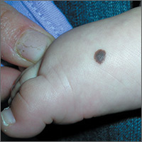
Brown spot on right foot

The FP recognized this as a benign congenital nevus.
While most congenital nevi are visible at birth, there are some that appear in the first year of life and are known as tardive congenital nevi. The FP used a dermatoscope to look at this nevus and found that its features were benign.
The parents wondered whether this needed to be removed to prevent it from becoming skin cancer in the future. The FP reassured them that the risk of melanoma from this one nevus was too small to warrant a prophylactic surgical excision. The parents agreed to the standard 6-month immunizations.
Photos and text for Photo Rounds Friday courtesy of Richard P. Usatine, MD. This case was adapted from: Smith, M. Congenital nevi. In: Usatine R, Smith M, Mayeaux EJ, et al. Color Atlas of Family Medicine. 2nd ed. New York, NY: McGraw-Hill; 2013:953-957.
To learn more about the Color Atlas of Family Medicine, see: www.amazon.com/Color-Family-Medicine-Richard-Usatine/dp/0071769641/.
You can now get the second edition of the Color Atlas of Family Medicine as an app by clicking on this link: usatinemedia.com.