User login
Transplantation palliative care: The time is ripe
Over 10 years ago, a challenge was made in a surgical publication for increased collaboration between the fields of transplantation and palliative care.1
Since that time not much progress has been made bringing these fields together in a consistent way that would mutually benefit patients and the specialties. However, other progress has been made, particularly in the field of palliative care, which could brighten the prospects and broaden the opportunities to accomplish collaboration between palliative care and transplantation.
Growth of palliative services
During the past decade there has been a robust proliferation of hospital-based palliative care programs in the United States. In all, 67% of U.S. hospitals with 50 or more beds report palliative care teams, up from 63% in 2011 and 53% in 2008.

Only a decade ago, critical care and palliative care were generally considered mutually exclusive. Evidence is trickling in to suggest that this is no longer the case. Although palliative care was not an integral part of critical care at that time, patients, families, and even practitioners began to demand these services. Cook and Rocker have eloquently advocated the rightful place of palliative care in the ICU.2
Studies in recent years have shown that the integration of palliative care into critical care decreases in length of ICU and hospital stay, decreases costs, enhances patient/family satisfaction, and promotes a more rapid consensus about goals of care, without increasing mortality. The ICU experience to date could be considered a reassuring precedent for transplantation palliative care.
Integration of palliative care with transplantation
Early palliative care intervention has been shown to improve symptom burden and depression scores in end-stage liver disease patients awaiting transplant. In addition, early palliative care consultation in conjunction with cancer treatment has been associated with increased survival in non–small-cell lung cancer patients. It has been demonstrated that early integration of palliative care in the surgical ICU alongside disease-directed curative care can be accomplished without change in mortality, while improving end-of-life practice in liver transplant patients.3

What palliative care can do for transplant patients
What does palliative care mean for the person (and family) awaiting transplantation? For the cirrhotic patient with cachexia, ascites, and encephalopathy, it means access to the services of a team trained in the management of these symptoms. Palliative care teams can also provide psychosocial and spiritual support for patients and families who are intimidated by the complex navigation of the health care system and the existential threat that end-stage organ failure presents to them. Skilled palliative care and services can be the difference between failing and extended life with a higher quality of life for these very sick patients
Resuscitation of a patient, whether through restoration of organ function or interdicting the progression of disease, begins with resuscitation of hope. Nothing achieves this more quickly than amelioration of burdensome symptoms for the patient and family.
The barriers for transplant surgeons and teams referring and incorporating palliative care services in their practices are multiple and profound. The unique dilemma facing the transplant team is to balance the treatment of the failing organ, the treatment of the patient (and family and friends), and the best use of the graft, a precious gift of society.
Palliative surgery has been defined as any invasive procedure in which the main intention is to mitigate physical symptoms in patients with noncurable disease without causing premature death. The very success of transplantation over the past 3 decades has obscured our memory of transplantation as a type of palliative surgery. It is a well-known axiom of reconstructive surgery that the reconstructed site should be compared to what was there, not to “normal.” Even in the current era of improved immunosuppression and posttransplant support services, one could hardly describe even a successful transplant patient’s experience as “normal.” These patients’ lives may be extended and/or enhanced but they need palliative care before, during, and after transplantation. The growing availability of trained palliative care clinicians and teams, the increased familiarity of palliative and end-of-life care to surgical residents and fellows, and quality metrics measuring palliative care outcomes will provide reassurance and guidance to address reservations about the convergence of the two seemingly opposite realities.
A modest proposal
We propose that palliative care be presented to the entire spectrum of transplantation care: on the ward, in the ICU, and after transplantation. More specific “triggers” for palliative care for referral of transplant patients should be identified. Wentlandt et al.4 have described a promising model for an ambulatory clinic, which provides early, integrated palliative care to patients awaiting and receiving organ transplantation. In addition, we propose an application for grant funding for a conference and eventual formation of a work group of transplant surgeons and team members, palliative care clinicians, and patient/families who have experienced one of the aspects of the transplant spectrum. We await the subspecialty certification in hospice and palliative medicine of a transplant surgeon. Outside of transplantation, every other surgical specialty in the United States has diplomates certified in hospice and palliative medicine. We await the benefits that will accrue from research about the merging of these fields.
1. Molmenti EP, Dunn GP: Transplantation and palliative care: The convergence of two seemingly opposite realities. Surg Clin North Am. 2005;85:373-82.
2. Cook D, Rocker G. Dying with dignity in the intensive care unit. N Engl J Med. 2014;370:2506-14.
3. Lamba S, Murphy P, McVicker S, Smith JH, and Mosenthal AC. Changing end-of-life care practice for liver transplant patients: structured palliative care intervention in the surgical intensive care unit. J Pain Symptom Manage. 2012; 44(4):508-19.
4. Wentlandt, K., Dall’Osto, A., Freeman, N., Le, L. W., Kaya, E., Ross, H., Singer, L. G., Abbey, S., Clarke, H. and Zimmermann, C. (2016), The Transplant Palliative Care Clinic: An early palliative care model for patients in a transplant program. Clin Transplant. 2016 Nov 4; doi: 10.1111/ctr.12838.
Dr. Azoulay is a transplantation specialist of Assistance Publique – Hôpitaux de Paris, and the University of Paris. Dr. Dunn is medical director of the Palliative Care Consultation Service at the University of Pittsburgh Medical Center Hamot, and vice-chair of the ACS Committee on Surgical Palliative Care.
Over 10 years ago, a challenge was made in a surgical publication for increased collaboration between the fields of transplantation and palliative care.1
Since that time not much progress has been made bringing these fields together in a consistent way that would mutually benefit patients and the specialties. However, other progress has been made, particularly in the field of palliative care, which could brighten the prospects and broaden the opportunities to accomplish collaboration between palliative care and transplantation.
Growth of palliative services
During the past decade there has been a robust proliferation of hospital-based palliative care programs in the United States. In all, 67% of U.S. hospitals with 50 or more beds report palliative care teams, up from 63% in 2011 and 53% in 2008.
Only a decade ago, critical care and palliative care were generally considered mutually exclusive. Evidence is trickling in to suggest that this is no longer the case. Although palliative care was not an integral part of critical care at that time, patients, families, and even practitioners began to demand these services. Cook and Rocker have eloquently advocated the rightful place of palliative care in the ICU.2
Studies in recent years have shown that the integration of palliative care into critical care decreases in length of ICU and hospital stay, decreases costs, enhances patient/family satisfaction, and promotes a more rapid consensus about goals of care, without increasing mortality. The ICU experience to date could be considered a reassuring precedent for transplantation palliative care.
Integration of palliative care with transplantation
Early palliative care intervention has been shown to improve symptom burden and depression scores in end-stage liver disease patients awaiting transplant. In addition, early palliative care consultation in conjunction with cancer treatment has been associated with increased survival in non–small-cell lung cancer patients. It has been demonstrated that early integration of palliative care in the surgical ICU alongside disease-directed curative care can be accomplished without change in mortality, while improving end-of-life practice in liver transplant patients.3
What palliative care can do for transplant patients
What does palliative care mean for the person (and family) awaiting transplantation? For the cirrhotic patient with cachexia, ascites, and encephalopathy, it means access to the services of a team trained in the management of these symptoms. Palliative care teams can also provide psychosocial and spiritual support for patients and families who are intimidated by the complex navigation of the health care system and the existential threat that end-stage organ failure presents to them. Skilled palliative care and services can be the difference between failing and extended life with a higher quality of life for these very sick patients
Resuscitation of a patient, whether through restoration of organ function or interdicting the progression of disease, begins with resuscitation of hope. Nothing achieves this more quickly than amelioration of burdensome symptoms for the patient and family.
The barriers for transplant surgeons and teams referring and incorporating palliative care services in their practices are multiple and profound. The unique dilemma facing the transplant team is to balance the treatment of the failing organ, the treatment of the patient (and family and friends), and the best use of the graft, a precious gift of society.
Palliative surgery has been defined as any invasive procedure in which the main intention is to mitigate physical symptoms in patients with noncurable disease without causing premature death. The very success of transplantation over the past 3 decades has obscured our memory of transplantation as a type of palliative surgery. It is a well-known axiom of reconstructive surgery that the reconstructed site should be compared to what was there, not to “normal.” Even in the current era of improved immunosuppression and posttransplant support services, one could hardly describe even a successful transplant patient’s experience as “normal.” These patients’ lives may be extended and/or enhanced but they need palliative care before, during, and after transplantation. The growing availability of trained palliative care clinicians and teams, the increased familiarity of palliative and end-of-life care to surgical residents and fellows, and quality metrics measuring palliative care outcomes will provide reassurance and guidance to address reservations about the convergence of the two seemingly opposite realities.
A modest proposal
We propose that palliative care be presented to the entire spectrum of transplantation care: on the ward, in the ICU, and after transplantation. More specific “triggers” for palliative care for referral of transplant patients should be identified. Wentlandt et al.4 have described a promising model for an ambulatory clinic, which provides early, integrated palliative care to patients awaiting and receiving organ transplantation. In addition, we propose an application for grant funding for a conference and eventual formation of a work group of transplant surgeons and team members, palliative care clinicians, and patient/families who have experienced one of the aspects of the transplant spectrum. We await the subspecialty certification in hospice and palliative medicine of a transplant surgeon. Outside of transplantation, every other surgical specialty in the United States has diplomates certified in hospice and palliative medicine. We await the benefits that will accrue from research about the merging of these fields.
1. Molmenti EP, Dunn GP: Transplantation and palliative care: The convergence of two seemingly opposite realities. Surg Clin North Am. 2005;85:373-82.
2. Cook D, Rocker G. Dying with dignity in the intensive care unit. N Engl J Med. 2014;370:2506-14.
3. Lamba S, Murphy P, McVicker S, Smith JH, and Mosenthal AC. Changing end-of-life care practice for liver transplant patients: structured palliative care intervention in the surgical intensive care unit. J Pain Symptom Manage. 2012; 44(4):508-19.
4. Wentlandt, K., Dall’Osto, A., Freeman, N., Le, L. W., Kaya, E., Ross, H., Singer, L. G., Abbey, S., Clarke, H. and Zimmermann, C. (2016), The Transplant Palliative Care Clinic: An early palliative care model for patients in a transplant program. Clin Transplant. 2016 Nov 4; doi: 10.1111/ctr.12838.
Dr. Azoulay is a transplantation specialist of Assistance Publique – Hôpitaux de Paris, and the University of Paris. Dr. Dunn is medical director of the Palliative Care Consultation Service at the University of Pittsburgh Medical Center Hamot, and vice-chair of the ACS Committee on Surgical Palliative Care.
Over 10 years ago, a challenge was made in a surgical publication for increased collaboration between the fields of transplantation and palliative care.1
Since that time not much progress has been made bringing these fields together in a consistent way that would mutually benefit patients and the specialties. However, other progress has been made, particularly in the field of palliative care, which could brighten the prospects and broaden the opportunities to accomplish collaboration between palliative care and transplantation.
Growth of palliative services
During the past decade there has been a robust proliferation of hospital-based palliative care programs in the United States. In all, 67% of U.S. hospitals with 50 or more beds report palliative care teams, up from 63% in 2011 and 53% in 2008.
Only a decade ago, critical care and palliative care were generally considered mutually exclusive. Evidence is trickling in to suggest that this is no longer the case. Although palliative care was not an integral part of critical care at that time, patients, families, and even practitioners began to demand these services. Cook and Rocker have eloquently advocated the rightful place of palliative care in the ICU.2
Studies in recent years have shown that the integration of palliative care into critical care decreases in length of ICU and hospital stay, decreases costs, enhances patient/family satisfaction, and promotes a more rapid consensus about goals of care, without increasing mortality. The ICU experience to date could be considered a reassuring precedent for transplantation palliative care.
Integration of palliative care with transplantation
Early palliative care intervention has been shown to improve symptom burden and depression scores in end-stage liver disease patients awaiting transplant. In addition, early palliative care consultation in conjunction with cancer treatment has been associated with increased survival in non–small-cell lung cancer patients. It has been demonstrated that early integration of palliative care in the surgical ICU alongside disease-directed curative care can be accomplished without change in mortality, while improving end-of-life practice in liver transplant patients.3
What palliative care can do for transplant patients
What does palliative care mean for the person (and family) awaiting transplantation? For the cirrhotic patient with cachexia, ascites, and encephalopathy, it means access to the services of a team trained in the management of these symptoms. Palliative care teams can also provide psychosocial and spiritual support for patients and families who are intimidated by the complex navigation of the health care system and the existential threat that end-stage organ failure presents to them. Skilled palliative care and services can be the difference between failing and extended life with a higher quality of life for these very sick patients
Resuscitation of a patient, whether through restoration of organ function or interdicting the progression of disease, begins with resuscitation of hope. Nothing achieves this more quickly than amelioration of burdensome symptoms for the patient and family.
The barriers for transplant surgeons and teams referring and incorporating palliative care services in their practices are multiple and profound. The unique dilemma facing the transplant team is to balance the treatment of the failing organ, the treatment of the patient (and family and friends), and the best use of the graft, a precious gift of society.
Palliative surgery has been defined as any invasive procedure in which the main intention is to mitigate physical symptoms in patients with noncurable disease without causing premature death. The very success of transplantation over the past 3 decades has obscured our memory of transplantation as a type of palliative surgery. It is a well-known axiom of reconstructive surgery that the reconstructed site should be compared to what was there, not to “normal.” Even in the current era of improved immunosuppression and posttransplant support services, one could hardly describe even a successful transplant patient’s experience as “normal.” These patients’ lives may be extended and/or enhanced but they need palliative care before, during, and after transplantation. The growing availability of trained palliative care clinicians and teams, the increased familiarity of palliative and end-of-life care to surgical residents and fellows, and quality metrics measuring palliative care outcomes will provide reassurance and guidance to address reservations about the convergence of the two seemingly opposite realities.
A modest proposal
We propose that palliative care be presented to the entire spectrum of transplantation care: on the ward, in the ICU, and after transplantation. More specific “triggers” for palliative care for referral of transplant patients should be identified. Wentlandt et al.4 have described a promising model for an ambulatory clinic, which provides early, integrated palliative care to patients awaiting and receiving organ transplantation. In addition, we propose an application for grant funding for a conference and eventual formation of a work group of transplant surgeons and team members, palliative care clinicians, and patient/families who have experienced one of the aspects of the transplant spectrum. We await the subspecialty certification in hospice and palliative medicine of a transplant surgeon. Outside of transplantation, every other surgical specialty in the United States has diplomates certified in hospice and palliative medicine. We await the benefits that will accrue from research about the merging of these fields.
1. Molmenti EP, Dunn GP: Transplantation and palliative care: The convergence of two seemingly opposite realities. Surg Clin North Am. 2005;85:373-82.
2. Cook D, Rocker G. Dying with dignity in the intensive care unit. N Engl J Med. 2014;370:2506-14.
3. Lamba S, Murphy P, McVicker S, Smith JH, and Mosenthal AC. Changing end-of-life care practice for liver transplant patients: structured palliative care intervention in the surgical intensive care unit. J Pain Symptom Manage. 2012; 44(4):508-19.
4. Wentlandt, K., Dall’Osto, A., Freeman, N., Le, L. W., Kaya, E., Ross, H., Singer, L. G., Abbey, S., Clarke, H. and Zimmermann, C. (2016), The Transplant Palliative Care Clinic: An early palliative care model for patients in a transplant program. Clin Transplant. 2016 Nov 4; doi: 10.1111/ctr.12838.
Dr. Azoulay is a transplantation specialist of Assistance Publique – Hôpitaux de Paris, and the University of Paris. Dr. Dunn is medical director of the Palliative Care Consultation Service at the University of Pittsburgh Medical Center Hamot, and vice-chair of the ACS Committee on Surgical Palliative Care.

SVS Now Accepting Abstracts for VAM 2017
Abstracts for the 2017 Vascular Annual Meeting are now being accepted. The submission site opened Monday, Nov. 14 for the meeting, to be held May 31 to June 3, 2017, in San Diego. Plenary sessions and exhibits will be June 1 to 3.
Participants may submit abstracts into any of 14 categories and a number of presentation types, including videos. In 2016, organizers selected approximately two-thirds of the submitted abstracts, and this year the VAM Program Committee is seeking additional venues for people to present their work in, including more sessions and other presentation formats.
Click here for abstract guidelines and more information. Abstracts themselves may be submitted here.
Abstracts for the 2017 Vascular Annual Meeting are now being accepted. The submission site opened Monday, Nov. 14 for the meeting, to be held May 31 to June 3, 2017, in San Diego. Plenary sessions and exhibits will be June 1 to 3.
Participants may submit abstracts into any of 14 categories and a number of presentation types, including videos. In 2016, organizers selected approximately two-thirds of the submitted abstracts, and this year the VAM Program Committee is seeking additional venues for people to present their work in, including more sessions and other presentation formats.
Click here for abstract guidelines and more information. Abstracts themselves may be submitted here.
Abstracts for the 2017 Vascular Annual Meeting are now being accepted. The submission site opened Monday, Nov. 14 for the meeting, to be held May 31 to June 3, 2017, in San Diego. Plenary sessions and exhibits will be June 1 to 3.
Participants may submit abstracts into any of 14 categories and a number of presentation types, including videos. In 2016, organizers selected approximately two-thirds of the submitted abstracts, and this year the VAM Program Committee is seeking additional venues for people to present their work in, including more sessions and other presentation formats.
Click here for abstract guidelines and more information. Abstracts themselves may be submitted here.
Best Practices: Protecting Dry Vulnerable Skin with CeraVe® Healing Ointment
A supplement to Dermatology News. This advertising supplement is sponsored by Valeant Pharmaceuticals.

- Reinforcing the Skin Barrier
- NEA Seal of Acceptance
- A Preventative Approach to Dry, Cracked Skin
- CeraVe Ointment in the Clinical Setting
Faculty/Faculty Disclosure
Sheila Fallon Friedlander, MD
Professor of Clinical Dermatology & Pediatrics
Director, Pediatric Dermatology Fellowship Training Program
University of California at San Diego School of Medicine
Rady Children’s Hospital,
San Diego, California
Dr. Friedlander was compensated for her participation in the development of this article.
CeraVe is a registered trademark of Valeant Pharmaceuticals International, Inc. or its affiliates.
A supplement to Dermatology News. This advertising supplement is sponsored by Valeant Pharmaceuticals.
- Reinforcing the Skin Barrier
- NEA Seal of Acceptance
- A Preventative Approach to Dry, Cracked Skin
- CeraVe Ointment in the Clinical Setting
Faculty/Faculty Disclosure
Sheila Fallon Friedlander, MD
Professor of Clinical Dermatology & Pediatrics
Director, Pediatric Dermatology Fellowship Training Program
University of California at San Diego School of Medicine
Rady Children’s Hospital,
San Diego, California
Dr. Friedlander was compensated for her participation in the development of this article.
CeraVe is a registered trademark of Valeant Pharmaceuticals International, Inc. or its affiliates.
A supplement to Dermatology News. This advertising supplement is sponsored by Valeant Pharmaceuticals.
- Reinforcing the Skin Barrier
- NEA Seal of Acceptance
- A Preventative Approach to Dry, Cracked Skin
- CeraVe Ointment in the Clinical Setting
Faculty/Faculty Disclosure
Sheila Fallon Friedlander, MD
Professor of Clinical Dermatology & Pediatrics
Director, Pediatric Dermatology Fellowship Training Program
University of California at San Diego School of Medicine
Rady Children’s Hospital,
San Diego, California
Dr. Friedlander was compensated for her participation in the development of this article.
CeraVe is a registered trademark of Valeant Pharmaceuticals International, Inc. or its affiliates.

What Every Primary Care Clinician Should Know About Natriuretic Peptides
What Every Primary Care Clinician Should Know About Natriuretic Peptides
Primary care clinicians sit at the crossroads of multiple medical disciplines. Symptoms such as fatigue, dyspnea, weight gain, and lower extremity swelling rarely point to a single organ system. These overlapping presentations reflect complex cardiometabolic syndromes that require an integrated diagnostic approach.
Natriuretic peptides are biologically active hormones secreted in response to myocardial stress that can provide valuable diagnostic and prognostic information in this setting. Despite extensive validation and inclusion in major guidelines, B-type natriuretic peptide (BNP) and N-terminal pro–B-type natriuretic peptide (NT-proBNP) are often regarded as confirmatory tests for heart failure, limiting their broader clinical utility. When understood as part of an active hormonal network, however, natriuretic peptides reveal diagnostic and therapeutic potential well beyond heart failure.
PHYSIOLOGIC OVERVIEW
The natriuretic peptide system is a key endocrine mechanism for regulating cardiovascular and volume function. Ventricular myocytes release BNP and NT-proBNP in response to increased wall stretch, while atrial natriuretic peptide (ANP) arises primarily from atrial tissue under similar hemodynamic stress. These peptides reduce preload and afterload by promoting vasodilation, natriuresis, and suppression of the renin-angiotensin-aldosterone system and the sympathetic nervous system.1
C-type natriuretic peptide (CNP), synthesized in endothelial cells, performs a complementary but distinct role. It maintains endothelial health, inhibits fibrosis, and modulates vascular tone.2 Together, these peptides orchestrate rapid compensatory responses to pressure and volume overload. Their biologic actions are mediated by 3 receptor subtypes: natriuretic peptide receptor A (NPR-A), the primary receptor for ANP and BNP; natriuretic peptide receptor B (NPR-B), which mediates CNP’s vascular effects; and natriuretic peptide receptor C (NPR-C), once considered solely a clearance receptor but now recognized as having active intracellular signaling properties that regulate vascular homeostasis.2
Clinical Translation and Therapeutic Evolution
The physiologic understanding of natriuretic peptides transitioned into therapeutic application with the development of angiotensin receptor–neprilysin inhibitors (ARNIs). Neprilysin degrades natriuretic peptides, limiting their duration of action; inhibition therefore enhances natriuretic peptide-mediated effects. The PARADIGMHF trial demonstrated that sacubitril/valsartan significantly reduced mortality and hospitalization in patients with heart failure with reduced ejection fraction (HFrEF) compared with enalapril.3
However, findings from the PARAGONHF trial, which evaluated patients with heart failure with preserved ejection fraction (HFpEF), were less definitive.4 This divergence underscores that heart failure is a heterogeneous syndrome, requiring phenotype-specific management strategies.
NATRIURETIC PEPTIDE RESISTANCE
A persistent clinical challenge is natriuretic peptide resistance, a condition in which circulating natriuretic peptide levels are elevated, but their physiologic efficacy is diminished. This is particularly observed in patients with obesity, chronic kidney disease (CKD), and advanced heart failure. Mechanistic studies attribute this resistance to enhanced clearance through NPR-C and increased neprilysin activity.5
Understanding natriuretic peptide resistance helps clinicians more accurately interpret laboratory results. An elevated BNP level in a patient with obesity or CKD should not automatically be dismissed as a false-positive finding but instead viewed within the broader context of altered natriuretic peptide bioactivity.
Applications Beyond Heart Failure
Natriuretic peptides have diagnostic and prognostic roles in several conditions beyond traditional heart failure. In pulmonary embolism, chronic obstructive pulmonary disease (COPD), and acute respiratory distress syndrome (ARDS), elevated BNP or NT-proBNP levels often indicate right ventricular strain and portend worse outcomes. Similarly, in sepsis-induced cardiomyopathy, natriuretic peptides may serve as early markers of cardiac dysfunction.6
In valvular heart disease, BNP elevation can guide the timing of intervention. For example, a markedly elevated BNP in aortic stenosis may prompt earlier surgical evaluation even when symptoms remain subtle.1 For patients with CKD, natriuretic peptide interpretation requires adjustment because renal impairment reduces peptide clearance, leading to higher baseline values.
Nevertheless, by employing adjusted cutoffs or tracking serial trends, clinicians can preserve diagnostic precision. Moreover, ARNI therapy may confer renal protection by lowering intraglomerular pressure, providing a therapeutic advantage in cardiorenal syndromes.5
Emerging Peptide-Based Therapies
Recent advances in peptide pharmacology have yielded a new generation of synthetic natriuretic peptides and receptor modulators. Agents such as CRRL269 produce natriuresis with less hypotension, and NPA7 acts on both the natriuretic peptide and Mas receptor pathways to enhance cardiac and renal protection. C53, a selective activator of guanylyl cyclase-B (GC-B), shows potential for improving outcomes in HFpEF without major blood pressure reduction.7
Meanwhile, CNP analogs, such as vosoritide, approved for achondroplasia, have demonstrated antifibrotic and vascular benefits in preclinical studies.8 These developments highlight a growing recognition of the natriuretic peptide system as a therapeutic target across multiple organ systems, including metabolic and fibrotic diseases.
Genetic Insights and Precision Medicine
Genetic studies further illustrate how interindividual variation influences natriuretic peptide function. The rs5068 polymorphism in the ANP gene (NPPA) is associated with higher circulating natriuretic peptide levels and lower blood pressure, suggesting a naturally cardioprotective phenotype.9,10 Such findings may eventually inform natriuretic peptide-guided or genotype- based treatment strategies for hypertension and heart failure.
Beyond genetics, emerging evidence supports the use of natriuretic peptide profiling to identify salt-sensitive hypertension and early subclinical ventricular dysfunction, positioning natriuretic peptides as central tools for precision prevention in primary care.6
PRACTICAL CONSIDERATIONS FOR PRIMARY CARE
Natriuretic peptides provide a means for integrating cardiovascular physiology into daily decision-making for primary care clinicians. BNP and NT-proBNP testing can distinguish cardiac from noncardiac causes of dyspnea, gauge volume status in CKD, and monitor patients at risk for decompensation. Interpretation should account for age, obesity, renal function, and baseline comorbidities, with emphasis on trends rather than single values.
FUTURE IMPLICATIONS
In the coming years, as novel therapies and oral receptor modulators emerge, natriuretic peptides are likely to become not only diagnostic indicators but also therapeutic guides. This evolution will bridge the gap between physiology and personalized medicine, offering a new paradigm in cardiovascular care. Their use in primary care may improve early detection, risk stratification, and treatment optimization across a broad spectrum of cardiometabolic disorders, ushering in a new era of patient-centered care.
CONCLUSIONS
Natriuretic peptides are more than numbers on a laboratory report. They are biologically active hormones that provide real-time insights into cardiovascular, renal, and systemic stress. As new evidence, therapeutics, and genetic data expand the scope of natriuretic peptide science, primary care physicians are positioned to apply this knowledge for earlier diagnosis, more precise treatment, and better long-term outcomes. Understanding when and how to use natriuretic peptides and interpreting them in the full context of patient physiology should now be considered an essential skill in modern primary care.
- Potter LR, Yoder AR, Flora DR, et al. Natriuretic peptides: their structures, receptors, physiologic functions, and therapeutic applications. Handb Exp Pharmacol. 2009;191:341-366. doi:10.1007/978-3-540-68964-5_17
- Kuhn M. Molecular physiology of the natriuretic peptide system: new aspects on biochemistry and pathophysiology. Basic Res Cardiol. 2022;117:1-23. doi:10.1007/s00395-021-00919-y
- McMurray JJV, Packer M, Desai AS, et al. Angiotensin-neprilysin inhibition versus enalapril in heart failure. N Engl J Med. 2014;371:993-1004. doi:10.1056/NEJMoa1409077
- Solomon SD, McMurray JJV, Anand IS, et al. Angiotensin-neprilysin inhibition in heart failure with preserved ejection fraction. N Engl J Med. 2019;381:1609-1620. doi:10.1056/NEJMoa1908655
- Packer M, Lam CSP, Lund LH, et al. Mechanisms of natriuretic peptide resistance in heart failure. J Am Coll Cardiol. 2021;78:704-714. doi:10.1016/j.jacc.2021.05.050
- Srivastava A, Gupta A, Raman R, et al. Diagnostic and prognostic role of natriuretic peptides in pulmonary and systemic disorders. Cytokine. 2024;173:156417. doi:10.1016/j.cyto.2024.156417
- Zhang Y, Li W, Chen Q, et al. Designer natriuretic peptides for heart and kidney diseases: next-generation therapeutics. Nat Commun. 2023;14:1-14. doi:10.1038/s41467-023-37070-6
- Holtwick KM, Kuhn M, Scheller D, et al. Expanding roles of C-type natriuretic peptide signaling in cardiovascular and metabolic disease. Cardiovasc Res. 2023;119:140-152. doi:10.1093/cvr/cvac101
- Rame JE, Tam SW, McNamara D, et al. The common atrial natriuretic peptide gene variant rs5068 is associated with enhanced natriuretic peptide levels and reduced blood pressure in humans. Hypertension. 2007;49:857-864. doi:10.1161/01.HYP.0000259802.77768.48
- Tokudome T, Horio T, Kishimoto I, et al. C-type natriuretic peptide as a novel cardiac hormone. Circulation. 2008; 117: 2329-2337. doi:10.1161/CIRCULATIONAHA.107.757740
Primary care clinicians sit at the crossroads of multiple medical disciplines. Symptoms such as fatigue, dyspnea, weight gain, and lower extremity swelling rarely point to a single organ system. These overlapping presentations reflect complex cardiometabolic syndromes that require an integrated diagnostic approach.
Natriuretic peptides are biologically active hormones secreted in response to myocardial stress that can provide valuable diagnostic and prognostic information in this setting. Despite extensive validation and inclusion in major guidelines, B-type natriuretic peptide (BNP) and N-terminal pro–B-type natriuretic peptide (NT-proBNP) are often regarded as confirmatory tests for heart failure, limiting their broader clinical utility. When understood as part of an active hormonal network, however, natriuretic peptides reveal diagnostic and therapeutic potential well beyond heart failure.
PHYSIOLOGIC OVERVIEW
The natriuretic peptide system is a key endocrine mechanism for regulating cardiovascular and volume function. Ventricular myocytes release BNP and NT-proBNP in response to increased wall stretch, while atrial natriuretic peptide (ANP) arises primarily from atrial tissue under similar hemodynamic stress. These peptides reduce preload and afterload by promoting vasodilation, natriuresis, and suppression of the renin-angiotensin-aldosterone system and the sympathetic nervous system.1
C-type natriuretic peptide (CNP), synthesized in endothelial cells, performs a complementary but distinct role. It maintains endothelial health, inhibits fibrosis, and modulates vascular tone.2 Together, these peptides orchestrate rapid compensatory responses to pressure and volume overload. Their biologic actions are mediated by 3 receptor subtypes: natriuretic peptide receptor A (NPR-A), the primary receptor for ANP and BNP; natriuretic peptide receptor B (NPR-B), which mediates CNP’s vascular effects; and natriuretic peptide receptor C (NPR-C), once considered solely a clearance receptor but now recognized as having active intracellular signaling properties that regulate vascular homeostasis.2
Clinical Translation and Therapeutic Evolution
The physiologic understanding of natriuretic peptides transitioned into therapeutic application with the development of angiotensin receptor–neprilysin inhibitors (ARNIs). Neprilysin degrades natriuretic peptides, limiting their duration of action; inhibition therefore enhances natriuretic peptide-mediated effects. The PARADIGMHF trial demonstrated that sacubitril/valsartan significantly reduced mortality and hospitalization in patients with heart failure with reduced ejection fraction (HFrEF) compared with enalapril.3
However, findings from the PARAGONHF trial, which evaluated patients with heart failure with preserved ejection fraction (HFpEF), were less definitive.4 This divergence underscores that heart failure is a heterogeneous syndrome, requiring phenotype-specific management strategies.
NATRIURETIC PEPTIDE RESISTANCE
A persistent clinical challenge is natriuretic peptide resistance, a condition in which circulating natriuretic peptide levels are elevated, but their physiologic efficacy is diminished. This is particularly observed in patients with obesity, chronic kidney disease (CKD), and advanced heart failure. Mechanistic studies attribute this resistance to enhanced clearance through NPR-C and increased neprilysin activity.5
Understanding natriuretic peptide resistance helps clinicians more accurately interpret laboratory results. An elevated BNP level in a patient with obesity or CKD should not automatically be dismissed as a false-positive finding but instead viewed within the broader context of altered natriuretic peptide bioactivity.
Applications Beyond Heart Failure
Natriuretic peptides have diagnostic and prognostic roles in several conditions beyond traditional heart failure. In pulmonary embolism, chronic obstructive pulmonary disease (COPD), and acute respiratory distress syndrome (ARDS), elevated BNP or NT-proBNP levels often indicate right ventricular strain and portend worse outcomes. Similarly, in sepsis-induced cardiomyopathy, natriuretic peptides may serve as early markers of cardiac dysfunction.6
In valvular heart disease, BNP elevation can guide the timing of intervention. For example, a markedly elevated BNP in aortic stenosis may prompt earlier surgical evaluation even when symptoms remain subtle.1 For patients with CKD, natriuretic peptide interpretation requires adjustment because renal impairment reduces peptide clearance, leading to higher baseline values.
Nevertheless, by employing adjusted cutoffs or tracking serial trends, clinicians can preserve diagnostic precision. Moreover, ARNI therapy may confer renal protection by lowering intraglomerular pressure, providing a therapeutic advantage in cardiorenal syndromes.5
Emerging Peptide-Based Therapies
Recent advances in peptide pharmacology have yielded a new generation of synthetic natriuretic peptides and receptor modulators. Agents such as CRRL269 produce natriuresis with less hypotension, and NPA7 acts on both the natriuretic peptide and Mas receptor pathways to enhance cardiac and renal protection. C53, a selective activator of guanylyl cyclase-B (GC-B), shows potential for improving outcomes in HFpEF without major blood pressure reduction.7
Meanwhile, CNP analogs, such as vosoritide, approved for achondroplasia, have demonstrated antifibrotic and vascular benefits in preclinical studies.8 These developments highlight a growing recognition of the natriuretic peptide system as a therapeutic target across multiple organ systems, including metabolic and fibrotic diseases.
Genetic Insights and Precision Medicine
Genetic studies further illustrate how interindividual variation influences natriuretic peptide function. The rs5068 polymorphism in the ANP gene (NPPA) is associated with higher circulating natriuretic peptide levels and lower blood pressure, suggesting a naturally cardioprotective phenotype.9,10 Such findings may eventually inform natriuretic peptide-guided or genotype- based treatment strategies for hypertension and heart failure.
Beyond genetics, emerging evidence supports the use of natriuretic peptide profiling to identify salt-sensitive hypertension and early subclinical ventricular dysfunction, positioning natriuretic peptides as central tools for precision prevention in primary care.6
PRACTICAL CONSIDERATIONS FOR PRIMARY CARE
Natriuretic peptides provide a means for integrating cardiovascular physiology into daily decision-making for primary care clinicians. BNP and NT-proBNP testing can distinguish cardiac from noncardiac causes of dyspnea, gauge volume status in CKD, and monitor patients at risk for decompensation. Interpretation should account for age, obesity, renal function, and baseline comorbidities, with emphasis on trends rather than single values.
FUTURE IMPLICATIONS
In the coming years, as novel therapies and oral receptor modulators emerge, natriuretic peptides are likely to become not only diagnostic indicators but also therapeutic guides. This evolution will bridge the gap between physiology and personalized medicine, offering a new paradigm in cardiovascular care. Their use in primary care may improve early detection, risk stratification, and treatment optimization across a broad spectrum of cardiometabolic disorders, ushering in a new era of patient-centered care.
CONCLUSIONS
Natriuretic peptides are more than numbers on a laboratory report. They are biologically active hormones that provide real-time insights into cardiovascular, renal, and systemic stress. As new evidence, therapeutics, and genetic data expand the scope of natriuretic peptide science, primary care physicians are positioned to apply this knowledge for earlier diagnosis, more precise treatment, and better long-term outcomes. Understanding when and how to use natriuretic peptides and interpreting them in the full context of patient physiology should now be considered an essential skill in modern primary care.
Primary care clinicians sit at the crossroads of multiple medical disciplines. Symptoms such as fatigue, dyspnea, weight gain, and lower extremity swelling rarely point to a single organ system. These overlapping presentations reflect complex cardiometabolic syndromes that require an integrated diagnostic approach.
Natriuretic peptides are biologically active hormones secreted in response to myocardial stress that can provide valuable diagnostic and prognostic information in this setting. Despite extensive validation and inclusion in major guidelines, B-type natriuretic peptide (BNP) and N-terminal pro–B-type natriuretic peptide (NT-proBNP) are often regarded as confirmatory tests for heart failure, limiting their broader clinical utility. When understood as part of an active hormonal network, however, natriuretic peptides reveal diagnostic and therapeutic potential well beyond heart failure.
PHYSIOLOGIC OVERVIEW
The natriuretic peptide system is a key endocrine mechanism for regulating cardiovascular and volume function. Ventricular myocytes release BNP and NT-proBNP in response to increased wall stretch, while atrial natriuretic peptide (ANP) arises primarily from atrial tissue under similar hemodynamic stress. These peptides reduce preload and afterload by promoting vasodilation, natriuresis, and suppression of the renin-angiotensin-aldosterone system and the sympathetic nervous system.1
C-type natriuretic peptide (CNP), synthesized in endothelial cells, performs a complementary but distinct role. It maintains endothelial health, inhibits fibrosis, and modulates vascular tone.2 Together, these peptides orchestrate rapid compensatory responses to pressure and volume overload. Their biologic actions are mediated by 3 receptor subtypes: natriuretic peptide receptor A (NPR-A), the primary receptor for ANP and BNP; natriuretic peptide receptor B (NPR-B), which mediates CNP’s vascular effects; and natriuretic peptide receptor C (NPR-C), once considered solely a clearance receptor but now recognized as having active intracellular signaling properties that regulate vascular homeostasis.2
Clinical Translation and Therapeutic Evolution
The physiologic understanding of natriuretic peptides transitioned into therapeutic application with the development of angiotensin receptor–neprilysin inhibitors (ARNIs). Neprilysin degrades natriuretic peptides, limiting their duration of action; inhibition therefore enhances natriuretic peptide-mediated effects. The PARADIGMHF trial demonstrated that sacubitril/valsartan significantly reduced mortality and hospitalization in patients with heart failure with reduced ejection fraction (HFrEF) compared with enalapril.3
However, findings from the PARAGONHF trial, which evaluated patients with heart failure with preserved ejection fraction (HFpEF), were less definitive.4 This divergence underscores that heart failure is a heterogeneous syndrome, requiring phenotype-specific management strategies.
NATRIURETIC PEPTIDE RESISTANCE
A persistent clinical challenge is natriuretic peptide resistance, a condition in which circulating natriuretic peptide levels are elevated, but their physiologic efficacy is diminished. This is particularly observed in patients with obesity, chronic kidney disease (CKD), and advanced heart failure. Mechanistic studies attribute this resistance to enhanced clearance through NPR-C and increased neprilysin activity.5
Understanding natriuretic peptide resistance helps clinicians more accurately interpret laboratory results. An elevated BNP level in a patient with obesity or CKD should not automatically be dismissed as a false-positive finding but instead viewed within the broader context of altered natriuretic peptide bioactivity.
Applications Beyond Heart Failure
Natriuretic peptides have diagnostic and prognostic roles in several conditions beyond traditional heart failure. In pulmonary embolism, chronic obstructive pulmonary disease (COPD), and acute respiratory distress syndrome (ARDS), elevated BNP or NT-proBNP levels often indicate right ventricular strain and portend worse outcomes. Similarly, in sepsis-induced cardiomyopathy, natriuretic peptides may serve as early markers of cardiac dysfunction.6
In valvular heart disease, BNP elevation can guide the timing of intervention. For example, a markedly elevated BNP in aortic stenosis may prompt earlier surgical evaluation even when symptoms remain subtle.1 For patients with CKD, natriuretic peptide interpretation requires adjustment because renal impairment reduces peptide clearance, leading to higher baseline values.
Nevertheless, by employing adjusted cutoffs or tracking serial trends, clinicians can preserve diagnostic precision. Moreover, ARNI therapy may confer renal protection by lowering intraglomerular pressure, providing a therapeutic advantage in cardiorenal syndromes.5
Emerging Peptide-Based Therapies
Recent advances in peptide pharmacology have yielded a new generation of synthetic natriuretic peptides and receptor modulators. Agents such as CRRL269 produce natriuresis with less hypotension, and NPA7 acts on both the natriuretic peptide and Mas receptor pathways to enhance cardiac and renal protection. C53, a selective activator of guanylyl cyclase-B (GC-B), shows potential for improving outcomes in HFpEF without major blood pressure reduction.7
Meanwhile, CNP analogs, such as vosoritide, approved for achondroplasia, have demonstrated antifibrotic and vascular benefits in preclinical studies.8 These developments highlight a growing recognition of the natriuretic peptide system as a therapeutic target across multiple organ systems, including metabolic and fibrotic diseases.
Genetic Insights and Precision Medicine
Genetic studies further illustrate how interindividual variation influences natriuretic peptide function. The rs5068 polymorphism in the ANP gene (NPPA) is associated with higher circulating natriuretic peptide levels and lower blood pressure, suggesting a naturally cardioprotective phenotype.9,10 Such findings may eventually inform natriuretic peptide-guided or genotype- based treatment strategies for hypertension and heart failure.
Beyond genetics, emerging evidence supports the use of natriuretic peptide profiling to identify salt-sensitive hypertension and early subclinical ventricular dysfunction, positioning natriuretic peptides as central tools for precision prevention in primary care.6
PRACTICAL CONSIDERATIONS FOR PRIMARY CARE
Natriuretic peptides provide a means for integrating cardiovascular physiology into daily decision-making for primary care clinicians. BNP and NT-proBNP testing can distinguish cardiac from noncardiac causes of dyspnea, gauge volume status in CKD, and monitor patients at risk for decompensation. Interpretation should account for age, obesity, renal function, and baseline comorbidities, with emphasis on trends rather than single values.
FUTURE IMPLICATIONS
In the coming years, as novel therapies and oral receptor modulators emerge, natriuretic peptides are likely to become not only diagnostic indicators but also therapeutic guides. This evolution will bridge the gap between physiology and personalized medicine, offering a new paradigm in cardiovascular care. Their use in primary care may improve early detection, risk stratification, and treatment optimization across a broad spectrum of cardiometabolic disorders, ushering in a new era of patient-centered care.
CONCLUSIONS
Natriuretic peptides are more than numbers on a laboratory report. They are biologically active hormones that provide real-time insights into cardiovascular, renal, and systemic stress. As new evidence, therapeutics, and genetic data expand the scope of natriuretic peptide science, primary care physicians are positioned to apply this knowledge for earlier diagnosis, more precise treatment, and better long-term outcomes. Understanding when and how to use natriuretic peptides and interpreting them in the full context of patient physiology should now be considered an essential skill in modern primary care.
- Potter LR, Yoder AR, Flora DR, et al. Natriuretic peptides: their structures, receptors, physiologic functions, and therapeutic applications. Handb Exp Pharmacol. 2009;191:341-366. doi:10.1007/978-3-540-68964-5_17
- Kuhn M. Molecular physiology of the natriuretic peptide system: new aspects on biochemistry and pathophysiology. Basic Res Cardiol. 2022;117:1-23. doi:10.1007/s00395-021-00919-y
- McMurray JJV, Packer M, Desai AS, et al. Angiotensin-neprilysin inhibition versus enalapril in heart failure. N Engl J Med. 2014;371:993-1004. doi:10.1056/NEJMoa1409077
- Solomon SD, McMurray JJV, Anand IS, et al. Angiotensin-neprilysin inhibition in heart failure with preserved ejection fraction. N Engl J Med. 2019;381:1609-1620. doi:10.1056/NEJMoa1908655
- Packer M, Lam CSP, Lund LH, et al. Mechanisms of natriuretic peptide resistance in heart failure. J Am Coll Cardiol. 2021;78:704-714. doi:10.1016/j.jacc.2021.05.050
- Srivastava A, Gupta A, Raman R, et al. Diagnostic and prognostic role of natriuretic peptides in pulmonary and systemic disorders. Cytokine. 2024;173:156417. doi:10.1016/j.cyto.2024.156417
- Zhang Y, Li W, Chen Q, et al. Designer natriuretic peptides for heart and kidney diseases: next-generation therapeutics. Nat Commun. 2023;14:1-14. doi:10.1038/s41467-023-37070-6
- Holtwick KM, Kuhn M, Scheller D, et al. Expanding roles of C-type natriuretic peptide signaling in cardiovascular and metabolic disease. Cardiovasc Res. 2023;119:140-152. doi:10.1093/cvr/cvac101
- Rame JE, Tam SW, McNamara D, et al. The common atrial natriuretic peptide gene variant rs5068 is associated with enhanced natriuretic peptide levels and reduced blood pressure in humans. Hypertension. 2007;49:857-864. doi:10.1161/01.HYP.0000259802.77768.48
- Tokudome T, Horio T, Kishimoto I, et al. C-type natriuretic peptide as a novel cardiac hormone. Circulation. 2008; 117: 2329-2337. doi:10.1161/CIRCULATIONAHA.107.757740
- Potter LR, Yoder AR, Flora DR, et al. Natriuretic peptides: their structures, receptors, physiologic functions, and therapeutic applications. Handb Exp Pharmacol. 2009;191:341-366. doi:10.1007/978-3-540-68964-5_17
- Kuhn M. Molecular physiology of the natriuretic peptide system: new aspects on biochemistry and pathophysiology. Basic Res Cardiol. 2022;117:1-23. doi:10.1007/s00395-021-00919-y
- McMurray JJV, Packer M, Desai AS, et al. Angiotensin-neprilysin inhibition versus enalapril in heart failure. N Engl J Med. 2014;371:993-1004. doi:10.1056/NEJMoa1409077
- Solomon SD, McMurray JJV, Anand IS, et al. Angiotensin-neprilysin inhibition in heart failure with preserved ejection fraction. N Engl J Med. 2019;381:1609-1620. doi:10.1056/NEJMoa1908655
- Packer M, Lam CSP, Lund LH, et al. Mechanisms of natriuretic peptide resistance in heart failure. J Am Coll Cardiol. 2021;78:704-714. doi:10.1016/j.jacc.2021.05.050
- Srivastava A, Gupta A, Raman R, et al. Diagnostic and prognostic role of natriuretic peptides in pulmonary and systemic disorders. Cytokine. 2024;173:156417. doi:10.1016/j.cyto.2024.156417
- Zhang Y, Li W, Chen Q, et al. Designer natriuretic peptides for heart and kidney diseases: next-generation therapeutics. Nat Commun. 2023;14:1-14. doi:10.1038/s41467-023-37070-6
- Holtwick KM, Kuhn M, Scheller D, et al. Expanding roles of C-type natriuretic peptide signaling in cardiovascular and metabolic disease. Cardiovasc Res. 2023;119:140-152. doi:10.1093/cvr/cvac101
- Rame JE, Tam SW, McNamara D, et al. The common atrial natriuretic peptide gene variant rs5068 is associated with enhanced natriuretic peptide levels and reduced blood pressure in humans. Hypertension. 2007;49:857-864. doi:10.1161/01.HYP.0000259802.77768.48
- Tokudome T, Horio T, Kishimoto I, et al. C-type natriuretic peptide as a novel cardiac hormone. Circulation. 2008; 117: 2329-2337. doi:10.1161/CIRCULATIONAHA.107.757740
What Every Primary Care Clinician Should Know About Natriuretic Peptides
What Every Primary Care Clinician Should Know About Natriuretic Peptides

Association of Hemoglobin A1c With Outcomes Following Urologic Oncology Procedures in Veterans
Association of Hemoglobin A1c With Outcomes Following Urologic Oncology Procedures in Veterans
Diabetes mellitus (DM) is a well-known risk factor for postoperative complications, including infections and poor wound healing.1 Hemoglobin A1c (HbA1c) reflects a patient’s mean glucose level over the past 90 days. The American Diabetes Association recommends using HbA1c to monitor glycemic control.
Several meta-analyses and systematic reviews have demonstrated that elevated preoperative HbA1c levels are associated with higher rates of postoperative complications.2-5 An association between poor glycemic control and perioperative complications has been reported in cardiac, bariatric, abdominal, and orthopedic surgeries. In 1 prospective study, patients with HbA1c ≥ 7% had significantly higher incidences of postoperative infection and wound problems compared with patients with HbA1c 7%.6 In another study of 1775 patients with DM who underwent noncardiac surgery, patients with HbA1c > 8% had significantly longer hospital stays.7
The relationship between HbA1c and postoperative complications following urologic procedures, specifically oncologic surgeries, has not been as well researched. One study reported that uncontrolled preoperative DM was associated with complications following cystectomy.8 However, this study did not evaluate HbA1c levels as a marker. Another study reported an association between HbA1c and infection following penile prosthetic implants, with an HbA1c cutoff of 8.5% predicting increased risk of infection.9 Better understanding the association between HbA1c and postoperative complications following urologic oncology procedures may allow for better perioperative planning and improving clinical outcomes.
In this study, we examined the relationship between perioperative HbA1c levels and postoperative outcomes (complications, readmission, and death) in patients undergoing urologic oncology procedures (cystectomy, prostatectomy, and nephrectomy), while controlling for recognized clinical risk factors.
Methods
Data were compiled using the US Department of Veterans Affairs (VA) Corporate Data Warehouse (CDW) structured datasets within the VA Informatics and Computing Infrastructure (VINCI). The data in the VA CDW were aggregated from the VA electronic health record and curated for extraction and analysis.
The study examined veterans treated at any Veterans Health Administration facility from January 2000 to February 2024. Included patients had a diagnosis of prostate, bladder, or kidney cancer and ≥ 1 genitourinary surgery (cystectomy, prostatectomy, or nephrectomy) identified in the VA CDW using the ninth and 10th revisions of the International Classification of Diseases (ICD) codes. Demographic (age, sex, race, and ethnicity) and clinical data (body mass index, smoking status, and Charlson Comorbidity Index) were collected. HbA1c values at multiple time points within 90 days pre- and postsurgery were retrieved using Logical Observation Identifiers, Names, and Codes (LOINC), a widely used coding system for laboratory tests (Supplementary Table 1). Outcomes data were extracted including death date, hospital readmission, and different types of complications first diagnosed within 30 or 90 days postsurgery based on ICD diagnostic codes (Supplementary Table 2). Patients with missing HbA1c or DM data were excluded.
Statistical Analysis
The primary outcome variables included 30-day and 90-day complications, readmission, and death. The predictor variable was HbA1c level within 90 days of the procedure, which was categorized into 4 groups. The reference category was HbA1c < 5.7% and/or no history of DM. The next category fit the prediabetes (impaired glucose tolerance) definition, with HbA1c between 5.7% and 6.4%, and the last 2 categories were HbA1c values 6.5% to 8.9% and ≤ 9.0% to reflect uncontrolled DM. Associations between the different HbA1c level groupings and other variables are reported with χ2 tests. The final multivariable logistic regression models included several covariates including demographic data (age, sex, race, and ethnicity) and clinical data (body mass index, smoking status, and Charlson Comorbidity Index). Results are presented as odds ratios (ORs) and 95% CIs with a statistical significance threshold of P < .05. Statistical analyses were performed using Stata version 14.1.
Results
A total of 106,740 veterans who underwent cystectomy, prostatectomy, or nephrectomy from 2001 to 2023 were identified in VINCI. After excluding patients without demographic or clinical variables, 97,498 patients were included in our final cohort. The most common procedure was prostatectomy (n = 59,316). Most of the patients were male (99.1%) and White (67.3%) (Tables 1, 2, and 3).
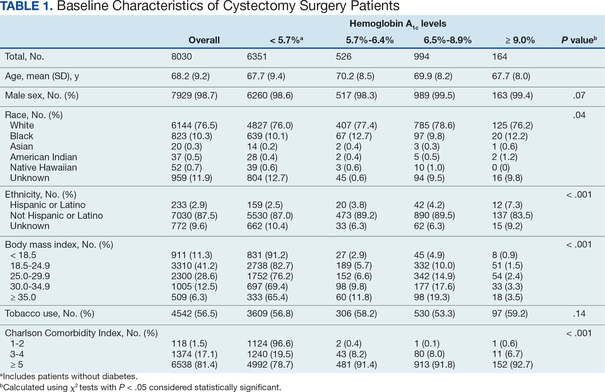
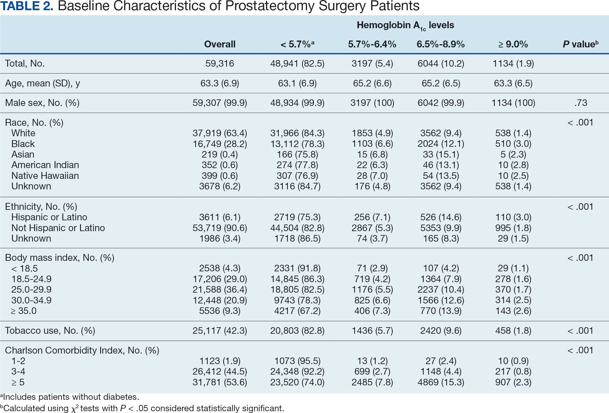
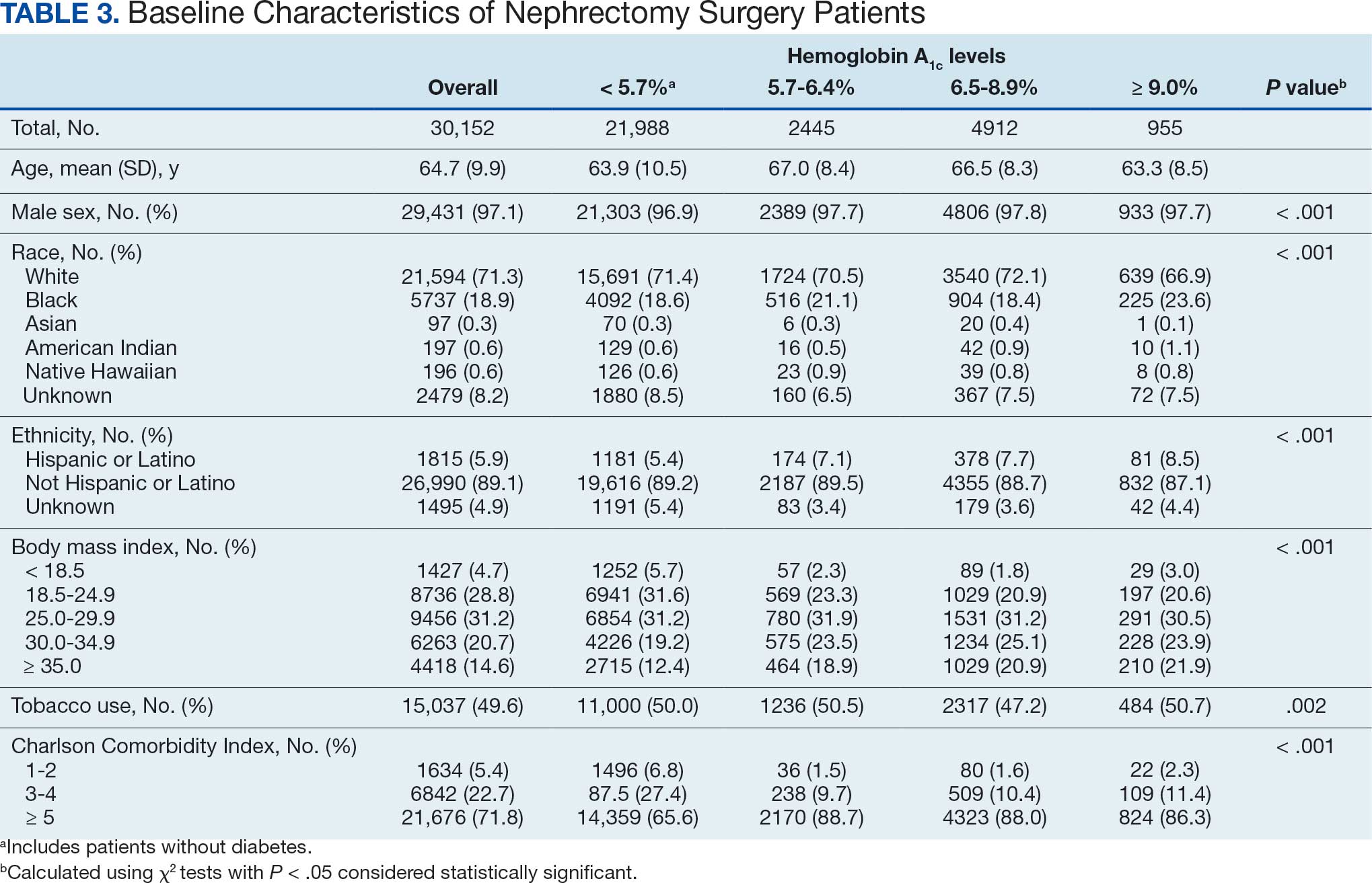
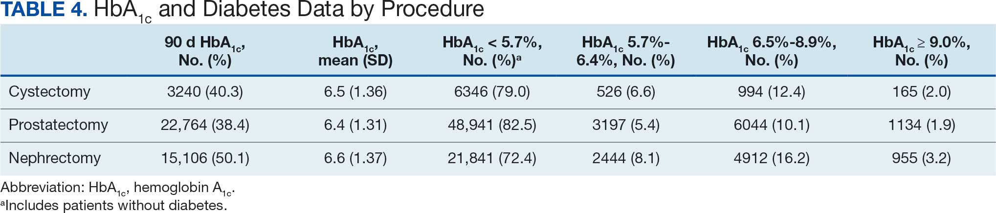
HbA1c values within 90 days of surgery were available in 42.2% of patients with a mean HbA1c value of 6.5% (Table 4). The prostatectomy group had the lowest proportion of patients with available HbA1c data (38.4%) but the highest proportion in the reference category (82.5%).
Multivariable logistic regression models incorporating categorical HbA1c values and other demographic and clinical variables were developed to predict all 30-day and 90-day outcomes (Tables 5 and 6). These multivariable models were developed for each procedure group.
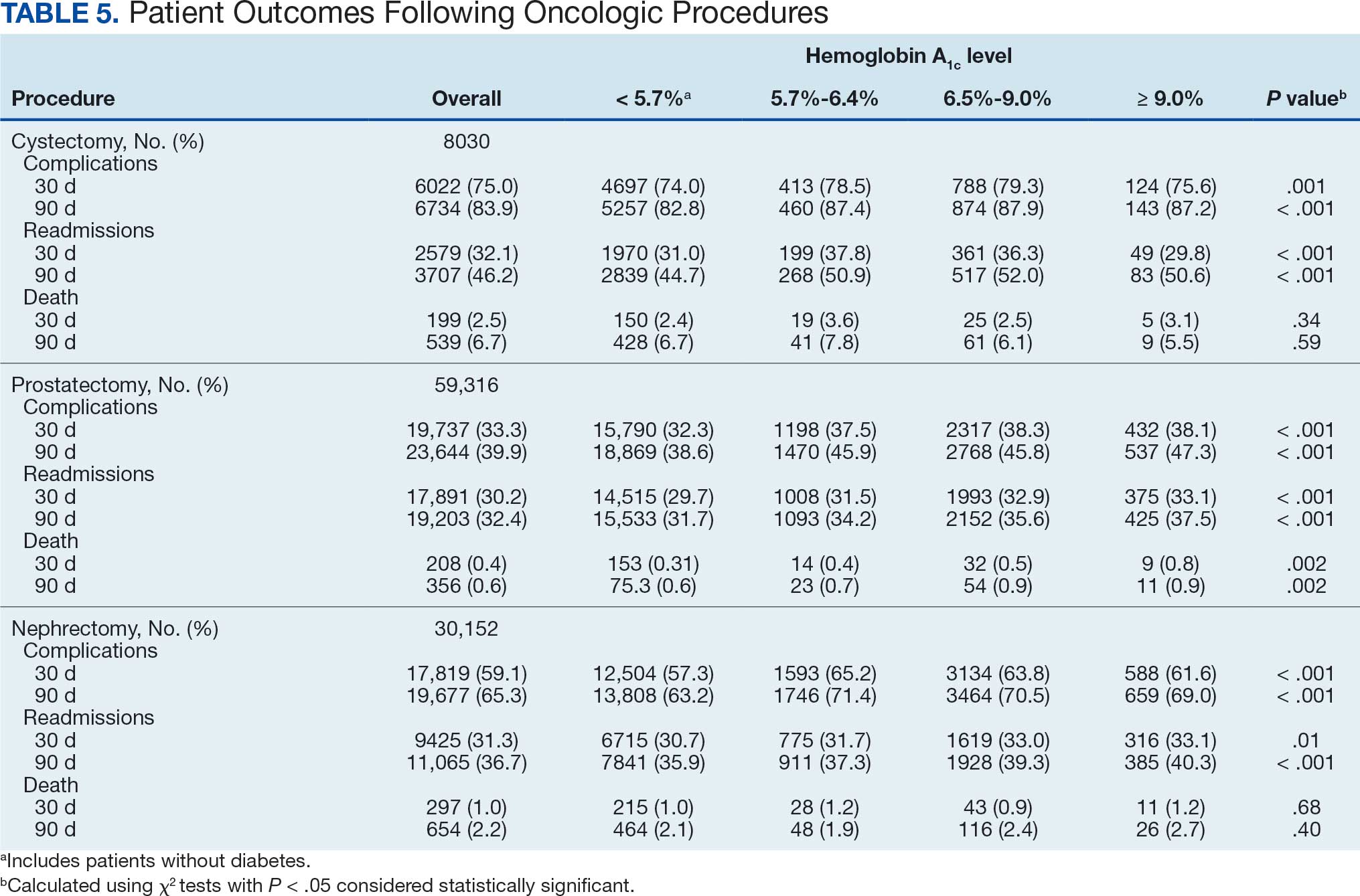
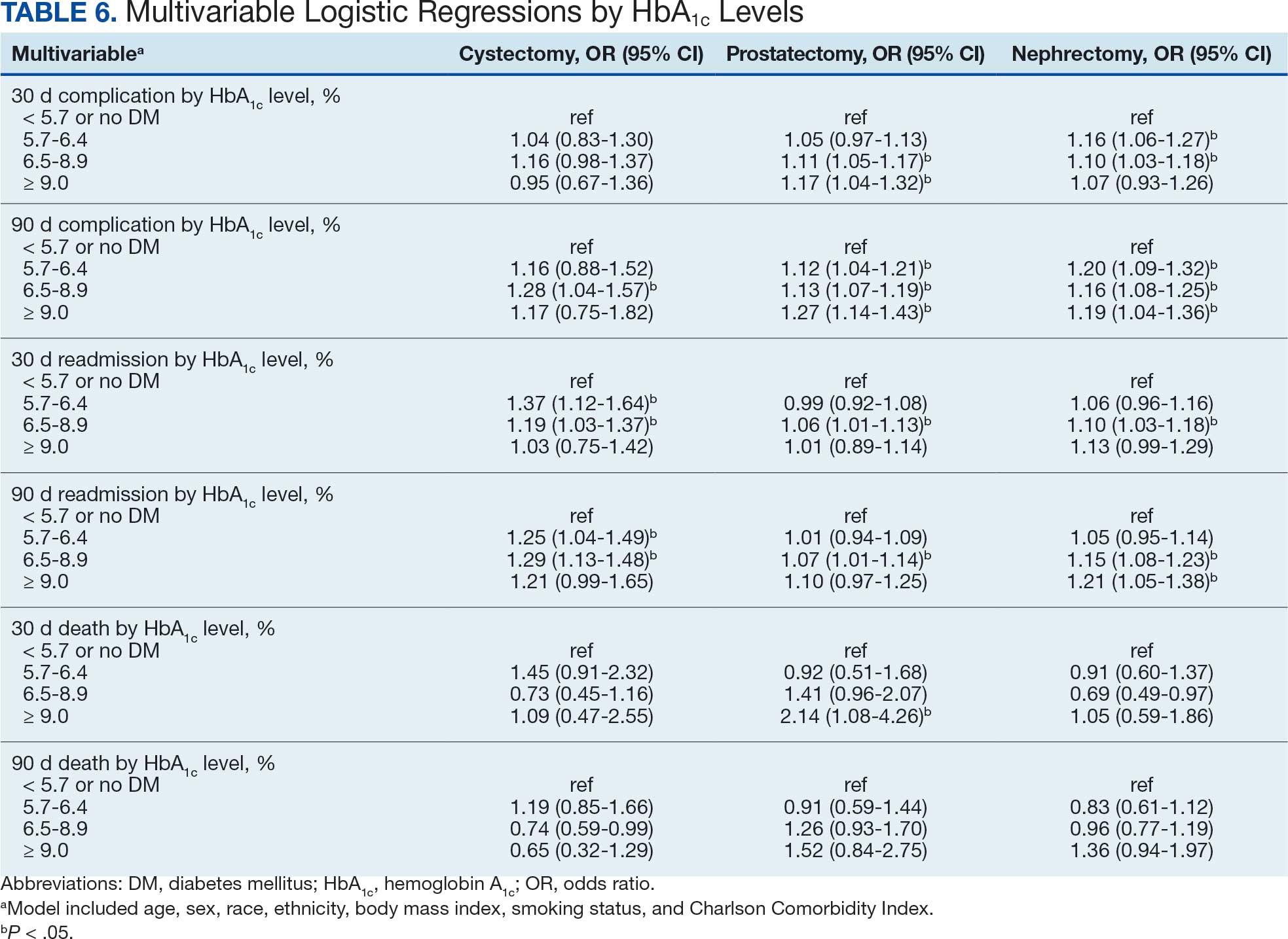
There was no significant association between HbA1c level and 30-day or 90-day complications or 30-day or 90-day deaths in the cystectomy group. Although the HbA1c 5.7% to 6.5% and 6.5% to 8.9% groups had significantly increased odds of 30-day and 90-day readmissions compared with the reference group, the association was not significant for the HbA1c > 9.0% group.
There were significant associations between higher HbA1c levels and 30-day and 90- day complications for patients who underwent prostatectomy. When compared with the reference group, the 90-day complications ORs increased progressively across each HbA1c group. The group with the highest odds of 90-day complications was the HbA1c ≥ 9.0% group (OR, 1.27; 95% CI, 1.14-1.43; P < .001). In the prostatectomy group, there was no significant trend between HbA1c and readmission rates. In patients who had undergone a prostatectomy, those with HbA1c ≥ 9.0% had significantly increased odds of 30-day mortality compared with the reference group (OR, 2.14; 95% CI, 1.08-4.26; P < .03). However, no significant association was observed between HbA1c and 90-day mortality.
For patients who underwent nephrectomy, the HbA1c 5.7% to 6.5% and HbA1c 6.5% to 9.0% groups had significantly increased odds of 30-day complications. All 3 HbA1c groups were significantly associated with 90-day complications following nephrectomy, with ORs ranging from 1.16 to 1.20 (OR, 1.20; 95% CI, 1.09-1.32 for HbA1c 5.7%-6.4%; OR, 1.16; 95% CI, 1.08- 1.25 for HbA1c 6.5%-8.9%; and OR, 1.19; 95% CI, 1.04-1.36; P = .04 for HbA1c ≥ 9.0%) of having complications within 90 days of surgery (OR, 1.19; 95% CI, 1.04-1.36; P = .04). Additionally, increasing HbA1c were associated with 90-day readmissions. At the highest level, HbA1c ≥ 9.0% was associated with 21% increased odds of readmission within 90 days of surgery (OR, 1.21; 95% CI, 1.05-1.38; P = .006). There was no association between HbA1c and mortality following nephrectomy surgery.
Discussion
Long-term impairment of glucose tolerance associated with DM is known to cause vascular, neurologic, and infectious complications. Several studies have demonstrated that elevated preoperative HbA1c levels are associated with increased postoperative complications across various surgical types. To our knowledge, this is the first study to examine the association between perioperative HbA1c and outcomes following cystectomy, prostatectomy, and nephrectomy. Using VINCI, we analyzed 106,740 veterans undergoing urologic oncology surgery and discovered that elevated perioperative HbA1c values were significantly associated with 30-day and 90-day complications following prostatectomy and nephrectomy. Additionally, we found a significant association between HbA1c ≥ 9.0% and 30-day death following prostatectomy. There was no significant association between HbA1c values and postoperative death following cystectomy or nephrectomy.
In our study, HbA1c values were categorized into < 5.7%, 5.7% to 6.4%, 6.5% to 8.9%, and ≥ 9.0%. We found significant associations with 30-day and 90-day complications following prostatectomy with the highest odds of 90-day complications in the HbA1c ≥ 9.0% group (OR, 1.27; 95% CI, 1.14-1.43; P < .001). We report similar findings for those undergoing nephrectomy. Those with HbA1c ≥ 9.0% were found to have 19% higher odds of 90-day complications compared with those in the reference group with HbA1c < 5.7% or no history of DM (OR, 1.19; 95% CI, 1.04-1.36; P = .043). Although HbA1c 6.5%-8.9% was significantly associated with 90-day complications following cystectomy (OR, 1.28; 95% CI, 1.04-1.57), no other significant associations were found between HbA1c categories and 30-day complications or death following cystectomy. It is unclear why a more consistent association does not exist across all HbA1c categories for this procedure, but this may be related to other unaccounted clinical factors influencing the outcomes.
The HbA1c ≥ 9.0% group had a significantly increased risk of 30-day death following prostatectomy in our study (OR, 2.14; 95% CI, 1.08-4.26; P = .03). However, we interpret these results with caution. The 30-day mortality after prostatectomy was low in the overall cohort (0.4%, 208 deaths) and in the HbA1c ≥ 9.0% group (0.8%, 9 deaths) and the 95% CI was wide. It will be important to verify this finding in additional cohorts.
Limitations
This was a retrospective study, which makes it difficult to control many variables that may have influenced postoperative outcomes. Additionally, the clinical characteristics and outcomes data rely on administratively coded data, which can be prone to error. Furthermore, while this was a large, national dataset, patients received care over a long period of time and at several different institutions, which may limit the generalizability of the findings to contemporary patient care. We also recognize that HbA1c values were not available in the perioperative period for all patients. We grouped those with missing HbA1c values into the reference category if they did not have a diagnostic code for DM. It is possible that some patients grouped into the reference category had undiagnosed DM or elevated HbA1c, which would theoretically lessen the magnitude of the findings in our multivariable analysis. Finally, despite our large cohort, 30-day and 90-day deaths were quite uncommon; larger datasets are needed to further investigate the association between perioperative HbA1c and mortality.
Conclusions
The results from this study suggest that elevated perioperative HbA1c is significantly associated with higher odds of 30-day and 90-day complications following prostatectomy and nephrectomy with the strongest associations noted for HbA1c ≥ 9.0%. Prospective investigation is warranted to determine whether reducing HbA1c values preoperatively would mitigate the risk of postoperative complications following urologic oncology surgeries.
- Sreedharan R, Khanna S, Shaw A. Perioperative glycemic management in adults presenting for elective cardiac and noncardiac surgery. Perioper Med (Lond). 2023;12:13. doi:10.1186/s13741-023-00302-6
- Dronge AS, Perkal MF, Kancir S, et al. Long-term glycemic control and postoperative infectious complications. Arch Surg. 2006;141:375-380. doi:10.1001/archsurg.141.4.375
- Yu A, Truong Q, Whitfield K, et al. Impact of preoperative haemoglobin A1c levels on postoperative outcomes in adults undergoing major noncardiac surgery: a systematic review. Diabet Med. 2024;41:e15380. doi:10.1111/dme.15380
- Schaschinger T, Niederegger T, Brandt J, et al. Preoperative hemoglobin A1C, glycemic status, and postoperative outcomes in general surgery. JAMA Surg. 2026;161:39-49. doi:10.1001/jamasurg.2025.4706
- Yong PH, Weinberg L, Torkamani N, et al. The presence of diabetes and higher HbA1c are independently associated with adverse outcomes after surgery. Diabetes Care. 2018;41:1172-1179. doi:10.2337/dc17-2304
- Chen P, Hallock KK, Mulvey CL, et al. The effect of elevated A1C on immediate postoperative complications: a prospective observational study. Clin Diabetes. 2018;36:128-132. doi:10.2337/cd17-0081
- Underwood P, Askari R, Hurwitz S, et al. Preoperative A1C and clinical outcomes in patients with diabetes undergoing major noncardiac surgical procedures. Diabetes Care. 2014;37:611-616. doi:10.2337/dc13-1929
- Faiena I, Dombrovskiy VY, Sultan RC, et al. Effect of uncontrolled diabetes on outcomes after cystectomy in patients with bladder cancer: a population-based study. Clin Genitourin Cancer. 2016;14:e509-e514. doi:10.1016/j.clgc.2016.02.004
- Habous M, Tal R, Tealab A, et al. Defining a glycated haemoglobin (HbA1c) level that predicts increased risk of penile implant infection. BJU Int. 2018;121:293-300. doi:10.1111/bju.14076
Diabetes mellitus (DM) is a well-known risk factor for postoperative complications, including infections and poor wound healing.1 Hemoglobin A1c (HbA1c) reflects a patient’s mean glucose level over the past 90 days. The American Diabetes Association recommends using HbA1c to monitor glycemic control.
Several meta-analyses and systematic reviews have demonstrated that elevated preoperative HbA1c levels are associated with higher rates of postoperative complications.2-5 An association between poor glycemic control and perioperative complications has been reported in cardiac, bariatric, abdominal, and orthopedic surgeries. In 1 prospective study, patients with HbA1c ≥ 7% had significantly higher incidences of postoperative infection and wound problems compared with patients with HbA1c 7%.6 In another study of 1775 patients with DM who underwent noncardiac surgery, patients with HbA1c > 8% had significantly longer hospital stays.7
The relationship between HbA1c and postoperative complications following urologic procedures, specifically oncologic surgeries, has not been as well researched. One study reported that uncontrolled preoperative DM was associated with complications following cystectomy.8 However, this study did not evaluate HbA1c levels as a marker. Another study reported an association between HbA1c and infection following penile prosthetic implants, with an HbA1c cutoff of 8.5% predicting increased risk of infection.9 Better understanding the association between HbA1c and postoperative complications following urologic oncology procedures may allow for better perioperative planning and improving clinical outcomes.
In this study, we examined the relationship between perioperative HbA1c levels and postoperative outcomes (complications, readmission, and death) in patients undergoing urologic oncology procedures (cystectomy, prostatectomy, and nephrectomy), while controlling for recognized clinical risk factors.
Methods
Data were compiled using the US Department of Veterans Affairs (VA) Corporate Data Warehouse (CDW) structured datasets within the VA Informatics and Computing Infrastructure (VINCI). The data in the VA CDW were aggregated from the VA electronic health record and curated for extraction and analysis.
The study examined veterans treated at any Veterans Health Administration facility from January 2000 to February 2024. Included patients had a diagnosis of prostate, bladder, or kidney cancer and ≥ 1 genitourinary surgery (cystectomy, prostatectomy, or nephrectomy) identified in the VA CDW using the ninth and 10th revisions of the International Classification of Diseases (ICD) codes. Demographic (age, sex, race, and ethnicity) and clinical data (body mass index, smoking status, and Charlson Comorbidity Index) were collected. HbA1c values at multiple time points within 90 days pre- and postsurgery were retrieved using Logical Observation Identifiers, Names, and Codes (LOINC), a widely used coding system for laboratory tests (Supplementary Table 1). Outcomes data were extracted including death date, hospital readmission, and different types of complications first diagnosed within 30 or 90 days postsurgery based on ICD diagnostic codes (Supplementary Table 2). Patients with missing HbA1c or DM data were excluded.
Statistical Analysis
The primary outcome variables included 30-day and 90-day complications, readmission, and death. The predictor variable was HbA1c level within 90 days of the procedure, which was categorized into 4 groups. The reference category was HbA1c < 5.7% and/or no history of DM. The next category fit the prediabetes (impaired glucose tolerance) definition, with HbA1c between 5.7% and 6.4%, and the last 2 categories were HbA1c values 6.5% to 8.9% and ≤ 9.0% to reflect uncontrolled DM. Associations between the different HbA1c level groupings and other variables are reported with χ2 tests. The final multivariable logistic regression models included several covariates including demographic data (age, sex, race, and ethnicity) and clinical data (body mass index, smoking status, and Charlson Comorbidity Index). Results are presented as odds ratios (ORs) and 95% CIs with a statistical significance threshold of P < .05. Statistical analyses were performed using Stata version 14.1.
Results
A total of 106,740 veterans who underwent cystectomy, prostatectomy, or nephrectomy from 2001 to 2023 were identified in VINCI. After excluding patients without demographic or clinical variables, 97,498 patients were included in our final cohort. The most common procedure was prostatectomy (n = 59,316). Most of the patients were male (99.1%) and White (67.3%) (Tables 1, 2, and 3).
HbA1c values within 90 days of surgery were available in 42.2% of patients with a mean HbA1c value of 6.5% (Table 4). The prostatectomy group had the lowest proportion of patients with available HbA1c data (38.4%) but the highest proportion in the reference category (82.5%).
Multivariable logistic regression models incorporating categorical HbA1c values and other demographic and clinical variables were developed to predict all 30-day and 90-day outcomes (Tables 5 and 6). These multivariable models were developed for each procedure group.
There was no significant association between HbA1c level and 30-day or 90-day complications or 30-day or 90-day deaths in the cystectomy group. Although the HbA1c 5.7% to 6.5% and 6.5% to 8.9% groups had significantly increased odds of 30-day and 90-day readmissions compared with the reference group, the association was not significant for the HbA1c > 9.0% group.
There were significant associations between higher HbA1c levels and 30-day and 90- day complications for patients who underwent prostatectomy. When compared with the reference group, the 90-day complications ORs increased progressively across each HbA1c group. The group with the highest odds of 90-day complications was the HbA1c ≥ 9.0% group (OR, 1.27; 95% CI, 1.14-1.43; P < .001). In the prostatectomy group, there was no significant trend between HbA1c and readmission rates. In patients who had undergone a prostatectomy, those with HbA1c ≥ 9.0% had significantly increased odds of 30-day mortality compared with the reference group (OR, 2.14; 95% CI, 1.08-4.26; P < .03). However, no significant association was observed between HbA1c and 90-day mortality.
For patients who underwent nephrectomy, the HbA1c 5.7% to 6.5% and HbA1c 6.5% to 9.0% groups had significantly increased odds of 30-day complications. All 3 HbA1c groups were significantly associated with 90-day complications following nephrectomy, with ORs ranging from 1.16 to 1.20 (OR, 1.20; 95% CI, 1.09-1.32 for HbA1c 5.7%-6.4%; OR, 1.16; 95% CI, 1.08- 1.25 for HbA1c 6.5%-8.9%; and OR, 1.19; 95% CI, 1.04-1.36; P = .04 for HbA1c ≥ 9.0%) of having complications within 90 days of surgery (OR, 1.19; 95% CI, 1.04-1.36; P = .04). Additionally, increasing HbA1c were associated with 90-day readmissions. At the highest level, HbA1c ≥ 9.0% was associated with 21% increased odds of readmission within 90 days of surgery (OR, 1.21; 95% CI, 1.05-1.38; P = .006). There was no association between HbA1c and mortality following nephrectomy surgery.
Discussion
Long-term impairment of glucose tolerance associated with DM is known to cause vascular, neurologic, and infectious complications. Several studies have demonstrated that elevated preoperative HbA1c levels are associated with increased postoperative complications across various surgical types. To our knowledge, this is the first study to examine the association between perioperative HbA1c and outcomes following cystectomy, prostatectomy, and nephrectomy. Using VINCI, we analyzed 106,740 veterans undergoing urologic oncology surgery and discovered that elevated perioperative HbA1c values were significantly associated with 30-day and 90-day complications following prostatectomy and nephrectomy. Additionally, we found a significant association between HbA1c ≥ 9.0% and 30-day death following prostatectomy. There was no significant association between HbA1c values and postoperative death following cystectomy or nephrectomy.
In our study, HbA1c values were categorized into < 5.7%, 5.7% to 6.4%, 6.5% to 8.9%, and ≥ 9.0%. We found significant associations with 30-day and 90-day complications following prostatectomy with the highest odds of 90-day complications in the HbA1c ≥ 9.0% group (OR, 1.27; 95% CI, 1.14-1.43; P < .001). We report similar findings for those undergoing nephrectomy. Those with HbA1c ≥ 9.0% were found to have 19% higher odds of 90-day complications compared with those in the reference group with HbA1c < 5.7% or no history of DM (OR, 1.19; 95% CI, 1.04-1.36; P = .043). Although HbA1c 6.5%-8.9% was significantly associated with 90-day complications following cystectomy (OR, 1.28; 95% CI, 1.04-1.57), no other significant associations were found between HbA1c categories and 30-day complications or death following cystectomy. It is unclear why a more consistent association does not exist across all HbA1c categories for this procedure, but this may be related to other unaccounted clinical factors influencing the outcomes.
The HbA1c ≥ 9.0% group had a significantly increased risk of 30-day death following prostatectomy in our study (OR, 2.14; 95% CI, 1.08-4.26; P = .03). However, we interpret these results with caution. The 30-day mortality after prostatectomy was low in the overall cohort (0.4%, 208 deaths) and in the HbA1c ≥ 9.0% group (0.8%, 9 deaths) and the 95% CI was wide. It will be important to verify this finding in additional cohorts.
Limitations
This was a retrospective study, which makes it difficult to control many variables that may have influenced postoperative outcomes. Additionally, the clinical characteristics and outcomes data rely on administratively coded data, which can be prone to error. Furthermore, while this was a large, national dataset, patients received care over a long period of time and at several different institutions, which may limit the generalizability of the findings to contemporary patient care. We also recognize that HbA1c values were not available in the perioperative period for all patients. We grouped those with missing HbA1c values into the reference category if they did not have a diagnostic code for DM. It is possible that some patients grouped into the reference category had undiagnosed DM or elevated HbA1c, which would theoretically lessen the magnitude of the findings in our multivariable analysis. Finally, despite our large cohort, 30-day and 90-day deaths were quite uncommon; larger datasets are needed to further investigate the association between perioperative HbA1c and mortality.
Conclusions
The results from this study suggest that elevated perioperative HbA1c is significantly associated with higher odds of 30-day and 90-day complications following prostatectomy and nephrectomy with the strongest associations noted for HbA1c ≥ 9.0%. Prospective investigation is warranted to determine whether reducing HbA1c values preoperatively would mitigate the risk of postoperative complications following urologic oncology surgeries.
Diabetes mellitus (DM) is a well-known risk factor for postoperative complications, including infections and poor wound healing.1 Hemoglobin A1c (HbA1c) reflects a patient’s mean glucose level over the past 90 days. The American Diabetes Association recommends using HbA1c to monitor glycemic control.
Several meta-analyses and systematic reviews have demonstrated that elevated preoperative HbA1c levels are associated with higher rates of postoperative complications.2-5 An association between poor glycemic control and perioperative complications has been reported in cardiac, bariatric, abdominal, and orthopedic surgeries. In 1 prospective study, patients with HbA1c ≥ 7% had significantly higher incidences of postoperative infection and wound problems compared with patients with HbA1c 7%.6 In another study of 1775 patients with DM who underwent noncardiac surgery, patients with HbA1c > 8% had significantly longer hospital stays.7
The relationship between HbA1c and postoperative complications following urologic procedures, specifically oncologic surgeries, has not been as well researched. One study reported that uncontrolled preoperative DM was associated with complications following cystectomy.8 However, this study did not evaluate HbA1c levels as a marker. Another study reported an association between HbA1c and infection following penile prosthetic implants, with an HbA1c cutoff of 8.5% predicting increased risk of infection.9 Better understanding the association between HbA1c and postoperative complications following urologic oncology procedures may allow for better perioperative planning and improving clinical outcomes.
In this study, we examined the relationship between perioperative HbA1c levels and postoperative outcomes (complications, readmission, and death) in patients undergoing urologic oncology procedures (cystectomy, prostatectomy, and nephrectomy), while controlling for recognized clinical risk factors.
Methods
Data were compiled using the US Department of Veterans Affairs (VA) Corporate Data Warehouse (CDW) structured datasets within the VA Informatics and Computing Infrastructure (VINCI). The data in the VA CDW were aggregated from the VA electronic health record and curated for extraction and analysis.
The study examined veterans treated at any Veterans Health Administration facility from January 2000 to February 2024. Included patients had a diagnosis of prostate, bladder, or kidney cancer and ≥ 1 genitourinary surgery (cystectomy, prostatectomy, or nephrectomy) identified in the VA CDW using the ninth and 10th revisions of the International Classification of Diseases (ICD) codes. Demographic (age, sex, race, and ethnicity) and clinical data (body mass index, smoking status, and Charlson Comorbidity Index) were collected. HbA1c values at multiple time points within 90 days pre- and postsurgery were retrieved using Logical Observation Identifiers, Names, and Codes (LOINC), a widely used coding system for laboratory tests (Supplementary Table 1). Outcomes data were extracted including death date, hospital readmission, and different types of complications first diagnosed within 30 or 90 days postsurgery based on ICD diagnostic codes (Supplementary Table 2). Patients with missing HbA1c or DM data were excluded.
Statistical Analysis
The primary outcome variables included 30-day and 90-day complications, readmission, and death. The predictor variable was HbA1c level within 90 days of the procedure, which was categorized into 4 groups. The reference category was HbA1c < 5.7% and/or no history of DM. The next category fit the prediabetes (impaired glucose tolerance) definition, with HbA1c between 5.7% and 6.4%, and the last 2 categories were HbA1c values 6.5% to 8.9% and ≤ 9.0% to reflect uncontrolled DM. Associations between the different HbA1c level groupings and other variables are reported with χ2 tests. The final multivariable logistic regression models included several covariates including demographic data (age, sex, race, and ethnicity) and clinical data (body mass index, smoking status, and Charlson Comorbidity Index). Results are presented as odds ratios (ORs) and 95% CIs with a statistical significance threshold of P < .05. Statistical analyses were performed using Stata version 14.1.
Results
A total of 106,740 veterans who underwent cystectomy, prostatectomy, or nephrectomy from 2001 to 2023 were identified in VINCI. After excluding patients without demographic or clinical variables, 97,498 patients were included in our final cohort. The most common procedure was prostatectomy (n = 59,316). Most of the patients were male (99.1%) and White (67.3%) (Tables 1, 2, and 3).
HbA1c values within 90 days of surgery were available in 42.2% of patients with a mean HbA1c value of 6.5% (Table 4). The prostatectomy group had the lowest proportion of patients with available HbA1c data (38.4%) but the highest proportion in the reference category (82.5%).
Multivariable logistic regression models incorporating categorical HbA1c values and other demographic and clinical variables were developed to predict all 30-day and 90-day outcomes (Tables 5 and 6). These multivariable models were developed for each procedure group.
There was no significant association between HbA1c level and 30-day or 90-day complications or 30-day or 90-day deaths in the cystectomy group. Although the HbA1c 5.7% to 6.5% and 6.5% to 8.9% groups had significantly increased odds of 30-day and 90-day readmissions compared with the reference group, the association was not significant for the HbA1c > 9.0% group.
There were significant associations between higher HbA1c levels and 30-day and 90- day complications for patients who underwent prostatectomy. When compared with the reference group, the 90-day complications ORs increased progressively across each HbA1c group. The group with the highest odds of 90-day complications was the HbA1c ≥ 9.0% group (OR, 1.27; 95% CI, 1.14-1.43; P < .001). In the prostatectomy group, there was no significant trend between HbA1c and readmission rates. In patients who had undergone a prostatectomy, those with HbA1c ≥ 9.0% had significantly increased odds of 30-day mortality compared with the reference group (OR, 2.14; 95% CI, 1.08-4.26; P < .03). However, no significant association was observed between HbA1c and 90-day mortality.
For patients who underwent nephrectomy, the HbA1c 5.7% to 6.5% and HbA1c 6.5% to 9.0% groups had significantly increased odds of 30-day complications. All 3 HbA1c groups were significantly associated with 90-day complications following nephrectomy, with ORs ranging from 1.16 to 1.20 (OR, 1.20; 95% CI, 1.09-1.32 for HbA1c 5.7%-6.4%; OR, 1.16; 95% CI, 1.08- 1.25 for HbA1c 6.5%-8.9%; and OR, 1.19; 95% CI, 1.04-1.36; P = .04 for HbA1c ≥ 9.0%) of having complications within 90 days of surgery (OR, 1.19; 95% CI, 1.04-1.36; P = .04). Additionally, increasing HbA1c were associated with 90-day readmissions. At the highest level, HbA1c ≥ 9.0% was associated with 21% increased odds of readmission within 90 days of surgery (OR, 1.21; 95% CI, 1.05-1.38; P = .006). There was no association between HbA1c and mortality following nephrectomy surgery.
Discussion
Long-term impairment of glucose tolerance associated with DM is known to cause vascular, neurologic, and infectious complications. Several studies have demonstrated that elevated preoperative HbA1c levels are associated with increased postoperative complications across various surgical types. To our knowledge, this is the first study to examine the association between perioperative HbA1c and outcomes following cystectomy, prostatectomy, and nephrectomy. Using VINCI, we analyzed 106,740 veterans undergoing urologic oncology surgery and discovered that elevated perioperative HbA1c values were significantly associated with 30-day and 90-day complications following prostatectomy and nephrectomy. Additionally, we found a significant association between HbA1c ≥ 9.0% and 30-day death following prostatectomy. There was no significant association between HbA1c values and postoperative death following cystectomy or nephrectomy.
In our study, HbA1c values were categorized into < 5.7%, 5.7% to 6.4%, 6.5% to 8.9%, and ≥ 9.0%. We found significant associations with 30-day and 90-day complications following prostatectomy with the highest odds of 90-day complications in the HbA1c ≥ 9.0% group (OR, 1.27; 95% CI, 1.14-1.43; P < .001). We report similar findings for those undergoing nephrectomy. Those with HbA1c ≥ 9.0% were found to have 19% higher odds of 90-day complications compared with those in the reference group with HbA1c < 5.7% or no history of DM (OR, 1.19; 95% CI, 1.04-1.36; P = .043). Although HbA1c 6.5%-8.9% was significantly associated with 90-day complications following cystectomy (OR, 1.28; 95% CI, 1.04-1.57), no other significant associations were found between HbA1c categories and 30-day complications or death following cystectomy. It is unclear why a more consistent association does not exist across all HbA1c categories for this procedure, but this may be related to other unaccounted clinical factors influencing the outcomes.
The HbA1c ≥ 9.0% group had a significantly increased risk of 30-day death following prostatectomy in our study (OR, 2.14; 95% CI, 1.08-4.26; P = .03). However, we interpret these results with caution. The 30-day mortality after prostatectomy was low in the overall cohort (0.4%, 208 deaths) and in the HbA1c ≥ 9.0% group (0.8%, 9 deaths) and the 95% CI was wide. It will be important to verify this finding in additional cohorts.
Limitations
This was a retrospective study, which makes it difficult to control many variables that may have influenced postoperative outcomes. Additionally, the clinical characteristics and outcomes data rely on administratively coded data, which can be prone to error. Furthermore, while this was a large, national dataset, patients received care over a long period of time and at several different institutions, which may limit the generalizability of the findings to contemporary patient care. We also recognize that HbA1c values were not available in the perioperative period for all patients. We grouped those with missing HbA1c values into the reference category if they did not have a diagnostic code for DM. It is possible that some patients grouped into the reference category had undiagnosed DM or elevated HbA1c, which would theoretically lessen the magnitude of the findings in our multivariable analysis. Finally, despite our large cohort, 30-day and 90-day deaths were quite uncommon; larger datasets are needed to further investigate the association between perioperative HbA1c and mortality.
Conclusions
The results from this study suggest that elevated perioperative HbA1c is significantly associated with higher odds of 30-day and 90-day complications following prostatectomy and nephrectomy with the strongest associations noted for HbA1c ≥ 9.0%. Prospective investigation is warranted to determine whether reducing HbA1c values preoperatively would mitigate the risk of postoperative complications following urologic oncology surgeries.
- Sreedharan R, Khanna S, Shaw A. Perioperative glycemic management in adults presenting for elective cardiac and noncardiac surgery. Perioper Med (Lond). 2023;12:13. doi:10.1186/s13741-023-00302-6
- Dronge AS, Perkal MF, Kancir S, et al. Long-term glycemic control and postoperative infectious complications. Arch Surg. 2006;141:375-380. doi:10.1001/archsurg.141.4.375
- Yu A, Truong Q, Whitfield K, et al. Impact of preoperative haemoglobin A1c levels on postoperative outcomes in adults undergoing major noncardiac surgery: a systematic review. Diabet Med. 2024;41:e15380. doi:10.1111/dme.15380
- Schaschinger T, Niederegger T, Brandt J, et al. Preoperative hemoglobin A1C, glycemic status, and postoperative outcomes in general surgery. JAMA Surg. 2026;161:39-49. doi:10.1001/jamasurg.2025.4706
- Yong PH, Weinberg L, Torkamani N, et al. The presence of diabetes and higher HbA1c are independently associated with adverse outcomes after surgery. Diabetes Care. 2018;41:1172-1179. doi:10.2337/dc17-2304
- Chen P, Hallock KK, Mulvey CL, et al. The effect of elevated A1C on immediate postoperative complications: a prospective observational study. Clin Diabetes. 2018;36:128-132. doi:10.2337/cd17-0081
- Underwood P, Askari R, Hurwitz S, et al. Preoperative A1C and clinical outcomes in patients with diabetes undergoing major noncardiac surgical procedures. Diabetes Care. 2014;37:611-616. doi:10.2337/dc13-1929
- Faiena I, Dombrovskiy VY, Sultan RC, et al. Effect of uncontrolled diabetes on outcomes after cystectomy in patients with bladder cancer: a population-based study. Clin Genitourin Cancer. 2016;14:e509-e514. doi:10.1016/j.clgc.2016.02.004
- Habous M, Tal R, Tealab A, et al. Defining a glycated haemoglobin (HbA1c) level that predicts increased risk of penile implant infection. BJU Int. 2018;121:293-300. doi:10.1111/bju.14076
- Sreedharan R, Khanna S, Shaw A. Perioperative glycemic management in adults presenting for elective cardiac and noncardiac surgery. Perioper Med (Lond). 2023;12:13. doi:10.1186/s13741-023-00302-6
- Dronge AS, Perkal MF, Kancir S, et al. Long-term glycemic control and postoperative infectious complications. Arch Surg. 2006;141:375-380. doi:10.1001/archsurg.141.4.375
- Yu A, Truong Q, Whitfield K, et al. Impact of preoperative haemoglobin A1c levels on postoperative outcomes in adults undergoing major noncardiac surgery: a systematic review. Diabet Med. 2024;41:e15380. doi:10.1111/dme.15380
- Schaschinger T, Niederegger T, Brandt J, et al. Preoperative hemoglobin A1C, glycemic status, and postoperative outcomes in general surgery. JAMA Surg. 2026;161:39-49. doi:10.1001/jamasurg.2025.4706
- Yong PH, Weinberg L, Torkamani N, et al. The presence of diabetes and higher HbA1c are independently associated with adverse outcomes after surgery. Diabetes Care. 2018;41:1172-1179. doi:10.2337/dc17-2304
- Chen P, Hallock KK, Mulvey CL, et al. The effect of elevated A1C on immediate postoperative complications: a prospective observational study. Clin Diabetes. 2018;36:128-132. doi:10.2337/cd17-0081
- Underwood P, Askari R, Hurwitz S, et al. Preoperative A1C and clinical outcomes in patients with diabetes undergoing major noncardiac surgical procedures. Diabetes Care. 2014;37:611-616. doi:10.2337/dc13-1929
- Faiena I, Dombrovskiy VY, Sultan RC, et al. Effect of uncontrolled diabetes on outcomes after cystectomy in patients with bladder cancer: a population-based study. Clin Genitourin Cancer. 2016;14:e509-e514. doi:10.1016/j.clgc.2016.02.004
- Habous M, Tal R, Tealab A, et al. Defining a glycated haemoglobin (HbA1c) level that predicts increased risk of penile implant infection. BJU Int. 2018;121:293-300. doi:10.1111/bju.14076
Association of Hemoglobin A1c With Outcomes Following Urologic Oncology Procedures in Veterans
Association of Hemoglobin A1c With Outcomes Following Urologic Oncology Procedures in Veterans

Vaccine to Prevent Pancreatic Cancer Shows Early Promise
Vaccine to Prevent Pancreatic Cancer Shows Early Promise
An off-the-shelf vaccine that targets common KRAS mutations appears safe and generates a durable immune response in people at a high risk for pancreatic cancer, according to a new phase 1 trial.
Researchers found that the experimental vaccine spurred KRAS-targeted immune responses in 18 of 20 trial participants, all of whom had a genetic predisposition to pancreatic ductal adenocarcinoma (PDAC) and evidence of pancreatic lesions.
After roughly 16 months of follow-up, none of the trial participants had developed cancer or high-risk lesions requiring surgery, and eight had partial or complete resolution of their pancreatic lesions.
Experts stressed that the findings, published online in Cancer Discovery, offer proof of concept that vaccinating against mutant KRAS can generate lasting immune responses.
It will take “many years” to show whether the strategy can prevent cancer, said study co-author Neeha Zaidi, MD, of Johns Hopkins University School of Medicine in Baltimore.
About 1 in 10 cases of PDAC are attributable to inherited genetic predisposition. Currently, people with such hereditary risk “can only ‘watch and wait’ with regular surveillance,” Zaidi noted.
“Options to proactively intercept disease progression are desperately needed,” she said.
A number of therapeutic vaccines for pancreatic cancer are under development, both personalized and off-the-shelf versions.
The vaccine used in the new trial, called mKRAS-VAX, targets six driver mutations in the KRAS gene that are present in about 90% of PDACs. The Hopkins team initially tested the vaccine as an adjuvant therapy, along with dual checkpoint blockade, in patients who’d undergone surgery for PDAC. In a phase 1 study published earlier this year, the vaccine elicited T-cell responses in nearly all of the 12 patients involved, and stronger T-cell response correlated with longer disease-free survival.
That prompted the researchers to investigate the vaccine’s preventive potential.
The new study is “a meaningful step” in that direction, said Alex Jaeger, PhD, molecular oncologist at Moffitt Cancer Center in Tampa, Florida, who was not involved in the work.
However, Jaeger told Medscape Medical News, many questions remain — most notably, can the strategy ultimately prevent cancer?
The trial included 20 people (median age, 66 years) with a hereditary predisposition to PDAC and radiographic evidence of ≥ 1 pancreatic lesion (typically a small cyst). Most had ≥ 1 first-degree relatives diagnosed with PDAC and harbored a germline mutation.
All participants received 4 subcutaneous doses of mKRAS-VAX given over 13 weeks. Eighteen (90%) were considered immune responders, showing an increase of at least 2.5 times in their pooled mutant KRAS-specific T-cell response.
T-cell responses rose by a median of > 18 times, and vaccine-induced mutant KRAS-specific T-cell clones were detectable for up to 2 years after vaccination. The responses were seen across a range of human leukocyte antigen alleles, which the researchers said supported their off-the-shelf approach.
The vaccine appeared to be generally safe. All treatment-related adverse events were grade 1 or 2, the most common being injection-site reactions (85%), self-limiting fatigue (70%), chills (40%), and flu-like symptoms (40%).
Zaidi said a follow-up trial is already underway. It will assess changes in precancer tissue to determine whether immune cells generated by the vaccine are not only detectable in the blood but also capable of infiltrating precancers.
They’re doing so, she noted, by vaccinating patients who need surgery for high-risk pancreatic cysts, which will allow them to collect both blood and tissue samples.
The study received funding from Stand Up to Cancer-Lustgarten Foundation, the National Institutes of Health, and the American Society of Clinical Oncology. Zaidi is cofounder of Adventris Pharmaceuticals, which has licensed a technology described in the study from Johns Hopkins University. Zaidi and the university are entitled to royalty distributions related to the technology. Jaeger reported having an equity stake in RyboDyn, a company developing immunotherapies.
A version of this article first appeared on Medscape.com.
An off-the-shelf vaccine that targets common KRAS mutations appears safe and generates a durable immune response in people at a high risk for pancreatic cancer, according to a new phase 1 trial.
Researchers found that the experimental vaccine spurred KRAS-targeted immune responses in 18 of 20 trial participants, all of whom had a genetic predisposition to pancreatic ductal adenocarcinoma (PDAC) and evidence of pancreatic lesions.
After roughly 16 months of follow-up, none of the trial participants had developed cancer or high-risk lesions requiring surgery, and eight had partial or complete resolution of their pancreatic lesions.
Experts stressed that the findings, published online in Cancer Discovery, offer proof of concept that vaccinating against mutant KRAS can generate lasting immune responses.
It will take “many years” to show whether the strategy can prevent cancer, said study co-author Neeha Zaidi, MD, of Johns Hopkins University School of Medicine in Baltimore.
About 1 in 10 cases of PDAC are attributable to inherited genetic predisposition. Currently, people with such hereditary risk “can only ‘watch and wait’ with regular surveillance,” Zaidi noted.
“Options to proactively intercept disease progression are desperately needed,” she said.
A number of therapeutic vaccines for pancreatic cancer are under development, both personalized and off-the-shelf versions.
The vaccine used in the new trial, called mKRAS-VAX, targets six driver mutations in the KRAS gene that are present in about 90% of PDACs. The Hopkins team initially tested the vaccine as an adjuvant therapy, along with dual checkpoint blockade, in patients who’d undergone surgery for PDAC. In a phase 1 study published earlier this year, the vaccine elicited T-cell responses in nearly all of the 12 patients involved, and stronger T-cell response correlated with longer disease-free survival.
That prompted the researchers to investigate the vaccine’s preventive potential.
The new study is “a meaningful step” in that direction, said Alex Jaeger, PhD, molecular oncologist at Moffitt Cancer Center in Tampa, Florida, who was not involved in the work.
However, Jaeger told Medscape Medical News, many questions remain — most notably, can the strategy ultimately prevent cancer?
The trial included 20 people (median age, 66 years) with a hereditary predisposition to PDAC and radiographic evidence of ≥ 1 pancreatic lesion (typically a small cyst). Most had ≥ 1 first-degree relatives diagnosed with PDAC and harbored a germline mutation.
All participants received 4 subcutaneous doses of mKRAS-VAX given over 13 weeks. Eighteen (90%) were considered immune responders, showing an increase of at least 2.5 times in their pooled mutant KRAS-specific T-cell response.
T-cell responses rose by a median of > 18 times, and vaccine-induced mutant KRAS-specific T-cell clones were detectable for up to 2 years after vaccination. The responses were seen across a range of human leukocyte antigen alleles, which the researchers said supported their off-the-shelf approach.
The vaccine appeared to be generally safe. All treatment-related adverse events were grade 1 or 2, the most common being injection-site reactions (85%), self-limiting fatigue (70%), chills (40%), and flu-like symptoms (40%).
Zaidi said a follow-up trial is already underway. It will assess changes in precancer tissue to determine whether immune cells generated by the vaccine are not only detectable in the blood but also capable of infiltrating precancers.
They’re doing so, she noted, by vaccinating patients who need surgery for high-risk pancreatic cysts, which will allow them to collect both blood and tissue samples.
The study received funding from Stand Up to Cancer-Lustgarten Foundation, the National Institutes of Health, and the American Society of Clinical Oncology. Zaidi is cofounder of Adventris Pharmaceuticals, which has licensed a technology described in the study from Johns Hopkins University. Zaidi and the university are entitled to royalty distributions related to the technology. Jaeger reported having an equity stake in RyboDyn, a company developing immunotherapies.
A version of this article first appeared on Medscape.com.
An off-the-shelf vaccine that targets common KRAS mutations appears safe and generates a durable immune response in people at a high risk for pancreatic cancer, according to a new phase 1 trial.
Researchers found that the experimental vaccine spurred KRAS-targeted immune responses in 18 of 20 trial participants, all of whom had a genetic predisposition to pancreatic ductal adenocarcinoma (PDAC) and evidence of pancreatic lesions.
After roughly 16 months of follow-up, none of the trial participants had developed cancer or high-risk lesions requiring surgery, and eight had partial or complete resolution of their pancreatic lesions.
Experts stressed that the findings, published online in Cancer Discovery, offer proof of concept that vaccinating against mutant KRAS can generate lasting immune responses.
It will take “many years” to show whether the strategy can prevent cancer, said study co-author Neeha Zaidi, MD, of Johns Hopkins University School of Medicine in Baltimore.
About 1 in 10 cases of PDAC are attributable to inherited genetic predisposition. Currently, people with such hereditary risk “can only ‘watch and wait’ with regular surveillance,” Zaidi noted.
“Options to proactively intercept disease progression are desperately needed,” she said.
A number of therapeutic vaccines for pancreatic cancer are under development, both personalized and off-the-shelf versions.
The vaccine used in the new trial, called mKRAS-VAX, targets six driver mutations in the KRAS gene that are present in about 90% of PDACs. The Hopkins team initially tested the vaccine as an adjuvant therapy, along with dual checkpoint blockade, in patients who’d undergone surgery for PDAC. In a phase 1 study published earlier this year, the vaccine elicited T-cell responses in nearly all of the 12 patients involved, and stronger T-cell response correlated with longer disease-free survival.
That prompted the researchers to investigate the vaccine’s preventive potential.
The new study is “a meaningful step” in that direction, said Alex Jaeger, PhD, molecular oncologist at Moffitt Cancer Center in Tampa, Florida, who was not involved in the work.
However, Jaeger told Medscape Medical News, many questions remain — most notably, can the strategy ultimately prevent cancer?
The trial included 20 people (median age, 66 years) with a hereditary predisposition to PDAC and radiographic evidence of ≥ 1 pancreatic lesion (typically a small cyst). Most had ≥ 1 first-degree relatives diagnosed with PDAC and harbored a germline mutation.
All participants received 4 subcutaneous doses of mKRAS-VAX given over 13 weeks. Eighteen (90%) were considered immune responders, showing an increase of at least 2.5 times in their pooled mutant KRAS-specific T-cell response.
T-cell responses rose by a median of > 18 times, and vaccine-induced mutant KRAS-specific T-cell clones were detectable for up to 2 years after vaccination. The responses were seen across a range of human leukocyte antigen alleles, which the researchers said supported their off-the-shelf approach.
The vaccine appeared to be generally safe. All treatment-related adverse events were grade 1 or 2, the most common being injection-site reactions (85%), self-limiting fatigue (70%), chills (40%), and flu-like symptoms (40%).
Zaidi said a follow-up trial is already underway. It will assess changes in precancer tissue to determine whether immune cells generated by the vaccine are not only detectable in the blood but also capable of infiltrating precancers.
They’re doing so, she noted, by vaccinating patients who need surgery for high-risk pancreatic cysts, which will allow them to collect both blood and tissue samples.
The study received funding from Stand Up to Cancer-Lustgarten Foundation, the National Institutes of Health, and the American Society of Clinical Oncology. Zaidi is cofounder of Adventris Pharmaceuticals, which has licensed a technology described in the study from Johns Hopkins University. Zaidi and the university are entitled to royalty distributions related to the technology. Jaeger reported having an equity stake in RyboDyn, a company developing immunotherapies.
A version of this article first appeared on Medscape.com.
Vaccine to Prevent Pancreatic Cancer Shows Early Promise
Vaccine to Prevent Pancreatic Cancer Shows Early Promise

Deprescribing of Proton Pump Inhibitors at the South Texas Veterans Health Care System
Deprescribing of Proton Pump Inhibitors at the South Texas Veterans Health Care System
Proton pump inhibitors (PPIs) are widely prescribed medications used to manage various disease states. A systematic review of global trends and practices in PPI use estimated that about 25% of adults use a PPI, accounting for > 28 million users worldwide.1 The duration of PPI therapy varies by indication; however, many patients continue therapy beyond the suggested period. According to the American Gastroenterological Association (AGA) 2022 PPI deprescribing guideline update, regular assessment of PPIs should be performed for ongoing indications, and patients without a definitive indication for chronic PPI use should be considered for safe deprescribing.2 Indications for chronic PPI use include a history of severe erosive esophagitis, esophageal ulcer, peptic stricture, dysphagia, Barrett esophagus, idiopathic pulmonary fibrosis, history of gastrointestinal (GI) bleeding, and GI protection (Table 1).2-4
While PPIs are generally considered safe for short-term use, long-term use has been associated with several adverse effects (AEs) and safety concerns.5 A meta-analysis found a significant risk for Clostridium difficile infections (CDI) in PPI users.6 The effect of PPIs on vitamin B12 deficiency remains controversial; however, there is evidence that vitamin B12 levels may be decreased when gastric acid is suppressed for prolonged periods.7,8 In addition, the use of PPIs may result in decreased absorption of magnesium and calcium. A meta-analysis to examine the association of PPI use and hypomagnesemia found that high-dose PPIs were associated with hypomagnesemia compared with low-dose PPIs.9 Another meta-analysis demonstrated a statistically significant increased risk of hypomagnesemia in patients with PPI use.10 Long-term PPI use has been associated with decreased calcium levels, resulting in osteoporosis and increased risk of bone fractures. Multiple longitudinal observational studies concluded that increased risk for osteoporosis is associated with chronic PPI use.11 Patients who use a PPI long term without a clearly documented indication for chronic PPI therapy may be at risk for unnecessary exposure to PPI-associated AEs.
The US Department of Veterans Affairs (VA) created a dashboard to identify patients with chronic PPI use who may benefit from deprescribing. The Randomized PPI De-Prescribing (RaPPID) Program Dashboard defines chronic PPI use as being prescribed PPI therapy for ≥ 90 days during the 120-day period prior to a scheduled primary care visit. Candidates for deprescribing include those on once-daily PPI with no clear indication for chronic use or with uncomplicated GERD (ie, no erosive esophagitis, stricture, dysphagia, Barrett esophagus), as well as those on a twice-daily PPI for any indication except Zollinger-Ellison syndrome.
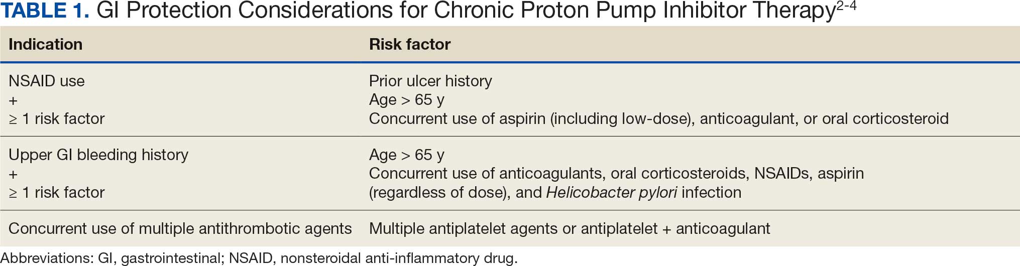
The dashboard categorizes deprescribing into 3 methods: (1) discontinuation of once-daily PPIs in the absence of a clear indication or in cases of uncomplicated GERD; (2) dose reduction for patients taking twice-daily PPIs for any non–Zollinger–Ellison indication; and (3) reduce and discontinue, which combines both for patients receiving twice-daily therapy who meet criteria for both strategies. A South Texas Veterans Health Care System (STVHCS) internal medication use evaluation using the VA RaPPID Program Dashboard identified 250 patients on a PPI and concluded that 56.9% of those patients were eligible for PPI deprescribing.12
This quality improvement (QI) project sought to assess PPI use and intervene to improve deprescribing rates at STVHCS. The intervention consisted of an educational in-service presentation delivered to primary care practitioners (PCPs) at a STVHCS patient aligned care team (PACT) collaborative meeting. Follow-up EHR reviews assessed whether PPI therapy was appropriately deprescribed after PCP appointments.
Methods
The primary objective of this QI project was to improve PPI prescribing practices at STVHCS PACT clinics to reduce PPI use in patients with an inappropriate indication by ≥ 10%. Secondary objectives included summarizing the use of vitamin supplementation (ie, cyanocobalamin, magnesium, calcium), reviewing AEs associated with PPI use (ie, CDI, fractures), and evaluating the occurrence of PPI reinitiation, recurrent GERD symptoms, and GI bleeding within 4 weeks of deprescribing.
On November 6, 2024, an in-service presentation was provided to PACT PCPs to educate and encourage the use of the VA RaPPID Program Dashboard, which creates a list of patients with a PCP appointment within 4 weeks of the current date who are eligible for PPI deprescribing. A patient list was generated from the VA RaPPID Program Dashboard on the morning before the in-service presentation. Four weeks following the in-service presentation and after all PCP appointments were conducted, a retrospective EHR review was conducted to assess outcomes. PPI deprescribing was determined collaboratively by the PCP and patient during the appointment, guided by clinical judgment and shared decision-making.
Patients were included if they had a PCP appointment between November 6, 2024, and December 3, 2024 (ie, within 4 weeks of in-service presentation), had an active prescription for an oral PPI from STVHCS and were identified as candidates by the VA RaPPID Program Dashboard for deprescribing as patients with chronic PPI use. Patients were excluded if they had an appropriate indication for chronic use of PPI, canceled or missed their PCP appointment, or failed previous de-escalation attempt (ie, worsening GERD symptoms or developed GI bleed after a deprescribing attempt).
Results
The VA RaPPID Program Dashboard identified 385 patients eligible for PPI deprescribing and 110 patients (29%) met the inclusion criteria and were reviewed. Most patients included were men, with a mean (SD) age of 56 years (13). Ninety-four patients (85%) had a daily PPI prescription with uncomplicated GERD at diagnosis and 16 patients (15%) took PPIs twice daily with uncomplicated GERD (Table 2). Seventy-two patients (65%) took omeprazole, 36 (33%) took pantoprazole, and 2 (2%) took esomeprazole. The dashboard identified 93 patients (85%) as candidates for PPI therapy discontinuation, 9 patients (8%) as candidates for PPI therapy reduction, and 8 patients (7%) as candidates for PPI therapy reduction and discontinuation.
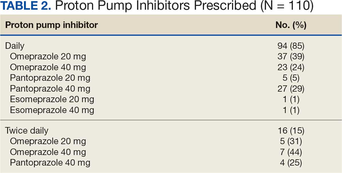
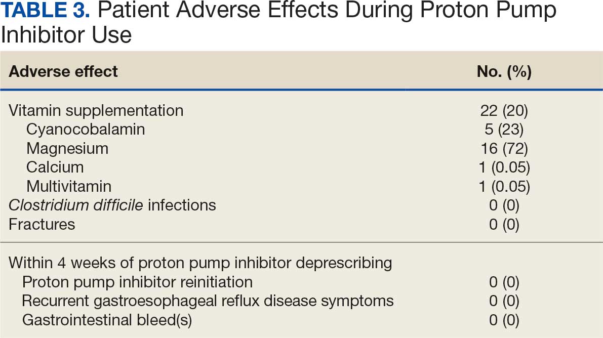
PPIs were deprescribed for 13 of 110 patients (12%). Of the 13 patients offered a trial of PPI deprescribing, 6 (46%) agreed to participate while 7 (54%) declined deprescribing (Figure). The 6 patients who agreed to trial deprescribing were initiated on an individualized tapering regimen.
Among all patients, 22 (20%) received vitamin supplementation during PPI therapy. Of those supplemented, 5 (23%) received cyanocobalamin, 16 (73%) received magnesium, 1 (1%) received calcium, and 1 (1%) received a multivitamin. One case of CDI was documented during PPI therapy and no fractures were observed.
The 6 patients who agreed to deprescribe PPIs successfully stopped use and were transitioned to an alternative agent (ie, famotidine) for GERD symptom management. Follow-up EHR review showed no patients reinitiated a PPI, experienced GI bleeding, or had documented recurrence of GERD symptoms within 4 weeks after deprescribing. (Table 3).
Discussion
This STVHCS QI project used the RaPPID Program Dashboard to increase PPI deprescribing rates. A 10% deprescribing goal was established for the single in-service presentation to maintain an attainable, conservative estimate of the expected impact. Six of the 13 patients (46%) successfully deprescribed their PPI, thereby meeting the project’s 10% deprescribing goal. Notably, 1 of the 6 patients was referred to a PACT clinical pharmacist practitioner (CPP) by their PCP for deprescribing and management of GERD symptoms. The patient transitioned from PPI to famotidine and had long-term follow-up care with their assigned PACT CPP, suggesting that greater involvement from PACT CPPs may lead to better adherence to PPI prescribing guidelines.
Our findings align with prior studies that assessed PPI deprescribing. In 2024, Rossi et al examined 66 studies evaluating various PPI deprescribing interventions among patients with mild illness. These included collaborative strategies involving physicians, pharmacists, and other health care professionals; clinical decision-making algorithms; and patient engagement. Rossi et al found that 24% to 67% of patients successfully deprescribed PPI and most patients (51%-88%) did not report any recurring symptoms of heartburn or acid regurgitation after discontinuing PPI therapy.13
In this QI project, famotidine was prescribed as an alternative agent for all 6 patients who successfully discontinued PPI. The AGA guidelines recommend as-needed H2-receptor antagonist use following PPI deprescribing to control symptoms in the short term and prevent immediate resumption of a PPI. Providing an alternative agent for GERD symptoms may encourage patient participation and improve the success of PPI deprescribing initiatives by mitigating concerns over symptom reoccurrence.
Limitations
Secondary outcomes were assessed only within a 4-week period following the PCP visit, and patients may have experienced AEs beyond the 4-week time frame, which may have resulted in their underestimation. It is possible patients may have independently restarted PPI therapy without reporting recurrent symptoms to their PCP, which could further confound the assessment of outcomes. Additionally, while the need for vitamin supplementation and the occurrence of a CDI was observed, it remains unclear whether these events were exclusively caused by PPI use. Potential confounding variables for vitamin supplementation include age and other clinical indications for vitamin supplementation (eg, osteoporosis, migraines, macrocytic anemia, malabsorption syndromes, or concurrent use of other medications associated with vitamin deficiencies). For the patient with CDI, potential confounders include antibiotic use, advanced age, travel history, immunosuppression, or prior GI surgery. These factors limit the ability to draw causal associations to PPI use.
Several factors may explain why deprescribing was only initiated within 12% of eligible patients. First, deprescribing attempts may not have been consistently documented in the PCP visit notes, which may have led to an underestimation of the number of patients who considered deprescribing. Furthermore, time constraints during PCP appointments may have hindered thorough education and documentation. Among patients for whom deprescribing was attempted, about half declined. This hesitancy may have been influenced by their prolonged history of use, perceived medication efficacy, and concern for symptom relapse.
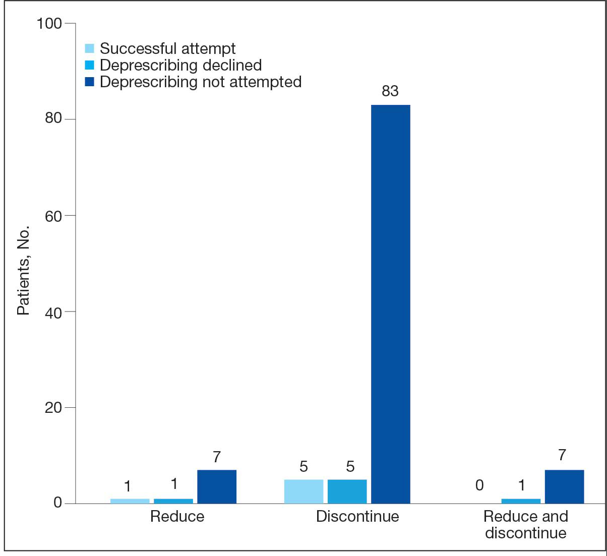
The proposed next step of this QI project would be to involve the PACT CPPs in the PPI deprescribing initiative during patient encounters and assess the impact of their involvement in deprescribing rates, reporting interval outcomes with long-term follow-up. Statistical analysis may also be beneficial to determine whether the observed deprescribing outcomes are statistically significant.
Conclusions
This QI project demonstrated that the use of the VA RaPPID Program Dashboard, in conjunction with targeted clinician education, can positively influence PPI deprescribing. Although a relatively small number of patients considered deprescribing, it was successful in patients who agreed to discontinue PPI therapy. These patients had no recurrence of short-term GERD symptoms or AEs. An H2-receptor antagonist may facilitate successful deprescribing for patients who agree to stop PPI use. The findings of this project highlight the importance of clinician engagement, shared decision-making, and thorough documentation of deprescribing attempts. Further efforts should focus on reinforcing the dashboard, involving PACT CPPs, and evaluating long-term outcomes to enhance the safety and appropriateness of PPI use in the veteran population.
Shanika LGT, Reynolds A, Pattison S, et al. Proton pump inhibitor use: systematic review of global trends and practices. Eur J Clin Pharmacol. 2023:1159-1172. doi:10.1007/s00228-023-03534-z
Targownik LE, Fisher DA, Saini SD. AGA clinical practice update on de-prescribing of proton pump inhibitors: expert review. Gastroenterology. 2022:1334-1342. doi:10.1053/j.gastro.2021.12.247
Lanza FL, Chan FKL, Quigley EMM. Guidelines for prevention of NSAID-related ulcer complications. Am J Gastroenterol. 2009:728-738. doi:10.1038/ajg.2009.115
Abraham NS, Hlatky MA, Antman EM, et al. ACCF/ACG/AHA 2010 expert consensus document on the concomitant use of proton pump inhibitors and thienopyridines: a focused update of the ACCF/ACG/AHA 2008 expert consensus document on reducing the gastrointestinal risks of antiplatelet therapy and NSAID use: a report of the American College of Cardiology Foundation Task Force on Expert Consensus Documents. Circulation. 2010:2619-2633. doi:10.1161/CIR.0b013e318202f701
Ament PW, Dicola DB, James ME. Reducing adverse effects of proton pump inhibitors. Am Fam Physician. 2012;86:66-70. Accessed June 18, 2026. https://www.aafp.org/pubs/afp/issues/2012/0701/p66.html
Cao F, Chen CX, Wang M, et al. Updated meta-analysis of controlled observational studies: proton-pump inhibitors and risk of Clostridium difficile infection. J Hosp Infect. 2018:4-13. doi:10.1016/j.jhin.2017.08.017
Losurdo G, Caccavo NLB, Indellicati G, et al. Effect of long-term proton pump inhibitor use on blood vitamins and minerals: a primary care setting study. J Clin Med. 2023:2910. doi:10.3390/jcm12082910
Laine L, Ahnen D, McClain C, et al. Review article: potential gastrointestinal effects of long-term acid suppression with proton pump inhibitors. Aliment Pharmacol Ther. 2000:651-668. doi:10.1046/j.1365-2036.2000.00768.x
Srinutta T, Chewcharat A, Takkavatakarn K, et al. Proton pump inhibitors and hypomagnesemia. Medicine (Baltimore). 2019:e17788. doi:10.1097/MD.0000000000017788
Cheungpasitporn W, Thongprayoon C, Kittanamongkolchai W, et al. Proton pump inhibitors linked to hypomagnesemia: a systematic review and meta-analysis of observational studies. Ren Fail. 2015:1237-1241.doi:10.3109/0886022X.2015.1057800
Thong BKS, Ima-Nirwana S, Chin KY. Proton pump inhibitors and fracture risk: a review of current evidence and mechanisms involved. Int J Environ Res Public Health. 2019:1571. doi:10.3390/ijerph16091571
Errisuriz K. Medication Use Evaluation: Proton Pump Inhibitor De-Prescribing. South Texas Veterans Health Care System; 2024. Unpublished report.
Rossi A, Perrella L, Scotti S, et al. Approaches to deprescribing proton pump inhibitors in clinical practice: a systematic review. J Clin Med. 2024:6283. doi:10.3390/jcm13206283
Proton pump inhibitors (PPIs) are widely prescribed medications used to manage various disease states. A systematic review of global trends and practices in PPI use estimated that about 25% of adults use a PPI, accounting for > 28 million users worldwide.1 The duration of PPI therapy varies by indication; however, many patients continue therapy beyond the suggested period. According to the American Gastroenterological Association (AGA) 2022 PPI deprescribing guideline update, regular assessment of PPIs should be performed for ongoing indications, and patients without a definitive indication for chronic PPI use should be considered for safe deprescribing.2 Indications for chronic PPI use include a history of severe erosive esophagitis, esophageal ulcer, peptic stricture, dysphagia, Barrett esophagus, idiopathic pulmonary fibrosis, history of gastrointestinal (GI) bleeding, and GI protection (Table 1).2-4
While PPIs are generally considered safe for short-term use, long-term use has been associated with several adverse effects (AEs) and safety concerns.5 A meta-analysis found a significant risk for Clostridium difficile infections (CDI) in PPI users.6 The effect of PPIs on vitamin B12 deficiency remains controversial; however, there is evidence that vitamin B12 levels may be decreased when gastric acid is suppressed for prolonged periods.7,8 In addition, the use of PPIs may result in decreased absorption of magnesium and calcium. A meta-analysis to examine the association of PPI use and hypomagnesemia found that high-dose PPIs were associated with hypomagnesemia compared with low-dose PPIs.9 Another meta-analysis demonstrated a statistically significant increased risk of hypomagnesemia in patients with PPI use.10 Long-term PPI use has been associated with decreased calcium levels, resulting in osteoporosis and increased risk of bone fractures. Multiple longitudinal observational studies concluded that increased risk for osteoporosis is associated with chronic PPI use.11 Patients who use a PPI long term without a clearly documented indication for chronic PPI therapy may be at risk for unnecessary exposure to PPI-associated AEs.
The US Department of Veterans Affairs (VA) created a dashboard to identify patients with chronic PPI use who may benefit from deprescribing. The Randomized PPI De-Prescribing (RaPPID) Program Dashboard defines chronic PPI use as being prescribed PPI therapy for ≥ 90 days during the 120-day period prior to a scheduled primary care visit. Candidates for deprescribing include those on once-daily PPI with no clear indication for chronic use or with uncomplicated GERD (ie, no erosive esophagitis, stricture, dysphagia, Barrett esophagus), as well as those on a twice-daily PPI for any indication except Zollinger-Ellison syndrome.
The dashboard categorizes deprescribing into 3 methods: (1) discontinuation of once-daily PPIs in the absence of a clear indication or in cases of uncomplicated GERD; (2) dose reduction for patients taking twice-daily PPIs for any non–Zollinger–Ellison indication; and (3) reduce and discontinue, which combines both for patients receiving twice-daily therapy who meet criteria for both strategies. A South Texas Veterans Health Care System (STVHCS) internal medication use evaluation using the VA RaPPID Program Dashboard identified 250 patients on a PPI and concluded that 56.9% of those patients were eligible for PPI deprescribing.12
This quality improvement (QI) project sought to assess PPI use and intervene to improve deprescribing rates at STVHCS. The intervention consisted of an educational in-service presentation delivered to primary care practitioners (PCPs) at a STVHCS patient aligned care team (PACT) collaborative meeting. Follow-up EHR reviews assessed whether PPI therapy was appropriately deprescribed after PCP appointments.
Methods
The primary objective of this QI project was to improve PPI prescribing practices at STVHCS PACT clinics to reduce PPI use in patients with an inappropriate indication by ≥ 10%. Secondary objectives included summarizing the use of vitamin supplementation (ie, cyanocobalamin, magnesium, calcium), reviewing AEs associated with PPI use (ie, CDI, fractures), and evaluating the occurrence of PPI reinitiation, recurrent GERD symptoms, and GI bleeding within 4 weeks of deprescribing.
On November 6, 2024, an in-service presentation was provided to PACT PCPs to educate and encourage the use of the VA RaPPID Program Dashboard, which creates a list of patients with a PCP appointment within 4 weeks of the current date who are eligible for PPI deprescribing. A patient list was generated from the VA RaPPID Program Dashboard on the morning before the in-service presentation. Four weeks following the in-service presentation and after all PCP appointments were conducted, a retrospective EHR review was conducted to assess outcomes. PPI deprescribing was determined collaboratively by the PCP and patient during the appointment, guided by clinical judgment and shared decision-making.
Patients were included if they had a PCP appointment between November 6, 2024, and December 3, 2024 (ie, within 4 weeks of in-service presentation), had an active prescription for an oral PPI from STVHCS and were identified as candidates by the VA RaPPID Program Dashboard for deprescribing as patients with chronic PPI use. Patients were excluded if they had an appropriate indication for chronic use of PPI, canceled or missed their PCP appointment, or failed previous de-escalation attempt (ie, worsening GERD symptoms or developed GI bleed after a deprescribing attempt).
Results
The VA RaPPID Program Dashboard identified 385 patients eligible for PPI deprescribing and 110 patients (29%) met the inclusion criteria and were reviewed. Most patients included were men, with a mean (SD) age of 56 years (13). Ninety-four patients (85%) had a daily PPI prescription with uncomplicated GERD at diagnosis and 16 patients (15%) took PPIs twice daily with uncomplicated GERD (Table 2). Seventy-two patients (65%) took omeprazole, 36 (33%) took pantoprazole, and 2 (2%) took esomeprazole. The dashboard identified 93 patients (85%) as candidates for PPI therapy discontinuation, 9 patients (8%) as candidates for PPI therapy reduction, and 8 patients (7%) as candidates for PPI therapy reduction and discontinuation.
PPIs were deprescribed for 13 of 110 patients (12%). Of the 13 patients offered a trial of PPI deprescribing, 6 (46%) agreed to participate while 7 (54%) declined deprescribing (Figure). The 6 patients who agreed to trial deprescribing were initiated on an individualized tapering regimen.
Among all patients, 22 (20%) received vitamin supplementation during PPI therapy. Of those supplemented, 5 (23%) received cyanocobalamin, 16 (73%) received magnesium, 1 (1%) received calcium, and 1 (1%) received a multivitamin. One case of CDI was documented during PPI therapy and no fractures were observed.
The 6 patients who agreed to deprescribe PPIs successfully stopped use and were transitioned to an alternative agent (ie, famotidine) for GERD symptom management. Follow-up EHR review showed no patients reinitiated a PPI, experienced GI bleeding, or had documented recurrence of GERD symptoms within 4 weeks after deprescribing. (Table 3).
Discussion
This STVHCS QI project used the RaPPID Program Dashboard to increase PPI deprescribing rates. A 10% deprescribing goal was established for the single in-service presentation to maintain an attainable, conservative estimate of the expected impact. Six of the 13 patients (46%) successfully deprescribed their PPI, thereby meeting the project’s 10% deprescribing goal. Notably, 1 of the 6 patients was referred to a PACT clinical pharmacist practitioner (CPP) by their PCP for deprescribing and management of GERD symptoms. The patient transitioned from PPI to famotidine and had long-term follow-up care with their assigned PACT CPP, suggesting that greater involvement from PACT CPPs may lead to better adherence to PPI prescribing guidelines.
Our findings align with prior studies that assessed PPI deprescribing. In 2024, Rossi et al examined 66 studies evaluating various PPI deprescribing interventions among patients with mild illness. These included collaborative strategies involving physicians, pharmacists, and other health care professionals; clinical decision-making algorithms; and patient engagement. Rossi et al found that 24% to 67% of patients successfully deprescribed PPI and most patients (51%-88%) did not report any recurring symptoms of heartburn or acid regurgitation after discontinuing PPI therapy.13
In this QI project, famotidine was prescribed as an alternative agent for all 6 patients who successfully discontinued PPI. The AGA guidelines recommend as-needed H2-receptor antagonist use following PPI deprescribing to control symptoms in the short term and prevent immediate resumption of a PPI. Providing an alternative agent for GERD symptoms may encourage patient participation and improve the success of PPI deprescribing initiatives by mitigating concerns over symptom reoccurrence.
Limitations
Secondary outcomes were assessed only within a 4-week period following the PCP visit, and patients may have experienced AEs beyond the 4-week time frame, which may have resulted in their underestimation. It is possible patients may have independently restarted PPI therapy without reporting recurrent symptoms to their PCP, which could further confound the assessment of outcomes. Additionally, while the need for vitamin supplementation and the occurrence of a CDI was observed, it remains unclear whether these events were exclusively caused by PPI use. Potential confounding variables for vitamin supplementation include age and other clinical indications for vitamin supplementation (eg, osteoporosis, migraines, macrocytic anemia, malabsorption syndromes, or concurrent use of other medications associated with vitamin deficiencies). For the patient with CDI, potential confounders include antibiotic use, advanced age, travel history, immunosuppression, or prior GI surgery. These factors limit the ability to draw causal associations to PPI use.
Several factors may explain why deprescribing was only initiated within 12% of eligible patients. First, deprescribing attempts may not have been consistently documented in the PCP visit notes, which may have led to an underestimation of the number of patients who considered deprescribing. Furthermore, time constraints during PCP appointments may have hindered thorough education and documentation. Among patients for whom deprescribing was attempted, about half declined. This hesitancy may have been influenced by their prolonged history of use, perceived medication efficacy, and concern for symptom relapse.
The proposed next step of this QI project would be to involve the PACT CPPs in the PPI deprescribing initiative during patient encounters and assess the impact of their involvement in deprescribing rates, reporting interval outcomes with long-term follow-up. Statistical analysis may also be beneficial to determine whether the observed deprescribing outcomes are statistically significant.
Conclusions
This QI project demonstrated that the use of the VA RaPPID Program Dashboard, in conjunction with targeted clinician education, can positively influence PPI deprescribing. Although a relatively small number of patients considered deprescribing, it was successful in patients who agreed to discontinue PPI therapy. These patients had no recurrence of short-term GERD symptoms or AEs. An H2-receptor antagonist may facilitate successful deprescribing for patients who agree to stop PPI use. The findings of this project highlight the importance of clinician engagement, shared decision-making, and thorough documentation of deprescribing attempts. Further efforts should focus on reinforcing the dashboard, involving PACT CPPs, and evaluating long-term outcomes to enhance the safety and appropriateness of PPI use in the veteran population.
Proton pump inhibitors (PPIs) are widely prescribed medications used to manage various disease states. A systematic review of global trends and practices in PPI use estimated that about 25% of adults use a PPI, accounting for > 28 million users worldwide.1 The duration of PPI therapy varies by indication; however, many patients continue therapy beyond the suggested period. According to the American Gastroenterological Association (AGA) 2022 PPI deprescribing guideline update, regular assessment of PPIs should be performed for ongoing indications, and patients without a definitive indication for chronic PPI use should be considered for safe deprescribing.2 Indications for chronic PPI use include a history of severe erosive esophagitis, esophageal ulcer, peptic stricture, dysphagia, Barrett esophagus, idiopathic pulmonary fibrosis, history of gastrointestinal (GI) bleeding, and GI protection (Table 1).2-4
While PPIs are generally considered safe for short-term use, long-term use has been associated with several adverse effects (AEs) and safety concerns.5 A meta-analysis found a significant risk for Clostridium difficile infections (CDI) in PPI users.6 The effect of PPIs on vitamin B12 deficiency remains controversial; however, there is evidence that vitamin B12 levels may be decreased when gastric acid is suppressed for prolonged periods.7,8 In addition, the use of PPIs may result in decreased absorption of magnesium and calcium. A meta-analysis to examine the association of PPI use and hypomagnesemia found that high-dose PPIs were associated with hypomagnesemia compared with low-dose PPIs.9 Another meta-analysis demonstrated a statistically significant increased risk of hypomagnesemia in patients with PPI use.10 Long-term PPI use has been associated with decreased calcium levels, resulting in osteoporosis and increased risk of bone fractures. Multiple longitudinal observational studies concluded that increased risk for osteoporosis is associated with chronic PPI use.11 Patients who use a PPI long term without a clearly documented indication for chronic PPI therapy may be at risk for unnecessary exposure to PPI-associated AEs.
The US Department of Veterans Affairs (VA) created a dashboard to identify patients with chronic PPI use who may benefit from deprescribing. The Randomized PPI De-Prescribing (RaPPID) Program Dashboard defines chronic PPI use as being prescribed PPI therapy for ≥ 90 days during the 120-day period prior to a scheduled primary care visit. Candidates for deprescribing include those on once-daily PPI with no clear indication for chronic use or with uncomplicated GERD (ie, no erosive esophagitis, stricture, dysphagia, Barrett esophagus), as well as those on a twice-daily PPI for any indication except Zollinger-Ellison syndrome.
The dashboard categorizes deprescribing into 3 methods: (1) discontinuation of once-daily PPIs in the absence of a clear indication or in cases of uncomplicated GERD; (2) dose reduction for patients taking twice-daily PPIs for any non–Zollinger–Ellison indication; and (3) reduce and discontinue, which combines both for patients receiving twice-daily therapy who meet criteria for both strategies. A South Texas Veterans Health Care System (STVHCS) internal medication use evaluation using the VA RaPPID Program Dashboard identified 250 patients on a PPI and concluded that 56.9% of those patients were eligible for PPI deprescribing.12
This quality improvement (QI) project sought to assess PPI use and intervene to improve deprescribing rates at STVHCS. The intervention consisted of an educational in-service presentation delivered to primary care practitioners (PCPs) at a STVHCS patient aligned care team (PACT) collaborative meeting. Follow-up EHR reviews assessed whether PPI therapy was appropriately deprescribed after PCP appointments.
Methods
The primary objective of this QI project was to improve PPI prescribing practices at STVHCS PACT clinics to reduce PPI use in patients with an inappropriate indication by ≥ 10%. Secondary objectives included summarizing the use of vitamin supplementation (ie, cyanocobalamin, magnesium, calcium), reviewing AEs associated with PPI use (ie, CDI, fractures), and evaluating the occurrence of PPI reinitiation, recurrent GERD symptoms, and GI bleeding within 4 weeks of deprescribing.
On November 6, 2024, an in-service presentation was provided to PACT PCPs to educate and encourage the use of the VA RaPPID Program Dashboard, which creates a list of patients with a PCP appointment within 4 weeks of the current date who are eligible for PPI deprescribing. A patient list was generated from the VA RaPPID Program Dashboard on the morning before the in-service presentation. Four weeks following the in-service presentation and after all PCP appointments were conducted, a retrospective EHR review was conducted to assess outcomes. PPI deprescribing was determined collaboratively by the PCP and patient during the appointment, guided by clinical judgment and shared decision-making.
Patients were included if they had a PCP appointment between November 6, 2024, and December 3, 2024 (ie, within 4 weeks of in-service presentation), had an active prescription for an oral PPI from STVHCS and were identified as candidates by the VA RaPPID Program Dashboard for deprescribing as patients with chronic PPI use. Patients were excluded if they had an appropriate indication for chronic use of PPI, canceled or missed their PCP appointment, or failed previous de-escalation attempt (ie, worsening GERD symptoms or developed GI bleed after a deprescribing attempt).
Results
The VA RaPPID Program Dashboard identified 385 patients eligible for PPI deprescribing and 110 patients (29%) met the inclusion criteria and were reviewed. Most patients included were men, with a mean (SD) age of 56 years (13). Ninety-four patients (85%) had a daily PPI prescription with uncomplicated GERD at diagnosis and 16 patients (15%) took PPIs twice daily with uncomplicated GERD (Table 2). Seventy-two patients (65%) took omeprazole, 36 (33%) took pantoprazole, and 2 (2%) took esomeprazole. The dashboard identified 93 patients (85%) as candidates for PPI therapy discontinuation, 9 patients (8%) as candidates for PPI therapy reduction, and 8 patients (7%) as candidates for PPI therapy reduction and discontinuation.
PPIs were deprescribed for 13 of 110 patients (12%). Of the 13 patients offered a trial of PPI deprescribing, 6 (46%) agreed to participate while 7 (54%) declined deprescribing (Figure). The 6 patients who agreed to trial deprescribing were initiated on an individualized tapering regimen.
Among all patients, 22 (20%) received vitamin supplementation during PPI therapy. Of those supplemented, 5 (23%) received cyanocobalamin, 16 (73%) received magnesium, 1 (1%) received calcium, and 1 (1%) received a multivitamin. One case of CDI was documented during PPI therapy and no fractures were observed.
The 6 patients who agreed to deprescribe PPIs successfully stopped use and were transitioned to an alternative agent (ie, famotidine) for GERD symptom management. Follow-up EHR review showed no patients reinitiated a PPI, experienced GI bleeding, or had documented recurrence of GERD symptoms within 4 weeks after deprescribing. (Table 3).
Discussion
This STVHCS QI project used the RaPPID Program Dashboard to increase PPI deprescribing rates. A 10% deprescribing goal was established for the single in-service presentation to maintain an attainable, conservative estimate of the expected impact. Six of the 13 patients (46%) successfully deprescribed their PPI, thereby meeting the project’s 10% deprescribing goal. Notably, 1 of the 6 patients was referred to a PACT clinical pharmacist practitioner (CPP) by their PCP for deprescribing and management of GERD symptoms. The patient transitioned from PPI to famotidine and had long-term follow-up care with their assigned PACT CPP, suggesting that greater involvement from PACT CPPs may lead to better adherence to PPI prescribing guidelines.
Our findings align with prior studies that assessed PPI deprescribing. In 2024, Rossi et al examined 66 studies evaluating various PPI deprescribing interventions among patients with mild illness. These included collaborative strategies involving physicians, pharmacists, and other health care professionals; clinical decision-making algorithms; and patient engagement. Rossi et al found that 24% to 67% of patients successfully deprescribed PPI and most patients (51%-88%) did not report any recurring symptoms of heartburn or acid regurgitation after discontinuing PPI therapy.13
In this QI project, famotidine was prescribed as an alternative agent for all 6 patients who successfully discontinued PPI. The AGA guidelines recommend as-needed H2-receptor antagonist use following PPI deprescribing to control symptoms in the short term and prevent immediate resumption of a PPI. Providing an alternative agent for GERD symptoms may encourage patient participation and improve the success of PPI deprescribing initiatives by mitigating concerns over symptom reoccurrence.
Limitations
Secondary outcomes were assessed only within a 4-week period following the PCP visit, and patients may have experienced AEs beyond the 4-week time frame, which may have resulted in their underestimation. It is possible patients may have independently restarted PPI therapy without reporting recurrent symptoms to their PCP, which could further confound the assessment of outcomes. Additionally, while the need for vitamin supplementation and the occurrence of a CDI was observed, it remains unclear whether these events were exclusively caused by PPI use. Potential confounding variables for vitamin supplementation include age and other clinical indications for vitamin supplementation (eg, osteoporosis, migraines, macrocytic anemia, malabsorption syndromes, or concurrent use of other medications associated with vitamin deficiencies). For the patient with CDI, potential confounders include antibiotic use, advanced age, travel history, immunosuppression, or prior GI surgery. These factors limit the ability to draw causal associations to PPI use.
Several factors may explain why deprescribing was only initiated within 12% of eligible patients. First, deprescribing attempts may not have been consistently documented in the PCP visit notes, which may have led to an underestimation of the number of patients who considered deprescribing. Furthermore, time constraints during PCP appointments may have hindered thorough education and documentation. Among patients for whom deprescribing was attempted, about half declined. This hesitancy may have been influenced by their prolonged history of use, perceived medication efficacy, and concern for symptom relapse.
The proposed next step of this QI project would be to involve the PACT CPPs in the PPI deprescribing initiative during patient encounters and assess the impact of their involvement in deprescribing rates, reporting interval outcomes with long-term follow-up. Statistical analysis may also be beneficial to determine whether the observed deprescribing outcomes are statistically significant.
Conclusions
This QI project demonstrated that the use of the VA RaPPID Program Dashboard, in conjunction with targeted clinician education, can positively influence PPI deprescribing. Although a relatively small number of patients considered deprescribing, it was successful in patients who agreed to discontinue PPI therapy. These patients had no recurrence of short-term GERD symptoms or AEs. An H2-receptor antagonist may facilitate successful deprescribing for patients who agree to stop PPI use. The findings of this project highlight the importance of clinician engagement, shared decision-making, and thorough documentation of deprescribing attempts. Further efforts should focus on reinforcing the dashboard, involving PACT CPPs, and evaluating long-term outcomes to enhance the safety and appropriateness of PPI use in the veteran population.
Shanika LGT, Reynolds A, Pattison S, et al. Proton pump inhibitor use: systematic review of global trends and practices. Eur J Clin Pharmacol. 2023:1159-1172. doi:10.1007/s00228-023-03534-z
Targownik LE, Fisher DA, Saini SD. AGA clinical practice update on de-prescribing of proton pump inhibitors: expert review. Gastroenterology. 2022:1334-1342. doi:10.1053/j.gastro.2021.12.247
Lanza FL, Chan FKL, Quigley EMM. Guidelines for prevention of NSAID-related ulcer complications. Am J Gastroenterol. 2009:728-738. doi:10.1038/ajg.2009.115
Abraham NS, Hlatky MA, Antman EM, et al. ACCF/ACG/AHA 2010 expert consensus document on the concomitant use of proton pump inhibitors and thienopyridines: a focused update of the ACCF/ACG/AHA 2008 expert consensus document on reducing the gastrointestinal risks of antiplatelet therapy and NSAID use: a report of the American College of Cardiology Foundation Task Force on Expert Consensus Documents. Circulation. 2010:2619-2633. doi:10.1161/CIR.0b013e318202f701
Ament PW, Dicola DB, James ME. Reducing adverse effects of proton pump inhibitors. Am Fam Physician. 2012;86:66-70. Accessed June 18, 2026. https://www.aafp.org/pubs/afp/issues/2012/0701/p66.html
Cao F, Chen CX, Wang M, et al. Updated meta-analysis of controlled observational studies: proton-pump inhibitors and risk of Clostridium difficile infection. J Hosp Infect. 2018:4-13. doi:10.1016/j.jhin.2017.08.017
Losurdo G, Caccavo NLB, Indellicati G, et al. Effect of long-term proton pump inhibitor use on blood vitamins and minerals: a primary care setting study. J Clin Med. 2023:2910. doi:10.3390/jcm12082910
Laine L, Ahnen D, McClain C, et al. Review article: potential gastrointestinal effects of long-term acid suppression with proton pump inhibitors. Aliment Pharmacol Ther. 2000:651-668. doi:10.1046/j.1365-2036.2000.00768.x
Srinutta T, Chewcharat A, Takkavatakarn K, et al. Proton pump inhibitors and hypomagnesemia. Medicine (Baltimore). 2019:e17788. doi:10.1097/MD.0000000000017788
Cheungpasitporn W, Thongprayoon C, Kittanamongkolchai W, et al. Proton pump inhibitors linked to hypomagnesemia: a systematic review and meta-analysis of observational studies. Ren Fail. 2015:1237-1241.doi:10.3109/0886022X.2015.1057800
Thong BKS, Ima-Nirwana S, Chin KY. Proton pump inhibitors and fracture risk: a review of current evidence and mechanisms involved. Int J Environ Res Public Health. 2019:1571. doi:10.3390/ijerph16091571
Errisuriz K. Medication Use Evaluation: Proton Pump Inhibitor De-Prescribing. South Texas Veterans Health Care System; 2024. Unpublished report.
Rossi A, Perrella L, Scotti S, et al. Approaches to deprescribing proton pump inhibitors in clinical practice: a systematic review. J Clin Med. 2024:6283. doi:10.3390/jcm13206283
Shanika LGT, Reynolds A, Pattison S, et al. Proton pump inhibitor use: systematic review of global trends and practices. Eur J Clin Pharmacol. 2023:1159-1172. doi:10.1007/s00228-023-03534-z
Targownik LE, Fisher DA, Saini SD. AGA clinical practice update on de-prescribing of proton pump inhibitors: expert review. Gastroenterology. 2022:1334-1342. doi:10.1053/j.gastro.2021.12.247
Lanza FL, Chan FKL, Quigley EMM. Guidelines for prevention of NSAID-related ulcer complications. Am J Gastroenterol. 2009:728-738. doi:10.1038/ajg.2009.115
Abraham NS, Hlatky MA, Antman EM, et al. ACCF/ACG/AHA 2010 expert consensus document on the concomitant use of proton pump inhibitors and thienopyridines: a focused update of the ACCF/ACG/AHA 2008 expert consensus document on reducing the gastrointestinal risks of antiplatelet therapy and NSAID use: a report of the American College of Cardiology Foundation Task Force on Expert Consensus Documents. Circulation. 2010:2619-2633. doi:10.1161/CIR.0b013e318202f701
Ament PW, Dicola DB, James ME. Reducing adverse effects of proton pump inhibitors. Am Fam Physician. 2012;86:66-70. Accessed June 18, 2026. https://www.aafp.org/pubs/afp/issues/2012/0701/p66.html
Cao F, Chen CX, Wang M, et al. Updated meta-analysis of controlled observational studies: proton-pump inhibitors and risk of Clostridium difficile infection. J Hosp Infect. 2018:4-13. doi:10.1016/j.jhin.2017.08.017
Losurdo G, Caccavo NLB, Indellicati G, et al. Effect of long-term proton pump inhibitor use on blood vitamins and minerals: a primary care setting study. J Clin Med. 2023:2910. doi:10.3390/jcm12082910
Laine L, Ahnen D, McClain C, et al. Review article: potential gastrointestinal effects of long-term acid suppression with proton pump inhibitors. Aliment Pharmacol Ther. 2000:651-668. doi:10.1046/j.1365-2036.2000.00768.x
Srinutta T, Chewcharat A, Takkavatakarn K, et al. Proton pump inhibitors and hypomagnesemia. Medicine (Baltimore). 2019:e17788. doi:10.1097/MD.0000000000017788
Cheungpasitporn W, Thongprayoon C, Kittanamongkolchai W, et al. Proton pump inhibitors linked to hypomagnesemia: a systematic review and meta-analysis of observational studies. Ren Fail. 2015:1237-1241.doi:10.3109/0886022X.2015.1057800
Thong BKS, Ima-Nirwana S, Chin KY. Proton pump inhibitors and fracture risk: a review of current evidence and mechanisms involved. Int J Environ Res Public Health. 2019:1571. doi:10.3390/ijerph16091571
Errisuriz K. Medication Use Evaluation: Proton Pump Inhibitor De-Prescribing. South Texas Veterans Health Care System; 2024. Unpublished report.
Rossi A, Perrella L, Scotti S, et al. Approaches to deprescribing proton pump inhibitors in clinical practice: a systematic review. J Clin Med. 2024:6283. doi:10.3390/jcm13206283
Deprescribing of Proton Pump Inhibitors at the South Texas Veterans Health Care System
Deprescribing of Proton Pump Inhibitors at the South Texas Veterans Health Care System

Cyclical Topical Oxygen Therapy of Trauma-Induced Venous Leg Wounds in Patients With Diabetes
Cyclical Topical Oxygen Therapy of Trauma-Induced Venous Leg Wounds in Patients With Diabetes
Lower extremity venous ulcerations impact the health, well-being, and associated costs of patient care. These ulcerations are associated with comorbidities such as diabetes mellitus and chronic venous insufficiency or may be related to trauma. Often, the standard of care fails to effectively and completely heal ulcers, causing them to become stalled, chronic, and require ≥ 1 month of treatment.
Healthy skin partial pressure of oxygen (PO2) approximates 100 mm Hg, but prolonged periods of poor blood perfusion from vascular damage can cause the PO2 within a wound bed to become as low as 10 mm Hg.1 Deprivation of oxygen impairs biological processes essential for bacterial control and growth factor activity, contributing to wound chronicity.2-5 Exposure to oxygen not only upregulates growth factors essential for maintaining a well-vascularized wound core, but also aids enzymatic reactions responsible for healing the skin that are sensitive to PO2 levels.3,6,7 These physiologic processes can be maximized by raising oxygen level concentrations by means of oxygen therapy.8
Hyperbaric oxygen therapy (HBOT) uses a pressurized system to deliver concentrated oxygen that is diffused in the lungs, resulting in increased blood oxygen levels for delivery to bodily tissues. Full chamber HBOT can accelerate the wound healing process. Some disadvantages of HBOT include its high cost, the possibility of oxygen toxicity, and travel time to facilities for daily therapy sessions that last an average of 1.5 hours and may continue for ≤ 60 sessions.7,9 Thus, alternatives to HBOT have been developed, including topical oxygen therapy (TOT), which uses portable bags or chambers that deliver concentrated oxygen applied directly over ulcers, allowing oxygen to diffuse directly into the injured tissue. Combined with the standard of care and regular debridement, TOT saturates the wound base and surrounding tissues with oxygen, thereby inducing growth factors that promote angiogenesis as well as upregulation of enzymatic reactions that create a reparative environment.6
In a study conducted in pigs, Fries et al induced full-thickness wounds that were allowed to heal by secondary intention. They demonstrated that wounds treated with TOT not only healed in fewer days but also revealed a higher presence of vascular endothelial growth factor, greater density of vascular structures, and more histologically mature healing tissues when compared with control wounds exposed to room air.2
The effectiveness of TOT in ulcer healing has been previously reported. Frykberg et al performed a multicenter randomized controlled trial (RCT) comparing TOT with sham therapy to determine the efficacy of TOT in diabetic foot ulcers (DFUs) that had previously failed treatment with the standard of care. The RCT demonstrated that wounds treated with TOT had a 4.5-fold greater likelihood of complete epithelialization within 12 weeks compared with the sham group.3 In a multicenter RCT, Al-Jalodi et al found that 56% of DFUs treated with TOT remained epithelialized at 12 months compared with 27% of ulcers treated with the standard of care alone. Al Jalodi et al argued that TOT may increase the durability of DFU closure.8 In addition, a small prospective cohort study biopsied 23 DFUs treated with TOT and noted increased cytokines and growth factors, as well as increased transcutaneous oxygen measurement perfusion after only 1 week of treatment compared with wounds treated with the standard of care.9
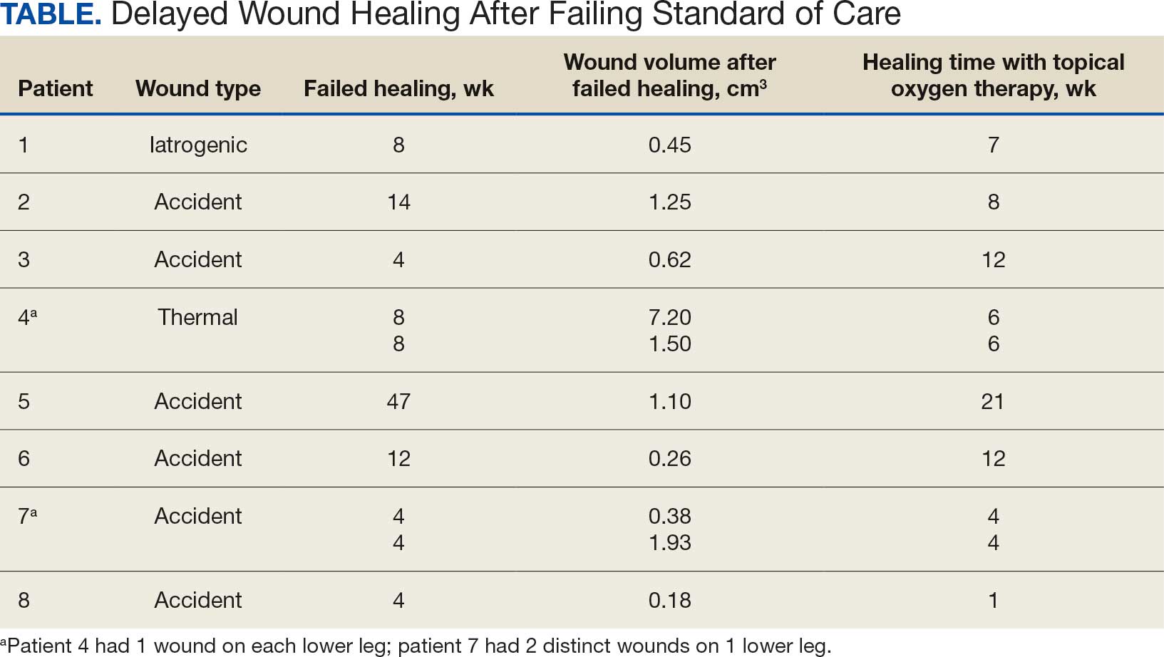
Yellin et al reported that patients with DFU treated with TOT had reduced hospital lengths of stay and a lower amputation rate compared with patients who did not receive TOT modality.10 Another study compared conventional compression dressings with TOT in managing 132 recalcitrant venous ulcers. In that study, Nataraj et al found that 76% of ulcers treated with TOT had completely healed at 12 weeks compared with 46% of those treated with compression only.11 Furthermore, numerous systematic reviews and meta-analyses have analyzed the efficacy of TOT, concluding that this modality not only improves healing among ulcers of varying severity when compared with the standard of care but also contributes to the durability of healed ulcers and thus helps improve patient quality of life by preventing recurrence.2,12-18
Intermittent cyclical compression topical oxygen therapy (cTOT) is a modality that provides concentrated oxygen at a rate of 10 L/min. cTOT cycles twice a minute and applies noncontact compression directly to ulcerations between 10 and 50 millibars (7.5-37.5 mm Hg). In a study of 8 patients with leg ulcers, transcutaneous oxygen tension was higher after treatment with intermittent pneumatic compression (42.7 mm Hg) when compared with baseline values. The authors postulated that intermittent compression decreased interstitial fluid volume and venous stasis, both of which lead to increased cutaneous oxygenation. The data suggested that removal of carbon dioxide from tissues and enhancement of cutaneous cyclical oxygenation promoted ulcer healing.19To the best of our knowledge, there are no published studies that have evaluated the efficacy of cTOT for traumatic injuries while also considering existing comorbidities such as diabetes mellitus and venous insufficiency in veterans. The aim of this study was to demonstrate the efficacy of cTOT in the setting of traumatic wounds that failed the standard of care and were complicated by venous insufficiency and diabetes mellitus. We hypothesized that cTOT enhances healing of trauma-induced venous leg wounds in patients with diabetes mellitus, achieving full epithelialization within 12 weeks after failing the standard of care.
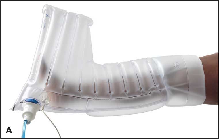
B, controller. Source: Advanced Oxygen Therapy Inc. Reprinted with permission.
Methods
Institutional review board approval was granted prior to commencement of this single-center retrospective review at the Veterans Affairs Illiana Healthcare System (VAIHS) in Danville, Illinois. Electronic health records between May 1, 2016, and July 31, 2021, were examined to find patients treated with cTOT using the Advanced Oxygen Therapy Inc. Topical Wound Oxygen (TWO2) system (Figure). Data collected included demographics, follow-up, and outcomes.
Inclusion criteria included patients who failed standard wound care for ≥ 4 weeks with venous insufficiency, trauma to the lower legs by means of iatrogenic injury, accidental injury, thermal injury/burn, or diabetes mellitus determined to be an active problem and a hemoglobin A1c (HbA1c) obtained within 3 months of wound treatment. The VAIHS standard of care was weekly visits to a wound care physician, physician assistant, or a nurse practitioner who performed sharp wound debridement and applied multilayer compression therapy. Patients with confirmed history of intermittent claudication, stroke, absent pedal pulses, abnormal blood pressure, deep vein thrombosis, pulmonary embolism, revascularization of the lower extremity, trauma fractures of the affected leg, phlebitis, pregnancy, coagulopathy, infection, or history of malignancy were excluded.
Patients who failed the standard of care for ≥ 4 weeks were enrolled in the cTOT program, after which they began weekly visits to an outpatient wound healing center where visits consisted of sharp wound debridement, wound measurement, and application of compressive dressing to the affected lower extremity. Following wound-healing center visits, the subjects received cTOT at home according to the manufacturer’s protocol, which consisted of ≤ 90-minute sessions, 5 to 7 times weekly. The wounds were measured from initial presentation to complete epithelialization, and the patients were followed for ≤ 27 months.
Results
Sixty-nine patients underwent cTOT between May 1, 2016, and July 31, 2021, and 8 patients with 10 wounds met the inclusion criteria. The patients had a mean age of 76 years, BMI of 27, and HbA1c of 8.0%. All wounds completely healed following cTOT. Prior to the use of cTOT, the included patients had a mean 11.3 weeks of failed healing attempts when receiving standard of care therapy. Following cTOT implementation, the mean time to healing was 8.1 weeks (Table). One wound recurred after 10 weeks in a patient with uncontrolled diabetes mellitus. One patient died of causes unrelated to the wound and was excluded from follow-up. All other wounds remained closed and healed after a mean end follow-up period of 110 weeks through June 2023.
Discussion
Traumatic wounds are commonly seen in the emergency department (ED), accounting for 5.4% of all ED visits.20-22 According to the National Hospital Ambulatory Medical Care Survey, 14.6% of 117 million ED visits in 2007 involved lower-extremity wounds, costing the health care system > $25 billion annually.23 Kulkarni et al reported that 44% of patients experienced leg ulcerations secondary to trauma.24 Traumatic wounds that lack epithelialization > 4 weeks are categorized as chronic, placing a large social and financial impact on patient health and well-being.
This case series supports the benefits of cTOT and demonstrates improved wound-closure rates with sustained healing compared with standard care alone, corroborating findings reported by the American Diabetes Association (ADA).23 The ADA cited high-quality RCTs and systematic reviews supporting the healing benefits of cTOT for chronic DFUs through the upregulation of essential growth factors.24
The current literature offers many examples of the healing benefits of TOT for arterial ulcerations. Benefits include faster healing with longer lasting reparative ulcer tissue, prevention of amputation and improved quality of life. However, there is a lack of literature on the use of cTOT for venous stasis traumatic wounds, which prompted this study. Our case series presents evidence that cTOT enhances healing of trauma-induced venous leg wounds in male veterans with diabetes mellitus within 12 weeks after failure of the standard of care. Other risk factors that were recorded but not included in our analysis included tobacco use, chronic kidney disease, chronic obstructive pulmonary disease, use of cellular-based tissue/collagen products, and neuropathy.
This case series provides evidence that cTOT can be a beneficial and cost-effective treatment. Economic considerations are derived from retrospective cohort analyses and a published decision-analytic Markov model representing modeled estimates and need to be interpreted in that context rather than as findings from prospective comparative cost analyses healing.25,26
The authors did not conduct an independent health economic evaluation as part of this review. Wound recurrence, hospitalizations and lower extremity amputations represent major cost drivers in chronic wound care. In addition, repeat ED admissions, surgical intervention, rehabilitation, prosthetics, and long-term disability contribute to economic burden and negative psychosocial and quality of life impacts on patients and caregivers. The reductions in hospitalization (≤ 88%) and amputation (≤ 73%) observed in real-world TWO2 cohorts suggest significant potential downstream cost savings. A recent Markov model analysis suggested that TWO2 may be cost-favorable compared with standard care, with modeled estimates indicating lower total 2-year costs and improved quality-adjusted life years. These projections are based on modeling assumptions and require confirmation through prospective economic evaluations comparing TWO2 directly with negative pressure wound therapy, HBOT, and skin substitutes across longer time horizons.
Limitations
The findings of this case series were small and lacked statistical power with only 8 male patients. Literature gaps also exist regarding the specific effects of TOT on traumatic wounds. This review was limited by its single-center location.
Conclusions
This case series suggests that cTOT may be an effective adjuvant therapy in managing trauma-induced venous leg wounds and supports consideration of early implementation for expedited epithelialization. However, further studies with larger sample sizes are required.
Tawfick WA, Sultan S. Technical and clinical outcome of topical wound oxygen in comparison to conventional compression dressings in the management of refractory nonhealing venous ulcers. Vasc Endovascular Surg. 2013:30-37. doi:10.1177/1538574412467684
Fries RB, Wallace WA, Roy S, et al. Dermal excisional wound healing in pigs following treatment with topically applied pure oxygen. Mutat Res. 2005:172-181. doi:10.1016/j.mrfmmm.2005.02.023
Frykberg RG. Topical wound oxygen therapy in the treatment of chronic diabetic foot ulcers. Medicina (Kaunas). 2021:917. doi:10.3390/medicina57090917
Sun XK, Li R, Yang XL, et al. Efficacy and safety of topical oxygen therapy for diabetic foot ulcers: an updated systematic review and meta-analysis. Int Wound J. 2022:2200-2209. doi:10.1111/iwj.13830
Lavery LA, Killeen AL, Farrar D, et al. The effect of continuous diffusion of oxygen treatment on cytokines, perfusion, bacterial load, and healing in patients with diabetic foot ulcers. Int Wound J. 2020:1986-1995. doi:10.1111/iwj.13490
Frykberg RG, Franks PJ, Edmonds M, et al. A multinational, multicenter, randomized, double-blinded, placebo-controlled trial to evaluate the efficacy of cyclical topical wound oxygen (TWO2) therapy in the treatment of chronic diabetic foot ulcers: the TWO2 study. Diabetes Care. 2020:616-624. doi:10.2337/dc19-0476
Sun XK, Li R, Yang XL, et al. Efficacy and safety of topical oxygen therapy for diabetic foot ulcers: an updated systematic review and meta-analysis. Int Wound J. 2022:2200-2209. doi:10.1111/iwj.13830
Al-Jalodi O, Kupcella M, Breisinger K, et al. A multicenter clinical trial evaluating the durability of diabetic foot ulcer healing in ulcers treated with topical oxygen and standard of care versus standard of care alone 1 year post healing. Int Wound J. 2022:1838-1842. doi:10.1111/iwj.13789
Carter MJ, Frykberg RG, Oropallo A, et al. Efficacy of topical wound oxygen therapy in healing chronic diabetic foot ulcers: systematic review and meta-analysis. Adv Wound Care (New Rochelle). 2023;12:177-186. doi:10.1089/wound.2022.0041
Yellin JI, Gaebler JA, Zhou FF, et al. Reduced hospitalizations and amputations in patients with diabetic foot ulcers treated with cyclical pressurized topical wound oxygen therapy: real-world outcomes. Adv Wound Care (New Rochelle). 2022:657-665. doi:10.1089/wound.2021.0118
Nataraj M, Maiya AG, Karkada G, et al. Application of topical oxygen therapy in healing dynamics of diabetic foot ulcers: a systematic review. Rev Diabet Stud. 2019:74-82. doi:10.1900/RDS.2019.15.74
Thanigaimani S, Singh T, Golledge J. Topical oxygen therapy for diabetes-related foot ulcers: a systematic review and meta-analysis. Diabet Med. 2021:e14585. doi:10.1111/dme.14585
Frykberg RG. Topical wound oxygen therapy in the treatment of chronic diabetic foot ulcers. Medicina (Kaunas). 2021:917. doi:10.3390/medicina57090917
Oropallo AR, Serena TE, Armstrong DG, et al. Molecular biomarkers of oxygen therapy in patients with diabetic foot ulcers. Biomolecules. 2021:925. doi:10.3390/biom11070925
Lavery LA, Killeen AL, Farrar D, et al. The effect of continuous diffusion of oxygen treatment on cytokines, perfusion, bacterial load, and healing in patients with diabetic foot ulcers. Int Wound J. 2020:1986-1995. doi:10.1111/iwj.13490
Kahle B, Hermanns HJ, Gallenkemper G. Evidence-based treatment of chronic leg ulcers. Dtsch Arztebl Int. 2011:231-237. doi:10.3238/arztebl.2011.0231
Ortega MA, Fraile-Martinez O, García-Montero C, et al. A general overview on hyperbaric oxygen therapy: applications, mechanisms, and translational opportunities. Medicina (Kaunas). 2021:864. doi:10.3390/medicina57090864
Sayadi LR, Banyard DA, Ziegler ME, et al. Topical oxygen therapy and micro/nanobubbles: a new modality for tissue oxygen delivery. Int Wound J. 2018:363-374. doi:10.1111/iwj.12873
Chen AH, Frangos SG, Kilaru S, et al. Intermittent pneumatic compression devices: physiologic mechanisms of action. Eur J Vasc Endovasc Surg. 2001:383-392. doi:10.1053/ejvs.2001.1348
Prevaldi C, Paolillo C, Locatelli C, et al. Management of traumatic wounds in the emergency department: position paper from the Academy of Emergency Medicine and Care (AcEMC) and the World Society of Emergency Surgery (WSES). World J Emerg Surg. 2016:30. doi:10.1186/s13017-016-0084-3
Han G, Ceilley R. Chronic wound healing: a review of current management and treatments. Adv Ther. 2017:599-610. doi:10.1007/s12325-017-0478-y
Kamal S, Sehgal A, Shahi P, et al. Topical oxygen therapy in acute traumatic musculoskeletal wounds of the foot and ankle. J Wound Care. 2023:92-97. doi:10.12968/jowc.2023.32.2.92
Lambers K, Ootes D, Ring D. Incidence of patients with lower extremity injuries presenting to US emergency departments by anatomic region, disease category, and age. Clin Orthop Relat Res. 2012:284-290. doi:10.1007/s11999-011-1982-z
Kulkarni SR, Gohel MS, Whyman MR, et al. Significance of limb trauma as an initiating factor in chronic leg ulceration. Phlebology. 2008:130-136.
Kerr M, Wild D, Edmonds M, et al. Cost effectiveness of topical wound oxygen therapy for chronic diabetic foot ulcers. J Diabetes Complications. 2025;39:109016. doi:10.1016/j.jdiacomp.2025.109016.
Yellin JI, Gaebler JA, Zhou FF, et al. Reduced hospitalizations and amputations in patients with diabetic foot ulcers treated with cyclical pressurized topical wound oxygen therapy: real-world outcomes. Adv Wound Care (New Rochelle). 2022;11:657-665. doi:10.1089/wound.2021.0118
Lower extremity venous ulcerations impact the health, well-being, and associated costs of patient care. These ulcerations are associated with comorbidities such as diabetes mellitus and chronic venous insufficiency or may be related to trauma. Often, the standard of care fails to effectively and completely heal ulcers, causing them to become stalled, chronic, and require ≥ 1 month of treatment.
Healthy skin partial pressure of oxygen (PO2) approximates 100 mm Hg, but prolonged periods of poor blood perfusion from vascular damage can cause the PO2 within a wound bed to become as low as 10 mm Hg.1 Deprivation of oxygen impairs biological processes essential for bacterial control and growth factor activity, contributing to wound chronicity.2-5 Exposure to oxygen not only upregulates growth factors essential for maintaining a well-vascularized wound core, but also aids enzymatic reactions responsible for healing the skin that are sensitive to PO2 levels.3,6,7 These physiologic processes can be maximized by raising oxygen level concentrations by means of oxygen therapy.8
Hyperbaric oxygen therapy (HBOT) uses a pressurized system to deliver concentrated oxygen that is diffused in the lungs, resulting in increased blood oxygen levels for delivery to bodily tissues. Full chamber HBOT can accelerate the wound healing process. Some disadvantages of HBOT include its high cost, the possibility of oxygen toxicity, and travel time to facilities for daily therapy sessions that last an average of 1.5 hours and may continue for ≤ 60 sessions.7,9 Thus, alternatives to HBOT have been developed, including topical oxygen therapy (TOT), which uses portable bags or chambers that deliver concentrated oxygen applied directly over ulcers, allowing oxygen to diffuse directly into the injured tissue. Combined with the standard of care and regular debridement, TOT saturates the wound base and surrounding tissues with oxygen, thereby inducing growth factors that promote angiogenesis as well as upregulation of enzymatic reactions that create a reparative environment.6
In a study conducted in pigs, Fries et al induced full-thickness wounds that were allowed to heal by secondary intention. They demonstrated that wounds treated with TOT not only healed in fewer days but also revealed a higher presence of vascular endothelial growth factor, greater density of vascular structures, and more histologically mature healing tissues when compared with control wounds exposed to room air.2
The effectiveness of TOT in ulcer healing has been previously reported. Frykberg et al performed a multicenter randomized controlled trial (RCT) comparing TOT with sham therapy to determine the efficacy of TOT in diabetic foot ulcers (DFUs) that had previously failed treatment with the standard of care. The RCT demonstrated that wounds treated with TOT had a 4.5-fold greater likelihood of complete epithelialization within 12 weeks compared with the sham group.3 In a multicenter RCT, Al-Jalodi et al found that 56% of DFUs treated with TOT remained epithelialized at 12 months compared with 27% of ulcers treated with the standard of care alone. Al Jalodi et al argued that TOT may increase the durability of DFU closure.8 In addition, a small prospective cohort study biopsied 23 DFUs treated with TOT and noted increased cytokines and growth factors, as well as increased transcutaneous oxygen measurement perfusion after only 1 week of treatment compared with wounds treated with the standard of care.9
Yellin et al reported that patients with DFU treated with TOT had reduced hospital lengths of stay and a lower amputation rate compared with patients who did not receive TOT modality.10 Another study compared conventional compression dressings with TOT in managing 132 recalcitrant venous ulcers. In that study, Nataraj et al found that 76% of ulcers treated with TOT had completely healed at 12 weeks compared with 46% of those treated with compression only.11 Furthermore, numerous systematic reviews and meta-analyses have analyzed the efficacy of TOT, concluding that this modality not only improves healing among ulcers of varying severity when compared with the standard of care but also contributes to the durability of healed ulcers and thus helps improve patient quality of life by preventing recurrence.2,12-18
Intermittent cyclical compression topical oxygen therapy (cTOT) is a modality that provides concentrated oxygen at a rate of 10 L/min. cTOT cycles twice a minute and applies noncontact compression directly to ulcerations between 10 and 50 millibars (7.5-37.5 mm Hg). In a study of 8 patients with leg ulcers, transcutaneous oxygen tension was higher after treatment with intermittent pneumatic compression (42.7 mm Hg) when compared with baseline values. The authors postulated that intermittent compression decreased interstitial fluid volume and venous stasis, both of which lead to increased cutaneous oxygenation. The data suggested that removal of carbon dioxide from tissues and enhancement of cutaneous cyclical oxygenation promoted ulcer healing.19To the best of our knowledge, there are no published studies that have evaluated the efficacy of cTOT for traumatic injuries while also considering existing comorbidities such as diabetes mellitus and venous insufficiency in veterans. The aim of this study was to demonstrate the efficacy of cTOT in the setting of traumatic wounds that failed the standard of care and were complicated by venous insufficiency and diabetes mellitus. We hypothesized that cTOT enhances healing of trauma-induced venous leg wounds in patients with diabetes mellitus, achieving full epithelialization within 12 weeks after failing the standard of care.
B, controller. Source: Advanced Oxygen Therapy Inc. Reprinted with permission.
Methods
Institutional review board approval was granted prior to commencement of this single-center retrospective review at the Veterans Affairs Illiana Healthcare System (VAIHS) in Danville, Illinois. Electronic health records between May 1, 2016, and July 31, 2021, were examined to find patients treated with cTOT using the Advanced Oxygen Therapy Inc. Topical Wound Oxygen (TWO2) system (Figure). Data collected included demographics, follow-up, and outcomes.
Inclusion criteria included patients who failed standard wound care for ≥ 4 weeks with venous insufficiency, trauma to the lower legs by means of iatrogenic injury, accidental injury, thermal injury/burn, or diabetes mellitus determined to be an active problem and a hemoglobin A1c (HbA1c) obtained within 3 months of wound treatment. The VAIHS standard of care was weekly visits to a wound care physician, physician assistant, or a nurse practitioner who performed sharp wound debridement and applied multilayer compression therapy. Patients with confirmed history of intermittent claudication, stroke, absent pedal pulses, abnormal blood pressure, deep vein thrombosis, pulmonary embolism, revascularization of the lower extremity, trauma fractures of the affected leg, phlebitis, pregnancy, coagulopathy, infection, or history of malignancy were excluded.
Patients who failed the standard of care for ≥ 4 weeks were enrolled in the cTOT program, after which they began weekly visits to an outpatient wound healing center where visits consisted of sharp wound debridement, wound measurement, and application of compressive dressing to the affected lower extremity. Following wound-healing center visits, the subjects received cTOT at home according to the manufacturer’s protocol, which consisted of ≤ 90-minute sessions, 5 to 7 times weekly. The wounds were measured from initial presentation to complete epithelialization, and the patients were followed for ≤ 27 months.
Results
Sixty-nine patients underwent cTOT between May 1, 2016, and July 31, 2021, and 8 patients with 10 wounds met the inclusion criteria. The patients had a mean age of 76 years, BMI of 27, and HbA1c of 8.0%. All wounds completely healed following cTOT. Prior to the use of cTOT, the included patients had a mean 11.3 weeks of failed healing attempts when receiving standard of care therapy. Following cTOT implementation, the mean time to healing was 8.1 weeks (Table). One wound recurred after 10 weeks in a patient with uncontrolled diabetes mellitus. One patient died of causes unrelated to the wound and was excluded from follow-up. All other wounds remained closed and healed after a mean end follow-up period of 110 weeks through June 2023.
Discussion
Traumatic wounds are commonly seen in the emergency department (ED), accounting for 5.4% of all ED visits.20-22 According to the National Hospital Ambulatory Medical Care Survey, 14.6% of 117 million ED visits in 2007 involved lower-extremity wounds, costing the health care system > $25 billion annually.23 Kulkarni et al reported that 44% of patients experienced leg ulcerations secondary to trauma.24 Traumatic wounds that lack epithelialization > 4 weeks are categorized as chronic, placing a large social and financial impact on patient health and well-being.
This case series supports the benefits of cTOT and demonstrates improved wound-closure rates with sustained healing compared with standard care alone, corroborating findings reported by the American Diabetes Association (ADA).23 The ADA cited high-quality RCTs and systematic reviews supporting the healing benefits of cTOT for chronic DFUs through the upregulation of essential growth factors.24
The current literature offers many examples of the healing benefits of TOT for arterial ulcerations. Benefits include faster healing with longer lasting reparative ulcer tissue, prevention of amputation and improved quality of life. However, there is a lack of literature on the use of cTOT for venous stasis traumatic wounds, which prompted this study. Our case series presents evidence that cTOT enhances healing of trauma-induced venous leg wounds in male veterans with diabetes mellitus within 12 weeks after failure of the standard of care. Other risk factors that were recorded but not included in our analysis included tobacco use, chronic kidney disease, chronic obstructive pulmonary disease, use of cellular-based tissue/collagen products, and neuropathy.
This case series provides evidence that cTOT can be a beneficial and cost-effective treatment. Economic considerations are derived from retrospective cohort analyses and a published decision-analytic Markov model representing modeled estimates and need to be interpreted in that context rather than as findings from prospective comparative cost analyses healing.25,26
The authors did not conduct an independent health economic evaluation as part of this review. Wound recurrence, hospitalizations and lower extremity amputations represent major cost drivers in chronic wound care. In addition, repeat ED admissions, surgical intervention, rehabilitation, prosthetics, and long-term disability contribute to economic burden and negative psychosocial and quality of life impacts on patients and caregivers. The reductions in hospitalization (≤ 88%) and amputation (≤ 73%) observed in real-world TWO2 cohorts suggest significant potential downstream cost savings. A recent Markov model analysis suggested that TWO2 may be cost-favorable compared with standard care, with modeled estimates indicating lower total 2-year costs and improved quality-adjusted life years. These projections are based on modeling assumptions and require confirmation through prospective economic evaluations comparing TWO2 directly with negative pressure wound therapy, HBOT, and skin substitutes across longer time horizons.
Limitations
The findings of this case series were small and lacked statistical power with only 8 male patients. Literature gaps also exist regarding the specific effects of TOT on traumatic wounds. This review was limited by its single-center location.
Conclusions
This case series suggests that cTOT may be an effective adjuvant therapy in managing trauma-induced venous leg wounds and supports consideration of early implementation for expedited epithelialization. However, further studies with larger sample sizes are required.
Lower extremity venous ulcerations impact the health, well-being, and associated costs of patient care. These ulcerations are associated with comorbidities such as diabetes mellitus and chronic venous insufficiency or may be related to trauma. Often, the standard of care fails to effectively and completely heal ulcers, causing them to become stalled, chronic, and require ≥ 1 month of treatment.
Healthy skin partial pressure of oxygen (PO2) approximates 100 mm Hg, but prolonged periods of poor blood perfusion from vascular damage can cause the PO2 within a wound bed to become as low as 10 mm Hg.1 Deprivation of oxygen impairs biological processes essential for bacterial control and growth factor activity, contributing to wound chronicity.2-5 Exposure to oxygen not only upregulates growth factors essential for maintaining a well-vascularized wound core, but also aids enzymatic reactions responsible for healing the skin that are sensitive to PO2 levels.3,6,7 These physiologic processes can be maximized by raising oxygen level concentrations by means of oxygen therapy.8
Hyperbaric oxygen therapy (HBOT) uses a pressurized system to deliver concentrated oxygen that is diffused in the lungs, resulting in increased blood oxygen levels for delivery to bodily tissues. Full chamber HBOT can accelerate the wound healing process. Some disadvantages of HBOT include its high cost, the possibility of oxygen toxicity, and travel time to facilities for daily therapy sessions that last an average of 1.5 hours and may continue for ≤ 60 sessions.7,9 Thus, alternatives to HBOT have been developed, including topical oxygen therapy (TOT), which uses portable bags or chambers that deliver concentrated oxygen applied directly over ulcers, allowing oxygen to diffuse directly into the injured tissue. Combined with the standard of care and regular debridement, TOT saturates the wound base and surrounding tissues with oxygen, thereby inducing growth factors that promote angiogenesis as well as upregulation of enzymatic reactions that create a reparative environment.6
In a study conducted in pigs, Fries et al induced full-thickness wounds that were allowed to heal by secondary intention. They demonstrated that wounds treated with TOT not only healed in fewer days but also revealed a higher presence of vascular endothelial growth factor, greater density of vascular structures, and more histologically mature healing tissues when compared with control wounds exposed to room air.2
The effectiveness of TOT in ulcer healing has been previously reported. Frykberg et al performed a multicenter randomized controlled trial (RCT) comparing TOT with sham therapy to determine the efficacy of TOT in diabetic foot ulcers (DFUs) that had previously failed treatment with the standard of care. The RCT demonstrated that wounds treated with TOT had a 4.5-fold greater likelihood of complete epithelialization within 12 weeks compared with the sham group.3 In a multicenter RCT, Al-Jalodi et al found that 56% of DFUs treated with TOT remained epithelialized at 12 months compared with 27% of ulcers treated with the standard of care alone. Al Jalodi et al argued that TOT may increase the durability of DFU closure.8 In addition, a small prospective cohort study biopsied 23 DFUs treated with TOT and noted increased cytokines and growth factors, as well as increased transcutaneous oxygen measurement perfusion after only 1 week of treatment compared with wounds treated with the standard of care.9
Yellin et al reported that patients with DFU treated with TOT had reduced hospital lengths of stay and a lower amputation rate compared with patients who did not receive TOT modality.10 Another study compared conventional compression dressings with TOT in managing 132 recalcitrant venous ulcers. In that study, Nataraj et al found that 76% of ulcers treated with TOT had completely healed at 12 weeks compared with 46% of those treated with compression only.11 Furthermore, numerous systematic reviews and meta-analyses have analyzed the efficacy of TOT, concluding that this modality not only improves healing among ulcers of varying severity when compared with the standard of care but also contributes to the durability of healed ulcers and thus helps improve patient quality of life by preventing recurrence.2,12-18
Intermittent cyclical compression topical oxygen therapy (cTOT) is a modality that provides concentrated oxygen at a rate of 10 L/min. cTOT cycles twice a minute and applies noncontact compression directly to ulcerations between 10 and 50 millibars (7.5-37.5 mm Hg). In a study of 8 patients with leg ulcers, transcutaneous oxygen tension was higher after treatment with intermittent pneumatic compression (42.7 mm Hg) when compared with baseline values. The authors postulated that intermittent compression decreased interstitial fluid volume and venous stasis, both of which lead to increased cutaneous oxygenation. The data suggested that removal of carbon dioxide from tissues and enhancement of cutaneous cyclical oxygenation promoted ulcer healing.19To the best of our knowledge, there are no published studies that have evaluated the efficacy of cTOT for traumatic injuries while also considering existing comorbidities such as diabetes mellitus and venous insufficiency in veterans. The aim of this study was to demonstrate the efficacy of cTOT in the setting of traumatic wounds that failed the standard of care and were complicated by venous insufficiency and diabetes mellitus. We hypothesized that cTOT enhances healing of trauma-induced venous leg wounds in patients with diabetes mellitus, achieving full epithelialization within 12 weeks after failing the standard of care.
B, controller. Source: Advanced Oxygen Therapy Inc. Reprinted with permission.
Methods
Institutional review board approval was granted prior to commencement of this single-center retrospective review at the Veterans Affairs Illiana Healthcare System (VAIHS) in Danville, Illinois. Electronic health records between May 1, 2016, and July 31, 2021, were examined to find patients treated with cTOT using the Advanced Oxygen Therapy Inc. Topical Wound Oxygen (TWO2) system (Figure). Data collected included demographics, follow-up, and outcomes.
Inclusion criteria included patients who failed standard wound care for ≥ 4 weeks with venous insufficiency, trauma to the lower legs by means of iatrogenic injury, accidental injury, thermal injury/burn, or diabetes mellitus determined to be an active problem and a hemoglobin A1c (HbA1c) obtained within 3 months of wound treatment. The VAIHS standard of care was weekly visits to a wound care physician, physician assistant, or a nurse practitioner who performed sharp wound debridement and applied multilayer compression therapy. Patients with confirmed history of intermittent claudication, stroke, absent pedal pulses, abnormal blood pressure, deep vein thrombosis, pulmonary embolism, revascularization of the lower extremity, trauma fractures of the affected leg, phlebitis, pregnancy, coagulopathy, infection, or history of malignancy were excluded.
Patients who failed the standard of care for ≥ 4 weeks were enrolled in the cTOT program, after which they began weekly visits to an outpatient wound healing center where visits consisted of sharp wound debridement, wound measurement, and application of compressive dressing to the affected lower extremity. Following wound-healing center visits, the subjects received cTOT at home according to the manufacturer’s protocol, which consisted of ≤ 90-minute sessions, 5 to 7 times weekly. The wounds were measured from initial presentation to complete epithelialization, and the patients were followed for ≤ 27 months.
Results
Sixty-nine patients underwent cTOT between May 1, 2016, and July 31, 2021, and 8 patients with 10 wounds met the inclusion criteria. The patients had a mean age of 76 years, BMI of 27, and HbA1c of 8.0%. All wounds completely healed following cTOT. Prior to the use of cTOT, the included patients had a mean 11.3 weeks of failed healing attempts when receiving standard of care therapy. Following cTOT implementation, the mean time to healing was 8.1 weeks (Table). One wound recurred after 10 weeks in a patient with uncontrolled diabetes mellitus. One patient died of causes unrelated to the wound and was excluded from follow-up. All other wounds remained closed and healed after a mean end follow-up period of 110 weeks through June 2023.
Discussion
Traumatic wounds are commonly seen in the emergency department (ED), accounting for 5.4% of all ED visits.20-22 According to the National Hospital Ambulatory Medical Care Survey, 14.6% of 117 million ED visits in 2007 involved lower-extremity wounds, costing the health care system > $25 billion annually.23 Kulkarni et al reported that 44% of patients experienced leg ulcerations secondary to trauma.24 Traumatic wounds that lack epithelialization > 4 weeks are categorized as chronic, placing a large social and financial impact on patient health and well-being.
This case series supports the benefits of cTOT and demonstrates improved wound-closure rates with sustained healing compared with standard care alone, corroborating findings reported by the American Diabetes Association (ADA).23 The ADA cited high-quality RCTs and systematic reviews supporting the healing benefits of cTOT for chronic DFUs through the upregulation of essential growth factors.24
The current literature offers many examples of the healing benefits of TOT for arterial ulcerations. Benefits include faster healing with longer lasting reparative ulcer tissue, prevention of amputation and improved quality of life. However, there is a lack of literature on the use of cTOT for venous stasis traumatic wounds, which prompted this study. Our case series presents evidence that cTOT enhances healing of trauma-induced venous leg wounds in male veterans with diabetes mellitus within 12 weeks after failure of the standard of care. Other risk factors that were recorded but not included in our analysis included tobacco use, chronic kidney disease, chronic obstructive pulmonary disease, use of cellular-based tissue/collagen products, and neuropathy.
This case series provides evidence that cTOT can be a beneficial and cost-effective treatment. Economic considerations are derived from retrospective cohort analyses and a published decision-analytic Markov model representing modeled estimates and need to be interpreted in that context rather than as findings from prospective comparative cost analyses healing.25,26
The authors did not conduct an independent health economic evaluation as part of this review. Wound recurrence, hospitalizations and lower extremity amputations represent major cost drivers in chronic wound care. In addition, repeat ED admissions, surgical intervention, rehabilitation, prosthetics, and long-term disability contribute to economic burden and negative psychosocial and quality of life impacts on patients and caregivers. The reductions in hospitalization (≤ 88%) and amputation (≤ 73%) observed in real-world TWO2 cohorts suggest significant potential downstream cost savings. A recent Markov model analysis suggested that TWO2 may be cost-favorable compared with standard care, with modeled estimates indicating lower total 2-year costs and improved quality-adjusted life years. These projections are based on modeling assumptions and require confirmation through prospective economic evaluations comparing TWO2 directly with negative pressure wound therapy, HBOT, and skin substitutes across longer time horizons.
Limitations
The findings of this case series were small and lacked statistical power with only 8 male patients. Literature gaps also exist regarding the specific effects of TOT on traumatic wounds. This review was limited by its single-center location.
Conclusions
This case series suggests that cTOT may be an effective adjuvant therapy in managing trauma-induced venous leg wounds and supports consideration of early implementation for expedited epithelialization. However, further studies with larger sample sizes are required.
Tawfick WA, Sultan S. Technical and clinical outcome of topical wound oxygen in comparison to conventional compression dressings in the management of refractory nonhealing venous ulcers. Vasc Endovascular Surg. 2013:30-37. doi:10.1177/1538574412467684
Fries RB, Wallace WA, Roy S, et al. Dermal excisional wound healing in pigs following treatment with topically applied pure oxygen. Mutat Res. 2005:172-181. doi:10.1016/j.mrfmmm.2005.02.023
Frykberg RG. Topical wound oxygen therapy in the treatment of chronic diabetic foot ulcers. Medicina (Kaunas). 2021:917. doi:10.3390/medicina57090917
Sun XK, Li R, Yang XL, et al. Efficacy and safety of topical oxygen therapy for diabetic foot ulcers: an updated systematic review and meta-analysis. Int Wound J. 2022:2200-2209. doi:10.1111/iwj.13830
Lavery LA, Killeen AL, Farrar D, et al. The effect of continuous diffusion of oxygen treatment on cytokines, perfusion, bacterial load, and healing in patients with diabetic foot ulcers. Int Wound J. 2020:1986-1995. doi:10.1111/iwj.13490
Frykberg RG, Franks PJ, Edmonds M, et al. A multinational, multicenter, randomized, double-blinded, placebo-controlled trial to evaluate the efficacy of cyclical topical wound oxygen (TWO2) therapy in the treatment of chronic diabetic foot ulcers: the TWO2 study. Diabetes Care. 2020:616-624. doi:10.2337/dc19-0476
Sun XK, Li R, Yang XL, et al. Efficacy and safety of topical oxygen therapy for diabetic foot ulcers: an updated systematic review and meta-analysis. Int Wound J. 2022:2200-2209. doi:10.1111/iwj.13830
Al-Jalodi O, Kupcella M, Breisinger K, et al. A multicenter clinical trial evaluating the durability of diabetic foot ulcer healing in ulcers treated with topical oxygen and standard of care versus standard of care alone 1 year post healing. Int Wound J. 2022:1838-1842. doi:10.1111/iwj.13789
Carter MJ, Frykberg RG, Oropallo A, et al. Efficacy of topical wound oxygen therapy in healing chronic diabetic foot ulcers: systematic review and meta-analysis. Adv Wound Care (New Rochelle). 2023;12:177-186. doi:10.1089/wound.2022.0041
Yellin JI, Gaebler JA, Zhou FF, et al. Reduced hospitalizations and amputations in patients with diabetic foot ulcers treated with cyclical pressurized topical wound oxygen therapy: real-world outcomes. Adv Wound Care (New Rochelle). 2022:657-665. doi:10.1089/wound.2021.0118
Nataraj M, Maiya AG, Karkada G, et al. Application of topical oxygen therapy in healing dynamics of diabetic foot ulcers: a systematic review. Rev Diabet Stud. 2019:74-82. doi:10.1900/RDS.2019.15.74
Thanigaimani S, Singh T, Golledge J. Topical oxygen therapy for diabetes-related foot ulcers: a systematic review and meta-analysis. Diabet Med. 2021:e14585. doi:10.1111/dme.14585
Frykberg RG. Topical wound oxygen therapy in the treatment of chronic diabetic foot ulcers. Medicina (Kaunas). 2021:917. doi:10.3390/medicina57090917
Oropallo AR, Serena TE, Armstrong DG, et al. Molecular biomarkers of oxygen therapy in patients with diabetic foot ulcers. Biomolecules. 2021:925. doi:10.3390/biom11070925
Lavery LA, Killeen AL, Farrar D, et al. The effect of continuous diffusion of oxygen treatment on cytokines, perfusion, bacterial load, and healing in patients with diabetic foot ulcers. Int Wound J. 2020:1986-1995. doi:10.1111/iwj.13490
Kahle B, Hermanns HJ, Gallenkemper G. Evidence-based treatment of chronic leg ulcers. Dtsch Arztebl Int. 2011:231-237. doi:10.3238/arztebl.2011.0231
Ortega MA, Fraile-Martinez O, García-Montero C, et al. A general overview on hyperbaric oxygen therapy: applications, mechanisms, and translational opportunities. Medicina (Kaunas). 2021:864. doi:10.3390/medicina57090864
Sayadi LR, Banyard DA, Ziegler ME, et al. Topical oxygen therapy and micro/nanobubbles: a new modality for tissue oxygen delivery. Int Wound J. 2018:363-374. doi:10.1111/iwj.12873
Chen AH, Frangos SG, Kilaru S, et al. Intermittent pneumatic compression devices: physiologic mechanisms of action. Eur J Vasc Endovasc Surg. 2001:383-392. doi:10.1053/ejvs.2001.1348
Prevaldi C, Paolillo C, Locatelli C, et al. Management of traumatic wounds in the emergency department: position paper from the Academy of Emergency Medicine and Care (AcEMC) and the World Society of Emergency Surgery (WSES). World J Emerg Surg. 2016:30. doi:10.1186/s13017-016-0084-3
Han G, Ceilley R. Chronic wound healing: a review of current management and treatments. Adv Ther. 2017:599-610. doi:10.1007/s12325-017-0478-y
Kamal S, Sehgal A, Shahi P, et al. Topical oxygen therapy in acute traumatic musculoskeletal wounds of the foot and ankle. J Wound Care. 2023:92-97. doi:10.12968/jowc.2023.32.2.92
Lambers K, Ootes D, Ring D. Incidence of patients with lower extremity injuries presenting to US emergency departments by anatomic region, disease category, and age. Clin Orthop Relat Res. 2012:284-290. doi:10.1007/s11999-011-1982-z
Kulkarni SR, Gohel MS, Whyman MR, et al. Significance of limb trauma as an initiating factor in chronic leg ulceration. Phlebology. 2008:130-136.
Kerr M, Wild D, Edmonds M, et al. Cost effectiveness of topical wound oxygen therapy for chronic diabetic foot ulcers. J Diabetes Complications. 2025;39:109016. doi:10.1016/j.jdiacomp.2025.109016.
Yellin JI, Gaebler JA, Zhou FF, et al. Reduced hospitalizations and amputations in patients with diabetic foot ulcers treated with cyclical pressurized topical wound oxygen therapy: real-world outcomes. Adv Wound Care (New Rochelle). 2022;11:657-665. doi:10.1089/wound.2021.0118
Tawfick WA, Sultan S. Technical and clinical outcome of topical wound oxygen in comparison to conventional compression dressings in the management of refractory nonhealing venous ulcers. Vasc Endovascular Surg. 2013:30-37. doi:10.1177/1538574412467684
Fries RB, Wallace WA, Roy S, et al. Dermal excisional wound healing in pigs following treatment with topically applied pure oxygen. Mutat Res. 2005:172-181. doi:10.1016/j.mrfmmm.2005.02.023
Frykberg RG. Topical wound oxygen therapy in the treatment of chronic diabetic foot ulcers. Medicina (Kaunas). 2021:917. doi:10.3390/medicina57090917
Sun XK, Li R, Yang XL, et al. Efficacy and safety of topical oxygen therapy for diabetic foot ulcers: an updated systematic review and meta-analysis. Int Wound J. 2022:2200-2209. doi:10.1111/iwj.13830
Lavery LA, Killeen AL, Farrar D, et al. The effect of continuous diffusion of oxygen treatment on cytokines, perfusion, bacterial load, and healing in patients with diabetic foot ulcers. Int Wound J. 2020:1986-1995. doi:10.1111/iwj.13490
Frykberg RG, Franks PJ, Edmonds M, et al. A multinational, multicenter, randomized, double-blinded, placebo-controlled trial to evaluate the efficacy of cyclical topical wound oxygen (TWO2) therapy in the treatment of chronic diabetic foot ulcers: the TWO2 study. Diabetes Care. 2020:616-624. doi:10.2337/dc19-0476
Sun XK, Li R, Yang XL, et al. Efficacy and safety of topical oxygen therapy for diabetic foot ulcers: an updated systematic review and meta-analysis. Int Wound J. 2022:2200-2209. doi:10.1111/iwj.13830
Al-Jalodi O, Kupcella M, Breisinger K, et al. A multicenter clinical trial evaluating the durability of diabetic foot ulcer healing in ulcers treated with topical oxygen and standard of care versus standard of care alone 1 year post healing. Int Wound J. 2022:1838-1842. doi:10.1111/iwj.13789
Carter MJ, Frykberg RG, Oropallo A, et al. Efficacy of topical wound oxygen therapy in healing chronic diabetic foot ulcers: systematic review and meta-analysis. Adv Wound Care (New Rochelle). 2023;12:177-186. doi:10.1089/wound.2022.0041
Yellin JI, Gaebler JA, Zhou FF, et al. Reduced hospitalizations and amputations in patients with diabetic foot ulcers treated with cyclical pressurized topical wound oxygen therapy: real-world outcomes. Adv Wound Care (New Rochelle). 2022:657-665. doi:10.1089/wound.2021.0118
Nataraj M, Maiya AG, Karkada G, et al. Application of topical oxygen therapy in healing dynamics of diabetic foot ulcers: a systematic review. Rev Diabet Stud. 2019:74-82. doi:10.1900/RDS.2019.15.74
Thanigaimani S, Singh T, Golledge J. Topical oxygen therapy for diabetes-related foot ulcers: a systematic review and meta-analysis. Diabet Med. 2021:e14585. doi:10.1111/dme.14585
Frykberg RG. Topical wound oxygen therapy in the treatment of chronic diabetic foot ulcers. Medicina (Kaunas). 2021:917. doi:10.3390/medicina57090917
Oropallo AR, Serena TE, Armstrong DG, et al. Molecular biomarkers of oxygen therapy in patients with diabetic foot ulcers. Biomolecules. 2021:925. doi:10.3390/biom11070925
Lavery LA, Killeen AL, Farrar D, et al. The effect of continuous diffusion of oxygen treatment on cytokines, perfusion, bacterial load, and healing in patients with diabetic foot ulcers. Int Wound J. 2020:1986-1995. doi:10.1111/iwj.13490
Kahle B, Hermanns HJ, Gallenkemper G. Evidence-based treatment of chronic leg ulcers. Dtsch Arztebl Int. 2011:231-237. doi:10.3238/arztebl.2011.0231
Ortega MA, Fraile-Martinez O, García-Montero C, et al. A general overview on hyperbaric oxygen therapy: applications, mechanisms, and translational opportunities. Medicina (Kaunas). 2021:864. doi:10.3390/medicina57090864
Sayadi LR, Banyard DA, Ziegler ME, et al. Topical oxygen therapy and micro/nanobubbles: a new modality for tissue oxygen delivery. Int Wound J. 2018:363-374. doi:10.1111/iwj.12873
Chen AH, Frangos SG, Kilaru S, et al. Intermittent pneumatic compression devices: physiologic mechanisms of action. Eur J Vasc Endovasc Surg. 2001:383-392. doi:10.1053/ejvs.2001.1348
Prevaldi C, Paolillo C, Locatelli C, et al. Management of traumatic wounds in the emergency department: position paper from the Academy of Emergency Medicine and Care (AcEMC) and the World Society of Emergency Surgery (WSES). World J Emerg Surg. 2016:30. doi:10.1186/s13017-016-0084-3
Han G, Ceilley R. Chronic wound healing: a review of current management and treatments. Adv Ther. 2017:599-610. doi:10.1007/s12325-017-0478-y
Kamal S, Sehgal A, Shahi P, et al. Topical oxygen therapy in acute traumatic musculoskeletal wounds of the foot and ankle. J Wound Care. 2023:92-97. doi:10.12968/jowc.2023.32.2.92
Lambers K, Ootes D, Ring D. Incidence of patients with lower extremity injuries presenting to US emergency departments by anatomic region, disease category, and age. Clin Orthop Relat Res. 2012:284-290. doi:10.1007/s11999-011-1982-z
Kulkarni SR, Gohel MS, Whyman MR, et al. Significance of limb trauma as an initiating factor in chronic leg ulceration. Phlebology. 2008:130-136.
Kerr M, Wild D, Edmonds M, et al. Cost effectiveness of topical wound oxygen therapy for chronic diabetic foot ulcers. J Diabetes Complications. 2025;39:109016. doi:10.1016/j.jdiacomp.2025.109016.
Yellin JI, Gaebler JA, Zhou FF, et al. Reduced hospitalizations and amputations in patients with diabetic foot ulcers treated with cyclical pressurized topical wound oxygen therapy: real-world outcomes. Adv Wound Care (New Rochelle). 2022;11:657-665. doi:10.1089/wound.2021.0118
Cyclical Topical Oxygen Therapy of Trauma-Induced Venous Leg Wounds in Patients With Diabetes
Cyclical Topical Oxygen Therapy of Trauma-Induced Venous Leg Wounds in Patients With Diabetes