User login
Bariatric surgery may curtail colorectal cancer risk
Bariatric surgery was associated with a significant reduction in the risk of colorectal cancer among obese adults in a retrospective study of more than 1 million individuals.
Although some studies have suggested that bariatric surgery may reduce the risk of obesity-associated cancers, such as colorectal cancer, other studies have shown an increased colorectal cancer risk after surgery, according to Laurent Bailly, MD, of Université Côte d’Azur in Nice, France, and colleagues.
In a study published in JAMA Surgery, Dr. Bailly and colleagues compared the incidence of colorectal cancer in obese patients who underwent bariatric surgery with the incidence in obese patients who did not have surgery and the incidence in the general population.
Using the French National Health Insurance Information System database, the researchers identified 1,045,348 obese adults aged 50-75 years who had no colorectal cancer at baseline. Of these patients, 74,131 underwent bariatric surgery and 971,217 did not. The mean age was 57.3 years in the surgery group and 63.4 years in the nonsurgery group.
The mean follow-up period was 6.2 years for patients who underwent adjustable gastric banding, 5.5 years for those with sleeve gastrectomy, 5.7 years for those who underwent gastric bypass, and 5.3 years for the nonsurgery group.
Results
Overall, the colorectal cancer rate was 0.6% in the surgery group and 1.3% in the nonsurgery group (P < .001).
The researchers calculated standardized incidence ratios (SIRs) to compare the risk of colorectal cancer in the study population with the risk among the French general population; in other words, the number of observed colorectal cancer cases divided by the number of expected cases.
In the surgery group, 423 cases of colorectal cancer were observed and 428 cases were expected, which leads to an SIR of 1.0. In the nonsurgery group, 12,629 cases were observed and 9,417 cases were expected, leading to an SIR of 1.34.
These results suggest patients in the nonsurgery group had a 34% higher risk of colorectal cancer compared with the general population, whereas the risk in the surgery group was similar to that in the general population.
Patients who underwent either gastric bypass or sleeve gastrectomy had fewer new colorectal cancer diagnoses (0.5% for both) compared with patients who had adjustable gastric banding (0.7%).
The researchers noted that this study was limited by several factors, including the retrospective, observational design and potential selection bias among surgery patients. However, the results were strengthened by the large study population and long-term follow-up.
Putting results into context
The authors of an invited commentary noted that this study is supported by results from a retrospective, U.S.-based study, which indicated that bariatric surgery has a “protective effect” against colorectal cancer (Ann Surg. 2019 Jan;269[1]:95-101).
However, these results conflict with other retrospective studies. A study of Nordic patients suggested that bariatric surgery is associated with an increased risk of colon cancer but perhaps not rectal cancer (Int J Cancer. 2019. doi: 10.1002/ijc.32770).
And a study of English patients showed an increased risk of colorectal cancer in patients who underwent gastric bypass but not in those who underwent gastric banding or sleeve gastrectomy (Br J Surg. 2018;105(12):1650-7).
These conflicting results “imply that the jury is still out on whether bariatric surgery increases or decreases” the risk of colorectal cancer, the commentators wrote. They added that future studies “must account for differences in study population (i.e., race/ethnicity and national origin), mechanistic variation in bariatric surgical type, and length of follow-up, while also distinguishing between rectal and colon cancer before the case is settled.”
This study had no outside sponsorship, and the researchers and commentators had no financial conflicts to disclose.
SOURCE: Bailly L et al. JAMA Surg. 2020 Mar 11. doi:10.1001/jamasurg.2020.0089; Davidson LE et al. JAMA Surg. 2020 Mar. 11. doi:10.1001/jamasurg.2020.0090.
Bariatric surgery was associated with a significant reduction in the risk of colorectal cancer among obese adults in a retrospective study of more than 1 million individuals.
Although some studies have suggested that bariatric surgery may reduce the risk of obesity-associated cancers, such as colorectal cancer, other studies have shown an increased colorectal cancer risk after surgery, according to Laurent Bailly, MD, of Université Côte d’Azur in Nice, France, and colleagues.
In a study published in JAMA Surgery, Dr. Bailly and colleagues compared the incidence of colorectal cancer in obese patients who underwent bariatric surgery with the incidence in obese patients who did not have surgery and the incidence in the general population.
Using the French National Health Insurance Information System database, the researchers identified 1,045,348 obese adults aged 50-75 years who had no colorectal cancer at baseline. Of these patients, 74,131 underwent bariatric surgery and 971,217 did not. The mean age was 57.3 years in the surgery group and 63.4 years in the nonsurgery group.
The mean follow-up period was 6.2 years for patients who underwent adjustable gastric banding, 5.5 years for those with sleeve gastrectomy, 5.7 years for those who underwent gastric bypass, and 5.3 years for the nonsurgery group.
Results
Overall, the colorectal cancer rate was 0.6% in the surgery group and 1.3% in the nonsurgery group (P < .001).
The researchers calculated standardized incidence ratios (SIRs) to compare the risk of colorectal cancer in the study population with the risk among the French general population; in other words, the number of observed colorectal cancer cases divided by the number of expected cases.
In the surgery group, 423 cases of colorectal cancer were observed and 428 cases were expected, which leads to an SIR of 1.0. In the nonsurgery group, 12,629 cases were observed and 9,417 cases were expected, leading to an SIR of 1.34.
These results suggest patients in the nonsurgery group had a 34% higher risk of colorectal cancer compared with the general population, whereas the risk in the surgery group was similar to that in the general population.
Patients who underwent either gastric bypass or sleeve gastrectomy had fewer new colorectal cancer diagnoses (0.5% for both) compared with patients who had adjustable gastric banding (0.7%).
The researchers noted that this study was limited by several factors, including the retrospective, observational design and potential selection bias among surgery patients. However, the results were strengthened by the large study population and long-term follow-up.
Putting results into context
The authors of an invited commentary noted that this study is supported by results from a retrospective, U.S.-based study, which indicated that bariatric surgery has a “protective effect” against colorectal cancer (Ann Surg. 2019 Jan;269[1]:95-101).
However, these results conflict with other retrospective studies. A study of Nordic patients suggested that bariatric surgery is associated with an increased risk of colon cancer but perhaps not rectal cancer (Int J Cancer. 2019. doi: 10.1002/ijc.32770).
And a study of English patients showed an increased risk of colorectal cancer in patients who underwent gastric bypass but not in those who underwent gastric banding or sleeve gastrectomy (Br J Surg. 2018;105(12):1650-7).
These conflicting results “imply that the jury is still out on whether bariatric surgery increases or decreases” the risk of colorectal cancer, the commentators wrote. They added that future studies “must account for differences in study population (i.e., race/ethnicity and national origin), mechanistic variation in bariatric surgical type, and length of follow-up, while also distinguishing between rectal and colon cancer before the case is settled.”
This study had no outside sponsorship, and the researchers and commentators had no financial conflicts to disclose.
SOURCE: Bailly L et al. JAMA Surg. 2020 Mar 11. doi:10.1001/jamasurg.2020.0089; Davidson LE et al. JAMA Surg. 2020 Mar. 11. doi:10.1001/jamasurg.2020.0090.
Bariatric surgery was associated with a significant reduction in the risk of colorectal cancer among obese adults in a retrospective study of more than 1 million individuals.
Although some studies have suggested that bariatric surgery may reduce the risk of obesity-associated cancers, such as colorectal cancer, other studies have shown an increased colorectal cancer risk after surgery, according to Laurent Bailly, MD, of Université Côte d’Azur in Nice, France, and colleagues.
In a study published in JAMA Surgery, Dr. Bailly and colleagues compared the incidence of colorectal cancer in obese patients who underwent bariatric surgery with the incidence in obese patients who did not have surgery and the incidence in the general population.
Using the French National Health Insurance Information System database, the researchers identified 1,045,348 obese adults aged 50-75 years who had no colorectal cancer at baseline. Of these patients, 74,131 underwent bariatric surgery and 971,217 did not. The mean age was 57.3 years in the surgery group and 63.4 years in the nonsurgery group.
The mean follow-up period was 6.2 years for patients who underwent adjustable gastric banding, 5.5 years for those with sleeve gastrectomy, 5.7 years for those who underwent gastric bypass, and 5.3 years for the nonsurgery group.
Results
Overall, the colorectal cancer rate was 0.6% in the surgery group and 1.3% in the nonsurgery group (P < .001).
The researchers calculated standardized incidence ratios (SIRs) to compare the risk of colorectal cancer in the study population with the risk among the French general population; in other words, the number of observed colorectal cancer cases divided by the number of expected cases.
In the surgery group, 423 cases of colorectal cancer were observed and 428 cases were expected, which leads to an SIR of 1.0. In the nonsurgery group, 12,629 cases were observed and 9,417 cases were expected, leading to an SIR of 1.34.
These results suggest patients in the nonsurgery group had a 34% higher risk of colorectal cancer compared with the general population, whereas the risk in the surgery group was similar to that in the general population.
Patients who underwent either gastric bypass or sleeve gastrectomy had fewer new colorectal cancer diagnoses (0.5% for both) compared with patients who had adjustable gastric banding (0.7%).
The researchers noted that this study was limited by several factors, including the retrospective, observational design and potential selection bias among surgery patients. However, the results were strengthened by the large study population and long-term follow-up.
Putting results into context
The authors of an invited commentary noted that this study is supported by results from a retrospective, U.S.-based study, which indicated that bariatric surgery has a “protective effect” against colorectal cancer (Ann Surg. 2019 Jan;269[1]:95-101).
However, these results conflict with other retrospective studies. A study of Nordic patients suggested that bariatric surgery is associated with an increased risk of colon cancer but perhaps not rectal cancer (Int J Cancer. 2019. doi: 10.1002/ijc.32770).
And a study of English patients showed an increased risk of colorectal cancer in patients who underwent gastric bypass but not in those who underwent gastric banding or sleeve gastrectomy (Br J Surg. 2018;105(12):1650-7).
These conflicting results “imply that the jury is still out on whether bariatric surgery increases or decreases” the risk of colorectal cancer, the commentators wrote. They added that future studies “must account for differences in study population (i.e., race/ethnicity and national origin), mechanistic variation in bariatric surgical type, and length of follow-up, while also distinguishing between rectal and colon cancer before the case is settled.”
This study had no outside sponsorship, and the researchers and commentators had no financial conflicts to disclose.
SOURCE: Bailly L et al. JAMA Surg. 2020 Mar 11. doi:10.1001/jamasurg.2020.0089; Davidson LE et al. JAMA Surg. 2020 Mar. 11. doi:10.1001/jamasurg.2020.0090.
FROM JAMA SURGERY
Rare Neurological Disease Special Report

Our sixth annual Rare Neurological Disease Special Report is our biggest issue yet. It is very gratifying to know we are part of the rare disease community and witness to some of the exciting developments that are transforming this field. There are many newly approved therapies highlighted in the articles in this issue, as well as brief profiles of a number of research groups within the NIH’s Rare Diseases Clinical Research Network and an intriguing profile of how the Netflix show Diagnosis used crowdsourcing to solve medical mysteries, many of which involved rare neurologic conditions. That’s just a sampling of what this issue has to offer. There are too many articles to mention each one, but I hope you take the time to read the entire issue.
—Glenn S. Williams, vice president, group editor, Neurology Reviews
Our sixth annual Rare Neurological Disease Special Report is our biggest issue yet. It is very gratifying to know we are part of the rare disease community and witness to some of the exciting developments that are transforming this field. There are many newly approved therapies highlighted in the articles in this issue, as well as brief profiles of a number of research groups within the NIH’s Rare Diseases Clinical Research Network and an intriguing profile of how the Netflix show Diagnosis used crowdsourcing to solve medical mysteries, many of which involved rare neurologic conditions. That’s just a sampling of what this issue has to offer. There are too many articles to mention each one, but I hope you take the time to read the entire issue.
—Glenn S. Williams, vice president, group editor, Neurology Reviews
Our sixth annual Rare Neurological Disease Special Report is our biggest issue yet. It is very gratifying to know we are part of the rare disease community and witness to some of the exciting developments that are transforming this field. There are many newly approved therapies highlighted in the articles in this issue, as well as brief profiles of a number of research groups within the NIH’s Rare Diseases Clinical Research Network and an intriguing profile of how the Netflix show Diagnosis used crowdsourcing to solve medical mysteries, many of which involved rare neurologic conditions. That’s just a sampling of what this issue has to offer. There are too many articles to mention each one, but I hope you take the time to read the entire issue.
—Glenn S. Williams, vice president, group editor, Neurology Reviews
Microbiome profiling ready to take personalized medicine to next level
Standards and technology are now available for microbiome profiling to take personalized medicine to the next level, but prospective trials are needed to realize this possibility, according to a leading expert.
The need for prospective microbiomics trials is increasing with the incidence of immune disorders, many of which have been linked with disturbances in the gut, reported Jöel Doré, PhD, at the annual Gut Microbiota for Health World Summit.
“In spite of considerable progress in medicine, together with hygiene, antibiotics, and vaccination developments, we are still seeing an increasing incidence – uncontrolled, that started over 60 years ago – of immune-mediated conditions,” said Dr. Doré, research director at the French National Research Institute for Agriculture, Food, and the Environment.
According to the World Health Organization, one out of four people will be affected by such a disorder in their lifetime, and the incidence rate of some conditions is accelerating faster than others, with geographical distributions that suggest environmental risk factors.
“The rate of incidence of autism in the U.S.A. is a quite scary exponential curve, where less than 1 birth per 5,000 was [affected] in the 1970s, where today it is 1 birth out of 50,” Dr. Doré said at the meeting sponsored by the American Gastroenterological Association and the European Society for Neurogastroenterology and Motility. “Prevention is an urgent need, and possibly if we do not manage to understand what’s going on, human longevity might be at stake.”
Multiple studies have shown that transferring microbiota from humans with immune disorders to healthy mice can induce clinical signs of immune disorders, he said. And between humans, fecal transplants from healthy donors have reduced symptoms in patients with conditions such as inflammatory bowel disease.
While these studies support the link between microbe-host relationships and immune function, most of the underlying mechanisms remain unknown, Dr. Doré said. He highlighted the fact that the complex network of interactions involved separates microbiomics research from conventional approaches to disease.
“I want to stress the fact that clinical trials [in the past] have been designed in the situation where infection was the problem, [but] infection is essentially a linear thing; one agent, one risk, one disease,” Dr. Doré said. “What we are dealing with – with the increasing incidence of immune disorders – is host-microbe interaction at the center of everything, and alteration of host-microbe leading to risk, which may lead to disease. But host-microbe interaction is under the control of a vast number of environmental aspects ... so tools to deal with innovation and translation in a totally different or systemic configuration have yet to be invented.”
According to Dr. Doré, to develop clinical applications, research procedures must first be standardized.
“To be of use for the clinician and general practitioner, microbiome profiling will have to rely on pipelines of standardized preanalytical and analytical procedures,” he said. “This starts from sample collection and shipment.”
For the past 5 years, Dr. Doré and colleagues have been working to standardize procedures with a number of organizations around the world, and progress has been made.
“Today we have very good standards for shotgun sequencing,” he said.
With standards solidifying, microbiomics may lead to new clinical strategies for a range of conditions, even beyond immune disorders, Dr. Doré said. He noted that, as a relatively simple measure, gene richness in the microbiome may be used as a health stratifier. Studies have shown that low gene count has been associated with more severe metabolic and inflammatory traits among overweight patients, a lack of response to low-calorie diets among overweight and obese patients, severity of related conditions and risk of mortality among patients with liver cirrhosis, and poorer responses to immunotherapy among patients with cancer.
Certain patterns of flora may be prognostic, Dr. Doré said, citing a study by Gopalakrishnan et al. that involved 112 melanoma patients, in which those with a high abundance of Faecalibacterium had significantly longer progression-free survival than patients with a low abundance of the same bacteria. Further, a multivariate model showed that a high abundance of Faecalibacterium was the strongest predictor of response to immunotherapy, (hazard ratio, 2.95; P = .03), followed closely by prior immunotherapy (HR, 2.87; P = .03). In contrast, patients with a high abundance of Bacteroidales had shorter progression-free survival than patients with a low abundance of the same bacteria.
Dr. Doré also referred to one of the first interventional microbiomics studies in oncology. Mohty et al. conducted the ODYSSEE phase 1b/2a trial involving 25 patients with acute myeloid leukemia, in which patients were given autologous fecal microbiota transplants after induction chemotherapy and antibiotics. The treatment recovered 90% of original microbiota, and the estimated 1-year overall survival rate was 84%, compared with a historical rate of 70%.
The ODYSSEE study serves as proof of concept that microbiomics may eventually offer the next level of personalized medicine, Dr. Doré said. And now, with standards and technology available, researchers can move forward.
Dr. Doré disclosed relationships with BioFortis, Janssen, Sanofi, and other pharmaceutical companies.
Standards and technology are now available for microbiome profiling to take personalized medicine to the next level, but prospective trials are needed to realize this possibility, according to a leading expert.
The need for prospective microbiomics trials is increasing with the incidence of immune disorders, many of which have been linked with disturbances in the gut, reported Jöel Doré, PhD, at the annual Gut Microbiota for Health World Summit.
“In spite of considerable progress in medicine, together with hygiene, antibiotics, and vaccination developments, we are still seeing an increasing incidence – uncontrolled, that started over 60 years ago – of immune-mediated conditions,” said Dr. Doré, research director at the French National Research Institute for Agriculture, Food, and the Environment.
According to the World Health Organization, one out of four people will be affected by such a disorder in their lifetime, and the incidence rate of some conditions is accelerating faster than others, with geographical distributions that suggest environmental risk factors.
“The rate of incidence of autism in the U.S.A. is a quite scary exponential curve, where less than 1 birth per 5,000 was [affected] in the 1970s, where today it is 1 birth out of 50,” Dr. Doré said at the meeting sponsored by the American Gastroenterological Association and the European Society for Neurogastroenterology and Motility. “Prevention is an urgent need, and possibly if we do not manage to understand what’s going on, human longevity might be at stake.”
Multiple studies have shown that transferring microbiota from humans with immune disorders to healthy mice can induce clinical signs of immune disorders, he said. And between humans, fecal transplants from healthy donors have reduced symptoms in patients with conditions such as inflammatory bowel disease.
While these studies support the link between microbe-host relationships and immune function, most of the underlying mechanisms remain unknown, Dr. Doré said. He highlighted the fact that the complex network of interactions involved separates microbiomics research from conventional approaches to disease.
“I want to stress the fact that clinical trials [in the past] have been designed in the situation where infection was the problem, [but] infection is essentially a linear thing; one agent, one risk, one disease,” Dr. Doré said. “What we are dealing with – with the increasing incidence of immune disorders – is host-microbe interaction at the center of everything, and alteration of host-microbe leading to risk, which may lead to disease. But host-microbe interaction is under the control of a vast number of environmental aspects ... so tools to deal with innovation and translation in a totally different or systemic configuration have yet to be invented.”
According to Dr. Doré, to develop clinical applications, research procedures must first be standardized.
“To be of use for the clinician and general practitioner, microbiome profiling will have to rely on pipelines of standardized preanalytical and analytical procedures,” he said. “This starts from sample collection and shipment.”
For the past 5 years, Dr. Doré and colleagues have been working to standardize procedures with a number of organizations around the world, and progress has been made.
“Today we have very good standards for shotgun sequencing,” he said.
With standards solidifying, microbiomics may lead to new clinical strategies for a range of conditions, even beyond immune disorders, Dr. Doré said. He noted that, as a relatively simple measure, gene richness in the microbiome may be used as a health stratifier. Studies have shown that low gene count has been associated with more severe metabolic and inflammatory traits among overweight patients, a lack of response to low-calorie diets among overweight and obese patients, severity of related conditions and risk of mortality among patients with liver cirrhosis, and poorer responses to immunotherapy among patients with cancer.
Certain patterns of flora may be prognostic, Dr. Doré said, citing a study by Gopalakrishnan et al. that involved 112 melanoma patients, in which those with a high abundance of Faecalibacterium had significantly longer progression-free survival than patients with a low abundance of the same bacteria. Further, a multivariate model showed that a high abundance of Faecalibacterium was the strongest predictor of response to immunotherapy, (hazard ratio, 2.95; P = .03), followed closely by prior immunotherapy (HR, 2.87; P = .03). In contrast, patients with a high abundance of Bacteroidales had shorter progression-free survival than patients with a low abundance of the same bacteria.
Dr. Doré also referred to one of the first interventional microbiomics studies in oncology. Mohty et al. conducted the ODYSSEE phase 1b/2a trial involving 25 patients with acute myeloid leukemia, in which patients were given autologous fecal microbiota transplants after induction chemotherapy and antibiotics. The treatment recovered 90% of original microbiota, and the estimated 1-year overall survival rate was 84%, compared with a historical rate of 70%.
The ODYSSEE study serves as proof of concept that microbiomics may eventually offer the next level of personalized medicine, Dr. Doré said. And now, with standards and technology available, researchers can move forward.
Dr. Doré disclosed relationships with BioFortis, Janssen, Sanofi, and other pharmaceutical companies.
Standards and technology are now available for microbiome profiling to take personalized medicine to the next level, but prospective trials are needed to realize this possibility, according to a leading expert.
The need for prospective microbiomics trials is increasing with the incidence of immune disorders, many of which have been linked with disturbances in the gut, reported Jöel Doré, PhD, at the annual Gut Microbiota for Health World Summit.
“In spite of considerable progress in medicine, together with hygiene, antibiotics, and vaccination developments, we are still seeing an increasing incidence – uncontrolled, that started over 60 years ago – of immune-mediated conditions,” said Dr. Doré, research director at the French National Research Institute for Agriculture, Food, and the Environment.
According to the World Health Organization, one out of four people will be affected by such a disorder in their lifetime, and the incidence rate of some conditions is accelerating faster than others, with geographical distributions that suggest environmental risk factors.
“The rate of incidence of autism in the U.S.A. is a quite scary exponential curve, where less than 1 birth per 5,000 was [affected] in the 1970s, where today it is 1 birth out of 50,” Dr. Doré said at the meeting sponsored by the American Gastroenterological Association and the European Society for Neurogastroenterology and Motility. “Prevention is an urgent need, and possibly if we do not manage to understand what’s going on, human longevity might be at stake.”
Multiple studies have shown that transferring microbiota from humans with immune disorders to healthy mice can induce clinical signs of immune disorders, he said. And between humans, fecal transplants from healthy donors have reduced symptoms in patients with conditions such as inflammatory bowel disease.
While these studies support the link between microbe-host relationships and immune function, most of the underlying mechanisms remain unknown, Dr. Doré said. He highlighted the fact that the complex network of interactions involved separates microbiomics research from conventional approaches to disease.
“I want to stress the fact that clinical trials [in the past] have been designed in the situation where infection was the problem, [but] infection is essentially a linear thing; one agent, one risk, one disease,” Dr. Doré said. “What we are dealing with – with the increasing incidence of immune disorders – is host-microbe interaction at the center of everything, and alteration of host-microbe leading to risk, which may lead to disease. But host-microbe interaction is under the control of a vast number of environmental aspects ... so tools to deal with innovation and translation in a totally different or systemic configuration have yet to be invented.”
According to Dr. Doré, to develop clinical applications, research procedures must first be standardized.
“To be of use for the clinician and general practitioner, microbiome profiling will have to rely on pipelines of standardized preanalytical and analytical procedures,” he said. “This starts from sample collection and shipment.”
For the past 5 years, Dr. Doré and colleagues have been working to standardize procedures with a number of organizations around the world, and progress has been made.
“Today we have very good standards for shotgun sequencing,” he said.
With standards solidifying, microbiomics may lead to new clinical strategies for a range of conditions, even beyond immune disorders, Dr. Doré said. He noted that, as a relatively simple measure, gene richness in the microbiome may be used as a health stratifier. Studies have shown that low gene count has been associated with more severe metabolic and inflammatory traits among overweight patients, a lack of response to low-calorie diets among overweight and obese patients, severity of related conditions and risk of mortality among patients with liver cirrhosis, and poorer responses to immunotherapy among patients with cancer.
Certain patterns of flora may be prognostic, Dr. Doré said, citing a study by Gopalakrishnan et al. that involved 112 melanoma patients, in which those with a high abundance of Faecalibacterium had significantly longer progression-free survival than patients with a low abundance of the same bacteria. Further, a multivariate model showed that a high abundance of Faecalibacterium was the strongest predictor of response to immunotherapy, (hazard ratio, 2.95; P = .03), followed closely by prior immunotherapy (HR, 2.87; P = .03). In contrast, patients with a high abundance of Bacteroidales had shorter progression-free survival than patients with a low abundance of the same bacteria.
Dr. Doré also referred to one of the first interventional microbiomics studies in oncology. Mohty et al. conducted the ODYSSEE phase 1b/2a trial involving 25 patients with acute myeloid leukemia, in which patients were given autologous fecal microbiota transplants after induction chemotherapy and antibiotics. The treatment recovered 90% of original microbiota, and the estimated 1-year overall survival rate was 84%, compared with a historical rate of 70%.
The ODYSSEE study serves as proof of concept that microbiomics may eventually offer the next level of personalized medicine, Dr. Doré said. And now, with standards and technology available, researchers can move forward.
Dr. Doré disclosed relationships with BioFortis, Janssen, Sanofi, and other pharmaceutical companies.
EXPERT ANALYSIS FROM GMFH 2020
Rapid Development of Perifolliculitis Following Mesotherapy
To the Editor:
Mesotherapy, also known as intradermotherapy, is a cosmetic procedure in which multiple intradermal or subcutaneous injections of homeopathic substances, vitamins, chemicals, and plant extracts are administered.1 First conceived in Europe, mesotherapy is not approved by the US Food and Drug Administration but is gaining popularity in the United States as an alternative cosmetic procedure for various purposes, including lipolysis, body contouring, stretch marks, acne scars, actinic damage, and skin rejuvenation.1,2 We report a case of a healthy woman who developed perifolliculitis, transaminitis, and neutropenia 2 weeks after mesotherapy administration to the face, neck, and chest. We also review other potential side effects of this procedure.
A 36-year-old woman with no notable medical history presented to the emergency department with a worsening pruritic and painful rash on the face, chest, and neck of 2 weeks’ duration. The rash had developed 3 days after the patient received mesotherapy with an unknown substance for cosmetic rejuvenation; the rash was localized only to the injection sites. She did not note any fever, chills, nausea, vomiting, diarrhea, headache, arthralgia, or upper respiratory tract symptoms. She further denied starting any new medications, herbal products, or topical therapies apart from the procedure she had received 2 weeks prior.
The patient was found to be in no acute distress and vital signs were stable. Laboratory testing was remarkable for elevations in alanine aminotransferase (62 U/L [reference range, 10–40 U/L]) and aspartate aminotransferase (72 U/L [reference range 10–30 U/L]). Moreover, she had an absolute neutrophil count of 0.5×10³ cells/µL (reference range 1.8–8.0×10³ cells/µL). An electrolyte panel, creatinine level, and urinalysis were normal. Physical examination revealed numerous 4- to 5-mm erythematous papules in a gridlike distribution across the face, neck, and chest (Figure 1). No pustules or nodules were present. There was no discharge, crust, excoriations, or secondary lesions. Additionally, there was no lymphadenopathy and no mucous membrane or ocular involvement.

A 4-mm punch biopsy from a representative papule on the right lateral aspect of the neck demonstrated a perifollicular and perivascular lymphohistiocytic infiltrate with some focal granulomatous changes. No polarizable foreign body material was found (Figure 2). Bacterial, fungal, mycobacterial, and skin cultures were obtained, and results were all negative after several weeks.
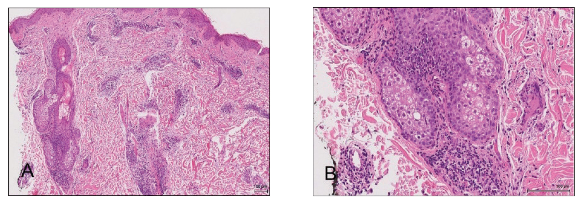
A diagnosis of perifolliculitis from the mesotherapy procedure was on the top of the differential vs a fast-growing mycobacterial or granulomatous reaction. The patient was started on a prednisone taper at 40 mg once daily tapered down completely over 3 weeks in addition to triamcinolone cream 0.1% applied 2 to 4 times daily as needed. Although she did not return to our outpatient clinic for follow-up, she informed us that her rash had improved 1 month after starting the prednisone taper. She was later lost to follow-up. It is unclear if the transaminitis and neutropenia were related to the materials injected during the mesotherapy procedure or from long-standing health issues.
Mesotherapy promises aesthetic benefits through a minimally invasive procedure and therefore is rapidly gaining popularity in aesthetic spas and treatment centers. Due to the lack of regulation in treatment protocols and substances used, there have been numerous reported cases of adverse side effects following mesotherapy, such as pain, allergic reactions, urticaria, panniculitis, ulceration, hair loss, necrosis, paraffinoma, cutaneous tuberculosis, and rapidly growing nontuberculous mycobacterial infections.1-5 More serious side effects also have been reported, such as permanent scarring, deformities, delirium, and massive subcutaneous emphysema (Table).2,4-18
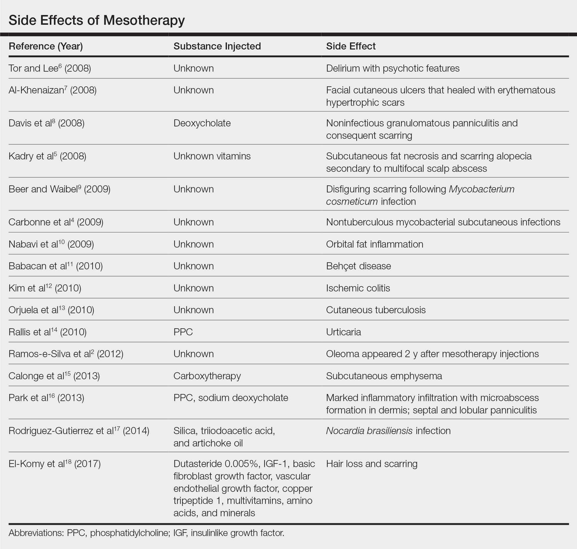
Given the potential complications of mesotherapy documented in the literature, we believe clinical investigations and trials must be performed to appropriately assess the safety and efficacy of this potentially hazardous procedure. Because there currently is insufficient research showing why certain patients are developing these adverse side effects, aesthetic spas and treatment centers should inform patients of all potential side effects associated with mesotherapy for the patient to make an informed decision about the procedure. Mesotherapy should be a point of focus for both the US Food and Drug Administration and researchers to determine its efficacy, safety, and standardization of the procedure.
- Bishara AS, Ibrahim AE, Dibo SA. Cosmetic mesotherapy: between scientific evidence, science fiction, and lucrative business. Aesth Plast Surg. 2008;32:842-849.
- Ramos-e-Silva M, Pereira AL, Ramos-e-Silva S, et al. Oleoma: a rare complication of mesotherapy for cellulite. Int J Dermatol. 2012;51:162-167.
- Rotunda AM, Kolodney MS. Mesotherapy and phosphatidylcholine injections: historical clarification and review. Dermatol Surg. 2006;32:465-480.
- Carbonne A, Brossier F, Arnaud I, et al. Outbreak of nontuberculous mycobacterial subcutaneous infections related to multiple mesotherapy injections. J Clin Microbiol. 2009;47:1961-1964.
- Kadry R, Hamadah I, Al-Issa A, et al. Multifocal scalp abscess with subcutaneous fat necrosis and scarring alopecia as a complication of scalp mesotherapy. J Drugs Dermatol. 2008;7:72-73.
- Tor PC, Lee TS. Delirium with psychotic features possibly associated with mesotherapy. Psychosomatics. 2008;49:273-274.
- Al-Khenaizan S. Facial cutaneous ulcers following mesotherapy. Dermatol Surg. 2008;34:832-834.
- Davis MD, Wright TI, Shehan JM. A complication of mesotherapy: noninfectious granulomatous panniculitis. Arch Dermatol. 2008;144:808-809.
- Beer K, Waibel J. Disfiguring scarring following mesotherapy-associated Mycobacterium cosmeticum infection. J Drugs Dermatol. 2009;8:391-393.
- Nabavi CB, Minckler DS, Tao JP. Histologic features of mesotherapy-induced orbital fat inflammation. Opthalmic Plast Reconstr Surg. 2009;25:69-70.
- Babacan T, Onat AM, Pehlivan Y, et al. A case of Behçet’s disease diagnosed by the panniculitis after mesotherapy. Rheumatol Int. 2010;30:1657-1659.
- Kim JB, Moon W, Park SJ, et al. Ischemic colitis after mesotherapy combined with anti-obesity medications. World J Gastroenterol. 2010;16:1537-1540.
- Orjuela D, Puerto G, Mejia G, et al. Cutaneous tuberculosis after mesotherapy: report of six cases. Biomedica. 2010;30:321-326.
- Rallis E, Kintzoglou S, Moussatou V, et al. Mesotherapy-induced urticaria. Dermatol Surg. 2010;36:1355-1356.
- Calonge WM, Lesbros-Pantoflickova D, Hodina M, et al. Massive subcutaneous emphysema after carbon dioxide mesotherapy. Aesthetic Plast Surg. 2013;37:194-197.
- Park EJ, Kim HS, Kim M, et al. Histological changes after treatment for localized fat deposits with phosphatidylcholine and sodium deoxycholate. J Cosmet Dermatol. 2013;3:240-243.
- Rodriguez-Gutierrez G, Toussaint S, Hernandez-Castro R, et al. Norcardia brasiliensis infection: an emergent suppurative granuloma after mesotherapy. Int J Dermatol. 2014;53:888-890.
- El-Komy M, Hassan A, Tawdy A, et al. Hair loss at injection sites of mesotherapy for alopecia [published online February 3, 2017]. J Cosmet Dermatol. 2017;16:E28-E30.
To the Editor:
Mesotherapy, also known as intradermotherapy, is a cosmetic procedure in which multiple intradermal or subcutaneous injections of homeopathic substances, vitamins, chemicals, and plant extracts are administered.1 First conceived in Europe, mesotherapy is not approved by the US Food and Drug Administration but is gaining popularity in the United States as an alternative cosmetic procedure for various purposes, including lipolysis, body contouring, stretch marks, acne scars, actinic damage, and skin rejuvenation.1,2 We report a case of a healthy woman who developed perifolliculitis, transaminitis, and neutropenia 2 weeks after mesotherapy administration to the face, neck, and chest. We also review other potential side effects of this procedure.
A 36-year-old woman with no notable medical history presented to the emergency department with a worsening pruritic and painful rash on the face, chest, and neck of 2 weeks’ duration. The rash had developed 3 days after the patient received mesotherapy with an unknown substance for cosmetic rejuvenation; the rash was localized only to the injection sites. She did not note any fever, chills, nausea, vomiting, diarrhea, headache, arthralgia, or upper respiratory tract symptoms. She further denied starting any new medications, herbal products, or topical therapies apart from the procedure she had received 2 weeks prior.
The patient was found to be in no acute distress and vital signs were stable. Laboratory testing was remarkable for elevations in alanine aminotransferase (62 U/L [reference range, 10–40 U/L]) and aspartate aminotransferase (72 U/L [reference range 10–30 U/L]). Moreover, she had an absolute neutrophil count of 0.5×10³ cells/µL (reference range 1.8–8.0×10³ cells/µL). An electrolyte panel, creatinine level, and urinalysis were normal. Physical examination revealed numerous 4- to 5-mm erythematous papules in a gridlike distribution across the face, neck, and chest (Figure 1). No pustules or nodules were present. There was no discharge, crust, excoriations, or secondary lesions. Additionally, there was no lymphadenopathy and no mucous membrane or ocular involvement.
A 4-mm punch biopsy from a representative papule on the right lateral aspect of the neck demonstrated a perifollicular and perivascular lymphohistiocytic infiltrate with some focal granulomatous changes. No polarizable foreign body material was found (Figure 2). Bacterial, fungal, mycobacterial, and skin cultures were obtained, and results were all negative after several weeks.
A diagnosis of perifolliculitis from the mesotherapy procedure was on the top of the differential vs a fast-growing mycobacterial or granulomatous reaction. The patient was started on a prednisone taper at 40 mg once daily tapered down completely over 3 weeks in addition to triamcinolone cream 0.1% applied 2 to 4 times daily as needed. Although she did not return to our outpatient clinic for follow-up, she informed us that her rash had improved 1 month after starting the prednisone taper. She was later lost to follow-up. It is unclear if the transaminitis and neutropenia were related to the materials injected during the mesotherapy procedure or from long-standing health issues.
Mesotherapy promises aesthetic benefits through a minimally invasive procedure and therefore is rapidly gaining popularity in aesthetic spas and treatment centers. Due to the lack of regulation in treatment protocols and substances used, there have been numerous reported cases of adverse side effects following mesotherapy, such as pain, allergic reactions, urticaria, panniculitis, ulceration, hair loss, necrosis, paraffinoma, cutaneous tuberculosis, and rapidly growing nontuberculous mycobacterial infections.1-5 More serious side effects also have been reported, such as permanent scarring, deformities, delirium, and massive subcutaneous emphysema (Table).2,4-18
Given the potential complications of mesotherapy documented in the literature, we believe clinical investigations and trials must be performed to appropriately assess the safety and efficacy of this potentially hazardous procedure. Because there currently is insufficient research showing why certain patients are developing these adverse side effects, aesthetic spas and treatment centers should inform patients of all potential side effects associated with mesotherapy for the patient to make an informed decision about the procedure. Mesotherapy should be a point of focus for both the US Food and Drug Administration and researchers to determine its efficacy, safety, and standardization of the procedure.
To the Editor:
Mesotherapy, also known as intradermotherapy, is a cosmetic procedure in which multiple intradermal or subcutaneous injections of homeopathic substances, vitamins, chemicals, and plant extracts are administered.1 First conceived in Europe, mesotherapy is not approved by the US Food and Drug Administration but is gaining popularity in the United States as an alternative cosmetic procedure for various purposes, including lipolysis, body contouring, stretch marks, acne scars, actinic damage, and skin rejuvenation.1,2 We report a case of a healthy woman who developed perifolliculitis, transaminitis, and neutropenia 2 weeks after mesotherapy administration to the face, neck, and chest. We also review other potential side effects of this procedure.
A 36-year-old woman with no notable medical history presented to the emergency department with a worsening pruritic and painful rash on the face, chest, and neck of 2 weeks’ duration. The rash had developed 3 days after the patient received mesotherapy with an unknown substance for cosmetic rejuvenation; the rash was localized only to the injection sites. She did not note any fever, chills, nausea, vomiting, diarrhea, headache, arthralgia, or upper respiratory tract symptoms. She further denied starting any new medications, herbal products, or topical therapies apart from the procedure she had received 2 weeks prior.
The patient was found to be in no acute distress and vital signs were stable. Laboratory testing was remarkable for elevations in alanine aminotransferase (62 U/L [reference range, 10–40 U/L]) and aspartate aminotransferase (72 U/L [reference range 10–30 U/L]). Moreover, she had an absolute neutrophil count of 0.5×10³ cells/µL (reference range 1.8–8.0×10³ cells/µL). An electrolyte panel, creatinine level, and urinalysis were normal. Physical examination revealed numerous 4- to 5-mm erythematous papules in a gridlike distribution across the face, neck, and chest (Figure 1). No pustules or nodules were present. There was no discharge, crust, excoriations, or secondary lesions. Additionally, there was no lymphadenopathy and no mucous membrane or ocular involvement.
A 4-mm punch biopsy from a representative papule on the right lateral aspect of the neck demonstrated a perifollicular and perivascular lymphohistiocytic infiltrate with some focal granulomatous changes. No polarizable foreign body material was found (Figure 2). Bacterial, fungal, mycobacterial, and skin cultures were obtained, and results were all negative after several weeks.
A diagnosis of perifolliculitis from the mesotherapy procedure was on the top of the differential vs a fast-growing mycobacterial or granulomatous reaction. The patient was started on a prednisone taper at 40 mg once daily tapered down completely over 3 weeks in addition to triamcinolone cream 0.1% applied 2 to 4 times daily as needed. Although she did not return to our outpatient clinic for follow-up, she informed us that her rash had improved 1 month after starting the prednisone taper. She was later lost to follow-up. It is unclear if the transaminitis and neutropenia were related to the materials injected during the mesotherapy procedure or from long-standing health issues.
Mesotherapy promises aesthetic benefits through a minimally invasive procedure and therefore is rapidly gaining popularity in aesthetic spas and treatment centers. Due to the lack of regulation in treatment protocols and substances used, there have been numerous reported cases of adverse side effects following mesotherapy, such as pain, allergic reactions, urticaria, panniculitis, ulceration, hair loss, necrosis, paraffinoma, cutaneous tuberculosis, and rapidly growing nontuberculous mycobacterial infections.1-5 More serious side effects also have been reported, such as permanent scarring, deformities, delirium, and massive subcutaneous emphysema (Table).2,4-18
Given the potential complications of mesotherapy documented in the literature, we believe clinical investigations and trials must be performed to appropriately assess the safety and efficacy of this potentially hazardous procedure. Because there currently is insufficient research showing why certain patients are developing these adverse side effects, aesthetic spas and treatment centers should inform patients of all potential side effects associated with mesotherapy for the patient to make an informed decision about the procedure. Mesotherapy should be a point of focus for both the US Food and Drug Administration and researchers to determine its efficacy, safety, and standardization of the procedure.
- Bishara AS, Ibrahim AE, Dibo SA. Cosmetic mesotherapy: between scientific evidence, science fiction, and lucrative business. Aesth Plast Surg. 2008;32:842-849.
- Ramos-e-Silva M, Pereira AL, Ramos-e-Silva S, et al. Oleoma: a rare complication of mesotherapy for cellulite. Int J Dermatol. 2012;51:162-167.
- Rotunda AM, Kolodney MS. Mesotherapy and phosphatidylcholine injections: historical clarification and review. Dermatol Surg. 2006;32:465-480.
- Carbonne A, Brossier F, Arnaud I, et al. Outbreak of nontuberculous mycobacterial subcutaneous infections related to multiple mesotherapy injections. J Clin Microbiol. 2009;47:1961-1964.
- Kadry R, Hamadah I, Al-Issa A, et al. Multifocal scalp abscess with subcutaneous fat necrosis and scarring alopecia as a complication of scalp mesotherapy. J Drugs Dermatol. 2008;7:72-73.
- Tor PC, Lee TS. Delirium with psychotic features possibly associated with mesotherapy. Psychosomatics. 2008;49:273-274.
- Al-Khenaizan S. Facial cutaneous ulcers following mesotherapy. Dermatol Surg. 2008;34:832-834.
- Davis MD, Wright TI, Shehan JM. A complication of mesotherapy: noninfectious granulomatous panniculitis. Arch Dermatol. 2008;144:808-809.
- Beer K, Waibel J. Disfiguring scarring following mesotherapy-associated Mycobacterium cosmeticum infection. J Drugs Dermatol. 2009;8:391-393.
- Nabavi CB, Minckler DS, Tao JP. Histologic features of mesotherapy-induced orbital fat inflammation. Opthalmic Plast Reconstr Surg. 2009;25:69-70.
- Babacan T, Onat AM, Pehlivan Y, et al. A case of Behçet’s disease diagnosed by the panniculitis after mesotherapy. Rheumatol Int. 2010;30:1657-1659.
- Kim JB, Moon W, Park SJ, et al. Ischemic colitis after mesotherapy combined with anti-obesity medications. World J Gastroenterol. 2010;16:1537-1540.
- Orjuela D, Puerto G, Mejia G, et al. Cutaneous tuberculosis after mesotherapy: report of six cases. Biomedica. 2010;30:321-326.
- Rallis E, Kintzoglou S, Moussatou V, et al. Mesotherapy-induced urticaria. Dermatol Surg. 2010;36:1355-1356.
- Calonge WM, Lesbros-Pantoflickova D, Hodina M, et al. Massive subcutaneous emphysema after carbon dioxide mesotherapy. Aesthetic Plast Surg. 2013;37:194-197.
- Park EJ, Kim HS, Kim M, et al. Histological changes after treatment for localized fat deposits with phosphatidylcholine and sodium deoxycholate. J Cosmet Dermatol. 2013;3:240-243.
- Rodriguez-Gutierrez G, Toussaint S, Hernandez-Castro R, et al. Norcardia brasiliensis infection: an emergent suppurative granuloma after mesotherapy. Int J Dermatol. 2014;53:888-890.
- El-Komy M, Hassan A, Tawdy A, et al. Hair loss at injection sites of mesotherapy for alopecia [published online February 3, 2017]. J Cosmet Dermatol. 2017;16:E28-E30.
- Bishara AS, Ibrahim AE, Dibo SA. Cosmetic mesotherapy: between scientific evidence, science fiction, and lucrative business. Aesth Plast Surg. 2008;32:842-849.
- Ramos-e-Silva M, Pereira AL, Ramos-e-Silva S, et al. Oleoma: a rare complication of mesotherapy for cellulite. Int J Dermatol. 2012;51:162-167.
- Rotunda AM, Kolodney MS. Mesotherapy and phosphatidylcholine injections: historical clarification and review. Dermatol Surg. 2006;32:465-480.
- Carbonne A, Brossier F, Arnaud I, et al. Outbreak of nontuberculous mycobacterial subcutaneous infections related to multiple mesotherapy injections. J Clin Microbiol. 2009;47:1961-1964.
- Kadry R, Hamadah I, Al-Issa A, et al. Multifocal scalp abscess with subcutaneous fat necrosis and scarring alopecia as a complication of scalp mesotherapy. J Drugs Dermatol. 2008;7:72-73.
- Tor PC, Lee TS. Delirium with psychotic features possibly associated with mesotherapy. Psychosomatics. 2008;49:273-274.
- Al-Khenaizan S. Facial cutaneous ulcers following mesotherapy. Dermatol Surg. 2008;34:832-834.
- Davis MD, Wright TI, Shehan JM. A complication of mesotherapy: noninfectious granulomatous panniculitis. Arch Dermatol. 2008;144:808-809.
- Beer K, Waibel J. Disfiguring scarring following mesotherapy-associated Mycobacterium cosmeticum infection. J Drugs Dermatol. 2009;8:391-393.
- Nabavi CB, Minckler DS, Tao JP. Histologic features of mesotherapy-induced orbital fat inflammation. Opthalmic Plast Reconstr Surg. 2009;25:69-70.
- Babacan T, Onat AM, Pehlivan Y, et al. A case of Behçet’s disease diagnosed by the panniculitis after mesotherapy. Rheumatol Int. 2010;30:1657-1659.
- Kim JB, Moon W, Park SJ, et al. Ischemic colitis after mesotherapy combined with anti-obesity medications. World J Gastroenterol. 2010;16:1537-1540.
- Orjuela D, Puerto G, Mejia G, et al. Cutaneous tuberculosis after mesotherapy: report of six cases. Biomedica. 2010;30:321-326.
- Rallis E, Kintzoglou S, Moussatou V, et al. Mesotherapy-induced urticaria. Dermatol Surg. 2010;36:1355-1356.
- Calonge WM, Lesbros-Pantoflickova D, Hodina M, et al. Massive subcutaneous emphysema after carbon dioxide mesotherapy. Aesthetic Plast Surg. 2013;37:194-197.
- Park EJ, Kim HS, Kim M, et al. Histological changes after treatment for localized fat deposits with phosphatidylcholine and sodium deoxycholate. J Cosmet Dermatol. 2013;3:240-243.
- Rodriguez-Gutierrez G, Toussaint S, Hernandez-Castro R, et al. Norcardia brasiliensis infection: an emergent suppurative granuloma after mesotherapy. Int J Dermatol. 2014;53:888-890.
- El-Komy M, Hassan A, Tawdy A, et al. Hair loss at injection sites of mesotherapy for alopecia [published online February 3, 2017]. J Cosmet Dermatol. 2017;16:E28-E30.
Practice Points
- Mesotherapy—the delivery of vitamins, chemicals, and plant extracts directly into the dermis via injections—is a common procedure performed in both medical and nonmedical settings for cosmetic rejuvenation.
- Complications can occur from mesotherapy treatment.
- Patients should be advised to seek medical care with US Food and Drug Administration–approved cosmetic techniques and substances only

Human Immunodeficiency Virus Infection in a Hepatitis B Virus–Positive Psoriasis Patient Treated With Ustekinumab
To the Editor:
The incidence of psoriasis in human immunodeficiency virus (HIV)–infected patients is similar to the general population, but it usually becomes more severe as immunosuppression increases. Additionally, it tends to be more resistant to conventional therapies, and the incidence and severity of psoriatic arthropathy is increased. Psoriasis often worsens at the time of HIV primary infection.1 We describe a case of a patient with hepatitis B virus (HBV) whose severe plaque psoriasis was controlled on ustekinumab; he was later diagnosed with HIV infection.
A 42-year-old man with HBV treated with entecavir (HBV DNA viral load, <20 copies/mL [inactive carrier, <2000 copies/mL]) presented to our dermatology unit with severe plaque psoriasis (psoriasis area and severity index 23) that caused notable psychologic difficulties such as anxiety and depression. Treatment was attempted with cyclosporine; acitretin; psoralen plus UVA; infliximab; adalimumab; and eventually ustekinumab (45 mg every 3 months), which controlled the condition well (psoriasis area and severity index 0) in an almost completely sustained manner.
Serologic tests requested at one of his analytical control appointments 2 years after initiating treatment with ustekinumab revealed he was HIV positive. The patient reported unprotected sexual intercourse 4 months prior. He was referred to the infectious disease unit and was classified in subtype A1 of HIV infection (CD4 count, 583 cells/µL [reference range, 500-1200 cells/µL]; viral load, 159,268 copies/mL [rapid progression to AIDS, >100,000 copies/mL]). Treatment was initiated with raltegravir, ritonavir, darunavir, and abacavir; tolerance was suitable. Because of the patient’s history of severe psoriasis, treatment with ustekinumab was maintained. Normally, treatment with this drug would be contraindicated in patients with HIV, as it can lead to viral reactivation. Four years after his HIV diagnosis, neither the patient’s cutaneous nor HIV-associated condition had worsened.
For patients with HIV and mild or moderate psoriasis, topical therapies (eg, corticosteroids, vitamin D analogues, tazarotene) are recommended, similar to patients who are HIV negative. Human immunodeficiency virus–positive patients with severe psoriasis who do not respond to topical treatment should receive phototherapy (UVB or psoralen plus UVA) or acitretin along with their antiretroviral drugs.2 In refractory cases, immunosuppressants, including cyclosporine, methotrexate, or tumor necrosis factor α inhibitors, might be used, though experience with them is limited.3,4 Maintaining antiretroviral therapy and prophylaxis against opportunist disease is important in patients who receive such immunosuppressants, as is close monitoring of the viral load.
Ustekinumab is an IL-12/IL-23 monoclonal antibody indicated for the treatment of moderate to severe plaque psoriasis, active psoriatic arthritis, and inflammatory bowel disease. It is contraindicated in patients with clinically important active infections, such as HBV and hepatitis C virus infections.5 However, it was shown to be safe in a group of 18 patients with HBV who had received antiviral prophylaxis6; a degree of reactivation was observed in similar patients who received no such prophylaxis and in others with hepatitis C virus infection.7 The simultaneous use of methotrexate with ustekinumab in the treatment of psoriatic arthritis does not appear to affect the safety of the latter drug.8 Paparizos et al9 described a patient with HIV controlled with antiretroviral drugs who was treated with ustekinumab for psoriasis; no adverse effects were observed.
We report the case of a patient with HBV and psoriasis who was treated with ustekinumab and later became infected with HIV. His ustekinumab treatment was maintained without subsequent cutaneous or systemic complications.
- Menon K, Van Voorhees V, Bebo B, et al. Psoriasis in patients with HIV infection: from the Medical Board of the National Psoriasis Foundation. J Am Acad Dermatol. 2010;62:291-299.
- Chiricozzi A, Saraceno R, Cannizzaro MV. Complete resolution of erythrodermic psoriasis in an HIV and HCV patient unresponsive to antipsoriatic treatments after highly active antiretroviral therapy. Dermatology. 2012;225:333-337.
- Barco D, Puig L, Alomar A. Treatment of moderate-severe psoriasis with etanercept in patients with chronic human immunodeficiency virus infection. Actas Dermosifiliogr. 2010;101(suppl 1):77-81.
- Lindsey SF, Weiss J, Lee ES, et al. Treatment of severe psoriasis and psoriatic arthritis with adalimumab in an HIV positive patient. J Drugs Dermatol. 2014;13:869-871.
- Rustin MH. Long-term safety of biologics in the treatment of moderate to severe plaque psoriasis: review of the current data. Br J Dermatol. 2012;167(suppl 3):3-11.
- Navarro R, Vilarrasa E, Herranz P, et al. Safety and effectiveness of ustekinumab and antitumour necrosis factor therapy in patients with psoriasis and chronic viral hepatitis B or C: a retrospective, multicentre study in a clinical setting. Br J Dermatol. 2013;168:609-616.
- Chiu HY, Chen CH, Wu MS, et al. The safety profile of ustekinumab in the treatment of patients with psoriasis and concurrent hepatitis B or C. Br J Dermatol. 2013;169:1295-1303.
- Weitz JE, Ritchlin CT. Ustekinumab: targeting the IL-17 pathway to improve outcomes in psoriatic arthritis. Expert Opin Biol Ther. 2014;14:515-526.
- Paparizos V, Rallis E, Kirsten L, et al. Ustekinumab for the treatment of HIV psoriasis. J Dermatol Treat. 2012;23:398-399.
To the Editor:
The incidence of psoriasis in human immunodeficiency virus (HIV)–infected patients is similar to the general population, but it usually becomes more severe as immunosuppression increases. Additionally, it tends to be more resistant to conventional therapies, and the incidence and severity of psoriatic arthropathy is increased. Psoriasis often worsens at the time of HIV primary infection.1 We describe a case of a patient with hepatitis B virus (HBV) whose severe plaque psoriasis was controlled on ustekinumab; he was later diagnosed with HIV infection.
A 42-year-old man with HBV treated with entecavir (HBV DNA viral load, <20 copies/mL [inactive carrier, <2000 copies/mL]) presented to our dermatology unit with severe plaque psoriasis (psoriasis area and severity index 23) that caused notable psychologic difficulties such as anxiety and depression. Treatment was attempted with cyclosporine; acitretin; psoralen plus UVA; infliximab; adalimumab; and eventually ustekinumab (45 mg every 3 months), which controlled the condition well (psoriasis area and severity index 0) in an almost completely sustained manner.
Serologic tests requested at one of his analytical control appointments 2 years after initiating treatment with ustekinumab revealed he was HIV positive. The patient reported unprotected sexual intercourse 4 months prior. He was referred to the infectious disease unit and was classified in subtype A1 of HIV infection (CD4 count, 583 cells/µL [reference range, 500-1200 cells/µL]; viral load, 159,268 copies/mL [rapid progression to AIDS, >100,000 copies/mL]). Treatment was initiated with raltegravir, ritonavir, darunavir, and abacavir; tolerance was suitable. Because of the patient’s history of severe psoriasis, treatment with ustekinumab was maintained. Normally, treatment with this drug would be contraindicated in patients with HIV, as it can lead to viral reactivation. Four years after his HIV diagnosis, neither the patient’s cutaneous nor HIV-associated condition had worsened.
For patients with HIV and mild or moderate psoriasis, topical therapies (eg, corticosteroids, vitamin D analogues, tazarotene) are recommended, similar to patients who are HIV negative. Human immunodeficiency virus–positive patients with severe psoriasis who do not respond to topical treatment should receive phototherapy (UVB or psoralen plus UVA) or acitretin along with their antiretroviral drugs.2 In refractory cases, immunosuppressants, including cyclosporine, methotrexate, or tumor necrosis factor α inhibitors, might be used, though experience with them is limited.3,4 Maintaining antiretroviral therapy and prophylaxis against opportunist disease is important in patients who receive such immunosuppressants, as is close monitoring of the viral load.
Ustekinumab is an IL-12/IL-23 monoclonal antibody indicated for the treatment of moderate to severe plaque psoriasis, active psoriatic arthritis, and inflammatory bowel disease. It is contraindicated in patients with clinically important active infections, such as HBV and hepatitis C virus infections.5 However, it was shown to be safe in a group of 18 patients with HBV who had received antiviral prophylaxis6; a degree of reactivation was observed in similar patients who received no such prophylaxis and in others with hepatitis C virus infection.7 The simultaneous use of methotrexate with ustekinumab in the treatment of psoriatic arthritis does not appear to affect the safety of the latter drug.8 Paparizos et al9 described a patient with HIV controlled with antiretroviral drugs who was treated with ustekinumab for psoriasis; no adverse effects were observed.
We report the case of a patient with HBV and psoriasis who was treated with ustekinumab and later became infected with HIV. His ustekinumab treatment was maintained without subsequent cutaneous or systemic complications.
To the Editor:
The incidence of psoriasis in human immunodeficiency virus (HIV)–infected patients is similar to the general population, but it usually becomes more severe as immunosuppression increases. Additionally, it tends to be more resistant to conventional therapies, and the incidence and severity of psoriatic arthropathy is increased. Psoriasis often worsens at the time of HIV primary infection.1 We describe a case of a patient with hepatitis B virus (HBV) whose severe plaque psoriasis was controlled on ustekinumab; he was later diagnosed with HIV infection.
A 42-year-old man with HBV treated with entecavir (HBV DNA viral load, <20 copies/mL [inactive carrier, <2000 copies/mL]) presented to our dermatology unit with severe plaque psoriasis (psoriasis area and severity index 23) that caused notable psychologic difficulties such as anxiety and depression. Treatment was attempted with cyclosporine; acitretin; psoralen plus UVA; infliximab; adalimumab; and eventually ustekinumab (45 mg every 3 months), which controlled the condition well (psoriasis area and severity index 0) in an almost completely sustained manner.
Serologic tests requested at one of his analytical control appointments 2 years after initiating treatment with ustekinumab revealed he was HIV positive. The patient reported unprotected sexual intercourse 4 months prior. He was referred to the infectious disease unit and was classified in subtype A1 of HIV infection (CD4 count, 583 cells/µL [reference range, 500-1200 cells/µL]; viral load, 159,268 copies/mL [rapid progression to AIDS, >100,000 copies/mL]). Treatment was initiated with raltegravir, ritonavir, darunavir, and abacavir; tolerance was suitable. Because of the patient’s history of severe psoriasis, treatment with ustekinumab was maintained. Normally, treatment with this drug would be contraindicated in patients with HIV, as it can lead to viral reactivation. Four years after his HIV diagnosis, neither the patient’s cutaneous nor HIV-associated condition had worsened.
For patients with HIV and mild or moderate psoriasis, topical therapies (eg, corticosteroids, vitamin D analogues, tazarotene) are recommended, similar to patients who are HIV negative. Human immunodeficiency virus–positive patients with severe psoriasis who do not respond to topical treatment should receive phototherapy (UVB or psoralen plus UVA) or acitretin along with their antiretroviral drugs.2 In refractory cases, immunosuppressants, including cyclosporine, methotrexate, or tumor necrosis factor α inhibitors, might be used, though experience with them is limited.3,4 Maintaining antiretroviral therapy and prophylaxis against opportunist disease is important in patients who receive such immunosuppressants, as is close monitoring of the viral load.
Ustekinumab is an IL-12/IL-23 monoclonal antibody indicated for the treatment of moderate to severe plaque psoriasis, active psoriatic arthritis, and inflammatory bowel disease. It is contraindicated in patients with clinically important active infections, such as HBV and hepatitis C virus infections.5 However, it was shown to be safe in a group of 18 patients with HBV who had received antiviral prophylaxis6; a degree of reactivation was observed in similar patients who received no such prophylaxis and in others with hepatitis C virus infection.7 The simultaneous use of methotrexate with ustekinumab in the treatment of psoriatic arthritis does not appear to affect the safety of the latter drug.8 Paparizos et al9 described a patient with HIV controlled with antiretroviral drugs who was treated with ustekinumab for psoriasis; no adverse effects were observed.
We report the case of a patient with HBV and psoriasis who was treated with ustekinumab and later became infected with HIV. His ustekinumab treatment was maintained without subsequent cutaneous or systemic complications.
- Menon K, Van Voorhees V, Bebo B, et al. Psoriasis in patients with HIV infection: from the Medical Board of the National Psoriasis Foundation. J Am Acad Dermatol. 2010;62:291-299.
- Chiricozzi A, Saraceno R, Cannizzaro MV. Complete resolution of erythrodermic psoriasis in an HIV and HCV patient unresponsive to antipsoriatic treatments after highly active antiretroviral therapy. Dermatology. 2012;225:333-337.
- Barco D, Puig L, Alomar A. Treatment of moderate-severe psoriasis with etanercept in patients with chronic human immunodeficiency virus infection. Actas Dermosifiliogr. 2010;101(suppl 1):77-81.
- Lindsey SF, Weiss J, Lee ES, et al. Treatment of severe psoriasis and psoriatic arthritis with adalimumab in an HIV positive patient. J Drugs Dermatol. 2014;13:869-871.
- Rustin MH. Long-term safety of biologics in the treatment of moderate to severe plaque psoriasis: review of the current data. Br J Dermatol. 2012;167(suppl 3):3-11.
- Navarro R, Vilarrasa E, Herranz P, et al. Safety and effectiveness of ustekinumab and antitumour necrosis factor therapy in patients with psoriasis and chronic viral hepatitis B or C: a retrospective, multicentre study in a clinical setting. Br J Dermatol. 2013;168:609-616.
- Chiu HY, Chen CH, Wu MS, et al. The safety profile of ustekinumab in the treatment of patients with psoriasis and concurrent hepatitis B or C. Br J Dermatol. 2013;169:1295-1303.
- Weitz JE, Ritchlin CT. Ustekinumab: targeting the IL-17 pathway to improve outcomes in psoriatic arthritis. Expert Opin Biol Ther. 2014;14:515-526.
- Paparizos V, Rallis E, Kirsten L, et al. Ustekinumab for the treatment of HIV psoriasis. J Dermatol Treat. 2012;23:398-399.
- Menon K, Van Voorhees V, Bebo B, et al. Psoriasis in patients with HIV infection: from the Medical Board of the National Psoriasis Foundation. J Am Acad Dermatol. 2010;62:291-299.
- Chiricozzi A, Saraceno R, Cannizzaro MV. Complete resolution of erythrodermic psoriasis in an HIV and HCV patient unresponsive to antipsoriatic treatments after highly active antiretroviral therapy. Dermatology. 2012;225:333-337.
- Barco D, Puig L, Alomar A. Treatment of moderate-severe psoriasis with etanercept in patients with chronic human immunodeficiency virus infection. Actas Dermosifiliogr. 2010;101(suppl 1):77-81.
- Lindsey SF, Weiss J, Lee ES, et al. Treatment of severe psoriasis and psoriatic arthritis with adalimumab in an HIV positive patient. J Drugs Dermatol. 2014;13:869-871.
- Rustin MH. Long-term safety of biologics in the treatment of moderate to severe plaque psoriasis: review of the current data. Br J Dermatol. 2012;167(suppl 3):3-11.
- Navarro R, Vilarrasa E, Herranz P, et al. Safety and effectiveness of ustekinumab and antitumour necrosis factor therapy in patients with psoriasis and chronic viral hepatitis B or C: a retrospective, multicentre study in a clinical setting. Br J Dermatol. 2013;168:609-616.
- Chiu HY, Chen CH, Wu MS, et al. The safety profile of ustekinumab in the treatment of patients with psoriasis and concurrent hepatitis B or C. Br J Dermatol. 2013;169:1295-1303.
- Weitz JE, Ritchlin CT. Ustekinumab: targeting the IL-17 pathway to improve outcomes in psoriatic arthritis. Expert Opin Biol Ther. 2014;14:515-526.
- Paparizos V, Rallis E, Kirsten L, et al. Ustekinumab for the treatment of HIV psoriasis. J Dermatol Treat. 2012;23:398-399.
Practice Points
- Psoriasis in patients with human immunodeficiency virus (HIV) tends to be more resistant to conventional therapies.
- Experience is limited in the use of immunosuppressants and biologics to treat psoriasis in HIV patients.
- Maintaining antiretroviral therapy and prophylaxis against opportunist disease is important in HIV patients who receive biologics, as is close monitoring of the viral load.
COVID-19 update: Transmission 5% or less among close contacts
The transmission rate of coronavirus disease 2019 (COVID-19) was 1%-5% among 38,000 Chinese people in close contact with infected patients, according to the chief epidemiologist of the Chinese Centers for Disease Control and Prevention, Beijing, Zunyou Wu, MD, PhD, who gave an update on the epidemic at the Conference on Retroviruses & Opportunistic Infections.
The rate of spread to family members – the driver of the infection in China – was 10% early in the outbreak, but fell to 3% with quicker recognition and isolation. The overall numbers are lower than might have been expected, and an important insight for clinicians trying to contain the outbreak in the United States.
, but their ability to spread the infection dropped after that, Dr. Wu and others said at a special COVID-19 session at the meeting, which was scheduled to be in Boston, but was held online instead because of concerns about spreading the virus. The session has been posted.
Transmission from presymptomatic people is rare. Shedding persists to some degree for 7-12 days in mild/moderate cases, but 2 weeks or more in severe cases.
Dr. Wu said the numbers in China are moving in the right direction, which means that containment efforts there have worked.
The virus emerged in Wuhan, the capital of Hubei province in central China, in connection with a wildlife food market in December 2019. Bats are thought to be the reservoir, with perhaps an intermediate step between civet cats and raccoon dogs. Officials shut down the market.
Essentially, the entire population of China, more than a billion people, was told to stay home for 10 days to interrupt the transmission cycle after the virus spread throughout the country in a few weeks, and almost 60 million people in Hubei were put behind a cordon sanitaire, where they have been for 50 days and will remain “for a while,” Dr. Wu said.
It’s led to a steep drop in new cases and deaths in China since mid-February; both are now more common outside China than inside, and international numbers are lower than they were at the peak in China.
Meanwhile, there’s been no evidence of perinatal transmission; the virus has not been detected in amniotic fluid, cord blood, neonatal throat swabs, or breast milk. Maternal morbidity appears to be similar to uninfected women. “The data around pregnancy are reassuring,” said John Brooks, MD, chief medical officers for HIV/AIDS prevention at the Centers for Disease Control and Prevention, Atlanta, who has been involved with CDC’s containment efforts.
There’s no data yet for immunocompromised people, but for people with HIV, he said, “we think the risk of severe illness would be greater” with lower CD4 counts and unsuppressed viral loads. “People living with HIV should take precautions against this new virus,” including having at least a 30-day supply of HIV medications; keeping up flu and pneumonia vaccinations; and having a care plan if quarantined. Setting up telemedicine might be a good idea.
The usual incubation period for COVID-19 is 4-6 days but can be longer. Recovery time is about 2 weeks in mild cases and 3-6 weeks in more severe cases. People who die do so within 2 months of symptom onset.
The most common symptoms among hospitalized patients in China are fever, dry cough, fatigue, and headache. Truly asymptomatic cases are not common; most go on to develop symptoms. There have been reports of diarrhea before other symptoms by a day or two, but it’s probably a red herring. The virus has been isolated from stool, but there is no evidence of fecal-oral transmission, Dr. Wu said.
Eighty percent of COVID-19 cases are mild or moderate and most patients recover spontaneously, especially middle aged and younger people. There is no meaningful difference in distribution between the sexes.
There are limited pediatric data perhaps due to underreporting, “but we know [children] experience milder illness than adults,” the CDC’s Dr. Brooks said.
He pegged the latest case fatality estimate at 0.5% to 3.5%, which is considerably higher than seasonal flu, but might well drop as more mild cases are detected and added to the denominator, he said.
For now, death rates top 5% in adults over 60 years old and climb further with increasing age, approaching 16% in people 80 years or older. Patients with hypertension, diabetes, cardiovascular disease, and chronic respiratory illness are at increased risk. The ultimate cause of death is acute respiratory distress syndrome, said Ralph Baric, PhD, a coronavirus expert and epidemiology professor at the University of North Carolina, Chapel Hill, who also presented at the meeting.
Several drug and vaccine candidates are under study for the infection. An intriguing possibility is that angiotensin converting enzyme (ACE) inhibitors might help. Hypertension is a known risk factor for severe infection; the virus makes use of ACE receptor pathways to infect airway epithelial cells; and there have been reports of ACE inhibitors having effect against the virus that caused severe acute respiratory syndrome (SARS), another coronavirus outbreak in 2003.
“I think it’s a very good idea to go back and re-explore use of these drugs,” Dr. Baric said.
The presenters didn’t have any relevant disclosures.
The transmission rate of coronavirus disease 2019 (COVID-19) was 1%-5% among 38,000 Chinese people in close contact with infected patients, according to the chief epidemiologist of the Chinese Centers for Disease Control and Prevention, Beijing, Zunyou Wu, MD, PhD, who gave an update on the epidemic at the Conference on Retroviruses & Opportunistic Infections.
The rate of spread to family members – the driver of the infection in China – was 10% early in the outbreak, but fell to 3% with quicker recognition and isolation. The overall numbers are lower than might have been expected, and an important insight for clinicians trying to contain the outbreak in the United States.
, but their ability to spread the infection dropped after that, Dr. Wu and others said at a special COVID-19 session at the meeting, which was scheduled to be in Boston, but was held online instead because of concerns about spreading the virus. The session has been posted.
Transmission from presymptomatic people is rare. Shedding persists to some degree for 7-12 days in mild/moderate cases, but 2 weeks or more in severe cases.
Dr. Wu said the numbers in China are moving in the right direction, which means that containment efforts there have worked.
The virus emerged in Wuhan, the capital of Hubei province in central China, in connection with a wildlife food market in December 2019. Bats are thought to be the reservoir, with perhaps an intermediate step between civet cats and raccoon dogs. Officials shut down the market.
Essentially, the entire population of China, more than a billion people, was told to stay home for 10 days to interrupt the transmission cycle after the virus spread throughout the country in a few weeks, and almost 60 million people in Hubei were put behind a cordon sanitaire, where they have been for 50 days and will remain “for a while,” Dr. Wu said.
It’s led to a steep drop in new cases and deaths in China since mid-February; both are now more common outside China than inside, and international numbers are lower than they were at the peak in China.
Meanwhile, there’s been no evidence of perinatal transmission; the virus has not been detected in amniotic fluid, cord blood, neonatal throat swabs, or breast milk. Maternal morbidity appears to be similar to uninfected women. “The data around pregnancy are reassuring,” said John Brooks, MD, chief medical officers for HIV/AIDS prevention at the Centers for Disease Control and Prevention, Atlanta, who has been involved with CDC’s containment efforts.
There’s no data yet for immunocompromised people, but for people with HIV, he said, “we think the risk of severe illness would be greater” with lower CD4 counts and unsuppressed viral loads. “People living with HIV should take precautions against this new virus,” including having at least a 30-day supply of HIV medications; keeping up flu and pneumonia vaccinations; and having a care plan if quarantined. Setting up telemedicine might be a good idea.
The usual incubation period for COVID-19 is 4-6 days but can be longer. Recovery time is about 2 weeks in mild cases and 3-6 weeks in more severe cases. People who die do so within 2 months of symptom onset.
The most common symptoms among hospitalized patients in China are fever, dry cough, fatigue, and headache. Truly asymptomatic cases are not common; most go on to develop symptoms. There have been reports of diarrhea before other symptoms by a day or two, but it’s probably a red herring. The virus has been isolated from stool, but there is no evidence of fecal-oral transmission, Dr. Wu said.
Eighty percent of COVID-19 cases are mild or moderate and most patients recover spontaneously, especially middle aged and younger people. There is no meaningful difference in distribution between the sexes.
There are limited pediatric data perhaps due to underreporting, “but we know [children] experience milder illness than adults,” the CDC’s Dr. Brooks said.
He pegged the latest case fatality estimate at 0.5% to 3.5%, which is considerably higher than seasonal flu, but might well drop as more mild cases are detected and added to the denominator, he said.
For now, death rates top 5% in adults over 60 years old and climb further with increasing age, approaching 16% in people 80 years or older. Patients with hypertension, diabetes, cardiovascular disease, and chronic respiratory illness are at increased risk. The ultimate cause of death is acute respiratory distress syndrome, said Ralph Baric, PhD, a coronavirus expert and epidemiology professor at the University of North Carolina, Chapel Hill, who also presented at the meeting.
Several drug and vaccine candidates are under study for the infection. An intriguing possibility is that angiotensin converting enzyme (ACE) inhibitors might help. Hypertension is a known risk factor for severe infection; the virus makes use of ACE receptor pathways to infect airway epithelial cells; and there have been reports of ACE inhibitors having effect against the virus that caused severe acute respiratory syndrome (SARS), another coronavirus outbreak in 2003.
“I think it’s a very good idea to go back and re-explore use of these drugs,” Dr. Baric said.
The presenters didn’t have any relevant disclosures.
The transmission rate of coronavirus disease 2019 (COVID-19) was 1%-5% among 38,000 Chinese people in close contact with infected patients, according to the chief epidemiologist of the Chinese Centers for Disease Control and Prevention, Beijing, Zunyou Wu, MD, PhD, who gave an update on the epidemic at the Conference on Retroviruses & Opportunistic Infections.
The rate of spread to family members – the driver of the infection in China – was 10% early in the outbreak, but fell to 3% with quicker recognition and isolation. The overall numbers are lower than might have been expected, and an important insight for clinicians trying to contain the outbreak in the United States.
, but their ability to spread the infection dropped after that, Dr. Wu and others said at a special COVID-19 session at the meeting, which was scheduled to be in Boston, but was held online instead because of concerns about spreading the virus. The session has been posted.
Transmission from presymptomatic people is rare. Shedding persists to some degree for 7-12 days in mild/moderate cases, but 2 weeks or more in severe cases.
Dr. Wu said the numbers in China are moving in the right direction, which means that containment efforts there have worked.
The virus emerged in Wuhan, the capital of Hubei province in central China, in connection with a wildlife food market in December 2019. Bats are thought to be the reservoir, with perhaps an intermediate step between civet cats and raccoon dogs. Officials shut down the market.
Essentially, the entire population of China, more than a billion people, was told to stay home for 10 days to interrupt the transmission cycle after the virus spread throughout the country in a few weeks, and almost 60 million people in Hubei were put behind a cordon sanitaire, where they have been for 50 days and will remain “for a while,” Dr. Wu said.
It’s led to a steep drop in new cases and deaths in China since mid-February; both are now more common outside China than inside, and international numbers are lower than they were at the peak in China.
Meanwhile, there’s been no evidence of perinatal transmission; the virus has not been detected in amniotic fluid, cord blood, neonatal throat swabs, or breast milk. Maternal morbidity appears to be similar to uninfected women. “The data around pregnancy are reassuring,” said John Brooks, MD, chief medical officers for HIV/AIDS prevention at the Centers for Disease Control and Prevention, Atlanta, who has been involved with CDC’s containment efforts.
There’s no data yet for immunocompromised people, but for people with HIV, he said, “we think the risk of severe illness would be greater” with lower CD4 counts and unsuppressed viral loads. “People living with HIV should take precautions against this new virus,” including having at least a 30-day supply of HIV medications; keeping up flu and pneumonia vaccinations; and having a care plan if quarantined. Setting up telemedicine might be a good idea.
The usual incubation period for COVID-19 is 4-6 days but can be longer. Recovery time is about 2 weeks in mild cases and 3-6 weeks in more severe cases. People who die do so within 2 months of symptom onset.
The most common symptoms among hospitalized patients in China are fever, dry cough, fatigue, and headache. Truly asymptomatic cases are not common; most go on to develop symptoms. There have been reports of diarrhea before other symptoms by a day or two, but it’s probably a red herring. The virus has been isolated from stool, but there is no evidence of fecal-oral transmission, Dr. Wu said.
Eighty percent of COVID-19 cases are mild or moderate and most patients recover spontaneously, especially middle aged and younger people. There is no meaningful difference in distribution between the sexes.
There are limited pediatric data perhaps due to underreporting, “but we know [children] experience milder illness than adults,” the CDC’s Dr. Brooks said.
He pegged the latest case fatality estimate at 0.5% to 3.5%, which is considerably higher than seasonal flu, but might well drop as more mild cases are detected and added to the denominator, he said.
For now, death rates top 5% in adults over 60 years old and climb further with increasing age, approaching 16% in people 80 years or older. Patients with hypertension, diabetes, cardiovascular disease, and chronic respiratory illness are at increased risk. The ultimate cause of death is acute respiratory distress syndrome, said Ralph Baric, PhD, a coronavirus expert and epidemiology professor at the University of North Carolina, Chapel Hill, who also presented at the meeting.
Several drug and vaccine candidates are under study for the infection. An intriguing possibility is that angiotensin converting enzyme (ACE) inhibitors might help. Hypertension is a known risk factor for severe infection; the virus makes use of ACE receptor pathways to infect airway epithelial cells; and there have been reports of ACE inhibitors having effect against the virus that caused severe acute respiratory syndrome (SARS), another coronavirus outbreak in 2003.
“I think it’s a very good idea to go back and re-explore use of these drugs,” Dr. Baric said.
The presenters didn’t have any relevant disclosures.
FROM CROI 2020
Serum GFAP may indicate disease severity in NMOSD
WEST PALM BEACH, FLA. – (NMOSD), according to research presented at the meeting held by the Americas Committee for Treatment and Research in Multiple Sclerosis. Treatment with inebilizumab decreases serum GFAP concentrations and reduces the risk of an NMOSD attack, the investigators said.
In NMOSD, autoantibodies often target aquaporin-4, a protein expressed on astrocytes. This immune response destroys astrocytes and increases levels of GFAP in cerebrospinal fluid and serum. In the N-MOmentum trial, inebilizumab reduced the risk of attacks by 73%, compared with placebo, in patients with NMOSD. Bruce Cree, MD, PhD, professor of neurology at the University of California, San Francisco, Weill Institute for Neurosciences and colleagues analyzed the data from this trial to examine the relationship between serum GFAP concentrations and disease activity.
In the study, the investigators prospectively collected 1,260 serum samples from 215 participants, 25 healthy donors, and 23 patients with relapsing-remitting multiple sclerosis (MS). Serum GFAP concentration was measured using a single-molecule array assay. The researchers and an independent adjudication committee evaluated attacks during the 28-week randomized controlled study period using predefined criteria.
The median serum GFAP concentration was higher in patients with NMOSD (128.3 pg/mL) than in patients with relapsing-remitting MS (97.5 pg/mL) and healthy controls (73.3 pg/mL). At baseline, 29% of patients with NMOSD and 9% of patients with MS had a serum GFAP concentration that was three or more standard deviations above the mean level in healthy donors.
During the study, patients with a high baseline concentration of serum GFAP were three times more likely to have an adjudicated NMOSD attack than patients with lower serum GFAP concentrations. Concentrations of serum GFAP increased within 1 week of an adjudicated attack during the randomized controlled period. Furthermore, serum GFAP concentration was significantly higher in patients with major adjudicated attacks, compared with those who had minor adjudicated attacks. This finding was true for all organ domains, including for attacks that affected only the optic nerve.
Compared with placebo, inebilizumab reduced the risk of an adjudicated attack by 79% in patients with a normal baseline serum GFAP concentration and by 61% in patients with an elevated baseline serum GFAP concentration. The increase in serum GFAP concentration following an adjudicated attack was significant in patients receiving placebo, but not in patients receiving inebilizumab. Seven samples taken during an adjudicated attack from patients receiving inebilizumab had serum GFAP concentrations within the normal range. One sample taken during an attack from a patient receiving placebo had a serum GFAP concentration within the normal range. In addition, serum GFAP concentrations decreased from baseline among patients receiving inebilizumab who did not have an adjudicated attack.
“These observations suggest that serum GFAP could be a clinically useful biomarker of disease activity and increased attack risk in NMOSD,” said Dr. Cree and colleagues.
Viela Bio, which is developing inebilizumab, and MedImmune sponsored the study. Dr. Cree has received compensation for consulting services that he provided to Alexion, Atara, Biogen, EMD Serono, Novartis, and TG Therapeutics.
SOURCE: Hartung H et al. ACTRIMS 2020, Abstract P205.
WEST PALM BEACH, FLA. – (NMOSD), according to research presented at the meeting held by the Americas Committee for Treatment and Research in Multiple Sclerosis. Treatment with inebilizumab decreases serum GFAP concentrations and reduces the risk of an NMOSD attack, the investigators said.
In NMOSD, autoantibodies often target aquaporin-4, a protein expressed on astrocytes. This immune response destroys astrocytes and increases levels of GFAP in cerebrospinal fluid and serum. In the N-MOmentum trial, inebilizumab reduced the risk of attacks by 73%, compared with placebo, in patients with NMOSD. Bruce Cree, MD, PhD, professor of neurology at the University of California, San Francisco, Weill Institute for Neurosciences and colleagues analyzed the data from this trial to examine the relationship between serum GFAP concentrations and disease activity.
In the study, the investigators prospectively collected 1,260 serum samples from 215 participants, 25 healthy donors, and 23 patients with relapsing-remitting multiple sclerosis (MS). Serum GFAP concentration was measured using a single-molecule array assay. The researchers and an independent adjudication committee evaluated attacks during the 28-week randomized controlled study period using predefined criteria.
The median serum GFAP concentration was higher in patients with NMOSD (128.3 pg/mL) than in patients with relapsing-remitting MS (97.5 pg/mL) and healthy controls (73.3 pg/mL). At baseline, 29% of patients with NMOSD and 9% of patients with MS had a serum GFAP concentration that was three or more standard deviations above the mean level in healthy donors.
During the study, patients with a high baseline concentration of serum GFAP were three times more likely to have an adjudicated NMOSD attack than patients with lower serum GFAP concentrations. Concentrations of serum GFAP increased within 1 week of an adjudicated attack during the randomized controlled period. Furthermore, serum GFAP concentration was significantly higher in patients with major adjudicated attacks, compared with those who had minor adjudicated attacks. This finding was true for all organ domains, including for attacks that affected only the optic nerve.
Compared with placebo, inebilizumab reduced the risk of an adjudicated attack by 79% in patients with a normal baseline serum GFAP concentration and by 61% in patients with an elevated baseline serum GFAP concentration. The increase in serum GFAP concentration following an adjudicated attack was significant in patients receiving placebo, but not in patients receiving inebilizumab. Seven samples taken during an adjudicated attack from patients receiving inebilizumab had serum GFAP concentrations within the normal range. One sample taken during an attack from a patient receiving placebo had a serum GFAP concentration within the normal range. In addition, serum GFAP concentrations decreased from baseline among patients receiving inebilizumab who did not have an adjudicated attack.
“These observations suggest that serum GFAP could be a clinically useful biomarker of disease activity and increased attack risk in NMOSD,” said Dr. Cree and colleagues.
Viela Bio, which is developing inebilizumab, and MedImmune sponsored the study. Dr. Cree has received compensation for consulting services that he provided to Alexion, Atara, Biogen, EMD Serono, Novartis, and TG Therapeutics.
SOURCE: Hartung H et al. ACTRIMS 2020, Abstract P205.
WEST PALM BEACH, FLA. – (NMOSD), according to research presented at the meeting held by the Americas Committee for Treatment and Research in Multiple Sclerosis. Treatment with inebilizumab decreases serum GFAP concentrations and reduces the risk of an NMOSD attack, the investigators said.
In NMOSD, autoantibodies often target aquaporin-4, a protein expressed on astrocytes. This immune response destroys astrocytes and increases levels of GFAP in cerebrospinal fluid and serum. In the N-MOmentum trial, inebilizumab reduced the risk of attacks by 73%, compared with placebo, in patients with NMOSD. Bruce Cree, MD, PhD, professor of neurology at the University of California, San Francisco, Weill Institute for Neurosciences and colleagues analyzed the data from this trial to examine the relationship between serum GFAP concentrations and disease activity.
In the study, the investigators prospectively collected 1,260 serum samples from 215 participants, 25 healthy donors, and 23 patients with relapsing-remitting multiple sclerosis (MS). Serum GFAP concentration was measured using a single-molecule array assay. The researchers and an independent adjudication committee evaluated attacks during the 28-week randomized controlled study period using predefined criteria.
The median serum GFAP concentration was higher in patients with NMOSD (128.3 pg/mL) than in patients with relapsing-remitting MS (97.5 pg/mL) and healthy controls (73.3 pg/mL). At baseline, 29% of patients with NMOSD and 9% of patients with MS had a serum GFAP concentration that was three or more standard deviations above the mean level in healthy donors.
During the study, patients with a high baseline concentration of serum GFAP were three times more likely to have an adjudicated NMOSD attack than patients with lower serum GFAP concentrations. Concentrations of serum GFAP increased within 1 week of an adjudicated attack during the randomized controlled period. Furthermore, serum GFAP concentration was significantly higher in patients with major adjudicated attacks, compared with those who had minor adjudicated attacks. This finding was true for all organ domains, including for attacks that affected only the optic nerve.
Compared with placebo, inebilizumab reduced the risk of an adjudicated attack by 79% in patients with a normal baseline serum GFAP concentration and by 61% in patients with an elevated baseline serum GFAP concentration. The increase in serum GFAP concentration following an adjudicated attack was significant in patients receiving placebo, but not in patients receiving inebilizumab. Seven samples taken during an adjudicated attack from patients receiving inebilizumab had serum GFAP concentrations within the normal range. One sample taken during an attack from a patient receiving placebo had a serum GFAP concentration within the normal range. In addition, serum GFAP concentrations decreased from baseline among patients receiving inebilizumab who did not have an adjudicated attack.
“These observations suggest that serum GFAP could be a clinically useful biomarker of disease activity and increased attack risk in NMOSD,” said Dr. Cree and colleagues.
Viela Bio, which is developing inebilizumab, and MedImmune sponsored the study. Dr. Cree has received compensation for consulting services that he provided to Alexion, Atara, Biogen, EMD Serono, Novartis, and TG Therapeutics.
SOURCE: Hartung H et al. ACTRIMS 2020, Abstract P205.
REPORTING FROM ACTRIMS FORUM 2020
Younger children can safely visit HSCT recipients (sometimes)
ORLANDO – For visits to hematopoietic stem cell transplant recipients, the kids are alright – assuming that they’re not sniffling, coughing, or running a fever, that is.
Since their blood and marrow transplant (BMT) unit began allowing child visits to patients in 2018, there have been no increases in either health care–associated infections or average length of stay, and patient satisfaction has markedly improved, reported Mylene R. de Vera, RN, and colleagues from the Greenbaum Cancer Center at the University of Maryland Medical Center in Baltimore.
Although her center had previously had a policy restricting child visitors out of concerns about transmission of infections, “we did a literature search and found that child visitor restriction is really neither evidence based nor recommended by any professional organization,” she said in an interview at the annual Transplantation & Cellular Therapy Meetings.
“At the same time, I also informally surveyed 10 hospitals and BMT centers nationally, and found that 70% of them allow child visitation,” she added at the meeting held by the American Society for Blood and Marrow Transplantation and the Center for International Blood and Marrow Transplant Research.
An informal survey by this reporter found wide variations in practice. For example, Massachusetts General Hospital Cancer Center policy allows only children 12 and older to visit, but other hospitals have somewhat less stringent policies.
“Our policy is that for children under 12, they need to A) be screened, and B) need to get approval from the attending stem-cell physician,” said Reggie E. Duerst, MD, director of the Stem Cell Transplant Program at Ann & Robert H. Lurie Children’s Hospital of Chicago.
Dr. Duerst, who was not involved in the study, said in an interview seeking objective commentary that his center doesn’t especially encourage visits from younger children, on the understanding that, as every parent knows, the odds are higher that children under 12 may be carrying a respiratory virus, compared with adolescents.
He added that visits from children might be beneficial for patients who are more critically ill or at high risk for dying, but again, only if the child has been carefully screened.
At Memorial Sloan Kettering Cancer Center in New York, children are allowed to visit transplant recipients strictly on a case-by-case basis, and only after consultation with and approval from transplant unit staff, an MSKCC spokesman said.
In an interview, Ms. de Vera cited a 2016 study in the American Journal of Critical Care by Mini Jacob, RN, and colleagues from Emory University in Atlanta, who reported that, among family members visiting patients in an intensive care unit with a continuous visitation policy, open visitation was ranked as the second-most important need.
Beginning in 2018, Ms. de Vera and colleagues sought to promote a patient-centered policy by implementing guidelines that would allow safe visitation by children to BMT recipients.
The process included the aforementioned literature search, survey of other centers, and a staff survey, which showed that 70% of staff said they believed that allowing child visitation would support the goal of patient-centered care. The survey also showed, however, that 40% of staff thought that healthy children presented a higher risk of infection to immunocompromised patients, and 50% agreed with the statement that having children on the unit would be disruptive to care.
In August of 2019, all BMT unit staff members took part in an in-service training session designed “to align perception with evidence-based practice.” The training included detailed information and procedures for screening would-be child visitors for potential risks prior to allowing them on the unit.
The one-page screening tool asks about current or recent infections; coughing, sneezing, etc.; and other health-related issues, including whether the child is up to date on vaccinations.
The following month, the BMT unit implemented its new child visitation policy, and by the end of 2019 the unit had logged a total of 71 child visits.
As noted, there were no increases in either health care–associated infections or in the average length of stay, In fact, from April to September 2019 the average length of stay was 13 days, compared with 18 days during the 6-month period in 2018, before child visits were allowed.
In addition, a Hospital Consumer Assessment of Healthcare Providers and Systems (HCAHPS) patient-satisfaction survey showed that the scores on “taking preferences into account” increased to 92% during October through December 2018, compared with 33% from July through September of the same year.
Interestingly, patient-reported satisfaction also fell by half from January to March 2019, when child visits were suspended as part of hospital-wide restrictions during the influenza season.
A survey of staff conducted a year after the child visitation policy was implemented showed a relative 37% increase in reported awareness that the visits promote patient-centered care, a 35% drop in the reported belief that healthy child visitors pose higher risk of infection, and a more than 100% relative decline in the perception that child visitors would upset BMT unit routine.
“I just wanted to prove to my coworkers that child visitation really did not affect length of stay or health care–associated infections, and at the same time may have a positive effect on patient satisfaction,” Ms. de Vera said.
The study was internally supported. All persons interviewed reported no conflicts of interest to disclose.
SOURCE: de Vera MR et al. TCT 2020, Abstract 588.
ORLANDO – For visits to hematopoietic stem cell transplant recipients, the kids are alright – assuming that they’re not sniffling, coughing, or running a fever, that is.
Since their blood and marrow transplant (BMT) unit began allowing child visits to patients in 2018, there have been no increases in either health care–associated infections or average length of stay, and patient satisfaction has markedly improved, reported Mylene R. de Vera, RN, and colleagues from the Greenbaum Cancer Center at the University of Maryland Medical Center in Baltimore.
Although her center had previously had a policy restricting child visitors out of concerns about transmission of infections, “we did a literature search and found that child visitor restriction is really neither evidence based nor recommended by any professional organization,” she said in an interview at the annual Transplantation & Cellular Therapy Meetings.
“At the same time, I also informally surveyed 10 hospitals and BMT centers nationally, and found that 70% of them allow child visitation,” she added at the meeting held by the American Society for Blood and Marrow Transplantation and the Center for International Blood and Marrow Transplant Research.
An informal survey by this reporter found wide variations in practice. For example, Massachusetts General Hospital Cancer Center policy allows only children 12 and older to visit, but other hospitals have somewhat less stringent policies.
“Our policy is that for children under 12, they need to A) be screened, and B) need to get approval from the attending stem-cell physician,” said Reggie E. Duerst, MD, director of the Stem Cell Transplant Program at Ann & Robert H. Lurie Children’s Hospital of Chicago.
Dr. Duerst, who was not involved in the study, said in an interview seeking objective commentary that his center doesn’t especially encourage visits from younger children, on the understanding that, as every parent knows, the odds are higher that children under 12 may be carrying a respiratory virus, compared with adolescents.
He added that visits from children might be beneficial for patients who are more critically ill or at high risk for dying, but again, only if the child has been carefully screened.
At Memorial Sloan Kettering Cancer Center in New York, children are allowed to visit transplant recipients strictly on a case-by-case basis, and only after consultation with and approval from transplant unit staff, an MSKCC spokesman said.
In an interview, Ms. de Vera cited a 2016 study in the American Journal of Critical Care by Mini Jacob, RN, and colleagues from Emory University in Atlanta, who reported that, among family members visiting patients in an intensive care unit with a continuous visitation policy, open visitation was ranked as the second-most important need.
Beginning in 2018, Ms. de Vera and colleagues sought to promote a patient-centered policy by implementing guidelines that would allow safe visitation by children to BMT recipients.
The process included the aforementioned literature search, survey of other centers, and a staff survey, which showed that 70% of staff said they believed that allowing child visitation would support the goal of patient-centered care. The survey also showed, however, that 40% of staff thought that healthy children presented a higher risk of infection to immunocompromised patients, and 50% agreed with the statement that having children on the unit would be disruptive to care.
In August of 2019, all BMT unit staff members took part in an in-service training session designed “to align perception with evidence-based practice.” The training included detailed information and procedures for screening would-be child visitors for potential risks prior to allowing them on the unit.
The one-page screening tool asks about current or recent infections; coughing, sneezing, etc.; and other health-related issues, including whether the child is up to date on vaccinations.
The following month, the BMT unit implemented its new child visitation policy, and by the end of 2019 the unit had logged a total of 71 child visits.
As noted, there were no increases in either health care–associated infections or in the average length of stay, In fact, from April to September 2019 the average length of stay was 13 days, compared with 18 days during the 6-month period in 2018, before child visits were allowed.
In addition, a Hospital Consumer Assessment of Healthcare Providers and Systems (HCAHPS) patient-satisfaction survey showed that the scores on “taking preferences into account” increased to 92% during October through December 2018, compared with 33% from July through September of the same year.
Interestingly, patient-reported satisfaction also fell by half from January to March 2019, when child visits were suspended as part of hospital-wide restrictions during the influenza season.
A survey of staff conducted a year after the child visitation policy was implemented showed a relative 37% increase in reported awareness that the visits promote patient-centered care, a 35% drop in the reported belief that healthy child visitors pose higher risk of infection, and a more than 100% relative decline in the perception that child visitors would upset BMT unit routine.
“I just wanted to prove to my coworkers that child visitation really did not affect length of stay or health care–associated infections, and at the same time may have a positive effect on patient satisfaction,” Ms. de Vera said.
The study was internally supported. All persons interviewed reported no conflicts of interest to disclose.
SOURCE: de Vera MR et al. TCT 2020, Abstract 588.
ORLANDO – For visits to hematopoietic stem cell transplant recipients, the kids are alright – assuming that they’re not sniffling, coughing, or running a fever, that is.
Since their blood and marrow transplant (BMT) unit began allowing child visits to patients in 2018, there have been no increases in either health care–associated infections or average length of stay, and patient satisfaction has markedly improved, reported Mylene R. de Vera, RN, and colleagues from the Greenbaum Cancer Center at the University of Maryland Medical Center in Baltimore.
Although her center had previously had a policy restricting child visitors out of concerns about transmission of infections, “we did a literature search and found that child visitor restriction is really neither evidence based nor recommended by any professional organization,” she said in an interview at the annual Transplantation & Cellular Therapy Meetings.
“At the same time, I also informally surveyed 10 hospitals and BMT centers nationally, and found that 70% of them allow child visitation,” she added at the meeting held by the American Society for Blood and Marrow Transplantation and the Center for International Blood and Marrow Transplant Research.
An informal survey by this reporter found wide variations in practice. For example, Massachusetts General Hospital Cancer Center policy allows only children 12 and older to visit, but other hospitals have somewhat less stringent policies.
“Our policy is that for children under 12, they need to A) be screened, and B) need to get approval from the attending stem-cell physician,” said Reggie E. Duerst, MD, director of the Stem Cell Transplant Program at Ann & Robert H. Lurie Children’s Hospital of Chicago.
Dr. Duerst, who was not involved in the study, said in an interview seeking objective commentary that his center doesn’t especially encourage visits from younger children, on the understanding that, as every parent knows, the odds are higher that children under 12 may be carrying a respiratory virus, compared with adolescents.
He added that visits from children might be beneficial for patients who are more critically ill or at high risk for dying, but again, only if the child has been carefully screened.
At Memorial Sloan Kettering Cancer Center in New York, children are allowed to visit transplant recipients strictly on a case-by-case basis, and only after consultation with and approval from transplant unit staff, an MSKCC spokesman said.
In an interview, Ms. de Vera cited a 2016 study in the American Journal of Critical Care by Mini Jacob, RN, and colleagues from Emory University in Atlanta, who reported that, among family members visiting patients in an intensive care unit with a continuous visitation policy, open visitation was ranked as the second-most important need.
Beginning in 2018, Ms. de Vera and colleagues sought to promote a patient-centered policy by implementing guidelines that would allow safe visitation by children to BMT recipients.
The process included the aforementioned literature search, survey of other centers, and a staff survey, which showed that 70% of staff said they believed that allowing child visitation would support the goal of patient-centered care. The survey also showed, however, that 40% of staff thought that healthy children presented a higher risk of infection to immunocompromised patients, and 50% agreed with the statement that having children on the unit would be disruptive to care.
In August of 2019, all BMT unit staff members took part in an in-service training session designed “to align perception with evidence-based practice.” The training included detailed information and procedures for screening would-be child visitors for potential risks prior to allowing them on the unit.
The one-page screening tool asks about current or recent infections; coughing, sneezing, etc.; and other health-related issues, including whether the child is up to date on vaccinations.
The following month, the BMT unit implemented its new child visitation policy, and by the end of 2019 the unit had logged a total of 71 child visits.
As noted, there were no increases in either health care–associated infections or in the average length of stay, In fact, from April to September 2019 the average length of stay was 13 days, compared with 18 days during the 6-month period in 2018, before child visits were allowed.
In addition, a Hospital Consumer Assessment of Healthcare Providers and Systems (HCAHPS) patient-satisfaction survey showed that the scores on “taking preferences into account” increased to 92% during October through December 2018, compared with 33% from July through September of the same year.
Interestingly, patient-reported satisfaction also fell by half from January to March 2019, when child visits were suspended as part of hospital-wide restrictions during the influenza season.
A survey of staff conducted a year after the child visitation policy was implemented showed a relative 37% increase in reported awareness that the visits promote patient-centered care, a 35% drop in the reported belief that healthy child visitors pose higher risk of infection, and a more than 100% relative decline in the perception that child visitors would upset BMT unit routine.
“I just wanted to prove to my coworkers that child visitation really did not affect length of stay or health care–associated infections, and at the same time may have a positive effect on patient satisfaction,” Ms. de Vera said.
The study was internally supported. All persons interviewed reported no conflicts of interest to disclose.
SOURCE: de Vera MR et al. TCT 2020, Abstract 588.
REPORTING FROM TCT 2020
Under the influence
I don’t know how successful you have been at getting your adolescent patients to follow your suggestions, but I would guess that my batting average was in the low 100s. Even when I try stepping off my soapbox to involve the patient in a nonjudgmental dialogue, my successes pale in comparison to my failures.

Just looking at our national statistics for obesity, it’s pretty obvious that we are all doing a pretty rotten job of modifying our patients behaviors. You could point to a few encouraging numbers but they are few and far between. You could claim correctly that by the time a child reaches preschool, the die is already cast, throw up your arms, and not even raise the subject of diet with your overweight teenage patients.
A recent article in the journal Appetite hints at a group of strategies for molding patient behavior that so far have gotten very little attention from physicians (“Do perceived norms of social media users eating habits and preferences predict our own food consumption and BMI?” Appetite. 2020 Jan 18. doi: 10.1016/j.appet.2020.104611). Researchers at the department of psychology at Ashton University in Birmingham, England, surveyed more than 350 college-age students asking them about the dietary preference of their Facebook contacts and their own dietary habits. What the investigators found was that respondents who perceived their peers ate a healthy diet ate a healthier diet. Conversely, if the respondents thought their social media contacts ate junk food, they reported eating more of an unhealthy diet themselves.
In other words, it appears that, through social media, we have the potential to influence the eating habits of our patients’ peers. Before we get too excited, it should be pointed out that this study from England wasn’t of a long enough duration to demonstrate an effect on body mass index. And another study of 176 children recently published in Pediatrics found that while influencer marketing of unhealthy foods increased children’s immediate food intake, the equivalent marketing of healthy foods had no effect (“Social influencer marketing and children’s food intake: A randomized trial.” Pediatrics. 2019 Apr 1. doi: 10.1542/peds.2018-2554).
Not being terribly aware of the whos, whats, and wheres of influencers, I did a little bit of Internet searching at the Influencer Marketing hub and learned that influencers comes in all shapes and sizes, from “nanoinfluencers” who have acknowledged expertise and a very small Internet following numbering as few as a hundred to “megainfluencers” who have more than a million followers and might charge large entities a million dollars for a single post. The influencer’s content could appear as a blog, a YouTube video, a podcast, or simply a social media post.

The field of influencer marketing is new and growing exponentially. This initiative could come in the form of an office dedicated to Influencer Marketing created by the American Academy of Pediatrics. That group could search for megainfluencers who might be funded by the academy. But it also could develop a handbook for individual practitioners and groups to help them identify nano- and micro- (1,000-40,000 followers) influencers in their own practices.
You probably don’t ask your patients about their social media habits other than to caution them about time management. Maybe it’s time to dig a little deeper. You may find that you have a potent influencer hidden in your practice. She or he might just be willing to spread a good word or two for you.
Dr. Wilkoff practiced primary care pediatrics in Brunswick, Maine for nearly 40 years. He has authored several books on behavioral pediatrics, including “How to Say No to Your Toddler.” Email him at pdnews@mdedge.com.
I don’t know how successful you have been at getting your adolescent patients to follow your suggestions, but I would guess that my batting average was in the low 100s. Even when I try stepping off my soapbox to involve the patient in a nonjudgmental dialogue, my successes pale in comparison to my failures.
Just looking at our national statistics for obesity, it’s pretty obvious that we are all doing a pretty rotten job of modifying our patients behaviors. You could point to a few encouraging numbers but they are few and far between. You could claim correctly that by the time a child reaches preschool, the die is already cast, throw up your arms, and not even raise the subject of diet with your overweight teenage patients.
A recent article in the journal Appetite hints at a group of strategies for molding patient behavior that so far have gotten very little attention from physicians (“Do perceived norms of social media users eating habits and preferences predict our own food consumption and BMI?” Appetite. 2020 Jan 18. doi: 10.1016/j.appet.2020.104611). Researchers at the department of psychology at Ashton University in Birmingham, England, surveyed more than 350 college-age students asking them about the dietary preference of their Facebook contacts and their own dietary habits. What the investigators found was that respondents who perceived their peers ate a healthy diet ate a healthier diet. Conversely, if the respondents thought their social media contacts ate junk food, they reported eating more of an unhealthy diet themselves.
In other words, it appears that, through social media, we have the potential to influence the eating habits of our patients’ peers. Before we get too excited, it should be pointed out that this study from England wasn’t of a long enough duration to demonstrate an effect on body mass index. And another study of 176 children recently published in Pediatrics found that while influencer marketing of unhealthy foods increased children’s immediate food intake, the equivalent marketing of healthy foods had no effect (“Social influencer marketing and children’s food intake: A randomized trial.” Pediatrics. 2019 Apr 1. doi: 10.1542/peds.2018-2554).
Not being terribly aware of the whos, whats, and wheres of influencers, I did a little bit of Internet searching at the Influencer Marketing hub and learned that influencers comes in all shapes and sizes, from “nanoinfluencers” who have acknowledged expertise and a very small Internet following numbering as few as a hundred to “megainfluencers” who have more than a million followers and might charge large entities a million dollars for a single post. The influencer’s content could appear as a blog, a YouTube video, a podcast, or simply a social media post.
The field of influencer marketing is new and growing exponentially. This initiative could come in the form of an office dedicated to Influencer Marketing created by the American Academy of Pediatrics. That group could search for megainfluencers who might be funded by the academy. But it also could develop a handbook for individual practitioners and groups to help them identify nano- and micro- (1,000-40,000 followers) influencers in their own practices.
You probably don’t ask your patients about their social media habits other than to caution them about time management. Maybe it’s time to dig a little deeper. You may find that you have a potent influencer hidden in your practice. She or he might just be willing to spread a good word or two for you.
Dr. Wilkoff practiced primary care pediatrics in Brunswick, Maine for nearly 40 years. He has authored several books on behavioral pediatrics, including “How to Say No to Your Toddler.” Email him at pdnews@mdedge.com.
I don’t know how successful you have been at getting your adolescent patients to follow your suggestions, but I would guess that my batting average was in the low 100s. Even when I try stepping off my soapbox to involve the patient in a nonjudgmental dialogue, my successes pale in comparison to my failures.
Just looking at our national statistics for obesity, it’s pretty obvious that we are all doing a pretty rotten job of modifying our patients behaviors. You could point to a few encouraging numbers but they are few and far between. You could claim correctly that by the time a child reaches preschool, the die is already cast, throw up your arms, and not even raise the subject of diet with your overweight teenage patients.
A recent article in the journal Appetite hints at a group of strategies for molding patient behavior that so far have gotten very little attention from physicians (“Do perceived norms of social media users eating habits and preferences predict our own food consumption and BMI?” Appetite. 2020 Jan 18. doi: 10.1016/j.appet.2020.104611). Researchers at the department of psychology at Ashton University in Birmingham, England, surveyed more than 350 college-age students asking them about the dietary preference of their Facebook contacts and their own dietary habits. What the investigators found was that respondents who perceived their peers ate a healthy diet ate a healthier diet. Conversely, if the respondents thought their social media contacts ate junk food, they reported eating more of an unhealthy diet themselves.
In other words, it appears that, through social media, we have the potential to influence the eating habits of our patients’ peers. Before we get too excited, it should be pointed out that this study from England wasn’t of a long enough duration to demonstrate an effect on body mass index. And another study of 176 children recently published in Pediatrics found that while influencer marketing of unhealthy foods increased children’s immediate food intake, the equivalent marketing of healthy foods had no effect (“Social influencer marketing and children’s food intake: A randomized trial.” Pediatrics. 2019 Apr 1. doi: 10.1542/peds.2018-2554).
Not being terribly aware of the whos, whats, and wheres of influencers, I did a little bit of Internet searching at the Influencer Marketing hub and learned that influencers comes in all shapes and sizes, from “nanoinfluencers” who have acknowledged expertise and a very small Internet following numbering as few as a hundred to “megainfluencers” who have more than a million followers and might charge large entities a million dollars for a single post. The influencer’s content could appear as a blog, a YouTube video, a podcast, or simply a social media post.
The field of influencer marketing is new and growing exponentially. This initiative could come in the form of an office dedicated to Influencer Marketing created by the American Academy of Pediatrics. That group could search for megainfluencers who might be funded by the academy. But it also could develop a handbook for individual practitioners and groups to help them identify nano- and micro- (1,000-40,000 followers) influencers in their own practices.
You probably don’t ask your patients about their social media habits other than to caution them about time management. Maybe it’s time to dig a little deeper. You may find that you have a potent influencer hidden in your practice. She or he might just be willing to spread a good word or two for you.
Dr. Wilkoff practiced primary care pediatrics in Brunswick, Maine for nearly 40 years. He has authored several books on behavioral pediatrics, including “How to Say No to Your Toddler.” Email him at pdnews@mdedge.com.
Probably okay to skip SIBO breath test before starting rifaximin in IBS-D
MAUI, HAWAII – , according to IBS expert Lin Chang, MD, a professor and vice chief of the division of digestive diseases at the University of California, Los Angeles.

Rifaximin is an antibiotic that works in the intestines but is not absorbed by the body, and is approved for irritable bowel syndrome with diarrhea (IBS-D). It’s a second-line option for moderate illness when diet, OTC medications, and other easier options don’t do the trick. Endpoints were met by about 10% more patients on rifaximin than placebo in randomized trials, perhaps by resetting gut microbiota.
“It was a small therapeutic gain, but it was statistically significant,” Dr. Chang said at the Gastroenterology Updates, IBD, Liver Disease Conference. In her own practice, she said it sometimes helps even with bloating and abdominal distension, two of the most vexing problems in IBS-D.
However, symptoms come back after 3-6 months, and prices approach $2,000 for the 2-week IBS-D course. Insurance doesn’t always fully cover it, and the Food and Drug Administration has capped treatments at three.
That’s left providers wondering what to do when people ask for rifaximin after seeing it advertised and eager for some way to predict if it will work or not. Some clinicians have turned to a breath test for small-intestinal bacterial overgrowth (SIBO) before prescribing rifaximin because there is evidence that SIBO contributes to IBS-D, but others have not. It’s a contentious issue in IBS medicine, Dr. Chang said.
A recent open-label study funded by rifaximin maker Salix Pharmaceuticals made a case for testing. Among 93 IBS-D patients, 60% with a positive breath test at baseline had reduced pain, diarrhea, and other symptoms after 550 mg three times daily for 2 weeks, versus 26% with a negative test (Am J Gastroenterol. 2019 Dec;114[12]:1886-93).
Even so, when asked after her IBS presentation, Dr. Chang said she doesn’t breath test because it doesn’t make sense. Even with positive results, “you don’t really know if they have SIBO or not. Sometimes healthy controls have a positive test, and some people with IBS who end up responding to rifaximin have a negative breath test. It’s up to you, [but] it doesn’t correlate” with outcomes, she said.
Indeed, a letter in response to the study made a strong case that nine of the subjects should have been counted as having a negative, not positive, breath test, and if they had, the difference in outcome would have disappeared (Am J Gastroenterol. 2020 Mar 2. doi: 10.14309/ajg.0000000000000569).
Dr. Chang has opted for an empiric approach. “I just treat patients who meet the criteria used in the clinical trials,” such as moderate abdominal pain and bloating, she said (for example, N Engl J Med. 2011 Jan 6;364[1]:22-32).
And she doesn’t push if people can’t afford rifaximin. Some turn to other antibiotics, like metronidazole (Flagyl) or ciprofloxacin (Cipro), which are absorbed in the GI tract, but Dr. Chang said symptoms will recur after a few months regardless of the antibiotic. “You don’t want all these young women to be given antibiotics over and over again. Their symptoms are just going to come back, and you are going to have to learn how to treat them anyway,” she said.
Instead, she moves on to other options when patients are at the point where they need prescription pharmaceuticals. One of her top choices is the tricyclic antidepressant amitriptyline. It helps with the pain; the anticholinergic effects counter the diarrhea; and the sedative effects help patients who have a hard time sleeping. Sleep problems make IBS-D symptoms worse, Dr. Chang said.
MAUI, HAWAII – , according to IBS expert Lin Chang, MD, a professor and vice chief of the division of digestive diseases at the University of California, Los Angeles.
Rifaximin is an antibiotic that works in the intestines but is not absorbed by the body, and is approved for irritable bowel syndrome with diarrhea (IBS-D). It’s a second-line option for moderate illness when diet, OTC medications, and other easier options don’t do the trick. Endpoints were met by about 10% more patients on rifaximin than placebo in randomized trials, perhaps by resetting gut microbiota.
“It was a small therapeutic gain, but it was statistically significant,” Dr. Chang said at the Gastroenterology Updates, IBD, Liver Disease Conference. In her own practice, she said it sometimes helps even with bloating and abdominal distension, two of the most vexing problems in IBS-D.
However, symptoms come back after 3-6 months, and prices approach $2,000 for the 2-week IBS-D course. Insurance doesn’t always fully cover it, and the Food and Drug Administration has capped treatments at three.
That’s left providers wondering what to do when people ask for rifaximin after seeing it advertised and eager for some way to predict if it will work or not. Some clinicians have turned to a breath test for small-intestinal bacterial overgrowth (SIBO) before prescribing rifaximin because there is evidence that SIBO contributes to IBS-D, but others have not. It’s a contentious issue in IBS medicine, Dr. Chang said.
A recent open-label study funded by rifaximin maker Salix Pharmaceuticals made a case for testing. Among 93 IBS-D patients, 60% with a positive breath test at baseline had reduced pain, diarrhea, and other symptoms after 550 mg three times daily for 2 weeks, versus 26% with a negative test (Am J Gastroenterol. 2019 Dec;114[12]:1886-93).
Even so, when asked after her IBS presentation, Dr. Chang said she doesn’t breath test because it doesn’t make sense. Even with positive results, “you don’t really know if they have SIBO or not. Sometimes healthy controls have a positive test, and some people with IBS who end up responding to rifaximin have a negative breath test. It’s up to you, [but] it doesn’t correlate” with outcomes, she said.
Indeed, a letter in response to the study made a strong case that nine of the subjects should have been counted as having a negative, not positive, breath test, and if they had, the difference in outcome would have disappeared (Am J Gastroenterol. 2020 Mar 2. doi: 10.14309/ajg.0000000000000569).
Dr. Chang has opted for an empiric approach. “I just treat patients who meet the criteria used in the clinical trials,” such as moderate abdominal pain and bloating, she said (for example, N Engl J Med. 2011 Jan 6;364[1]:22-32).
And she doesn’t push if people can’t afford rifaximin. Some turn to other antibiotics, like metronidazole (Flagyl) or ciprofloxacin (Cipro), which are absorbed in the GI tract, but Dr. Chang said symptoms will recur after a few months regardless of the antibiotic. “You don’t want all these young women to be given antibiotics over and over again. Their symptoms are just going to come back, and you are going to have to learn how to treat them anyway,” she said.
Instead, she moves on to other options when patients are at the point where they need prescription pharmaceuticals. One of her top choices is the tricyclic antidepressant amitriptyline. It helps with the pain; the anticholinergic effects counter the diarrhea; and the sedative effects help patients who have a hard time sleeping. Sleep problems make IBS-D symptoms worse, Dr. Chang said.
MAUI, HAWAII – , according to IBS expert Lin Chang, MD, a professor and vice chief of the division of digestive diseases at the University of California, Los Angeles.
Rifaximin is an antibiotic that works in the intestines but is not absorbed by the body, and is approved for irritable bowel syndrome with diarrhea (IBS-D). It’s a second-line option for moderate illness when diet, OTC medications, and other easier options don’t do the trick. Endpoints were met by about 10% more patients on rifaximin than placebo in randomized trials, perhaps by resetting gut microbiota.
“It was a small therapeutic gain, but it was statistically significant,” Dr. Chang said at the Gastroenterology Updates, IBD, Liver Disease Conference. In her own practice, she said it sometimes helps even with bloating and abdominal distension, two of the most vexing problems in IBS-D.
However, symptoms come back after 3-6 months, and prices approach $2,000 for the 2-week IBS-D course. Insurance doesn’t always fully cover it, and the Food and Drug Administration has capped treatments at three.
That’s left providers wondering what to do when people ask for rifaximin after seeing it advertised and eager for some way to predict if it will work or not. Some clinicians have turned to a breath test for small-intestinal bacterial overgrowth (SIBO) before prescribing rifaximin because there is evidence that SIBO contributes to IBS-D, but others have not. It’s a contentious issue in IBS medicine, Dr. Chang said.
A recent open-label study funded by rifaximin maker Salix Pharmaceuticals made a case for testing. Among 93 IBS-D patients, 60% with a positive breath test at baseline had reduced pain, diarrhea, and other symptoms after 550 mg three times daily for 2 weeks, versus 26% with a negative test (Am J Gastroenterol. 2019 Dec;114[12]:1886-93).
Even so, when asked after her IBS presentation, Dr. Chang said she doesn’t breath test because it doesn’t make sense. Even with positive results, “you don’t really know if they have SIBO or not. Sometimes healthy controls have a positive test, and some people with IBS who end up responding to rifaximin have a negative breath test. It’s up to you, [but] it doesn’t correlate” with outcomes, she said.
Indeed, a letter in response to the study made a strong case that nine of the subjects should have been counted as having a negative, not positive, breath test, and if they had, the difference in outcome would have disappeared (Am J Gastroenterol. 2020 Mar 2. doi: 10.14309/ajg.0000000000000569).
Dr. Chang has opted for an empiric approach. “I just treat patients who meet the criteria used in the clinical trials,” such as moderate abdominal pain and bloating, she said (for example, N Engl J Med. 2011 Jan 6;364[1]:22-32).
And she doesn’t push if people can’t afford rifaximin. Some turn to other antibiotics, like metronidazole (Flagyl) or ciprofloxacin (Cipro), which are absorbed in the GI tract, but Dr. Chang said symptoms will recur after a few months regardless of the antibiotic. “You don’t want all these young women to be given antibiotics over and over again. Their symptoms are just going to come back, and you are going to have to learn how to treat them anyway,” she said.
Instead, she moves on to other options when patients are at the point where they need prescription pharmaceuticals. One of her top choices is the tricyclic antidepressant amitriptyline. It helps with the pain; the anticholinergic effects counter the diarrhea; and the sedative effects help patients who have a hard time sleeping. Sleep problems make IBS-D symptoms worse, Dr. Chang said.
REPORTING FROM GUILD 2020