User login
COVID attacks DNA in heart, unlike flu, study says
, according to a study published in Immunology.
The study looked at the hearts of patients who died from COVID-19, the flu, and other causes. The findings could provide clues about why coronavirus has led to complications such as ongoing heart issues.
“We found a lot of DNA damage that was unique to the COVID-19 patients, which wasn’t present in the flu patients,” Arutha Kulasinghe, one of the lead study authors and a research fellow at the University of Queensland, Brisbane, Australia, told the Brisbane Times.
“So in this study, COVID-19 and flu look very different in the way they affect the heart,” he said.
Dr. Kulasinghe and colleagues analyzed the hearts of seven COVID-19 patients, two flu patients, and six patients who died from other causes. They used transcriptomic profiling, which looks at the DNA landscape of an organ, to investigate heart tissue from the patients.
Because of previous studies about heart problems associated with COVID-19, he and colleagues expected to find extreme inflammation in the heart. Instead, they found that inflammation signals had been suppressed in the heart, and markers for DNA damage and repair were much higher. They’re still unsure of the underlying cause.
“The indications here are that there’s DNA damage here, it’s not inflammation,” Dr. Kulasinghe said. “There’s something else going on that we need to figure out.”
The damage was similar to the way chronic diseases such as diabetes and cancer appear in the heart, he said, with heart tissue showing DNA damage signals.
Dr. Kulasinghe said he hopes other studies can build on the findings to develop risk models to understand which patients may face a higher risk of serious COVID-19 complications. In turn, this could help doctors provide early treatment. For instance, all seven COVID-19 patients had other chronic diseases, such as diabetes, hypertension, and heart disease.
“Ideally in the future, if you have cardiovascular disease, if you’re obese or have other complications, and you’ve got a signature in your blood that indicates you are at risk of severe disease, then we can risk-stratify patients when they are diagnosed,” he said.
The research is a preliminary step, Dr. Kulasinghe said, because of the small sample size. This type of study is often difficult to conduct because researchers have to wait for the availability of organs, as well as request permission from families for postmortem autopsies and biopsies, to be able to look at the effects on dead tissues.
“Our challenge now is to draw a clinical finding from this, which we can’t at this stage,” he added. “But it’s a really fundamental biological difference we’re observing [between COVID-19 and flu], which we need to validate with larger studies.”
A version of this article first appeared on WebMD.com.
, according to a study published in Immunology.
The study looked at the hearts of patients who died from COVID-19, the flu, and other causes. The findings could provide clues about why coronavirus has led to complications such as ongoing heart issues.
“We found a lot of DNA damage that was unique to the COVID-19 patients, which wasn’t present in the flu patients,” Arutha Kulasinghe, one of the lead study authors and a research fellow at the University of Queensland, Brisbane, Australia, told the Brisbane Times.
“So in this study, COVID-19 and flu look very different in the way they affect the heart,” he said.
Dr. Kulasinghe and colleagues analyzed the hearts of seven COVID-19 patients, two flu patients, and six patients who died from other causes. They used transcriptomic profiling, which looks at the DNA landscape of an organ, to investigate heart tissue from the patients.
Because of previous studies about heart problems associated with COVID-19, he and colleagues expected to find extreme inflammation in the heart. Instead, they found that inflammation signals had been suppressed in the heart, and markers for DNA damage and repair were much higher. They’re still unsure of the underlying cause.
“The indications here are that there’s DNA damage here, it’s not inflammation,” Dr. Kulasinghe said. “There’s something else going on that we need to figure out.”
The damage was similar to the way chronic diseases such as diabetes and cancer appear in the heart, he said, with heart tissue showing DNA damage signals.
Dr. Kulasinghe said he hopes other studies can build on the findings to develop risk models to understand which patients may face a higher risk of serious COVID-19 complications. In turn, this could help doctors provide early treatment. For instance, all seven COVID-19 patients had other chronic diseases, such as diabetes, hypertension, and heart disease.
“Ideally in the future, if you have cardiovascular disease, if you’re obese or have other complications, and you’ve got a signature in your blood that indicates you are at risk of severe disease, then we can risk-stratify patients when they are diagnosed,” he said.
The research is a preliminary step, Dr. Kulasinghe said, because of the small sample size. This type of study is often difficult to conduct because researchers have to wait for the availability of organs, as well as request permission from families for postmortem autopsies and biopsies, to be able to look at the effects on dead tissues.
“Our challenge now is to draw a clinical finding from this, which we can’t at this stage,” he added. “But it’s a really fundamental biological difference we’re observing [between COVID-19 and flu], which we need to validate with larger studies.”
A version of this article first appeared on WebMD.com.
, according to a study published in Immunology.
The study looked at the hearts of patients who died from COVID-19, the flu, and other causes. The findings could provide clues about why coronavirus has led to complications such as ongoing heart issues.
“We found a lot of DNA damage that was unique to the COVID-19 patients, which wasn’t present in the flu patients,” Arutha Kulasinghe, one of the lead study authors and a research fellow at the University of Queensland, Brisbane, Australia, told the Brisbane Times.
“So in this study, COVID-19 and flu look very different in the way they affect the heart,” he said.
Dr. Kulasinghe and colleagues analyzed the hearts of seven COVID-19 patients, two flu patients, and six patients who died from other causes. They used transcriptomic profiling, which looks at the DNA landscape of an organ, to investigate heart tissue from the patients.
Because of previous studies about heart problems associated with COVID-19, he and colleagues expected to find extreme inflammation in the heart. Instead, they found that inflammation signals had been suppressed in the heart, and markers for DNA damage and repair were much higher. They’re still unsure of the underlying cause.
“The indications here are that there’s DNA damage here, it’s not inflammation,” Dr. Kulasinghe said. “There’s something else going on that we need to figure out.”
The damage was similar to the way chronic diseases such as diabetes and cancer appear in the heart, he said, with heart tissue showing DNA damage signals.
Dr. Kulasinghe said he hopes other studies can build on the findings to develop risk models to understand which patients may face a higher risk of serious COVID-19 complications. In turn, this could help doctors provide early treatment. For instance, all seven COVID-19 patients had other chronic diseases, such as diabetes, hypertension, and heart disease.
“Ideally in the future, if you have cardiovascular disease, if you’re obese or have other complications, and you’ve got a signature in your blood that indicates you are at risk of severe disease, then we can risk-stratify patients when they are diagnosed,” he said.
The research is a preliminary step, Dr. Kulasinghe said, because of the small sample size. This type of study is often difficult to conduct because researchers have to wait for the availability of organs, as well as request permission from families for postmortem autopsies and biopsies, to be able to look at the effects on dead tissues.
“Our challenge now is to draw a clinical finding from this, which we can’t at this stage,” he added. “But it’s a really fundamental biological difference we’re observing [between COVID-19 and flu], which we need to validate with larger studies.”
A version of this article first appeared on WebMD.com.
FROM IMMUNOLOGY
Ancient DNA discoveries lead to Nobel Prize in medicine and help explain how humans evolved
It all started with a 40,000-year-old bone. That Neanderthal bone contained enough DNA that Dr. Pääbo could start decades of research showing us modern humans, Homo sapiens, are genetically distinct from other now-extinct hominins such as Neanderthals and Denisovans.
The connection to physiology and medicine comes from the genes some modern humans carry from these ancient relatives. For example, Neanderthal genes transferred to Homo sapiens can explain how our immune systems react to different infections. Dr. Pääbo also discovered that a copy of a gene from another extinct relative, the Denisovans, gives modern-day Tibetans a survival advantage for living at high altitudes.
“Svante Pääbo accomplished something seemingly impossible: sequencing the genome of the Neanderthal, an extinct relative of present-day humans. He also made the sensational discovery of a previously unknown hominin, Denisova,” the Nobel Committee stated in a news release announcing the award. “Importantly, [Dr.] Pääbo also found that gene transfer had occurred from these now extinct hominins to Homo sapiens following the migration out of Africa around 70,000 years ago.”
Dr. Pääbo’s scientific discoveries only happened by pairing ancient DNA evidence with modern technology. An initial discovery occurred when Dr. Pääbo worked at the University of Munich (Germany), where he sequenced mitochondrial DNA from the 40,000-year-old Neanderthal bone. The sequencing of the human genome in the 1990s coupled with genomic DNA recovered from ancient bones allowed Dr. Pääbo to compare our DNA with that of Neanderthals and Denisovans. A lot of this work happened at the Max Planck Institute for Evolutionary Anthropology in Leipzig, Germany, which Dr. Pääbo founded in 1999 and where he still works today.
In 2008, a different 40,000-year-old fragment from a finger bone was discovered in a Denisova cave in the southern part of Siberia. Fortunately for Dr. Pääbo and his team, the DNA was well preserved and could be sequenced.
“The results caused a sensation: the DNA sequence was unique when compared to all known sequences from Neanderthals and present-day humans,” the Nobel Committee said.
In a big picture sense, Dr. Pääbo’s research helps answer where modern-day humans come from and how we interacted with our hominin relatives tens of thousands of years ago. His research is so pioneering that it created a new field of science called paleogenomics, the Nobel Committee noted.
“On behalf of The Physiological Society, I am delighted to congratulate Svante Pääbo for being awarded the Nobel Prize. His pioneering research shines a light on the physiology that makes humans unique from their ancestors,” David Paterson, DPhil, president of The Physiological Society, said in a statement.
“This is an important science discovery in evolutionary biology,” Dr. Paterson added. “Ascribing physiological function to highly conserved mitochondrial genes has been important in our understanding in high-altitude acclimatization as populations move and adapt to new environments, and how genetic variants affect us on a day-to-day basis in health and disease.”
A version of this article first appeared on Medscape.com.
It all started with a 40,000-year-old bone. That Neanderthal bone contained enough DNA that Dr. Pääbo could start decades of research showing us modern humans, Homo sapiens, are genetically distinct from other now-extinct hominins such as Neanderthals and Denisovans.
The connection to physiology and medicine comes from the genes some modern humans carry from these ancient relatives. For example, Neanderthal genes transferred to Homo sapiens can explain how our immune systems react to different infections. Dr. Pääbo also discovered that a copy of a gene from another extinct relative, the Denisovans, gives modern-day Tibetans a survival advantage for living at high altitudes.
“Svante Pääbo accomplished something seemingly impossible: sequencing the genome of the Neanderthal, an extinct relative of present-day humans. He also made the sensational discovery of a previously unknown hominin, Denisova,” the Nobel Committee stated in a news release announcing the award. “Importantly, [Dr.] Pääbo also found that gene transfer had occurred from these now extinct hominins to Homo sapiens following the migration out of Africa around 70,000 years ago.”
Dr. Pääbo’s scientific discoveries only happened by pairing ancient DNA evidence with modern technology. An initial discovery occurred when Dr. Pääbo worked at the University of Munich (Germany), where he sequenced mitochondrial DNA from the 40,000-year-old Neanderthal bone. The sequencing of the human genome in the 1990s coupled with genomic DNA recovered from ancient bones allowed Dr. Pääbo to compare our DNA with that of Neanderthals and Denisovans. A lot of this work happened at the Max Planck Institute for Evolutionary Anthropology in Leipzig, Germany, which Dr. Pääbo founded in 1999 and where he still works today.
In 2008, a different 40,000-year-old fragment from a finger bone was discovered in a Denisova cave in the southern part of Siberia. Fortunately for Dr. Pääbo and his team, the DNA was well preserved and could be sequenced.
“The results caused a sensation: the DNA sequence was unique when compared to all known sequences from Neanderthals and present-day humans,” the Nobel Committee said.
In a big picture sense, Dr. Pääbo’s research helps answer where modern-day humans come from and how we interacted with our hominin relatives tens of thousands of years ago. His research is so pioneering that it created a new field of science called paleogenomics, the Nobel Committee noted.
“On behalf of The Physiological Society, I am delighted to congratulate Svante Pääbo for being awarded the Nobel Prize. His pioneering research shines a light on the physiology that makes humans unique from their ancestors,” David Paterson, DPhil, president of The Physiological Society, said in a statement.
“This is an important science discovery in evolutionary biology,” Dr. Paterson added. “Ascribing physiological function to highly conserved mitochondrial genes has been important in our understanding in high-altitude acclimatization as populations move and adapt to new environments, and how genetic variants affect us on a day-to-day basis in health and disease.”
A version of this article first appeared on Medscape.com.
It all started with a 40,000-year-old bone. That Neanderthal bone contained enough DNA that Dr. Pääbo could start decades of research showing us modern humans, Homo sapiens, are genetically distinct from other now-extinct hominins such as Neanderthals and Denisovans.
The connection to physiology and medicine comes from the genes some modern humans carry from these ancient relatives. For example, Neanderthal genes transferred to Homo sapiens can explain how our immune systems react to different infections. Dr. Pääbo also discovered that a copy of a gene from another extinct relative, the Denisovans, gives modern-day Tibetans a survival advantage for living at high altitudes.
“Svante Pääbo accomplished something seemingly impossible: sequencing the genome of the Neanderthal, an extinct relative of present-day humans. He also made the sensational discovery of a previously unknown hominin, Denisova,” the Nobel Committee stated in a news release announcing the award. “Importantly, [Dr.] Pääbo also found that gene transfer had occurred from these now extinct hominins to Homo sapiens following the migration out of Africa around 70,000 years ago.”
Dr. Pääbo’s scientific discoveries only happened by pairing ancient DNA evidence with modern technology. An initial discovery occurred when Dr. Pääbo worked at the University of Munich (Germany), where he sequenced mitochondrial DNA from the 40,000-year-old Neanderthal bone. The sequencing of the human genome in the 1990s coupled with genomic DNA recovered from ancient bones allowed Dr. Pääbo to compare our DNA with that of Neanderthals and Denisovans. A lot of this work happened at the Max Planck Institute for Evolutionary Anthropology in Leipzig, Germany, which Dr. Pääbo founded in 1999 and where he still works today.
In 2008, a different 40,000-year-old fragment from a finger bone was discovered in a Denisova cave in the southern part of Siberia. Fortunately for Dr. Pääbo and his team, the DNA was well preserved and could be sequenced.
“The results caused a sensation: the DNA sequence was unique when compared to all known sequences from Neanderthals and present-day humans,” the Nobel Committee said.
In a big picture sense, Dr. Pääbo’s research helps answer where modern-day humans come from and how we interacted with our hominin relatives tens of thousands of years ago. His research is so pioneering that it created a new field of science called paleogenomics, the Nobel Committee noted.
“On behalf of The Physiological Society, I am delighted to congratulate Svante Pääbo for being awarded the Nobel Prize. His pioneering research shines a light on the physiology that makes humans unique from their ancestors,” David Paterson, DPhil, president of The Physiological Society, said in a statement.
“This is an important science discovery in evolutionary biology,” Dr. Paterson added. “Ascribing physiological function to highly conserved mitochondrial genes has been important in our understanding in high-altitude acclimatization as populations move and adapt to new environments, and how genetic variants affect us on a day-to-day basis in health and disease.”
A version of this article first appeared on Medscape.com.
Physician bias may prevent quality care for patients with disabilities
For Tara Lagu, MD, the realization that the health care system was broken for patients with disabilities came when a woman she had been treating seemed to keep ignoring Dr. Lagu’s request to see a urologist.
When Dr. Lagu asked the patient’s two attentive daughters about the delay, their response surprised her. The women said they couldn’t find a urologist who was willing to see a patient in a wheelchair.
Surprised and a bit doubtful, Dr. Lagu checked around. She found that, indeed, the only way to get her patient in to see the type of physician required was to send her by ambulance.
“It opened my eyes to how hard it is for patients with disabilities to navigate the health care system,” Dr. Lagu said.
Dr. Lagu, director of the Center for Health Services and Outcomes Research at Northwestern University in Chicago, decided to take a closer look at how her colleagues in medicine care for – or not, as the case proved – the roughly one in four American adults, and millions of children, with disabilities.
In a series of three focus groups, Dr. Lagu and colleagues identified a range of obstacles – including some physician attitudes – that prevent people with disabilities from getting adequate care.
For the study, published in Health Affairs, the researchers interviewed 22 physicians in three groups: Nonrural primary care physicians, rural primary care physicians, and specialists in rheumatology, neurology, obstetrics/gynecology, orthopedics, and ophthalmology.
During the interviews, conducted in the fall of 2018, participants were asked about providing care for five specific types of disabilities: mobility, hearing, vision, mental health, and intellectual limitations.
Lack of experience, logistics often cited
Some physicians admitted that limited resources and training left them without the space and necessary knowledge to properly care for patients with disabilities. They felt they lacked the expertise or exposure to care for individuals with disabilities, nor did they have enough time and space to properly accommodate these patients, according to the researchers. Some said they struggled to coordinate care for individuals with disabilities and did not know which types of accessible equipment, such as adjustable tables and chair scales, were needed or how to use them.
Several physicians also noted that they are inadequately reimbursed for the special accommodations – including additional staff, equipment, and time – required to care for these patients. One primary care physician said he hired a sign-language interpreter for a patient but the bill for the services exceeded the amount insurance reimbursed. As a result, he said, he spent $30 of his own money per visit to see the patient.
Because of these limitations, some physicians in the focus groups said they try to turn away patients with disabilities. Both specialists and general practitioners said they had told patients with disabilities that they didn’t feel they could provide the care needed, and suggested they look elsewhere. A few were surprisingly – even upsettingly – honest, Dr. Lagu said, making statements such as: “I am not the doctor for you.”
‘We really need a rewrite’
Previous work has shown that people with disabilities have worse health outcomes, such as undetected cancer, obesity, and cardiovascular disease.
But “the disability itself isn’t what leads to worse outcomes,” said Allison Kessler, MD, section chief of the Renée Crown Center for Spinal Cord Innovation and associate director of the Shirley Ryan AbilityLab in Chicago*. This study does a good job at highlighting “the need for change on multiple levels,” said Dr. Kessler, who was not a member of the study team.
“People with disabilities have all these disparities in access and outcomes. We’ve never understood why. I think the why is complicated,” Dr. Lagu added. “I think this study suggests some of the negative outcomes are due to explicit bias.”
“It’s also clear that the current framework of health care in the United States does not lend to allowing physicians and medical providers the time needed to adequately address patient issues – those with disabilities or just multiple complex problems,” Colin O’Reilly, DO, vice president and chief medical officer at Children’s Specialized Hospital, an acute rehabilitation facility affiliated with RWJBarnabas Health, in New Brunswick, N.J. “We really need a rewrite.”
However, Dr. O’Reilly said, such a small study population with no control group and no mention of physician resources makes it difficult to come to a strong conclusion about physician bias and discriminatory attitudes against individuals with disabilities.
Dr. Lagu agreed, saying this research “is not conclusive in any way.” The excuses doctors use to discharge patients with disabilities, such as “we don’t accept your insurance,” “we aren’t taking new patients,” and “we can’t provide you with the appropriate care,” could be legitimate, the study authors wrote. But the “disparities in care for people with disabilities suggest that there is a pattern of more frequently denying care to them than people without a disability,” they added.
Dr. Kessler said many of her patients have told her they experience barriers to care. Some say finding an office with the necessary equipment is a challenge or that they often don’t feel welcome.
The Americans With Disabilities Act (ADA) is a federal civil rights law that prohibits discrimination against individuals with disabilities in all public and private places that are open to the general public, including medical offices.
“It is difficult to enforce the ADA in medical settings,” the researchers noted. “Explanations physicians gave in this study could, for any single case of denying care, be legitimate.” Knowing whether a particular instance of denial of care represents discrimination related to disability is “nearly impossible,” they wrote.
All the experts agreed that the study adds valuable insight into an ongoing health disparity. And while system and policy changes are required, Dr. Kessler said, individual physicians can take steps to improve the situation.
A physician in an academic setting can look at the curriculum and the medical school and see about increasing exposure to patients with disabilities earlier in training. In a practice, physicians can retrain staff to ask every patient if an accommodation is needed. “Each one of those changes can only help us move our system in the right direction,” Dr. Kessler said.
The study was supported by a grant from the Eunice Kennedy Shriver National Institute of Child Health and Human Development.
*Correction, 10/5/22: This article includes a corrected title for Dr. Allison Kessler.
A version of this article first appeared on Medscape.com.
For Tara Lagu, MD, the realization that the health care system was broken for patients with disabilities came when a woman she had been treating seemed to keep ignoring Dr. Lagu’s request to see a urologist.
When Dr. Lagu asked the patient’s two attentive daughters about the delay, their response surprised her. The women said they couldn’t find a urologist who was willing to see a patient in a wheelchair.
Surprised and a bit doubtful, Dr. Lagu checked around. She found that, indeed, the only way to get her patient in to see the type of physician required was to send her by ambulance.
“It opened my eyes to how hard it is for patients with disabilities to navigate the health care system,” Dr. Lagu said.
Dr. Lagu, director of the Center for Health Services and Outcomes Research at Northwestern University in Chicago, decided to take a closer look at how her colleagues in medicine care for – or not, as the case proved – the roughly one in four American adults, and millions of children, with disabilities.
In a series of three focus groups, Dr. Lagu and colleagues identified a range of obstacles – including some physician attitudes – that prevent people with disabilities from getting adequate care.
For the study, published in Health Affairs, the researchers interviewed 22 physicians in three groups: Nonrural primary care physicians, rural primary care physicians, and specialists in rheumatology, neurology, obstetrics/gynecology, orthopedics, and ophthalmology.
During the interviews, conducted in the fall of 2018, participants were asked about providing care for five specific types of disabilities: mobility, hearing, vision, mental health, and intellectual limitations.
Lack of experience, logistics often cited
Some physicians admitted that limited resources and training left them without the space and necessary knowledge to properly care for patients with disabilities. They felt they lacked the expertise or exposure to care for individuals with disabilities, nor did they have enough time and space to properly accommodate these patients, according to the researchers. Some said they struggled to coordinate care for individuals with disabilities and did not know which types of accessible equipment, such as adjustable tables and chair scales, were needed or how to use them.
Several physicians also noted that they are inadequately reimbursed for the special accommodations – including additional staff, equipment, and time – required to care for these patients. One primary care physician said he hired a sign-language interpreter for a patient but the bill for the services exceeded the amount insurance reimbursed. As a result, he said, he spent $30 of his own money per visit to see the patient.
Because of these limitations, some physicians in the focus groups said they try to turn away patients with disabilities. Both specialists and general practitioners said they had told patients with disabilities that they didn’t feel they could provide the care needed, and suggested they look elsewhere. A few were surprisingly – even upsettingly – honest, Dr. Lagu said, making statements such as: “I am not the doctor for you.”
‘We really need a rewrite’
Previous work has shown that people with disabilities have worse health outcomes, such as undetected cancer, obesity, and cardiovascular disease.
But “the disability itself isn’t what leads to worse outcomes,” said Allison Kessler, MD, section chief of the Renée Crown Center for Spinal Cord Innovation and associate director of the Shirley Ryan AbilityLab in Chicago*. This study does a good job at highlighting “the need for change on multiple levels,” said Dr. Kessler, who was not a member of the study team.
“People with disabilities have all these disparities in access and outcomes. We’ve never understood why. I think the why is complicated,” Dr. Lagu added. “I think this study suggests some of the negative outcomes are due to explicit bias.”
“It’s also clear that the current framework of health care in the United States does not lend to allowing physicians and medical providers the time needed to adequately address patient issues – those with disabilities or just multiple complex problems,” Colin O’Reilly, DO, vice president and chief medical officer at Children’s Specialized Hospital, an acute rehabilitation facility affiliated with RWJBarnabas Health, in New Brunswick, N.J. “We really need a rewrite.”
However, Dr. O’Reilly said, such a small study population with no control group and no mention of physician resources makes it difficult to come to a strong conclusion about physician bias and discriminatory attitudes against individuals with disabilities.
Dr. Lagu agreed, saying this research “is not conclusive in any way.” The excuses doctors use to discharge patients with disabilities, such as “we don’t accept your insurance,” “we aren’t taking new patients,” and “we can’t provide you with the appropriate care,” could be legitimate, the study authors wrote. But the “disparities in care for people with disabilities suggest that there is a pattern of more frequently denying care to them than people without a disability,” they added.
Dr. Kessler said many of her patients have told her they experience barriers to care. Some say finding an office with the necessary equipment is a challenge or that they often don’t feel welcome.
The Americans With Disabilities Act (ADA) is a federal civil rights law that prohibits discrimination against individuals with disabilities in all public and private places that are open to the general public, including medical offices.
“It is difficult to enforce the ADA in medical settings,” the researchers noted. “Explanations physicians gave in this study could, for any single case of denying care, be legitimate.” Knowing whether a particular instance of denial of care represents discrimination related to disability is “nearly impossible,” they wrote.
All the experts agreed that the study adds valuable insight into an ongoing health disparity. And while system and policy changes are required, Dr. Kessler said, individual physicians can take steps to improve the situation.
A physician in an academic setting can look at the curriculum and the medical school and see about increasing exposure to patients with disabilities earlier in training. In a practice, physicians can retrain staff to ask every patient if an accommodation is needed. “Each one of those changes can only help us move our system in the right direction,” Dr. Kessler said.
The study was supported by a grant from the Eunice Kennedy Shriver National Institute of Child Health and Human Development.
*Correction, 10/5/22: This article includes a corrected title for Dr. Allison Kessler.
A version of this article first appeared on Medscape.com.
For Tara Lagu, MD, the realization that the health care system was broken for patients with disabilities came when a woman she had been treating seemed to keep ignoring Dr. Lagu’s request to see a urologist.
When Dr. Lagu asked the patient’s two attentive daughters about the delay, their response surprised her. The women said they couldn’t find a urologist who was willing to see a patient in a wheelchair.
Surprised and a bit doubtful, Dr. Lagu checked around. She found that, indeed, the only way to get her patient in to see the type of physician required was to send her by ambulance.
“It opened my eyes to how hard it is for patients with disabilities to navigate the health care system,” Dr. Lagu said.
Dr. Lagu, director of the Center for Health Services and Outcomes Research at Northwestern University in Chicago, decided to take a closer look at how her colleagues in medicine care for – or not, as the case proved – the roughly one in four American adults, and millions of children, with disabilities.
In a series of three focus groups, Dr. Lagu and colleagues identified a range of obstacles – including some physician attitudes – that prevent people with disabilities from getting adequate care.
For the study, published in Health Affairs, the researchers interviewed 22 physicians in three groups: Nonrural primary care physicians, rural primary care physicians, and specialists in rheumatology, neurology, obstetrics/gynecology, orthopedics, and ophthalmology.
During the interviews, conducted in the fall of 2018, participants were asked about providing care for five specific types of disabilities: mobility, hearing, vision, mental health, and intellectual limitations.
Lack of experience, logistics often cited
Some physicians admitted that limited resources and training left them without the space and necessary knowledge to properly care for patients with disabilities. They felt they lacked the expertise or exposure to care for individuals with disabilities, nor did they have enough time and space to properly accommodate these patients, according to the researchers. Some said they struggled to coordinate care for individuals with disabilities and did not know which types of accessible equipment, such as adjustable tables and chair scales, were needed or how to use them.
Several physicians also noted that they are inadequately reimbursed for the special accommodations – including additional staff, equipment, and time – required to care for these patients. One primary care physician said he hired a sign-language interpreter for a patient but the bill for the services exceeded the amount insurance reimbursed. As a result, he said, he spent $30 of his own money per visit to see the patient.
Because of these limitations, some physicians in the focus groups said they try to turn away patients with disabilities. Both specialists and general practitioners said they had told patients with disabilities that they didn’t feel they could provide the care needed, and suggested they look elsewhere. A few were surprisingly – even upsettingly – honest, Dr. Lagu said, making statements such as: “I am not the doctor for you.”
‘We really need a rewrite’
Previous work has shown that people with disabilities have worse health outcomes, such as undetected cancer, obesity, and cardiovascular disease.
But “the disability itself isn’t what leads to worse outcomes,” said Allison Kessler, MD, section chief of the Renée Crown Center for Spinal Cord Innovation and associate director of the Shirley Ryan AbilityLab in Chicago*. This study does a good job at highlighting “the need for change on multiple levels,” said Dr. Kessler, who was not a member of the study team.
“People with disabilities have all these disparities in access and outcomes. We’ve never understood why. I think the why is complicated,” Dr. Lagu added. “I think this study suggests some of the negative outcomes are due to explicit bias.”
“It’s also clear that the current framework of health care in the United States does not lend to allowing physicians and medical providers the time needed to adequately address patient issues – those with disabilities or just multiple complex problems,” Colin O’Reilly, DO, vice president and chief medical officer at Children’s Specialized Hospital, an acute rehabilitation facility affiliated with RWJBarnabas Health, in New Brunswick, N.J. “We really need a rewrite.”
However, Dr. O’Reilly said, such a small study population with no control group and no mention of physician resources makes it difficult to come to a strong conclusion about physician bias and discriminatory attitudes against individuals with disabilities.
Dr. Lagu agreed, saying this research “is not conclusive in any way.” The excuses doctors use to discharge patients with disabilities, such as “we don’t accept your insurance,” “we aren’t taking new patients,” and “we can’t provide you with the appropriate care,” could be legitimate, the study authors wrote. But the “disparities in care for people with disabilities suggest that there is a pattern of more frequently denying care to them than people without a disability,” they added.
Dr. Kessler said many of her patients have told her they experience barriers to care. Some say finding an office with the necessary equipment is a challenge or that they often don’t feel welcome.
The Americans With Disabilities Act (ADA) is a federal civil rights law that prohibits discrimination against individuals with disabilities in all public and private places that are open to the general public, including medical offices.
“It is difficult to enforce the ADA in medical settings,” the researchers noted. “Explanations physicians gave in this study could, for any single case of denying care, be legitimate.” Knowing whether a particular instance of denial of care represents discrimination related to disability is “nearly impossible,” they wrote.
All the experts agreed that the study adds valuable insight into an ongoing health disparity. And while system and policy changes are required, Dr. Kessler said, individual physicians can take steps to improve the situation.
A physician in an academic setting can look at the curriculum and the medical school and see about increasing exposure to patients with disabilities earlier in training. In a practice, physicians can retrain staff to ask every patient if an accommodation is needed. “Each one of those changes can only help us move our system in the right direction,” Dr. Kessler said.
The study was supported by a grant from the Eunice Kennedy Shriver National Institute of Child Health and Human Development.
*Correction, 10/5/22: This article includes a corrected title for Dr. Allison Kessler.
A version of this article first appeared on Medscape.com.

Turned away from urgent care – and toward a big ER bill
Frankie Cook remembers last year’s car crash only in flashes.
She was driving a friend home from high school on a winding road outside Rome, Ga. She saw standing water from a recent rain. She tried to slow down but lost control of her car on a big curve. “The car flipped about three times,” Frankie said. “We spun around and went off the side of this hill. My car was on its side, and the back end was crushed up into a tree.”
Frankie said the air bags deployed and both passengers were wearing seat belts, so she was left with just a headache when her father, Russell Cook, came to pick her up from the crash site.
Frankie, then a high school junior, worried she might have a concussion that could affect her performance on an upcoming Advanced Placement exam, so she and her father decided to stop by an urgent care center near their house to get her checked out. They didn’t make it past the front desk.
“‘We don’t take third-party insurance,’” Russell said the receptionist at Atrium Health Floyd Urgent Care Rome told him, though he wasn’t sure what she meant. “She told me, like, three times.”
The problem didn’t seem to be that the clinic lacked the medical expertise to evaluate Frankie. Rather, the Cooks seemed to be confronting a reimbursement policy that is often used by urgent care centers to avoid waiting for payments from car insurance settlements.
Russell was told to take Frankie to an emergency room, which by law must see all patients regardless of such issues. The nearest one, at Atrium Health Floyd Medical Center, was about a mile down the road and was owned by the same hospital system as the urgent care center.
There, Russell said, a doctor looked Frankie over “for just a few minutes,” did precautionary CT scans of her head and body, and sent her home with advice to “take some Tylenol” and rest. She did not have a concussion or serious head injury and was able to take her AP exam on time.
Then the bill came.
The patient: Frankie Cook, 18, now a first-year college student from Rome, Ga.
Medical services: A medical evaluation and two CT scans.
Service provider: Atrium Health Floyd, a hospital system with urgent care centers in northwestern Georgia and northeastern Alabama.
Total bill: $17,005 for an emergency room visit; it was later adjusted to $11,805 after a duplicate charge was removed.
What gives: The Cooks hit a hazard in the health care system after Frankie’s car struck that tree: More and more hospital systems own urgent care centers, which have limits on whom they treat – for both financial and medical reasons.
Russell was pretty upset after he received such a large bill, especially when he had tried to make a quick, inexpensive trip to the clinic. He said Frankie’s grandmother was seen at an urgent care center after a car wreck and walked out with a bill for just a few hundred dollars.
“That’s kind of what I was expecting,” he said. “She just really needed to be looked over.”
So why was Frankie turned away from an urgent care center?
Lou Ellen Horwitz, CEO of the Urgent Care Association, said it’s a pretty standard policy for urgent care centers not to treat injuries that result from car crashes, even minor ones. “Generally, as a rule, they do not take care of car accident victims regardless of the extent of their injuries, because it is going to go through that auto insurance claims process before the provider gets paid,” she said.
Ms. Horwitz said urgent care centers – even ones owned by big health systems – often operate on thin margins and can’t wait months and months for an auto insurance company to pay out a claim. She said “unfortunately” people tend to learn about such policies when they show up expecting care.
Fold in the complicated relationship between health and auto insurance companies and you have what Barak D. Richman, a health care policy professor at Duke University’s law school, called “the wildly complex world that we live in.”
“Each product has its own specifications about where to go and what it covers. Each one is incredibly difficult and complex to administer,” he said. “And each one imposes mistakes on the system.”
Atrium Health did not respond to repeated requests for comment on Frankie’s case.
Ms. Horwitz dismissed the idea that a health system might push people in car wrecks from urgent care centers to emergency rooms to make more money off them. Still, auto insurance generally pays more than health insurance for the same services.
Mr. Richman remained skeptical.
“At the risk of sounding a little too cynical, there are always dollar signs when a health care provider sees a patient come through the door,” Mr. Richman said.
Ateev Mehrotra, MD, professor of health care policy at Harvard Medical School, Boston, said it was likely strategic for the urgent care center to be right down the street from the ER. Part of the strategy makes sense medically, he said, “because if a bad thing happens, you want to get them to some place with more skill really quickly.”
But he also said urgent care centers are “one of the most effective ways” for a health system to generate new revenue, creating a pipeline of new patients to visit its hospitals and later see doctors for testing and follow-up.
Dr. Mehrotra said urgent care centers are not bound by the Emergency Medical Treatment and Labor Act, a federal law known as EMTALA that requires hospitals to stabilize patients regardless of their ability to pay.
At the time of Frankie’s visit, both the urgent care center and emergency room were owned by Floyd health system, which operated a handful of hospitals and clinics in northwestern Georgia and northeastern Alabama. Since then, Floyd has merged with Atrium Health – a larger, North Carolina–based company that operates dozens of hospitals across the Southeast.
Frankie got a CT scan of her head and body in the emergency room, tests KHN confirmed she couldn’t have gotten at the urgent care center regardless of whether the test was medically necessary or just part of a protocol for people in car wrecks who complain of a headache.
Resolution: Sixteen months have passed since Frankie Cook’s hospital visit, and Russell has delayed paying any of the bill on advice he got from a family friend who’s an attorney. After insurance covered its share, the Cooks’ portion came to $1,042.
Getting to that number has been a frustrating process, Russell said. He heard about the initial $17,005 bill in a letter from a lawyer representing the hospital – another unnerving wrinkle of Frankie’s care resulting from the car wreck. The Cooks then had to pursue a lengthy appeal process to get a $5,200 duplicate charge removed from the bill.
Anthem Blue Cross Blue Shield, the Cooks’ insurer, paid $4,006 of the claim. It said in a statement that it’s “committed to providing access to high-quality medical care for our members. This matter was reviewed in accordance with our clinical guidelines, and the billed claims were processed accordingly.”
“It’s not going to put us out on the street,” Russell said of the $1,042 balance, “but we’ve got expenses like everybody else.”
He added, “I would have loved a $200 urgent care visit, but that ship has sailed.”
The takeaway: It’s important to remember that urgent care centers aren’t governed by the same laws as emergency rooms and that they can be more selective about whom they treat. Sometimes their reasons are financial, not clinical.
It’s not uncommon for urgent care centers – even ones in large health systems – to turn away people who have been in car wrecks because of the complications that car insurance settlements create.
Although urgent care visits are less expensive than going to an emergency room, the clinics often can’t offer the same level of care. And you might have to pay the cost of an urgent care visit just to find out you need follow-up care in the emergency room. Then you could be stuck with two bills.
KHN (Kaiser Health News) is a national newsroom that produces in-depth journalism about health issues. Together with Policy Analysis and Polling, KHN is one of the three major operating programs at KFF (Kaiser Family Foundation). KFF is an endowed nonprofit organization providing information on health issues to the nation.
Frankie Cook remembers last year’s car crash only in flashes.
She was driving a friend home from high school on a winding road outside Rome, Ga. She saw standing water from a recent rain. She tried to slow down but lost control of her car on a big curve. “The car flipped about three times,” Frankie said. “We spun around and went off the side of this hill. My car was on its side, and the back end was crushed up into a tree.”
Frankie said the air bags deployed and both passengers were wearing seat belts, so she was left with just a headache when her father, Russell Cook, came to pick her up from the crash site.
Frankie, then a high school junior, worried she might have a concussion that could affect her performance on an upcoming Advanced Placement exam, so she and her father decided to stop by an urgent care center near their house to get her checked out. They didn’t make it past the front desk.
“‘We don’t take third-party insurance,’” Russell said the receptionist at Atrium Health Floyd Urgent Care Rome told him, though he wasn’t sure what she meant. “She told me, like, three times.”
The problem didn’t seem to be that the clinic lacked the medical expertise to evaluate Frankie. Rather, the Cooks seemed to be confronting a reimbursement policy that is often used by urgent care centers to avoid waiting for payments from car insurance settlements.
Russell was told to take Frankie to an emergency room, which by law must see all patients regardless of such issues. The nearest one, at Atrium Health Floyd Medical Center, was about a mile down the road and was owned by the same hospital system as the urgent care center.
There, Russell said, a doctor looked Frankie over “for just a few minutes,” did precautionary CT scans of her head and body, and sent her home with advice to “take some Tylenol” and rest. She did not have a concussion or serious head injury and was able to take her AP exam on time.
Then the bill came.
The patient: Frankie Cook, 18, now a first-year college student from Rome, Ga.
Medical services: A medical evaluation and two CT scans.
Service provider: Atrium Health Floyd, a hospital system with urgent care centers in northwestern Georgia and northeastern Alabama.
Total bill: $17,005 for an emergency room visit; it was later adjusted to $11,805 after a duplicate charge was removed.
What gives: The Cooks hit a hazard in the health care system after Frankie’s car struck that tree: More and more hospital systems own urgent care centers, which have limits on whom they treat – for both financial and medical reasons.
Russell was pretty upset after he received such a large bill, especially when he had tried to make a quick, inexpensive trip to the clinic. He said Frankie’s grandmother was seen at an urgent care center after a car wreck and walked out with a bill for just a few hundred dollars.
“That’s kind of what I was expecting,” he said. “She just really needed to be looked over.”
So why was Frankie turned away from an urgent care center?
Lou Ellen Horwitz, CEO of the Urgent Care Association, said it’s a pretty standard policy for urgent care centers not to treat injuries that result from car crashes, even minor ones. “Generally, as a rule, they do not take care of car accident victims regardless of the extent of their injuries, because it is going to go through that auto insurance claims process before the provider gets paid,” she said.
Ms. Horwitz said urgent care centers – even ones owned by big health systems – often operate on thin margins and can’t wait months and months for an auto insurance company to pay out a claim. She said “unfortunately” people tend to learn about such policies when they show up expecting care.
Fold in the complicated relationship between health and auto insurance companies and you have what Barak D. Richman, a health care policy professor at Duke University’s law school, called “the wildly complex world that we live in.”
“Each product has its own specifications about where to go and what it covers. Each one is incredibly difficult and complex to administer,” he said. “And each one imposes mistakes on the system.”
Atrium Health did not respond to repeated requests for comment on Frankie’s case.
Ms. Horwitz dismissed the idea that a health system might push people in car wrecks from urgent care centers to emergency rooms to make more money off them. Still, auto insurance generally pays more than health insurance for the same services.
Mr. Richman remained skeptical.
“At the risk of sounding a little too cynical, there are always dollar signs when a health care provider sees a patient come through the door,” Mr. Richman said.
Ateev Mehrotra, MD, professor of health care policy at Harvard Medical School, Boston, said it was likely strategic for the urgent care center to be right down the street from the ER. Part of the strategy makes sense medically, he said, “because if a bad thing happens, you want to get them to some place with more skill really quickly.”
But he also said urgent care centers are “one of the most effective ways” for a health system to generate new revenue, creating a pipeline of new patients to visit its hospitals and later see doctors for testing and follow-up.
Dr. Mehrotra said urgent care centers are not bound by the Emergency Medical Treatment and Labor Act, a federal law known as EMTALA that requires hospitals to stabilize patients regardless of their ability to pay.
At the time of Frankie’s visit, both the urgent care center and emergency room were owned by Floyd health system, which operated a handful of hospitals and clinics in northwestern Georgia and northeastern Alabama. Since then, Floyd has merged with Atrium Health – a larger, North Carolina–based company that operates dozens of hospitals across the Southeast.
Frankie got a CT scan of her head and body in the emergency room, tests KHN confirmed she couldn’t have gotten at the urgent care center regardless of whether the test was medically necessary or just part of a protocol for people in car wrecks who complain of a headache.
Resolution: Sixteen months have passed since Frankie Cook’s hospital visit, and Russell has delayed paying any of the bill on advice he got from a family friend who’s an attorney. After insurance covered its share, the Cooks’ portion came to $1,042.
Getting to that number has been a frustrating process, Russell said. He heard about the initial $17,005 bill in a letter from a lawyer representing the hospital – another unnerving wrinkle of Frankie’s care resulting from the car wreck. The Cooks then had to pursue a lengthy appeal process to get a $5,200 duplicate charge removed from the bill.
Anthem Blue Cross Blue Shield, the Cooks’ insurer, paid $4,006 of the claim. It said in a statement that it’s “committed to providing access to high-quality medical care for our members. This matter was reviewed in accordance with our clinical guidelines, and the billed claims were processed accordingly.”
“It’s not going to put us out on the street,” Russell said of the $1,042 balance, “but we’ve got expenses like everybody else.”
He added, “I would have loved a $200 urgent care visit, but that ship has sailed.”
The takeaway: It’s important to remember that urgent care centers aren’t governed by the same laws as emergency rooms and that they can be more selective about whom they treat. Sometimes their reasons are financial, not clinical.
It’s not uncommon for urgent care centers – even ones in large health systems – to turn away people who have been in car wrecks because of the complications that car insurance settlements create.
Although urgent care visits are less expensive than going to an emergency room, the clinics often can’t offer the same level of care. And you might have to pay the cost of an urgent care visit just to find out you need follow-up care in the emergency room. Then you could be stuck with two bills.
KHN (Kaiser Health News) is a national newsroom that produces in-depth journalism about health issues. Together with Policy Analysis and Polling, KHN is one of the three major operating programs at KFF (Kaiser Family Foundation). KFF is an endowed nonprofit organization providing information on health issues to the nation.
Frankie Cook remembers last year’s car crash only in flashes.
She was driving a friend home from high school on a winding road outside Rome, Ga. She saw standing water from a recent rain. She tried to slow down but lost control of her car on a big curve. “The car flipped about three times,” Frankie said. “We spun around and went off the side of this hill. My car was on its side, and the back end was crushed up into a tree.”
Frankie said the air bags deployed and both passengers were wearing seat belts, so she was left with just a headache when her father, Russell Cook, came to pick her up from the crash site.
Frankie, then a high school junior, worried she might have a concussion that could affect her performance on an upcoming Advanced Placement exam, so she and her father decided to stop by an urgent care center near their house to get her checked out. They didn’t make it past the front desk.
“‘We don’t take third-party insurance,’” Russell said the receptionist at Atrium Health Floyd Urgent Care Rome told him, though he wasn’t sure what she meant. “She told me, like, three times.”
The problem didn’t seem to be that the clinic lacked the medical expertise to evaluate Frankie. Rather, the Cooks seemed to be confronting a reimbursement policy that is often used by urgent care centers to avoid waiting for payments from car insurance settlements.
Russell was told to take Frankie to an emergency room, which by law must see all patients regardless of such issues. The nearest one, at Atrium Health Floyd Medical Center, was about a mile down the road and was owned by the same hospital system as the urgent care center.
There, Russell said, a doctor looked Frankie over “for just a few minutes,” did precautionary CT scans of her head and body, and sent her home with advice to “take some Tylenol” and rest. She did not have a concussion or serious head injury and was able to take her AP exam on time.
Then the bill came.
The patient: Frankie Cook, 18, now a first-year college student from Rome, Ga.
Medical services: A medical evaluation and two CT scans.
Service provider: Atrium Health Floyd, a hospital system with urgent care centers in northwestern Georgia and northeastern Alabama.
Total bill: $17,005 for an emergency room visit; it was later adjusted to $11,805 after a duplicate charge was removed.
What gives: The Cooks hit a hazard in the health care system after Frankie’s car struck that tree: More and more hospital systems own urgent care centers, which have limits on whom they treat – for both financial and medical reasons.
Russell was pretty upset after he received such a large bill, especially when he had tried to make a quick, inexpensive trip to the clinic. He said Frankie’s grandmother was seen at an urgent care center after a car wreck and walked out with a bill for just a few hundred dollars.
“That’s kind of what I was expecting,” he said. “She just really needed to be looked over.”
So why was Frankie turned away from an urgent care center?
Lou Ellen Horwitz, CEO of the Urgent Care Association, said it’s a pretty standard policy for urgent care centers not to treat injuries that result from car crashes, even minor ones. “Generally, as a rule, they do not take care of car accident victims regardless of the extent of their injuries, because it is going to go through that auto insurance claims process before the provider gets paid,” she said.
Ms. Horwitz said urgent care centers – even ones owned by big health systems – often operate on thin margins and can’t wait months and months for an auto insurance company to pay out a claim. She said “unfortunately” people tend to learn about such policies when they show up expecting care.
Fold in the complicated relationship between health and auto insurance companies and you have what Barak D. Richman, a health care policy professor at Duke University’s law school, called “the wildly complex world that we live in.”
“Each product has its own specifications about where to go and what it covers. Each one is incredibly difficult and complex to administer,” he said. “And each one imposes mistakes on the system.”
Atrium Health did not respond to repeated requests for comment on Frankie’s case.
Ms. Horwitz dismissed the idea that a health system might push people in car wrecks from urgent care centers to emergency rooms to make more money off them. Still, auto insurance generally pays more than health insurance for the same services.
Mr. Richman remained skeptical.
“At the risk of sounding a little too cynical, there are always dollar signs when a health care provider sees a patient come through the door,” Mr. Richman said.
Ateev Mehrotra, MD, professor of health care policy at Harvard Medical School, Boston, said it was likely strategic for the urgent care center to be right down the street from the ER. Part of the strategy makes sense medically, he said, “because if a bad thing happens, you want to get them to some place with more skill really quickly.”
But he also said urgent care centers are “one of the most effective ways” for a health system to generate new revenue, creating a pipeline of new patients to visit its hospitals and later see doctors for testing and follow-up.
Dr. Mehrotra said urgent care centers are not bound by the Emergency Medical Treatment and Labor Act, a federal law known as EMTALA that requires hospitals to stabilize patients regardless of their ability to pay.
At the time of Frankie’s visit, both the urgent care center and emergency room were owned by Floyd health system, which operated a handful of hospitals and clinics in northwestern Georgia and northeastern Alabama. Since then, Floyd has merged with Atrium Health – a larger, North Carolina–based company that operates dozens of hospitals across the Southeast.
Frankie got a CT scan of her head and body in the emergency room, tests KHN confirmed she couldn’t have gotten at the urgent care center regardless of whether the test was medically necessary or just part of a protocol for people in car wrecks who complain of a headache.
Resolution: Sixteen months have passed since Frankie Cook’s hospital visit, and Russell has delayed paying any of the bill on advice he got from a family friend who’s an attorney. After insurance covered its share, the Cooks’ portion came to $1,042.
Getting to that number has been a frustrating process, Russell said. He heard about the initial $17,005 bill in a letter from a lawyer representing the hospital – another unnerving wrinkle of Frankie’s care resulting from the car wreck. The Cooks then had to pursue a lengthy appeal process to get a $5,200 duplicate charge removed from the bill.
Anthem Blue Cross Blue Shield, the Cooks’ insurer, paid $4,006 of the claim. It said in a statement that it’s “committed to providing access to high-quality medical care for our members. This matter was reviewed in accordance with our clinical guidelines, and the billed claims were processed accordingly.”
“It’s not going to put us out on the street,” Russell said of the $1,042 balance, “but we’ve got expenses like everybody else.”
He added, “I would have loved a $200 urgent care visit, but that ship has sailed.”
The takeaway: It’s important to remember that urgent care centers aren’t governed by the same laws as emergency rooms and that they can be more selective about whom they treat. Sometimes their reasons are financial, not clinical.
It’s not uncommon for urgent care centers – even ones in large health systems – to turn away people who have been in car wrecks because of the complications that car insurance settlements create.
Although urgent care visits are less expensive than going to an emergency room, the clinics often can’t offer the same level of care. And you might have to pay the cost of an urgent care visit just to find out you need follow-up care in the emergency room. Then you could be stuck with two bills.
KHN (Kaiser Health News) is a national newsroom that produces in-depth journalism about health issues. Together with Policy Analysis and Polling, KHN is one of the three major operating programs at KFF (Kaiser Family Foundation). KFF is an endowed nonprofit organization providing information on health issues to the nation.
The hunt for N-acetylcysteine: Medicine or dietary supplement?
Medicine or dietary supplement? N-acetylcysteine (NAC) is marketed as both and in 2021, the supplement abruptly became difficult to find, causing distress to people who had been using it for a variety of conditions. The story behind its disappearance is one of a cat-and-mouse chase between manufacturers, advocacy agencies, and the Food and Drug Administration.
NAC is a medication that was approved by the FDA in 1963. It has two FDA-approved uses: To prevent hepatotoxicity after overdose with acetaminophen, administered intravenously or by mouth, and as a mucolytic agent – previously available as Mucomyst, now available only as a generic – given by inhaler or nebulizer for pulmonary illnesses. Since the 1990s, NAC has been labeled by manufacturers as a dietary supplement. It is a derivative of the amino acid L-cysteine and a precursor to glutathione, an antioxidant.

Studies have had small sample sizes, and the supplement has not been considered an “off label” use of an already available medication. People have purchased NAC from retail drug and big box stores, Amazon, and online companies. So what’s the problem?
In July 2020, the FDA issued a warning letter to Purple Biosciences LLC, because of claims on the company’s website that the product “Purple Tree,” sold by Amazon, could cure hangovers that result from alcohol intoxication. The letter discussed justification for why a hangover is a disease, and goes on to note:
Based on the product label on your website, it appears that you intend to market your Purple Tree® product, which contains N-acetyl-L-cysteine (NAC), as a dietary supplement. However, even if your product labeling did not have therapeutic claims that make your product an unapproved new drug, your product could not be a dietary supplement, because it does not meet the definition of dietary supplement ... products containing that article are outside the definition of a dietary supplement, unless before such approval that article was marketed as a dietary supplement or as a food. NAC was approved as a new drug under section 505 of the Act [21 U.S.C. § 355] on September 14, 1963.
The issue here is that because NAC was first approved as a drug in 1963, it cannot be marketed as a supplement. If it had been marketed as a supplement before it was approved as a drug by the FDA, then it could remain on the market. The fact that it had been sold as a supplement since the 1990s and could be classified as an “old dietary ingredient” according to the Dietary Supplement Health and Education Act of 1994 did not seem to matter. Following the warning letter, NAC was pulled from shelves and websites.
Citizen petitions allow people and organizations to request that the FDA change their policy. During summer 2021, there were two citizen petitions – one from the Council for Responsible Nutrition (CRN) and another from the Natural Products Association (NPA) – asking that the FDA look at policy around NAC. In response, in November 2021, the FDA put out a request for more information about how long NAC had been used and if there were safety concerns, to determine if making it a lawful supplement would be appropriate. CRN promptly released a response that they were “extremely dissatisfied” and felt this was an unnecessary tactic to delay a decision. Two members of Congress wrote letters to the FDA in support of leaving NAC as an available supplement. Congressman Jeff Duncan noted that there were over 1,170 products containing NAC.
The FDA continued to issue responses. On March 31, 2022, the agency formally denied the requests of the two citizen petitions, and 3 weeks later the Guidance for Industry: Policy Regarding N-acetyl-L-cysteine was released, saying that the FDA would “... exercise enforcement discretion with respect to the sale and distribution of certain products that contain NAC.” While NAC was still not considered a supplement, no safety issues had been identified to date. Thus, while the FDA investigation continues, the agency will essentially look the other way.
The agency will consider rulemaking to include NAC as a supplement, a process that may take years. In a notice of final guidance released in August 2022, the FDA reiterated, “unless we identify safety-related concerns during our ongoing review, FDA intends to exercise enforcement discretion until either of the following occurs: we complete notice-and-comment rulemaking to allow the use of NAC in or as a dietary supplement (should we move forward with such proceedings) or we deny the NPA citizen petition’s request for rulemaking.”
It’s a win for the consumer who wants the supplement, and a half-win for the supplement manufacturers and their advocacy organizations who would like NAC to be an official dietary supplement. But just to be clear, the issue is one of a technicality: If NAC had been marketed as a supplement before it was a drug, it would just be a dietary supplement without all the controversy and scrutiny. It was not pulled because of a clinical concern.
For now, NAC is again readily available.
Dr. Miller is a coauthor of “Committed: The Battle Over Involuntary Psychiatric Care” (Johns Hopkins University Press, 2016). She has a private practice and is assistant professor of psychiatry and behavioral sciences at Johns Hopkins University, Baltimore. She has disclosed no relevant financial relationships.
Medicine or dietary supplement? N-acetylcysteine (NAC) is marketed as both and in 2021, the supplement abruptly became difficult to find, causing distress to people who had been using it for a variety of conditions. The story behind its disappearance is one of a cat-and-mouse chase between manufacturers, advocacy agencies, and the Food and Drug Administration.
NAC is a medication that was approved by the FDA in 1963. It has two FDA-approved uses: To prevent hepatotoxicity after overdose with acetaminophen, administered intravenously or by mouth, and as a mucolytic agent – previously available as Mucomyst, now available only as a generic – given by inhaler or nebulizer for pulmonary illnesses. Since the 1990s, NAC has been labeled by manufacturers as a dietary supplement. It is a derivative of the amino acid L-cysteine and a precursor to glutathione, an antioxidant.
Studies have had small sample sizes, and the supplement has not been considered an “off label” use of an already available medication. People have purchased NAC from retail drug and big box stores, Amazon, and online companies. So what’s the problem?
In July 2020, the FDA issued a warning letter to Purple Biosciences LLC, because of claims on the company’s website that the product “Purple Tree,” sold by Amazon, could cure hangovers that result from alcohol intoxication. The letter discussed justification for why a hangover is a disease, and goes on to note:
Based on the product label on your website, it appears that you intend to market your Purple Tree® product, which contains N-acetyl-L-cysteine (NAC), as a dietary supplement. However, even if your product labeling did not have therapeutic claims that make your product an unapproved new drug, your product could not be a dietary supplement, because it does not meet the definition of dietary supplement ... products containing that article are outside the definition of a dietary supplement, unless before such approval that article was marketed as a dietary supplement or as a food. NAC was approved as a new drug under section 505 of the Act [21 U.S.C. § 355] on September 14, 1963.
The issue here is that because NAC was first approved as a drug in 1963, it cannot be marketed as a supplement. If it had been marketed as a supplement before it was approved as a drug by the FDA, then it could remain on the market. The fact that it had been sold as a supplement since the 1990s and could be classified as an “old dietary ingredient” according to the Dietary Supplement Health and Education Act of 1994 did not seem to matter. Following the warning letter, NAC was pulled from shelves and websites.
Citizen petitions allow people and organizations to request that the FDA change their policy. During summer 2021, there were two citizen petitions – one from the Council for Responsible Nutrition (CRN) and another from the Natural Products Association (NPA) – asking that the FDA look at policy around NAC. In response, in November 2021, the FDA put out a request for more information about how long NAC had been used and if there were safety concerns, to determine if making it a lawful supplement would be appropriate. CRN promptly released a response that they were “extremely dissatisfied” and felt this was an unnecessary tactic to delay a decision. Two members of Congress wrote letters to the FDA in support of leaving NAC as an available supplement. Congressman Jeff Duncan noted that there were over 1,170 products containing NAC.
The FDA continued to issue responses. On March 31, 2022, the agency formally denied the requests of the two citizen petitions, and 3 weeks later the Guidance for Industry: Policy Regarding N-acetyl-L-cysteine was released, saying that the FDA would “... exercise enforcement discretion with respect to the sale and distribution of certain products that contain NAC.” While NAC was still not considered a supplement, no safety issues had been identified to date. Thus, while the FDA investigation continues, the agency will essentially look the other way.
The agency will consider rulemaking to include NAC as a supplement, a process that may take years. In a notice of final guidance released in August 2022, the FDA reiterated, “unless we identify safety-related concerns during our ongoing review, FDA intends to exercise enforcement discretion until either of the following occurs: we complete notice-and-comment rulemaking to allow the use of NAC in or as a dietary supplement (should we move forward with such proceedings) or we deny the NPA citizen petition’s request for rulemaking.”
It’s a win for the consumer who wants the supplement, and a half-win for the supplement manufacturers and their advocacy organizations who would like NAC to be an official dietary supplement. But just to be clear, the issue is one of a technicality: If NAC had been marketed as a supplement before it was a drug, it would just be a dietary supplement without all the controversy and scrutiny. It was not pulled because of a clinical concern.
For now, NAC is again readily available.
Dr. Miller is a coauthor of “Committed: The Battle Over Involuntary Psychiatric Care” (Johns Hopkins University Press, 2016). She has a private practice and is assistant professor of psychiatry and behavioral sciences at Johns Hopkins University, Baltimore. She has disclosed no relevant financial relationships.
Medicine or dietary supplement? N-acetylcysteine (NAC) is marketed as both and in 2021, the supplement abruptly became difficult to find, causing distress to people who had been using it for a variety of conditions. The story behind its disappearance is one of a cat-and-mouse chase between manufacturers, advocacy agencies, and the Food and Drug Administration.
NAC is a medication that was approved by the FDA in 1963. It has two FDA-approved uses: To prevent hepatotoxicity after overdose with acetaminophen, administered intravenously or by mouth, and as a mucolytic agent – previously available as Mucomyst, now available only as a generic – given by inhaler or nebulizer for pulmonary illnesses. Since the 1990s, NAC has been labeled by manufacturers as a dietary supplement. It is a derivative of the amino acid L-cysteine and a precursor to glutathione, an antioxidant.
Studies have had small sample sizes, and the supplement has not been considered an “off label” use of an already available medication. People have purchased NAC from retail drug and big box stores, Amazon, and online companies. So what’s the problem?
In July 2020, the FDA issued a warning letter to Purple Biosciences LLC, because of claims on the company’s website that the product “Purple Tree,” sold by Amazon, could cure hangovers that result from alcohol intoxication. The letter discussed justification for why a hangover is a disease, and goes on to note:
Based on the product label on your website, it appears that you intend to market your Purple Tree® product, which contains N-acetyl-L-cysteine (NAC), as a dietary supplement. However, even if your product labeling did not have therapeutic claims that make your product an unapproved new drug, your product could not be a dietary supplement, because it does not meet the definition of dietary supplement ... products containing that article are outside the definition of a dietary supplement, unless before such approval that article was marketed as a dietary supplement or as a food. NAC was approved as a new drug under section 505 of the Act [21 U.S.C. § 355] on September 14, 1963.
The issue here is that because NAC was first approved as a drug in 1963, it cannot be marketed as a supplement. If it had been marketed as a supplement before it was approved as a drug by the FDA, then it could remain on the market. The fact that it had been sold as a supplement since the 1990s and could be classified as an “old dietary ingredient” according to the Dietary Supplement Health and Education Act of 1994 did not seem to matter. Following the warning letter, NAC was pulled from shelves and websites.
Citizen petitions allow people and organizations to request that the FDA change their policy. During summer 2021, there were two citizen petitions – one from the Council for Responsible Nutrition (CRN) and another from the Natural Products Association (NPA) – asking that the FDA look at policy around NAC. In response, in November 2021, the FDA put out a request for more information about how long NAC had been used and if there were safety concerns, to determine if making it a lawful supplement would be appropriate. CRN promptly released a response that they were “extremely dissatisfied” and felt this was an unnecessary tactic to delay a decision. Two members of Congress wrote letters to the FDA in support of leaving NAC as an available supplement. Congressman Jeff Duncan noted that there were over 1,170 products containing NAC.
The FDA continued to issue responses. On March 31, 2022, the agency formally denied the requests of the two citizen petitions, and 3 weeks later the Guidance for Industry: Policy Regarding N-acetyl-L-cysteine was released, saying that the FDA would “... exercise enforcement discretion with respect to the sale and distribution of certain products that contain NAC.” While NAC was still not considered a supplement, no safety issues had been identified to date. Thus, while the FDA investigation continues, the agency will essentially look the other way.
The agency will consider rulemaking to include NAC as a supplement, a process that may take years. In a notice of final guidance released in August 2022, the FDA reiterated, “unless we identify safety-related concerns during our ongoing review, FDA intends to exercise enforcement discretion until either of the following occurs: we complete notice-and-comment rulemaking to allow the use of NAC in or as a dietary supplement (should we move forward with such proceedings) or we deny the NPA citizen petition’s request for rulemaking.”
It’s a win for the consumer who wants the supplement, and a half-win for the supplement manufacturers and their advocacy organizations who would like NAC to be an official dietary supplement. But just to be clear, the issue is one of a technicality: If NAC had been marketed as a supplement before it was a drug, it would just be a dietary supplement without all the controversy and scrutiny. It was not pulled because of a clinical concern.
For now, NAC is again readily available.
Dr. Miller is a coauthor of “Committed: The Battle Over Involuntary Psychiatric Care” (Johns Hopkins University Press, 2016). She has a private practice and is assistant professor of psychiatry and behavioral sciences at Johns Hopkins University, Baltimore. She has disclosed no relevant financial relationships.

Had my patient come in today, we may have had other options
Decades ago I saw a patient with non–small cell lung cancer (NSCLC) whose tumor was sent out for next-generation sequencing only to find a HER2 mutation. What to do? Had my patient come in today, we may have had other options.
Multiple studies have shown that trastuzumab (Herceptin, Genentech), as assessed by HER2 overexpression or amplification, has been shown to have essentially no efficacy benefit in NSCLC alone or in combination with chemotherapy. In fact, a randomized, phase 2 study of gemcitabine-cisplatin with or without trastuzumab in HER2 mutation–positive NSCLC essentially showed no difference between gemcitabine-cisplatin or gemcitabine-cisplatin with trastuzumab.
NSCLC has become the poster child for targeted therapies. After all, NSCLC makes up about 85% of all lung cancer cases, some of which are driven by gene mutations or other genetic abnormalities like translocation, fusion, or amplification. Seven of these genetic alterations have Food and Drug Administration–approved targeted drugs: EGFR, ALK, ROS1, BRAF V6006, RET, KRAS, MET, and NTRK fusions. And, now we have a new one: HER2.
In August, the FDA granted accelerated approval of trastuzumab deruxtecan (T-DXd) (Enhertu, Daiichi Sankyo) for the second-line treatment of NSCLC patients with HER alterations. T-DXd is a humanized anti-HER antibody linked to a topoisomerase 1 inhibitor. When given intravenously, the antibody portion of the molecule binds to cells with a mutated HER2 on the surface. The molecule is taken up by the cancer cell and the linker between the antibody and the chemotherapy drug is broken, so the drug will be delivered very specifically only to cancer cells that have a mutated HER2. In theory, they will only target cells with HER alterations and thus should have less toxicity.
Unlike other driver mutations, HER mutations are relatively rare. Roughly 3% of nonsquamous NSCLC tumors carry mutations in the HER2 gene, and they are associated with female sex, never-smokers, and a poor prognosis. Accelerated approved by the FDA was based on data from the DESTINY-Lung 02 phase 2 trial. An interim efficacy analysis of this trial reported an overall response rate to trastuzumab deruxtecan (at 5.4 mg/kg every 3 weeks) of 57.7% in 52 patients. Median duration of response was 8.7 months. Data are also available from the DESTINY-Lung-01 clinical trial, published in the New England Journal of Medicine, in which 91 patients with metastatic HER2-mutant NSCLC that was refractory to standard treatment were treated with T-DXd (at 6.4 mg/kg every 3 weeks). The investigators reported a 55% objective response rate, a median duration of response of 9.3 months, a median progression-free survival (PFS) of 8.2 months, and a median overall survival of almost 18 months.
Companion tests
Biomarker testing is obviously a must in these cases. The FDA-approved companion diagnostic tests to detect HER2 mutations: Life Companion tests, Technologies Corporation’s Oncomine Dx Target Test for use in lung tissue, and Guardant Health’s Guardant360 CDx for use on plasma samples. The agency notes that, if no mutation is detected in a plasma specimen, the tumor tissue should be tested.
Other approvals
T-DXd is also approved for advanced breast and gastric patients who are HER-2 positive. Of note, the majority of HER2-positive NSCLC have HER2 mutations, whereas the majority of HER2-positive breast and gastric cancers have HER2 amplification (increased copy number) or overexpression (increased protein expression).
T-DXd is approved for unresectable or metastatic HER2-positive breast cancer patients who have received a prior anti-HER2–based regimen in the metastatic setting, or in the neoadjuvant or adjuvant setting and have developed disease recurrence during or within 6 months of completing therapy. DESTINY-Breast01 enrolled breast cancer patients who had received two or more prior anti-HER2 therapies in the metastatic setting, and reported a response rate of 60.3% with a median duration of response of 14.8 months.
For patients with locally advanced or metastatic HER2-positive gastric cancer who have received two or more prior therapies, including a trastuzumab-based regimen, approval was based on a randomized, phase 3 study comparing 6.4 mg/kg of T-DXd with physician’s choice – either irinotecan or paclitaxel. Overall survival was 12.5 months in the T-DXd arm, compared with 8.4 months in the irinotecan or paclitaxel arm (hazard ratio, 0.59). Response rates were 40.5% and 11.3%, respectively. Median PFS was 5.6 months in the T-DXd arm, compared with a median PFS of 3.5 months in the chemotherapy arm.
Trastuzumab emtansine vs. trastuzumab deruxtecan
Trastuzumab emtansine (T-DM1, ado-trastuzumab emtansine, Kadcyla) is another antibody-drug conjugate consisting of the humanized monoclonal antibody trastuzumab covalently linked to the antimicrotubule agent DM1. It is also approved for advanced breast cancer patients with HER2-positive disease. Although no studies comparing T-DXd with trastuzumab emtansine have been conducted in lung cancer patients, a randomized, phase 3 trial in patients with HER2-positive advanced breast cancer comparing the two reported an overall response rate of 79.7% of the patients who received trastuzumab deruxtecan and 34.2% of those who received trastuzumab emtansine. Drug-related interstitial lung disease (ILD) occurred in 10.5% of the patients in the trastuzumab deruxtecan group and in 1.9% of those in the trastuzumab emtansine group; at 12 months, 75.8% of the patients in the trastuzumab deruxtecan were alive without progression, compared with 34.1% of those receiving trastuzumab emtansine.
ILDs
In DESTINY-Lung01, ILD occurred in 26% of patients and resulted in death in two patients. Increased rates of ILD were more commonly observed at higher dose levels. Of 491 patients with unresectable or metastatic HER2-positive breast cancer treated with 5.4 mg/kg of T-TDx, ILD occurred in 13% of patients. Fatal outcomes caused by ILD and/or pneumonitis occurred in 1.4% of patients. Median time to first onset was 5.5 months (range, 1.1-20.8 months). In DESTINY-Gastric01, of the 125 patients with locally advanced or metastatic HER2-positive gastric or gastroesophageal junction adenocarcinoma treated with 6.4 mg/kg, T-DXd ILD occurred in 10% of patients. Median time to first onset was 2.8 months (range, 1.2-21.0 months).
Chemotherapy-like adverse effects
Other adverse events are more typically seen with cytotoxic agents and are presumably related to the release of the topoisomerase inhibitor into the blood stream. Although common (occurring in 97% of patients), these adverse events are generally mild (grade 1 or 2). Nausea was reported in about two-thirds of patients. Other side effects occurring in 20% or more of patients included vomiting, decreased appetite, alopecia, and constipation and diarrhea, musculoskeletal pain, and respiratory infections. Laboratory abnormalities occurred in 20% or more of patients included myelosuppression, increased AST, ALT, alkaline phosphatase, and hypokalemia (28%). Grade 3 or higher drug-related adverse events were observed in 46% of patients, with the most common being neutropenia and anemia which was observed in 19% and 10% of patients in the DESTINY-LUNG-01 trial.
Dr. Schiller is a medical oncologist and founding member of Oncologists United for Climate and Health. She is a former board member of the International Association for the Study of Lung Cancer and a current board member of the Lung Cancer Research Foundation.
Decades ago I saw a patient with non–small cell lung cancer (NSCLC) whose tumor was sent out for next-generation sequencing only to find a HER2 mutation. What to do? Had my patient come in today, we may have had other options.
Multiple studies have shown that trastuzumab (Herceptin, Genentech), as assessed by HER2 overexpression or amplification, has been shown to have essentially no efficacy benefit in NSCLC alone or in combination with chemotherapy. In fact, a randomized, phase 2 study of gemcitabine-cisplatin with or without trastuzumab in HER2 mutation–positive NSCLC essentially showed no difference between gemcitabine-cisplatin or gemcitabine-cisplatin with trastuzumab.
NSCLC has become the poster child for targeted therapies. After all, NSCLC makes up about 85% of all lung cancer cases, some of which are driven by gene mutations or other genetic abnormalities like translocation, fusion, or amplification. Seven of these genetic alterations have Food and Drug Administration–approved targeted drugs: EGFR, ALK, ROS1, BRAF V6006, RET, KRAS, MET, and NTRK fusions. And, now we have a new one: HER2.
In August, the FDA granted accelerated approval of trastuzumab deruxtecan (T-DXd) (Enhertu, Daiichi Sankyo) for the second-line treatment of NSCLC patients with HER alterations. T-DXd is a humanized anti-HER antibody linked to a topoisomerase 1 inhibitor. When given intravenously, the antibody portion of the molecule binds to cells with a mutated HER2 on the surface. The molecule is taken up by the cancer cell and the linker between the antibody and the chemotherapy drug is broken, so the drug will be delivered very specifically only to cancer cells that have a mutated HER2. In theory, they will only target cells with HER alterations and thus should have less toxicity.
Unlike other driver mutations, HER mutations are relatively rare. Roughly 3% of nonsquamous NSCLC tumors carry mutations in the HER2 gene, and they are associated with female sex, never-smokers, and a poor prognosis. Accelerated approved by the FDA was based on data from the DESTINY-Lung 02 phase 2 trial. An interim efficacy analysis of this trial reported an overall response rate to trastuzumab deruxtecan (at 5.4 mg/kg every 3 weeks) of 57.7% in 52 patients. Median duration of response was 8.7 months. Data are also available from the DESTINY-Lung-01 clinical trial, published in the New England Journal of Medicine, in which 91 patients with metastatic HER2-mutant NSCLC that was refractory to standard treatment were treated with T-DXd (at 6.4 mg/kg every 3 weeks). The investigators reported a 55% objective response rate, a median duration of response of 9.3 months, a median progression-free survival (PFS) of 8.2 months, and a median overall survival of almost 18 months.
Companion tests
Biomarker testing is obviously a must in these cases. The FDA-approved companion diagnostic tests to detect HER2 mutations: Life Companion tests, Technologies Corporation’s Oncomine Dx Target Test for use in lung tissue, and Guardant Health’s Guardant360 CDx for use on plasma samples. The agency notes that, if no mutation is detected in a plasma specimen, the tumor tissue should be tested.
Other approvals
T-DXd is also approved for advanced breast and gastric patients who are HER-2 positive. Of note, the majority of HER2-positive NSCLC have HER2 mutations, whereas the majority of HER2-positive breast and gastric cancers have HER2 amplification (increased copy number) or overexpression (increased protein expression).
T-DXd is approved for unresectable or metastatic HER2-positive breast cancer patients who have received a prior anti-HER2–based regimen in the metastatic setting, or in the neoadjuvant or adjuvant setting and have developed disease recurrence during or within 6 months of completing therapy. DESTINY-Breast01 enrolled breast cancer patients who had received two or more prior anti-HER2 therapies in the metastatic setting, and reported a response rate of 60.3% with a median duration of response of 14.8 months.
For patients with locally advanced or metastatic HER2-positive gastric cancer who have received two or more prior therapies, including a trastuzumab-based regimen, approval was based on a randomized, phase 3 study comparing 6.4 mg/kg of T-DXd with physician’s choice – either irinotecan or paclitaxel. Overall survival was 12.5 months in the T-DXd arm, compared with 8.4 months in the irinotecan or paclitaxel arm (hazard ratio, 0.59). Response rates were 40.5% and 11.3%, respectively. Median PFS was 5.6 months in the T-DXd arm, compared with a median PFS of 3.5 months in the chemotherapy arm.
Trastuzumab emtansine vs. trastuzumab deruxtecan
Trastuzumab emtansine (T-DM1, ado-trastuzumab emtansine, Kadcyla) is another antibody-drug conjugate consisting of the humanized monoclonal antibody trastuzumab covalently linked to the antimicrotubule agent DM1. It is also approved for advanced breast cancer patients with HER2-positive disease. Although no studies comparing T-DXd with trastuzumab emtansine have been conducted in lung cancer patients, a randomized, phase 3 trial in patients with HER2-positive advanced breast cancer comparing the two reported an overall response rate of 79.7% of the patients who received trastuzumab deruxtecan and 34.2% of those who received trastuzumab emtansine. Drug-related interstitial lung disease (ILD) occurred in 10.5% of the patients in the trastuzumab deruxtecan group and in 1.9% of those in the trastuzumab emtansine group; at 12 months, 75.8% of the patients in the trastuzumab deruxtecan were alive without progression, compared with 34.1% of those receiving trastuzumab emtansine.
ILDs
In DESTINY-Lung01, ILD occurred in 26% of patients and resulted in death in two patients. Increased rates of ILD were more commonly observed at higher dose levels. Of 491 patients with unresectable or metastatic HER2-positive breast cancer treated with 5.4 mg/kg of T-TDx, ILD occurred in 13% of patients. Fatal outcomes caused by ILD and/or pneumonitis occurred in 1.4% of patients. Median time to first onset was 5.5 months (range, 1.1-20.8 months). In DESTINY-Gastric01, of the 125 patients with locally advanced or metastatic HER2-positive gastric or gastroesophageal junction adenocarcinoma treated with 6.4 mg/kg, T-DXd ILD occurred in 10% of patients. Median time to first onset was 2.8 months (range, 1.2-21.0 months).
Chemotherapy-like adverse effects
Other adverse events are more typically seen with cytotoxic agents and are presumably related to the release of the topoisomerase inhibitor into the blood stream. Although common (occurring in 97% of patients), these adverse events are generally mild (grade 1 or 2). Nausea was reported in about two-thirds of patients. Other side effects occurring in 20% or more of patients included vomiting, decreased appetite, alopecia, and constipation and diarrhea, musculoskeletal pain, and respiratory infections. Laboratory abnormalities occurred in 20% or more of patients included myelosuppression, increased AST, ALT, alkaline phosphatase, and hypokalemia (28%). Grade 3 or higher drug-related adverse events were observed in 46% of patients, with the most common being neutropenia and anemia which was observed in 19% and 10% of patients in the DESTINY-LUNG-01 trial.
Dr. Schiller is a medical oncologist and founding member of Oncologists United for Climate and Health. She is a former board member of the International Association for the Study of Lung Cancer and a current board member of the Lung Cancer Research Foundation.
Decades ago I saw a patient with non–small cell lung cancer (NSCLC) whose tumor was sent out for next-generation sequencing only to find a HER2 mutation. What to do? Had my patient come in today, we may have had other options.
Multiple studies have shown that trastuzumab (Herceptin, Genentech), as assessed by HER2 overexpression or amplification, has been shown to have essentially no efficacy benefit in NSCLC alone or in combination with chemotherapy. In fact, a randomized, phase 2 study of gemcitabine-cisplatin with or without trastuzumab in HER2 mutation–positive NSCLC essentially showed no difference between gemcitabine-cisplatin or gemcitabine-cisplatin with trastuzumab.
NSCLC has become the poster child for targeted therapies. After all, NSCLC makes up about 85% of all lung cancer cases, some of which are driven by gene mutations or other genetic abnormalities like translocation, fusion, or amplification. Seven of these genetic alterations have Food and Drug Administration–approved targeted drugs: EGFR, ALK, ROS1, BRAF V6006, RET, KRAS, MET, and NTRK fusions. And, now we have a new one: HER2.
In August, the FDA granted accelerated approval of trastuzumab deruxtecan (T-DXd) (Enhertu, Daiichi Sankyo) for the second-line treatment of NSCLC patients with HER alterations. T-DXd is a humanized anti-HER antibody linked to a topoisomerase 1 inhibitor. When given intravenously, the antibody portion of the molecule binds to cells with a mutated HER2 on the surface. The molecule is taken up by the cancer cell and the linker between the antibody and the chemotherapy drug is broken, so the drug will be delivered very specifically only to cancer cells that have a mutated HER2. In theory, they will only target cells with HER alterations and thus should have less toxicity.
Unlike other driver mutations, HER mutations are relatively rare. Roughly 3% of nonsquamous NSCLC tumors carry mutations in the HER2 gene, and they are associated with female sex, never-smokers, and a poor prognosis. Accelerated approved by the FDA was based on data from the DESTINY-Lung 02 phase 2 trial. An interim efficacy analysis of this trial reported an overall response rate to trastuzumab deruxtecan (at 5.4 mg/kg every 3 weeks) of 57.7% in 52 patients. Median duration of response was 8.7 months. Data are also available from the DESTINY-Lung-01 clinical trial, published in the New England Journal of Medicine, in which 91 patients with metastatic HER2-mutant NSCLC that was refractory to standard treatment were treated with T-DXd (at 6.4 mg/kg every 3 weeks). The investigators reported a 55% objective response rate, a median duration of response of 9.3 months, a median progression-free survival (PFS) of 8.2 months, and a median overall survival of almost 18 months.
Companion tests
Biomarker testing is obviously a must in these cases. The FDA-approved companion diagnostic tests to detect HER2 mutations: Life Companion tests, Technologies Corporation’s Oncomine Dx Target Test for use in lung tissue, and Guardant Health’s Guardant360 CDx for use on plasma samples. The agency notes that, if no mutation is detected in a plasma specimen, the tumor tissue should be tested.
Other approvals
T-DXd is also approved for advanced breast and gastric patients who are HER-2 positive. Of note, the majority of HER2-positive NSCLC have HER2 mutations, whereas the majority of HER2-positive breast and gastric cancers have HER2 amplification (increased copy number) or overexpression (increased protein expression).
T-DXd is approved for unresectable or metastatic HER2-positive breast cancer patients who have received a prior anti-HER2–based regimen in the metastatic setting, or in the neoadjuvant or adjuvant setting and have developed disease recurrence during or within 6 months of completing therapy. DESTINY-Breast01 enrolled breast cancer patients who had received two or more prior anti-HER2 therapies in the metastatic setting, and reported a response rate of 60.3% with a median duration of response of 14.8 months.
For patients with locally advanced or metastatic HER2-positive gastric cancer who have received two or more prior therapies, including a trastuzumab-based regimen, approval was based on a randomized, phase 3 study comparing 6.4 mg/kg of T-DXd with physician’s choice – either irinotecan or paclitaxel. Overall survival was 12.5 months in the T-DXd arm, compared with 8.4 months in the irinotecan or paclitaxel arm (hazard ratio, 0.59). Response rates were 40.5% and 11.3%, respectively. Median PFS was 5.6 months in the T-DXd arm, compared with a median PFS of 3.5 months in the chemotherapy arm.
Trastuzumab emtansine vs. trastuzumab deruxtecan
Trastuzumab emtansine (T-DM1, ado-trastuzumab emtansine, Kadcyla) is another antibody-drug conjugate consisting of the humanized monoclonal antibody trastuzumab covalently linked to the antimicrotubule agent DM1. It is also approved for advanced breast cancer patients with HER2-positive disease. Although no studies comparing T-DXd with trastuzumab emtansine have been conducted in lung cancer patients, a randomized, phase 3 trial in patients with HER2-positive advanced breast cancer comparing the two reported an overall response rate of 79.7% of the patients who received trastuzumab deruxtecan and 34.2% of those who received trastuzumab emtansine. Drug-related interstitial lung disease (ILD) occurred in 10.5% of the patients in the trastuzumab deruxtecan group and in 1.9% of those in the trastuzumab emtansine group; at 12 months, 75.8% of the patients in the trastuzumab deruxtecan were alive without progression, compared with 34.1% of those receiving trastuzumab emtansine.
ILDs
In DESTINY-Lung01, ILD occurred in 26% of patients and resulted in death in two patients. Increased rates of ILD were more commonly observed at higher dose levels. Of 491 patients with unresectable or metastatic HER2-positive breast cancer treated with 5.4 mg/kg of T-TDx, ILD occurred in 13% of patients. Fatal outcomes caused by ILD and/or pneumonitis occurred in 1.4% of patients. Median time to first onset was 5.5 months (range, 1.1-20.8 months). In DESTINY-Gastric01, of the 125 patients with locally advanced or metastatic HER2-positive gastric or gastroesophageal junction adenocarcinoma treated with 6.4 mg/kg, T-DXd ILD occurred in 10% of patients. Median time to first onset was 2.8 months (range, 1.2-21.0 months).
Chemotherapy-like adverse effects
Other adverse events are more typically seen with cytotoxic agents and are presumably related to the release of the topoisomerase inhibitor into the blood stream. Although common (occurring in 97% of patients), these adverse events are generally mild (grade 1 or 2). Nausea was reported in about two-thirds of patients. Other side effects occurring in 20% or more of patients included vomiting, decreased appetite, alopecia, and constipation and diarrhea, musculoskeletal pain, and respiratory infections. Laboratory abnormalities occurred in 20% or more of patients included myelosuppression, increased AST, ALT, alkaline phosphatase, and hypokalemia (28%). Grade 3 or higher drug-related adverse events were observed in 46% of patients, with the most common being neutropenia and anemia which was observed in 19% and 10% of patients in the DESTINY-LUNG-01 trial.
Dr. Schiller is a medical oncologist and founding member of Oncologists United for Climate and Health. She is a former board member of the International Association for the Study of Lung Cancer and a current board member of the Lung Cancer Research Foundation.
Deep brain stimulation effective for OCD, but barriers persist
“DBS is a viable option for treatment-resistant OCD that can be expected to produce significant clinical benefit in about two out of three cases,” study investigator Wayne Goodman, MD, chair, department of psychiatry and behavioral sciences, Baylor College of Medicine, Houston, said in a statement.

However, “challenges in access still prevent many eligible individuals from getting this life-improving therapy,” co-investigator Sameer Sheth, MD, PhD, vice chair of research, department of neurosurgery at Baylor, told this news organization.
The study was published online in the Journal of Neurology, Neurosurgery and Psychiatry.
50% reduction in symptoms
The analysis included 34 studies conducted from 2005 to 2021, including 9 randomized controlled trials (RCTs) and 25 non-RCTs, involving 352 patients with treatment-resistant OCD.
Both RCTs and non-RCTs had a predominantly low risk of bias.
The results show an average 14.3-point, or 47%, reduction (P < .01) in Yale-Brown Obsessive-Compulsive Scale scores with DBS at last follow-up, with no significant difference between RCTs and non-RCTs.
Two-thirds (66%) of patients fully responded to the DBS at last follow-up, the authors found.
DBS for treatment-resistant OCD also had a “strong” effect on comorbid depression, with 47% of patients considered “full responders” relative to their preoperative (baseline) depression status and an additional 16% considered partial responders (with a 30%-49% reduction in pre/post-treatment depressive symptoms).
“The demonstrated effects of DBS in this report are even more impressive when one considers that these patients have failed numerous behavioral and pharmacological therapies,” said study investigator Eric Storch, PhD, professor and vice chair for the department of psychiatry and behavioral sciences at Baylor.
The rate of hardware-related complications was roughly 8% and infection rate was about 4% – in line with other data.
This study “offers hope for patients with severe symptoms of OCD whose disorder did not respond to a range of conventional therapies,” Dr. Goodman said.
The bigger story
Dr. Sheth said the challenges in getting appropriate OCD patients access to DBS are multifactorial.
“Psychiatrists and general practitioners and even patients are not aware of it, and insurance company policies are often out of date and ignorant of recent data such as those in this study,” Dr. Sheth explained.
“Hopefully, improved awareness in the future will reverse these trends and lead to increased access for patients in need of this therapy,” Dr. Sheth said.
Access to DBS for OCD is clearly the “bigger story” here, Brian Kopell, MD, who was not involved in the study, told this news organization.
This meta-analysis “confirms what all of us that do this with some regularity know – that DBS for OCD can be extremely helpful in patients who are refractory to standard OCD therapies,” said Dr. Kopell, department of neurosurgery and director, Center for Neuromodulation, Mount Sinai Health System, New York.
Yet, there is a dramatic difference in getting reimbursement for DBS in a case of dystonia vs. OCD.
In the United States, DBS has humanitarian device exemption for use in both dystonia and OCD, Dr. Kopell noted.
“Yet because dystonia is a movement disorder, I can get DBS for dystonia paid for by most private insurance – no big deal,” Dr. Kopell said.
“But OCD, because it’s deemed a psychiatric disorder, is treated like the redheaded stepchild and it’s monumentally hard to get insurance to pay for it – and if you can’t pay for it, you can’t do it. Simple as that,” he added.
The study was supported by the McNair Foundation and the Dana Foundation. Dr. Storch is a consultant for Biohaven and owns stock in nView. Dr. Sheth is a consultant for Boston Scientific, NeuroPace, Abbott, and Zimmer Biomet. Dr. Kopell reports no relevant financial relationships.
A version of this article first appeared on Medscape.com.
“DBS is a viable option for treatment-resistant OCD that can be expected to produce significant clinical benefit in about two out of three cases,” study investigator Wayne Goodman, MD, chair, department of psychiatry and behavioral sciences, Baylor College of Medicine, Houston, said in a statement.
However, “challenges in access still prevent many eligible individuals from getting this life-improving therapy,” co-investigator Sameer Sheth, MD, PhD, vice chair of research, department of neurosurgery at Baylor, told this news organization.
The study was published online in the Journal of Neurology, Neurosurgery and Psychiatry.
50% reduction in symptoms
The analysis included 34 studies conducted from 2005 to 2021, including 9 randomized controlled trials (RCTs) and 25 non-RCTs, involving 352 patients with treatment-resistant OCD.
Both RCTs and non-RCTs had a predominantly low risk of bias.
The results show an average 14.3-point, or 47%, reduction (P < .01) in Yale-Brown Obsessive-Compulsive Scale scores with DBS at last follow-up, with no significant difference between RCTs and non-RCTs.
Two-thirds (66%) of patients fully responded to the DBS at last follow-up, the authors found.
DBS for treatment-resistant OCD also had a “strong” effect on comorbid depression, with 47% of patients considered “full responders” relative to their preoperative (baseline) depression status and an additional 16% considered partial responders (with a 30%-49% reduction in pre/post-treatment depressive symptoms).
“The demonstrated effects of DBS in this report are even more impressive when one considers that these patients have failed numerous behavioral and pharmacological therapies,” said study investigator Eric Storch, PhD, professor and vice chair for the department of psychiatry and behavioral sciences at Baylor.
The rate of hardware-related complications was roughly 8% and infection rate was about 4% – in line with other data.
This study “offers hope for patients with severe symptoms of OCD whose disorder did not respond to a range of conventional therapies,” Dr. Goodman said.
The bigger story
Dr. Sheth said the challenges in getting appropriate OCD patients access to DBS are multifactorial.
“Psychiatrists and general practitioners and even patients are not aware of it, and insurance company policies are often out of date and ignorant of recent data such as those in this study,” Dr. Sheth explained.
“Hopefully, improved awareness in the future will reverse these trends and lead to increased access for patients in need of this therapy,” Dr. Sheth said.
Access to DBS for OCD is clearly the “bigger story” here, Brian Kopell, MD, who was not involved in the study, told this news organization.
This meta-analysis “confirms what all of us that do this with some regularity know – that DBS for OCD can be extremely helpful in patients who are refractory to standard OCD therapies,” said Dr. Kopell, department of neurosurgery and director, Center for Neuromodulation, Mount Sinai Health System, New York.
Yet, there is a dramatic difference in getting reimbursement for DBS in a case of dystonia vs. OCD.
In the United States, DBS has humanitarian device exemption for use in both dystonia and OCD, Dr. Kopell noted.
“Yet because dystonia is a movement disorder, I can get DBS for dystonia paid for by most private insurance – no big deal,” Dr. Kopell said.
“But OCD, because it’s deemed a psychiatric disorder, is treated like the redheaded stepchild and it’s monumentally hard to get insurance to pay for it – and if you can’t pay for it, you can’t do it. Simple as that,” he added.
The study was supported by the McNair Foundation and the Dana Foundation. Dr. Storch is a consultant for Biohaven and owns stock in nView. Dr. Sheth is a consultant for Boston Scientific, NeuroPace, Abbott, and Zimmer Biomet. Dr. Kopell reports no relevant financial relationships.
A version of this article first appeared on Medscape.com.
“DBS is a viable option for treatment-resistant OCD that can be expected to produce significant clinical benefit in about two out of three cases,” study investigator Wayne Goodman, MD, chair, department of psychiatry and behavioral sciences, Baylor College of Medicine, Houston, said in a statement.
However, “challenges in access still prevent many eligible individuals from getting this life-improving therapy,” co-investigator Sameer Sheth, MD, PhD, vice chair of research, department of neurosurgery at Baylor, told this news organization.
The study was published online in the Journal of Neurology, Neurosurgery and Psychiatry.
50% reduction in symptoms
The analysis included 34 studies conducted from 2005 to 2021, including 9 randomized controlled trials (RCTs) and 25 non-RCTs, involving 352 patients with treatment-resistant OCD.
Both RCTs and non-RCTs had a predominantly low risk of bias.
The results show an average 14.3-point, or 47%, reduction (P < .01) in Yale-Brown Obsessive-Compulsive Scale scores with DBS at last follow-up, with no significant difference between RCTs and non-RCTs.
Two-thirds (66%) of patients fully responded to the DBS at last follow-up, the authors found.
DBS for treatment-resistant OCD also had a “strong” effect on comorbid depression, with 47% of patients considered “full responders” relative to their preoperative (baseline) depression status and an additional 16% considered partial responders (with a 30%-49% reduction in pre/post-treatment depressive symptoms).
“The demonstrated effects of DBS in this report are even more impressive when one considers that these patients have failed numerous behavioral and pharmacological therapies,” said study investigator Eric Storch, PhD, professor and vice chair for the department of psychiatry and behavioral sciences at Baylor.
The rate of hardware-related complications was roughly 8% and infection rate was about 4% – in line with other data.
This study “offers hope for patients with severe symptoms of OCD whose disorder did not respond to a range of conventional therapies,” Dr. Goodman said.
The bigger story
Dr. Sheth said the challenges in getting appropriate OCD patients access to DBS are multifactorial.
“Psychiatrists and general practitioners and even patients are not aware of it, and insurance company policies are often out of date and ignorant of recent data such as those in this study,” Dr. Sheth explained.
“Hopefully, improved awareness in the future will reverse these trends and lead to increased access for patients in need of this therapy,” Dr. Sheth said.
Access to DBS for OCD is clearly the “bigger story” here, Brian Kopell, MD, who was not involved in the study, told this news organization.
This meta-analysis “confirms what all of us that do this with some regularity know – that DBS for OCD can be extremely helpful in patients who are refractory to standard OCD therapies,” said Dr. Kopell, department of neurosurgery and director, Center for Neuromodulation, Mount Sinai Health System, New York.
Yet, there is a dramatic difference in getting reimbursement for DBS in a case of dystonia vs. OCD.
In the United States, DBS has humanitarian device exemption for use in both dystonia and OCD, Dr. Kopell noted.
“Yet because dystonia is a movement disorder, I can get DBS for dystonia paid for by most private insurance – no big deal,” Dr. Kopell said.
“But OCD, because it’s deemed a psychiatric disorder, is treated like the redheaded stepchild and it’s monumentally hard to get insurance to pay for it – and if you can’t pay for it, you can’t do it. Simple as that,” he added.
The study was supported by the McNair Foundation and the Dana Foundation. Dr. Storch is a consultant for Biohaven and owns stock in nView. Dr. Sheth is a consultant for Boston Scientific, NeuroPace, Abbott, and Zimmer Biomet. Dr. Kopell reports no relevant financial relationships.
A version of this article first appeared on Medscape.com.
FROM JOURNAL OF NEUROLOGY, NEUROSURGERY AND PSYCHIATRY

Strong link found between enterovirus and type 1 diabetes
STOCKHOLM – Enterovirus infection appears to be strongly linked to both type 1 diabetes and islet cell autoantibodies, new research suggests.
The strength of the relationship, particularly within the first month of type 1 diabetes diagnosis, “further supports the rationale for development of enterovirus-targeted vaccines and antiviral therapy to prevent and reduce the impact of type 1 diabetes,” according to lead investigator Sonia Isaacs, MD, of the department of pediatrics and child health at the University of New South Wales, Sydney, Australia.
Enteroviruses are a large family of viruses responsible for many infections in children. These live in the intestinal tract but can cause a wide variety of illnesses. There are more than 70 different strains, which include the group A and group B coxsackieviruses, the polioviruses, hepatitis A virus, and several strains that just go by the name enterovirus.
Dr. Isaacs presented the data, from a meta-analysis of studies using modern molecular techniques, at the annual meeting of the European Association for the Study of Diabetes.
The findings raise the question of whether people should be routinely tested for enterovirus at the time of type 1 diabetes diagnosis, she said during her presentation.
Asked by this news organization about the implications for first-degree relatives of people with type 1 diabetes, Dr. Isaacs said that they are “definitely a population to watch out for,” with regard to enteroviral infections. “Type 1 diabetes is very diverse and has different endotypes. Different environmental factors may be implicated in these different endotypes, and it may be that the enteroviruses are quite important in the first-degree relative group.”
Asked to comment, session moderator Kamlesh Khunti, MD, PhD, told this news organization that the data were “compelling,” particularly in the short term after type 1 diabetes diagnosis. “It seems that there may be plausibility for enterovirus associated with the development of type 1 diabetes ... Are there methods by which we can reduce this risk with either antivirals or vaccinations? I think that needs to be tested.”
And in regard to first-degree relatives, “I think that’s the group to go for because the association is so highly correlated. I think that’s the group worth testing with any interventions,” said Dr. Khunti, professor of primary care diabetes and vascular medicine at the University of Leicester, England.
Link stronger a month after diagnosis, in close relatives, in Europe
The new meta-analysis is an update to a prior review published in 2011 by Dr. Isaacs’ group, which found that people with islet cell autoimmunity were more than four times as likely as were controls to have an enterovirus infection, and people with type 1 diabetes were almost 10 times as likely.
This new analysis focuses on studies using more modern molecular techniques for detecting viruses, including high throughput sequencing and single-cell technologies.
The analysis identified 60 studies with a total of 12,077 participants, of whom 900 had islet autoimmunity, 5,081 had type 1 diabetes, and 6,096 were controls. Thirty-five of the studies were from Europe, while others were from the United States, Asia, and the Middle East.
Of 16 studies examining enterovirus infection in islet autoimmunity, cases with islet autoimmunity were twice as likely to have an enterovirus infection at any time point compared to controls, a significant difference (odds ratio [OR], 2.07, P = .002.)
Among 48 studies reporting enterovirus infection in type 1 diabetes, those with type 1 diabetes were eight times as likely to have an enterovirus infection compared with controls (OR, 8.0, P < .00001).
In 25 studies including 2,977 participants with onset of type 1 diabetes within the prior month, those individuals were more than 16 times more likely to present with an enterovirus infection (OR, 16.2, P < .00001).
“The strength of this is association is greater than previously reported by both us and others,” Dr. Isaacs noted.
The association between enterovirus infection and islet autoimmunity was greater in individuals who later progressed to type 1 diabetes, with odds ratio 5.1 vs. 2.0 for those who didn’t. The association was most evident at or shortly after seroconversion (5.1), was stronger in Europe (3.2) than in other regions (1.9), and was stronger among those with a first-degree relative with type 1 diabetes (9.8) than those recruited via a high-risk human leukocyte antigen (HLA), in whom the relationship wasn’t significant.
Having multiple or consecutive enteroviral infections was also associated with islet autoimmunity (2.0).
With type 1 diabetes, the relationship with enterovirus was greater in children (9.0) than in adults (4.1), and was greater for type 1 diabetes onset within 1 year (13.8) and within 1 month (16.2) than for those with established type 1 diabetes (7.0). Here, too, the relationship was stronger in Europe (10.2) than outside Europe (7.5).
The link with type 1 diabetes and enterovirus was particularly strong for those with both a first-degree relative and a high-risk HLA (141.4).
The relationship with type 1 diabetes was significant for enterovirus species A (3.7), B (12.7) and C (13.8), including coxsackie virus genotypes, but not D.
“Future studies should focus on characterizing enterovirus genomes in at-risk cohorts rather than just the presence or absence of the virus,” Dr. Isaacs said.
However, she added, “type 1 diabetes is such a heterogenous condition, viruses may be implicated more in one type than another. It’s important that we start to look into this.”
Dr. Isaacs reports no relevant financial relationships. Dr. Khunti disclosed ties with AstraZeneca, Novartis, Novo Nordisk, Sanofi-Aventis, Lilly, Merck Sharp & Dohme, Boehringer Ingelheim, Bayer, Berlin-Chemie AG / Menarini Group, Janssen, and Napp.
A version of this article first appeared on Medscape.com.
STOCKHOLM – Enterovirus infection appears to be strongly linked to both type 1 diabetes and islet cell autoantibodies, new research suggests.
The strength of the relationship, particularly within the first month of type 1 diabetes diagnosis, “further supports the rationale for development of enterovirus-targeted vaccines and antiviral therapy to prevent and reduce the impact of type 1 diabetes,” according to lead investigator Sonia Isaacs, MD, of the department of pediatrics and child health at the University of New South Wales, Sydney, Australia.
Enteroviruses are a large family of viruses responsible for many infections in children. These live in the intestinal tract but can cause a wide variety of illnesses. There are more than 70 different strains, which include the group A and group B coxsackieviruses, the polioviruses, hepatitis A virus, and several strains that just go by the name enterovirus.
Dr. Isaacs presented the data, from a meta-analysis of studies using modern molecular techniques, at the annual meeting of the European Association for the Study of Diabetes.
The findings raise the question of whether people should be routinely tested for enterovirus at the time of type 1 diabetes diagnosis, she said during her presentation.
Asked by this news organization about the implications for first-degree relatives of people with type 1 diabetes, Dr. Isaacs said that they are “definitely a population to watch out for,” with regard to enteroviral infections. “Type 1 diabetes is very diverse and has different endotypes. Different environmental factors may be implicated in these different endotypes, and it may be that the enteroviruses are quite important in the first-degree relative group.”
Asked to comment, session moderator Kamlesh Khunti, MD, PhD, told this news organization that the data were “compelling,” particularly in the short term after type 1 diabetes diagnosis. “It seems that there may be plausibility for enterovirus associated with the development of type 1 diabetes ... Are there methods by which we can reduce this risk with either antivirals or vaccinations? I think that needs to be tested.”
And in regard to first-degree relatives, “I think that’s the group to go for because the association is so highly correlated. I think that’s the group worth testing with any interventions,” said Dr. Khunti, professor of primary care diabetes and vascular medicine at the University of Leicester, England.
Link stronger a month after diagnosis, in close relatives, in Europe
The new meta-analysis is an update to a prior review published in 2011 by Dr. Isaacs’ group, which found that people with islet cell autoimmunity were more than four times as likely as were controls to have an enterovirus infection, and people with type 1 diabetes were almost 10 times as likely.
This new analysis focuses on studies using more modern molecular techniques for detecting viruses, including high throughput sequencing and single-cell technologies.
The analysis identified 60 studies with a total of 12,077 participants, of whom 900 had islet autoimmunity, 5,081 had type 1 diabetes, and 6,096 were controls. Thirty-five of the studies were from Europe, while others were from the United States, Asia, and the Middle East.
Of 16 studies examining enterovirus infection in islet autoimmunity, cases with islet autoimmunity were twice as likely to have an enterovirus infection at any time point compared to controls, a significant difference (odds ratio [OR], 2.07, P = .002.)
Among 48 studies reporting enterovirus infection in type 1 diabetes, those with type 1 diabetes were eight times as likely to have an enterovirus infection compared with controls (OR, 8.0, P < .00001).
In 25 studies including 2,977 participants with onset of type 1 diabetes within the prior month, those individuals were more than 16 times more likely to present with an enterovirus infection (OR, 16.2, P < .00001).
“The strength of this is association is greater than previously reported by both us and others,” Dr. Isaacs noted.
The association between enterovirus infection and islet autoimmunity was greater in individuals who later progressed to type 1 diabetes, with odds ratio 5.1 vs. 2.0 for those who didn’t. The association was most evident at or shortly after seroconversion (5.1), was stronger in Europe (3.2) than in other regions (1.9), and was stronger among those with a first-degree relative with type 1 diabetes (9.8) than those recruited via a high-risk human leukocyte antigen (HLA), in whom the relationship wasn’t significant.
Having multiple or consecutive enteroviral infections was also associated with islet autoimmunity (2.0).
With type 1 diabetes, the relationship with enterovirus was greater in children (9.0) than in adults (4.1), and was greater for type 1 diabetes onset within 1 year (13.8) and within 1 month (16.2) than for those with established type 1 diabetes (7.0). Here, too, the relationship was stronger in Europe (10.2) than outside Europe (7.5).
The link with type 1 diabetes and enterovirus was particularly strong for those with both a first-degree relative and a high-risk HLA (141.4).
The relationship with type 1 diabetes was significant for enterovirus species A (3.7), B (12.7) and C (13.8), including coxsackie virus genotypes, but not D.
“Future studies should focus on characterizing enterovirus genomes in at-risk cohorts rather than just the presence or absence of the virus,” Dr. Isaacs said.
However, she added, “type 1 diabetes is such a heterogenous condition, viruses may be implicated more in one type than another. It’s important that we start to look into this.”
Dr. Isaacs reports no relevant financial relationships. Dr. Khunti disclosed ties with AstraZeneca, Novartis, Novo Nordisk, Sanofi-Aventis, Lilly, Merck Sharp & Dohme, Boehringer Ingelheim, Bayer, Berlin-Chemie AG / Menarini Group, Janssen, and Napp.
A version of this article first appeared on Medscape.com.
STOCKHOLM – Enterovirus infection appears to be strongly linked to both type 1 diabetes and islet cell autoantibodies, new research suggests.
The strength of the relationship, particularly within the first month of type 1 diabetes diagnosis, “further supports the rationale for development of enterovirus-targeted vaccines and antiviral therapy to prevent and reduce the impact of type 1 diabetes,” according to lead investigator Sonia Isaacs, MD, of the department of pediatrics and child health at the University of New South Wales, Sydney, Australia.
Enteroviruses are a large family of viruses responsible for many infections in children. These live in the intestinal tract but can cause a wide variety of illnesses. There are more than 70 different strains, which include the group A and group B coxsackieviruses, the polioviruses, hepatitis A virus, and several strains that just go by the name enterovirus.
Dr. Isaacs presented the data, from a meta-analysis of studies using modern molecular techniques, at the annual meeting of the European Association for the Study of Diabetes.
The findings raise the question of whether people should be routinely tested for enterovirus at the time of type 1 diabetes diagnosis, she said during her presentation.
Asked by this news organization about the implications for first-degree relatives of people with type 1 diabetes, Dr. Isaacs said that they are “definitely a population to watch out for,” with regard to enteroviral infections. “Type 1 diabetes is very diverse and has different endotypes. Different environmental factors may be implicated in these different endotypes, and it may be that the enteroviruses are quite important in the first-degree relative group.”
Asked to comment, session moderator Kamlesh Khunti, MD, PhD, told this news organization that the data were “compelling,” particularly in the short term after type 1 diabetes diagnosis. “It seems that there may be plausibility for enterovirus associated with the development of type 1 diabetes ... Are there methods by which we can reduce this risk with either antivirals or vaccinations? I think that needs to be tested.”
And in regard to first-degree relatives, “I think that’s the group to go for because the association is so highly correlated. I think that’s the group worth testing with any interventions,” said Dr. Khunti, professor of primary care diabetes and vascular medicine at the University of Leicester, England.
Link stronger a month after diagnosis, in close relatives, in Europe
The new meta-analysis is an update to a prior review published in 2011 by Dr. Isaacs’ group, which found that people with islet cell autoimmunity were more than four times as likely as were controls to have an enterovirus infection, and people with type 1 diabetes were almost 10 times as likely.
This new analysis focuses on studies using more modern molecular techniques for detecting viruses, including high throughput sequencing and single-cell technologies.
The analysis identified 60 studies with a total of 12,077 participants, of whom 900 had islet autoimmunity, 5,081 had type 1 diabetes, and 6,096 were controls. Thirty-five of the studies were from Europe, while others were from the United States, Asia, and the Middle East.
Of 16 studies examining enterovirus infection in islet autoimmunity, cases with islet autoimmunity were twice as likely to have an enterovirus infection at any time point compared to controls, a significant difference (odds ratio [OR], 2.07, P = .002.)
Among 48 studies reporting enterovirus infection in type 1 diabetes, those with type 1 diabetes were eight times as likely to have an enterovirus infection compared with controls (OR, 8.0, P < .00001).
In 25 studies including 2,977 participants with onset of type 1 diabetes within the prior month, those individuals were more than 16 times more likely to present with an enterovirus infection (OR, 16.2, P < .00001).
“The strength of this is association is greater than previously reported by both us and others,” Dr. Isaacs noted.
The association between enterovirus infection and islet autoimmunity was greater in individuals who later progressed to type 1 diabetes, with odds ratio 5.1 vs. 2.0 for those who didn’t. The association was most evident at or shortly after seroconversion (5.1), was stronger in Europe (3.2) than in other regions (1.9), and was stronger among those with a first-degree relative with type 1 diabetes (9.8) than those recruited via a high-risk human leukocyte antigen (HLA), in whom the relationship wasn’t significant.
Having multiple or consecutive enteroviral infections was also associated with islet autoimmunity (2.0).
With type 1 diabetes, the relationship with enterovirus was greater in children (9.0) than in adults (4.1), and was greater for type 1 diabetes onset within 1 year (13.8) and within 1 month (16.2) than for those with established type 1 diabetes (7.0). Here, too, the relationship was stronger in Europe (10.2) than outside Europe (7.5).
The link with type 1 diabetes and enterovirus was particularly strong for those with both a first-degree relative and a high-risk HLA (141.4).
The relationship with type 1 diabetes was significant for enterovirus species A (3.7), B (12.7) and C (13.8), including coxsackie virus genotypes, but not D.
“Future studies should focus on characterizing enterovirus genomes in at-risk cohorts rather than just the presence or absence of the virus,” Dr. Isaacs said.
However, she added, “type 1 diabetes is such a heterogenous condition, viruses may be implicated more in one type than another. It’s important that we start to look into this.”
Dr. Isaacs reports no relevant financial relationships. Dr. Khunti disclosed ties with AstraZeneca, Novartis, Novo Nordisk, Sanofi-Aventis, Lilly, Merck Sharp & Dohme, Boehringer Ingelheim, Bayer, Berlin-Chemie AG / Menarini Group, Janssen, and Napp.
A version of this article first appeared on Medscape.com.
AT EASD 2022
Cutaneous Eruption in an Immunocompromised Patient
The Diagnosis: Secondary Syphilis
Histopathology revealed a lichenoid interface dermatitis with psoriasiform hyperplasia (Figure 1A). A single spirochete was identified using immunohistochemical staining (Figure 1B). Laboratory workup revealed positive IgG and IgM treponemal antibodies and reactive rapid plasma reagin titer of 1:2048. A VDRL test performed on a cerebrospinal fluid specimen also was reactive at 1:8. A diagnosis of secondary syphilis with neurologic involvement was made, and the patient was treated with intravenous penicillin G for 14 days. Following treatment, his rapid plasma reagin decreased 4-fold with an improvement in his ocular and cutaneous symptoms.
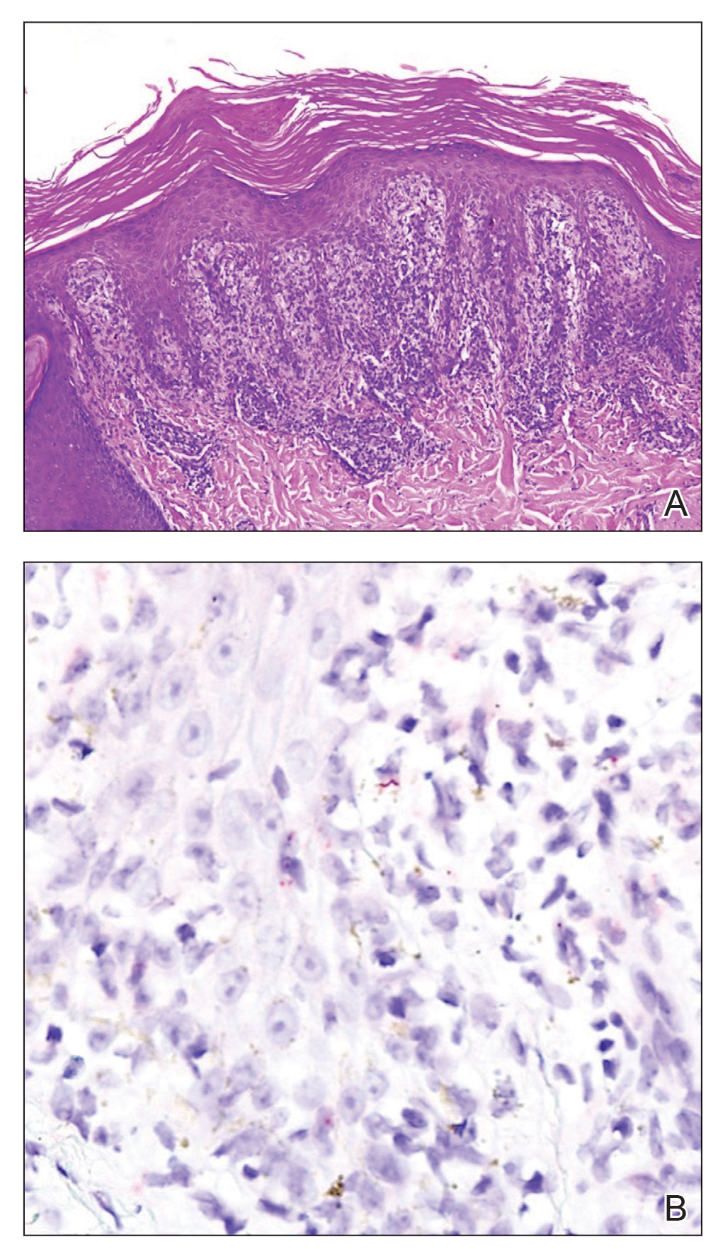
Mucocutaneus manifestations of secondary syphilis are multitudinous. As in our patient, the classic presentation is a generalized morbilliform and papulosquamous eruption involving the palms (Figure 2) and soles. Split papules at the oral commissures, mucosal patches, and condyloma lata are the characteristic mucosal lesions of secondary syphilis.1 Patchy nonscarring alopecia is not uncommon and can be the only manifestation of secondary syphilis.2 The histopathologic features of secondary syphilis vary depending on the location and type of the skin eruption. Psoriasiform or lichenoid changes commonly occur in the epidermis and dermoepidermal junction.3 The dermal inflammatory patterns that have been described include granulomatous, nodular, and superficial and deep perivascular inflammation. The infiltrate often is composed of lymphocytes, plasma cells, and histocytes. Reactive endothelial cells and perineural plasma cell infiltrates also are common histologic features.3,4 Spirochetes can be identified in most cases using immunohistochemical staining; however, the absence of spirochetes does not exclude syphilis.3 The sensitivity of immunohistochemical staining in secondary syphilis is reported to be 71% to 100% with a very high specificity.5 The treatment for all stages of syphilis is benzathine penicillin G, and the route of administration and duration of treatment depend on the stage of disease.6

A broad differential diagnosis must be considered when encountering skin eruptions in patients with HIV. Psoriasis usually presents as circumscribed erythematous plaques with dry and silvery scaling and a predilection for the extensor surfaces of the limbs, sacrum, scalp, and nails. Nail manifestations include distal onycholysis, irregular pitting, oil spots, salmon patches, and subungual hyperkeratosis. Alopecia occasionally may be seen within scalp lesions7; however, the constellation of alopecia with a moth-eaten appearance, subungual hyperkeratosis, papulosquamous eruption, and split papules was more suggestive of secondary syphilis in our patient. In immunocompromised patients, crusted scabies can be considered for the diagnosis of papulosquamous eruptions involving the palms and soles. It often presents with symmetric, mildly pruritic, psoriasiform dermatitis that favors acral sites, but widespread involvement can be observed.8 Areas of the scalp and face can be affected in infants, elderly patients, and immunocompromised individuals. Unlike in secondary syphilis, patchy alopecia, split papules, and ocular symptoms typically are not observed in scabies.
Sarcoidosis is common in Black individuals, and similar to syphilis, it is considered a great imitator of other dermatologic diseases. Frequently, it presents as redviolaceous papules, nodules, or plaques; however, rare variants including psoriasiform, ichthyosiform, verrucous, and lichenoid skin eruptions can occur. Nail dystrophy, split papules, and alopecia also have been observed.9 Ocular involvement is common and frequently presents as uveitis.10 The pathologic hallmark of sarcoidosis is noncaseating granulomatous inflammation, which also may occur in syphilitic lesions9; however, a papulosquamous eruption involving the palms and soles, positive serology, and the finding of interface lichenoid dermatitis with psoriasiform hyperplasia confirmed the diagnosis of secondary syphilis in our patient. Pityriasis rubra pilaris is a rare papulosquamous disorder that can be associated with HIV (type VI/HIVassociated follicular syndrome). It presents with generalized red-orange keratotic papules and often is associated with acne conglobata, hidradenitis suppurativa, and lichen spinulosus.11 Unlike in secondary syphilis, patchy alopecia, split papules, and ocular symptoms typically are not observed in pityriasis rubra pilaris.
This case highlights many classical findings of secondary syphilis and demonstrates that, while helpful, routine skin biopsy may not be required. Treatment should be guided by clinical presentation and serologic testing while reserving skin biopsy for equivocal cases.
- Forrestel AK, Kovarik CL, Katz KA. Sexually acquired syphilis: historical aspects, microbiology, epidemiology, and clinical manifestations. J Am Acad Dermatol. 2020;82:1-14.
- Balagula Y, Mattei PL, Wisco OJ, et al. The great imitator revisited: the spectrum of atypical cutaneous manifestations of secondary syphilis. Int J Dermatol. 2014;53:1434-1441.
- Hoang MP, High WA, Molberg KH. Secondary syphilis: a histologic and immunohistochemical evaluation. J Cutan Pathol. 2004; 31:595-599.
- Flamm A, Parikh K, Xie Q, et al. Histologic features of secondary syphilis: a multicenter retrospective review. J Am Acad Dermatol. 2015;73:1025-1030.
- Forrestel AK, Kovarik CL, Katz KA. Sexually acquired syphilis: laboratory diagnosis, management, and prevention [published online February 8, 2020]. J Am Acad Dermatol. 2020;82:17-28.
- Ghanem KG, Ram S, Rice PA. The modern epidemic of syphilis. N Engl J Med. 2020;382:845-854.
- Boehncke WH, Schön MP. Psoriasis. Lancet. 2015;386:983-994.
- Karthikeyan K. Crusted scabies. Indian J Dermatol Venereol Leprol. 2009;75:340-347.
- Haimovic A, Sanchez M, Judson MA, et al. Sarcoidosis: a comprehensive review and update for the dermatologist: part I. cutaneous disease. J Am Acad Dermatol. 2012;66:699.e1-718.
- Haimovic A, Sanchez M, Judson MA, et al. Sarcoidosis: a comprehensive review and update for the dermatologist: part II. extracutaneous disease. J Am Acad Dermatol. 2012;66:719.e1-730.
- Miralles E, Núñez M, De Las Heras M, et al. Pityriasis rubra pilaris and human immunodeficiency virus infection. Br J Dermatol. 1995;133:990-993.
The Diagnosis: Secondary Syphilis
Histopathology revealed a lichenoid interface dermatitis with psoriasiform hyperplasia (Figure 1A). A single spirochete was identified using immunohistochemical staining (Figure 1B). Laboratory workup revealed positive IgG and IgM treponemal antibodies and reactive rapid plasma reagin titer of 1:2048. A VDRL test performed on a cerebrospinal fluid specimen also was reactive at 1:8. A diagnosis of secondary syphilis with neurologic involvement was made, and the patient was treated with intravenous penicillin G for 14 days. Following treatment, his rapid plasma reagin decreased 4-fold with an improvement in his ocular and cutaneous symptoms.
Mucocutaneus manifestations of secondary syphilis are multitudinous. As in our patient, the classic presentation is a generalized morbilliform and papulosquamous eruption involving the palms (Figure 2) and soles. Split papules at the oral commissures, mucosal patches, and condyloma lata are the characteristic mucosal lesions of secondary syphilis.1 Patchy nonscarring alopecia is not uncommon and can be the only manifestation of secondary syphilis.2 The histopathologic features of secondary syphilis vary depending on the location and type of the skin eruption. Psoriasiform or lichenoid changes commonly occur in the epidermis and dermoepidermal junction.3 The dermal inflammatory patterns that have been described include granulomatous, nodular, and superficial and deep perivascular inflammation. The infiltrate often is composed of lymphocytes, plasma cells, and histocytes. Reactive endothelial cells and perineural plasma cell infiltrates also are common histologic features.3,4 Spirochetes can be identified in most cases using immunohistochemical staining; however, the absence of spirochetes does not exclude syphilis.3 The sensitivity of immunohistochemical staining in secondary syphilis is reported to be 71% to 100% with a very high specificity.5 The treatment for all stages of syphilis is benzathine penicillin G, and the route of administration and duration of treatment depend on the stage of disease.6
A broad differential diagnosis must be considered when encountering skin eruptions in patients with HIV. Psoriasis usually presents as circumscribed erythematous plaques with dry and silvery scaling and a predilection for the extensor surfaces of the limbs, sacrum, scalp, and nails. Nail manifestations include distal onycholysis, irregular pitting, oil spots, salmon patches, and subungual hyperkeratosis. Alopecia occasionally may be seen within scalp lesions7; however, the constellation of alopecia with a moth-eaten appearance, subungual hyperkeratosis, papulosquamous eruption, and split papules was more suggestive of secondary syphilis in our patient. In immunocompromised patients, crusted scabies can be considered for the diagnosis of papulosquamous eruptions involving the palms and soles. It often presents with symmetric, mildly pruritic, psoriasiform dermatitis that favors acral sites, but widespread involvement can be observed.8 Areas of the scalp and face can be affected in infants, elderly patients, and immunocompromised individuals. Unlike in secondary syphilis, patchy alopecia, split papules, and ocular symptoms typically are not observed in scabies.
Sarcoidosis is common in Black individuals, and similar to syphilis, it is considered a great imitator of other dermatologic diseases. Frequently, it presents as redviolaceous papules, nodules, or plaques; however, rare variants including psoriasiform, ichthyosiform, verrucous, and lichenoid skin eruptions can occur. Nail dystrophy, split papules, and alopecia also have been observed.9 Ocular involvement is common and frequently presents as uveitis.10 The pathologic hallmark of sarcoidosis is noncaseating granulomatous inflammation, which also may occur in syphilitic lesions9; however, a papulosquamous eruption involving the palms and soles, positive serology, and the finding of interface lichenoid dermatitis with psoriasiform hyperplasia confirmed the diagnosis of secondary syphilis in our patient. Pityriasis rubra pilaris is a rare papulosquamous disorder that can be associated with HIV (type VI/HIVassociated follicular syndrome). It presents with generalized red-orange keratotic papules and often is associated with acne conglobata, hidradenitis suppurativa, and lichen spinulosus.11 Unlike in secondary syphilis, patchy alopecia, split papules, and ocular symptoms typically are not observed in pityriasis rubra pilaris.
This case highlights many classical findings of secondary syphilis and demonstrates that, while helpful, routine skin biopsy may not be required. Treatment should be guided by clinical presentation and serologic testing while reserving skin biopsy for equivocal cases.
The Diagnosis: Secondary Syphilis
Histopathology revealed a lichenoid interface dermatitis with psoriasiform hyperplasia (Figure 1A). A single spirochete was identified using immunohistochemical staining (Figure 1B). Laboratory workup revealed positive IgG and IgM treponemal antibodies and reactive rapid plasma reagin titer of 1:2048. A VDRL test performed on a cerebrospinal fluid specimen also was reactive at 1:8. A diagnosis of secondary syphilis with neurologic involvement was made, and the patient was treated with intravenous penicillin G for 14 days. Following treatment, his rapid plasma reagin decreased 4-fold with an improvement in his ocular and cutaneous symptoms.
Mucocutaneus manifestations of secondary syphilis are multitudinous. As in our patient, the classic presentation is a generalized morbilliform and papulosquamous eruption involving the palms (Figure 2) and soles. Split papules at the oral commissures, mucosal patches, and condyloma lata are the characteristic mucosal lesions of secondary syphilis.1 Patchy nonscarring alopecia is not uncommon and can be the only manifestation of secondary syphilis.2 The histopathologic features of secondary syphilis vary depending on the location and type of the skin eruption. Psoriasiform or lichenoid changes commonly occur in the epidermis and dermoepidermal junction.3 The dermal inflammatory patterns that have been described include granulomatous, nodular, and superficial and deep perivascular inflammation. The infiltrate often is composed of lymphocytes, plasma cells, and histocytes. Reactive endothelial cells and perineural plasma cell infiltrates also are common histologic features.3,4 Spirochetes can be identified in most cases using immunohistochemical staining; however, the absence of spirochetes does not exclude syphilis.3 The sensitivity of immunohistochemical staining in secondary syphilis is reported to be 71% to 100% with a very high specificity.5 The treatment for all stages of syphilis is benzathine penicillin G, and the route of administration and duration of treatment depend on the stage of disease.6
A broad differential diagnosis must be considered when encountering skin eruptions in patients with HIV. Psoriasis usually presents as circumscribed erythematous plaques with dry and silvery scaling and a predilection for the extensor surfaces of the limbs, sacrum, scalp, and nails. Nail manifestations include distal onycholysis, irregular pitting, oil spots, salmon patches, and subungual hyperkeratosis. Alopecia occasionally may be seen within scalp lesions7; however, the constellation of alopecia with a moth-eaten appearance, subungual hyperkeratosis, papulosquamous eruption, and split papules was more suggestive of secondary syphilis in our patient. In immunocompromised patients, crusted scabies can be considered for the diagnosis of papulosquamous eruptions involving the palms and soles. It often presents with symmetric, mildly pruritic, psoriasiform dermatitis that favors acral sites, but widespread involvement can be observed.8 Areas of the scalp and face can be affected in infants, elderly patients, and immunocompromised individuals. Unlike in secondary syphilis, patchy alopecia, split papules, and ocular symptoms typically are not observed in scabies.
Sarcoidosis is common in Black individuals, and similar to syphilis, it is considered a great imitator of other dermatologic diseases. Frequently, it presents as redviolaceous papules, nodules, or plaques; however, rare variants including psoriasiform, ichthyosiform, verrucous, and lichenoid skin eruptions can occur. Nail dystrophy, split papules, and alopecia also have been observed.9 Ocular involvement is common and frequently presents as uveitis.10 The pathologic hallmark of sarcoidosis is noncaseating granulomatous inflammation, which also may occur in syphilitic lesions9; however, a papulosquamous eruption involving the palms and soles, positive serology, and the finding of interface lichenoid dermatitis with psoriasiform hyperplasia confirmed the diagnosis of secondary syphilis in our patient. Pityriasis rubra pilaris is a rare papulosquamous disorder that can be associated with HIV (type VI/HIVassociated follicular syndrome). It presents with generalized red-orange keratotic papules and often is associated with acne conglobata, hidradenitis suppurativa, and lichen spinulosus.11 Unlike in secondary syphilis, patchy alopecia, split papules, and ocular symptoms typically are not observed in pityriasis rubra pilaris.
This case highlights many classical findings of secondary syphilis and demonstrates that, while helpful, routine skin biopsy may not be required. Treatment should be guided by clinical presentation and serologic testing while reserving skin biopsy for equivocal cases.
- Forrestel AK, Kovarik CL, Katz KA. Sexually acquired syphilis: historical aspects, microbiology, epidemiology, and clinical manifestations. J Am Acad Dermatol. 2020;82:1-14.
- Balagula Y, Mattei PL, Wisco OJ, et al. The great imitator revisited: the spectrum of atypical cutaneous manifestations of secondary syphilis. Int J Dermatol. 2014;53:1434-1441.
- Hoang MP, High WA, Molberg KH. Secondary syphilis: a histologic and immunohistochemical evaluation. J Cutan Pathol. 2004; 31:595-599.
- Flamm A, Parikh K, Xie Q, et al. Histologic features of secondary syphilis: a multicenter retrospective review. J Am Acad Dermatol. 2015;73:1025-1030.
- Forrestel AK, Kovarik CL, Katz KA. Sexually acquired syphilis: laboratory diagnosis, management, and prevention [published online February 8, 2020]. J Am Acad Dermatol. 2020;82:17-28.
- Ghanem KG, Ram S, Rice PA. The modern epidemic of syphilis. N Engl J Med. 2020;382:845-854.
- Boehncke WH, Schön MP. Psoriasis. Lancet. 2015;386:983-994.
- Karthikeyan K. Crusted scabies. Indian J Dermatol Venereol Leprol. 2009;75:340-347.
- Haimovic A, Sanchez M, Judson MA, et al. Sarcoidosis: a comprehensive review and update for the dermatologist: part I. cutaneous disease. J Am Acad Dermatol. 2012;66:699.e1-718.
- Haimovic A, Sanchez M, Judson MA, et al. Sarcoidosis: a comprehensive review and update for the dermatologist: part II. extracutaneous disease. J Am Acad Dermatol. 2012;66:719.e1-730.
- Miralles E, Núñez M, De Las Heras M, et al. Pityriasis rubra pilaris and human immunodeficiency virus infection. Br J Dermatol. 1995;133:990-993.
- Forrestel AK, Kovarik CL, Katz KA. Sexually acquired syphilis: historical aspects, microbiology, epidemiology, and clinical manifestations. J Am Acad Dermatol. 2020;82:1-14.
- Balagula Y, Mattei PL, Wisco OJ, et al. The great imitator revisited: the spectrum of atypical cutaneous manifestations of secondary syphilis. Int J Dermatol. 2014;53:1434-1441.
- Hoang MP, High WA, Molberg KH. Secondary syphilis: a histologic and immunohistochemical evaluation. J Cutan Pathol. 2004; 31:595-599.
- Flamm A, Parikh K, Xie Q, et al. Histologic features of secondary syphilis: a multicenter retrospective review. J Am Acad Dermatol. 2015;73:1025-1030.
- Forrestel AK, Kovarik CL, Katz KA. Sexually acquired syphilis: laboratory diagnosis, management, and prevention [published online February 8, 2020]. J Am Acad Dermatol. 2020;82:17-28.
- Ghanem KG, Ram S, Rice PA. The modern epidemic of syphilis. N Engl J Med. 2020;382:845-854.
- Boehncke WH, Schön MP. Psoriasis. Lancet. 2015;386:983-994.
- Karthikeyan K. Crusted scabies. Indian J Dermatol Venereol Leprol. 2009;75:340-347.
- Haimovic A, Sanchez M, Judson MA, et al. Sarcoidosis: a comprehensive review and update for the dermatologist: part I. cutaneous disease. J Am Acad Dermatol. 2012;66:699.e1-718.
- Haimovic A, Sanchez M, Judson MA, et al. Sarcoidosis: a comprehensive review and update for the dermatologist: part II. extracutaneous disease. J Am Acad Dermatol. 2012;66:719.e1-730.
- Miralles E, Núñez M, De Las Heras M, et al. Pityriasis rubra pilaris and human immunodeficiency virus infection. Br J Dermatol. 1995;133:990-993.
A 29-year-old Black man with long-standing untreated HIV presented with mildly pruritic, scaly plaques on the palms and soles of 2 weeks’ duration. His medical history was notable for primary syphilis treated approximately 1 year prior. A review of symptoms was positive for blurry vision and floaters but negative for constitutional symptoms. Physical examination revealed well-defined scaly plaques over the palms, soles, and elbows with subungual hyperkeratosis. Patches of nonscarring alopecia over the scalp and split papules at the oral commissures also were noted. There were no palpable lymph nodes or genital involvement. Eye examination showed conjunctival injection and 20 cells per field in the vitreous humor. Laboratory evaluation revealed an HIV viral load of 31,623 copies/mL and a CD4 count of 47 cells/μL (reference range, 362–1531 cells/μL). A shave biopsy of the left elbow was performed for histopathologic evaluation.
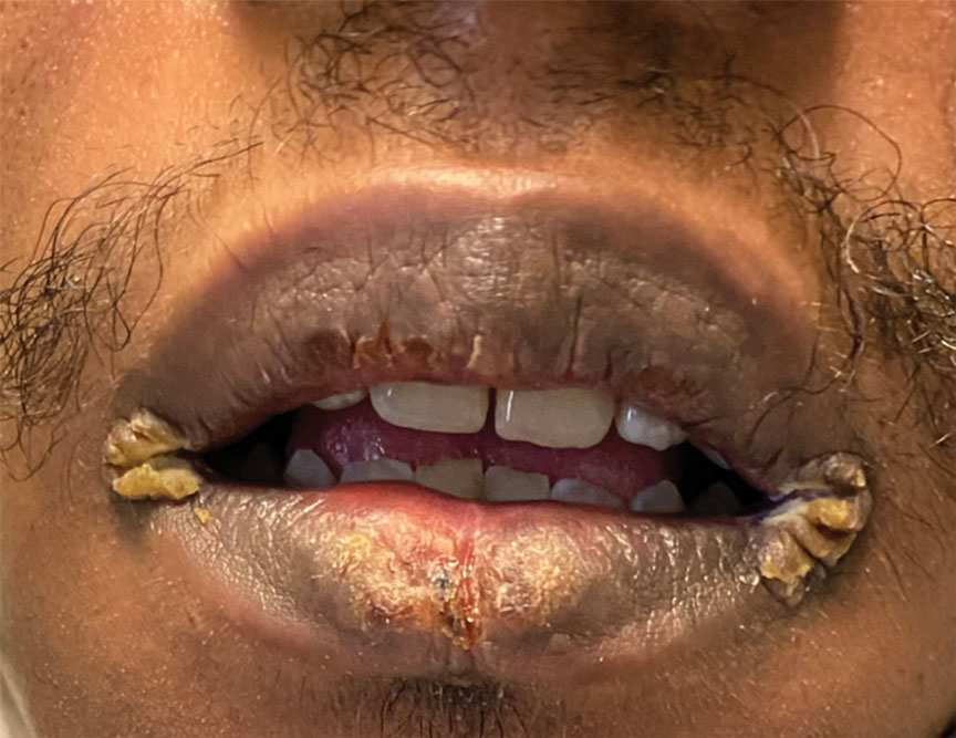

BREEZE-AD-PEDS: First data for baricitinib in childhood eczema reported
The oral Janus kinase
After 16 weeks of treatment, the primary endpoint – an Investigators Global Assessment (IGA) score of 0 or 1 with at least a 2-point improvement from baseline – was met by 41.7% of patients given 2 mg (those younger than age 10) or 4 mg of baricitinib (those aged 10-17 years), the highest dose studied in each of those two age groups.
By comparison, the primary endpoint was met in 16.4% of children in the placebo group (P < .001).
Baricitinib is approved for the treatment of AD in adults in many countries, Antonio Torrelo, MD, of the Hospital Infantil Niño Jesús, Madrid, said at the annual congress of the European Academy of Dermatology and Venereology. It was approved by the U.S. Food and Drug Administration for treating adults with severe alopecia areata in June and is under FDA review for the treatment of AD.
The phase 3 BREEZE-AD-PEDS trial
BREEZE-AD-PEDS was a randomized, double-blind trial that evaluated the safety and efficacy of baricitinib in 483 children and adolescents with moderate to severe AD. Participants were aged 2-17 years. Those aged 2-5 years had been diagnosed with AD for at least 6 months; if they were older, they had been diagnosed for at least 12 months.
Three dosing levels of baricitinib were tested: 121 patients were given a low dose, which was 0.5 mg/day in children aged 2 to less than 10 years and 1 mg/day in those aged 10 to less than 18 years. A medium dose – 1 mg/day in the younger children and 2 mg/day in the older children – was given to 120 children, while a high dose – 2 mg/day and 4 mg/day, respectively – was given to another 120 children.
Topical treatments were permitted, although for entry into the trial, participants had to have had an inadequate response to steroids and an inadequate or no response to topical calcineurin inhibitors. In all groups, age, gender, race, geographic region, age at diagnosis of AD, and duration of AD “were more or less similar,” Dr. Torello said.
Good results, but only with highest dose
The primary IGA endpoint was reached by 25.8% of children in the medium-dose group and by 18.2% in the low-dose group. Neither result was statistically significant in comparison with placebo (16.4%).
When breaking down the results between different ages, “the results in the IGA scores are consistent in both age subgroups – below 10 years and over 10 years,” Dr. Torello noted. The results are also consistent across body weights (< 20 kg, 20-60 kg, and > 60 kg), he added.
Among those treated with the high dose of baricitinib, Eczema Area and Severity Index (EASI) 75% and 95% improvement scores were reached in 52.5% and 30% of patients, respectively. Corresponding figures for the medium dose were 40% and 21.7%; for the low baricitinib dose, 32.2% and 11.6%; and for placebo, 32% and 12.3%. Again, only the results for the highest baricitinib dose were significant in comparison with placebo.
A similar pattern was seen for improvement in itch, and there was a 75% improvement in Scoring Atopic Dermatitis (SCORAD75) results.
Safety of baricitinib in children
The labeling for JAK inhibitors that have been approved to date, including baricitinib, include a boxed warning regarding risks for thrombosis, major adverse cardiovascular events, and all-cause mortality. The warning is based on use by patients with rheumatoid arthritis.
Dr. Torello summarized baricitinib’s safety profile in the trial as being “consistent with the well-known safety profile for baricitinib in adults with moderate to severe atopic dermatitis.”
In the study, no severe adverse effects were noted, and no new safety signals were observed, he said. The rate of any treatment-emergent effect among patients was around 50% and was similar across all baricitinib and placebo groups. Study discontinuations because of a side effect were more frequent in the placebo arm (1.6% of patients) than in the baricitinib low-, medium-, and high-dose arms (0.8%, 0%, and 0.8%, respectively).
There were no cases of deep-vein thrombosis, pulmonary embolism, or other adverse effects of special interest, including major adverse cardiovascular events, gastrointestinal perforations, and opportunistic infections, Dr. Torrelo said.
No patient experienced elevations in liver enzyme levels, although there were some cases of elevated creatinine phosphokinase levels (16% in the placebo group and 19% in the baricitinib arms altogether) that were not from muscle injury. There was a possible increase in low-density cholesterol level (3.3% of those taking placebo vs. 10.1% of baricitinib-treated patients).
Is there a role for baricitinib?
“Baricitinib is a potential therapeutic option with a favorable benefit-to-risk profile for children between 2 and 18 years who have moderate to severe atopic dermatitis, and candidates for systemic therapy,” Dr. Torrelo said. “No single drug is capable to treat every patient with atopic dermatitis,” he added in discussing the possible place of baricitinib in pediatric practice.
“There are patients who do not respond to dupilumab, who apparently respond later to JAK inhibitors,” he noted.
“We are trying to work phenotypically, trying to learn what kind of patients – especially children who have a more heterogeneous disease than adults – can be better treated with JAK inhibitors or dupilumab.” There may be other important considerations in choosing a treatment in children, Dr. Torrelo said, including that JAK inhibitors can be given orally, while dupilumab is administered by injection.
Asked to comment on the results, Jashin J. Wu, MD, founder and CEO of the Dermatology Research and Education Foundation in Irvine, Calif., pointed out that “only the higher dose is significantly more effective than placebo.”
In his view, “the potentially severe adverse events are not worth the risk compared to more effective agents, such as dupilumab, in this pediatric population,” added Dr. Wu, who recently authored a review of the role of JAK inhibitors in skin disease. He was not involved with the baricitinib study.
The study was funded by Eli Lilly in collaboration with Incyte. Dr. Torello has participated in advisory boards and/or has served as a principal investigator in clinical trials for AbbVie, Eli Lilly and Company, Novartis, Pfizer, Pierre Fabre, and Sanofi. Dr. Wu has been an investigator, consultant, or speaker for multiple pharmaceutical companies.
A version of this article first appeared on Medscape.com.
The oral Janus kinase
After 16 weeks of treatment, the primary endpoint – an Investigators Global Assessment (IGA) score of 0 or 1 with at least a 2-point improvement from baseline – was met by 41.7% of patients given 2 mg (those younger than age 10) or 4 mg of baricitinib (those aged 10-17 years), the highest dose studied in each of those two age groups.
By comparison, the primary endpoint was met in 16.4% of children in the placebo group (P < .001).
Baricitinib is approved for the treatment of AD in adults in many countries, Antonio Torrelo, MD, of the Hospital Infantil Niño Jesús, Madrid, said at the annual congress of the European Academy of Dermatology and Venereology. It was approved by the U.S. Food and Drug Administration for treating adults with severe alopecia areata in June and is under FDA review for the treatment of AD.
The phase 3 BREEZE-AD-PEDS trial
BREEZE-AD-PEDS was a randomized, double-blind trial that evaluated the safety and efficacy of baricitinib in 483 children and adolescents with moderate to severe AD. Participants were aged 2-17 years. Those aged 2-5 years had been diagnosed with AD for at least 6 months; if they were older, they had been diagnosed for at least 12 months.
Three dosing levels of baricitinib were tested: 121 patients were given a low dose, which was 0.5 mg/day in children aged 2 to less than 10 years and 1 mg/day in those aged 10 to less than 18 years. A medium dose – 1 mg/day in the younger children and 2 mg/day in the older children – was given to 120 children, while a high dose – 2 mg/day and 4 mg/day, respectively – was given to another 120 children.
Topical treatments were permitted, although for entry into the trial, participants had to have had an inadequate response to steroids and an inadequate or no response to topical calcineurin inhibitors. In all groups, age, gender, race, geographic region, age at diagnosis of AD, and duration of AD “were more or less similar,” Dr. Torello said.
Good results, but only with highest dose
The primary IGA endpoint was reached by 25.8% of children in the medium-dose group and by 18.2% in the low-dose group. Neither result was statistically significant in comparison with placebo (16.4%).
When breaking down the results between different ages, “the results in the IGA scores are consistent in both age subgroups – below 10 years and over 10 years,” Dr. Torello noted. The results are also consistent across body weights (< 20 kg, 20-60 kg, and > 60 kg), he added.
Among those treated with the high dose of baricitinib, Eczema Area and Severity Index (EASI) 75% and 95% improvement scores were reached in 52.5% and 30% of patients, respectively. Corresponding figures for the medium dose were 40% and 21.7%; for the low baricitinib dose, 32.2% and 11.6%; and for placebo, 32% and 12.3%. Again, only the results for the highest baricitinib dose were significant in comparison with placebo.
A similar pattern was seen for improvement in itch, and there was a 75% improvement in Scoring Atopic Dermatitis (SCORAD75) results.
Safety of baricitinib in children
The labeling for JAK inhibitors that have been approved to date, including baricitinib, include a boxed warning regarding risks for thrombosis, major adverse cardiovascular events, and all-cause mortality. The warning is based on use by patients with rheumatoid arthritis.
Dr. Torello summarized baricitinib’s safety profile in the trial as being “consistent with the well-known safety profile for baricitinib in adults with moderate to severe atopic dermatitis.”
In the study, no severe adverse effects were noted, and no new safety signals were observed, he said. The rate of any treatment-emergent effect among patients was around 50% and was similar across all baricitinib and placebo groups. Study discontinuations because of a side effect were more frequent in the placebo arm (1.6% of patients) than in the baricitinib low-, medium-, and high-dose arms (0.8%, 0%, and 0.8%, respectively).
There were no cases of deep-vein thrombosis, pulmonary embolism, or other adverse effects of special interest, including major adverse cardiovascular events, gastrointestinal perforations, and opportunistic infections, Dr. Torrelo said.
No patient experienced elevations in liver enzyme levels, although there were some cases of elevated creatinine phosphokinase levels (16% in the placebo group and 19% in the baricitinib arms altogether) that were not from muscle injury. There was a possible increase in low-density cholesterol level (3.3% of those taking placebo vs. 10.1% of baricitinib-treated patients).
Is there a role for baricitinib?
“Baricitinib is a potential therapeutic option with a favorable benefit-to-risk profile for children between 2 and 18 years who have moderate to severe atopic dermatitis, and candidates for systemic therapy,” Dr. Torrelo said. “No single drug is capable to treat every patient with atopic dermatitis,” he added in discussing the possible place of baricitinib in pediatric practice.
“There are patients who do not respond to dupilumab, who apparently respond later to JAK inhibitors,” he noted.
“We are trying to work phenotypically, trying to learn what kind of patients – especially children who have a more heterogeneous disease than adults – can be better treated with JAK inhibitors or dupilumab.” There may be other important considerations in choosing a treatment in children, Dr. Torrelo said, including that JAK inhibitors can be given orally, while dupilumab is administered by injection.
Asked to comment on the results, Jashin J. Wu, MD, founder and CEO of the Dermatology Research and Education Foundation in Irvine, Calif., pointed out that “only the higher dose is significantly more effective than placebo.”
In his view, “the potentially severe adverse events are not worth the risk compared to more effective agents, such as dupilumab, in this pediatric population,” added Dr. Wu, who recently authored a review of the role of JAK inhibitors in skin disease. He was not involved with the baricitinib study.
The study was funded by Eli Lilly in collaboration with Incyte. Dr. Torello has participated in advisory boards and/or has served as a principal investigator in clinical trials for AbbVie, Eli Lilly and Company, Novartis, Pfizer, Pierre Fabre, and Sanofi. Dr. Wu has been an investigator, consultant, or speaker for multiple pharmaceutical companies.
A version of this article first appeared on Medscape.com.
The oral Janus kinase
After 16 weeks of treatment, the primary endpoint – an Investigators Global Assessment (IGA) score of 0 or 1 with at least a 2-point improvement from baseline – was met by 41.7% of patients given 2 mg (those younger than age 10) or 4 mg of baricitinib (those aged 10-17 years), the highest dose studied in each of those two age groups.
By comparison, the primary endpoint was met in 16.4% of children in the placebo group (P < .001).
Baricitinib is approved for the treatment of AD in adults in many countries, Antonio Torrelo, MD, of the Hospital Infantil Niño Jesús, Madrid, said at the annual congress of the European Academy of Dermatology and Venereology. It was approved by the U.S. Food and Drug Administration for treating adults with severe alopecia areata in June and is under FDA review for the treatment of AD.
The phase 3 BREEZE-AD-PEDS trial
BREEZE-AD-PEDS was a randomized, double-blind trial that evaluated the safety and efficacy of baricitinib in 483 children and adolescents with moderate to severe AD. Participants were aged 2-17 years. Those aged 2-5 years had been diagnosed with AD for at least 6 months; if they were older, they had been diagnosed for at least 12 months.
Three dosing levels of baricitinib were tested: 121 patients were given a low dose, which was 0.5 mg/day in children aged 2 to less than 10 years and 1 mg/day in those aged 10 to less than 18 years. A medium dose – 1 mg/day in the younger children and 2 mg/day in the older children – was given to 120 children, while a high dose – 2 mg/day and 4 mg/day, respectively – was given to another 120 children.
Topical treatments were permitted, although for entry into the trial, participants had to have had an inadequate response to steroids and an inadequate or no response to topical calcineurin inhibitors. In all groups, age, gender, race, geographic region, age at diagnosis of AD, and duration of AD “were more or less similar,” Dr. Torello said.
Good results, but only with highest dose
The primary IGA endpoint was reached by 25.8% of children in the medium-dose group and by 18.2% in the low-dose group. Neither result was statistically significant in comparison with placebo (16.4%).
When breaking down the results between different ages, “the results in the IGA scores are consistent in both age subgroups – below 10 years and over 10 years,” Dr. Torello noted. The results are also consistent across body weights (< 20 kg, 20-60 kg, and > 60 kg), he added.
Among those treated with the high dose of baricitinib, Eczema Area and Severity Index (EASI) 75% and 95% improvement scores were reached in 52.5% and 30% of patients, respectively. Corresponding figures for the medium dose were 40% and 21.7%; for the low baricitinib dose, 32.2% and 11.6%; and for placebo, 32% and 12.3%. Again, only the results for the highest baricitinib dose were significant in comparison with placebo.
A similar pattern was seen for improvement in itch, and there was a 75% improvement in Scoring Atopic Dermatitis (SCORAD75) results.
Safety of baricitinib in children
The labeling for JAK inhibitors that have been approved to date, including baricitinib, include a boxed warning regarding risks for thrombosis, major adverse cardiovascular events, and all-cause mortality. The warning is based on use by patients with rheumatoid arthritis.
Dr. Torello summarized baricitinib’s safety profile in the trial as being “consistent with the well-known safety profile for baricitinib in adults with moderate to severe atopic dermatitis.”
In the study, no severe adverse effects were noted, and no new safety signals were observed, he said. The rate of any treatment-emergent effect among patients was around 50% and was similar across all baricitinib and placebo groups. Study discontinuations because of a side effect were more frequent in the placebo arm (1.6% of patients) than in the baricitinib low-, medium-, and high-dose arms (0.8%, 0%, and 0.8%, respectively).
There were no cases of deep-vein thrombosis, pulmonary embolism, or other adverse effects of special interest, including major adverse cardiovascular events, gastrointestinal perforations, and opportunistic infections, Dr. Torrelo said.
No patient experienced elevations in liver enzyme levels, although there were some cases of elevated creatinine phosphokinase levels (16% in the placebo group and 19% in the baricitinib arms altogether) that were not from muscle injury. There was a possible increase in low-density cholesterol level (3.3% of those taking placebo vs. 10.1% of baricitinib-treated patients).
Is there a role for baricitinib?
“Baricitinib is a potential therapeutic option with a favorable benefit-to-risk profile for children between 2 and 18 years who have moderate to severe atopic dermatitis, and candidates for systemic therapy,” Dr. Torrelo said. “No single drug is capable to treat every patient with atopic dermatitis,” he added in discussing the possible place of baricitinib in pediatric practice.
“There are patients who do not respond to dupilumab, who apparently respond later to JAK inhibitors,” he noted.
“We are trying to work phenotypically, trying to learn what kind of patients – especially children who have a more heterogeneous disease than adults – can be better treated with JAK inhibitors or dupilumab.” There may be other important considerations in choosing a treatment in children, Dr. Torrelo said, including that JAK inhibitors can be given orally, while dupilumab is administered by injection.
Asked to comment on the results, Jashin J. Wu, MD, founder and CEO of the Dermatology Research and Education Foundation in Irvine, Calif., pointed out that “only the higher dose is significantly more effective than placebo.”
In his view, “the potentially severe adverse events are not worth the risk compared to more effective agents, such as dupilumab, in this pediatric population,” added Dr. Wu, who recently authored a review of the role of JAK inhibitors in skin disease. He was not involved with the baricitinib study.
The study was funded by Eli Lilly in collaboration with Incyte. Dr. Torello has participated in advisory boards and/or has served as a principal investigator in clinical trials for AbbVie, Eli Lilly and Company, Novartis, Pfizer, Pierre Fabre, and Sanofi. Dr. Wu has been an investigator, consultant, or speaker for multiple pharmaceutical companies.
A version of this article first appeared on Medscape.com.
FROM THE EADV CONGRESS