User login
What is an “early and accurate” diagnosis?
For the last few weeks, the eye-grabber at the top of the American Academy of Pediatrics shopAAP email has been “Early and Accurate Diagnosis.” The unstated claim is that a practitioner who subscribes to one of their continuing education products will improve his or her chances of making an early and accurate diagnosis that “Also Cures Missed School, Soccer Practice, and Music Lessons.” The tagline, Early and Accurate Diagnosis, got me ruminating.

What exactly is an accurate diagnosis? And how does one define an early diagnosis? These are not merely questions of semantics. An honest attempt to answer them scratches through the surface of some serious issues facing a primary care physician.
Who are the judges deciding whether a physician’s diagnosis is accurate? Should it be a panel of academic physicians, most of who are specialists and subspecialists, and who are most comfortable seeing patients with array of signs and symptoms that your patient has presented? Or, should it be a collection of your primary care peers working with limited resources miles away from a tertiary care center?
Is there such a thing as a diagnosis that is close enough? How often is it important that your diagnosis is spot on? Is it like a high school algebra problem in which you could get partial credit for showing how you arrived at the not-quite-right-answer? It really makes a difference only when you start acting (or, in some cases, not acting) on your diagnosis.
Let’s be honest. How often have you made the wrong diagnosis and the patient got better with your management plan? Your therapy may have worked for Diagnosis A even though you were targeting Diagnosis B. Or, more likely, the patient was going to get better without any intervention.
Don’t get me wrong. I think a correct diagnosis can be, and often is, extremely important, but it is really the patient who is the judge of whether you got it right. He doesn’t care what you called it. He is happy knowing that he got better and you didn’t hurt him.
Now, what about that “early” piece? Again, the patient might have something to say about this. You may have made the correct diagnosis but because your productivity is limited by a clunky EMR or your appointment desk does a poor job of triage, the patient was forced to wait an unconscionable amount of time to be seen.
A timely diagnosis certainly is important in many situations. But particularly, early in your career, you may not have the experience to make those quick one look and you’ve got it right diagnoses. These are times to come clean and tell the patient that you aren’t sure what they have. Of course, you might want to choose a better phrase than, “I don’t have clue.”
If I had been asked to write the AAP’s tag line, I would have chosen “efficient” instead of early. If you made the correct diagnosis and it was reasonably timely but you ordered a barrage of unnecessary and expensive tests that inconvenienced the patient, you should have done a better job.
Finally, if you make the correct and early diagnosis but deliver it to the patient poorly, your therapy may not work. Again, it boils down to being an artful and caring physician.
Dr. Wilkoff practiced primary care pediatrics in Brunswick, Maine for nearly 40 years. He has authored several books on behavioral pediatrics, including “How to Say No to Your Toddler.” Email him at pdnews@mdedge.com.
For the last few weeks, the eye-grabber at the top of the American Academy of Pediatrics shopAAP email has been “Early and Accurate Diagnosis.” The unstated claim is that a practitioner who subscribes to one of their continuing education products will improve his or her chances of making an early and accurate diagnosis that “Also Cures Missed School, Soccer Practice, and Music Lessons.” The tagline, Early and Accurate Diagnosis, got me ruminating.
What exactly is an accurate diagnosis? And how does one define an early diagnosis? These are not merely questions of semantics. An honest attempt to answer them scratches through the surface of some serious issues facing a primary care physician.
Who are the judges deciding whether a physician’s diagnosis is accurate? Should it be a panel of academic physicians, most of who are specialists and subspecialists, and who are most comfortable seeing patients with array of signs and symptoms that your patient has presented? Or, should it be a collection of your primary care peers working with limited resources miles away from a tertiary care center?
Is there such a thing as a diagnosis that is close enough? How often is it important that your diagnosis is spot on? Is it like a high school algebra problem in which you could get partial credit for showing how you arrived at the not-quite-right-answer? It really makes a difference only when you start acting (or, in some cases, not acting) on your diagnosis.
Let’s be honest. How often have you made the wrong diagnosis and the patient got better with your management plan? Your therapy may have worked for Diagnosis A even though you were targeting Diagnosis B. Or, more likely, the patient was going to get better without any intervention.
Don’t get me wrong. I think a correct diagnosis can be, and often is, extremely important, but it is really the patient who is the judge of whether you got it right. He doesn’t care what you called it. He is happy knowing that he got better and you didn’t hurt him.
Now, what about that “early” piece? Again, the patient might have something to say about this. You may have made the correct diagnosis but because your productivity is limited by a clunky EMR or your appointment desk does a poor job of triage, the patient was forced to wait an unconscionable amount of time to be seen.
A timely diagnosis certainly is important in many situations. But particularly, early in your career, you may not have the experience to make those quick one look and you’ve got it right diagnoses. These are times to come clean and tell the patient that you aren’t sure what they have. Of course, you might want to choose a better phrase than, “I don’t have clue.”
If I had been asked to write the AAP’s tag line, I would have chosen “efficient” instead of early. If you made the correct diagnosis and it was reasonably timely but you ordered a barrage of unnecessary and expensive tests that inconvenienced the patient, you should have done a better job.
Finally, if you make the correct and early diagnosis but deliver it to the patient poorly, your therapy may not work. Again, it boils down to being an artful and caring physician.
Dr. Wilkoff practiced primary care pediatrics in Brunswick, Maine for nearly 40 years. He has authored several books on behavioral pediatrics, including “How to Say No to Your Toddler.” Email him at pdnews@mdedge.com.
How does caring affect the placebo effect?
How thorough are you when you prescribe medication? You check the patient’s list of allergies and current medications. You make sure that the dose is appropriate for the patient’s weight. Hopefully, you spend a minute or 2 describing the most common side effects. You prescribe the correct amount of medication and an appropriate number of refills. If you think you can distill it into one or two sentences, you also explain the medication’s mechanism of action. That is if you understand it yourself.

What about placebos? How often do you believe that your patient has gotten better because of the placebo effect? Do you ever intentionally recommend or prescribe a placebo? Do you share with the patient that there is no current explanation of why the treatment you are recommending should work? Or, do you just play dumb?
Whether you admit to being a frequent prescriber of placebos or not you should take the 20 minutes it will take to read a New York Times article titled “What if the Placebo Effect Isn’t a Trick” (Gary Greenberg, Nov 7, 2018). You will learn a bit about the history of the placebo effect including some recent functional MRI studies that have uncovered consistent brain activity patterns in subjects that respond to placebos.
You will read about some exciting research indicating that certain people with a genomic variant of an enzyme that has been shown to affect the response to painkillers generally have the weakest response to placebo. While in some studies the association between the patient’s response and the level of the enzyme is the reverse, Kathryn Hall, PhD, the molecular biologist overseeing these studies, feels that at this point in her research the fact that there is an association that varies with genotype is a critical finding. She suspects that the placebo effect and the drug operate on the same biochemical highway that includes this enzyme and that “clinician warmth” is particularly effective in patients with a certain genotype.
Ted Kaptchuk, who heads up Harvard Medical School’s Program in Placebo Studies and the Therapeutic Encounter and has collaborated with Dr. Hall, hypothesizes “that the placebo effect is a biological response to an act of caring.” Is Dr. Hall’s work the first step in defining that response?
What does all of this new information mean for us as care dispensers? I think it means that caring is important and can make a critical difference if we have chosen a patient with the favorable genome. Of course, how are we to know whether we are working with such a patient? All the caring in the world may not change the outcome if we have selected incorrectly.
And then there is the other side of the practitioner-patient relationship and the definition and quantification of “caring.” Are there practitioners who are so inept and/or devoid of caring that even patients with the most favorable genome are not going to respond to their attempts at dispensing placebos?
Are there some practitioners who are born with a knack for caring? Can it be taught? Do we select for the quality of caring with the Medical College Admission Test (MCAT)? Do we weed out those who obviously don’t have it during their training?
Is caring a finite resource that can be exhausted? Is it affected by sleep deprivation or marital troubles at home? Or hours sitting in front of a computer screen? I suspect I know the answers to some of these questions. But what I do know for sure is that the placebo effect is real and is just another example that practicing medicine is more of an art than a science.
Dr. Wilkoff practiced primary care pediatrics in Brunswick, Maine, for nearly 40 years. He has authored several books on behavioral pediatrics, including “How to Say No to Your Toddler.” Email him at pdnews@mdedge.com.
How thorough are you when you prescribe medication? You check the patient’s list of allergies and current medications. You make sure that the dose is appropriate for the patient’s weight. Hopefully, you spend a minute or 2 describing the most common side effects. You prescribe the correct amount of medication and an appropriate number of refills. If you think you can distill it into one or two sentences, you also explain the medication’s mechanism of action. That is if you understand it yourself.
What about placebos? How often do you believe that your patient has gotten better because of the placebo effect? Do you ever intentionally recommend or prescribe a placebo? Do you share with the patient that there is no current explanation of why the treatment you are recommending should work? Or, do you just play dumb?
Whether you admit to being a frequent prescriber of placebos or not you should take the 20 minutes it will take to read a New York Times article titled “What if the Placebo Effect Isn’t a Trick” (Gary Greenberg, Nov 7, 2018). You will learn a bit about the history of the placebo effect including some recent functional MRI studies that have uncovered consistent brain activity patterns in subjects that respond to placebos.
You will read about some exciting research indicating that certain people with a genomic variant of an enzyme that has been shown to affect the response to painkillers generally have the weakest response to placebo. While in some studies the association between the patient’s response and the level of the enzyme is the reverse, Kathryn Hall, PhD, the molecular biologist overseeing these studies, feels that at this point in her research the fact that there is an association that varies with genotype is a critical finding. She suspects that the placebo effect and the drug operate on the same biochemical highway that includes this enzyme and that “clinician warmth” is particularly effective in patients with a certain genotype.
Ted Kaptchuk, who heads up Harvard Medical School’s Program in Placebo Studies and the Therapeutic Encounter and has collaborated with Dr. Hall, hypothesizes “that the placebo effect is a biological response to an act of caring.” Is Dr. Hall’s work the first step in defining that response?
What does all of this new information mean for us as care dispensers? I think it means that caring is important and can make a critical difference if we have chosen a patient with the favorable genome. Of course, how are we to know whether we are working with such a patient? All the caring in the world may not change the outcome if we have selected incorrectly.
And then there is the other side of the practitioner-patient relationship and the definition and quantification of “caring.” Are there practitioners who are so inept and/or devoid of caring that even patients with the most favorable genome are not going to respond to their attempts at dispensing placebos?
Are there some practitioners who are born with a knack for caring? Can it be taught? Do we select for the quality of caring with the Medical College Admission Test (MCAT)? Do we weed out those who obviously don’t have it during their training?
Is caring a finite resource that can be exhausted? Is it affected by sleep deprivation or marital troubles at home? Or hours sitting in front of a computer screen? I suspect I know the answers to some of these questions. But what I do know for sure is that the placebo effect is real and is just another example that practicing medicine is more of an art than a science.
Dr. Wilkoff practiced primary care pediatrics in Brunswick, Maine, for nearly 40 years. He has authored several books on behavioral pediatrics, including “How to Say No to Your Toddler.” Email him at pdnews@mdedge.com.

Obesity linked to 10% of childhood asthma
Around one-quarter of new asthma cases in children with obesity may be attributable to their obesity, according to research published in Pediatrics.
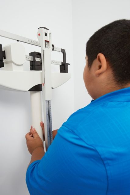
Jason E. Lang, MD, MPH, of Duke University, Durham, N.C., and his coauthors used the PEDSnet clinical data research network to conduct a retrospective cohort study of 507,496 children aged 2-17 years from 2009-2015, comparing the incidence of asthma in overweight and obese children to the incidence in healthy weight children.
The overall rate of new diagnoses of asthma was 2.4 per 1,000 patient years among normal-weight children and 3.2 per 1,000 patient years among obese children.
After adjustment for factors such as age, ethnicity, insurance status, sex, allergic rhinitis, food allergy, and proton pump inhibitor use, overweight children had a 17% higher risk of incident asthma, and obese children had a 26% higher risk of asthma, compared with children of normal weight. The relative risk of spirometry-confirmed asthma was 29% higher in obese children compared with normal-weight children, and the association between obesity and asthma persisted even when a second asthma encounter was required for the diagnosis.
Overall, the authors estimated that 23%-25% of clinically diagnosed asthma in children with obesity could be specifically attributed to obesity, and that in the overall population of children 10% of asthma was attributable to obesity.
“Currently, there are few known preventable risk factors that can be used to reduce childhood asthma,” wrote Dr. Lang and his coauthors. “With these data, it is suggested that reducing the onset of obesity in childhood would significantly reduce the public health burden of asthma in children.”
They noted that with current estimates of U.S. pediatric asthma prevalence being around 6-8 million cases, obesity could therefore account for up to 1 million of these cases.
The study was funded by the Patient-Centered Outcomes Research Institute, the Nemours Children’s Hospital and Nemours Children’s Health System. One author declared advisory board positions and consultancies with the pharmaceutical industry. The remaining researchers said they had no conflicts of interest.
SOURCE: Lang J et al. Pediatrics. 2018 Dec;142(6):e20182119.
While there has long been recognition of an association between childhood obesity and childhood asthma, the incidence of pediatric obesity–related asthma has not been well known. This study therefore addresses that gap in knowledge, and does so among children with a range of racial and ethnic backgrounds and while addressing potential confounders such as comorbidities and sociodemographic variables.
The findings show how significant a contribution obesity makes to the burden of childhood asthma, and also points to the potential increase in childhood asthma incidence that may arise with the increase in childhood obesity.
However, the good news is that this offers what may be the first modifiable risk factor for childhood asthma, which presents an opportunity for primary prevention of this common childhood condition.
Deepa Rastogi, MBBS, MS, is from the department of pediatrics at the Albert Einstein College of Medicine, New York. These comments are taken from an editorial (Pediatrics. 2018 Dec;142(6):e20182979.). No conflicts of interest were declared.
Around one-quarter of new asthma cases in children with obesity may be attributable to their obesity, according to research published in Pediatrics.
Jason E. Lang, MD, MPH, of Duke University, Durham, N.C., and his coauthors used the PEDSnet clinical data research network to conduct a retrospective cohort study of 507,496 children aged 2-17 years from 2009-2015, comparing the incidence of asthma in overweight and obese children to the incidence in healthy weight children.
The overall rate of new diagnoses of asthma was 2.4 per 1,000 patient years among normal-weight children and 3.2 per 1,000 patient years among obese children.
After adjustment for factors such as age, ethnicity, insurance status, sex, allergic rhinitis, food allergy, and proton pump inhibitor use, overweight children had a 17% higher risk of incident asthma, and obese children had a 26% higher risk of asthma, compared with children of normal weight. The relative risk of spirometry-confirmed asthma was 29% higher in obese children compared with normal-weight children, and the association between obesity and asthma persisted even when a second asthma encounter was required for the diagnosis.
Overall, the authors estimated that 23%-25% of clinically diagnosed asthma in children with obesity could be specifically attributed to obesity, and that in the overall population of children 10% of asthma was attributable to obesity.
“Currently, there are few known preventable risk factors that can be used to reduce childhood asthma,” wrote Dr. Lang and his coauthors. “With these data, it is suggested that reducing the onset of obesity in childhood would significantly reduce the public health burden of asthma in children.”
They noted that with current estimates of U.S. pediatric asthma prevalence being around 6-8 million cases, obesity could therefore account for up to 1 million of these cases.
The study was funded by the Patient-Centered Outcomes Research Institute, the Nemours Children’s Hospital and Nemours Children’s Health System. One author declared advisory board positions and consultancies with the pharmaceutical industry. The remaining researchers said they had no conflicts of interest.
SOURCE: Lang J et al. Pediatrics. 2018 Dec;142(6):e20182119.
FROM PEDIATRICS
Key clinical point: Obesity may be responsible for around 10% of childhood asthma.
Major finding: Obesity in children is associated with a 26% higher risk of asthma compared with normal-weight children.
Study details: A retrospective cohort study of 507,496 children.
Disclosures: The study was funded by the Patient-Centered Outcomes Research Institute and the Nemours Children’s Hospital and Nemours Children’s Health System. One author declared advisory board positions and consultancies with the pharmaceutical industry.
Source: Lang J et al. Pediatrics. 2018 Dec;142(6):e20182119. doi: 10.1542/peds.2018- 2119
Emapalumab safe, effective in pediatric primary hemophagocytic lymphohistiocytosis
, according to Franco Locatelli, MD, of the department of pediatric hematology and oncology at Ospedale Pediatrico Bambino Gesù, Rome.
The recently approved agent should be considered a new therapeutic option for this rare and life-threatening syndrome because of its targeted mode of action, Dr. Locatelli and his coinvestigators reported at the annual meeting of the American Society of Hematology.
Multiple lines of evidence have pointed to interferon gamma as a “rational target” in this disease, and elevated levels of interferon gamma are consistently observed in patients with HLH, Dr. Locatelli said in a press conference at the meeting.
Emapalumab binds to its target with high affinity, recognizing both free and receptor-bound interferon gamma, he added.
Primary HLH is a rare, life-threatening syndrome of hyperinflammation, characterized by prolonged fever, cytopenias, and splenomegaly and hepatomegaly, among other clinical manifestations, Dr. Locatelli said.
In the open-label, single-arm, pivotal study, 34 children with primary HLH were treated: 7 who were treatment naive and 27 who had failed conventional HLH therapy.
The patients received emapalumab intravenously with concomitant dexamethasone for up to 8 weeks, or extended to the point of allogeneic hematopoietic stem cell transplantation (HSCT), if needed.
The study met its primary endpoint of overall response rate higher than 40%, Dr. Locatelli reported. The overall response rate was 64.7% for all 34 treated patients (95% confidence interval, 46% to 80%; P = .0031), and 63% for the 27 patients who had failed prior therapy (95% CI, 42% to 81%; P = .0134), reported data show.
Response was rapid, occurring at a median of 8 days after starting emapalumab, and patients were in response for a median of 75% of days during treatment, Dr. Locatelli said.
Common adverse events in the study included infections, infusion-related reactions, pyrexia, and hypertension, while one patient had disseminated histoplasmosis that resolved with appropriate treatment, according to investigators.
In light of these results, the Food and Drug Administration approved emapalumab on Nov. 20, 2018, for the treatment of pediatric and adult patients with primary HLH with refractory, recurrent or progressive disease, or intolerance to conventional HLH treatments.
There is “certainly room for enlarging the indication” to first-line treatment of HLH once a sufficient number of previously untreated patients have been treated with the monoclonal antibody, Dr. Locatelli said.
However, a randomized trial would not be feasible, he said. “It’s a very rare disease, and it would be almost impossible to run a prospective, randomized trial in a reasonable period of time.”
The study described by Dr. Locatelli was sponsored by Novimmune. Study authors provided disclosures related to Sobi, Novimmune, Rocket Pharmaceuticals, Inc., AB2Bio, Novartis, Eli Lilly, Sanofi, UCB, Pfizer, and Abbvie. Two authors reported employment with Novimmune.
SOURCE: Locatelli F et al. ASH 2018; Abstract LBA-6.
FROM ASH 2018
Key clinical point: Emapalumab, an interferon gamma-blocking antibody, controls disease activity and has a favorable safety profile in patients with primary hemophagocytic lymphohistiocytosis.
Major finding: The overall response rate was 64.7% for all 34 treated patients (95% CI, 46%-80%; P = .0031), and 63% for the 27 patients who had failed prior therapy (95% CI, 42%-81%; P = .0134).
Study details: In the open-label, single-arm, pivotal study, 34 children with primary HLH were treated: 7 who were treatment naive and 27 who had failed conventional HLH therapy.
Disclosures: The study described by Dr. Locatelli was sponsored by Novimmune. Study authors provided disclosures related to Sobi, Novimmune, Rocket Pharmaceuticals, AB2Bio, Novartis, Eli Lilly, Sanofi, UCB, Pfizer, and Abbvie. Two authors reported employment with Novimmune.
Source: Locatelli F et al. ASH 2018; Abstract LBA-6.
Prenatal, postnatal neuroimaging IDs most Zika-related brain injuries
Prenatal ultrasound can identify most abnormalities in fetuses exposed to Zika virus during pregnancy, and neuroimaging after birth can detect infant exposure in cases that appeared normal on prenatal ultrasound, according to research published in JAMA Pediatrics.

“Absence of prolonged maternal viremia did not have predictive associations with normal fetal or neonatal brain imaging,” Sarah B. Mulkey, MD, PhD, from the division of fetal and transitional medicine at Children’s National Health System, in Washington, and her colleagues wrote. “Postnatal imaging can detect changes not seen on fetal imaging, supporting the current CDC [Centers for Disease Control and Prevention] recommendation for postnatal cranial [ultrasound].”
Dr. Mulkey and her colleagues performed a prospective cohort analysis of 82 pregnant women from Colombia and the United States who had clinical evidence of probable exposure to the Zika virus through travel (U.S. cases, 2 patients), physician referral, or community cases during June 2016-June 2017. Pregnant women underwent fetal MRI or ultrasound during the second or third trimesters between 4 weeks and 10 weeks after symptom onset, with infants undergoing brain MRI and cranial ultrasound after birth.
Of those 82 pregnancies, there were 80 live births, 1 case of termination because of severe fetal brain abnormalities, and 1 near-term fetal death of unknown cause. There was one death 3 days after birth and one instance of neurosurgical intervention from encephalocele. The researchers found 3 of 82 cases (4%) displayed fetal abnormalities from MRI, which consisted of 2 cases of heterotopias and malformations in cortical development and 1 case with parietal encephalocele, Chiari II malformation, and microcephaly. One infant had a normal ultrasound despite abnormalities displayed on fetal MRI.
After birth, of the 79 infants with normal ultrasound results, 53 infants underwent a postnatal brain MRI and Dr. Mulkey and her associates found 7 cases with mild abnormalities (13%). There were 57 infants who underwent cranial ultrasound, which yielded 21 cases of lenticulostriate vasculopathy, choroid plexus cysts, germinolytic/subependymal cysts, and/or calcification; these were poorly characterized by MRI.
“Normal fetal imaging had predictive associations with normal postnatal imaging or mild postnatal imaging findings unlikely to be of significant clinical consequence,” they said.
Nonetheless, “there is a need for long-term follow-up to assess the neurodevelopmental significance of these early neuroimaging findings, both normal and abnormal; such studies are in progress,” Dr. Mulkey and her colleagues said.
The researchers noted the timing of maternal infections and symptoms as well as the Zika testing, ultrasound, and MRI performance, technique during fetal MRI, and incomplete prenatal testing in the cohort as limitations in the study.
This study was funded in part by Children’s National Health System and by a philanthropic gift from the Ikaria Healthcare Fund. Dr. Mulkey received research support from the Thrasher Research Fund and is supported by awards from the National Institutes of Health National Center for Advancing Translational Sciences. The other authors reported no relevant conflicts of interest.
SOURCE: Mulkey SB et al. JAMA Pediatr. 2018 Nov. 26. doi: 10.1001/jamapediatrics.2018.4138.
While the study by Mulkey et al. adds to the body of evidence of prenatal and postnatal brain abnormalities, there are still many unanswered questions about the Zika virus and how to handle its unique diagnostic and clinical challenges, Margaret A. Honein, PhD, MPH, and Denise J. Jamieson, MD, MPH, wrote in a related editorial.
For example, Centers for Disease Control and Prevention recommendations state that infants with possible Zika exposure should receive an ophthalmologic and ultrasonographic examination at 1 month, and if the hearing test used otoacoustic emissions methods only, an automated auditory brainstem response test should be administered. While Mulkey et al. examined brain abnormalities in utero and in infants, it is not clear whether all CDC guidelines were followed in these cases.
In addition, because there is no reliable way to determine whether infants acquired Zika virus through the mother or through vertical transmission, assessing the proportion of congenitally infected infants or vertical-transmission infected infants who have neurodevelopmental disabilities and defects is not possible, they said. More longitudinal studies are needed to study the effects of the Zika virus and to prepare for the next outbreak.
“Zika was affecting pregnant women and their infants years before its teratogenic effect was recognized, and Zika will remain a serious risk to pregnant women and their infants until we have a safe vaccine that can fully prevent Zika virus infection during pregnancy,” they said. “Until then, ongoing public health efforts are essential to protect mothers and babies from this threat and ensure all disabilities associated with Zika virus infection are promptly identified, so that timely interventions can be provided.”
Dr. Honein is from the National Center on Birth Defects and Developmental Disabilities at the Centers for Disease Control and Prevention, and Dr. Jamieson is from the department of gynecology & obstetrics at Emory University School of Medicine, Atlanta. These comments summarize their editorial in response to Mulkey et al. (JAMA Pediatr. 2018 Nov. 26. doi: 10.1001/jamapediatrics.2018.4164). They reported no relevant conflicts of interest.
Prenatal ultrasound can identify most abnormalities in fetuses exposed to Zika virus during pregnancy, and neuroimaging after birth can detect infant exposure in cases that appeared normal on prenatal ultrasound, according to research published in JAMA Pediatrics.
“Absence of prolonged maternal viremia did not have predictive associations with normal fetal or neonatal brain imaging,” Sarah B. Mulkey, MD, PhD, from the division of fetal and transitional medicine at Children’s National Health System, in Washington, and her colleagues wrote. “Postnatal imaging can detect changes not seen on fetal imaging, supporting the current CDC [Centers for Disease Control and Prevention] recommendation for postnatal cranial [ultrasound].”
Dr. Mulkey and her colleagues performed a prospective cohort analysis of 82 pregnant women from Colombia and the United States who had clinical evidence of probable exposure to the Zika virus through travel (U.S. cases, 2 patients), physician referral, or community cases during June 2016-June 2017. Pregnant women underwent fetal MRI or ultrasound during the second or third trimesters between 4 weeks and 10 weeks after symptom onset, with infants undergoing brain MRI and cranial ultrasound after birth.
Of those 82 pregnancies, there were 80 live births, 1 case of termination because of severe fetal brain abnormalities, and 1 near-term fetal death of unknown cause. There was one death 3 days after birth and one instance of neurosurgical intervention from encephalocele. The researchers found 3 of 82 cases (4%) displayed fetal abnormalities from MRI, which consisted of 2 cases of heterotopias and malformations in cortical development and 1 case with parietal encephalocele, Chiari II malformation, and microcephaly. One infant had a normal ultrasound despite abnormalities displayed on fetal MRI.
After birth, of the 79 infants with normal ultrasound results, 53 infants underwent a postnatal brain MRI and Dr. Mulkey and her associates found 7 cases with mild abnormalities (13%). There were 57 infants who underwent cranial ultrasound, which yielded 21 cases of lenticulostriate vasculopathy, choroid plexus cysts, germinolytic/subependymal cysts, and/or calcification; these were poorly characterized by MRI.
“Normal fetal imaging had predictive associations with normal postnatal imaging or mild postnatal imaging findings unlikely to be of significant clinical consequence,” they said.
Nonetheless, “there is a need for long-term follow-up to assess the neurodevelopmental significance of these early neuroimaging findings, both normal and abnormal; such studies are in progress,” Dr. Mulkey and her colleagues said.
The researchers noted the timing of maternal infections and symptoms as well as the Zika testing, ultrasound, and MRI performance, technique during fetal MRI, and incomplete prenatal testing in the cohort as limitations in the study.
This study was funded in part by Children’s National Health System and by a philanthropic gift from the Ikaria Healthcare Fund. Dr. Mulkey received research support from the Thrasher Research Fund and is supported by awards from the National Institutes of Health National Center for Advancing Translational Sciences. The other authors reported no relevant conflicts of interest.
SOURCE: Mulkey SB et al. JAMA Pediatr. 2018 Nov. 26. doi: 10.1001/jamapediatrics.2018.4138.
FROM JAMA PEDIATRICS
Key clinical point:
Major finding: In 82 pregnant women, prenatal neuroimaging identified fetal abnormalities in 3 cases, while postnatal neuroimaging in 53 of the remaining 79 cases yielded an additional 7 cases with mild abnormalities.
Study details: A prospective longitudinal cohort study of 82 pregnant women with clinical evidence of probable Zika infection in Colombia and the United States.
Disclosures: This study was funded in part by Children’s National Health System and by a philanthropic gift from the Ikaria Healthcare Fund. Dr Mulkey received research support from the Thrasher Research Fund and is supported by awards from the National Institutes of Health National Center for Advancing Translational Sciences. The other authors reported no relevant conflicts of interest.
Source: Mulkey SB et al. JAMA Pediatr. 2018 Nov. 26; doi: 10.1001/jamapediatrics.2018.4138.
Gestational, umbilical cord vitamin D levels don’t predict atopic disease in offspring
according to study results published in the journal Allergy.

Áine Hennessy, PhD, from the School of Food and Nutritional Sciences at the University College Cork (Ireland), and her colleagues performed a prospective cohort study of 1,537 women in the Cork BASELINE Birth Cohort Study who underwent measurement of serum 25-hydroxyvitamin D (25[OH]D) from maternal sera followed by measurement of 25(OH)D in umbilical cord blood (1,050 cases). They then measured the prevalence of eczema, food allergy, allergic rhinitis, and asthma in infants at aged 2 and 5 years.
The researchers found at 2 years old, 5% of infants had persistent eczema, 4% of infants had a food allergy and 8% of infants had aeroallergen sensitization. At age 5 years, 15% of infants had asthma, while 5% had allergic rhinitis. Mothers whose children went on to have atopy did not differ in their 25(OH)D levels at 15 weeks’ gestation (mean 58.4 nmol/L vs. 58.5 nmol/L) or in the levels in umbilical cord blood (mean 35.2 nmol/L and 35.4 nmol/L).
Of the women in the cohort, 74% ranged in age from 25 to 34 years; 49% reported a personal history of allergy and 37% reported a paternal allergy. The mean birth weight of the infants was 3,458 g; infants were breastfed for mean 11.9 weeks, 73% of infants were breastfeeding by the time they left the hospital and 45% of infants were breastfeeding by age 2 months.
Limitations of the study included that parental atopy status was self-reported and that the researchers noted they did not examine genetic variants of immunoglobulin E synthesis or vitamin D receptor polymorphisms.
“To fully characterize relationships between intrauterine vitamin D exposure and allergic disease, analysis of well‐constructed, large‐scale prospective cohorts of maternal‐infant dyads, which take due consideration of an individual’s inherited risk, early‐life exposures and environmental confounders, is still needed,” Dr. Hennessy and her colleagues wrote.
The study was funded by grants from the European Commission, Ireland Health Research Board, National Children’s Research Centre, Food Standards Agency and Science Foundation Ireland. The authors report no relevant conflicts of interest.
SOURCE: Hennessy A et al. Allergy. 2018 Aug 7. doi: 10.1111/all.13590.
according to study results published in the journal Allergy.
Áine Hennessy, PhD, from the School of Food and Nutritional Sciences at the University College Cork (Ireland), and her colleagues performed a prospective cohort study of 1,537 women in the Cork BASELINE Birth Cohort Study who underwent measurement of serum 25-hydroxyvitamin D (25[OH]D) from maternal sera followed by measurement of 25(OH)D in umbilical cord blood (1,050 cases). They then measured the prevalence of eczema, food allergy, allergic rhinitis, and asthma in infants at aged 2 and 5 years.
The researchers found at 2 years old, 5% of infants had persistent eczema, 4% of infants had a food allergy and 8% of infants had aeroallergen sensitization. At age 5 years, 15% of infants had asthma, while 5% had allergic rhinitis. Mothers whose children went on to have atopy did not differ in their 25(OH)D levels at 15 weeks’ gestation (mean 58.4 nmol/L vs. 58.5 nmol/L) or in the levels in umbilical cord blood (mean 35.2 nmol/L and 35.4 nmol/L).
Of the women in the cohort, 74% ranged in age from 25 to 34 years; 49% reported a personal history of allergy and 37% reported a paternal allergy. The mean birth weight of the infants was 3,458 g; infants were breastfed for mean 11.9 weeks, 73% of infants were breastfeeding by the time they left the hospital and 45% of infants were breastfeeding by age 2 months.
Limitations of the study included that parental atopy status was self-reported and that the researchers noted they did not examine genetic variants of immunoglobulin E synthesis or vitamin D receptor polymorphisms.
“To fully characterize relationships between intrauterine vitamin D exposure and allergic disease, analysis of well‐constructed, large‐scale prospective cohorts of maternal‐infant dyads, which take due consideration of an individual’s inherited risk, early‐life exposures and environmental confounders, is still needed,” Dr. Hennessy and her colleagues wrote.
The study was funded by grants from the European Commission, Ireland Health Research Board, National Children’s Research Centre, Food Standards Agency and Science Foundation Ireland. The authors report no relevant conflicts of interest.
SOURCE: Hennessy A et al. Allergy. 2018 Aug 7. doi: 10.1111/all.13590.
FROM ALLERGY
Key clinical point: There was no association between prevalence of atopic disease and vitamin D levels measured in maternal sera during pregnancy or in umbilical cord blood.
Major finding: Maternal vitamin D levels at 15 weeks of gestation (mean 58.4 nmol/L vs. 58.5 nmol/L) and concentrations in umbilical cord blood (mean 35.2 nmol/L and 35.4 nmol/L) were not associated with such atopic diseases as eczema, food allergy, asthma, and allergic rhinitis in children.
Study details: A prospective group of 1,537 women and infant pairs from the Cork BASELINE Birth Cohort Study.
Disclosures: This study was funded by grants from the European Commission, Ireland Health Research Board, National Children’s Research Centre, Food Standards Agency and Science Foundation Ireland. The authors report no relevant conflicts of interest.
Source: Hennessy A et al. Allergy 2018 Aug 7. doi:10.1111/all.13590.

CDC: Acute flaccid myelitis on the decline for 2018
, according to the Centers for Disease Control and Prevention.
![]()
Through Nov. 30, 134 cases of AFM in 33 states have been confirmed out of the 299 reported to the CDC. That represents “an increase of 18 confirmed cases from the previous week, but most of the latest confirmed AFM cases occurred in September and October,” the CDC reported Dec. 3.
There has been a pattern of increased AFM cases every other year for the previous 4 years: 120 cases in 2014, 22 cases in 2015, 149 cases in 2016, and 33 cases in 2017. “Most cases are reported between August and October, and a marked reduction in cases is seen in November. That pattern appears to be repeating in 2018 because states have reported fewer [persons under investigation] over the past couple of weeks. CDC expects this decline to continue,” the statement said.
The 16 confirmed cases in Texas are the most for any state this year, followed by Colorado with 15; Ohio with 10; and Illinois, New Jersey, and Washington with 9 each. California and Florida have not had any confirmed cases as of Nov. 30. Since 2014, over 90% of all confirmed AFM cases have occurred in children, the CDC noted.
More information on AFM is available at a CDC website for health care professionals.
Adults with Congenital Heart Disease: The Critical Transition from Pediatric to Adult Care
From the Greenville Health System, Greenville, SC.
Abstract
- Objective: To review the management of patients with congenital heart disease (CHD) transitioning from pediatric to adult care.
- Methods: Review of the literature.
- Results: Persons with CHD require close monitoring and evaluation throughout life to address the physiologic consequences of acquired cardiopulmonary, gastrointestinal, and renal disease in the setting of underlying congenital heart lesions. During the transition from pediatric to adult cardiology, a high proportion of patients are lost to follow up or have long gaps in care after leaving pediatric cardiology, which can lead to poor outcomes. Care of the adult with CHD requires close coordination between the patient’s primary care physician), cardiologist, adult CHD specialist, and other specialists. The transition process for CHD patients begin at 12 years of age, with a goal of discussing future expectations of the child’s education, employment, and independent living. Successful transition programs use a systematic approach to addressing the medical, psychosocial, and educational/vocational needs of the adolescent as he or she moves from the family-centered pediatric to the patient-centered adult health care system.
- Conclusion: The transition from pediatric to adult care in ACHD patients is best provided through a comprehensive transition program that begins in early adolescence and enables patients to take charge of their disease process in adulthood, allowing them to maximize their quality of life and societal contributions.
Keywords: adult; congenital heart defects; complications; disease management; patient care team.
The population of adults with congenital heart disease (CHD) in developed countries has grown at an exponential rate in the past 4 decades. With advances in medical care and surgical interventions, the proportion of pediatric patients reaching adulthood has increased from 15% in the 1930s-60s to more than 95% for patients with mild to moderate complexity CHD. The rate of survival to adulthood for patients with severely complex CHD remains lower at around 56%.1
There are now more adult than pediatric patients with CHD in the United States. Because adult CHD (ACHD) patients have increased morbidity and mortality in their young adult years, it is imperative for all providers to understand and address the long-terms needs of this population. Unfortunately, adults with CHD do not always receive adequate health care, frequently because they are lost to follow-up, particularly during their adolescent years when they are expected to gain independence in their medical management. As will be discussed, CHD is a chronic illness fraught with numerous expected and unexpected complications that require close monitoring and re-interventions. Effectively anticipating and addressing these complications requires a standardized and comprehensive process of transition from the pediatric to the adult population to ensure maximal quality of life.
Epidemiology
The actual prevalence of ACHD in the United States is unknown, as a national database of persons with CHD has not been established.2 In contrast, Europe and China have maintained databases that enable ongoing monitoring of the evolving CHD epidemiology in those regions.3,4 The best estimates of the U.S. incidence and prevalence of ACHD stem from extrapolations from Canadian data. According to this data, there were more than 1.2 million adults with ACHD in the United States in 2012, with an anticipated 5% annual increase.1,5 However, the limitations of such extrapolations must be noted, as the Canadian population does not perfectly mirror that of the United States. Canada has lower infant mortality and adult obesity rates, and the United States has larger African American and Hispanic populations.6 Also, the juxtaposition of universal access to health care in Canada and the socioeconomic class–dependent access in the United States causes variations in care and outcomes of ACHD between the 2 populations. These differing genetic and social backgrounds may change the incidence of CHD by affecting maternal-fetal health.7
The 32nd Bethesda Conference on “Care of the Adult with Congenital Heart Disease” in 2000 was tasked with characterizing the ACHD population in the United States. This project found a prevalence similar to that of the Canadian extrapolation and showed that among persons with ACHD in the United States, 45% have mild disease, 37% moderate disease, and 13% severe disease.8
Characterizing the true incidence of CHD in the United States also has proven difficult because of variations in the definitions and methods used to detect lesions across the multiple studies that have looked at this matter. The estimated incidence of CHD, grouped according to severity, is 2.5 to 3 per 1000 live births for severe CHD, and from 3 to 13 per 1000 live births for moderately severe forms.9 When all forms are considered, including minor CHD (which includes tiny muscular ventricular septal defect [VSDs] present at birth and other trivial lesions), the total incidence of CHD rises to 75 per 1000 live births.9 CHD is one of the most common chronic illnesses in young adults with special health care needs.
Complications in Adulthood
The ACHD population represents a diverse population in terms of severity of CHD, history of surgical/catheter-based interventions, and socioeconomic status. However, a unifying clinical concern for these patients is their increased risk for morbidity and mortality in the young adult years. Despite the tremendous advances in the field over previous decades, mortality in this population in adulthood is estimated to be up to 7 times higher compared to age-matched peers.10,11 For many patients, palliative CHD interventions result in a significant drop in early morbidity and mortality but frequently lead to delayed morbidity from secondary multi-organ complications as these patients transition from pediatric to adult care. For example, due to the chronic low flow and low cardiac output state created by Fontan palliations, patients are at risk for diastolic dysfunction, arrhythmias, thrombotic events, protein-losing enteropathy, and cirrhosis/congestive hepatopathy, among other chronic conditions. These patients require frequent follow up and management by a multidisciplinary team including a primary care provider and various specialty groups.
Cardiac Disease
The most common causes of death in ACHD patients are heart failure (27%) and sudden cardiac death (19%), which occur at mean ages of 48 years and 39 years, respectively.10 The form of heart failure in ACHD patients is related to subsystemic right ventricle (RV) dysfunction, coronary under-perfusion, residual shunts, and residual progressive valve regurgitation. One of the more common examples of this is seen in palliated Tetralogy of Fallot patients who have undergone a transannular patch as a neonate. These patients are frequently left with significant pulmonary regurgitation leading to RV dilation, RV failure, and subsequent left ventricle (LV) failure. Another common example is the patient with dextro-transposition of the great arteries (DTGA) status post atrial switch who has a subsystemic morphologic RV. These patients will often develop significant RV dysfunction related to the chronic high pressures associated with systemic circulation.
Arrhythmias are a major contributor to morbidity and mortality in this population and are the most common reason patients present back into care. Difficult to control, multifocal intra-atrial re-entrant tachycardia is extremely common in ACHD, with an estimated 50% of all patients developing atrial arrhythmia by age 55. A recent study determined that the risk of atrial fibrillation in individuals with CHD was 22 times higher than that in age-matched controls, with the highest risk being seen in patients with conotruncal defects. Furthermore 10% of these patients develop heart failure.12 The risk for, incidence of, and type of arrhythmia is associated with the severity of the congenital heart lesions, as well as the type and timing of surgical interventions. Later age of repair has been associated with an increased likelihood of arrhythmias.13 Tetralogy of Fallot is an example of a moderately complex congenital heart lesion and is the most common cyanotic congenital heart lesion. In these individuals, the risk for atrial tachycardias, ventricular tachycardias, and need for a pacemaker is much higher than in age-matched peers.14 This includes an increased risk of sudden cardiac death, with many of these patients requiring placement of an implantable cardioverter defibrillator.
Pulmonary Disease
There is a 44% to 56% prevalence of restrictive pulmonary disease in the ACHD population, compared to 9% in the general non-CHD adult population. The incidence of pulmonary hypertension is also significantly higher in the ACHD population. The etiology for development of pulmonary hypertension is multifactorial, including chronic thromboembolic disease, left-sided heart disease, longstanding left to right shunts, and obstructive sleep apnea. These conditions have a significant impact on survival, as moderate/severe lung function impairment is an independent predictor of survival. Patients with shunt lesions are at risk of developing pulmonary arterial hypertension later in life,1 which quadruples the risk of all-cause mortality and more than triples the risk of cardiovascular mortality.7
Liver Disease
Hepatic morbidity associated with palliated CHD is often related to prior surgical interventions. The most common morbidities include chronic hepatitis C and liver failure from chronic under-perfusion and passive congestion, especially following Fontan palliation. Long term, these complications can lead to cirrhosis and hepatocellular carcinoma.15-18 Unfortunately, hepatic morbidity often precludes patients from having a surgical intervention, complicating the management of a population with baseline significantly increased need for surgical re-intervention.
Renal Disease
Approximately 50% of the ACHD population has some degree of renal dysfunction, with a higher incidence in cyanotic CHD.19 The American College of Cardiology/American Heart Association (ACC/AHA) recommends routine assessment of renal function in all adults with moderate and severe CHD due to its association with a poor prognosis in the ACHD population.1 In the immediate cardiac postoperative period, acute kidney injury leads to an eightfold increase in mortality.20 Over the longer term, there is a fivefold increase in mortality with moderate to severe renal impairment and a twofold increase with mild renal impairment compared to those with normal renal function.21
Acquired Cardiovascular Disease
As the ACHD patient ages, acquired cardiovascular disease becomes a significant issue. Approximately 80% of adults with CHD have at least 1 cardiovascular risk factor,22 though overall there is a relative lack of specific data regarding the U.S. population. Surveillance of the Canadian CHD population older than 65 years shows a 47% prevalence of hypertension,23 with increased risk in certain conditions such as aortic coarctation and renal disease associated with CHD. Although studies on the increased risk of diabetes mellitus in the ACHD population have yielded conflicting results,22,24 there is evidence of abnormal glucose metabolism in ACHD patients, which is a predictor of cardiac morbidity and mortality.25,26 The incidence of hyperlipidemia in U.S. ACHD patients is estimated to be at least as high as that of the general population.1 These factors combine with abnormalities in the myocardial substrate, hemodynamic abnormalities, arrhythmias, and sequelae of surgical repairs to confer an increased risk of ischemic heart disease and cerebrovascular disease in the ACHD population.15,27 One large case-control cohort study showed that the risk for ischemic heart disease was 16.5 times higher in patients with CHD as compared with non-CHD patients, with the highest incidence being in those with conotruncal defects and severe non-conotruncal defects. Interestingly, hypertension and diabetes were less common among CHD patients with ischemic heart disease than among non-CHD patients with ischemic heart disease.28
Adults with CHD have an increased risk for cerebrovascular disease compared with the general population, and cerebrovascular disease appears to occur at a younger age.29 The risk of ischemic stroke in individuals with ACHD younger than 55 years is 9 to 12 times higher than that in the general population. As in the general population, the incidence of ischemic stroke in ACHD patients increases with age, and in those older than 55 years, the incidence remains 2 to 4 times higher than in the general population.30,31
Clearly, complications arising from therapeutic interventions in CHD patients contribute significantly to morbidity/mortality in adult life, which underscores the need for life-long follow up and prevention of lapses in care.
The Transition from Pediatric to Adult Care
The monitoring and evaluation of CHD patients throughout life requires close coordination between the patient’s primary care physician, cardiologist, ACHD specialist, and other specialists, as appropriate. The timing of routine follow-up appointments is largely dependent on the severity of the congenital heart lesion and clinical status of the individual patient. Routine surveillance often includes cardiac imaging, preconception/genetic counseling, Holter screenings for arrhythmia, laboratory testing, and titration of medication. Unfortunately, only 30% of adults with CHD receive the recommended cardiac care.32
Children with chronic conditions transitioning to adulthood frequently experience a drop off in coordinated services as they transition from pediatric to adult medicine. Adult institutions often have less multidisciplinary support staff in the form of social workers and case management.33 Furthermore, a recent systematic review of articles that outlined the transition process from pediatric to adult cardiology in the CHD population showed that a high proportion of patients were either lost to follow up or had long gaps in care after leaving pediatric cardiology, with the first lapse in care commonly occurring at approximately age 19 years.28,34 A 2004 study showed that only 48% of adolescents with CHD underwent successful transition.35 A multicenter study of 922 ACHD patients found a gap in care lasting longer than 3 years in 42%, with 8% having gaps exceeding 10 years.36 Another study showed that lapses exceeding 2 years occurred in 63% of patients, with a median duration of lapse of medical care of 10 years. The most common reasons for lapse in care were: being told that cardiac follow up was not required (33%); being discharged from a children’s hospital without appropriate follow up plans in place (23%); being aware of need for follow up but having no symptoms (19%); lack of insurance (18%); and ignoring follow up recommendations for fear of receiving bad news (7%).37 Moreover, living independently from one’s parents was independently associated with a lapse in care, and patients with moderate complexity defects were more likely to experience a lapse than those with high complexity defects.
In the absence of a structured transition program, there is often delayed or inadequate care, which can result in significant emotional and financial stress on families and increased stress on the health care system.38 Inadequate, incomplete, or nonexistent transition and transfer for care has been shown to lead to poor health outcomes. Patients who experienced a lapse in care were 3 times more likely to require urgent cardiac intervention and to have an adverse outcome.37 The urgent interventions required by these patients included pulmonary valve replacement, mitral and tricuspid valve repair/replacement, VSD closure, pulmonary artery stenting, Fontan revision, and pacemaker/defibrillator placement.37 Clearly, there is significant room for improvement in the transition process of patients with CHD.
Best Practices in Transitioning CHD Patients to Adulthood
The overarching goal of pediatric to adult care CHD transition programs is to empower the patient and their support system to assume ownership of the disease process in order to maximize quality of life, life expectancy, and productivity.39 This involves ensuring that the patient has a thorough understanding of their diagnosis, heart anatomy, prior cardiac interventions, limitations imposed upon them by their condition, and the frequency of their anticipated follow-up care. The components of a successful transition program include a systematic approach to addressing the medical, psychosocial, and educational/vocational needs of the adolescent as he or she moves from the family-centered pediatric to the patient-centered adult health care system.40 The visits during the transition period are also an opportunity to discuss reproductive issues and the need for planning pregnancies for women with CHD. The goal is to encourage autonomy and promote ownership of their medical condition to the best of their social-cognitive ability. Adolescents should be encouraged to speak alone with their doctor to foster independence and self-management in their disease process; this has been shown to be protective against failure in transition.32 They should be encouraged to start calling their doctors, requesting refills, and making appointments.
The ACC/AHA appropriately recommend that the transition process for CHD patients begin at 12 years of age, with a goal of discussing future expectations of the child’s education, employment, and independent living.41 As part of this process, it is important that the practitioner educate the child and the family of the need for lifelong surveillance. The exact timing of the transition process is heavily influenced by a number of factors, including the degree of dependence of the child on their guardians, the severity of the congenital heart lesion, and the anticipated short- and long-term prognosis. However, regardless of these circumstances a reasonable age of transition into adult services should be established early on so that an expectation remains in place and the family is adequately prepared.
The challenge of learning how to navigate the adult health care system is as daunting for the transitioning patient as the medical consequences of their disease process. It is critical for patients to have easy access to social workers and case managers, ideally in the setting of a medical home, to connect them to community resources as needed. It is incredibly important that patients consider vocational options and training along with planning their insurance and/or disability qualifications as they move into adulthood. Establishing guardianship is also an important consideration for young adults with CHD who have remained dependent on their guardians.
Towards this end, the AHA/ACC has developed a curriculum that outlines the core principles that should be addressed before the patient moves to the ACHD clinic.27 The transition program should be flexible to accommodate for the patient’s degree of development, and the transfer should not occur before the adolescent has demonstrated the ability to independently manage their own health care to the greatest possible extent.
The ideal transition occurs through the auspices of a medical home that can coordinate the multiple subspecialists involved in the patient’s care. However, what often occurs is that a patient transitions from the pediatric cardiologist’s care before transitioning from pediatric to adult primary care. Prior to transition, the pediatric cardiologist should identify a cardiac destination at an ACHD center. This must be done in conjunction with the pediatrician, who will help identify an internist to take over the patient’s primary care and continue the coordination via the medical home. Information regarding the patient’s complete medical history, medication lists, exercise prescriptions, dietary restrictions, anesthetic issues, functional status, diagnostic studies, and comorbidities should be compiled in a health summary.40 To aid the process of transitioning, the ACC has developed several tools that may be used during the transition process, including self-knowledge assessments and medical summary templates.42
The Primary Care Provider’s Role and the Medical Home
Ensuring adequate care during the transition period requires close coordination between the patient’s various subspecialists. It is vital to avoid multiple subspecialists providing care without knowledge of each other’s treatments, as the treatment course for each ACHD patient is dependent on their unique history of prior therapies.27 The role of the primary care physician in establishing a “medical home” in this setting, as defined by the American Academy of Pediatrics Policy Statement, is exceedingly important.43 In this structure, the primary care physician maintains an easily accessible, centralized, and comprehensive record of the patient’s entire medical history, including surgical and medical treatments of both cardiac and noncardiac issues. Establishing the medical home framework is crucial, as it has been shown to lead to better outcomes in transitioning youth with special health care needs.44
With the establishment of this centralized care, the primary care physician must be able to negotiate the various medications prescribed by subspecialists and monitor for drug levels, adverse effects, and drug-drug interactions. ACHD patients also need regular monitoring and care aside from the care related to their chronic disease. Medical issues of particular importance to the ACHD patient include vaccinations, cholesterol and hypertension screening, cancer screening, and nutritional counseling. The primary care physician is responsible for addressing both the cardiac and noncardiac needs of the patient, ensuring that the patient truly receives comprehensive care. Thorough knowledge of a patient’s unique medical/surgical history will enable the primary care physician to adequately triage and appropriately refer for the development of a new symptom in an ACHD patient. On the other end of the spectrum, the patient’s subspecialists must maintain accurate and up to date information regarding their patient and transmit this to the patient’s medical home.
ACHD Centers
ACHD centers are an important part of any ACHD patient’s clinical team. Regardless of the complexity of the heart defect, there is tremendous value in the education and anticipatory guidance ACHD centers provide for their patients. The providers at these centers are often board-certified ACHD physicians who will work within a multidisciplinary team that includes mid-level practitioners, electrophysiology physicians, high-risk obstetrics/gynecology physicians, pulmonologists, and hepatologists. Each center differs in terms of their on-site interventional capacity and experience. However, the ACHD provider community is highly capable in directing patients who require interventions to centers of excellence, where there is proven quality in congenital surgical and interventional outcomes. ACHD centers often serve as the portals of reentry into care and are critical for providing and coordinating the complex care of each patient. Regular follow-up at these centers will ensure that patients receive adequate management of complications as they arise and preventive care against acquired heart disease.
The timing of follow-up at ACHD centers varies according to the complexity of heart disease. Individuals with simple CHD should be evaluated at an ACHD center at least once to determine the need for further follow-up. Patients with moderate and complex CHD must be monitored at a minimum of every 12 to 24 months, whereas very complex CHD should be monitored every 6 to 12 months.23 The frequency with which the young adult population moves may hinder adequate continuity of care and long-term follow up; a searchable directory of ACHD clinics in the United States and Canada can be found at www.achaheart.org/your-heart/clinic-directory/clinic-listings/.
Managing Specific Issues in the Transitioning Patient
Arrhythmias and Heart Failure
As mentioned, arrhythmias in the ACHD population are extremely common, the most frequent being atrial arrhythmias, especially in patients who have undergone single-ventricle repairs. Patients with late repair of an atrial septal defect have a high incidence of supraventricular tachycardia, which can be treated with catheter ablation procedures.45,46 Pacemaker implantation is another therapeutic option, especially in those who have undergone atrial surgery (ie, Mustard or Senning repairs). In these individuals, particularly in adolescents, abdominal implantation of a pacemaker generator may lessen the psychological impact of the external appearance of the pacemaker. In this population avoiding blunt contact sports (ie, tackle football, wrestling) is also important.28 It is critical that adult and pediatric electrophysiologists work together in the care and management of these complex, recurrent arrhythmias.
As noted above, many ACHD patients will require surgical or catheter-based interventions (as high as 40% in 1 study),47 and many encounter late-onset morbidity as a sequela of interventions earlier in life or as a result of failure of these interventions. The key for adult cardiologists and ACHD providers is delineating the reversible causes (eg, residual shunts, progressive valve regurgitation, and recoarctation) through routine intermittent surveillance, including echocardiograms, magnetic resonance imaging, and cardiac catherization, so that heart failure and arrhythmias in these patients can be identified, treated, and even prevented.
Pregnancy
Pregnancy is the most common reason for women to reenter care. Pregnancy is associated with significant hemodynamic changes, resulting in an increase in cardiac output to up to 150% of pre-pregnancy levels at 32 weeks, and up to 180% during labor. The outcome of pregnancy in patients with CHD is favorable in most instances provided that functional class systemic ventricular function is good. Accordingly, pregnancy is contraindicated in instances of severe pulmonary arterial hypertension (eg, Eisenmenger’s physiology), systemic ventricular dysfunction, and severe left-sided obstructions (eg, aortic or mitral stenosis). It is therefore imperative for health care providers to address the risks of pregnancy and the need for contraception with women who have CHD and are of reproductive age. The AHA advises beginning this conversation at 12 years of age and recommends that counseling be provided by health care providers knowledgeable in both CHD and adolescent health.27 Given the thrombotic potential of estrogen-containing contraception, the selection of contraception for women with ACHD who are seeking birth control requires discussion between the health care provider and patient. Though there have been limited studies performed on the use of contraception in women with CHD, a British working group has developed a consensus statement regarding contraceptive use in women with heart disease based on the World Health Organization format.48,49
Surgical Procedures
The need for operative interventions and re-interventions, both cardiac and noncardiac, in many CHD populations is considerable. Regardless of the type of procedure, these patients should receive a comprehensive preoperative risk assessment as well as appropriate intraoperative and postoperative management, ideally at a center equipped to meet their unique needs. Approaching the surgical procedure under the guidance of an interdisciplinary team that includes an ACHD specialist, anesthesiologist, and surgeon ensures that critical issues for appropriate management are not overlooked.
The preoperative risk assessment should be aimed at identifying and minimizing major risk factors. Historical factors to consider include the congenital lesion, outcomes of prior surgeries, history of syncope or arrhythmias, and the presence of pulmonary disease, among others.27 If the patient has a pacemaker or defibrillator, this should be interrogated prior to the planned procedure to ensure proper functioning. The preoperative evaluation should include consultation with a cardiologist experienced in the care of adolescents with CHD. Cardiac medications should be continued until the time of surgery and restarted as soon after the procedure as possible. Periods without anticoagulation should be minimized if indicated at baseline, and may require substituting warfarin with heparin in the preoperative period. The need for endocarditis prophylaxis must be considered as well; antibacterial prophylaxis prior to dental surgery, respiratory tract procedures, and procedures on infected skin and musculoskeletal structures is recommended in individuals with prosthetic heart valves, previous infective endocarditis, unrepaired CHD, repaired CHD with prosthetic material for the first 6 months after surgery, repaired CHD with residual defects, and valvulopathy after cardiac transplantation.50
Fluid management is important intraoperatively and post procedure, particularly in individuals who are preload dependent at baseline (eg, patients who have had Fontan palliation). Mechanical ventilation strategies with high positive end-expiratory pressure and tidal volume may decrease systemic venous return and should be monitored closely. Early mobilization and pulmonary toilet post extubation is advised to avoid pulmonary infection.
Exercise Capacity and Restrictions
The ability to exercise is an important factor in the quality of life of ACHD patients, especially in the adolescent period when participation in school and recreational athletics oftentimes functions as a social institution. Exercise ability is influenced by both real limitations imposed by limited cardiopulmonary reserve as a result of underlying pathology and by misconceptions of and anxiety about their ability to safely participate in these activities. There is evidence of diminished aerobic activity in all groups with CHD. However, symptomatic restrictions account for only approximately 30% of all barriers to exercise,51 and some studies have shown that exercise training programs can improve functional capacity and some standards of quality of life in CHD patients, in addition to the general health benefits associated with obesity prevention.52
Recommendations regarding exercise capacity are often addressed at primary care visits, and should be reinforced by the patient’s cardiologist. In general, most patients with repaired or mild defects can engage in moderate- to high-intensity exercise; those with more complex defects, cyanosis, or arrhythmias should be evaluated by an ACHD specialist to determine an appropriate level of activity.27 The “exercise prescription” provided to the patient should include type of exercise tolerated as well as heart rate goals and limits. In patients with extremely limited exercise capacity, a cardiac rehabilitation program can be beneficial. The presence of significant pulmonary hypertension, cyanosis or aortic stenosis, symptomatic arrhythmias, or evidence of myocardial dysfunction usually restricts the degree of exercise; full recommendations by activity and lesion type can be found in the guidelines proposed by the 36th Bethesda Conference.53 The importance of serial and regular evaluations is emphasized in these guidelines due to changing hemodynamic status of the patient over time as their cardiac lesions evolve and new complications arise.
Social and Psychological Impact of Chronic Illness
Living with a chronic disease can have a psychological impact on the child and transitioning adolescent. Frequent hospitalizations, physician visits, medical tests, and management of medical emergencies take a toll on the patient’s self-image and self-esteem, particularly during their formative adolescent years. Adolescents with CHD often feel “different” from their peers due to their condition,54 causing them to withhold disclosures about their heart disease to others out of fear of its impact on personal and professional relationships. Recent studies have shown that children and adolescents with CHD are at risk of internalizing problems and exhibiting behavior problems;55 they are also more likely to have impaired quality of life secondary to their increased incidence of psychosocial difficulties.56 The social and physical debility often experienced by patients with ACHD leads to a higher incidence of depression and anxiety in this population.57 Studies have shown that ACHD patients are interested in psychological treatment and peer support of their mood and anxiety disorders.58
At least some degree of the mental health issues ACHD patients experience is thought to have a physiological basis and be related to early cyanosis and neonatal surgical bypass duration. Prolonged duration of deep hypothermic circulatory arrest (DHCA) during corrective surgery is associated with reduced social competence, and has been found to be an independent risk factor for anxiety, depression, aggressive behavior, and attention deficiencies.59 In other studies, DHCA has been associated with decreased intellectual ability and worse fine motor skills, memory, and visuospatial skills, among other neurodevelopmental outcomes.60-62 Psychiatric disorders have also been associated with genetic syndromes like DiGeorge syndrome.63 This impacts executive function, leading to missed appointments, delay in clinical visits, and medication noncompliance. Given the potential for worse outcomes and risk of transition failure, primary care providers should routinely evaluate CHD patients for mood disorders and neurocognitive delay.
Social Determinants of Health and Medical Legal Partnerships
Social determinants of health and workplace discrimination play a large role in determining the ability of individuals with CHD to achieve adequate health care and maintain gainful employment. Individuals with CHD often face significant challenges as they prepare to enter the workforce, including discrimination within the workplace and maintaining employment through medical emergencies. Studies have shown that while educational milestones are similar between patients with and without CHD, those with CHD are much less likely to be employed.64 Challenges facing adolescents as they enter the workforce include hiring discrimination, physical challenges imposed by functional limitations, and misunderstanding of disease process and actual functional capacity. Career counseling is therefore an integral part of the transitioning process and should be started in early adolescence to allow for full assessment of mental, physical, and social abilities.65
Medical-legal partnerships (MLPs) can be extremely beneficial to the CHD population adversely affected by social determinants of health and workplace discrimination. These partnerships integrate lawyers into health care to address legal problems that create and perpetuate poor health; on a broader scale, these partnerships can advance and support public policy changes that improve population health.66
The major social determinants of health addressed by MLPs are income supports/insurance, housing/utilities, employment/education, legal status, and personal/family stability (summarized in the mnemonic I-HELP).67 Some of the more specific areas in which MLPs may assist in the delivery of care to CHD patients include case management, translation services, health literacy, and legal aid/legal services. ACHD patients also often experience a significant loss of services, including physical, occupational, and speech therapy and nutrition services, as adult clinics may not be prepared to provide these services. While physicians can best address the individual patient’s health, members of the legal system can address the systemic ailments that propagate that patient’s recurrent hospitalizations and other use of medical resources. Members of the legal system are present onsite in health care settings and participate in clinical meetings, which allows a coordinated and comprehensive screening for social needs that may harm a patient’s health.
Loss of insurance coverage is a major issue for transitioning patients; while adolescents with complex medical conditions are eligible for Medicaid to help cover the significant cost of their health care that goes beyond the abilities of private insurance, this eligibility ends when the patient turns 21. Additionally, the Social Security Administration re-determines supplemental security income (SSI) eligibility when the patient turns 18, and about one-third of patients lose their SSI benefits. Without appropriate guidance in navigating the nuances of insurance, many patients are at risk of losing coverage for their health care expenditures as they transition. Uninsured adults with a chronic condition are 8 times more likely to have unmet medical needs and 6 times more likely to have no access to routine care than insured young adults, with a 35% likelihood of the unmet medical need being due to cost.68 Undoubtedly, linability to pay for health care contributes to the lack of follow-up in the adult population, and MLPs may be a valuable tool to aid in ameliorating this problem.
Studies have shown that when legal services are used to address the social determinants of health, patients with chronic illnesses such as asthma and sickle cell disease have reduced hospital admissions.69,70 Other studies have shown utilization of MLPs has reduced spending on the care of high-need, high-use patients.71 According to a 2016 national survey of health care organizations conducted by the National Center for Medical-Legal Partnership, 39% clinicians reported improved compliance with medical treatment and 66% reported improved health outcomes after their patients received MLP services.72 Families referred to MLPs have shown increased access to health care, food, and income resources, and two-thirds reported improved child health and well-being.73 Given the numerous challenges faced by patients with CHD, involving MLPs as a part of both the transition process and the patient-centered medical home benefits these patients greatly and allows them to maximize their quality of life.
Conclusion
As more patients are living to adulthood with CHD, there is an increasing need for long-term care and adequate follow up, especially regarding the need for re-intervention and management of physiologic consequences of acquired cardiopulmonary, gastrointestinal, and renal disease in the setting of underlying congenital heart lesions. Beyond the purely medical aspects of the individual’s long-term management, psychosocial issues must be addressed, including preparing the individual for future employment and family counseling. Crucial to this process is the implementation of a comprehensive transition that begins in early adolescence and enables patients to take charge of their disease process in adulthood and ultimately enables them to maximize their quality of life and societal contributions. Towards this end, the role of MLPs may be important in ensuring that local, state, and federal policies that promote health harming norms are addressed.
Acknowledgments: We thank Dr. Frances ‘Kitty’ O’Hare and Bobbie Lewis for inviting us to submit this review; Dr. Russ Kolarik, Current Med-Peds Residency Program Director and Former President of the National Med-Peds Program Directors Association; and Dr. Peter Tilkemeier, Chairman, Department of Internal Medicine at Greenville Health System, for his unending support of our ACHD program. We also thank our patients, whose resounding resilience in the face of ongoing medical and psychosocial challenges remains our daily inspiration.
Corresponding author: Manisha S. Patel, MD, Department of Medicine and Pediatrics, Division of Cardiology, University of South Carolina School of Medicine, Columbia, SC; mpatel@ghs.org.
Financial disclosures: None.
1. Bhatt AB, Foster E, Kuehl K, et al. Congenital heart disease in the older adult: a scientific Statement from the American Heart Association. Circulation. 2015;26;131:1884-1931.
2. Gilboa SM, Devine OJ, Kucik JE, et al. Congenital heart defects in the United States: estimating the magnitude of the affected population in 2010. Circulation. 2016;134:101-109.
3. Dolk H, Loane M, Garne E. Congenital heart defects in Europe. Circulation. 2011;123:841-849.
4. Qu Y, Liu X, Zhuang J, et al. Incidence of congenital heart disease: the 9-year experience of the Guangdog Registry of Congenital Heart Disease, China. PloS One. 2016;11:e0159257.
5. Brickner ME, Hillis LD, Lange RA. Congenital heart disease in adults. N Engl J Med.
6. McGee Banks C. Variations in diversity in the United States and Canada. 2010. http://www.canadianstudies.isp.msu.edu/docs/Cherry%20McGee%20Banks.pdf.
7. van der Linde D, Konings EE, Slager MA, et al. Birth prevalence of congenital heart disease worldwide: a systematic review and meta-analysis. J Am Coll Cardiol. 2011;58:2241-2247.
8. Warnes CA, Liberthson R, Danielson GK, et al. Task force 1: the changing profile of congenital heart disease in adult life. J Am Coll Cardiol. 2001;37:1170-1175.
9. Hoffman JIE, Kaplan S. The incidence of congenital heart disease. J Am Coll Cardiol. 2002;39;1890-1900.
10. Verheugt CL, Uiterwaal CS, van der Velde ET, et al. Mortality in adult congenital heart disease. Eur Heart J. 2010;31:1220-1229.
11. Greutmann M, Tobler D, Kovacs AH, et al. Increasing mortality burden among adults with complex congenital heart disease. Congenit Heart Dis. 2015;10:117-127.
12. Mandalenakis Z, Rosengren A, Lappas G, et al. Atrial fibrillation burden in young patients with congenital heart disease. Circulation. 2018;137:928-937.
13. Fishberger SB, Wernovsky G, Gentles TL, et al. Factors that influence the development of atrial flutter after the Fontan operation. J Thorac Cardiovasc Surg. 1997;113:80–86.
14. Khairy P, Aboulhosn J, Gurvitz M; AARC. Arrhythmia burden in adults with surgically repaired tetralogy of Fallot. Circulation. 2010;122:868-875.
15. Ewe SH, Tan JL. Hepatotocellular carcinoma—a rare complication post Fontan operation. Congenit Heart Dis. 2009;4:103-106.
16. Ghaferi AA, Hutchins GM. Progression of liver pathology in patients undergoing the Fontan procedure: Chronic passive congestion, cardiac cirrhosis, hepatic adenoma, and hepatocellular carcinoma. J Thorac Cardiovasc Surg. 2005;129:1348-1352.
17. Saliba T, Dorkhom S, O’Reilly EM, et al. Hepatocellular carcinoma in two patients with cardiac cirrhosis. Eur J Gastroenterol Hepatol. 2010;22:889-891.
18. Wang A, Book W, McConnell M, et al. Prevalence of hepatitis C infection in adult patients who underwent congenital heart surgery prior to screening in 1992. Am J Cardiol. 2006;100:1307-1309.
19. Dimopoulos K, Diller GP, Koltsida E, et al. Prevalence, predictors, and prognostic value of renal dysfunction in adults with congenital heart disease. Circulation. 2008;117:2320-2328.
20. Karkouti K, Wijeysundera DN, Yau TM, et al. Acute kidney injury after cardiac surgery: focus on modifiable risk factors. Circulation. 2009;119:495-502.
21. Dimopoulos K, Diller GP, Koltsida E, et al. Prevalence, predictors, and prognostic value of renal dysfunction in adults with congenital heart disease. Circulation. 2008;117:2320-2328.
22. Moons P, Van Deyk K, Dedroog D, et al. Prevalence of cardiovascular risk factors in adults with congenital heart disease. Eur J Cardiovasc Prev Rehabil. 2006;13:612-616.
23. Afilalo J, Therrien J, Pilote L, et al. Geriatric congenital heart disease: burden of disease and predictors of mortality. J Am Coll Cardiol. 2011;58:1509-1515.
24. Billett J, Cowie MR, Gatzoulis MA, et al. Comorbidity, healthcare utilisation and process of care measures in patients with congenital heart disease in the UK: cross-sectional, population-based study with case-control analysis. Heart. 2008;94:1194-1199.
25. Hait G, Corpus M, Lamarre FR, et al. Alteration of glucose and insulin metabolism in congenital heart disease. Circulation. 1972;46:333-346.
26. Ohuchi H, Miyamoto Y, Yamamoto M, et al. High prevalence of abnormal glucose metabolism in young adult patients with complex congenital heart disease. Am Heart J. 2009;158:30-39.
27. Lui GK, Fernandes S, McElhinney DB. Management of cardiovascular risk factors in adults with congenital heart disease. J Am Heart Assoc. 2014;3(6):e001076.
28. Fedchenko M, Mandalenakis Z, Rosegren A, et al. Ischemic heart disease in children and young adults with congenital heart disease in Sweden. Int J Cardiol. 2017;248:143-148.
29. Mandalenakis Z, Rosengren A, Lappas G, et al. Ischemic stroke in children and young adults with congenital heart disease. J Am Heart Assoc. 2016. 23;5(2):e003071..
30. Hoffmann A, Chockalingam P, Balint OH, et al. Cerebrovascular accidents in adult patients with congenital heart disease. Heart. 2010;96:1223-1226.
31. Lanz J, Brophy JM, Therrien J, et al. Stroke in adults with congenital heart disease: incidence, cumulative risk, and predictors. Circulation. 2015;132:2385-2394
32. Sable C, Foster E, Uzark K, et al; on behalf of the American Heart Association Congenital Heart Defects Committee of the Council on Cardiovascular Disease in the Young, Council on Cardiovascular Nursing, Council on Clinical Cardiology, and Council on Peripheral Vascular Disease. Best practices in managing transition to adulthood for adolescents with congenital heart disease: the transition process and medical and psychosocial issues: a scientific statement from the American Heart Association. Circulation. 2011;123:1454-1485.
33. Steinway C, Gable J, Jan S. Transitioning to adult care: supporting youth with special health care needs. Children’s Hospital of Philadelphia: Policylab Evidence to Action in Brief. Spring 2017.
34. Heery E, Sheehan AM, While AE, Coyne I. Experiences and outcomes of transition from pediatric to adult health care services for young people with congenital heart disease: a systematic review. Congenit Heart Dis. 2015;10413-427.
35. Reid GJ, Irvine MJ, McCrindle BW, et al. Prevalence and correlates of successful transfer from pediatric to adult health care among a cohort of young adults with complex congenital heart defects. Pediatrics. 2004;113(part 1):e197-e205.
36. Gurvitz M, Valente AM, Broberg C, et al; AARCC. Prevalence and predictors of gaps in care among adult congenital heart disease patients (The Health, Education and Access Research Trial). J Am Coll Cardiol. 2013;61:2180-2184.
37. Yeung E, Kay J, Roosevelt GE, et al. Lapse of care as a predictor for morbidity in adults in congenital heart disease. Int J Cardiol. 2008;125:62-65.
38. Meadows AK, Bosco V, Tong E, et al. Transition and transfer from pediatric to adult care of young adults with complex congenital heart disease. Current Cardiol Rep. 2009; 11;4;291-297.
39. lum RW, Garell D, Hodgman CH, et al. Transition from child-centered to adult health-care systems for adolescents with chronic conditions: a position paper of the Society for Adolescent Medicine. J Adolesc Health. 1993;14:570-576.
40. Rosen DS, Blum RW, Britto M, et al; Society for Adolescent Medicine. Transition to adult health care for adolescents and young adults with chronic conditions: position paper of the Society for Adolescent Medicine. J Adolesc Health. 2003;33:309-311.
41. Reiss JG, Gibson RW, Walker LR. Health care transition: youth, family, and provider perspectives. Pediatrics. 2005;115:112-120.
42. Congenital heart disease transition tools. American College of Cardiology. www.acc.org/membership/sections-and-councils/adult-congenital-and-pediatric-cardiology-section/resources/chdtransitiontools. Accessed November 1, 2018.
43. American Academy of Pediatrics Medical Home Initiatives for Children With Special Needs Project Advisory Committee. Policy statement: organizational principles to guide and define the child health care system and/or improve the health of all children. Pediatrics. 2004;113(suppl):1545-1547.
44. Lotstein DS, McPherson M, Strickland B, Newacheck PW. Transition planning for youth with special health care needs: results from the National Survey of Children with Special Health Care Needs. Pediatrics. 2005;115:1562-1568.
45. Gatzoulis MA, Freeman MA, Siu SC, et al. Atrial arrhythmia after surgical closure of atrial septal defects in adults. N Engl J Med. 1999;340:839-846.
46. Triedman JK, Bergau DM, Saul JP, et al. Efficacy of radiofrequency ablation for control of intraatrial reentrant tachycardia in patients with congenital heart disease. J Am Coll Cardiol. 1997;30:1032-1038.
47. Zomer AC, Verheugt CL, Vaartjes I, et al. Surgery in adults with congenital heart disease. Circulation. 2011;124:2195-2201.
48. Silversides CK, Sermer M, Siu SC. Choosing the best contraceptive method for the adult with congenital heart disease. Curr Cardiol Rep. 2009;11:298-305.
49. Thorne S, Nelson-Piercy C, MacGregor A, et al. Pregnancy and contraception in heart disease and pulmonary arterial hypertension. J Fam Plann Reprod Health Care. 2006;32:75-81.
50. Wilson W, Taubert KA, Gewitz M, et al. Prevention of infective endocarditis: guidelines from the American Heart Association: a guideline from the American Heart Association Rheumatic Fever, Endocarditis, and Kawasaki Disease Committee, Council on Cardiovascular Disease in the Young, and the Council on Clinical Cardiology, Council on Cardiovascular Surgery and Anesthesia, and the Quality of Care and Outcomes Research Interdisciplinary Working Group [published correction appears in Circulation. 2007;116:e376–e377]. Circulation. 2007;116:1736-1754.
51. Warnes CA, et al. ACC/AHA 2008 guidelines for the management of adults with congenital heart disease: executive summary: a report of the American College of Cardiology/American Heart Association Task Force on Practice Guidelines (Writing Committee to Develop Guidelines for the Management of Adults With Congenital Heart Disease): developed in collaboration with the American Society of Echocardiography, Heart Rhythm Society, International Society for Adult Congenital Heart Disease, Society for Cardiovascular Angiography and Interventions, and Society of Thoracic Surgeons. Circulation. 2008;118:2395-2451.
52. Curran TJ, Rhodes J. Cardiac rehabilitation improves heart rate recovery following peak exercise in children with repaired congenital heart disease. Pediatr Cardiol. 2007;28:276-279.
53. Graham TP Jr, Driscoll DJ, Gersony WM, et al Task force 2: congenital heart disease. J Am Coll Cardiol. 2005;45:1326-1333.
54. Tong EM, Sparacino PS, Messias DK, et al. Growing up with congenital heart disease: the dilemmas of adolescents and young adults. Cardiol Young. 1998;8:303-309.
55. Karsdorp PA, Everaerd W, Kindt M, Mulder BJ. Psychological and cognitive functioning in children and adolescents with congenital heart disease: a meta-analysis. J Pediatr Psychol. 2007;32:527-541.
56. Kovacs AH, Moons P. Psychosocial functioning and quality of life in adults with congenital heart disease and heart failure. Heart Fail Clin. 2014;10:35-42.
57. Bromberg JI, Beasley PJ, D’Angelo EJ, et al. Depression and anxiety in adults with congenital heart disease: a pilot study. Heart Lung. 2003;32:105–10.
58. Kovacs AH, Bendell KL, Colman J, et al. Adults with congenital heart disease: psychological needs and treatment preferences. Congenit Heart Dis. 2009;4:139-146
59. Hovels-Gurich HH, Konrad K, Wiesner M, et al. Long term behavioural outcome after neonatal arterial switch operation for transposition of the great arteries. Arch Dis Child. 2002;87:506–510.
60. Forbess JM, Visconti KJ, Hancock-Friesen C, et al. Neurodevelopmental outcome after congenital heart surgery: results from an institutional registry. Circulation. 2002;106:195-102.
61. Bellinger DC, Wypij D, duPlessis AJ, et al. Neurodevelopmental status at eight years in children with dextro-transposition of the great arteries: the Boston Circulatory Arrest Trial. J Thorac Cardiovasc Surg. 2003;126:1385-1396.
62. Wypij D, Newburger JW, Rappaport LA, et al. The effect of duration of deep hypothermic circulatory arrest in infant heart surgery on late neurodevelopment: the Boston Circulatory Arrest Trial. J Thorac Cardiovasc Surg. 2003;126:1397-1403.
63. Tang SX, Yi JJ, Calkins ME, et al. Psychiatric disorders in 22q11.2 deletion syndrome are prevalent but undertreated. Pscychol Med 2017;44:1267-1277.
64. Simko LC, McGinnis KA, Schembri J. Educational needs of adults with congenital heart disease. J Cardiovasc Nurs. 2006;21:85-94.
65. Foster E, Graham TP Jr, Driscoll DJ, et al. Task force 2: special health care needs of adults with congenital heart disease. J Am Coll Cardiol. 2001;37:1176-1183.
66. Sandel M, Hansen M, Kahn R, et al. Medical-legal partnerships: transforming primary care by addressing the legal needs of vulnerable populations. Health Aff. 2010;29:1697-1705.
67. The National Center for Medical-Legal Partnership. medical-legalpartnership.org. Accessed November 1, 2018.
68. Callahan ST, Cooper WO. Access to health care for young adults with disabling chronic conditions. Arch Pediatr Adolesc Med. 2006;160:178-182.
69. Pettignano R, Caley SB, Bliss LR. Medical-legal partnership: impact on patients with sickle cell disease. Pediatrics. 2011;128:1482-1488.
70. Klein MD, Beck AF, Henize AW, et al. Doctors and lawyers collaborating to HeLP children--outcomes from a successful partnership between professions. J Health Care Poor Underserved. 2013;24:1063-1073.
71. Martin J, Martin A, Schultz C, Sandel M. Embedding civil legal aid in care of high-utilizing patients using medical-legal partnership. Health Affairs blog. 22 April 2015. www.healthaffairs.org/do/10.1377/hblog20150422.047143/full. Accessed November 1, 2018.
72. Regenstein M, Sharac J, Williamson A. The state of the medical legal partnership field: findings from the 2016 National Center for Medical-Legal Partnership Surveys. August 2017.
73. Weintraub D, Rodgers MA, Botcheva L, et al. Pilot study of medical-legal partnership to address social and legal needs of patients. J Health Care Poor Underserved. 2010;21(2 Suppl):157-168.
From the Greenville Health System, Greenville, SC.
Abstract
- Objective: To review the management of patients with congenital heart disease (CHD) transitioning from pediatric to adult care.
- Methods: Review of the literature.
- Results: Persons with CHD require close monitoring and evaluation throughout life to address the physiologic consequences of acquired cardiopulmonary, gastrointestinal, and renal disease in the setting of underlying congenital heart lesions. During the transition from pediatric to adult cardiology, a high proportion of patients are lost to follow up or have long gaps in care after leaving pediatric cardiology, which can lead to poor outcomes. Care of the adult with CHD requires close coordination between the patient’s primary care physician), cardiologist, adult CHD specialist, and other specialists. The transition process for CHD patients begin at 12 years of age, with a goal of discussing future expectations of the child’s education, employment, and independent living. Successful transition programs use a systematic approach to addressing the medical, psychosocial, and educational/vocational needs of the adolescent as he or she moves from the family-centered pediatric to the patient-centered adult health care system.
- Conclusion: The transition from pediatric to adult care in ACHD patients is best provided through a comprehensive transition program that begins in early adolescence and enables patients to take charge of their disease process in adulthood, allowing them to maximize their quality of life and societal contributions.
Keywords: adult; congenital heart defects; complications; disease management; patient care team.
The population of adults with congenital heart disease (CHD) in developed countries has grown at an exponential rate in the past 4 decades. With advances in medical care and surgical interventions, the proportion of pediatric patients reaching adulthood has increased from 15% in the 1930s-60s to more than 95% for patients with mild to moderate complexity CHD. The rate of survival to adulthood for patients with severely complex CHD remains lower at around 56%.1
There are now more adult than pediatric patients with CHD in the United States. Because adult CHD (ACHD) patients have increased morbidity and mortality in their young adult years, it is imperative for all providers to understand and address the long-terms needs of this population. Unfortunately, adults with CHD do not always receive adequate health care, frequently because they are lost to follow-up, particularly during their adolescent years when they are expected to gain independence in their medical management. As will be discussed, CHD is a chronic illness fraught with numerous expected and unexpected complications that require close monitoring and re-interventions. Effectively anticipating and addressing these complications requires a standardized and comprehensive process of transition from the pediatric to the adult population to ensure maximal quality of life.
Epidemiology
The actual prevalence of ACHD in the United States is unknown, as a national database of persons with CHD has not been established.2 In contrast, Europe and China have maintained databases that enable ongoing monitoring of the evolving CHD epidemiology in those regions.3,4 The best estimates of the U.S. incidence and prevalence of ACHD stem from extrapolations from Canadian data. According to this data, there were more than 1.2 million adults with ACHD in the United States in 2012, with an anticipated 5% annual increase.1,5 However, the limitations of such extrapolations must be noted, as the Canadian population does not perfectly mirror that of the United States. Canada has lower infant mortality and adult obesity rates, and the United States has larger African American and Hispanic populations.6 Also, the juxtaposition of universal access to health care in Canada and the socioeconomic class–dependent access in the United States causes variations in care and outcomes of ACHD between the 2 populations. These differing genetic and social backgrounds may change the incidence of CHD by affecting maternal-fetal health.7
The 32nd Bethesda Conference on “Care of the Adult with Congenital Heart Disease” in 2000 was tasked with characterizing the ACHD population in the United States. This project found a prevalence similar to that of the Canadian extrapolation and showed that among persons with ACHD in the United States, 45% have mild disease, 37% moderate disease, and 13% severe disease.8
Characterizing the true incidence of CHD in the United States also has proven difficult because of variations in the definitions and methods used to detect lesions across the multiple studies that have looked at this matter. The estimated incidence of CHD, grouped according to severity, is 2.5 to 3 per 1000 live births for severe CHD, and from 3 to 13 per 1000 live births for moderately severe forms.9 When all forms are considered, including minor CHD (which includes tiny muscular ventricular septal defect [VSDs] present at birth and other trivial lesions), the total incidence of CHD rises to 75 per 1000 live births.9 CHD is one of the most common chronic illnesses in young adults with special health care needs.
Complications in Adulthood
The ACHD population represents a diverse population in terms of severity of CHD, history of surgical/catheter-based interventions, and socioeconomic status. However, a unifying clinical concern for these patients is their increased risk for morbidity and mortality in the young adult years. Despite the tremendous advances in the field over previous decades, mortality in this population in adulthood is estimated to be up to 7 times higher compared to age-matched peers.10,11 For many patients, palliative CHD interventions result in a significant drop in early morbidity and mortality but frequently lead to delayed morbidity from secondary multi-organ complications as these patients transition from pediatric to adult care. For example, due to the chronic low flow and low cardiac output state created by Fontan palliations, patients are at risk for diastolic dysfunction, arrhythmias, thrombotic events, protein-losing enteropathy, and cirrhosis/congestive hepatopathy, among other chronic conditions. These patients require frequent follow up and management by a multidisciplinary team including a primary care provider and various specialty groups.
Cardiac Disease
The most common causes of death in ACHD patients are heart failure (27%) and sudden cardiac death (19%), which occur at mean ages of 48 years and 39 years, respectively.10 The form of heart failure in ACHD patients is related to subsystemic right ventricle (RV) dysfunction, coronary under-perfusion, residual shunts, and residual progressive valve regurgitation. One of the more common examples of this is seen in palliated Tetralogy of Fallot patients who have undergone a transannular patch as a neonate. These patients are frequently left with significant pulmonary regurgitation leading to RV dilation, RV failure, and subsequent left ventricle (LV) failure. Another common example is the patient with dextro-transposition of the great arteries (DTGA) status post atrial switch who has a subsystemic morphologic RV. These patients will often develop significant RV dysfunction related to the chronic high pressures associated with systemic circulation.
Arrhythmias are a major contributor to morbidity and mortality in this population and are the most common reason patients present back into care. Difficult to control, multifocal intra-atrial re-entrant tachycardia is extremely common in ACHD, with an estimated 50% of all patients developing atrial arrhythmia by age 55. A recent study determined that the risk of atrial fibrillation in individuals with CHD was 22 times higher than that in age-matched controls, with the highest risk being seen in patients with conotruncal defects. Furthermore 10% of these patients develop heart failure.12 The risk for, incidence of, and type of arrhythmia is associated with the severity of the congenital heart lesions, as well as the type and timing of surgical interventions. Later age of repair has been associated with an increased likelihood of arrhythmias.13 Tetralogy of Fallot is an example of a moderately complex congenital heart lesion and is the most common cyanotic congenital heart lesion. In these individuals, the risk for atrial tachycardias, ventricular tachycardias, and need for a pacemaker is much higher than in age-matched peers.14 This includes an increased risk of sudden cardiac death, with many of these patients requiring placement of an implantable cardioverter defibrillator.
Pulmonary Disease
There is a 44% to 56% prevalence of restrictive pulmonary disease in the ACHD population, compared to 9% in the general non-CHD adult population. The incidence of pulmonary hypertension is also significantly higher in the ACHD population. The etiology for development of pulmonary hypertension is multifactorial, including chronic thromboembolic disease, left-sided heart disease, longstanding left to right shunts, and obstructive sleep apnea. These conditions have a significant impact on survival, as moderate/severe lung function impairment is an independent predictor of survival. Patients with shunt lesions are at risk of developing pulmonary arterial hypertension later in life,1 which quadruples the risk of all-cause mortality and more than triples the risk of cardiovascular mortality.7
Liver Disease
Hepatic morbidity associated with palliated CHD is often related to prior surgical interventions. The most common morbidities include chronic hepatitis C and liver failure from chronic under-perfusion and passive congestion, especially following Fontan palliation. Long term, these complications can lead to cirrhosis and hepatocellular carcinoma.15-18 Unfortunately, hepatic morbidity often precludes patients from having a surgical intervention, complicating the management of a population with baseline significantly increased need for surgical re-intervention.
Renal Disease
Approximately 50% of the ACHD population has some degree of renal dysfunction, with a higher incidence in cyanotic CHD.19 The American College of Cardiology/American Heart Association (ACC/AHA) recommends routine assessment of renal function in all adults with moderate and severe CHD due to its association with a poor prognosis in the ACHD population.1 In the immediate cardiac postoperative period, acute kidney injury leads to an eightfold increase in mortality.20 Over the longer term, there is a fivefold increase in mortality with moderate to severe renal impairment and a twofold increase with mild renal impairment compared to those with normal renal function.21
Acquired Cardiovascular Disease
As the ACHD patient ages, acquired cardiovascular disease becomes a significant issue. Approximately 80% of adults with CHD have at least 1 cardiovascular risk factor,22 though overall there is a relative lack of specific data regarding the U.S. population. Surveillance of the Canadian CHD population older than 65 years shows a 47% prevalence of hypertension,23 with increased risk in certain conditions such as aortic coarctation and renal disease associated with CHD. Although studies on the increased risk of diabetes mellitus in the ACHD population have yielded conflicting results,22,24 there is evidence of abnormal glucose metabolism in ACHD patients, which is a predictor of cardiac morbidity and mortality.25,26 The incidence of hyperlipidemia in U.S. ACHD patients is estimated to be at least as high as that of the general population.1 These factors combine with abnormalities in the myocardial substrate, hemodynamic abnormalities, arrhythmias, and sequelae of surgical repairs to confer an increased risk of ischemic heart disease and cerebrovascular disease in the ACHD population.15,27 One large case-control cohort study showed that the risk for ischemic heart disease was 16.5 times higher in patients with CHD as compared with non-CHD patients, with the highest incidence being in those with conotruncal defects and severe non-conotruncal defects. Interestingly, hypertension and diabetes were less common among CHD patients with ischemic heart disease than among non-CHD patients with ischemic heart disease.28
Adults with CHD have an increased risk for cerebrovascular disease compared with the general population, and cerebrovascular disease appears to occur at a younger age.29 The risk of ischemic stroke in individuals with ACHD younger than 55 years is 9 to 12 times higher than that in the general population. As in the general population, the incidence of ischemic stroke in ACHD patients increases with age, and in those older than 55 years, the incidence remains 2 to 4 times higher than in the general population.30,31
Clearly, complications arising from therapeutic interventions in CHD patients contribute significantly to morbidity/mortality in adult life, which underscores the need for life-long follow up and prevention of lapses in care.
The Transition from Pediatric to Adult Care
The monitoring and evaluation of CHD patients throughout life requires close coordination between the patient’s primary care physician, cardiologist, ACHD specialist, and other specialists, as appropriate. The timing of routine follow-up appointments is largely dependent on the severity of the congenital heart lesion and clinical status of the individual patient. Routine surveillance often includes cardiac imaging, preconception/genetic counseling, Holter screenings for arrhythmia, laboratory testing, and titration of medication. Unfortunately, only 30% of adults with CHD receive the recommended cardiac care.32
Children with chronic conditions transitioning to adulthood frequently experience a drop off in coordinated services as they transition from pediatric to adult medicine. Adult institutions often have less multidisciplinary support staff in the form of social workers and case management.33 Furthermore, a recent systematic review of articles that outlined the transition process from pediatric to adult cardiology in the CHD population showed that a high proportion of patients were either lost to follow up or had long gaps in care after leaving pediatric cardiology, with the first lapse in care commonly occurring at approximately age 19 years.28,34 A 2004 study showed that only 48% of adolescents with CHD underwent successful transition.35 A multicenter study of 922 ACHD patients found a gap in care lasting longer than 3 years in 42%, with 8% having gaps exceeding 10 years.36 Another study showed that lapses exceeding 2 years occurred in 63% of patients, with a median duration of lapse of medical care of 10 years. The most common reasons for lapse in care were: being told that cardiac follow up was not required (33%); being discharged from a children’s hospital without appropriate follow up plans in place (23%); being aware of need for follow up but having no symptoms (19%); lack of insurance (18%); and ignoring follow up recommendations for fear of receiving bad news (7%).37 Moreover, living independently from one’s parents was independently associated with a lapse in care, and patients with moderate complexity defects were more likely to experience a lapse than those with high complexity defects.
In the absence of a structured transition program, there is often delayed or inadequate care, which can result in significant emotional and financial stress on families and increased stress on the health care system.38 Inadequate, incomplete, or nonexistent transition and transfer for care has been shown to lead to poor health outcomes. Patients who experienced a lapse in care were 3 times more likely to require urgent cardiac intervention and to have an adverse outcome.37 The urgent interventions required by these patients included pulmonary valve replacement, mitral and tricuspid valve repair/replacement, VSD closure, pulmonary artery stenting, Fontan revision, and pacemaker/defibrillator placement.37 Clearly, there is significant room for improvement in the transition process of patients with CHD.
Best Practices in Transitioning CHD Patients to Adulthood
The overarching goal of pediatric to adult care CHD transition programs is to empower the patient and their support system to assume ownership of the disease process in order to maximize quality of life, life expectancy, and productivity.39 This involves ensuring that the patient has a thorough understanding of their diagnosis, heart anatomy, prior cardiac interventions, limitations imposed upon them by their condition, and the frequency of their anticipated follow-up care. The components of a successful transition program include a systematic approach to addressing the medical, psychosocial, and educational/vocational needs of the adolescent as he or she moves from the family-centered pediatric to the patient-centered adult health care system.40 The visits during the transition period are also an opportunity to discuss reproductive issues and the need for planning pregnancies for women with CHD. The goal is to encourage autonomy and promote ownership of their medical condition to the best of their social-cognitive ability. Adolescents should be encouraged to speak alone with their doctor to foster independence and self-management in their disease process; this has been shown to be protective against failure in transition.32 They should be encouraged to start calling their doctors, requesting refills, and making appointments.
The ACC/AHA appropriately recommend that the transition process for CHD patients begin at 12 years of age, with a goal of discussing future expectations of the child’s education, employment, and independent living.41 As part of this process, it is important that the practitioner educate the child and the family of the need for lifelong surveillance. The exact timing of the transition process is heavily influenced by a number of factors, including the degree of dependence of the child on their guardians, the severity of the congenital heart lesion, and the anticipated short- and long-term prognosis. However, regardless of these circumstances a reasonable age of transition into adult services should be established early on so that an expectation remains in place and the family is adequately prepared.
The challenge of learning how to navigate the adult health care system is as daunting for the transitioning patient as the medical consequences of their disease process. It is critical for patients to have easy access to social workers and case managers, ideally in the setting of a medical home, to connect them to community resources as needed. It is incredibly important that patients consider vocational options and training along with planning their insurance and/or disability qualifications as they move into adulthood. Establishing guardianship is also an important consideration for young adults with CHD who have remained dependent on their guardians.
Towards this end, the AHA/ACC has developed a curriculum that outlines the core principles that should be addressed before the patient moves to the ACHD clinic.27 The transition program should be flexible to accommodate for the patient’s degree of development, and the transfer should not occur before the adolescent has demonstrated the ability to independently manage their own health care to the greatest possible extent.
The ideal transition occurs through the auspices of a medical home that can coordinate the multiple subspecialists involved in the patient’s care. However, what often occurs is that a patient transitions from the pediatric cardiologist’s care before transitioning from pediatric to adult primary care. Prior to transition, the pediatric cardiologist should identify a cardiac destination at an ACHD center. This must be done in conjunction with the pediatrician, who will help identify an internist to take over the patient’s primary care and continue the coordination via the medical home. Information regarding the patient’s complete medical history, medication lists, exercise prescriptions, dietary restrictions, anesthetic issues, functional status, diagnostic studies, and comorbidities should be compiled in a health summary.40 To aid the process of transitioning, the ACC has developed several tools that may be used during the transition process, including self-knowledge assessments and medical summary templates.42
The Primary Care Provider’s Role and the Medical Home
Ensuring adequate care during the transition period requires close coordination between the patient’s various subspecialists. It is vital to avoid multiple subspecialists providing care without knowledge of each other’s treatments, as the treatment course for each ACHD patient is dependent on their unique history of prior therapies.27 The role of the primary care physician in establishing a “medical home” in this setting, as defined by the American Academy of Pediatrics Policy Statement, is exceedingly important.43 In this structure, the primary care physician maintains an easily accessible, centralized, and comprehensive record of the patient’s entire medical history, including surgical and medical treatments of both cardiac and noncardiac issues. Establishing the medical home framework is crucial, as it has been shown to lead to better outcomes in transitioning youth with special health care needs.44
With the establishment of this centralized care, the primary care physician must be able to negotiate the various medications prescribed by subspecialists and monitor for drug levels, adverse effects, and drug-drug interactions. ACHD patients also need regular monitoring and care aside from the care related to their chronic disease. Medical issues of particular importance to the ACHD patient include vaccinations, cholesterol and hypertension screening, cancer screening, and nutritional counseling. The primary care physician is responsible for addressing both the cardiac and noncardiac needs of the patient, ensuring that the patient truly receives comprehensive care. Thorough knowledge of a patient’s unique medical/surgical history will enable the primary care physician to adequately triage and appropriately refer for the development of a new symptom in an ACHD patient. On the other end of the spectrum, the patient’s subspecialists must maintain accurate and up to date information regarding their patient and transmit this to the patient’s medical home.
ACHD Centers
ACHD centers are an important part of any ACHD patient’s clinical team. Regardless of the complexity of the heart defect, there is tremendous value in the education and anticipatory guidance ACHD centers provide for their patients. The providers at these centers are often board-certified ACHD physicians who will work within a multidisciplinary team that includes mid-level practitioners, electrophysiology physicians, high-risk obstetrics/gynecology physicians, pulmonologists, and hepatologists. Each center differs in terms of their on-site interventional capacity and experience. However, the ACHD provider community is highly capable in directing patients who require interventions to centers of excellence, where there is proven quality in congenital surgical and interventional outcomes. ACHD centers often serve as the portals of reentry into care and are critical for providing and coordinating the complex care of each patient. Regular follow-up at these centers will ensure that patients receive adequate management of complications as they arise and preventive care against acquired heart disease.
The timing of follow-up at ACHD centers varies according to the complexity of heart disease. Individuals with simple CHD should be evaluated at an ACHD center at least once to determine the need for further follow-up. Patients with moderate and complex CHD must be monitored at a minimum of every 12 to 24 months, whereas very complex CHD should be monitored every 6 to 12 months.23 The frequency with which the young adult population moves may hinder adequate continuity of care and long-term follow up; a searchable directory of ACHD clinics in the United States and Canada can be found at www.achaheart.org/your-heart/clinic-directory/clinic-listings/.
Managing Specific Issues in the Transitioning Patient
Arrhythmias and Heart Failure
As mentioned, arrhythmias in the ACHD population are extremely common, the most frequent being atrial arrhythmias, especially in patients who have undergone single-ventricle repairs. Patients with late repair of an atrial septal defect have a high incidence of supraventricular tachycardia, which can be treated with catheter ablation procedures.45,46 Pacemaker implantation is another therapeutic option, especially in those who have undergone atrial surgery (ie, Mustard or Senning repairs). In these individuals, particularly in adolescents, abdominal implantation of a pacemaker generator may lessen the psychological impact of the external appearance of the pacemaker. In this population avoiding blunt contact sports (ie, tackle football, wrestling) is also important.28 It is critical that adult and pediatric electrophysiologists work together in the care and management of these complex, recurrent arrhythmias.
As noted above, many ACHD patients will require surgical or catheter-based interventions (as high as 40% in 1 study),47 and many encounter late-onset morbidity as a sequela of interventions earlier in life or as a result of failure of these interventions. The key for adult cardiologists and ACHD providers is delineating the reversible causes (eg, residual shunts, progressive valve regurgitation, and recoarctation) through routine intermittent surveillance, including echocardiograms, magnetic resonance imaging, and cardiac catherization, so that heart failure and arrhythmias in these patients can be identified, treated, and even prevented.
Pregnancy
Pregnancy is the most common reason for women to reenter care. Pregnancy is associated with significant hemodynamic changes, resulting in an increase in cardiac output to up to 150% of pre-pregnancy levels at 32 weeks, and up to 180% during labor. The outcome of pregnancy in patients with CHD is favorable in most instances provided that functional class systemic ventricular function is good. Accordingly, pregnancy is contraindicated in instances of severe pulmonary arterial hypertension (eg, Eisenmenger’s physiology), systemic ventricular dysfunction, and severe left-sided obstructions (eg, aortic or mitral stenosis). It is therefore imperative for health care providers to address the risks of pregnancy and the need for contraception with women who have CHD and are of reproductive age. The AHA advises beginning this conversation at 12 years of age and recommends that counseling be provided by health care providers knowledgeable in both CHD and adolescent health.27 Given the thrombotic potential of estrogen-containing contraception, the selection of contraception for women with ACHD who are seeking birth control requires discussion between the health care provider and patient. Though there have been limited studies performed on the use of contraception in women with CHD, a British working group has developed a consensus statement regarding contraceptive use in women with heart disease based on the World Health Organization format.48,49
Surgical Procedures
The need for operative interventions and re-interventions, both cardiac and noncardiac, in many CHD populations is considerable. Regardless of the type of procedure, these patients should receive a comprehensive preoperative risk assessment as well as appropriate intraoperative and postoperative management, ideally at a center equipped to meet their unique needs. Approaching the surgical procedure under the guidance of an interdisciplinary team that includes an ACHD specialist, anesthesiologist, and surgeon ensures that critical issues for appropriate management are not overlooked.
The preoperative risk assessment should be aimed at identifying and minimizing major risk factors. Historical factors to consider include the congenital lesion, outcomes of prior surgeries, history of syncope or arrhythmias, and the presence of pulmonary disease, among others.27 If the patient has a pacemaker or defibrillator, this should be interrogated prior to the planned procedure to ensure proper functioning. The preoperative evaluation should include consultation with a cardiologist experienced in the care of adolescents with CHD. Cardiac medications should be continued until the time of surgery and restarted as soon after the procedure as possible. Periods without anticoagulation should be minimized if indicated at baseline, and may require substituting warfarin with heparin in the preoperative period. The need for endocarditis prophylaxis must be considered as well; antibacterial prophylaxis prior to dental surgery, respiratory tract procedures, and procedures on infected skin and musculoskeletal structures is recommended in individuals with prosthetic heart valves, previous infective endocarditis, unrepaired CHD, repaired CHD with prosthetic material for the first 6 months after surgery, repaired CHD with residual defects, and valvulopathy after cardiac transplantation.50
Fluid management is important intraoperatively and post procedure, particularly in individuals who are preload dependent at baseline (eg, patients who have had Fontan palliation). Mechanical ventilation strategies with high positive end-expiratory pressure and tidal volume may decrease systemic venous return and should be monitored closely. Early mobilization and pulmonary toilet post extubation is advised to avoid pulmonary infection.
Exercise Capacity and Restrictions
The ability to exercise is an important factor in the quality of life of ACHD patients, especially in the adolescent period when participation in school and recreational athletics oftentimes functions as a social institution. Exercise ability is influenced by both real limitations imposed by limited cardiopulmonary reserve as a result of underlying pathology and by misconceptions of and anxiety about their ability to safely participate in these activities. There is evidence of diminished aerobic activity in all groups with CHD. However, symptomatic restrictions account for only approximately 30% of all barriers to exercise,51 and some studies have shown that exercise training programs can improve functional capacity and some standards of quality of life in CHD patients, in addition to the general health benefits associated with obesity prevention.52
Recommendations regarding exercise capacity are often addressed at primary care visits, and should be reinforced by the patient’s cardiologist. In general, most patients with repaired or mild defects can engage in moderate- to high-intensity exercise; those with more complex defects, cyanosis, or arrhythmias should be evaluated by an ACHD specialist to determine an appropriate level of activity.27 The “exercise prescription” provided to the patient should include type of exercise tolerated as well as heart rate goals and limits. In patients with extremely limited exercise capacity, a cardiac rehabilitation program can be beneficial. The presence of significant pulmonary hypertension, cyanosis or aortic stenosis, symptomatic arrhythmias, or evidence of myocardial dysfunction usually restricts the degree of exercise; full recommendations by activity and lesion type can be found in the guidelines proposed by the 36th Bethesda Conference.53 The importance of serial and regular evaluations is emphasized in these guidelines due to changing hemodynamic status of the patient over time as their cardiac lesions evolve and new complications arise.
Social and Psychological Impact of Chronic Illness
Living with a chronic disease can have a psychological impact on the child and transitioning adolescent. Frequent hospitalizations, physician visits, medical tests, and management of medical emergencies take a toll on the patient’s self-image and self-esteem, particularly during their formative adolescent years. Adolescents with CHD often feel “different” from their peers due to their condition,54 causing them to withhold disclosures about their heart disease to others out of fear of its impact on personal and professional relationships. Recent studies have shown that children and adolescents with CHD are at risk of internalizing problems and exhibiting behavior problems;55 they are also more likely to have impaired quality of life secondary to their increased incidence of psychosocial difficulties.56 The social and physical debility often experienced by patients with ACHD leads to a higher incidence of depression and anxiety in this population.57 Studies have shown that ACHD patients are interested in psychological treatment and peer support of their mood and anxiety disorders.58
At least some degree of the mental health issues ACHD patients experience is thought to have a physiological basis and be related to early cyanosis and neonatal surgical bypass duration. Prolonged duration of deep hypothermic circulatory arrest (DHCA) during corrective surgery is associated with reduced social competence, and has been found to be an independent risk factor for anxiety, depression, aggressive behavior, and attention deficiencies.59 In other studies, DHCA has been associated with decreased intellectual ability and worse fine motor skills, memory, and visuospatial skills, among other neurodevelopmental outcomes.60-62 Psychiatric disorders have also been associated with genetic syndromes like DiGeorge syndrome.63 This impacts executive function, leading to missed appointments, delay in clinical visits, and medication noncompliance. Given the potential for worse outcomes and risk of transition failure, primary care providers should routinely evaluate CHD patients for mood disorders and neurocognitive delay.
Social Determinants of Health and Medical Legal Partnerships
Social determinants of health and workplace discrimination play a large role in determining the ability of individuals with CHD to achieve adequate health care and maintain gainful employment. Individuals with CHD often face significant challenges as they prepare to enter the workforce, including discrimination within the workplace and maintaining employment through medical emergencies. Studies have shown that while educational milestones are similar between patients with and without CHD, those with CHD are much less likely to be employed.64 Challenges facing adolescents as they enter the workforce include hiring discrimination, physical challenges imposed by functional limitations, and misunderstanding of disease process and actual functional capacity. Career counseling is therefore an integral part of the transitioning process and should be started in early adolescence to allow for full assessment of mental, physical, and social abilities.65
Medical-legal partnerships (MLPs) can be extremely beneficial to the CHD population adversely affected by social determinants of health and workplace discrimination. These partnerships integrate lawyers into health care to address legal problems that create and perpetuate poor health; on a broader scale, these partnerships can advance and support public policy changes that improve population health.66
The major social determinants of health addressed by MLPs are income supports/insurance, housing/utilities, employment/education, legal status, and personal/family stability (summarized in the mnemonic I-HELP).67 Some of the more specific areas in which MLPs may assist in the delivery of care to CHD patients include case management, translation services, health literacy, and legal aid/legal services. ACHD patients also often experience a significant loss of services, including physical, occupational, and speech therapy and nutrition services, as adult clinics may not be prepared to provide these services. While physicians can best address the individual patient’s health, members of the legal system can address the systemic ailments that propagate that patient’s recurrent hospitalizations and other use of medical resources. Members of the legal system are present onsite in health care settings and participate in clinical meetings, which allows a coordinated and comprehensive screening for social needs that may harm a patient’s health.
Loss of insurance coverage is a major issue for transitioning patients; while adolescents with complex medical conditions are eligible for Medicaid to help cover the significant cost of their health care that goes beyond the abilities of private insurance, this eligibility ends when the patient turns 21. Additionally, the Social Security Administration re-determines supplemental security income (SSI) eligibility when the patient turns 18, and about one-third of patients lose their SSI benefits. Without appropriate guidance in navigating the nuances of insurance, many patients are at risk of losing coverage for their health care expenditures as they transition. Uninsured adults with a chronic condition are 8 times more likely to have unmet medical needs and 6 times more likely to have no access to routine care than insured young adults, with a 35% likelihood of the unmet medical need being due to cost.68 Undoubtedly, linability to pay for health care contributes to the lack of follow-up in the adult population, and MLPs may be a valuable tool to aid in ameliorating this problem.
Studies have shown that when legal services are used to address the social determinants of health, patients with chronic illnesses such as asthma and sickle cell disease have reduced hospital admissions.69,70 Other studies have shown utilization of MLPs has reduced spending on the care of high-need, high-use patients.71 According to a 2016 national survey of health care organizations conducted by the National Center for Medical-Legal Partnership, 39% clinicians reported improved compliance with medical treatment and 66% reported improved health outcomes after their patients received MLP services.72 Families referred to MLPs have shown increased access to health care, food, and income resources, and two-thirds reported improved child health and well-being.73 Given the numerous challenges faced by patients with CHD, involving MLPs as a part of both the transition process and the patient-centered medical home benefits these patients greatly and allows them to maximize their quality of life.
Conclusion
As more patients are living to adulthood with CHD, there is an increasing need for long-term care and adequate follow up, especially regarding the need for re-intervention and management of physiologic consequences of acquired cardiopulmonary, gastrointestinal, and renal disease in the setting of underlying congenital heart lesions. Beyond the purely medical aspects of the individual’s long-term management, psychosocial issues must be addressed, including preparing the individual for future employment and family counseling. Crucial to this process is the implementation of a comprehensive transition that begins in early adolescence and enables patients to take charge of their disease process in adulthood and ultimately enables them to maximize their quality of life and societal contributions. Towards this end, the role of MLPs may be important in ensuring that local, state, and federal policies that promote health harming norms are addressed.
Acknowledgments: We thank Dr. Frances ‘Kitty’ O’Hare and Bobbie Lewis for inviting us to submit this review; Dr. Russ Kolarik, Current Med-Peds Residency Program Director and Former President of the National Med-Peds Program Directors Association; and Dr. Peter Tilkemeier, Chairman, Department of Internal Medicine at Greenville Health System, for his unending support of our ACHD program. We also thank our patients, whose resounding resilience in the face of ongoing medical and psychosocial challenges remains our daily inspiration.
Corresponding author: Manisha S. Patel, MD, Department of Medicine and Pediatrics, Division of Cardiology, University of South Carolina School of Medicine, Columbia, SC; mpatel@ghs.org.
Financial disclosures: None.
1. Bhatt AB, Foster E, Kuehl K, et al. Congenital heart disease in the older adult: a scientific Statement from the American Heart Association. Circulation. 2015;26;131:1884-1931.
2. Gilboa SM, Devine OJ, Kucik JE, et al. Congenital heart defects in the United States: estimating the magnitude of the affected population in 2010. Circulation. 2016;134:101-109.
3. Dolk H, Loane M, Garne E. Congenital heart defects in Europe. Circulation. 2011;123:841-849.
4. Qu Y, Liu X, Zhuang J, et al. Incidence of congenital heart disease: the 9-year experience of the Guangdog Registry of Congenital Heart Disease, China. PloS One. 2016;11:e0159257.
5. Brickner ME, Hillis LD, Lange RA. Congenital heart disease in adults. N Engl J Med.
6. McGee Banks C. Variations in diversity in the United States and Canada. 2010. http://www.canadianstudies.isp.msu.edu/docs/Cherry%20McGee%20Banks.pdf.
7. van der Linde D, Konings EE, Slager MA, et al. Birth prevalence of congenital heart disease worldwide: a systematic review and meta-analysis. J Am Coll Cardiol. 2011;58:2241-2247.
8. Warnes CA, Liberthson R, Danielson GK, et al. Task force 1: the changing profile of congenital heart disease in adult life. J Am Coll Cardiol. 2001;37:1170-1175.
9. Hoffman JIE, Kaplan S. The incidence of congenital heart disease. J Am Coll Cardiol. 2002;39;1890-1900.
10. Verheugt CL, Uiterwaal CS, van der Velde ET, et al. Mortality in adult congenital heart disease. Eur Heart J. 2010;31:1220-1229.
11. Greutmann M, Tobler D, Kovacs AH, et al. Increasing mortality burden among adults with complex congenital heart disease. Congenit Heart Dis. 2015;10:117-127.
12. Mandalenakis Z, Rosengren A, Lappas G, et al. Atrial fibrillation burden in young patients with congenital heart disease. Circulation. 2018;137:928-937.
13. Fishberger SB, Wernovsky G, Gentles TL, et al. Factors that influence the development of atrial flutter after the Fontan operation. J Thorac Cardiovasc Surg. 1997;113:80–86.
14. Khairy P, Aboulhosn J, Gurvitz M; AARC. Arrhythmia burden in adults with surgically repaired tetralogy of Fallot. Circulation. 2010;122:868-875.
15. Ewe SH, Tan JL. Hepatotocellular carcinoma—a rare complication post Fontan operation. Congenit Heart Dis. 2009;4:103-106.
16. Ghaferi AA, Hutchins GM. Progression of liver pathology in patients undergoing the Fontan procedure: Chronic passive congestion, cardiac cirrhosis, hepatic adenoma, and hepatocellular carcinoma. J Thorac Cardiovasc Surg. 2005;129:1348-1352.
17. Saliba T, Dorkhom S, O’Reilly EM, et al. Hepatocellular carcinoma in two patients with cardiac cirrhosis. Eur J Gastroenterol Hepatol. 2010;22:889-891.
18. Wang A, Book W, McConnell M, et al. Prevalence of hepatitis C infection in adult patients who underwent congenital heart surgery prior to screening in 1992. Am J Cardiol. 2006;100:1307-1309.
19. Dimopoulos K, Diller GP, Koltsida E, et al. Prevalence, predictors, and prognostic value of renal dysfunction in adults with congenital heart disease. Circulation. 2008;117:2320-2328.
20. Karkouti K, Wijeysundera DN, Yau TM, et al. Acute kidney injury after cardiac surgery: focus on modifiable risk factors. Circulation. 2009;119:495-502.
21. Dimopoulos K, Diller GP, Koltsida E, et al. Prevalence, predictors, and prognostic value of renal dysfunction in adults with congenital heart disease. Circulation. 2008;117:2320-2328.
22. Moons P, Van Deyk K, Dedroog D, et al. Prevalence of cardiovascular risk factors in adults with congenital heart disease. Eur J Cardiovasc Prev Rehabil. 2006;13:612-616.
23. Afilalo J, Therrien J, Pilote L, et al. Geriatric congenital heart disease: burden of disease and predictors of mortality. J Am Coll Cardiol. 2011;58:1509-1515.
24. Billett J, Cowie MR, Gatzoulis MA, et al. Comorbidity, healthcare utilisation and process of care measures in patients with congenital heart disease in the UK: cross-sectional, population-based study with case-control analysis. Heart. 2008;94:1194-1199.
25. Hait G, Corpus M, Lamarre FR, et al. Alteration of glucose and insulin metabolism in congenital heart disease. Circulation. 1972;46:333-346.
26. Ohuchi H, Miyamoto Y, Yamamoto M, et al. High prevalence of abnormal glucose metabolism in young adult patients with complex congenital heart disease. Am Heart J. 2009;158:30-39.
27. Lui GK, Fernandes S, McElhinney DB. Management of cardiovascular risk factors in adults with congenital heart disease. J Am Heart Assoc. 2014;3(6):e001076.
28. Fedchenko M, Mandalenakis Z, Rosegren A, et al. Ischemic heart disease in children and young adults with congenital heart disease in Sweden. Int J Cardiol. 2017;248:143-148.
29. Mandalenakis Z, Rosengren A, Lappas G, et al. Ischemic stroke in children and young adults with congenital heart disease. J Am Heart Assoc. 2016. 23;5(2):e003071..
30. Hoffmann A, Chockalingam P, Balint OH, et al. Cerebrovascular accidents in adult patients with congenital heart disease. Heart. 2010;96:1223-1226.
31. Lanz J, Brophy JM, Therrien J, et al. Stroke in adults with congenital heart disease: incidence, cumulative risk, and predictors. Circulation. 2015;132:2385-2394
32. Sable C, Foster E, Uzark K, et al; on behalf of the American Heart Association Congenital Heart Defects Committee of the Council on Cardiovascular Disease in the Young, Council on Cardiovascular Nursing, Council on Clinical Cardiology, and Council on Peripheral Vascular Disease. Best practices in managing transition to adulthood for adolescents with congenital heart disease: the transition process and medical and psychosocial issues: a scientific statement from the American Heart Association. Circulation. 2011;123:1454-1485.
33. Steinway C, Gable J, Jan S. Transitioning to adult care: supporting youth with special health care needs. Children’s Hospital of Philadelphia: Policylab Evidence to Action in Brief. Spring 2017.
34. Heery E, Sheehan AM, While AE, Coyne I. Experiences and outcomes of transition from pediatric to adult health care services for young people with congenital heart disease: a systematic review. Congenit Heart Dis. 2015;10413-427.
35. Reid GJ, Irvine MJ, McCrindle BW, et al. Prevalence and correlates of successful transfer from pediatric to adult health care among a cohort of young adults with complex congenital heart defects. Pediatrics. 2004;113(part 1):e197-e205.
36. Gurvitz M, Valente AM, Broberg C, et al; AARCC. Prevalence and predictors of gaps in care among adult congenital heart disease patients (The Health, Education and Access Research Trial). J Am Coll Cardiol. 2013;61:2180-2184.
37. Yeung E, Kay J, Roosevelt GE, et al. Lapse of care as a predictor for morbidity in adults in congenital heart disease. Int J Cardiol. 2008;125:62-65.
38. Meadows AK, Bosco V, Tong E, et al. Transition and transfer from pediatric to adult care of young adults with complex congenital heart disease. Current Cardiol Rep. 2009; 11;4;291-297.
39. lum RW, Garell D, Hodgman CH, et al. Transition from child-centered to adult health-care systems for adolescents with chronic conditions: a position paper of the Society for Adolescent Medicine. J Adolesc Health. 1993;14:570-576.
40. Rosen DS, Blum RW, Britto M, et al; Society for Adolescent Medicine. Transition to adult health care for adolescents and young adults with chronic conditions: position paper of the Society for Adolescent Medicine. J Adolesc Health. 2003;33:309-311.
41. Reiss JG, Gibson RW, Walker LR. Health care transition: youth, family, and provider perspectives. Pediatrics. 2005;115:112-120.
42. Congenital heart disease transition tools. American College of Cardiology. www.acc.org/membership/sections-and-councils/adult-congenital-and-pediatric-cardiology-section/resources/chdtransitiontools. Accessed November 1, 2018.
43. American Academy of Pediatrics Medical Home Initiatives for Children With Special Needs Project Advisory Committee. Policy statement: organizational principles to guide and define the child health care system and/or improve the health of all children. Pediatrics. 2004;113(suppl):1545-1547.
44. Lotstein DS, McPherson M, Strickland B, Newacheck PW. Transition planning for youth with special health care needs: results from the National Survey of Children with Special Health Care Needs. Pediatrics. 2005;115:1562-1568.
45. Gatzoulis MA, Freeman MA, Siu SC, et al. Atrial arrhythmia after surgical closure of atrial septal defects in adults. N Engl J Med. 1999;340:839-846.
46. Triedman JK, Bergau DM, Saul JP, et al. Efficacy of radiofrequency ablation for control of intraatrial reentrant tachycardia in patients with congenital heart disease. J Am Coll Cardiol. 1997;30:1032-1038.
47. Zomer AC, Verheugt CL, Vaartjes I, et al. Surgery in adults with congenital heart disease. Circulation. 2011;124:2195-2201.
48. Silversides CK, Sermer M, Siu SC. Choosing the best contraceptive method for the adult with congenital heart disease. Curr Cardiol Rep. 2009;11:298-305.
49. Thorne S, Nelson-Piercy C, MacGregor A, et al. Pregnancy and contraception in heart disease and pulmonary arterial hypertension. J Fam Plann Reprod Health Care. 2006;32:75-81.
50. Wilson W, Taubert KA, Gewitz M, et al. Prevention of infective endocarditis: guidelines from the American Heart Association: a guideline from the American Heart Association Rheumatic Fever, Endocarditis, and Kawasaki Disease Committee, Council on Cardiovascular Disease in the Young, and the Council on Clinical Cardiology, Council on Cardiovascular Surgery and Anesthesia, and the Quality of Care and Outcomes Research Interdisciplinary Working Group [published correction appears in Circulation. 2007;116:e376–e377]. Circulation. 2007;116:1736-1754.
51. Warnes CA, et al. ACC/AHA 2008 guidelines for the management of adults with congenital heart disease: executive summary: a report of the American College of Cardiology/American Heart Association Task Force on Practice Guidelines (Writing Committee to Develop Guidelines for the Management of Adults With Congenital Heart Disease): developed in collaboration with the American Society of Echocardiography, Heart Rhythm Society, International Society for Adult Congenital Heart Disease, Society for Cardiovascular Angiography and Interventions, and Society of Thoracic Surgeons. Circulation. 2008;118:2395-2451.
52. Curran TJ, Rhodes J. Cardiac rehabilitation improves heart rate recovery following peak exercise in children with repaired congenital heart disease. Pediatr Cardiol. 2007;28:276-279.
53. Graham TP Jr, Driscoll DJ, Gersony WM, et al Task force 2: congenital heart disease. J Am Coll Cardiol. 2005;45:1326-1333.
54. Tong EM, Sparacino PS, Messias DK, et al. Growing up with congenital heart disease: the dilemmas of adolescents and young adults. Cardiol Young. 1998;8:303-309.
55. Karsdorp PA, Everaerd W, Kindt M, Mulder BJ. Psychological and cognitive functioning in children and adolescents with congenital heart disease: a meta-analysis. J Pediatr Psychol. 2007;32:527-541.
56. Kovacs AH, Moons P. Psychosocial functioning and quality of life in adults with congenital heart disease and heart failure. Heart Fail Clin. 2014;10:35-42.
57. Bromberg JI, Beasley PJ, D’Angelo EJ, et al. Depression and anxiety in adults with congenital heart disease: a pilot study. Heart Lung. 2003;32:105–10.
58. Kovacs AH, Bendell KL, Colman J, et al. Adults with congenital heart disease: psychological needs and treatment preferences. Congenit Heart Dis. 2009;4:139-146
59. Hovels-Gurich HH, Konrad K, Wiesner M, et al. Long term behavioural outcome after neonatal arterial switch operation for transposition of the great arteries. Arch Dis Child. 2002;87:506–510.
60. Forbess JM, Visconti KJ, Hancock-Friesen C, et al. Neurodevelopmental outcome after congenital heart surgery: results from an institutional registry. Circulation. 2002;106:195-102.
61. Bellinger DC, Wypij D, duPlessis AJ, et al. Neurodevelopmental status at eight years in children with dextro-transposition of the great arteries: the Boston Circulatory Arrest Trial. J Thorac Cardiovasc Surg. 2003;126:1385-1396.
62. Wypij D, Newburger JW, Rappaport LA, et al. The effect of duration of deep hypothermic circulatory arrest in infant heart surgery on late neurodevelopment: the Boston Circulatory Arrest Trial. J Thorac Cardiovasc Surg. 2003;126:1397-1403.
63. Tang SX, Yi JJ, Calkins ME, et al. Psychiatric disorders in 22q11.2 deletion syndrome are prevalent but undertreated. Pscychol Med 2017;44:1267-1277.
64. Simko LC, McGinnis KA, Schembri J. Educational needs of adults with congenital heart disease. J Cardiovasc Nurs. 2006;21:85-94.
65. Foster E, Graham TP Jr, Driscoll DJ, et al. Task force 2: special health care needs of adults with congenital heart disease. J Am Coll Cardiol. 2001;37:1176-1183.
66. Sandel M, Hansen M, Kahn R, et al. Medical-legal partnerships: transforming primary care by addressing the legal needs of vulnerable populations. Health Aff. 2010;29:1697-1705.
67. The National Center for Medical-Legal Partnership. medical-legalpartnership.org. Accessed November 1, 2018.
68. Callahan ST, Cooper WO. Access to health care for young adults with disabling chronic conditions. Arch Pediatr Adolesc Med. 2006;160:178-182.
69. Pettignano R, Caley SB, Bliss LR. Medical-legal partnership: impact on patients with sickle cell disease. Pediatrics. 2011;128:1482-1488.
70. Klein MD, Beck AF, Henize AW, et al. Doctors and lawyers collaborating to HeLP children--outcomes from a successful partnership between professions. J Health Care Poor Underserved. 2013;24:1063-1073.
71. Martin J, Martin A, Schultz C, Sandel M. Embedding civil legal aid in care of high-utilizing patients using medical-legal partnership. Health Affairs blog. 22 April 2015. www.healthaffairs.org/do/10.1377/hblog20150422.047143/full. Accessed November 1, 2018.
72. Regenstein M, Sharac J, Williamson A. The state of the medical legal partnership field: findings from the 2016 National Center for Medical-Legal Partnership Surveys. August 2017.
73. Weintraub D, Rodgers MA, Botcheva L, et al. Pilot study of medical-legal partnership to address social and legal needs of patients. J Health Care Poor Underserved. 2010;21(2 Suppl):157-168.
Patients with epilepsy may develop tolerance to CBD-enriched oil
NEW ORLEANS – according to a study presented at the annual meeting of the American Epilepsy Society.
“CBD is a good option for children and adults with certain kinds of epilepsy, but as with antiepileptic drugs, it can become less effective over time, and the dose may need to be increased to manage the seizures,” said Shimrit Uliel-Sibony, MD, lead author of the study and head of the pediatric epilepsy service at Tel Aviv Sourasky Medical Center’s Dana-Dwek Children’s Hospital.
Prior studies have found that the efficacy of cannabinoids may wane when used for pain management. Efficacy also declines in animals with seizures.
To assess the tolerance rate of cannabinoids in the treatment of children and adults with epilepsy, researchers in Israel conducted a prospective review of 92 consecutive patients with treatment-resistant epilepsy. Patients were aged 1-37 years (mean age, 11.8 years) and were treated with cannabis oil extract during March 1, 2014–Dec. 31, 2017. The researchers defined tolerance as the need to increase the dose by at least 30% following a reduction in efficacy, or a more than 30% reduction in treatment response.
The patients had various forms of epilepsy (e.g., Dravet syndrome, Lennox-Gastaut syndrome, and epilepsy caused by stroke) and used cannabis oil extract for an average of 19.8 months. Of the 84 patients included in the tolerance analysis, 21 patients (25%) developed tolerance after an average of 7.3 months (range, 1-24 months) at an average dose of 12.6 mg/kg per day. After patients with tolerance received an increased dose, 4 patients returned to their previous response levels, and 10 patients had a response that was “satisfying but less than [the] prior response level,” Dr. Uliel-Sibony and colleagues said.
About a third of patients discontinued treatment because of side effects or lack of efficacy. Side effects included sleepiness, nausea, decreased appetite, and vomiting. In addition, seizures worsened in two patients, and one patient had signs of psychosis; treatment was stopped immediately in those three patients.
The investigators had no disclosures and received no funding for this study.
SOURCE: Uliel-Sibony S et al., AES 2018, Abstract 2.233.
REPORTING FROM AES 2018
Key clinical point: Cannabis oil extract may become less effective, and the dose may need to be increased to manage seizures.
Major finding: About a quarter of patients who received cannabis oil extract developed tolerance.
Study details: Prospective review of 92 consecutive patients with treatment-resistant epilepsy.
Disclosures: The investigators had no disclosures and received no funding for this study.
Source: Uliel-Sibony S et al. AES 2018, Abstract 2.233.
Stem cell transplant after CAR T cells may reduce B-ALL relapse risk
SAN DIEGO – A hematopoietic cell transplant following chimeric antigen receptor (CAR) T-cell therapy for B-cell acute lymphocytic leukemia (B-ALL) may reduce late relapse risk in certain patients, a retrospective analysis suggests.
Corinne Summers, MD, of Seattle Children’s Hospital, and her colleagues evaluated the potential benefits of allogeneic hematopoietic cell transplant (HCT) in 50 pediatric and young adult B-ALL patients who had sustained leukemic remission after receiving SCRI-CAR19v1, a CD19-specific CAR T-cell product.
Leukemia-free survival was significantly improved for patients with no history of HCT who received CD19 CAR T-cell therapy followed by consolidative HCT, Dr. Summers reported at the annual meeting of the American Society of Hematology.
However, the benefits of consolidative HCT are unclear for patients with a history of HCT, Dr. Summers said at the meeting, noting that larger studies are needed.
In her video interview at ASH 2018, Dr. Summers talked more about the challenges of late leukemic relapse and the potential role of HCT after CAR T-cell therapy.
Dr. Summers reported no disclosures related to her presentation.
REPORTING FROM ASH 2018