User login
Understanding of capillary malformation characteristics continue to evolve
INDIANAPOLIS – The way Maria C. Garzon, MD, sees it,
“The challenge is, we also use that term to describe a diagnosis,” Dr. Garzon, professor of dermatology and pediatrics at Columbia University, New York, said at the annual meeting of the Society for Pediatric Dermatology. “We have imperfect terminology. We use many different terms like capillary nevi and vascular stain. Instead of port wine stain, we now use the term port wine birthmark, and old terms like nevus flammeus are still used. This leads to diagnostic confusion, and it’s a barrier to developing care guidelines.”
Some capillary malformations, she noted, are benign and fade away while others can cause disfigurement or herald significant medical issues.
Histologically, she continued, not all capillary malformations are composed of capillaries. “Some are composed of postcapillary venules,” she said. “There are also mixed type capillary malformations that include lymphatic tissue, and the capillary malformation of capillary malformation-arteriovenous malformation (CM-AVM) syndrome shares histologic features of evolving AVMs as opposed to classic port wine birthmarks.”
The most recent International Society for the Study of Vascular Anomalies Classification of Vascular Anomalies was published in 2018 and is currently being updated. Other proposed clinical classifications have been published, including one that is diagnosis-specific and includes 20 different types of capillary malformations (J Eur Acad Dermatol Venereol. 2015 29[12]:2295-305, Pediatr Dermatol. 2016;33[6]:570-84).
“There are also syndromic classifications. Another question relates to the role of genomics: Are we ready for a classification that’s based purely on genetic variants, or do we need to incorporate it into existing classifications?” Dr. Garzon said. “Novel testing technologies using cell-free DNA and digital droplet PCR may be used in the future to establish diagnoses.” Genetic variants are found within capillary malformations, and they tend to be associated with three major pathways: the RAS-MAPK/ERK pathway, the PI3K/Akt/mTOR pathway, and the G protein pathway.
The type of capillary malformation that dermatologists and pediatricians most commonly see is nevus simplex, which occurs in 20%-82% of neonates. Other terms used include angel’s kiss, stork bite, salmon patch, nevus flammeus simplex, fading vascular stain, medial telangiectatic nevus, and butterfly mark. “It’s important to differentiate this from a port wine birthmark,” Dr. Garzon said. “This can be challenging when the birthmark is a darker red color. I have cared for patients who were initially thought to have nevus simplex and later found to have Sturge-Weber syndrome.”
Typical locations of nevus simplex include the central forehead/glabella, eyelids, the nape of the neck, scalp (parietal and occipital), nose, lip area (including philtrum), and the back (lumbosacral area and upper back). Most lesions fade/disappear without treatment (J Am Acad Dermatol. 2020;63[5]:805-14). Rare genetic syndromes associated with exaggerated nevus simplex complex include macrocephaly-capillary malformation syndrome and Beckwith-Wiedemann syndrome, “which tells us that this is a heterogeneous group of patients,” she said.
Dr. Garzon added that it’s “incredibly common” to see an eczema flare occurring within a nevus simplex on the nape of the neck. These patients will have a patch of atopic dermatitis that doesn’t get better. “Beneath it is their nevus simplex,” she said. “Remind parents that even after treating the eczema, the pink patch is not going to go away” (Pediatr Rep. 2021;13[1]:131-4).
Meanwhile, the classic port wine birthmark is usually congenital, uniform, and darker red in color. It darkens with maturity and the pattern will correlate with embryonic vasculature. “I am very wary of acquired port wine lesions,” she added. “It’s been described with trauma-related lesions, but early morphea can also mimic a port wine birthmark. You will see this if you’re practicing pediatric dermatology.”
Nearly a decade ago researchers established a link between port wine birthmarks and genetic variants in the GNAQ gene. “We see this in GNA11 as well,” Dr. Garzon said. “These changes are found in isolated port wine stains, and in Sturge-Weber syndrome. We now know that GNAQ drives the formation of large blood vessels through angiopoietin-2,” she noted (Arterioscler Throm Vasc Biol. 2022;42[1]:e27-43).
In general, studies that have examined genotype-phenotype correlations have demonstrated that the classic port wine birthmark is associated with GNAQ while GNA11 variants can be associated with a more reticulated pattern. “But this is not as clearcut as it seems,” she said. Investigators of a recent study showed an association between hypertension and renal anomalies in patients with skin capillary malformations and mosaic GNAQ or GNA11 variants. “This is a new finding,” she said. “Investigators are working to understand this association.”
Port wine birthmarks with the highest risk of Sturge-Weber syndrome include those that involve the forehead, upper eyelid, the midline frontonasal area, the hemifacial area, and median sites. “Patients who have this should be evaluated at birth,” Dr. Garzon said. “You should not delay for 2 months. They should be evaluated by ophthalmology and neurology early.”
The other morphologies commonly seen are “geographic” well-demarcated capillary malformations, which are dark in color. These lesions can be seen in conditions that are associated with genetic variants in PIK3CA (PROS) and include classic Klippel-Trenaunay syndrome, CLOVES (congenital lipomatous overgrowth, vascular malformations, epidermal nevi, scoliosis/skeletal and spinal) syndrome, and CLAPO (capillary malformation of the lower lip, lymphatic malformation of the face and neck, asymmetry of the face and limbs, and partial or generalized overgrowth) syndrome.
“Reticulated stains are much more heterogeneous,” Dr. Garzon said. “They can be localized or widespread. When you see a patient with a widespread reticulated capillary malformation, think about diffuse capillary malformation with overgrowth (DCMO). This condition is clinically and genetically heterogenous with the affected tissue of some patients showing variants in GNA11 while others have variants in PIK3CA. Therefore, a thorough examination at presentation and long-term follow-up is very important.”
Dr. Garzon disclosed that she is a member of the executive board for the International Society for the Study of Vascular Anomalies.
AT SPD 2022
Two biologics equally effective for extraintestinal manifestations of IBD
Vedolizumab (Entyvio) and ustekinumab (Stelara) appear to be equally effective for extraintestinal manifestation (EIM) of inflammatory bowel disease (IBD), according to results of a retrospective study published online in Digestive and Liver Disease.
Between 25% and 40% of patients with IBD experience EIM, which reduces quality of life, according to the Crohn’s & Colitis Foundation. EIM commonly involves the joints, skin, bones, eyes, kidney, and liver. Anemia is another extraintestinal complication.
Until now, it’s been unclear whether vedolizumab and ustekinumab are equally effective for treating EIM.
Vedolizumab specifically targets the gastrointestinal tract, a potential disadvantage in reducing EIM, while ustekinumab is thought to have a systemic effect, a potential treatment advantage, Moran Livne-Margolin, MD, and colleagues, Chaim Sheba Medical Center, Ramat Gan, Israel, point out.
To investigate, they included 111 adults with IBD who were treated at the medical center between 2015 and 2021 – 53 with vedolizumab and 58 with ustekinumab. Before starting treatment, all of them had active EIM, most commonly arthralgia (84%).
After 6 weeks of treatment, 66% of patients in both groups had a clinical response to their intestinal disease.
After 14 and 26 weeks of treatment, clinical response rates were 59% and 50%, respectively, with vedolizumab, and 48% and 41%, respectively, with ustekinumab.
Over 52 weeks, both biologics were equally effective against the intestinal disease, with clinical response rates of 42% with vedolizumab and 44% with ustekinumab.
A similar pattern emerged when looking at improvement in EIM.
At week 6, 44% of patients taking vedolizumab and 35% taking ustekinumab had improvement in EIM, with no significant difference between the two biologics (P = .4).
At week 14, rates of improvement in EIM were 43% for vedolizumab and 33% for ustekinumab (P = .39); at 26 weeks, rates were 39% and 33%, respectively (P = .6); and at 52 weeks, rates were 34% and 36% (P = .9).
Researchers also found a significant positive correlation between improvement of the intestinal disease and clinical improvement of EIM at each time point.
Ustekinumab is usually preferred in patients with EIM, Dr. Livne-Margolin and colleagues note. But their findings “may raise some questions whether ustekinumab is, in fact, a better choice in those specific patients.”
Limitations of the study include its retrospective design and small cohort size.
Additionally, vedolizumab is given intravenously in the clinic and mandates patients to have a routine checkup every 1-2 months, whereas ustekinumab can be given at home. As a result, data were missing on some of the patients treated with ustekinumab during the follow-up.
Another limitation is that most of the patients had articular complaints with a small presentation of other EIM.
Also, most of the patients had Crohn’s disease, with only one patient with ulcerative colitis in the ustekinumab group, compared with 12 in the vedolizumab group.
Finally, patients treated with ustekinumab had more experience with anti-TNF treatment, compared with the vedolizumab group, which might have influenced the results with a negative bias toward ustekinumab.
The study had no specific funding. Three authors have disclosed relationships with Janssen, which makes ustekinumab.
A version of this article first appeared on Medscape.com.
FROM DIGESTIVE AND LIVER DISEASE
Remote assessment of atopic dermatitis is feasible with patient-provided images: Study
MONTREAL – , as well as the possibility of conducting remote clinical trials that would be less expensive and less burdensome for participants, according to investigators, who presented the study at the annual meeting of the International Society of Atopic Dermatitis.
Still, practical barriers need to be addressed, particularly the problem of image quality, noted study investigator Aviël Ragamin, MD, from the department of dermatology, Erasmus MC, University Medical Center, Rotterdam, the Netherlands.
“Good-quality images are crucial, [and] in our study, patients didn’t have any incentive to provide images because they had already received their medical consultation,” he explained. He suggested that this problem could be overcome by providing technical support for patients and compensation for trial participants.
The study included 87 children (median age, 7 years), who were assessed for AD severity at an academic outpatient clinic. The in-person visit included assessment with the Eczema Area and Severity Index (EASI) score, as well as the collection of whole-body clinical images. Parents were then asked to return home and to provide their own clinical images and self-administered EASI assessments of their child for comparison. Four raters were asked to rate all images twice and to compare in-clinic and self-administered EASI scores based on the images.
At the in-clinic visit, the median EASI score of the group was 8.8. The majority of patients had moderate (46.6%) or severe (14.8%) AD. Roughly 40% of the patients had darker skin (Fitzpatrick skin types IV–VI).
Using Spearman rank correlation of 1,534 in-clinic and 425 patient-provided images, the study found good inter- and intra-rater reliability for clinical image assessment and strong agreement between images and the in-clinic EASI scores. The top outliers in the assessment were individuals with either darker skin or significant postinflammatory hyperpigmentation, which are “the most difficult cases to rate, based on images,” Dr. Ragamin noted.
There was only moderate correlation between the in-clinic and self-administered EASI scores, with a significant number of patients either underestimating or overestimating their AD severity, he added.
Overall, the main problem with remote assessment seems to be the feasibility of patients providing images, said Dr. Ragamin. Only 36.8% of parents provided any images at all, and of these, 1 of 5 were deemed too blurry, leaving just 13 for final assessment, he explained.
“Pragmatically, it’s tricky,” said Aaron Drucker, MD, a dermatologist at Women’s College Hospital and associate professor at the University of Toronto, who was asked to comment on the study. “It takes long enough to do an EASI score in person, let alone looking through blurry pictures that take too long to load into your electronic medical record. We know it works, but when our hospital went virtual [during the COVID pandemic] ... most of my patients with chronic eczema weren’t even sending me pictures.”
Regarding the utility of remote, full-body photography in clinical practice, he said, “There’s too many feasibility hoops to jump through at this point. The most promise I see is for clinical trials, where it’s hard to get people to come in.”
Dr. Ragamin and Dr. Drucker have disclosed no relevant financial relationships.
A version of this article first appeared on Medscape.com.
Dupilumab-associated ocular surface disease in patients with AD: Unraveling the link
MONTREAL – , according to a study presented at the annual meeting of the International Society of Atopic Dermatitis.
In a prospective trial of 69 patients with AD starting dupilumab (Dupixent), baseline OSD was found in 91.3%, with about half of these patients reporting no symptoms, said investigator Roselie Achten, MD, from the National Expertise Center for Atopic Dermatitis at University Medical Center Utrecht, the Netherlands. Among these patients, ophthalmologic assessment revealed no OSD in 6 patients and mild OSD in 37 patients, but moderate and severe disease in 20 and 6 patients, respectively, she said, adding that 71% of the group also reported allergic conjunctivitis at baseline.
The patients enrolled in the study who started dupilumab were aged 36-38 years, with Eczema Area and Severity Index (EASI) scores of 14.7-16.5. Baseline ocular surface health was assessed with the Utrecht Ophthalmic Inflammatory and Allergic disease (UTOPIA) score. Tear fluid was collected to analyze biomarkers and dupilumab levels, and impression cytology was performed to collect conjunctival tissue cells for analysis of goblet cells. These measurements were repeated at 4 and 28 weeks after the start of therapy.
Over 28 weeks of treatment, 14.5% of patients experienced worsening of OSD, with worsening disease associated with a decline in the number of goblet cells. In addition, dupilumab treatment was associated with a significant decline in the production of Mucin5AC, suggesting a decline in function of the goblet cells. “Our hypothesis that the blocking effect of dupilumab on [interleukin-] IL-13 might lead to less goblet cells and less mucin production,” she explained.
In a subset of 48 patients, the researchers also detected significantly higher tear fluid dupilumab levels among those patients with more severe OSD, with comparable serum levels.
OSD has been reported in up to 34% of dupilumab-treated patients with AD and is the most frequently reported side-effect of this treatment, noted Dr. Achten. This side effect is not reported by other patients treated with dupilumab for other indications, she added, “suggesting that AD patients may have a predisposition to develop OSD during dupilumab treatment.”
Indeed, as recently noted by Vivian Shi, MD, from the department of dermatology, University of Arkansas for Medical Sciences, Little Rock, and colleagues, “for reasons not well understood, the incidence of conjunctivitis in dupilumab patients with asthma (0%-2.3%), chronic rhinosinusitis with nasal polyps (1.6%), or eosinophilic esophagitis (0%) is low to none; thus, patients with AD may be particularly susceptible.”
Dr. Achten said that dupilumab-treated patients with AD at her center are prescribed topical tacrolimus and ketotifen eye drops if they develop OSD.
Asked for comment, Melinda Gooderham, MD, who moderated the session, was impressed with the study. “I’d heard about the goblet cells, there were little bits of data here and there, but the tear analysis is something I hadn’t seen before. It was a nice series of experiments that pulled everything together,” she told this news organization. Dr. Gooderham, who is assistant professor at Queens University, in Kingston, Ontario, medical director at the SKiN Centre for Dermatology in Peterborough, Ontario, and consultant physician at Peterborough Regional Health Centre, said that she first began noticing dupilumab-related OSD as an early trial investigator for the drug. “When you put some patients on the drug it’s almost like tipping the balance – that little bit of mucin they’re dependent on is now reduced and it makes them more symptomatic,” she said.
Though she prescribes lubricating eye drops as prophylaxis for all her dupilumab-treated patients with AD, she recommends referring any patients who develop OSD to an ophthalmologist who is familiar with this specific side effect. “If they just see a random ophthalmologist who doesn’t know dupilumab, and doesn’t know the story around it, they could get any sort of diagnosis, or even be told to stop the medication altogether.”
The study was sponsored by Sanofi. Dr. Achten disclosed no other conflicts of interest. Dr. Gooderham is an investigator with Sanofi Genzyme for dupilumab.
A version of this article first appeared on Medscape.com.
FROM ISAD 2022
Evidence mounting that full-body emollients don’t prevent AD in at-risk babies
MONTREAL – , according to 5-year results of the BEEP randomized trial, reported at the annual meeting of the International Society of Atopic Dermatitis.
“So far, the science does not look convincing, and I am concerned about the possible harms,” commented senior investigator Hywel C. Williams, DSc, from the Centre of Evidence Based Dermatology, University of Nottingham (England).
The rate of AD at 2 years – the primary outcome of the BEEP trial – have already shown no benefit of either Diprobase cream or DoubleBase gel plus standard skin-care advice versus standard skin-care advice alone among 1,394 infants at high risk for developing AD. “These are children born to parents with a first-degree relative with eczema,” Dr. Williams explained.
At 2 years, 23% of the emollient group versus 25% of the control group developed eczema (adjusted relative risk, 0.95), and the parent-reported clinical skin infection rate was statistically increased (incidence rate ratio, 1.55). Despite these results, follow-up of BEEP was extended to 5 years to determine if there was a delayed benefit of emollients, both in AD prevention but also with other related disorders, he explained.
“Prevention is so much more logical than treating sick individuals with severe disease who present after a long chain of pathological events with expensive drugs. And even if you can’t primarily prevent eczema, even a small shift in the severity of distribution to the left has major public health implications,” Dr. Williams added. “And if you believe in the atopic march, then if you could prevent eczema, you might be able to prevent subsequent food allergy, asthma, and allergic rhinitis.”
The extension data was based on questionnaires at 3, 4, and 5 years documenting parental reports of doctor-diagnosed eczema and eczema severity, wheezing, allergic rhinitis, food allergy symptoms, and clinical diagnosis, as well as 5-year clinical diagnoses of asthma or allergic rhinitis. About 70% of parents returned their questionnaires at each point, showing no significant difference at 5 years for a clinical diagnosis of eczema (31% in the emollient group vs. 28% in controls), clinical diagnosis of food allergy (15% vs. 14%, respectively), or other outcomes.
“It’s a lovely hypothesis, but did we use the wrong emollients, or did we start it too late? Or should we start facing the possibility that maybe emollients really do not prevent eczema?” Dr. Williams commented, adding that he does not recommend use of emollients for AD prevention.
“There’s more research needed,” agreed panelist Eric Simpson, MD, professor of dermatology at Oregon Health & Science University, Portland, whose AD primary prevention CASCADE trial is expected to shed more light on the role of emollients in the near future. “And we can’t just ignore [another] randomized controlled trial that was done really well ... showing a positive effect,” he added, referring to the small, single-center STOP-AD trial.
“We’re always hoping, and it’s scientifically incredibly frustrating that none of this has borne out,” Aaron Drucker, MD, a dermatologist at Women’s College Hospital and associate professor at the University of Toronto, told this news organization. “It’s so appealing that emollients early in life would improve the skin barrier and then decrease likelihood of getting eczema. It’s great that there’s a new, large study from Dr. Simpson that is going to be coming out soon, so we’ll have another piece of this puzzle.”
Dr. Drucker said that although it sounds simple, there is much nuance in the question of emollients and skin barrier protection: “Who is the population that you ought to use the emollients in? What kind of emollient? How often and where? All of these things can influence potentially what the results of a trial might be. That’s where there’s still hope. I think the hope fades more and more as more evidence piles up.”
He added that although there currently is not enough evidence to recommend emollients for AD prevention, there is also not enough evidence of harm. “It’s nothing we should be afraid of,” Dr. Drucker advised.
Dr. Williams and Dr. Drucker report no relevant financial relationships.
A version of this article first appeared on Medscape.com.
AT ISAD 2022
Study finds systemic AD treatment relieves depressive symptoms along with skin symptoms
MONTREAL – presented at the annual meeting of the International Society of Atopic Dermatitis.
“Randomized, controlled, phase 3 studies have shown that systemic treatment of AD reduces depressive symptoms, but whether this holds true in real-world cohorts remains to be shown,” said study investigator Lina Ivert, MD, PhD, of the dermatology and venereology unit in the department of medicine at the Karolinska Institutet, Stockholm.
The study used data from SwedAD, a newly launched web-based Swedish national registry of patients with AD on systemic treatment between June 2017 and August 2021. Participants were followed at 6 and 12 months for the primary outcome of depressive symptoms using the Montgomery–Åsberg Depression Rating Scale–self-report (MADRS-S). Secondary outcomes included the Eczema Area and Severity Index (EASI) score, Patient-Oriented Eczema Measure (POEM), the Dermatology Life Quality Index (DLQI), and pruritus visual analog scale/numeric rating scale (VAS/NRS).
At baseline, 120 patients (median age, 39 years; 57.5% men) were started on dupilumab (n = 91), methotrexate (26), or cyclosporin (3). Although almost half had no depression at baseline, mild depression was present in 29.2%, with moderate and severe depression in 20% and 4.2%, respectively.
Among 59 patients with 6-month follow-up data (48 on dupilumab, 10 on methotrexate, 1 on cyclosporin), all nine depressive symptoms in MADRS-S improved significantly, with reduced sleep improving the most (from a median of 3 points to a median of 1 point). Similarly, overall MADRS-S scores improved (from a median of 14 points to a median of 5; P < .001), as did EASI scores (from a median of 20.5 to 2), POEM scores (from a median of 22 to 6), DLQI (from a median of 15 to 3), and pruritus scores (from a median of 7.1 to 1.8; all P < .001).
The analysis also found a strong correlation between the MADRS-S score and all of the secondary outcomes (P < .001 for all). All these improvements remained significant among the 36 patients with 12-month follow-up data.
“The median MADRS-S reduction also remained when we excluded eight patients who were on antidepressants during the study period, so these results cannot be explained by psychiatric medication,” noted Dr. Ivert, adding that three patients with severe suicide ideation at baseline improved their MADRS-S suicide item to less than 2 points. “So, this study taught us to look at the suicide item score and not only the total MADRS-S score,” she commented.
Comparing patients treated with dupilumab with those treated with methotrexate, the analysis showed that though baseline median MADRS-S scores did not differ significantly between them, there was a significant 6-month reduction in the dupilumab group but not in the methotrexate group.
Asked to comment on the findings, moderator Marissa Joseph, MD, a pediatric dermatologist at the University of Toronto, said that “the mental health effects of inflammatory skin conditions like atopic dermatitis are well known, but whether or not they are well explored in the patient-physician interaction is a whole other scenario.” There are time constraints, she said, adding, “it sometimes takes some deep-diving ... but exploring those types of symptoms is something we need to do more of, and the severity of the disease and reasons for treatment are not just what you can see.”
Dr. Joseph pointed out that taking the deep dive also involves being prepared for what comes up. “Once you’ve established there’s a mental health issue, what do you do then?” she said. “If you are a dermatologist, is that in your wheelhouse to address? There’s the education and connection piece for the physician, creating networks where – if you identify a patient who has an issue – who is a person I can send them to? We have these types of connections with infectious disease or with ophthalmologists if there are ocular symptoms, but mental health is one area where there may not be as much support for dermatologists.”
She noted that though all doctors learn how to screen for depression, “there’s the formulaic, yes/no answers, and then there’s the nuanced history-taking, creating a safe space, where the patient is going to answer you fulsomely ... and feel heard. Many of us know how to do that. The question is time.”
Dr. Ivert had no disclosures connected to this study. Dr. Joseph had no disclosures.
A version of this article first appeared on Medscape.com.
FROM ISAD 2022
Hair straighteners’ risk too small for docs to advise against their use
Clarissa Ghazi gets lye relaxers, which contain the chemical sodium hydroxide, applied to her hair two to three times a year.
A recent study that made headlines over a potential link between hair straighteners and uterine cancer is not going to make her stop.
“This study is not enough to cause me to say I’ll stay away from this because [the researchers] don’t prove that using relaxers causes cancer,” Ms. Ghazi said.
Indeed, primary care doctors are unlikely to address the increased risk of uterine cancer in women who frequently use hair straighteners that the study reported.
In the recently published paper on this research, the authors said that they found an 80% higher adjusted risk of uterine cancer among women who had ever “straightened,” “relaxed,” or used “hair pressing products” in the 12 months before enrolling in their study.
This finding is “real, but small,” says internist Douglas S. Paauw, MD, professor of medicine at the University of Washington in Seattle.
Dr. Paauw is among several primary care doctors interviewed for this story who expressed little concern about the implications of this research for their patients.
“Since we have hundreds of things we are supposed to discuss at our 20-minute clinic visits, this would not make the cut,” Dr. Paauw said.
While it’s good to be able to answer questions a patient might ask about this new research, the study does not prove anything, he said.
Alan Nelson, MD, an internist-endocrinologist and former special adviser to the CEO of the American College of Physicians, said while the study is well done, the number of actual cases of uterine cancer found was small.
One of the reasons he would not recommend discussing the study with patients is that the brands of hair products used to straighten hair in the study were not identified.
Alexandra White, PhD, lead author of the study, said participants were simply asked, “In the past 12 months, how frequently have you or someone else straightened or relaxed your hair, or used hair pressing products?”
The terms “straightened,” “relaxed,” and “hair pressing products” were not defined, and “some women may have interpreted the term ‘pressing products’ to mean nonchemical products” such as flat irons, Dr. White, head of the National Institute of Environmental Health Sciences’ Environment and Cancer Epidemiology group, said in an email.
Dermatologist Crystal Aguh, MD, associate professor of dermatology at Johns Hopkins University, Baltimore, tweeted the following advice in light of the new findings: “The overall risk of uterine cancer is quite low so it’s important to remember that. For now, if you want to change your routine, there’s no downside to decreasing your frequency of hair straightening to every 12 weeks or more, as that may lessen your risk.”
She also noted that “styles like relaxer, silk pressing, and keratin treatments should only be done by a professional, as this will decrease the likelihood of hair damage and scalp irritation.
“I also encourage women to look for hair products free of parabens and phthalates (which are generically listed as “fragrance”) on products to minimize exposure to hormone disrupting chemicals.”
Not ready to go curly
Ms. Ghazi said she decided to stop using keratin straighteners years ago after she learned they are made with several added ingredients. That includes the chemical formaldehyde, a known carcinogen, according to the American Cancer Society.
“People have been relaxing their hair for a very long time, and I feel more comfortable using [a relaxer] to straighten my hair than any of the others out there,” Ms. Ghazi said.
Janaki Ram, who has had her hair chemically straightened several times, said the findings have not made her worried that straightening will cause her to get uterine cancer specifically, but that they are a reminder that the chemicals in these products could harm her in some other way.
She said the new study findings, her knowledge of the damage straightening causes to hair, and the lengthy amount of time receiving a keratin treatment takes will lead her to reduce the frequency with which she gets her hair straightened.
“Going forward, I will have this done once a year instead of twice a year,” she said.
Dr. White, the author of the paper, said in an interview that the takeaway for consumers is that women who reported frequent use of hair straighteners/relaxers and pressing products were more than twice as likely to go on to develop uterine cancer compared to women who reported no use of these products in the previous year.
“However, uterine cancer is relatively rare, so these increases in risks are small,” she said. “Less frequent use of these products was not as strongly associated with risk, suggesting that decreasing use may be an option to reduce harmful exposure. Black women were the most frequent users of these products and therefore these findings are more relevant for Black women.”
In a statement, Dr. White noted, “We estimated that 1.64% of women who never used hair straighteners would go on to develop uterine cancer by the age of 70; but for frequent users, that risk goes up to 4.05%.”
The findings were based on the Sister Study, which enrolled women living in the United States, including Puerto Rico, between 2003 and 2009. Participants needed to have at least one sister who had been diagnosed with breast cancer, been breast cancer-free themselves, and aged 35-74 years. Women who reported a diagnosis of uterine cancer before enrollment, had an uncertain uterine cancer history, or had a hysterectomy were excluded from the study.
The researchers examined hair product usage and uterine cancer incidence during an 11-year period among 33 ,947 women. The analysis controlled for variables such as age, race, and risk factors. At baseline, participants were asked to complete a questionnaire on hair products use in the previous 12 months.
“One of the original aims of the study was to better understand the environmental and genetic causes of breast cancer, but we are also interested in studying ovarian cancer, uterine cancer, and many other cancers and chronic diseases,” Dr. White said.
A version of this article first appeared on WebMD.com.
An infant with a tender bump on her ear
A biopsy of the lesion was performed that showed a well-defined nodulocystic tumor composed of nests of basaloid cells that are undergoing trichilemmal keratinization. Shadow cells are seen as well as small areas of calcification. There is also a histiocytic infiltrate with multinucleated giant cells. The histologic diagnosis is of a pilomatrixoma.
Pilomatrixoma, also known as calcifying epithelioma of Malherbe, was first described in 1880, as a tumor of sebaceous gland origin. Later, in 1961, Robert Forbis Jr, MD, and Elson B. Helwig, MD, coined the term pilomatrixoma to describe the hair follicle matrix as the source of the tumor. Pilomatrixomas are commonly seen in the pediatric population, usually in children between 8 and 13 years of age. Our patient is one of the youngest described. The lesions are commonly seen on the face and neck in about 70% of the cases followed by the upper extremities, back, and legs. Clinically, the lesions appear as a firm dermal papule or nodule, which moves freely and may have associated erythema on the skin surface or a blueish gray hue on the underlying skin.

Most pilomatrixomas that have been studied have shown a mutation in Exon 3 of the beta-catenin gene (CTNNB1). The beta-catenin molecule is a subunit of the cadherin protein, which is part of an important pathway in the terminal hair follicle differentiation. Beta-catenin also plays an important role in the Wnt pathway, which regulates cell fate as well as early embryonic patterning. Beta-catenin is responsible for forming adhesion junctions among cells. There have also been immunohistochemical studies that have shown a BCL2 proto-oncogene overexpression to pilomatrixoma.
There are several genetic syndromes that have been associated with the presence of pilomatrixomas: Turner syndrome (XO chromosome abnormality associated with short stature and cardiac defects), Gardner syndrome (polyposis coli and colon and rectal cancer), myotonic dystrophy, Rubinstein-Taybi syndrome (characterized by broad thumbs and toes, short stature, distinctive facial features, and varying degrees of intellectual disability), and trisomy 9. On physical examination our patient didn’t present with any of the typical features or history that could suggest any of these syndromes. A close follow-up and evaluation by a geneticist was recommended because after the initial visit she developed a second lesion on the forehead.
The differential diagnosis for this lesion includes other cysts that may occur on the ear such as epidermal inclusion cyst or dermoid cysts, though these lesions do not tend to be as firm as pilomatrixomas are, which can help with the diagnosis. Dermoid cysts are made of dermal and epidermal components. They are usually present at birth and are commonly seen on the scalp and the periorbital face.
Keloids are rubbery nodules of scar tissue that can form on sites of trauma, and although the lesion occurred after she had her ears pierced, the consistency and rapid growth of the lesion as well as the pathological description made this benign fibrous growth less likely.
When pilomatrixomas are inflamed they can be confused with vascular growths: in this particular case, a hemangioma or another vascular tumor such as a tufted angioma or kaposiform hemangioendothelioma. An ultrasound of the lesion could have helped in the differential diagnosis of the lesion.
Pilomatrixomas can grow significantly and in some cases get inflamed or infected. Surgical management of pilomatrixomas is often required because the lesions do not regress spontaneously.
Dr. Matiz is a pediatric dermatologist at Southern California Permanente Medical Group, San Diego.
References
Forbis R Jr and Helwig EB. Arch Dermatol 1961;83:606-18.
Schwarz Y et al. Int J Pediatr Otorhinolaryngol. 2016 Jun;85:148-53.
A biopsy of the lesion was performed that showed a well-defined nodulocystic tumor composed of nests of basaloid cells that are undergoing trichilemmal keratinization. Shadow cells are seen as well as small areas of calcification. There is also a histiocytic infiltrate with multinucleated giant cells. The histologic diagnosis is of a pilomatrixoma.
Pilomatrixoma, also known as calcifying epithelioma of Malherbe, was first described in 1880, as a tumor of sebaceous gland origin. Later, in 1961, Robert Forbis Jr, MD, and Elson B. Helwig, MD, coined the term pilomatrixoma to describe the hair follicle matrix as the source of the tumor. Pilomatrixomas are commonly seen in the pediatric population, usually in children between 8 and 13 years of age. Our patient is one of the youngest described. The lesions are commonly seen on the face and neck in about 70% of the cases followed by the upper extremities, back, and legs. Clinically, the lesions appear as a firm dermal papule or nodule, which moves freely and may have associated erythema on the skin surface or a blueish gray hue on the underlying skin.
Most pilomatrixomas that have been studied have shown a mutation in Exon 3 of the beta-catenin gene (CTNNB1). The beta-catenin molecule is a subunit of the cadherin protein, which is part of an important pathway in the terminal hair follicle differentiation. Beta-catenin also plays an important role in the Wnt pathway, which regulates cell fate as well as early embryonic patterning. Beta-catenin is responsible for forming adhesion junctions among cells. There have also been immunohistochemical studies that have shown a BCL2 proto-oncogene overexpression to pilomatrixoma.
There are several genetic syndromes that have been associated with the presence of pilomatrixomas: Turner syndrome (XO chromosome abnormality associated with short stature and cardiac defects), Gardner syndrome (polyposis coli and colon and rectal cancer), myotonic dystrophy, Rubinstein-Taybi syndrome (characterized by broad thumbs and toes, short stature, distinctive facial features, and varying degrees of intellectual disability), and trisomy 9. On physical examination our patient didn’t present with any of the typical features or history that could suggest any of these syndromes. A close follow-up and evaluation by a geneticist was recommended because after the initial visit she developed a second lesion on the forehead.
The differential diagnosis for this lesion includes other cysts that may occur on the ear such as epidermal inclusion cyst or dermoid cysts, though these lesions do not tend to be as firm as pilomatrixomas are, which can help with the diagnosis. Dermoid cysts are made of dermal and epidermal components. They are usually present at birth and are commonly seen on the scalp and the periorbital face.
Keloids are rubbery nodules of scar tissue that can form on sites of trauma, and although the lesion occurred after she had her ears pierced, the consistency and rapid growth of the lesion as well as the pathological description made this benign fibrous growth less likely.
When pilomatrixomas are inflamed they can be confused with vascular growths: in this particular case, a hemangioma or another vascular tumor such as a tufted angioma or kaposiform hemangioendothelioma. An ultrasound of the lesion could have helped in the differential diagnosis of the lesion.
Pilomatrixomas can grow significantly and in some cases get inflamed or infected. Surgical management of pilomatrixomas is often required because the lesions do not regress spontaneously.
Dr. Matiz is a pediatric dermatologist at Southern California Permanente Medical Group, San Diego.
References
Forbis R Jr and Helwig EB. Arch Dermatol 1961;83:606-18.
Schwarz Y et al. Int J Pediatr Otorhinolaryngol. 2016 Jun;85:148-53.
A 4-month-old female was referred to our clinic for evaluation of a bump on the right ear. The lesion was first noted at 2 months of age as a little pimple. She was evaluated by her pediatrician and was treated with topical and oral antibiotics without resolution of the lesion. The bump continued to grow and seemed tender to palpation, so she was referred to dermatology for evaluation.
She was born via normal vaginal delivery at 40 weeks. Her mother has no medical conditions and the pregnancy was uneventful. She has been growing and developing well. She takes vitamin D and is currently breast fed.
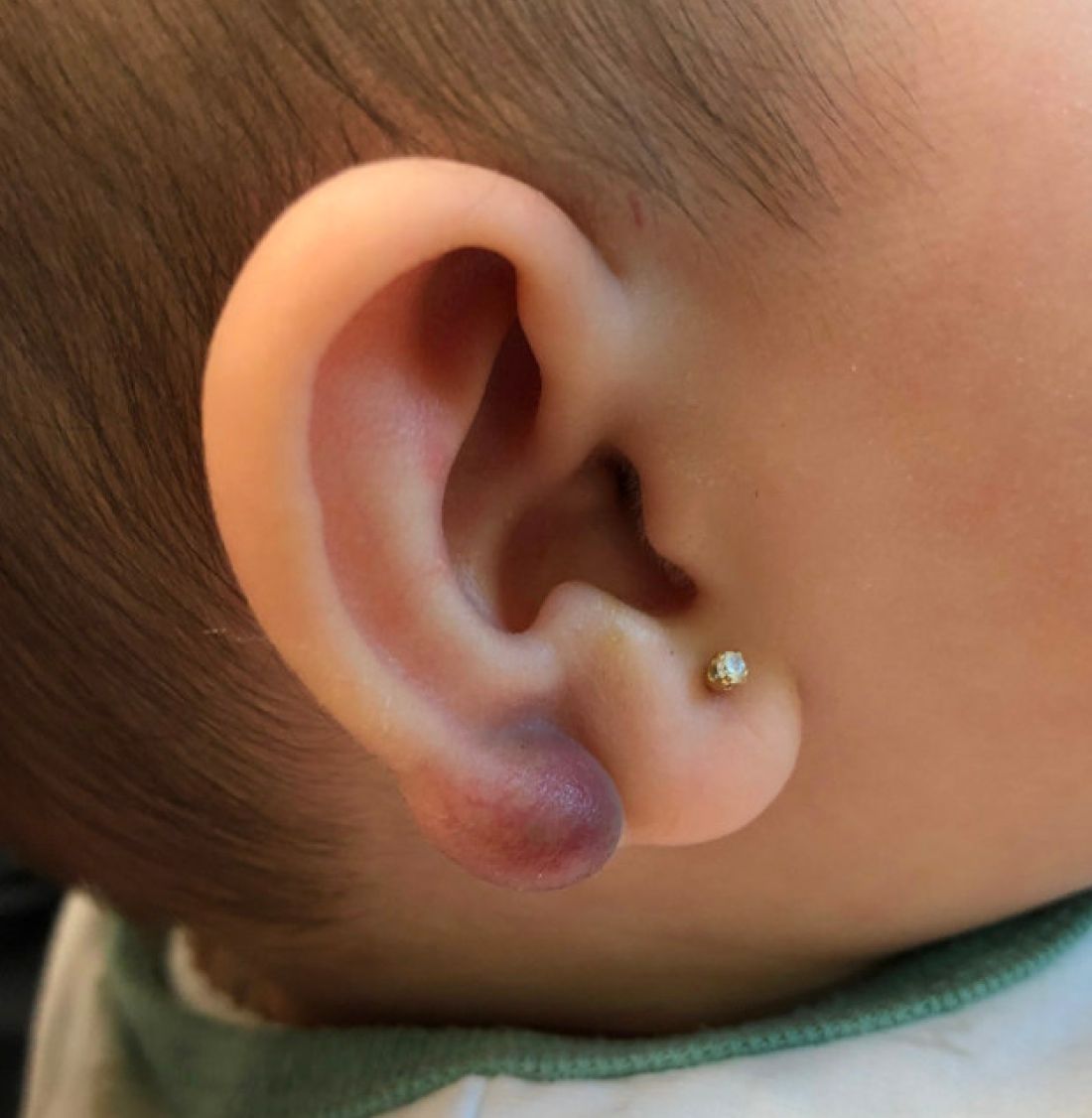
There have been no other family members with similar lesions. She had her ears pierced at a month of age without any complications.
On skin examination she has a firm red nodule on the right ear that appears slightly tender to touch. She has no other skin lesions of concern. She has normal muscle tone and there are no other abnormalities noted on the physical exam. She has no hepatomegaly, splenomegaly, or lymphadenopathy.
Sacral blistering

This patient had sustained multiple pressure injuries. The superior aspect of this image shows bullous change with intact dermis, which would classify that area of injury as a Stage 2 pressure injury.1 An older injury in the coccygeal area was through the dermis (Stage 3), with some eschar seen at the base, making that area unstageable.1 (There may have been deeper injury under the eschar.)
This patient was at heightened risk for pressure injury because of his paraplegia.2 Fortunately, he had some preserved sensation. However, his rotator cuff surgery made it harder for him to smoothly transfer to and from the wheelchair, leading to sheer forces against his skin. Social determinates of health care pose an additional risk for pressure injuries. Without a properly fitted wheelchair and cushion, there is an increased risk of localized pressure over both bony prominences and parts of the body that come into contact with mechanical elements of the wheelchair.
Treatment for all pressure injuries includes relief of pressure on the affected area. In this patient’s case, he had to stay in bed (and out of the wheelchair) so that he could heal.
This patient was very knowledgeable about his condition and pressure injuries. He had already arranged for a wheelchair fitting and a visit with the wound care team. His Stage 2 injury had a bullous change instead of absent epithelium, so rather than an adherent hydrocolloid dressing (which would likely remove the loosened epithelium), he was provided with a nonadherent dressing to the area, then an absorbent foam overdressing.
An image of the patient’s deeper sacral injury was shared with the wound care team, which recommended filling the area with a silver rope dressing for its absorptive filler and antibacterial properties. The area was then covered with an absorbent foam. The patient planned to follow up with the Wound Care Clinic for reevaluation and ongoing treatment.
Images and text courtesy of Daniel Stulberg, MD, FAAFP, Professor and Chair, Department of Family and Community Medicine, Western Michigan University Homer Stryker, MD School of Medicine Kalamazoo.
1. 2019 Guideline. The National Pressure Injury Advisory Panel. Accessed October 9, 2022. https://npiap.com/page/2019Guideline
2. Ricci JA, Bayer LR, Orgill DP. Evidence-based medicine: the evaluation and treatment of pressure injuries. Plast Reconstr Surg. 2017;139:275e-286e. doi: 10.1097/PRS.0000000000002850
This patient had sustained multiple pressure injuries. The superior aspect of this image shows bullous change with intact dermis, which would classify that area of injury as a Stage 2 pressure injury.1 An older injury in the coccygeal area was through the dermis (Stage 3), with some eschar seen at the base, making that area unstageable.1 (There may have been deeper injury under the eschar.)
This patient was at heightened risk for pressure injury because of his paraplegia.2 Fortunately, he had some preserved sensation. However, his rotator cuff surgery made it harder for him to smoothly transfer to and from the wheelchair, leading to sheer forces against his skin. Social determinates of health care pose an additional risk for pressure injuries. Without a properly fitted wheelchair and cushion, there is an increased risk of localized pressure over both bony prominences and parts of the body that come into contact with mechanical elements of the wheelchair.
Treatment for all pressure injuries includes relief of pressure on the affected area. In this patient’s case, he had to stay in bed (and out of the wheelchair) so that he could heal.
This patient was very knowledgeable about his condition and pressure injuries. He had already arranged for a wheelchair fitting and a visit with the wound care team. His Stage 2 injury had a bullous change instead of absent epithelium, so rather than an adherent hydrocolloid dressing (which would likely remove the loosened epithelium), he was provided with a nonadherent dressing to the area, then an absorbent foam overdressing.
An image of the patient’s deeper sacral injury was shared with the wound care team, which recommended filling the area with a silver rope dressing for its absorptive filler and antibacterial properties. The area was then covered with an absorbent foam. The patient planned to follow up with the Wound Care Clinic for reevaluation and ongoing treatment.
Images and text courtesy of Daniel Stulberg, MD, FAAFP, Professor and Chair, Department of Family and Community Medicine, Western Michigan University Homer Stryker, MD School of Medicine Kalamazoo.
1. 2019 Guideline. The National Pressure Injury Advisory Panel. Accessed October 9, 2022. https://npiap.com/page/2019Guideline
2. Ricci JA, Bayer LR, Orgill DP. Evidence-based medicine: the evaluation and treatment of pressure injuries. Plast Reconstr Surg. 2017;139:275e-286e. doi: 10.1097/PRS.0000000000002850

Newborn with white oral lesions

These lesions, called Bohn nodules, manifest on the buccal or lingual portion of the maxillary alveolar ridge and, less frequently, on the mandibular alveolar ridge. Because Bohn nodules are white and have a firm consistency, they are often confused with teeth. They can be differentiated by location, as teeth usually erupt from the distal aspect of the alveolar ridge.
Bohn nodules are epithelial cysts that are filled with keratin, which gives them their white color. They are caused by portions of epithelium that get trapped under surrounding epithelial cells. Bohn nodules usually resolve when the overlying epithelium ruptures and releases the keratinaceous material (usually by the time the child is 3 months of age).1
These nodules can be confused with neonatal or supernumerary teeth. Neonatal teeth can be abnormally small and pointed; they are true deciduous teeth that have erupted early. If they are removed, the child will not replace them until the time of their adult tooth eruption. Additionally, there are supernumerary teeth, which are extra teeth that are often abnormally shaped and loosely adherent. These abnormal teeth warrant extraction to avoid trauma to the tongue or aspiration.2
In this case, the family was advised regarding the benign nature of the nodules and the expectation that they would spontaneously resolve.
Images and text courtesy of Daniel Stulberg, MD, FAAFP, Professor and Chair, Department of Family and Community Medicine, Western Michigan University Homer Stryker, MD School of Medicine Kalamazoo.
1. Gupta N, Ramji S. Bohn's nodules: an under-recognised entity. Arch Dis Child Fetal Neonatal Ed. 2013;98:F464. doi: 10.1136/archdischild-2012-302922
2. DeSeta M, Holden E, Siddik D, et al. Natal and neonatal teeth: a review and case series. Br Dent J. 2022;232:449-453. doi: 10.1038/s41415-022-4091-3
These lesions, called Bohn nodules, manifest on the buccal or lingual portion of the maxillary alveolar ridge and, less frequently, on the mandibular alveolar ridge. Because Bohn nodules are white and have a firm consistency, they are often confused with teeth. They can be differentiated by location, as teeth usually erupt from the distal aspect of the alveolar ridge.
Bohn nodules are epithelial cysts that are filled with keratin, which gives them their white color. They are caused by portions of epithelium that get trapped under surrounding epithelial cells. Bohn nodules usually resolve when the overlying epithelium ruptures and releases the keratinaceous material (usually by the time the child is 3 months of age).1
These nodules can be confused with neonatal or supernumerary teeth. Neonatal teeth can be abnormally small and pointed; they are true deciduous teeth that have erupted early. If they are removed, the child will not replace them until the time of their adult tooth eruption. Additionally, there are supernumerary teeth, which are extra teeth that are often abnormally shaped and loosely adherent. These abnormal teeth warrant extraction to avoid trauma to the tongue or aspiration.2
In this case, the family was advised regarding the benign nature of the nodules and the expectation that they would spontaneously resolve.
Images and text courtesy of Daniel Stulberg, MD, FAAFP, Professor and Chair, Department of Family and Community Medicine, Western Michigan University Homer Stryker, MD School of Medicine Kalamazoo.
1. Gupta N, Ramji S. Bohn's nodules: an under-recognised entity. Arch Dis Child Fetal Neonatal Ed. 2013;98:F464. doi: 10.1136/archdischild-2012-302922
2. DeSeta M, Holden E, Siddik D, et al. Natal and neonatal teeth: a review and case series. Br Dent J. 2022;232:449-453. doi: 10.1038/s41415-022-4091-3
