Practicing evidence-based medicine, as obstetricians know, is not always possible when one does not have evidence due to lack of data or long-term experience in pregnancy. During the COVID-19 pandemic, the evidence changed so rapidly that we were compelled to alter our strategy frequently as we learned more about the impact of this disease on our vulnerable patient population. The COVID-19 pandemic taught us that, in unprecedented times, centering the safety of the patient, her child, and the health care team requires quick thinking, flexibility, and above all effective communication between team members.
Here, I share our institutional experience in providing practical obstetric care through various stages of the still-evolving COVID-19 pandemic. We based our strategy on guidance from the Centers for Disease Control and Prevention (CDC), the American College of Obstetricians and Gynecologists (ACOG),1,2 and the Society for Maternal-Fetal Medicine (SMFM).3-5 We were reminded yet again that the only constant is change and that timely but thoughtful adjustments were needed to keep up with the coronavirus.
Changes to prenatal care
Like many others, our institution has provided continued in-person outpatient prenatal care to both our low- and high-risk patients throughout each stage of the pandemic. While continuing to provide the necessary obstetric care, we made alterations to limit exposure and practice social distancing when possible.
Limiting patient support persons. One significant change was to restrict or limit support persons in the outpatient clinics based on guidelines reflecting community infection rates. Recognizing that this was not optimal for our patients’ emotional well-being, we needed to become more flexible in using technology to include family or support persons in prenatal visits and ultrasonography exams.
Altering test frequency. Using the guidance from SMFM,1 we changed the frequency of our antenatal testing and ultrasonography exams in the following ways: We increased the duration between indicated growth ultrasonography to every 4 weeks and decreased fetal antenatal testing to weekly, with twice-weekly testing continued for the highest-risk patients. Early first-trimester ultrasonography exams were limited and, when possible, delayed until after 10 to 12 weeks’ gestation or combined with other indications (nuchal translucency). Prenatal visits for low-risk patients were spaced out using existing models if the patient was amenable, especially in early pregnancy.
Adjusting staff assignments and using telehealth. In the early part of the pandemic, we divided into 2 groups to limit the number of clinicians at any one site: a dedicated group of outpatient clinicians who saw patients in the clinic only and a dedicated group of inpatient clinicians who staffed labor and delivery and the inpatient antepartum service. Additionally, our consultative maternal-fetal medicine service transitioned to a telehealth platform and performed the majority of consults remotely. Ultrasonography exams at various sites were read remotely and pertinent findings were communicated directly to patients via phone or the telehealth platform. Amniocentesis continued to be offered.
Responding to lower COVID-19 case numbers. When the number of COVID-19 cases decreased in the summer and fall of 2020, we returned to our prepandemic in-person practices, but we continued to offer telehealth visits as an option for patients who desired it. Patients were limited to one support person.
Shifting gears again. During the second surge of COVID-19 in our region, we used our experiences from the first to transition our practices to reduce in-person contact. Appointment frequency was decreased if appropriate, and we developed a tiered system of antenatal testing frequency based on risk factors. Visitors were again restricted, with exceptions made for extenuating circumstances. Consults were transitioned to telemedicine as appropriate and ultrasonography exams were read remotely when possible to limit exposures. Given the varied experiences with telemedicine and patient preferences, patients who desired in-person consult were (and are still) offered this option.
Some patients who were interested in telehealth but unable to access the technology were offered appointments via telehealth with the use of our clinic devices. Telemedicine increased our flexibility in offering consults as one provider could see patients at different office sites in one session. We continued our routine inpatient and outpatient coverage during this time as this kept our coverage options more flexible and expanded our obstetric backup plan in response to increased rates of community infection that affected both clinicians and patients.
Coordinating care for infected patients.One vital part of our prenatal care during the COVID-19 pandemic was to coordinate with our colleagues in medical specialties to provide outpatient care for patients with confirmed or suspected COVID-19 during their period of isolation or quarantine. Patients could be seen as outpatients in a dedicated space that used appropriate personal protective equipment (PPE) for not only prenatal care but also any needed in-person evaluation for COVID-19. Our obstetric clinicians and sonographers performed exams, antenatal testing (in the form of biophysical profiles), and indicated ultrasonography exams (such as umbilical artery Doppler studies and fetal growth assessments). This required a concerted effort and excellent communication between teams to provide the necessary care in the safest manner possible.
Continue to: Universal testing on labor and delivery...
Universal testing on labor and delivery
Not surprisingly, obstetric delivery volumes in our institution were not affected in the same way as elective surgery volumes. Our inpatient team continued to bring babies into the world at the same if not a higher rate than in prepandemic times. We continued elective inductions when space allowed. Our first COVID-19–positive patient was already at 40 weeks’ gestation when the result of her test, done due to exposure, was received. Creative effort among multiple specialties quickly developed her delivery plan, and she and her infant did well.
As data started coming out of the New York City obstetric experience, concern for preservation of the PPE supply and the potential for asymptomatic/presymptomatic patients led us, in consultation with our infectious disease colleagues, to institute universal testing for all antepartum and laboring patients. At first, all patients were tested on admission with our rapid in-house test. Eventually, we moved toward preoperative testing 3 to 5 days prior to scheduled cesarean deliveries in alignment with the surgical services when elective cases were reinstituted. Finally, we instituted preprocedure testing for all scheduled labor and delivery procedures, including inductions, cerclages, and fetal blood transfusions, while we still used rapid testing for patients who presented urgently or in labor.
We needed to address several considerations almost immediately after instituting universal testing, including:
what to do in case of patient refusal to be tested
which precautions to institute while awaiting test results
potential postponement of elective delivery if a patient tested positive, and
where best to deliver patients.
What we did at the beginning of the pandemic was not necessarily the same as we do in our current practice, and we expect that our procedures may need to change in the future. Derived from what we learned from others’ experience, we tailored our protocols to our own physical space, staffing capabilities, and testing limitations. We adjusted them often, with input from multiple services, based on updated policy, recommendation for isolation and quarantine durations, rates of community infection, and changes in the unit spaces. As with many things, one protocol did not fit every patient, necessitating case-by-case flexibility.
Delivery considerations
To answer some of the above questions, all patients who declined testing, were awaiting test results while in labor, or were in triage were placed in droplet and contact isolation on our unit, a practice we continue currently. Given the concern of potential aerosolization during the second stage of labor or during intubation, for any patients in those categories who required delivery, we limited the number of staff in their rooms as possible. Additional pediatric staff waited in close proximity of the room and were ready to come in if needed depending on fetal complications and gestational age. For delivery, all team members used full special pathogens precautions (N95 masks, face shields, gowns, and gloves).
Patients who were asymptomatic and tested negative for COVID-19 had and continue to have routine care from a PPE (standard gowns, gloves, face mask, and eye protection) and health care team perspective. We have allowed visitation of one support person per hospital stay for these patients throughout the pandemic.
For the majority of our experience during the pandemic, adult patients who tested positive for COVID-19 were cohorted within dedicated negative pressure units of varying levels of care. As these units included the same intensive care unit (ICU) we utilized in non-COVID times for critical obstetric patients, we had already operationalized their use and they were wired for our electronic fetal monitoring system. These rooms are adjacent to the main operating room (OR) complex, which allows for transition to a dedicated COVID-19 OR for cesarean delivery. We worked with the primary COVID-19 team, ICU team, anesthesia, and neonatal ICU team to develop a written protocol that detailed the care for our COVID-19–positive laboring and postpartum patients in this critical care COVID-19 unit.
For a time, admitted COVID-19–positive patients were not permitted to have support persons. The health care team therefore stepped in to be the patients’ support during the delivery of their child. Care of these patients required a great deal of coordination and communication between teams as well as the addition of a dedicated obstetric physician—separate from the regular labor and delivery team—assigned to care for these patients.
For pregnant patients in the emergency room or in the intermediate or floor COVID-19 units, portable fetal monitors and ultrasonography equipment were used for obstetric consults, fetal testing, and obstetrical ultrasonography as appropriate based on gestational age and medical conditions. Again, communication between teams was essential to provide seamless and timely patient care. Patients usually were admitted to the COVID-19 teams with maternal-fetal medicine or obstetric consult teams following daily; they were admitted and transferred to the ICU COVID-19 unit if delivery was necessary. To limit exposures whenever possible, coordinated care (such as exams and telephone evaluation) was performed outside of the room with the nursing and primary teams.
Continue to: Staying flexible to the changing COVID-19 environment...
Staying flexible to the changing COVID-19 environment
Postponed in-person visits. Whenever possible, deliveries that were not medically indicated and in-person outpatient care visits were postponed until isolation/quarantine precautions could be lifted to avoid the need for special pathogens precautions, separation of mother and infant, and visitor restrictions. We did not postpone any medically indicated deliveries or appropriate care due to COVID-19 alone. As the CDC guidelines changed regarding the timing of infectivity, we had to continually re-evaluate when a patient could return to regular outpatient care instead of the COVID-19 clinic and/or be delivered.
Mother-infant separation. As outlined in an article we wrote with our pediatric colleagues, originally all infants were immediately separated from their COVID-19–positive mothers, and delayed cord clamping was not performed.6 We adjusted our protocols as experience and data grew regarding the risk of transmission to the newborn from asymptomatic mothers and as updated recommendations were made by ACOG and the CDC. Currently, if desired, asymptomatic mothers are not separated from their well term infants. We practice our standard delayed cord clamping technique for all patients. Masking, hand hygiene, and physical distancing are used to reduce the risk of infection transmission. Breastfeeding is encouraged if the patient desires it, either directly using precautions or supported via pumping.
Reduced workplace exposure.Along with many others, we are even more cognizant of reducing the risk of workplace exposure; thus, we conduct our daily multidisciplinary huddle and physician transition of care sign-outs. We use multiple rooms for our larger group with secure video chats, and we limit huddles to a single representative from each specialty.
Medication protocols. Early in the pandemic in our area, we limited antenatal corticosteroids for fetal lung maturity to patients who were at less than 34 weeks’ gestation, per ACOG recommendations, carefully considering necessity in the critically ill. Now, we continue to administer antenatal steroids according to our usual protocols up to 36 6/7 weeks, per ACOG and SMFM recommendations, regardless of illness severity.7 Nonsteroidal anti-inflammatory drug use, once limited in COVID-19–positive patients, are now used again. Additionally, we had a comprehensive venous thromboembolism (VTE) prophylaxis protocol for our obstetric patients, and we have added special consideration for prophylaxis for patients with moderate to severe illness or other VTE risk factors. While we do not perform routine circumcisions on infants of COVID-19–positive mothers, we have a process in place to provide that service after discharge when isolation precautions are lifted.
Labor accommodations. As COVID-19 cases increased in our hospital during recent months, we made one more significant change in our care protocols. To open up space in the ICU, we moved our care for asymptomatic COVID-19–positive laboring patients to our new labor and delivery unit with implemented special pathogens precautions. This is not revolutionary; many other hospitals did not have the same capability we did with our existing collaboration with the ICU for critical obstetric care. However, this change again required communication and collaboration among multiple care teams, agreement on the qualifications for delivery on labor and delivery versus in the ICU, and physical alteration of our unit to accommodate additional isolation precautions.
Visitor policy. Another change is that we have opened up the visitor policy to welcome an asymptomatic support person for the COVID-19–positive labor patient, giving special attention to adherence to isolation precautions. Our staff members have embraced this change as they have everything else, with cautious optimism and focus on keeping both the patients and the health care team safe. Our moderate to severely ill patients continue to be cared for in the COVID-19 unit in close collaboration with our infectious disease and ICU colleagues.
It’s all about teamwork
I hope I have given a clear example of our approach to providing obstetric care in the ever-changing landscape of the COVID-19 pandemic. We embraced this period of necessary change as practically and safely as possible for both our patients and our health care workers. We learned multiple times along the way that what seemed to be a good idea was not feasible, or not the ideal option, or that COVID-19 had changed the rules of the game again. Our team met daily if not more frequently, as we found we had to constantly adapt and change to each new challenge or new clinical scenario. When we struggled, it generally related to a gap in communication.
I am privileged to work with a dedicated, selfless, multidisciplinary team that rose to the occasion. They had the focused goal to provide the highest quality and safety in obstetric care while offering compassion and empathy for the experience of having a baby during a pandemic. ●
The author would like to acknowledge Danielle Prentice, DO, and Jaimie Maines, MD, for their manuscript review.
Key takeaways: Safe obstetric care during the COVID-19 pandemic
The requirement for reduced in-person contact due to the COVID-19 pandemic challenged our traditional obstetric care models. This led us to comprehensively incorporate technology for communication with patients and their families and to significantly alter how, where, and when we delivered prenatal care.
Both patients and clinicians needed to adjust to the impact of these changes, especially concerning visitor policies.
Early incorporation of universal COVID-19 testing for labor and antepartum patients was initially instituted to improve patient and staff safety and to preserve PPE. However, it quickly led to the need for various protocols for both anticipated and unanticipated clinical scenarios.
As new data emerged and the number of cases fluctuated throughout the pandemic, our approach and protocols necessitated flexibility: Our strategy for maternal and neonatal care early in the pandemic was not the same as our current approach, and it will likely change several more times before we are done.
One of the biggest challenges to our care team was maintaining standards of excellence and safety in obstetric care while also adhering to the physical barriers of isolation precautions and maintaining vigilance to reduce exposure risk during our routine workflow.
The physical and operational specifics of our institution determined our approach to obstetric care during COVID-19, in part because halfway through the pandemic we moved our maternity unit from the adult hospital to a new center within our children’s hospital.
The frequent changes in the knowledge of and recommendations for COVID-19 highlighted the importance of maintaining multidisciplinary communication on a daily, if not more frequent, basis.
References
American College of Obstetricians and Gynecologists. Practice advisory: novel coronavirus 2019 (COVID-19): summary of key updates (December 14, 2020). https://www.acog.org/clinical /clinical-guidance/practice-advisory/articles/2020/03/novel -coronavirus-2019. Accessed January 28, 2021.
American College of Obstetricians and Gynecologists. COVID19 FAQs for obstetrician-gynecologists, obstetrics. Washington, DC: ACOG; 2020. https://www.acog.org/clinical-information /physician-faqs/covid-19-faqs-for-ob-gyns-obstetrics. Accessed January 28, 2021.
Society for Maternal-Fetal Medicine. Coronavirus (COVID19) and pregnancy: what maternal-fetal medicine subspecialists need to know. Updated November 23, 2020. https: //s3.amazonaws.com/cdn.smfm.org/media/2589/COVID19 -What_MFMs_need_to_know_revision_11-23-20_final.pdf. Accessed January 28, 2021.
Society for Maternal-Fetal Medicine. Management considerations for pregnant patients with COVID-19. Updated January 7, 2021. https://s3.amazonaws.com/cdn.smfm.org /media/2668/SMFM_COVID_Management_of_COVID_pos _preg_patients_1-7-21_(final).pdf. Accessed January 28, 2021.
Society for Maternal-Fetal Medicine. COVID-19 ultrasound clinical practice suggestions. Updated October 20, 2020. https://s3.amazonaws.com/cdn.smfm.org/media/2550 /Ultrasound_Covid19_Suggestions_10-20-20_(final).pdf. Accessed January 28, 2020.
Amatya S, Corr TE, Gandhi CK, et al. Management of newborns exposed to mothers with confirmed or suspected COVID-19. J Perinatol. 2020;40:987-996.
American College of Obstetricians and Gynecologists. Committee opinion no 713: antenatal corticosteroid therapy for fetal maturation. Obstet Gynecol. 2017;130:e102-e109.
Dr. Pauli is Associate Professor and Chief, Division of Maternal-Fetal Medicine, Department of Obstetrics and Gynecology, Penn State College of Medicine, Milton S. Hershey Medical Center, Hershey, Pennsylvania. She is a member of the OBG Management Board of Editors.
The author reports no financial relationships relevant to this article.
Dr. Pauli is Associate Professor and Chief, Division of Maternal-Fetal Medicine, Department of Obstetrics and Gynecology, Penn State College of Medicine, Milton S. Hershey Medical Center, Hershey, Pennsylvania. She is a member of the OBG Management Board of Editors.
The author reports no financial relationships relevant to this article.
Author and Disclosure Information
Dr. Pauli is Associate Professor and Chief, Division of Maternal-Fetal Medicine, Department of Obstetrics and Gynecology, Penn State College of Medicine, Milton S. Hershey Medical Center, Hershey, Pennsylvania. She is a member of the OBG Management Board of Editors.
The author reports no financial relationships relevant to this article.
Practicing evidence-based medicine, as obstetricians know, is not always possible when one does not have evidence due to lack of data or long-term experience in pregnancy. During the COVID-19 pandemic, the evidence changed so rapidly that we were compelled to alter our strategy frequently as we learned more about the impact of this disease on our vulnerable patient population. The COVID-19 pandemic taught us that, in unprecedented times, centering the safety of the patient, her child, and the health care team requires quick thinking, flexibility, and above all effective communication between team members.
Here, I share our institutional experience in providing practical obstetric care through various stages of the still-evolving COVID-19 pandemic. We based our strategy on guidance from the Centers for Disease Control and Prevention (CDC), the American College of Obstetricians and Gynecologists (ACOG),1,2 and the Society for Maternal-Fetal Medicine (SMFM).3-5 We were reminded yet again that the only constant is change and that timely but thoughtful adjustments were needed to keep up with the coronavirus.
Changes to prenatal care
Like many others, our institution has provided continued in-person outpatient prenatal care to both our low- and high-risk patients throughout each stage of the pandemic. While continuing to provide the necessary obstetric care, we made alterations to limit exposure and practice social distancing when possible.
Limiting patient support persons. One significant change was to restrict or limit support persons in the outpatient clinics based on guidelines reflecting community infection rates. Recognizing that this was not optimal for our patients’ emotional well-being, we needed to become more flexible in using technology to include family or support persons in prenatal visits and ultrasonography exams.
Altering test frequency. Using the guidance from SMFM,1 we changed the frequency of our antenatal testing and ultrasonography exams in the following ways: We increased the duration between indicated growth ultrasonography to every 4 weeks and decreased fetal antenatal testing to weekly, with twice-weekly testing continued for the highest-risk patients. Early first-trimester ultrasonography exams were limited and, when possible, delayed until after 10 to 12 weeks’ gestation or combined with other indications (nuchal translucency). Prenatal visits for low-risk patients were spaced out using existing models if the patient was amenable, especially in early pregnancy.
Adjusting staff assignments and using telehealth. In the early part of the pandemic, we divided into 2 groups to limit the number of clinicians at any one site: a dedicated group of outpatient clinicians who saw patients in the clinic only and a dedicated group of inpatient clinicians who staffed labor and delivery and the inpatient antepartum service. Additionally, our consultative maternal-fetal medicine service transitioned to a telehealth platform and performed the majority of consults remotely. Ultrasonography exams at various sites were read remotely and pertinent findings were communicated directly to patients via phone or the telehealth platform. Amniocentesis continued to be offered.
Responding to lower COVID-19 case numbers. When the number of COVID-19 cases decreased in the summer and fall of 2020, we returned to our prepandemic in-person practices, but we continued to offer telehealth visits as an option for patients who desired it. Patients were limited to one support person.
Shifting gears again. During the second surge of COVID-19 in our region, we used our experiences from the first to transition our practices to reduce in-person contact. Appointment frequency was decreased if appropriate, and we developed a tiered system of antenatal testing frequency based on risk factors. Visitors were again restricted, with exceptions made for extenuating circumstances. Consults were transitioned to telemedicine as appropriate and ultrasonography exams were read remotely when possible to limit exposures. Given the varied experiences with telemedicine and patient preferences, patients who desired in-person consult were (and are still) offered this option.
Some patients who were interested in telehealth but unable to access the technology were offered appointments via telehealth with the use of our clinic devices. Telemedicine increased our flexibility in offering consults as one provider could see patients at different office sites in one session. We continued our routine inpatient and outpatient coverage during this time as this kept our coverage options more flexible and expanded our obstetric backup plan in response to increased rates of community infection that affected both clinicians and patients.
Coordinating care for infected patients.One vital part of our prenatal care during the COVID-19 pandemic was to coordinate with our colleagues in medical specialties to provide outpatient care for patients with confirmed or suspected COVID-19 during their period of isolation or quarantine. Patients could be seen as outpatients in a dedicated space that used appropriate personal protective equipment (PPE) for not only prenatal care but also any needed in-person evaluation for COVID-19. Our obstetric clinicians and sonographers performed exams, antenatal testing (in the form of biophysical profiles), and indicated ultrasonography exams (such as umbilical artery Doppler studies and fetal growth assessments). This required a concerted effort and excellent communication between teams to provide the necessary care in the safest manner possible.
Continue to: Universal testing on labor and delivery...
Universal testing on labor and delivery
Not surprisingly, obstetric delivery volumes in our institution were not affected in the same way as elective surgery volumes. Our inpatient team continued to bring babies into the world at the same if not a higher rate than in prepandemic times. We continued elective inductions when space allowed. Our first COVID-19–positive patient was already at 40 weeks’ gestation when the result of her test, done due to exposure, was received. Creative effort among multiple specialties quickly developed her delivery plan, and she and her infant did well.
As data started coming out of the New York City obstetric experience, concern for preservation of the PPE supply and the potential for asymptomatic/presymptomatic patients led us, in consultation with our infectious disease colleagues, to institute universal testing for all antepartum and laboring patients. At first, all patients were tested on admission with our rapid in-house test. Eventually, we moved toward preoperative testing 3 to 5 days prior to scheduled cesarean deliveries in alignment with the surgical services when elective cases were reinstituted. Finally, we instituted preprocedure testing for all scheduled labor and delivery procedures, including inductions, cerclages, and fetal blood transfusions, while we still used rapid testing for patients who presented urgently or in labor.
We needed to address several considerations almost immediately after instituting universal testing, including:
what to do in case of patient refusal to be tested
which precautions to institute while awaiting test results
potential postponement of elective delivery if a patient tested positive, and
where best to deliver patients.
What we did at the beginning of the pandemic was not necessarily the same as we do in our current practice, and we expect that our procedures may need to change in the future. Derived from what we learned from others’ experience, we tailored our protocols to our own physical space, staffing capabilities, and testing limitations. We adjusted them often, with input from multiple services, based on updated policy, recommendation for isolation and quarantine durations, rates of community infection, and changes in the unit spaces. As with many things, one protocol did not fit every patient, necessitating case-by-case flexibility.
Delivery considerations
To answer some of the above questions, all patients who declined testing, were awaiting test results while in labor, or were in triage were placed in droplet and contact isolation on our unit, a practice we continue currently. Given the concern of potential aerosolization during the second stage of labor or during intubation, for any patients in those categories who required delivery, we limited the number of staff in their rooms as possible. Additional pediatric staff waited in close proximity of the room and were ready to come in if needed depending on fetal complications and gestational age. For delivery, all team members used full special pathogens precautions (N95 masks, face shields, gowns, and gloves).
Patients who were asymptomatic and tested negative for COVID-19 had and continue to have routine care from a PPE (standard gowns, gloves, face mask, and eye protection) and health care team perspective. We have allowed visitation of one support person per hospital stay for these patients throughout the pandemic.
For the majority of our experience during the pandemic, adult patients who tested positive for COVID-19 were cohorted within dedicated negative pressure units of varying levels of care. As these units included the same intensive care unit (ICU) we utilized in non-COVID times for critical obstetric patients, we had already operationalized their use and they were wired for our electronic fetal monitoring system. These rooms are adjacent to the main operating room (OR) complex, which allows for transition to a dedicated COVID-19 OR for cesarean delivery. We worked with the primary COVID-19 team, ICU team, anesthesia, and neonatal ICU team to develop a written protocol that detailed the care for our COVID-19–positive laboring and postpartum patients in this critical care COVID-19 unit.
For a time, admitted COVID-19–positive patients were not permitted to have support persons. The health care team therefore stepped in to be the patients’ support during the delivery of their child. Care of these patients required a great deal of coordination and communication between teams as well as the addition of a dedicated obstetric physician—separate from the regular labor and delivery team—assigned to care for these patients.
For pregnant patients in the emergency room or in the intermediate or floor COVID-19 units, portable fetal monitors and ultrasonography equipment were used for obstetric consults, fetal testing, and obstetrical ultrasonography as appropriate based on gestational age and medical conditions. Again, communication between teams was essential to provide seamless and timely patient care. Patients usually were admitted to the COVID-19 teams with maternal-fetal medicine or obstetric consult teams following daily; they were admitted and transferred to the ICU COVID-19 unit if delivery was necessary. To limit exposures whenever possible, coordinated care (such as exams and telephone evaluation) was performed outside of the room with the nursing and primary teams.
Continue to: Staying flexible to the changing COVID-19 environment...
Staying flexible to the changing COVID-19 environment
Postponed in-person visits. Whenever possible, deliveries that were not medically indicated and in-person outpatient care visits were postponed until isolation/quarantine precautions could be lifted to avoid the need for special pathogens precautions, separation of mother and infant, and visitor restrictions. We did not postpone any medically indicated deliveries or appropriate care due to COVID-19 alone. As the CDC guidelines changed regarding the timing of infectivity, we had to continually re-evaluate when a patient could return to regular outpatient care instead of the COVID-19 clinic and/or be delivered.
Mother-infant separation. As outlined in an article we wrote with our pediatric colleagues, originally all infants were immediately separated from their COVID-19–positive mothers, and delayed cord clamping was not performed.6 We adjusted our protocols as experience and data grew regarding the risk of transmission to the newborn from asymptomatic mothers and as updated recommendations were made by ACOG and the CDC. Currently, if desired, asymptomatic mothers are not separated from their well term infants. We practice our standard delayed cord clamping technique for all patients. Masking, hand hygiene, and physical distancing are used to reduce the risk of infection transmission. Breastfeeding is encouraged if the patient desires it, either directly using precautions or supported via pumping.
Reduced workplace exposure.Along with many others, we are even more cognizant of reducing the risk of workplace exposure; thus, we conduct our daily multidisciplinary huddle and physician transition of care sign-outs. We use multiple rooms for our larger group with secure video chats, and we limit huddles to a single representative from each specialty.
Medication protocols. Early in the pandemic in our area, we limited antenatal corticosteroids for fetal lung maturity to patients who were at less than 34 weeks’ gestation, per ACOG recommendations, carefully considering necessity in the critically ill. Now, we continue to administer antenatal steroids according to our usual protocols up to 36 6/7 weeks, per ACOG and SMFM recommendations, regardless of illness severity.7 Nonsteroidal anti-inflammatory drug use, once limited in COVID-19–positive patients, are now used again. Additionally, we had a comprehensive venous thromboembolism (VTE) prophylaxis protocol for our obstetric patients, and we have added special consideration for prophylaxis for patients with moderate to severe illness or other VTE risk factors. While we do not perform routine circumcisions on infants of COVID-19–positive mothers, we have a process in place to provide that service after discharge when isolation precautions are lifted.
Labor accommodations. As COVID-19 cases increased in our hospital during recent months, we made one more significant change in our care protocols. To open up space in the ICU, we moved our care for asymptomatic COVID-19–positive laboring patients to our new labor and delivery unit with implemented special pathogens precautions. This is not revolutionary; many other hospitals did not have the same capability we did with our existing collaboration with the ICU for critical obstetric care. However, this change again required communication and collaboration among multiple care teams, agreement on the qualifications for delivery on labor and delivery versus in the ICU, and physical alteration of our unit to accommodate additional isolation precautions.
Visitor policy. Another change is that we have opened up the visitor policy to welcome an asymptomatic support person for the COVID-19–positive labor patient, giving special attention to adherence to isolation precautions. Our staff members have embraced this change as they have everything else, with cautious optimism and focus on keeping both the patients and the health care team safe. Our moderate to severely ill patients continue to be cared for in the COVID-19 unit in close collaboration with our infectious disease and ICU colleagues.
It’s all about teamwork
I hope I have given a clear example of our approach to providing obstetric care in the ever-changing landscape of the COVID-19 pandemic. We embraced this period of necessary change as practically and safely as possible for both our patients and our health care workers. We learned multiple times along the way that what seemed to be a good idea was not feasible, or not the ideal option, or that COVID-19 had changed the rules of the game again. Our team met daily if not more frequently, as we found we had to constantly adapt and change to each new challenge or new clinical scenario. When we struggled, it generally related to a gap in communication.
I am privileged to work with a dedicated, selfless, multidisciplinary team that rose to the occasion. They had the focused goal to provide the highest quality and safety in obstetric care while offering compassion and empathy for the experience of having a baby during a pandemic. ●
The author would like to acknowledge Danielle Prentice, DO, and Jaimie Maines, MD, for their manuscript review.
Key takeaways: Safe obstetric care during the COVID-19 pandemic
The requirement for reduced in-person contact due to the COVID-19 pandemic challenged our traditional obstetric care models. This led us to comprehensively incorporate technology for communication with patients and their families and to significantly alter how, where, and when we delivered prenatal care.
Both patients and clinicians needed to adjust to the impact of these changes, especially concerning visitor policies.
Early incorporation of universal COVID-19 testing for labor and antepartum patients was initially instituted to improve patient and staff safety and to preserve PPE. However, it quickly led to the need for various protocols for both anticipated and unanticipated clinical scenarios.
As new data emerged and the number of cases fluctuated throughout the pandemic, our approach and protocols necessitated flexibility: Our strategy for maternal and neonatal care early in the pandemic was not the same as our current approach, and it will likely change several more times before we are done.
One of the biggest challenges to our care team was maintaining standards of excellence and safety in obstetric care while also adhering to the physical barriers of isolation precautions and maintaining vigilance to reduce exposure risk during our routine workflow.
The physical and operational specifics of our institution determined our approach to obstetric care during COVID-19, in part because halfway through the pandemic we moved our maternity unit from the adult hospital to a new center within our children’s hospital.
The frequent changes in the knowledge of and recommendations for COVID-19 highlighted the importance of maintaining multidisciplinary communication on a daily, if not more frequent, basis.
Practicing evidence-based medicine, as obstetricians know, is not always possible when one does not have evidence due to lack of data or long-term experience in pregnancy. During the COVID-19 pandemic, the evidence changed so rapidly that we were compelled to alter our strategy frequently as we learned more about the impact of this disease on our vulnerable patient population. The COVID-19 pandemic taught us that, in unprecedented times, centering the safety of the patient, her child, and the health care team requires quick thinking, flexibility, and above all effective communication between team members.
Here, I share our institutional experience in providing practical obstetric care through various stages of the still-evolving COVID-19 pandemic. We based our strategy on guidance from the Centers for Disease Control and Prevention (CDC), the American College of Obstetricians and Gynecologists (ACOG),1,2 and the Society for Maternal-Fetal Medicine (SMFM).3-5 We were reminded yet again that the only constant is change and that timely but thoughtful adjustments were needed to keep up with the coronavirus.
Changes to prenatal care
Like many others, our institution has provided continued in-person outpatient prenatal care to both our low- and high-risk patients throughout each stage of the pandemic. While continuing to provide the necessary obstetric care, we made alterations to limit exposure and practice social distancing when possible.
Limiting patient support persons. One significant change was to restrict or limit support persons in the outpatient clinics based on guidelines reflecting community infection rates. Recognizing that this was not optimal for our patients’ emotional well-being, we needed to become more flexible in using technology to include family or support persons in prenatal visits and ultrasonography exams.
Altering test frequency. Using the guidance from SMFM,1 we changed the frequency of our antenatal testing and ultrasonography exams in the following ways: We increased the duration between indicated growth ultrasonography to every 4 weeks and decreased fetal antenatal testing to weekly, with twice-weekly testing continued for the highest-risk patients. Early first-trimester ultrasonography exams were limited and, when possible, delayed until after 10 to 12 weeks’ gestation or combined with other indications (nuchal translucency). Prenatal visits for low-risk patients were spaced out using existing models if the patient was amenable, especially in early pregnancy.
Adjusting staff assignments and using telehealth. In the early part of the pandemic, we divided into 2 groups to limit the number of clinicians at any one site: a dedicated group of outpatient clinicians who saw patients in the clinic only and a dedicated group of inpatient clinicians who staffed labor and delivery and the inpatient antepartum service. Additionally, our consultative maternal-fetal medicine service transitioned to a telehealth platform and performed the majority of consults remotely. Ultrasonography exams at various sites were read remotely and pertinent findings were communicated directly to patients via phone or the telehealth platform. Amniocentesis continued to be offered.
Responding to lower COVID-19 case numbers. When the number of COVID-19 cases decreased in the summer and fall of 2020, we returned to our prepandemic in-person practices, but we continued to offer telehealth visits as an option for patients who desired it. Patients were limited to one support person.
Shifting gears again. During the second surge of COVID-19 in our region, we used our experiences from the first to transition our practices to reduce in-person contact. Appointment frequency was decreased if appropriate, and we developed a tiered system of antenatal testing frequency based on risk factors. Visitors were again restricted, with exceptions made for extenuating circumstances. Consults were transitioned to telemedicine as appropriate and ultrasonography exams were read remotely when possible to limit exposures. Given the varied experiences with telemedicine and patient preferences, patients who desired in-person consult were (and are still) offered this option.
Some patients who were interested in telehealth but unable to access the technology were offered appointments via telehealth with the use of our clinic devices. Telemedicine increased our flexibility in offering consults as one provider could see patients at different office sites in one session. We continued our routine inpatient and outpatient coverage during this time as this kept our coverage options more flexible and expanded our obstetric backup plan in response to increased rates of community infection that affected both clinicians and patients.
Coordinating care for infected patients.One vital part of our prenatal care during the COVID-19 pandemic was to coordinate with our colleagues in medical specialties to provide outpatient care for patients with confirmed or suspected COVID-19 during their period of isolation or quarantine. Patients could be seen as outpatients in a dedicated space that used appropriate personal protective equipment (PPE) for not only prenatal care but also any needed in-person evaluation for COVID-19. Our obstetric clinicians and sonographers performed exams, antenatal testing (in the form of biophysical profiles), and indicated ultrasonography exams (such as umbilical artery Doppler studies and fetal growth assessments). This required a concerted effort and excellent communication between teams to provide the necessary care in the safest manner possible.
Continue to: Universal testing on labor and delivery...
Universal testing on labor and delivery
Not surprisingly, obstetric delivery volumes in our institution were not affected in the same way as elective surgery volumes. Our inpatient team continued to bring babies into the world at the same if not a higher rate than in prepandemic times. We continued elective inductions when space allowed. Our first COVID-19–positive patient was already at 40 weeks’ gestation when the result of her test, done due to exposure, was received. Creative effort among multiple specialties quickly developed her delivery plan, and she and her infant did well.
As data started coming out of the New York City obstetric experience, concern for preservation of the PPE supply and the potential for asymptomatic/presymptomatic patients led us, in consultation with our infectious disease colleagues, to institute universal testing for all antepartum and laboring patients. At first, all patients were tested on admission with our rapid in-house test. Eventually, we moved toward preoperative testing 3 to 5 days prior to scheduled cesarean deliveries in alignment with the surgical services when elective cases were reinstituted. Finally, we instituted preprocedure testing for all scheduled labor and delivery procedures, including inductions, cerclages, and fetal blood transfusions, while we still used rapid testing for patients who presented urgently or in labor.
We needed to address several considerations almost immediately after instituting universal testing, including:
what to do in case of patient refusal to be tested
which precautions to institute while awaiting test results
potential postponement of elective delivery if a patient tested positive, and
where best to deliver patients.
What we did at the beginning of the pandemic was not necessarily the same as we do in our current practice, and we expect that our procedures may need to change in the future. Derived from what we learned from others’ experience, we tailored our protocols to our own physical space, staffing capabilities, and testing limitations. We adjusted them often, with input from multiple services, based on updated policy, recommendation for isolation and quarantine durations, rates of community infection, and changes in the unit spaces. As with many things, one protocol did not fit every patient, necessitating case-by-case flexibility.
Delivery considerations
To answer some of the above questions, all patients who declined testing, were awaiting test results while in labor, or were in triage were placed in droplet and contact isolation on our unit, a practice we continue currently. Given the concern of potential aerosolization during the second stage of labor or during intubation, for any patients in those categories who required delivery, we limited the number of staff in their rooms as possible. Additional pediatric staff waited in close proximity of the room and were ready to come in if needed depending on fetal complications and gestational age. For delivery, all team members used full special pathogens precautions (N95 masks, face shields, gowns, and gloves).
Patients who were asymptomatic and tested negative for COVID-19 had and continue to have routine care from a PPE (standard gowns, gloves, face mask, and eye protection) and health care team perspective. We have allowed visitation of one support person per hospital stay for these patients throughout the pandemic.
For the majority of our experience during the pandemic, adult patients who tested positive for COVID-19 were cohorted within dedicated negative pressure units of varying levels of care. As these units included the same intensive care unit (ICU) we utilized in non-COVID times for critical obstetric patients, we had already operationalized their use and they were wired for our electronic fetal monitoring system. These rooms are adjacent to the main operating room (OR) complex, which allows for transition to a dedicated COVID-19 OR for cesarean delivery. We worked with the primary COVID-19 team, ICU team, anesthesia, and neonatal ICU team to develop a written protocol that detailed the care for our COVID-19–positive laboring and postpartum patients in this critical care COVID-19 unit.
For a time, admitted COVID-19–positive patients were not permitted to have support persons. The health care team therefore stepped in to be the patients’ support during the delivery of their child. Care of these patients required a great deal of coordination and communication between teams as well as the addition of a dedicated obstetric physician—separate from the regular labor and delivery team—assigned to care for these patients.
For pregnant patients in the emergency room or in the intermediate or floor COVID-19 units, portable fetal monitors and ultrasonography equipment were used for obstetric consults, fetal testing, and obstetrical ultrasonography as appropriate based on gestational age and medical conditions. Again, communication between teams was essential to provide seamless and timely patient care. Patients usually were admitted to the COVID-19 teams with maternal-fetal medicine or obstetric consult teams following daily; they were admitted and transferred to the ICU COVID-19 unit if delivery was necessary. To limit exposures whenever possible, coordinated care (such as exams and telephone evaluation) was performed outside of the room with the nursing and primary teams.
Continue to: Staying flexible to the changing COVID-19 environment...
Staying flexible to the changing COVID-19 environment
Postponed in-person visits. Whenever possible, deliveries that were not medically indicated and in-person outpatient care visits were postponed until isolation/quarantine precautions could be lifted to avoid the need for special pathogens precautions, separation of mother and infant, and visitor restrictions. We did not postpone any medically indicated deliveries or appropriate care due to COVID-19 alone. As the CDC guidelines changed regarding the timing of infectivity, we had to continually re-evaluate when a patient could return to regular outpatient care instead of the COVID-19 clinic and/or be delivered.
Mother-infant separation. As outlined in an article we wrote with our pediatric colleagues, originally all infants were immediately separated from their COVID-19–positive mothers, and delayed cord clamping was not performed.6 We adjusted our protocols as experience and data grew regarding the risk of transmission to the newborn from asymptomatic mothers and as updated recommendations were made by ACOG and the CDC. Currently, if desired, asymptomatic mothers are not separated from their well term infants. We practice our standard delayed cord clamping technique for all patients. Masking, hand hygiene, and physical distancing are used to reduce the risk of infection transmission. Breastfeeding is encouraged if the patient desires it, either directly using precautions or supported via pumping.
Reduced workplace exposure.Along with many others, we are even more cognizant of reducing the risk of workplace exposure; thus, we conduct our daily multidisciplinary huddle and physician transition of care sign-outs. We use multiple rooms for our larger group with secure video chats, and we limit huddles to a single representative from each specialty.
Medication protocols. Early in the pandemic in our area, we limited antenatal corticosteroids for fetal lung maturity to patients who were at less than 34 weeks’ gestation, per ACOG recommendations, carefully considering necessity in the critically ill. Now, we continue to administer antenatal steroids according to our usual protocols up to 36 6/7 weeks, per ACOG and SMFM recommendations, regardless of illness severity.7 Nonsteroidal anti-inflammatory drug use, once limited in COVID-19–positive patients, are now used again. Additionally, we had a comprehensive venous thromboembolism (VTE) prophylaxis protocol for our obstetric patients, and we have added special consideration for prophylaxis for patients with moderate to severe illness or other VTE risk factors. While we do not perform routine circumcisions on infants of COVID-19–positive mothers, we have a process in place to provide that service after discharge when isolation precautions are lifted.
Labor accommodations. As COVID-19 cases increased in our hospital during recent months, we made one more significant change in our care protocols. To open up space in the ICU, we moved our care for asymptomatic COVID-19–positive laboring patients to our new labor and delivery unit with implemented special pathogens precautions. This is not revolutionary; many other hospitals did not have the same capability we did with our existing collaboration with the ICU for critical obstetric care. However, this change again required communication and collaboration among multiple care teams, agreement on the qualifications for delivery on labor and delivery versus in the ICU, and physical alteration of our unit to accommodate additional isolation precautions.
Visitor policy. Another change is that we have opened up the visitor policy to welcome an asymptomatic support person for the COVID-19–positive labor patient, giving special attention to adherence to isolation precautions. Our staff members have embraced this change as they have everything else, with cautious optimism and focus on keeping both the patients and the health care team safe. Our moderate to severely ill patients continue to be cared for in the COVID-19 unit in close collaboration with our infectious disease and ICU colleagues.
It’s all about teamwork
I hope I have given a clear example of our approach to providing obstetric care in the ever-changing landscape of the COVID-19 pandemic. We embraced this period of necessary change as practically and safely as possible for both our patients and our health care workers. We learned multiple times along the way that what seemed to be a good idea was not feasible, or not the ideal option, or that COVID-19 had changed the rules of the game again. Our team met daily if not more frequently, as we found we had to constantly adapt and change to each new challenge or new clinical scenario. When we struggled, it generally related to a gap in communication.
I am privileged to work with a dedicated, selfless, multidisciplinary team that rose to the occasion. They had the focused goal to provide the highest quality and safety in obstetric care while offering compassion and empathy for the experience of having a baby during a pandemic. ●
The author would like to acknowledge Danielle Prentice, DO, and Jaimie Maines, MD, for their manuscript review.
Key takeaways: Safe obstetric care during the COVID-19 pandemic
The requirement for reduced in-person contact due to the COVID-19 pandemic challenged our traditional obstetric care models. This led us to comprehensively incorporate technology for communication with patients and their families and to significantly alter how, where, and when we delivered prenatal care.
Both patients and clinicians needed to adjust to the impact of these changes, especially concerning visitor policies.
Early incorporation of universal COVID-19 testing for labor and antepartum patients was initially instituted to improve patient and staff safety and to preserve PPE. However, it quickly led to the need for various protocols for both anticipated and unanticipated clinical scenarios.
As new data emerged and the number of cases fluctuated throughout the pandemic, our approach and protocols necessitated flexibility: Our strategy for maternal and neonatal care early in the pandemic was not the same as our current approach, and it will likely change several more times before we are done.
One of the biggest challenges to our care team was maintaining standards of excellence and safety in obstetric care while also adhering to the physical barriers of isolation precautions and maintaining vigilance to reduce exposure risk during our routine workflow.
The physical and operational specifics of our institution determined our approach to obstetric care during COVID-19, in part because halfway through the pandemic we moved our maternity unit from the adult hospital to a new center within our children’s hospital.
The frequent changes in the knowledge of and recommendations for COVID-19 highlighted the importance of maintaining multidisciplinary communication on a daily, if not more frequent, basis.
References
American College of Obstetricians and Gynecologists. Practice advisory: novel coronavirus 2019 (COVID-19): summary of key updates (December 14, 2020). https://www.acog.org/clinical /clinical-guidance/practice-advisory/articles/2020/03/novel -coronavirus-2019. Accessed January 28, 2021.
American College of Obstetricians and Gynecologists. COVID19 FAQs for obstetrician-gynecologists, obstetrics. Washington, DC: ACOG; 2020. https://www.acog.org/clinical-information /physician-faqs/covid-19-faqs-for-ob-gyns-obstetrics. Accessed January 28, 2021.
Society for Maternal-Fetal Medicine. Coronavirus (COVID19) and pregnancy: what maternal-fetal medicine subspecialists need to know. Updated November 23, 2020. https: //s3.amazonaws.com/cdn.smfm.org/media/2589/COVID19 -What_MFMs_need_to_know_revision_11-23-20_final.pdf. Accessed January 28, 2021.
Society for Maternal-Fetal Medicine. Management considerations for pregnant patients with COVID-19. Updated January 7, 2021. https://s3.amazonaws.com/cdn.smfm.org /media/2668/SMFM_COVID_Management_of_COVID_pos _preg_patients_1-7-21_(final).pdf. Accessed January 28, 2021.
Society for Maternal-Fetal Medicine. COVID-19 ultrasound clinical practice suggestions. Updated October 20, 2020. https://s3.amazonaws.com/cdn.smfm.org/media/2550 /Ultrasound_Covid19_Suggestions_10-20-20_(final).pdf. Accessed January 28, 2020.
Amatya S, Corr TE, Gandhi CK, et al. Management of newborns exposed to mothers with confirmed or suspected COVID-19. J Perinatol. 2020;40:987-996.
American College of Obstetricians and Gynecologists. Committee opinion no 713: antenatal corticosteroid therapy for fetal maturation. Obstet Gynecol. 2017;130:e102-e109.
References
American College of Obstetricians and Gynecologists. Practice advisory: novel coronavirus 2019 (COVID-19): summary of key updates (December 14, 2020). https://www.acog.org/clinical /clinical-guidance/practice-advisory/articles/2020/03/novel -coronavirus-2019. Accessed January 28, 2021.
American College of Obstetricians and Gynecologists. COVID19 FAQs for obstetrician-gynecologists, obstetrics. Washington, DC: ACOG; 2020. https://www.acog.org/clinical-information /physician-faqs/covid-19-faqs-for-ob-gyns-obstetrics. Accessed January 28, 2021.
Society for Maternal-Fetal Medicine. Coronavirus (COVID19) and pregnancy: what maternal-fetal medicine subspecialists need to know. Updated November 23, 2020. https: //s3.amazonaws.com/cdn.smfm.org/media/2589/COVID19 -What_MFMs_need_to_know_revision_11-23-20_final.pdf. Accessed January 28, 2021.
Society for Maternal-Fetal Medicine. Management considerations for pregnant patients with COVID-19. Updated January 7, 2021. https://s3.amazonaws.com/cdn.smfm.org /media/2668/SMFM_COVID_Management_of_COVID_pos _preg_patients_1-7-21_(final).pdf. Accessed January 28, 2021.
Society for Maternal-Fetal Medicine. COVID-19 ultrasound clinical practice suggestions. Updated October 20, 2020. https://s3.amazonaws.com/cdn.smfm.org/media/2550 /Ultrasound_Covid19_Suggestions_10-20-20_(final).pdf. Accessed January 28, 2020.
Amatya S, Corr TE, Gandhi CK, et al. Management of newborns exposed to mothers with confirmed or suspected COVID-19. J Perinatol. 2020;40:987-996.
American College of Obstetricians and Gynecologists. Committee opinion no 713: antenatal corticosteroid therapy for fetal maturation. Obstet Gynecol. 2017;130:e102-e109.
Hologic announces updates to its Brevera® Breast Biopsy System with CorLumina® Imaging Technology. The Brevera system is designed for use with the manufacturer’s Affirm® Prone biopsy guidance system.
Available since 2017, the Brevera system is designed to enhance the workflow for the breast biopsy procedure and overall patient experience by allowing for real-time imaging of sample acquisition. This feature avoids the need for the clinician to leave the patient exam room to verify tissue samples, saving time during the patient visit and allowing for more clinician-patient interaction. Brevera also combines tissue acquisition and real-time imaging verification with postbiopsy handling, with new functionality and simplified storage, including stowage of disposable needles, and improved waste management. The combination of the system improvements help to maintain the sample integrity, says Hologic, allowing for radiologists and technicians to handle the sample and then for the transfer to pathology. According to the manufacturer, the improved features offer facilities the potential to save an average of 13 minutes per procedure.
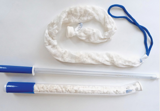
Although its research is in the pilot phase, Obstetrx, Inc. announces its XSTAT device has shown success in stopping postpartum hemorrhage (PPH) quickly. The device is a novel uterine tamponade “mini-sponge.” Contained in an applicator, XSTAT is comprised of compressed mini-sponges that are inserted into the postpartum uterus. Contained in a porous, distensible pouch, the sponges expand quickly, applying hemostatic pressure within the uterus. After a period of observation, the pouch is removed with gentle traction on the removal strand.
During a pilot study, reports Obstetrx, 9 patients, treated at the University Teaching Hospital in Lusaka, Zambia, did not respond to conventional PPH management options after vaginal birth but did respond, with bleeding resolved in 60 seconds and no adverse events, to the XSTAT device. The device was left in place for a mean time of 1 hour, and none of the patients required further surgical procedures or blood transfusions. The initial placement time of XSTAT (mean time to placement, 62 seconds) was faster than times reported for balloon uterine tamponade devices. The pilot study results were published in Obstetrics & Gynecology.
XSTAT is US Food and Drug Administration–approved to treat high-flow arterial bleeding in prehospital trauma settings, and Obstetrx is planning to submit for 510k clearance in 2022, after the conclusion of a follow-up PPH trial in 2021.
For more information, visit: https://www.obstetrx.com/.
Continue to: AI and ovulation prediction...
AI and ovulation prediction
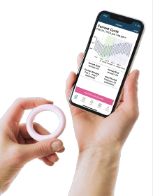
The Priya Fertility System, developed by Prima-Temp, will be available over the counter soon for couples who are trying to optimize their chances for pregnancy. The system consists of an intravaginal sensor and mobile app, the first of which measures a woman’s core body temperature and the second of which alerts the user of her fertility window to maximize conception.
A woman’s fertility window is typically the 5 days leading up to ovulation, with peak fertility in the 2 to 3 days before ovulation. There are other options for measuring that fertile window, including luteinizing hormone (LH) tests; however, Prima-Temp reports that Priya predicts the fertile window an average of 2.6 days before tests for LH. Utilizing continuous core body temperature measurement, Priya detects subtle changes in temperature patterns that occur prior to ovulation. The app portion of the technology stores and analyzes the temperature measurements, for a high-tech fertility alert system that also offers clinical diagnostic support. Potential users of the Priya system are able to sign up to receive it through the product’s website.
Hologic announces updates to its Brevera® Breast Biopsy System with CorLumina® Imaging Technology. The Brevera system is designed for use with the manufacturer’s Affirm® Prone biopsy guidance system.
Available since 2017, the Brevera system is designed to enhance the workflow for the breast biopsy procedure and overall patient experience by allowing for real-time imaging of sample acquisition. This feature avoids the need for the clinician to leave the patient exam room to verify tissue samples, saving time during the patient visit and allowing for more clinician-patient interaction. Brevera also combines tissue acquisition and real-time imaging verification with postbiopsy handling, with new functionality and simplified storage, including stowage of disposable needles, and improved waste management. The combination of the system improvements help to maintain the sample integrity, says Hologic, allowing for radiologists and technicians to handle the sample and then for the transfer to pathology. According to the manufacturer, the improved features offer facilities the potential to save an average of 13 minutes per procedure.
Although its research is in the pilot phase, Obstetrx, Inc. announces its XSTAT device has shown success in stopping postpartum hemorrhage (PPH) quickly. The device is a novel uterine tamponade “mini-sponge.” Contained in an applicator, XSTAT is comprised of compressed mini-sponges that are inserted into the postpartum uterus. Contained in a porous, distensible pouch, the sponges expand quickly, applying hemostatic pressure within the uterus. After a period of observation, the pouch is removed with gentle traction on the removal strand.
During a pilot study, reports Obstetrx, 9 patients, treated at the University Teaching Hospital in Lusaka, Zambia, did not respond to conventional PPH management options after vaginal birth but did respond, with bleeding resolved in 60 seconds and no adverse events, to the XSTAT device. The device was left in place for a mean time of 1 hour, and none of the patients required further surgical procedures or blood transfusions. The initial placement time of XSTAT (mean time to placement, 62 seconds) was faster than times reported for balloon uterine tamponade devices. The pilot study results were published in Obstetrics & Gynecology.
XSTAT is US Food and Drug Administration–approved to treat high-flow arterial bleeding in prehospital trauma settings, and Obstetrx is planning to submit for 510k clearance in 2022, after the conclusion of a follow-up PPH trial in 2021.
For more information, visit: https://www.obstetrx.com/.
Continue to: AI and ovulation prediction...
AI and ovulation prediction
The Priya Fertility System, developed by Prima-Temp, will be available over the counter soon for couples who are trying to optimize their chances for pregnancy. The system consists of an intravaginal sensor and mobile app, the first of which measures a woman’s core body temperature and the second of which alerts the user of her fertility window to maximize conception.
A woman’s fertility window is typically the 5 days leading up to ovulation, with peak fertility in the 2 to 3 days before ovulation. There are other options for measuring that fertile window, including luteinizing hormone (LH) tests; however, Prima-Temp reports that Priya predicts the fertile window an average of 2.6 days before tests for LH. Utilizing continuous core body temperature measurement, Priya detects subtle changes in temperature patterns that occur prior to ovulation. The app portion of the technology stores and analyzes the temperature measurements, for a high-tech fertility alert system that also offers clinical diagnostic support. Potential users of the Priya system are able to sign up to receive it through the product’s website.
Hologic announces updates to its Brevera® Breast Biopsy System with CorLumina® Imaging Technology. The Brevera system is designed for use with the manufacturer’s Affirm® Prone biopsy guidance system.
Available since 2017, the Brevera system is designed to enhance the workflow for the breast biopsy procedure and overall patient experience by allowing for real-time imaging of sample acquisition. This feature avoids the need for the clinician to leave the patient exam room to verify tissue samples, saving time during the patient visit and allowing for more clinician-patient interaction. Brevera also combines tissue acquisition and real-time imaging verification with postbiopsy handling, with new functionality and simplified storage, including stowage of disposable needles, and improved waste management. The combination of the system improvements help to maintain the sample integrity, says Hologic, allowing for radiologists and technicians to handle the sample and then for the transfer to pathology. According to the manufacturer, the improved features offer facilities the potential to save an average of 13 minutes per procedure.
Although its research is in the pilot phase, Obstetrx, Inc. announces its XSTAT device has shown success in stopping postpartum hemorrhage (PPH) quickly. The device is a novel uterine tamponade “mini-sponge.” Contained in an applicator, XSTAT is comprised of compressed mini-sponges that are inserted into the postpartum uterus. Contained in a porous, distensible pouch, the sponges expand quickly, applying hemostatic pressure within the uterus. After a period of observation, the pouch is removed with gentle traction on the removal strand.
During a pilot study, reports Obstetrx, 9 patients, treated at the University Teaching Hospital in Lusaka, Zambia, did not respond to conventional PPH management options after vaginal birth but did respond, with bleeding resolved in 60 seconds and no adverse events, to the XSTAT device. The device was left in place for a mean time of 1 hour, and none of the patients required further surgical procedures or blood transfusions. The initial placement time of XSTAT (mean time to placement, 62 seconds) was faster than times reported for balloon uterine tamponade devices. The pilot study results were published in Obstetrics & Gynecology.
XSTAT is US Food and Drug Administration–approved to treat high-flow arterial bleeding in prehospital trauma settings, and Obstetrx is planning to submit for 510k clearance in 2022, after the conclusion of a follow-up PPH trial in 2021.
For more information, visit: https://www.obstetrx.com/.
Continue to: AI and ovulation prediction...
AI and ovulation prediction
The Priya Fertility System, developed by Prima-Temp, will be available over the counter soon for couples who are trying to optimize their chances for pregnancy. The system consists of an intravaginal sensor and mobile app, the first of which measures a woman’s core body temperature and the second of which alerts the user of her fertility window to maximize conception.
A woman’s fertility window is typically the 5 days leading up to ovulation, with peak fertility in the 2 to 3 days before ovulation. There are other options for measuring that fertile window, including luteinizing hormone (LH) tests; however, Prima-Temp reports that Priya predicts the fertile window an average of 2.6 days before tests for LH. Utilizing continuous core body temperature measurement, Priya detects subtle changes in temperature patterns that occur prior to ovulation. The app portion of the technology stores and analyzes the temperature measurements, for a high-tech fertility alert system that also offers clinical diagnostic support. Potential users of the Priya system are able to sign up to receive it through the product’s website.
Patients who received telemedicine in an intensive care unit were less likely to die and more likely to have a shorter hospital stay than those who received standard ICU care without a 24-hour intensivist on-site, new data suggest.
Chiedozie I. Udeh, MD, staff intensivist with the Cleveland Clinic Foundation, presented results of a retrospective study of 153,987 consecutive ICU patients at the Critical Care Congress sponsored by the Society of Critical Care Medicine. .
Among the statistically significant findings were that 30-day mortality decreased by 18% (odds ratio, 0.82; 95% confidence interval, 0.77-0.87) and length of stay in the ICU decreased by 1.6 days in the telehealth model (95% CI, 1.5-1.7), compared with the traditional model. The total length of the average hospital stay was reduced by 2.1 days (95% CI, 1.9-2.4).
Patients in the study received ICU care at one of nine Cleveland Clinic hospitals between Jan. 1, 2010, and Dec. 31, 2019. Overall, 108,482 (70%) received ICU-telemedicine care during hours when an intensivist was not on-site.
Dr. Udeh said in an interview that only the largest academic centers typically have an intensivist on-site 24 hours a day. In the traditional model, critical care specialists may be on-site during the day but on call after hours.
In the tele-ICU model, in contrast, an intensivist – perhaps at a command center serving several hospitals – can observe and order treatments for patients remotely. The specialist has access to the patient’s medical record and test results, can monitor vital signs and visible changes, and can talk with both the patient and the nurse or other provider in the room.
Dr. Udeh said he suspects the 18% drop in mortality risk and the shorter hospital stay come from time saved. The physician doesn’t have to ask the nurse to look up health information and with constant monitoring can spot problems sooner or prevent them.
“You reduce a lot of the time from event to intervention or prevent an event by being more proactive,” Dr. Udeh said.
Ben Scott, MD, associate professor of anesthesiology and critical care at the University of Colorado at Denver, Aurora, said in an interview that his institution uses the tele-ICU model in several of the smaller hospitals there and is not surprised that Dr. Udeh’s team found such positive results. Dr. Scott was not involved in Dr. Udeh’s study.
“Most of us who have been working in this area and studying the results believe that these programs can make a big difference,” said Dr. Scott, vice chair for the SCCM tele-critical-care committee.
The smaller UC hospitals have ICU capability but not the census numbers to warrant 24-hour intensivist coverage. Of course, they do have 24-hour nursing coverage, and they typically use telemedicine when an intensivist is needed during the night, Dr. Scott said.
Hard to pinpoint telemedicine’s role
Dr. Scott said it’s hard to determine from studies how much telemedicine is influencing outcomes, compared with potentially confounding factors. A hospital with several ICUs might choose to send a patient to a certain ICU for a particular reason, which could confound comparisons.
The statistical techniques Dr. Udeh’s team used, however, helped account for confounding, Dr. Scott said. The extended years for the study and large patient sample also strengthen confidence in the results, he said.
The researchers found that several factors can increase an ICU patient’s risk of dying, including the reason for admission (such as cardiac arrest or sepsis), being admitted on a weekend, and the patient’s race. But they found that telemedicine might mitigate the effects of weekend admissions; the telemedicine patients admitted on a weekend in this study were no more likely to die than those admitted on a weekday.
The telemedicine model is especially important in areas without intensivists.
“If my only recourse is to send my patient out of town or out of state to another hospital, it’s a win-win,” Dr. Udeh said.
Regardless of the resources of individual hospitals, the national picture is clear, he said. “We just don’t have enough people trained in critical care to place an intensivist in every ICU 24/7.”
In late January, Santa Cruz Valley Regional Hospital in Green Valley, Ariz., temporarily shut down its ICU. The hospital CEO said the closure came because the hospital was unable to hire a pulmonologist.
Balancing cost issues
Cost issues with the tele-ICU have been a barrier for widespread adoption, Dr. Udeh said. He estimated that only about 15%-20% of hospitals incorporate the model.
Hospitals must pay for hardware and the telehealth service while still needing to have someone on staff available to come in if a physician’s presence is needed. And so far, those costs are not generally reimbursable by payers.
Hospitals must balance the costs with the potential for better outcomes and shorter stays, he said.
The model has benefits for the provider as well.
Dr. Udeh recounted being awakened by a call in the middle of the night and fighting off grogginess to quickly process information and make critical decisions.
But with the tele-ICU model, providers are awake for a specified shift and are periodically rounding on patients electronically with real-time access to health information.
Dr. Udeh said many of the tele-ICU platforms have decision support built in, with various degrees of complexity, so that the system might flag when a patient’s blood pressure is trending down, for example.
Although this research used prepandemic data, COVID-19 has highlighted the need for solutions to stretch ICU workforces.
Dr. Scott pointed out that in the pandemic, many hospitals that don’t have regular critical care services have had to take care of critically ill patients.
Having a telemedicine program can help bring that expertise to the bedside, he said.
Dr. Udeh, his coinvestigators, and Dr. Scott have disclosed no relevant financial relationships.
A version of this article first appeared on Medscape.com.
Patients who received telemedicine in an intensive care unit were less likely to die and more likely to have a shorter hospital stay than those who received standard ICU care without a 24-hour intensivist on-site, new data suggest.
Chiedozie I. Udeh, MD, staff intensivist with the Cleveland Clinic Foundation, presented results of a retrospective study of 153,987 consecutive ICU patients at the Critical Care Congress sponsored by the Society of Critical Care Medicine. .
Among the statistically significant findings were that 30-day mortality decreased by 18% (odds ratio, 0.82; 95% confidence interval, 0.77-0.87) and length of stay in the ICU decreased by 1.6 days in the telehealth model (95% CI, 1.5-1.7), compared with the traditional model. The total length of the average hospital stay was reduced by 2.1 days (95% CI, 1.9-2.4).
Patients in the study received ICU care at one of nine Cleveland Clinic hospitals between Jan. 1, 2010, and Dec. 31, 2019. Overall, 108,482 (70%) received ICU-telemedicine care during hours when an intensivist was not on-site.
Dr. Udeh said in an interview that only the largest academic centers typically have an intensivist on-site 24 hours a day. In the traditional model, critical care specialists may be on-site during the day but on call after hours.
In the tele-ICU model, in contrast, an intensivist – perhaps at a command center serving several hospitals – can observe and order treatments for patients remotely. The specialist has access to the patient’s medical record and test results, can monitor vital signs and visible changes, and can talk with both the patient and the nurse or other provider in the room.
Dr. Udeh said he suspects the 18% drop in mortality risk and the shorter hospital stay come from time saved. The physician doesn’t have to ask the nurse to look up health information and with constant monitoring can spot problems sooner or prevent them.
“You reduce a lot of the time from event to intervention or prevent an event by being more proactive,” Dr. Udeh said.
Ben Scott, MD, associate professor of anesthesiology and critical care at the University of Colorado at Denver, Aurora, said in an interview that his institution uses the tele-ICU model in several of the smaller hospitals there and is not surprised that Dr. Udeh’s team found such positive results. Dr. Scott was not involved in Dr. Udeh’s study.
“Most of us who have been working in this area and studying the results believe that these programs can make a big difference,” said Dr. Scott, vice chair for the SCCM tele-critical-care committee.
The smaller UC hospitals have ICU capability but not the census numbers to warrant 24-hour intensivist coverage. Of course, they do have 24-hour nursing coverage, and they typically use telemedicine when an intensivist is needed during the night, Dr. Scott said.
Hard to pinpoint telemedicine’s role
Dr. Scott said it’s hard to determine from studies how much telemedicine is influencing outcomes, compared with potentially confounding factors. A hospital with several ICUs might choose to send a patient to a certain ICU for a particular reason, which could confound comparisons.
The statistical techniques Dr. Udeh’s team used, however, helped account for confounding, Dr. Scott said. The extended years for the study and large patient sample also strengthen confidence in the results, he said.
The researchers found that several factors can increase an ICU patient’s risk of dying, including the reason for admission (such as cardiac arrest or sepsis), being admitted on a weekend, and the patient’s race. But they found that telemedicine might mitigate the effects of weekend admissions; the telemedicine patients admitted on a weekend in this study were no more likely to die than those admitted on a weekday.
The telemedicine model is especially important in areas without intensivists.
“If my only recourse is to send my patient out of town or out of state to another hospital, it’s a win-win,” Dr. Udeh said.
Regardless of the resources of individual hospitals, the national picture is clear, he said. “We just don’t have enough people trained in critical care to place an intensivist in every ICU 24/7.”
In late January, Santa Cruz Valley Regional Hospital in Green Valley, Ariz., temporarily shut down its ICU. The hospital CEO said the closure came because the hospital was unable to hire a pulmonologist.
Balancing cost issues
Cost issues with the tele-ICU have been a barrier for widespread adoption, Dr. Udeh said. He estimated that only about 15%-20% of hospitals incorporate the model.
Hospitals must pay for hardware and the telehealth service while still needing to have someone on staff available to come in if a physician’s presence is needed. And so far, those costs are not generally reimbursable by payers.
Hospitals must balance the costs with the potential for better outcomes and shorter stays, he said.
The model has benefits for the provider as well.
Dr. Udeh recounted being awakened by a call in the middle of the night and fighting off grogginess to quickly process information and make critical decisions.
But with the tele-ICU model, providers are awake for a specified shift and are periodically rounding on patients electronically with real-time access to health information.
Dr. Udeh said many of the tele-ICU platforms have decision support built in, with various degrees of complexity, so that the system might flag when a patient’s blood pressure is trending down, for example.
Although this research used prepandemic data, COVID-19 has highlighted the need for solutions to stretch ICU workforces.
Dr. Scott pointed out that in the pandemic, many hospitals that don’t have regular critical care services have had to take care of critically ill patients.
Having a telemedicine program can help bring that expertise to the bedside, he said.
Dr. Udeh, his coinvestigators, and Dr. Scott have disclosed no relevant financial relationships.
A version of this article first appeared on Medscape.com.
Patients who received telemedicine in an intensive care unit were less likely to die and more likely to have a shorter hospital stay than those who received standard ICU care without a 24-hour intensivist on-site, new data suggest.
Chiedozie I. Udeh, MD, staff intensivist with the Cleveland Clinic Foundation, presented results of a retrospective study of 153,987 consecutive ICU patients at the Critical Care Congress sponsored by the Society of Critical Care Medicine. .
Among the statistically significant findings were that 30-day mortality decreased by 18% (odds ratio, 0.82; 95% confidence interval, 0.77-0.87) and length of stay in the ICU decreased by 1.6 days in the telehealth model (95% CI, 1.5-1.7), compared with the traditional model. The total length of the average hospital stay was reduced by 2.1 days (95% CI, 1.9-2.4).
Patients in the study received ICU care at one of nine Cleveland Clinic hospitals between Jan. 1, 2010, and Dec. 31, 2019. Overall, 108,482 (70%) received ICU-telemedicine care during hours when an intensivist was not on-site.
Dr. Udeh said in an interview that only the largest academic centers typically have an intensivist on-site 24 hours a day. In the traditional model, critical care specialists may be on-site during the day but on call after hours.
In the tele-ICU model, in contrast, an intensivist – perhaps at a command center serving several hospitals – can observe and order treatments for patients remotely. The specialist has access to the patient’s medical record and test results, can monitor vital signs and visible changes, and can talk with both the patient and the nurse or other provider in the room.
Dr. Udeh said he suspects the 18% drop in mortality risk and the shorter hospital stay come from time saved. The physician doesn’t have to ask the nurse to look up health information and with constant monitoring can spot problems sooner or prevent them.
“You reduce a lot of the time from event to intervention or prevent an event by being more proactive,” Dr. Udeh said.
Ben Scott, MD, associate professor of anesthesiology and critical care at the University of Colorado at Denver, Aurora, said in an interview that his institution uses the tele-ICU model in several of the smaller hospitals there and is not surprised that Dr. Udeh’s team found such positive results. Dr. Scott was not involved in Dr. Udeh’s study.
“Most of us who have been working in this area and studying the results believe that these programs can make a big difference,” said Dr. Scott, vice chair for the SCCM tele-critical-care committee.
The smaller UC hospitals have ICU capability but not the census numbers to warrant 24-hour intensivist coverage. Of course, they do have 24-hour nursing coverage, and they typically use telemedicine when an intensivist is needed during the night, Dr. Scott said.
Hard to pinpoint telemedicine’s role
Dr. Scott said it’s hard to determine from studies how much telemedicine is influencing outcomes, compared with potentially confounding factors. A hospital with several ICUs might choose to send a patient to a certain ICU for a particular reason, which could confound comparisons.
The statistical techniques Dr. Udeh’s team used, however, helped account for confounding, Dr. Scott said. The extended years for the study and large patient sample also strengthen confidence in the results, he said.
The researchers found that several factors can increase an ICU patient’s risk of dying, including the reason for admission (such as cardiac arrest or sepsis), being admitted on a weekend, and the patient’s race. But they found that telemedicine might mitigate the effects of weekend admissions; the telemedicine patients admitted on a weekend in this study were no more likely to die than those admitted on a weekday.
The telemedicine model is especially important in areas without intensivists.
“If my only recourse is to send my patient out of town or out of state to another hospital, it’s a win-win,” Dr. Udeh said.
Regardless of the resources of individual hospitals, the national picture is clear, he said. “We just don’t have enough people trained in critical care to place an intensivist in every ICU 24/7.”
In late January, Santa Cruz Valley Regional Hospital in Green Valley, Ariz., temporarily shut down its ICU. The hospital CEO said the closure came because the hospital was unable to hire a pulmonologist.
Balancing cost issues
Cost issues with the tele-ICU have been a barrier for widespread adoption, Dr. Udeh said. He estimated that only about 15%-20% of hospitals incorporate the model.
Hospitals must pay for hardware and the telehealth service while still needing to have someone on staff available to come in if a physician’s presence is needed. And so far, those costs are not generally reimbursable by payers.
Hospitals must balance the costs with the potential for better outcomes and shorter stays, he said.
The model has benefits for the provider as well.
Dr. Udeh recounted being awakened by a call in the middle of the night and fighting off grogginess to quickly process information and make critical decisions.
But with the tele-ICU model, providers are awake for a specified shift and are periodically rounding on patients electronically with real-time access to health information.
Dr. Udeh said many of the tele-ICU platforms have decision support built in, with various degrees of complexity, so that the system might flag when a patient’s blood pressure is trending down, for example.
Although this research used prepandemic data, COVID-19 has highlighted the need for solutions to stretch ICU workforces.
Dr. Scott pointed out that in the pandemic, many hospitals that don’t have regular critical care services have had to take care of critically ill patients.
Having a telemedicine program can help bring that expertise to the bedside, he said.
Dr. Udeh, his coinvestigators, and Dr. Scott have disclosed no relevant financial relationships.
A version of this article first appeared on Medscape.com.
In this Update, we discuss several aspects of infertility and emerging technologic advances in treatment. We review an important infertility fact sheet recently issued by the World Health Organization (WHO) that provides a succinct overview of infertility causes, the rights of infertility patients, treatment challenges, and advocacy efforts. In addition, we discuss what the infertility literature reveals about reducing multiple birth rates and the technologic, financial, and social factors involved. Finally, we look at the molecular progress made in germline-editing technology and the myriad complications involved in its potential future translation to clinical phenotyping.
WHO recognizes the burden of infertility and addresses fertility care needs
World Health Organization (WHO). Infertility fact sheet. September 14, 2020. https://www.who.int/news-room/fact-sheets/detail/infertility. Accessed January 24, 2021.
The WHO published its first comprehensive infertility fact sheet in September 2020. This document is important because it validates infertility as a high-burden disease and disability that diminishes quality of life for up to 186 million individuals globally. The infertility fact sheet is a comprehensive yet focused quick read that addresses the causes of infertility, why infertility is important, challenges, and the WHO response.
Factors in infertility
Infertility is caused by different factors in women and men, yet sometimes it is unexplained, and its relative importance can vary from country to country. For women, tubal disorders (for example, postinfectious), uterine problems (fibroids, congenital), endometriosis, ovarian disorders (polycystic ovary syndrome, ovulation disorders), and endocrine imbalances are the most common factors.
For men, causes of infertility include obstruction of the reproductive tract (as after injuries or infection); hormonal disorders in the hypothalamus, pituitary, and/or testicles (for example, low testosterone); testicular failure to produce sperm (such as after cancer treatment); and abnormal sperm function and quality (low count, motility, or morphology).
Environmental and lifestyle factors— including smoking, obesity, alcohol, or toxins—can affect fertility.
Continue to: Recognizing all individuals’ fertility rights...
Recognizing all individuals’ fertility rights
The WHO infertility fact sheet makes strong statements, recognizing that individuals and couples have the right to decide the number, timing, and spacing of their children. Addressing infertility is therefore an important part of realizing the right of individuals and couples to found a family. This includes heterosexual couples, same-sex partners, older persons, individuals not in sexual relationships who might require infertility management and fertility care services, and notably marginalized populations.
Addressing infertility also can help mitigate gender inequality, which has significant negative social impacts on the lives of infertile individuals, especially women. Fertility education is important to reduce the fear of infertility and contraception use in those wanting pregnancy in the future.
In most countries the biggest challenges are availability, access, and quality of interventions to address infertility. This includes the United States, where only 1 in 4 individuals receive the fertility care they need. Lack of prioritization, ineffective public health strategies, inadequate funding, and costs are barriers. Health policies need to recognize that infertility is a disease that often can be prevented, thereby reducing future costs. Comprehensive awareness and education programs, laws and policies that regulate and ensure access and the human rights of all involved, are essential.
Advocacy efforts
To address infertility and fertility care, the WHO is committed to:
collaborate with partners on epidemiologic and etiologic research
facilitate policy dialogue globally to frame infertility within a legal and policy framework
support generation of data on the burden of infertility
develop guidelines
produce other documents of standards
collaborate with all stakeholders to strengthen political commitment and health system capacity, and
provide country-level technical support to develop or strengthen policies and services.
For your practice, this means that infertility is recognized as a disease that should receive its appropriate share of health care resources. Infertility and fertility care are the right of every individual according to their desires to found a family. Besides providing the best care you can to all your patients, including referring them when necessary, all health care clinicians should advocate on behalf of their patients to payors, policy makers, and the public the need to provide equitable laws, resources, and funding for infertility and fertility care.
WHAT THIS EVIDENCE MEANS FOR PRACTICE
Every person has the right to infertility and fertility care as endorsed by the recent WHO infertility fact sheet. To address this high-burden disease, all women’s health care clinicians should be aware of, equitably diagnose and treat, refer as necessary, and advocate for infertile individuals.
Continue to: Lessons learned in reducing multiple pregnancy rates in infertility treatment...
Lessons learned in reducing multiple pregnancy rates in infertility treatment
In the October 2020 issue of Fertility and Sterility, the Views and Reviews section included 5 articles on avoiding multiple live birth rates (LBRs) in assisted reproductive technologies (ART).1-5 International experts provided a comprehensive review of global multiple LBRs and their associated negative impact on maternal and perinatal outcomes, reasons for global variability, strategies to reduce multiples, single embryo transfer, and implications of funding and reporting. These international comparisons and recommendations are helpful and applicable to infertility care in the United States.3
The rise of multiple birth rates
During the first decade of in vitro fertilization (IVF), live birth rates were low, increasing to 14% in 1990. Multiple embryos needed to be transferred so that even these LBRs could be obtained. In the 1990s, however, laboratory technology improved rapidly, with increased implantation rates and subsequent rapid increases in LBR, but also with increased multiple birth rates (MBRs).
In the United States, clinic-specific reporting helped create competition among clinics for the best LBRs, and this led to MBRs of 30% and higher. Numerous studies documented the associated significantly increased morbidity and mortality of both mothers and babies. Similar situations occurred in many other countries while some, especially Nordic nations, Australia, New Zealand, and Japan, had twin rates of less than 10% or even 5% since the early 2000s. So why the difference?
The higher MBR is due largely to the transfer of more than one embryo. The immediate solution is therefore always to perform elective single embryo transfer (eSET). However, numerous factors affect the decision to perform eSET or not, and this ideal is far from being achieved. Older women, those with longer duration of infertility and/or failed treatment, often feel a time pressure and want to transfer more embryos. Of course, biologically this is reasonable because the number and quality of their embryos is lower. While attempts have been made to assess embryo quality with preimplantation genetic testing for aneuploidy, evidence that this increases the LBR is controversial except possibly in women aged 35 to 38 years. This is especially true when the cumulative LBR, that is, the number of live births after transfer of all embryos from an egg retrieval cycle, is the measured outcome.
The major factor that determines the frequency of eSET is financial. Affordability is the out-of-pocket cost (after insurance or other subsidy) as a percentage of disposable income, and it is the most important factor that determines whether eSET is performed. Less affordable treatment creates a financial incentive to transfer more than one embryo to maximize the pregnancy rates in fewer cycles.5 Other factors include whether the effectiveness of treatment, that is, LBR, is emphasized over safety, that is, MBR. In the United States, the Society for Assisted Reproductive Technology now reports cumulative LBR, singleton and multiple LBR, and preterm births as outcomes, thereby increasing the emphasis on eSET.
Sociologic, cultural, and religious factors also can affect the frequency of eSET. Even within the United States, great variation exists in values and beliefs regarding infertility treatment. It can be challenging to determine who makes decisions: the patient alone, the physician, the payor, professional guidelines, or laws. In many countries, including the United States, it is an amalgam of these.
Setting new goals
If the goal is to reduce the MBR, what should that rate be? In the past few years, the MBR in the United States has been reduced to approximately 10%. It is reasonable now to set a goal of 5% in the next several years. To do this, we can learn from countries that have been successful. The United States already has very high-quality clinical and laboratory services, knowledgeable physicians, and a reasonable regulatory environment. Improved technology, specifically embryo selection for transfer, and focus on adherence to established embryo transfer guidelines could help.
Many would argue that eSET essentially should be performed always in women younger than age 40 and in all women of any age with a known euploid embryo. The major problem that drives multiples is the lack of affordability, which can be addressed by increased subsidies from payors. Increased subsidies can result from legislative mandates or societal pressures on employers, either of which could be associated with requirements for eSET and/or reduced MBRs.
In your practice, you can now reassure your infertility patients that cumulative LBRs are excellent in the United States and that the risk of multiple pregnancy has been reduced dramatically. This should encourage more patients to accept and take advantage of this successful technology that has resulted in the birth of millions of babies globally. Further reduction of the MBR to 5% should be possible within a few years through education and advocacy by women’s health care clinicians that results in increased subsidies and more affordable IVF.
WHAT THIS EVIDENCE MEANS FOR PRACTICE The multiple birth rate in ART has been reduced to 10% in the United States through an increased understanding of the complex factors that affect embryo transfer practices globally. Further progress will depend primarily on increased subsidies that make ART more affordable.
Continue to: Genetics and ART...
Genetics and ART: Selection versus correction
Adashi EY, Cohen IG. The case for remedial germline editing—the long-term view. JAMA. 2020;323:1762-1763.
Rosenbaum L. The future of gene editing—toward scientific and social consensus. N Engl J Med. 2019;380:971-975.
Cyranoski D. The CRISPR-baby scandal: what’s next for human gene-editing. Nature. 2019;566:440-442.
de Wert G, Pennings G, Clarke A, et al; European Society of Human Genetics and European Society of Human Reproduction and Embryology. Human germline gene editing: recommendations of ESHG and ESHRE. Hum Reprod Open. 2018;hox025.
Following the completion of the Human Genome Project in 2003 and major technologic advancements in the subsequent years, the field of human genetics became the focal point of convergence for several distinct but interrelated disciplines: bioinformatics, computational biology, and sequencing technologies. As the result, individual human genomes can now be sequenced at a single base pair level, and with higher fidelity, at a fraction of the original cost and at a much faster speed.
This molecular progress, however, has not been accompanied by an equivalent clinical progress, because in a significant number of cases a defined and predictable clinical phenotype cannot be attributed to a detected molecular genotype. This has resulted in an overabundance of variants of uncertain significance. Variable expressivity, incomplete penetrance, epigenetics, mosaicism, and the polygenic nature of many human traits further complicate reliable interpretation and prognostication of the colossal amount of molecular genetic data that are being generated by the above-mentioned technologic advances.
Considering these limitations, at this juncture it is crucial to acknowledge that any attempts to prematurely commercialize these preclinical and research studies (such as polygenic risk scores for embryos) are perilous and have the potential to cause significant harm in terms of unnecessary stress and anxiety for intended parents as well as the potential for yet-unmapped societal and legal implications.
However, it is just a matter of time until more accurate clinical phenotyping catches up with molecular genotyping. As we get closer to this next historic milestone, precision medicine in the postnatal life (with regard to both diagnostics and therapeutics) and preimplantation genetic testing (PGT) at the prenatal stage for a much wider spectrum of conditions—including both monogenic and polygenic traits—may indeed become a reality.
The potential of germline editing
Specifically regarding PGT (which requires IVF), it is important to recognize that due to the limited and nonrenewable endowment of human oocytes (ovarian reserve), combined with the detrimental impact of advancing age on the quality of the remaining cohort as manifested by a higher risk of aneuploidy, the current clinical practice of trying to “select” a nonaffected embryo can be very inefficient. As a result, the intended parents pursuing such treatments may need to undergo multiple cycles of ovarian stimulation and oocyte retrieval.
A potential solution for genes associated with known diseases is the prospect of remedial germline editing by CRISPR–Cas9 technology or its future descendants. This would take advantage of the existing embryos to try to “correct” the defective gene instead of trying to “select” a normal embryo. These technologies are still in the early stages of development and are remotely distant from clinical applications. On the other hand, although germline gene editing, if actualized, would be a monumental breakthrough in the history of genetics and medicine, we must be cognizant of its serious legal, societal, and ethical ramifications, which are currently unknown. Furthermore, even at the biologic and technical level, the technology still is not advanced enough to reliably rule out off-target modifications, and the unintended clinical consequences of the on-target corrections have not been studied either.
Regulation of genetic modifications
Due to these myriad concerns and the lack of an existing appropriate regulatory framework and oversight for such interventions, current US law (since December 2015, through provisions in annual federal appropriations laws passed by Congress and renewed annually thereafter) bars the US Food and Drug Administration from considering any clinical trial application “in which a human embryo is intentionally created or modified to include a heritable genetic modification.” Notably, this moratorium also prohibits mitochondrial replacement technology (MRT), which is a less controversial and relatively better-studied innovation.
Mitochondrial genetic disorders caused by the mutations in mitochondrial DNA (versus nuclear DNA) are amenable to a specific treatment strategy aimed at substituting the defective maternal mitochondrial genome with the mitochondrial genome of an unaffected donor oocyte. This can be achieved via either pronuclear transfer, which involves isolation and transfer of the male and female pronuclei from an affected embryo to an enucleated normal donor embryo, or maternal spindle transfer, which involves isolation and transfer of the metaphase II spindle complex of an affected oocyte to an enucleated disease-free donor egg. It is noteworthy that in 2015 in the United Kingdom, Parliament expanded the definition of “permitted eggs and embryos” to include those “where unhealthy mitochondrial DNA is replaced by healthy mitochondrial DNA from a donor.” This thereby allows the UK Human Fertilisation and Embryology Authority to formally direct and oversee clinical trials in MRT.
Summing up
Although the future of assisted human reproduction cannot be clearly outlined at this time, it is likely to be radically different from the current state given these emerging applications at the intersection of ART and diagnostic and therapeutic genetics. To ensure that exploring this uncharted territory will ultimately be in the interest of humankind and civilization, proper regulatory oversight—after careful consideration of all ethical, societal, and legal implications—needs to be developed for all preclinical and clinical research in this field. Participatory public engagement must be an integrated part of this process. ●
WHAT THIS EVIDENCE MEANS FOR PRACTICE The field of human genetics has already transformed medicine. However, the convergence of the interrelated disciplines of bioinformatics, computational biology, sequencing technologies, and CRISPR–Cas9 technology is creating incredible new advances that will bring great benefits but also major societal challenges.
References
Farquhar C. Avoiding multiple pregnancies in assisted reproductive technologies: transferring one embryo at a time should be the norm. Fertil Steril. 2020;114:671-672.
Bergh C, Kamath MS, Wang R, et al. Strategies to reduce multiple pregnancies during medically assisted reproduction. Fertil Steril. 2020;114:673-679.
Adamson GD, Norman RJ. Why are multiple pregnancy rates and single embryo transfer rates so different globally, and what do we do about it? Fertil Steril. 2020;114:680-689.
Eapen A, Ryan GL, Ten Eyck P, et al. Current evidence supporting a goal of singletons: a review of maternal and neonatal outcomes associated with twin versus singleton pregnancies after in vitro fertilization and intracytoplasmic sperm injection. Fertil Steril. 2020;114: 690-714.
Chambers GM, Keller E, Choi S, et al. Funding and public reporting strategies for reducing multiple pregnancy from fertility treatments. Fertil Steril. 2020;114:715-721.
Dr. Adamson is Founder and CEO of Advanced Reproductive Care, Inc (ARC Fertility); Clinical Professor, ACF, at Stanford University School of Medicine; and Associate Clinical Professor at the University of California, San Francisco. He is also Director of Equal3 Fertility, APC in Cupertino, California.
M. Max Ezzati, MD
Dr. Ezzati is a Board-certified reproductive endocrinology and infertility (REI) specialist and the Medical Director of the Department of Reproductive Endocrinology and Infertility at Palo Alto Medical Foundation Fertility Physicians of Northern California.
Dr. Adamson reports that he is a consultant for Abbott and LabCorp and is a speaker for Abbott. Dr. Ezzati reports no financial relationships relevant to this article.
Dr. Adamson is Founder and CEO of Advanced Reproductive Care, Inc (ARC Fertility); Clinical Professor, ACF, at Stanford University School of Medicine; and Associate Clinical Professor at the University of California, San Francisco. He is also Director of Equal3 Fertility, APC in Cupertino, California.
M. Max Ezzati, MD
Dr. Ezzati is a Board-certified reproductive endocrinology and infertility (REI) specialist and the Medical Director of the Department of Reproductive Endocrinology and Infertility at Palo Alto Medical Foundation Fertility Physicians of Northern California.
Dr. Adamson reports that he is a consultant for Abbott and LabCorp and is a speaker for Abbott. Dr. Ezzati reports no financial relationships relevant to this article.
Author and Disclosure Information
G. David Adamson, MD
Dr. Adamson is Founder and CEO of Advanced Reproductive Care, Inc (ARC Fertility); Clinical Professor, ACF, at Stanford University School of Medicine; and Associate Clinical Professor at the University of California, San Francisco. He is also Director of Equal3 Fertility, APC in Cupertino, California.
M. Max Ezzati, MD
Dr. Ezzati is a Board-certified reproductive endocrinology and infertility (REI) specialist and the Medical Director of the Department of Reproductive Endocrinology and Infertility at Palo Alto Medical Foundation Fertility Physicians of Northern California.
Dr. Adamson reports that he is a consultant for Abbott and LabCorp and is a speaker for Abbott. Dr. Ezzati reports no financial relationships relevant to this article.
In this Update, we discuss several aspects of infertility and emerging technologic advances in treatment. We review an important infertility fact sheet recently issued by the World Health Organization (WHO) that provides a succinct overview of infertility causes, the rights of infertility patients, treatment challenges, and advocacy efforts. In addition, we discuss what the infertility literature reveals about reducing multiple birth rates and the technologic, financial, and social factors involved. Finally, we look at the molecular progress made in germline-editing technology and the myriad complications involved in its potential future translation to clinical phenotyping.
WHO recognizes the burden of infertility and addresses fertility care needs
World Health Organization (WHO). Infertility fact sheet. September 14, 2020. https://www.who.int/news-room/fact-sheets/detail/infertility. Accessed January 24, 2021.
The WHO published its first comprehensive infertility fact sheet in September 2020. This document is important because it validates infertility as a high-burden disease and disability that diminishes quality of life for up to 186 million individuals globally. The infertility fact sheet is a comprehensive yet focused quick read that addresses the causes of infertility, why infertility is important, challenges, and the WHO response.
Factors in infertility
Infertility is caused by different factors in women and men, yet sometimes it is unexplained, and its relative importance can vary from country to country. For women, tubal disorders (for example, postinfectious), uterine problems (fibroids, congenital), endometriosis, ovarian disorders (polycystic ovary syndrome, ovulation disorders), and endocrine imbalances are the most common factors.
For men, causes of infertility include obstruction of the reproductive tract (as after injuries or infection); hormonal disorders in the hypothalamus, pituitary, and/or testicles (for example, low testosterone); testicular failure to produce sperm (such as after cancer treatment); and abnormal sperm function and quality (low count, motility, or morphology).
Environmental and lifestyle factors— including smoking, obesity, alcohol, or toxins—can affect fertility.
Continue to: Recognizing all individuals’ fertility rights...
Recognizing all individuals’ fertility rights
The WHO infertility fact sheet makes strong statements, recognizing that individuals and couples have the right to decide the number, timing, and spacing of their children. Addressing infertility is therefore an important part of realizing the right of individuals and couples to found a family. This includes heterosexual couples, same-sex partners, older persons, individuals not in sexual relationships who might require infertility management and fertility care services, and notably marginalized populations.
Addressing infertility also can help mitigate gender inequality, which has significant negative social impacts on the lives of infertile individuals, especially women. Fertility education is important to reduce the fear of infertility and contraception use in those wanting pregnancy in the future.
In most countries the biggest challenges are availability, access, and quality of interventions to address infertility. This includes the United States, where only 1 in 4 individuals receive the fertility care they need. Lack of prioritization, ineffective public health strategies, inadequate funding, and costs are barriers. Health policies need to recognize that infertility is a disease that often can be prevented, thereby reducing future costs. Comprehensive awareness and education programs, laws and policies that regulate and ensure access and the human rights of all involved, are essential.
Advocacy efforts
To address infertility and fertility care, the WHO is committed to:
collaborate with partners on epidemiologic and etiologic research
facilitate policy dialogue globally to frame infertility within a legal and policy framework
support generation of data on the burden of infertility
develop guidelines
produce other documents of standards
collaborate with all stakeholders to strengthen political commitment and health system capacity, and
provide country-level technical support to develop or strengthen policies and services.
For your practice, this means that infertility is recognized as a disease that should receive its appropriate share of health care resources. Infertility and fertility care are the right of every individual according to their desires to found a family. Besides providing the best care you can to all your patients, including referring them when necessary, all health care clinicians should advocate on behalf of their patients to payors, policy makers, and the public the need to provide equitable laws, resources, and funding for infertility and fertility care.
WHAT THIS EVIDENCE MEANS FOR PRACTICE
Every person has the right to infertility and fertility care as endorsed by the recent WHO infertility fact sheet. To address this high-burden disease, all women’s health care clinicians should be aware of, equitably diagnose and treat, refer as necessary, and advocate for infertile individuals.
Continue to: Lessons learned in reducing multiple pregnancy rates in infertility treatment...
Lessons learned in reducing multiple pregnancy rates in infertility treatment
In the October 2020 issue of Fertility and Sterility, the Views and Reviews section included 5 articles on avoiding multiple live birth rates (LBRs) in assisted reproductive technologies (ART).1-5 International experts provided a comprehensive review of global multiple LBRs and their associated negative impact on maternal and perinatal outcomes, reasons for global variability, strategies to reduce multiples, single embryo transfer, and implications of funding and reporting. These international comparisons and recommendations are helpful and applicable to infertility care in the United States.3
The rise of multiple birth rates
During the first decade of in vitro fertilization (IVF), live birth rates were low, increasing to 14% in 1990. Multiple embryos needed to be transferred so that even these LBRs could be obtained. In the 1990s, however, laboratory technology improved rapidly, with increased implantation rates and subsequent rapid increases in LBR, but also with increased multiple birth rates (MBRs).
In the United States, clinic-specific reporting helped create competition among clinics for the best LBRs, and this led to MBRs of 30% and higher. Numerous studies documented the associated significantly increased morbidity and mortality of both mothers and babies. Similar situations occurred in many other countries while some, especially Nordic nations, Australia, New Zealand, and Japan, had twin rates of less than 10% or even 5% since the early 2000s. So why the difference?
The higher MBR is due largely to the transfer of more than one embryo. The immediate solution is therefore always to perform elective single embryo transfer (eSET). However, numerous factors affect the decision to perform eSET or not, and this ideal is far from being achieved. Older women, those with longer duration of infertility and/or failed treatment, often feel a time pressure and want to transfer more embryos. Of course, biologically this is reasonable because the number and quality of their embryos is lower. While attempts have been made to assess embryo quality with preimplantation genetic testing for aneuploidy, evidence that this increases the LBR is controversial except possibly in women aged 35 to 38 years. This is especially true when the cumulative LBR, that is, the number of live births after transfer of all embryos from an egg retrieval cycle, is the measured outcome.
The major factor that determines the frequency of eSET is financial. Affordability is the out-of-pocket cost (after insurance or other subsidy) as a percentage of disposable income, and it is the most important factor that determines whether eSET is performed. Less affordable treatment creates a financial incentive to transfer more than one embryo to maximize the pregnancy rates in fewer cycles.5 Other factors include whether the effectiveness of treatment, that is, LBR, is emphasized over safety, that is, MBR. In the United States, the Society for Assisted Reproductive Technology now reports cumulative LBR, singleton and multiple LBR, and preterm births as outcomes, thereby increasing the emphasis on eSET.
Sociologic, cultural, and religious factors also can affect the frequency of eSET. Even within the United States, great variation exists in values and beliefs regarding infertility treatment. It can be challenging to determine who makes decisions: the patient alone, the physician, the payor, professional guidelines, or laws. In many countries, including the United States, it is an amalgam of these.
Setting new goals
If the goal is to reduce the MBR, what should that rate be? In the past few years, the MBR in the United States has been reduced to approximately 10%. It is reasonable now to set a goal of 5% in the next several years. To do this, we can learn from countries that have been successful. The United States already has very high-quality clinical and laboratory services, knowledgeable physicians, and a reasonable regulatory environment. Improved technology, specifically embryo selection for transfer, and focus on adherence to established embryo transfer guidelines could help.
Many would argue that eSET essentially should be performed always in women younger than age 40 and in all women of any age with a known euploid embryo. The major problem that drives multiples is the lack of affordability, which can be addressed by increased subsidies from payors. Increased subsidies can result from legislative mandates or societal pressures on employers, either of which could be associated with requirements for eSET and/or reduced MBRs.
In your practice, you can now reassure your infertility patients that cumulative LBRs are excellent in the United States and that the risk of multiple pregnancy has been reduced dramatically. This should encourage more patients to accept and take advantage of this successful technology that has resulted in the birth of millions of babies globally. Further reduction of the MBR to 5% should be possible within a few years through education and advocacy by women’s health care clinicians that results in increased subsidies and more affordable IVF.
WHAT THIS EVIDENCE MEANS FOR PRACTICE The multiple birth rate in ART has been reduced to 10% in the United States through an increased understanding of the complex factors that affect embryo transfer practices globally. Further progress will depend primarily on increased subsidies that make ART more affordable.
Continue to: Genetics and ART...
Genetics and ART: Selection versus correction
Adashi EY, Cohen IG. The case for remedial germline editing—the long-term view. JAMA. 2020;323:1762-1763.
Rosenbaum L. The future of gene editing—toward scientific and social consensus. N Engl J Med. 2019;380:971-975.
Cyranoski D. The CRISPR-baby scandal: what’s next for human gene-editing. Nature. 2019;566:440-442.
de Wert G, Pennings G, Clarke A, et al; European Society of Human Genetics and European Society of Human Reproduction and Embryology. Human germline gene editing: recommendations of ESHG and ESHRE. Hum Reprod Open. 2018;hox025.
Following the completion of the Human Genome Project in 2003 and major technologic advancements in the subsequent years, the field of human genetics became the focal point of convergence for several distinct but interrelated disciplines: bioinformatics, computational biology, and sequencing technologies. As the result, individual human genomes can now be sequenced at a single base pair level, and with higher fidelity, at a fraction of the original cost and at a much faster speed.
This molecular progress, however, has not been accompanied by an equivalent clinical progress, because in a significant number of cases a defined and predictable clinical phenotype cannot be attributed to a detected molecular genotype. This has resulted in an overabundance of variants of uncertain significance. Variable expressivity, incomplete penetrance, epigenetics, mosaicism, and the polygenic nature of many human traits further complicate reliable interpretation and prognostication of the colossal amount of molecular genetic data that are being generated by the above-mentioned technologic advances.
Considering these limitations, at this juncture it is crucial to acknowledge that any attempts to prematurely commercialize these preclinical and research studies (such as polygenic risk scores for embryos) are perilous and have the potential to cause significant harm in terms of unnecessary stress and anxiety for intended parents as well as the potential for yet-unmapped societal and legal implications.
However, it is just a matter of time until more accurate clinical phenotyping catches up with molecular genotyping. As we get closer to this next historic milestone, precision medicine in the postnatal life (with regard to both diagnostics and therapeutics) and preimplantation genetic testing (PGT) at the prenatal stage for a much wider spectrum of conditions—including both monogenic and polygenic traits—may indeed become a reality.
The potential of germline editing
Specifically regarding PGT (which requires IVF), it is important to recognize that due to the limited and nonrenewable endowment of human oocytes (ovarian reserve), combined with the detrimental impact of advancing age on the quality of the remaining cohort as manifested by a higher risk of aneuploidy, the current clinical practice of trying to “select” a nonaffected embryo can be very inefficient. As a result, the intended parents pursuing such treatments may need to undergo multiple cycles of ovarian stimulation and oocyte retrieval.
A potential solution for genes associated with known diseases is the prospect of remedial germline editing by CRISPR–Cas9 technology or its future descendants. This would take advantage of the existing embryos to try to “correct” the defective gene instead of trying to “select” a normal embryo. These technologies are still in the early stages of development and are remotely distant from clinical applications. On the other hand, although germline gene editing, if actualized, would be a monumental breakthrough in the history of genetics and medicine, we must be cognizant of its serious legal, societal, and ethical ramifications, which are currently unknown. Furthermore, even at the biologic and technical level, the technology still is not advanced enough to reliably rule out off-target modifications, and the unintended clinical consequences of the on-target corrections have not been studied either.
Regulation of genetic modifications
Due to these myriad concerns and the lack of an existing appropriate regulatory framework and oversight for such interventions, current US law (since December 2015, through provisions in annual federal appropriations laws passed by Congress and renewed annually thereafter) bars the US Food and Drug Administration from considering any clinical trial application “in which a human embryo is intentionally created or modified to include a heritable genetic modification.” Notably, this moratorium also prohibits mitochondrial replacement technology (MRT), which is a less controversial and relatively better-studied innovation.
Mitochondrial genetic disorders caused by the mutations in mitochondrial DNA (versus nuclear DNA) are amenable to a specific treatment strategy aimed at substituting the defective maternal mitochondrial genome with the mitochondrial genome of an unaffected donor oocyte. This can be achieved via either pronuclear transfer, which involves isolation and transfer of the male and female pronuclei from an affected embryo to an enucleated normal donor embryo, or maternal spindle transfer, which involves isolation and transfer of the metaphase II spindle complex of an affected oocyte to an enucleated disease-free donor egg. It is noteworthy that in 2015 in the United Kingdom, Parliament expanded the definition of “permitted eggs and embryos” to include those “where unhealthy mitochondrial DNA is replaced by healthy mitochondrial DNA from a donor.” This thereby allows the UK Human Fertilisation and Embryology Authority to formally direct and oversee clinical trials in MRT.
Summing up
Although the future of assisted human reproduction cannot be clearly outlined at this time, it is likely to be radically different from the current state given these emerging applications at the intersection of ART and diagnostic and therapeutic genetics. To ensure that exploring this uncharted territory will ultimately be in the interest of humankind and civilization, proper regulatory oversight—after careful consideration of all ethical, societal, and legal implications—needs to be developed for all preclinical and clinical research in this field. Participatory public engagement must be an integrated part of this process. ●
WHAT THIS EVIDENCE MEANS FOR PRACTICE The field of human genetics has already transformed medicine. However, the convergence of the interrelated disciplines of bioinformatics, computational biology, sequencing technologies, and CRISPR–Cas9 technology is creating incredible new advances that will bring great benefits but also major societal challenges.
In this Update, we discuss several aspects of infertility and emerging technologic advances in treatment. We review an important infertility fact sheet recently issued by the World Health Organization (WHO) that provides a succinct overview of infertility causes, the rights of infertility patients, treatment challenges, and advocacy efforts. In addition, we discuss what the infertility literature reveals about reducing multiple birth rates and the technologic, financial, and social factors involved. Finally, we look at the molecular progress made in germline-editing technology and the myriad complications involved in its potential future translation to clinical phenotyping.
WHO recognizes the burden of infertility and addresses fertility care needs
World Health Organization (WHO). Infertility fact sheet. September 14, 2020. https://www.who.int/news-room/fact-sheets/detail/infertility. Accessed January 24, 2021.
The WHO published its first comprehensive infertility fact sheet in September 2020. This document is important because it validates infertility as a high-burden disease and disability that diminishes quality of life for up to 186 million individuals globally. The infertility fact sheet is a comprehensive yet focused quick read that addresses the causes of infertility, why infertility is important, challenges, and the WHO response.
Factors in infertility
Infertility is caused by different factors in women and men, yet sometimes it is unexplained, and its relative importance can vary from country to country. For women, tubal disorders (for example, postinfectious), uterine problems (fibroids, congenital), endometriosis, ovarian disorders (polycystic ovary syndrome, ovulation disorders), and endocrine imbalances are the most common factors.
For men, causes of infertility include obstruction of the reproductive tract (as after injuries or infection); hormonal disorders in the hypothalamus, pituitary, and/or testicles (for example, low testosterone); testicular failure to produce sperm (such as after cancer treatment); and abnormal sperm function and quality (low count, motility, or morphology).
Environmental and lifestyle factors— including smoking, obesity, alcohol, or toxins—can affect fertility.
Continue to: Recognizing all individuals’ fertility rights...
Recognizing all individuals’ fertility rights
The WHO infertility fact sheet makes strong statements, recognizing that individuals and couples have the right to decide the number, timing, and spacing of their children. Addressing infertility is therefore an important part of realizing the right of individuals and couples to found a family. This includes heterosexual couples, same-sex partners, older persons, individuals not in sexual relationships who might require infertility management and fertility care services, and notably marginalized populations.
Addressing infertility also can help mitigate gender inequality, which has significant negative social impacts on the lives of infertile individuals, especially women. Fertility education is important to reduce the fear of infertility and contraception use in those wanting pregnancy in the future.
In most countries the biggest challenges are availability, access, and quality of interventions to address infertility. This includes the United States, where only 1 in 4 individuals receive the fertility care they need. Lack of prioritization, ineffective public health strategies, inadequate funding, and costs are barriers. Health policies need to recognize that infertility is a disease that often can be prevented, thereby reducing future costs. Comprehensive awareness and education programs, laws and policies that regulate and ensure access and the human rights of all involved, are essential.
Advocacy efforts
To address infertility and fertility care, the WHO is committed to:
collaborate with partners on epidemiologic and etiologic research
facilitate policy dialogue globally to frame infertility within a legal and policy framework
support generation of data on the burden of infertility
develop guidelines
produce other documents of standards
collaborate with all stakeholders to strengthen political commitment and health system capacity, and
provide country-level technical support to develop or strengthen policies and services.
For your practice, this means that infertility is recognized as a disease that should receive its appropriate share of health care resources. Infertility and fertility care are the right of every individual according to their desires to found a family. Besides providing the best care you can to all your patients, including referring them when necessary, all health care clinicians should advocate on behalf of their patients to payors, policy makers, and the public the need to provide equitable laws, resources, and funding for infertility and fertility care.
WHAT THIS EVIDENCE MEANS FOR PRACTICE
Every person has the right to infertility and fertility care as endorsed by the recent WHO infertility fact sheet. To address this high-burden disease, all women’s health care clinicians should be aware of, equitably diagnose and treat, refer as necessary, and advocate for infertile individuals.
Continue to: Lessons learned in reducing multiple pregnancy rates in infertility treatment...
Lessons learned in reducing multiple pregnancy rates in infertility treatment
In the October 2020 issue of Fertility and Sterility, the Views and Reviews section included 5 articles on avoiding multiple live birth rates (LBRs) in assisted reproductive technologies (ART).1-5 International experts provided a comprehensive review of global multiple LBRs and their associated negative impact on maternal and perinatal outcomes, reasons for global variability, strategies to reduce multiples, single embryo transfer, and implications of funding and reporting. These international comparisons and recommendations are helpful and applicable to infertility care in the United States.3
The rise of multiple birth rates
During the first decade of in vitro fertilization (IVF), live birth rates were low, increasing to 14% in 1990. Multiple embryos needed to be transferred so that even these LBRs could be obtained. In the 1990s, however, laboratory technology improved rapidly, with increased implantation rates and subsequent rapid increases in LBR, but also with increased multiple birth rates (MBRs).
In the United States, clinic-specific reporting helped create competition among clinics for the best LBRs, and this led to MBRs of 30% and higher. Numerous studies documented the associated significantly increased morbidity and mortality of both mothers and babies. Similar situations occurred in many other countries while some, especially Nordic nations, Australia, New Zealand, and Japan, had twin rates of less than 10% or even 5% since the early 2000s. So why the difference?
The higher MBR is due largely to the transfer of more than one embryo. The immediate solution is therefore always to perform elective single embryo transfer (eSET). However, numerous factors affect the decision to perform eSET or not, and this ideal is far from being achieved. Older women, those with longer duration of infertility and/or failed treatment, often feel a time pressure and want to transfer more embryos. Of course, biologically this is reasonable because the number and quality of their embryos is lower. While attempts have been made to assess embryo quality with preimplantation genetic testing for aneuploidy, evidence that this increases the LBR is controversial except possibly in women aged 35 to 38 years. This is especially true when the cumulative LBR, that is, the number of live births after transfer of all embryos from an egg retrieval cycle, is the measured outcome.
The major factor that determines the frequency of eSET is financial. Affordability is the out-of-pocket cost (after insurance or other subsidy) as a percentage of disposable income, and it is the most important factor that determines whether eSET is performed. Less affordable treatment creates a financial incentive to transfer more than one embryo to maximize the pregnancy rates in fewer cycles.5 Other factors include whether the effectiveness of treatment, that is, LBR, is emphasized over safety, that is, MBR. In the United States, the Society for Assisted Reproductive Technology now reports cumulative LBR, singleton and multiple LBR, and preterm births as outcomes, thereby increasing the emphasis on eSET.
Sociologic, cultural, and religious factors also can affect the frequency of eSET. Even within the United States, great variation exists in values and beliefs regarding infertility treatment. It can be challenging to determine who makes decisions: the patient alone, the physician, the payor, professional guidelines, or laws. In many countries, including the United States, it is an amalgam of these.
Setting new goals
If the goal is to reduce the MBR, what should that rate be? In the past few years, the MBR in the United States has been reduced to approximately 10%. It is reasonable now to set a goal of 5% in the next several years. To do this, we can learn from countries that have been successful. The United States already has very high-quality clinical and laboratory services, knowledgeable physicians, and a reasonable regulatory environment. Improved technology, specifically embryo selection for transfer, and focus on adherence to established embryo transfer guidelines could help.
Many would argue that eSET essentially should be performed always in women younger than age 40 and in all women of any age with a known euploid embryo. The major problem that drives multiples is the lack of affordability, which can be addressed by increased subsidies from payors. Increased subsidies can result from legislative mandates or societal pressures on employers, either of which could be associated with requirements for eSET and/or reduced MBRs.
In your practice, you can now reassure your infertility patients that cumulative LBRs are excellent in the United States and that the risk of multiple pregnancy has been reduced dramatically. This should encourage more patients to accept and take advantage of this successful technology that has resulted in the birth of millions of babies globally. Further reduction of the MBR to 5% should be possible within a few years through education and advocacy by women’s health care clinicians that results in increased subsidies and more affordable IVF.
WHAT THIS EVIDENCE MEANS FOR PRACTICE The multiple birth rate in ART has been reduced to 10% in the United States through an increased understanding of the complex factors that affect embryo transfer practices globally. Further progress will depend primarily on increased subsidies that make ART more affordable.
Continue to: Genetics and ART...
Genetics and ART: Selection versus correction
Adashi EY, Cohen IG. The case for remedial germline editing—the long-term view. JAMA. 2020;323:1762-1763.
Rosenbaum L. The future of gene editing—toward scientific and social consensus. N Engl J Med. 2019;380:971-975.
Cyranoski D. The CRISPR-baby scandal: what’s next for human gene-editing. Nature. 2019;566:440-442.
de Wert G, Pennings G, Clarke A, et al; European Society of Human Genetics and European Society of Human Reproduction and Embryology. Human germline gene editing: recommendations of ESHG and ESHRE. Hum Reprod Open. 2018;hox025.
Following the completion of the Human Genome Project in 2003 and major technologic advancements in the subsequent years, the field of human genetics became the focal point of convergence for several distinct but interrelated disciplines: bioinformatics, computational biology, and sequencing technologies. As the result, individual human genomes can now be sequenced at a single base pair level, and with higher fidelity, at a fraction of the original cost and at a much faster speed.
This molecular progress, however, has not been accompanied by an equivalent clinical progress, because in a significant number of cases a defined and predictable clinical phenotype cannot be attributed to a detected molecular genotype. This has resulted in an overabundance of variants of uncertain significance. Variable expressivity, incomplete penetrance, epigenetics, mosaicism, and the polygenic nature of many human traits further complicate reliable interpretation and prognostication of the colossal amount of molecular genetic data that are being generated by the above-mentioned technologic advances.
Considering these limitations, at this juncture it is crucial to acknowledge that any attempts to prematurely commercialize these preclinical and research studies (such as polygenic risk scores for embryos) are perilous and have the potential to cause significant harm in terms of unnecessary stress and anxiety for intended parents as well as the potential for yet-unmapped societal and legal implications.
However, it is just a matter of time until more accurate clinical phenotyping catches up with molecular genotyping. As we get closer to this next historic milestone, precision medicine in the postnatal life (with regard to both diagnostics and therapeutics) and preimplantation genetic testing (PGT) at the prenatal stage for a much wider spectrum of conditions—including both monogenic and polygenic traits—may indeed become a reality.
The potential of germline editing
Specifically regarding PGT (which requires IVF), it is important to recognize that due to the limited and nonrenewable endowment of human oocytes (ovarian reserve), combined with the detrimental impact of advancing age on the quality of the remaining cohort as manifested by a higher risk of aneuploidy, the current clinical practice of trying to “select” a nonaffected embryo can be very inefficient. As a result, the intended parents pursuing such treatments may need to undergo multiple cycles of ovarian stimulation and oocyte retrieval.
A potential solution for genes associated with known diseases is the prospect of remedial germline editing by CRISPR–Cas9 technology or its future descendants. This would take advantage of the existing embryos to try to “correct” the defective gene instead of trying to “select” a normal embryo. These technologies are still in the early stages of development and are remotely distant from clinical applications. On the other hand, although germline gene editing, if actualized, would be a monumental breakthrough in the history of genetics and medicine, we must be cognizant of its serious legal, societal, and ethical ramifications, which are currently unknown. Furthermore, even at the biologic and technical level, the technology still is not advanced enough to reliably rule out off-target modifications, and the unintended clinical consequences of the on-target corrections have not been studied either.
Regulation of genetic modifications
Due to these myriad concerns and the lack of an existing appropriate regulatory framework and oversight for such interventions, current US law (since December 2015, through provisions in annual federal appropriations laws passed by Congress and renewed annually thereafter) bars the US Food and Drug Administration from considering any clinical trial application “in which a human embryo is intentionally created or modified to include a heritable genetic modification.” Notably, this moratorium also prohibits mitochondrial replacement technology (MRT), which is a less controversial and relatively better-studied innovation.
Mitochondrial genetic disorders caused by the mutations in mitochondrial DNA (versus nuclear DNA) are amenable to a specific treatment strategy aimed at substituting the defective maternal mitochondrial genome with the mitochondrial genome of an unaffected donor oocyte. This can be achieved via either pronuclear transfer, which involves isolation and transfer of the male and female pronuclei from an affected embryo to an enucleated normal donor embryo, or maternal spindle transfer, which involves isolation and transfer of the metaphase II spindle complex of an affected oocyte to an enucleated disease-free donor egg. It is noteworthy that in 2015 in the United Kingdom, Parliament expanded the definition of “permitted eggs and embryos” to include those “where unhealthy mitochondrial DNA is replaced by healthy mitochondrial DNA from a donor.” This thereby allows the UK Human Fertilisation and Embryology Authority to formally direct and oversee clinical trials in MRT.
Summing up
Although the future of assisted human reproduction cannot be clearly outlined at this time, it is likely to be radically different from the current state given these emerging applications at the intersection of ART and diagnostic and therapeutic genetics. To ensure that exploring this uncharted territory will ultimately be in the interest of humankind and civilization, proper regulatory oversight—after careful consideration of all ethical, societal, and legal implications—needs to be developed for all preclinical and clinical research in this field. Participatory public engagement must be an integrated part of this process. ●
WHAT THIS EVIDENCE MEANS FOR PRACTICE The field of human genetics has already transformed medicine. However, the convergence of the interrelated disciplines of bioinformatics, computational biology, sequencing technologies, and CRISPR–Cas9 technology is creating incredible new advances that will bring great benefits but also major societal challenges.
References
Farquhar C. Avoiding multiple pregnancies in assisted reproductive technologies: transferring one embryo at a time should be the norm. Fertil Steril. 2020;114:671-672.
Bergh C, Kamath MS, Wang R, et al. Strategies to reduce multiple pregnancies during medically assisted reproduction. Fertil Steril. 2020;114:673-679.
Adamson GD, Norman RJ. Why are multiple pregnancy rates and single embryo transfer rates so different globally, and what do we do about it? Fertil Steril. 2020;114:680-689.
Eapen A, Ryan GL, Ten Eyck P, et al. Current evidence supporting a goal of singletons: a review of maternal and neonatal outcomes associated with twin versus singleton pregnancies after in vitro fertilization and intracytoplasmic sperm injection. Fertil Steril. 2020;114: 690-714.
Chambers GM, Keller E, Choi S, et al. Funding and public reporting strategies for reducing multiple pregnancy from fertility treatments. Fertil Steril. 2020;114:715-721.
References
Farquhar C. Avoiding multiple pregnancies in assisted reproductive technologies: transferring one embryo at a time should be the norm. Fertil Steril. 2020;114:671-672.
Bergh C, Kamath MS, Wang R, et al. Strategies to reduce multiple pregnancies during medically assisted reproduction. Fertil Steril. 2020;114:673-679.
Adamson GD, Norman RJ. Why are multiple pregnancy rates and single embryo transfer rates so different globally, and what do we do about it? Fertil Steril. 2020;114:680-689.
Eapen A, Ryan GL, Ten Eyck P, et al. Current evidence supporting a goal of singletons: a review of maternal and neonatal outcomes associated with twin versus singleton pregnancies after in vitro fertilization and intracytoplasmic sperm injection. Fertil Steril. 2020;114: 690-714.
Chambers GM, Keller E, Choi S, et al. Funding and public reporting strategies for reducing multiple pregnancy from fertility treatments. Fertil Steril. 2020;114:715-721.
Psychiatrists with expertise in delusional infestation have some advice for dermatologists, infectious disease specialists, and primary care physicians who encounter affected patients: If you want to try to help them, initiate treatment yourself.
Dr. Peter Lepping
“If you see it, try and treat it. These patients are unlikely to agree to see a psychiatrist,” Peter Lepping, MD, said at the Entomology 2020 annual meeting.
Indeed, one of the hallmarks of delusional infestation (DI) is a refusal to even consider referral to a mental health professional, noted Dr. Lepping, a consultation-liaison psychiatrist at Bangor (Wales) University who, together with an infectious disease specialist, codirects one of the world’s few DI multispecialty referral clinics, located at the University of Liverpool School of Tropical Medicine.
That being said, he offered another piece of advice: “Accept that it is not easy to help these patients.”
Dr. Lepping was among a group of distinguished psychiatrists, dermatologists, entomologists, and a neurologist at the annual meeting who participated in a comprehensive session devoted to DI. The experts shared tips on making the diagnosis, establishing the rapport necessary to persuade affected patients to try taking a very-low-dose antipsychotic agent for their delusion, and how to achieve a high rate of therapeutic success. They also highlighted recent research advances in the field, including brain MRI evidence suggesting that impaired somatosensory neural networks mediate symptoms in DI, but not in nonsomatic delusional disorders.
COVID-19 pandemic triggers surge in DI
Entomologist Gail E. Ridge, PhD, has taken notes on all of her thousands of consultations with individuals with suspected DI since the late 1990s. A sharp jump in such contacts occurred during the Great Recession of 2008 in conjunction with the widespread social distress of job loss and threatened economic ruin. Now the same thing is happening as the catastrophic COVID-19 pandemic stretches on. Indeed, during the first 8 months of the pandemic she documented 500 interactions involving people with suspected DI. She’s learned to identify the clues, including a chattering mind, defensiveness, physician avoidance, and rigid body tension.
Courtesy Dr. Gale E. Ridge
Dr. Gale E. Ridge
“They’re fearful of judgment and suggestions of madness. And they’ll pounce on any perceived negativity. I never debunk beliefs; that can immediately backfire. If the medical profession was educated about DI, then many cases could be caught early. I, as the entomologist, and the mental health professionals are often last in line to be seen,” said Dr. Ridge, director of the Insect Information Office at the Connecticut Agricultural Experiment Station in New Haven.
She has noticed a recurring theme in her interactions with these patients: DI often starts with a real underlying medical condition, such as, for example, a cutaneous drug reaction, which over time, progresses to gain a psychiatric component. And she has found that a tipping point often occurs after roughly 6 months of unrelieved symptoms and sensations. Prior to that, affected individuals are concerned about their condition and will seek medical help in a genuine effort to understand what’s going on. They can be redirected. After about 6 months, however, Dr. Ridge has observed “they slide into the rabbit hole of fanaticism and despair.”
Arriving at the diagnosis
In the Diagnostic and Statistical Manual of Mental Disorders, Fifth Edition (DSM-5), DI is classified as a “delusional disorder, somatic type 297.1 F22.” The diagnosis requires that the delusion be present for at least 1 month, criteria for schizophrenia are not met, and the condition cannot be attributed to other medical or neuropsychiatric conditions.
“Many of these people are very high-functioning. I have corporate CEOs who fly in to see me in their private jets. At work, they’re king of their domain. At home, their family is falling apart because of their delusion,” said Dirk M. Elston, MD, professor and chair of the department of dermatology and dermatologic surgery at the Medical University of South Carolina, Charleston.
Dr. Dirk M. Elston
“These people suffer, and the people around them suffer,” he emphasized.
Dozens of medical conditions can cause intractable itching or biting sensations. Far and away at the top of the medical differential diagnosis is thyroid disease, given its high incidence and frequent presentation with anxiety and itch. Other possibilities that can readily be ruled out via lab tests include substance use – especially involving amphetamine/methamphetamine, cocaine, or opioids – liver or kidney disease, diabetes and other sources of peripheral neuropathy, polycythemia, dermatitis herpetiformis, and pemphigus, Dr. Elston said.
Scott A. Norton, MD, MPH, MSc, a dermatologist and preventive medicine specialist at the Uniformed Services University of the Health Sciences in Bethesda, Md., noted that a diagnosis of DI requires three elements: The presence of abnormal sensations in the skin, a patient’s tenacious conviction that the sensations are caused by an infestation, and a lack of supporting evidence for that conviction.
Dr. Scott A. Norton
Taking an accurate medical history can be a challenge in these patients because they are often so guarded. They won’t disclose that they’ve already seen other health care providers, or that they’ve been self-treating with OTC veterinary medicine products, such as high-dose topical or oral ivermectin. They’ll often even deny repeated scratching despite clear evidence to the contrary from the skin exam.
As a dermatologist, Dr. Norton considers his first task to be a search for evidence of an infestation. Scabies is usually the first diagnosis proposed to account for the uncomfortable skin sensations. The presentation can be subtle. While the classic teaching is that the telltale signs of infestation by Sarcoptes scabiei are burrows in the skin and a rash in the web spaces between the fingers, he finds these features are often absent or equivocal.
“I think there are two more reliable presentations of scabies: Check to see if there’s symmetric involvement of the volar or palm side of the wrists; if there isn’t, I’m skeptical of the diagnosis. And every male older than 1 year of age with scabies will have scabies nodules on their genitalia. If the penis, the glans, or the scrotum aren’t involved with the nodules, I discard scabies as a possible diagnosis and look for evidence of other skin conditions that can plausibly explain the sensations and skin lesions, like eczema, contact dermatitis, scalp folliculitis, or dry skin,” he said.
If he can’t find evidence of infestation, he next systematically looks for another dermatologic cause of the patient’s sensations. When that proves fruitless, he tries to determine if there might be a biomedical or neuropsychiatric cause, such as depression, anxiety, schizophrenia, or dementia.
Taking a personal hygiene history is helpful. Patients who believe they have an infestation may bathe or shower three to five times daily with harsh soaps, causing dry, inflamed, itchy and uncomfortable skin.
“Many patients are thrilled to hear the good news that the history, physical examination, and lab tests do not show an infestation and that we have another explanation to account for their unwanted sensations. However, there are some patients who vehemently reject that idea and immediately return to their unwavering, unalterable belief that they are in fact infested. At this point, the possible diagnosis of DI looms large,” the dermatologist said.
Clues suggestive of DI include a patient’s obsessive focus on collecting “specimens” of the offending pathogen in Ziplock bags for assessment during the office visit – “usually a mix of unhelpful household debris and environmental detritus” – and eager presentation of a lengthy and detailed infestation diary, Dr. Norton said.
“Among the most distinctive signs that the patient is detached from reality are the biologically implausible descriptions and explanations of the supposed attacking organism. It’s a fanciful amalgamation of mutable features, behaviors, and life cycles composed of a composite of taxonomically unrelated organisms – for example, fungal hyphae with wings – that shapeshift at will to evade detection,” he said.
Dr. Elston observed that DI skin lesions are typically excoriated, sometimes because of a patient’s systematic use of a sharp object in an effort to dig out the infestation.
“One of the clues is the angularity of the lesion,” the dermatologist noted. “We always say round-to-oval lesions suggest an inside job; angulated lesions suggest an outside job, like fingernail work. There’s often a row of good healing border showing there’s really nothing wrong with wound healing, but a fibrinoid base where the excoriations have occurred. And the lesions are often in various stages of healing.”
Don’t forget neuropathic itch in nondelusional individuals as a potential cause of sensations of infestation and self-injury due to relentless scratching, urged Anne Louise Oaklander, MD, PhD, associate professor of neurology, Harvard Medical School, Boston, who is director of the nerve unit and the neurodiagnostic skin biopsy lab at Massachusetts General Hospital, Boston.
Dr. Anne Louise Oaklander
“There’s no one cause of patients’ impressions that they may have insects. Let’s be sympathetic: It is a normal assumption that insects may be present if the skin itches. One problem is that when patients don’t get good medical diagnoses they make up their own explanations, and sometimes these include persistent ideas of infestation. Many of them don’t realize that their scratching is a cause, not a result, of their skin lesions,” said Dr. Oaklander, who has conducted pioneering research on unintentional self-injury due to neuropathic itch accompanied by loss of pain signaling.
“Rapport first, medication later”
“The office visits are typically difficult to conclude, but skills can be learned and make it much easier to help these people,” Dr. Elston said.
John Koo, MD, emphasized that establishing rapport is “by far” the most important part of managing patients with DI.
Dr. John Koo
“Rapport first, medication later. This may require multiple visits,” said Dr. Koo, professor of dermatology at the University of California, San Francisco, who is a board-certified psychiatrist.
He makes sure he walks into the examination room all smiles and positivity. Patients with DI are eager to expound on their ailment; he lets them talk for a while, then when the timing is right, he actively encourages them to shift their focus away from etiology to treatment.
Dr. Koo and coworkers have described a spectrum of mental fixation in DI ranging from having only crawling and biting sensations, progressing to holding an overvalued idea as to their cause, then on to DSM-5 somatic preoccupation, followed by becoming truly delusional, and finally terminal delusion, where the patient doesn’t care about getting better, but only wants the physician to agree there is an infestation (J Clin Exp Dermatol Res. 2014 Oct. 3. doi: 10.4172/2155-9554.1000241).
“You cannot argue with people with delusions. How you talk to them as a clinician depends on whether they are entirely delusional or not,” he advised. “I cannot agree with their ideation, but I can agree with their misery – and that’s how I make a connection.”
Declining a DI patient’s request for a skin biopsy when it’s obvious there is no infestation can lead to a counterproductive power struggle. Instead, Dr. Koo turns the patient request into an opportunity to form a verbal contract: “I ask, ‘If the result comes back negative, can you be open-minded about the possibility of other etiologies besides parasites?’ ”
As for Dr. Norton, when his schedule shows a patient is coming in for a first visit for a supposed skin infestation, he tells his staff to expect a lengthy session as he works at establishing a good relationship.
“When my patients arrive with bags of specimens, I ask them to select two or three that they’re most confident will have a creature in them. Then I bring a two-headed microscope into the exam room and ask the patient to join me in examining the material. It helps with rapport by showing that I genuinely want to determine if there’s an infestation,” he explained.
He then sends the specimens to a laboratory, which provides a full report of the findings.
In performing a skin biopsy in a patient with suspected DI, Dr. Norton routinely biopsies two sites so the patient can’t claim sampling error when the pathology report comes back with no pathogens or parasites found. Also, he asks the patient to choose biopsy sites with intact skin where he or she believes the infestation exists. There is no point in biopsying excoriated lesions because they often contain snagged textile fibers.
Another rapport-building strategy: “I try to design a treatment regimen that will palliate the uncomfortable sensations and help relieve the patient’s misery while we continue working towards treating those delusions,” Dr. Norton said.
This might entail cutting back to one lukewarm shower per day with gentle or no soap, coupled with moisturizing, oral antihistamines or doxepin for itch, topical corticosteroids for the associated inflammation, and oral or topical antibiotics for any secondary bacterial skin infection.
What he doesn’t recommend as a rapport-building strategy or simply in order to get the patient out of the office is offering a therapeutic trial of an antiparasitic agent. That’s counterproductive. It may reinforce the false belief of infestation, and when the medication doesn’t bring lasting belief, the patient may conclude the infestation is resistant to conventional treatment.
Dr. Koo tells affected patients that he suspects they have Morgellons syndrome. He doesn’t call it DI in their presence.
“These people would not like their condition to be called delusional,” he explained. “Morgellons is a more neutral term. I tell them it’s a mysterious condition, and that what I’m really interested in is in trying to get them out of their misery.”
Treatment tips
Dr. Koo’s first-line medication for DI is pimozide (Orap), which in the United States has the advantage of being approved only for Tourette syndrome; it’s an antipsychotic without the perceived stigma of a psychiatric indication.
“Many of these patients will not consider taking any medication that has any psychiatric indication,” he noted.
Low-dose pimozide is highly effective, according to Dr. Koo, who recommends starting at 0.5 mg to 1 mg/day, increasing by 0.5 mg/day every 2-4 weeks. The drug is usually effective at a dose of 3 mg/day or less. Once a patient’s symptoms become clear or almost clear, the patient is maintained on that dose for another 3-4 months, then tapered by 0.5 mg/day every 2-4 weeks.
“In 35 years of seeing a new patient on average every week or two, I’ve had only five patients with one recurrence and one patient with two recurrences. All six responded to repeat therapy,” Dr. Koo said.
Side effects at these low doses are “very rare,” he added. Diphenhydramine (Benadryl) at 25 mg up to four times daily is effective for complaints of stiffness or restlessness. Prolongation of the QT interval is a potential concern, but Dr. Koo has never encountered it despite routinely ordering ECGs for patients on pimozide with known heart disease or who are over age 50.
When a patient can’t tolerate pimozide, Dr. Koo’s second-line antipsychotic for DI is low-dose risperidone (Risperdal), which is also highly effective.
Dr. Lepping noted that the European situation is different. There, unlike in the United States, pimozide has regulatory approval as an antipsychotic, so it loses the advantage of being an under-the-radar neuroleptic. His go-to medication is the first-generation antipsychotic sulpiride (Dogmatil), which he finds has a more favorable side effect profile than pimozide, particularly in the elderly. (Sulpiride is not approved in the United States.)
In treating DI, he prefers more dopaminergic-focused antipsychotics over those covering a broader spectrum of receptors. His alternatives to sulpiride include risperidone and olanzapine, atypical antipsychotics. He explains to patients that just as aspirin is used in low doses for its antiplatelet effect and in higher doses for pain relief, these medications can help them feel better at much lower doses than for schizophrenia.
“Once we get some rapport and a trusting relationship going, we normally try to persuade people to basically try something against their better judgment. We know that they don’t believe in it, but you try to get them to at least try something because everything else has failed,” Dr. Lepping explained. “We tell them it’s a condition we have seen before, and we have seen these medications to be useful because they are good for their distress, they help with making them calmer, and they might help with their symptoms. We say, ‘What do you have to lose if you trust us?’
“About 60% of our patients take the medication and almost invariably they all get better,” the psychiatrist said. “The others we either lose to follow-up or they just refuse to take the medication.”
A patient’s first visit to the Liverpool multispecialty DI referral clinic is 1 hour long. “They know that in advance, and we very much stick to that hour. We say to people up front, ‘We have an hour – that’s a lot, but we don’t have more,’ ” he said.
The initial visit is typically followed by two to four 30-minute follow-up visits. Dr. Lepping recommends that when possible, patients with DI should be seen jointly by a psychiatrist and a nonpsychiatrist physician. He finds this approach leads to substantially better clinical outcomes than with a single health care provider.
“If you have two people in the clinic with the patient, when you get really annoyed and your amygdala really starts going, that’s the time when you can then turn to your colleague and say, ‘Oh yes, and Professor Squire, what do you have to say to that?’ So as you see the red mist rising in yourself because you’re getting so exasperated, you have the other person there to take over so you can calm down. And then the other person does the same. That can be really important to deescalate a heated situation,” Dr. Lepping explained.
Roughly 10% of patients with DI have what is termed folie à deux, where the delusion of infestation is shared by another person.
“Anecdotally, I would say those are much more difficult to treat,” said Jason S. Reichenberg, MD, MBA, professor of medicine (dermatology) at the University of Texas at Austin and president of the Ascension Medical Group Texas.
Dr. Jason S. Reichenberg
“It’s like getting somebody to quit smoking when everybody else in the house is still smoking. It’s very hard to convince a single family member that they’re wrong when everybody else in their family keeps telling them they’re right,” he said.
Recent advances in DI research
Dr. Lepping and coinvestigators at multispecialist DI clinics in London, Italy, and Moscow reported in an unusually large observational study of 236 affected patients that longer duration of untreated psychosis was associated with significantly worse clinical outcome. It’s a finding consistent with Dr. Koo’s construct of progressive stages of delusionality, and it underscores the need for early treatment.
“Having said that, improvement is still possible, even if people have had quite a long time of untreated psychosis,” Dr. Lepping said. The same study also showed that older age at illness onset was inversely associated with good outcome.
In another study, Dr. Lepping and colleagues reported that substance use involving amphetamines, cocaine, opioids, and other drugs that can cause itch was roughly twice as common in a group of patients with DI compared to the general population. “I highly recommend, if at all possible, a drug screen in suspected DI,” he said.
In a large survey of U.S. and Canadian veterinarians, Dr. Lepping and coinvestigators found that these practitioners not infrequently encountered delusional infestation among pet owners who claimed their dog or cat is infested when it’s not. This is called “delusion by proxy,” and it often leads to unwarranted animal euthanasia. Some of these pet owners claim they, too, are infested, which the investigators termed “double delusional infestation.”
MRI studies
Recent structural brain MRI studies support the concept that impaired somatosensory neural networks mediate the delusional symptoms of DI, but not in delusional disorders without somatic content. This was demonstrated in an MRI study by Dr. Lepping and others conducted in 18 patients with DI, 19 others with nonsomatic delusional disorders centered on themes of persecution or jealousy, and 20 healthy volunteers. The DI group had lower gray matter volume in prefrontal, thalamic, striatal, and insular regions of the brain compared to the other two groups.
Of note, mapping of the insula and dorsal striatum indicates they are part of the peripersonal space network, which integrates tactile and visual perceptions involving the area near the body surface. The insula also mediates feelings of pain and disgust.
Some of the same investigators have also recently reported brain MRI evidence specifically of cerebellar dysfunction in patients with DI, who displayed decreased gray matter volume in left lobule VIIa of the cerebellum and increased gray matter volume in bilateral lobule VIIa/crus II compared to patients with non-somatic delusions. This points to a role for impaired cerebellar neural networks related to somatosensory perception in patients with DI but not in those with non-somatic delusions.
Delusional infestation: What’s in a name?
Ekbom syndrome. Delusional parasitosis. Morgellons syndrome. These and other terms are increasingly giving way to ‘delusional infestation’ as the preferred moniker for the disorder. That’s in part because the delusional focus in patients with this condition has shifted over time. In the 19th century, for example, affected patients often attributed their infestation to typhus.
In contemporary practice, roughly one-quarter of affected patients think they are infested by small inanimate objects, most commonly fibers or threads emerging from the skin, rather than by parasites, insects, or worms. In a study of 148 consecutive European patients with suspected DI, Dr. Lepping and coinvestigators reported only 35% believed they were infested by parasites.
“The name ‘delusional infestation’ emphasizes the constantly changing pathogens and covers all present and future variations of the theme that are bound to occur,” Dr. Lepping observed.
All speakers reported having no conflicts of interest.
Psychiatrists with expertise in delusional infestation have some advice for dermatologists, infectious disease specialists, and primary care physicians who encounter affected patients: If you want to try to help them, initiate treatment yourself.
Dr. Peter Lepping
“If you see it, try and treat it. These patients are unlikely to agree to see a psychiatrist,” Peter Lepping, MD, said at the Entomology 2020 annual meeting.
Indeed, one of the hallmarks of delusional infestation (DI) is a refusal to even consider referral to a mental health professional, noted Dr. Lepping, a consultation-liaison psychiatrist at Bangor (Wales) University who, together with an infectious disease specialist, codirects one of the world’s few DI multispecialty referral clinics, located at the University of Liverpool School of Tropical Medicine.
That being said, he offered another piece of advice: “Accept that it is not easy to help these patients.”
Dr. Lepping was among a group of distinguished psychiatrists, dermatologists, entomologists, and a neurologist at the annual meeting who participated in a comprehensive session devoted to DI. The experts shared tips on making the diagnosis, establishing the rapport necessary to persuade affected patients to try taking a very-low-dose antipsychotic agent for their delusion, and how to achieve a high rate of therapeutic success. They also highlighted recent research advances in the field, including brain MRI evidence suggesting that impaired somatosensory neural networks mediate symptoms in DI, but not in nonsomatic delusional disorders.
COVID-19 pandemic triggers surge in DI
Entomologist Gail E. Ridge, PhD, has taken notes on all of her thousands of consultations with individuals with suspected DI since the late 1990s. A sharp jump in such contacts occurred during the Great Recession of 2008 in conjunction with the widespread social distress of job loss and threatened economic ruin. Now the same thing is happening as the catastrophic COVID-19 pandemic stretches on. Indeed, during the first 8 months of the pandemic she documented 500 interactions involving people with suspected DI. She’s learned to identify the clues, including a chattering mind, defensiveness, physician avoidance, and rigid body tension.
Courtesy Dr. Gale E. Ridge
Dr. Gale E. Ridge
“They’re fearful of judgment and suggestions of madness. And they’ll pounce on any perceived negativity. I never debunk beliefs; that can immediately backfire. If the medical profession was educated about DI, then many cases could be caught early. I, as the entomologist, and the mental health professionals are often last in line to be seen,” said Dr. Ridge, director of the Insect Information Office at the Connecticut Agricultural Experiment Station in New Haven.
She has noticed a recurring theme in her interactions with these patients: DI often starts with a real underlying medical condition, such as, for example, a cutaneous drug reaction, which over time, progresses to gain a psychiatric component. And she has found that a tipping point often occurs after roughly 6 months of unrelieved symptoms and sensations. Prior to that, affected individuals are concerned about their condition and will seek medical help in a genuine effort to understand what’s going on. They can be redirected. After about 6 months, however, Dr. Ridge has observed “they slide into the rabbit hole of fanaticism and despair.”
Arriving at the diagnosis
In the Diagnostic and Statistical Manual of Mental Disorders, Fifth Edition (DSM-5), DI is classified as a “delusional disorder, somatic type 297.1 F22.” The diagnosis requires that the delusion be present for at least 1 month, criteria for schizophrenia are not met, and the condition cannot be attributed to other medical or neuropsychiatric conditions.
“Many of these people are very high-functioning. I have corporate CEOs who fly in to see me in their private jets. At work, they’re king of their domain. At home, their family is falling apart because of their delusion,” said Dirk M. Elston, MD, professor and chair of the department of dermatology and dermatologic surgery at the Medical University of South Carolina, Charleston.
Dr. Dirk M. Elston
“These people suffer, and the people around them suffer,” he emphasized.
Dozens of medical conditions can cause intractable itching or biting sensations. Far and away at the top of the medical differential diagnosis is thyroid disease, given its high incidence and frequent presentation with anxiety and itch. Other possibilities that can readily be ruled out via lab tests include substance use – especially involving amphetamine/methamphetamine, cocaine, or opioids – liver or kidney disease, diabetes and other sources of peripheral neuropathy, polycythemia, dermatitis herpetiformis, and pemphigus, Dr. Elston said.
Scott A. Norton, MD, MPH, MSc, a dermatologist and preventive medicine specialist at the Uniformed Services University of the Health Sciences in Bethesda, Md., noted that a diagnosis of DI requires three elements: The presence of abnormal sensations in the skin, a patient’s tenacious conviction that the sensations are caused by an infestation, and a lack of supporting evidence for that conviction.
Dr. Scott A. Norton
Taking an accurate medical history can be a challenge in these patients because they are often so guarded. They won’t disclose that they’ve already seen other health care providers, or that they’ve been self-treating with OTC veterinary medicine products, such as high-dose topical or oral ivermectin. They’ll often even deny repeated scratching despite clear evidence to the contrary from the skin exam.
As a dermatologist, Dr. Norton considers his first task to be a search for evidence of an infestation. Scabies is usually the first diagnosis proposed to account for the uncomfortable skin sensations. The presentation can be subtle. While the classic teaching is that the telltale signs of infestation by Sarcoptes scabiei are burrows in the skin and a rash in the web spaces between the fingers, he finds these features are often absent or equivocal.
“I think there are two more reliable presentations of scabies: Check to see if there’s symmetric involvement of the volar or palm side of the wrists; if there isn’t, I’m skeptical of the diagnosis. And every male older than 1 year of age with scabies will have scabies nodules on their genitalia. If the penis, the glans, or the scrotum aren’t involved with the nodules, I discard scabies as a possible diagnosis and look for evidence of other skin conditions that can plausibly explain the sensations and skin lesions, like eczema, contact dermatitis, scalp folliculitis, or dry skin,” he said.
If he can’t find evidence of infestation, he next systematically looks for another dermatologic cause of the patient’s sensations. When that proves fruitless, he tries to determine if there might be a biomedical or neuropsychiatric cause, such as depression, anxiety, schizophrenia, or dementia.
Taking a personal hygiene history is helpful. Patients who believe they have an infestation may bathe or shower three to five times daily with harsh soaps, causing dry, inflamed, itchy and uncomfortable skin.
“Many patients are thrilled to hear the good news that the history, physical examination, and lab tests do not show an infestation and that we have another explanation to account for their unwanted sensations. However, there are some patients who vehemently reject that idea and immediately return to their unwavering, unalterable belief that they are in fact infested. At this point, the possible diagnosis of DI looms large,” the dermatologist said.
Clues suggestive of DI include a patient’s obsessive focus on collecting “specimens” of the offending pathogen in Ziplock bags for assessment during the office visit – “usually a mix of unhelpful household debris and environmental detritus” – and eager presentation of a lengthy and detailed infestation diary, Dr. Norton said.
“Among the most distinctive signs that the patient is detached from reality are the biologically implausible descriptions and explanations of the supposed attacking organism. It’s a fanciful amalgamation of mutable features, behaviors, and life cycles composed of a composite of taxonomically unrelated organisms – for example, fungal hyphae with wings – that shapeshift at will to evade detection,” he said.
Dr. Elston observed that DI skin lesions are typically excoriated, sometimes because of a patient’s systematic use of a sharp object in an effort to dig out the infestation.
“One of the clues is the angularity of the lesion,” the dermatologist noted. “We always say round-to-oval lesions suggest an inside job; angulated lesions suggest an outside job, like fingernail work. There’s often a row of good healing border showing there’s really nothing wrong with wound healing, but a fibrinoid base where the excoriations have occurred. And the lesions are often in various stages of healing.”
Don’t forget neuropathic itch in nondelusional individuals as a potential cause of sensations of infestation and self-injury due to relentless scratching, urged Anne Louise Oaklander, MD, PhD, associate professor of neurology, Harvard Medical School, Boston, who is director of the nerve unit and the neurodiagnostic skin biopsy lab at Massachusetts General Hospital, Boston.
Dr. Anne Louise Oaklander
“There’s no one cause of patients’ impressions that they may have insects. Let’s be sympathetic: It is a normal assumption that insects may be present if the skin itches. One problem is that when patients don’t get good medical diagnoses they make up their own explanations, and sometimes these include persistent ideas of infestation. Many of them don’t realize that their scratching is a cause, not a result, of their skin lesions,” said Dr. Oaklander, who has conducted pioneering research on unintentional self-injury due to neuropathic itch accompanied by loss of pain signaling.
“Rapport first, medication later”
“The office visits are typically difficult to conclude, but skills can be learned and make it much easier to help these people,” Dr. Elston said.
John Koo, MD, emphasized that establishing rapport is “by far” the most important part of managing patients with DI.
Dr. John Koo
“Rapport first, medication later. This may require multiple visits,” said Dr. Koo, professor of dermatology at the University of California, San Francisco, who is a board-certified psychiatrist.
He makes sure he walks into the examination room all smiles and positivity. Patients with DI are eager to expound on their ailment; he lets them talk for a while, then when the timing is right, he actively encourages them to shift their focus away from etiology to treatment.
Dr. Koo and coworkers have described a spectrum of mental fixation in DI ranging from having only crawling and biting sensations, progressing to holding an overvalued idea as to their cause, then on to DSM-5 somatic preoccupation, followed by becoming truly delusional, and finally terminal delusion, where the patient doesn’t care about getting better, but only wants the physician to agree there is an infestation (J Clin Exp Dermatol Res. 2014 Oct. 3. doi: 10.4172/2155-9554.1000241).
“You cannot argue with people with delusions. How you talk to them as a clinician depends on whether they are entirely delusional or not,” he advised. “I cannot agree with their ideation, but I can agree with their misery – and that’s how I make a connection.”
Declining a DI patient’s request for a skin biopsy when it’s obvious there is no infestation can lead to a counterproductive power struggle. Instead, Dr. Koo turns the patient request into an opportunity to form a verbal contract: “I ask, ‘If the result comes back negative, can you be open-minded about the possibility of other etiologies besides parasites?’ ”
As for Dr. Norton, when his schedule shows a patient is coming in for a first visit for a supposed skin infestation, he tells his staff to expect a lengthy session as he works at establishing a good relationship.
“When my patients arrive with bags of specimens, I ask them to select two or three that they’re most confident will have a creature in them. Then I bring a two-headed microscope into the exam room and ask the patient to join me in examining the material. It helps with rapport by showing that I genuinely want to determine if there’s an infestation,” he explained.
He then sends the specimens to a laboratory, which provides a full report of the findings.
In performing a skin biopsy in a patient with suspected DI, Dr. Norton routinely biopsies two sites so the patient can’t claim sampling error when the pathology report comes back with no pathogens or parasites found. Also, he asks the patient to choose biopsy sites with intact skin where he or she believes the infestation exists. There is no point in biopsying excoriated lesions because they often contain snagged textile fibers.
Another rapport-building strategy: “I try to design a treatment regimen that will palliate the uncomfortable sensations and help relieve the patient’s misery while we continue working towards treating those delusions,” Dr. Norton said.
This might entail cutting back to one lukewarm shower per day with gentle or no soap, coupled with moisturizing, oral antihistamines or doxepin for itch, topical corticosteroids for the associated inflammation, and oral or topical antibiotics for any secondary bacterial skin infection.
What he doesn’t recommend as a rapport-building strategy or simply in order to get the patient out of the office is offering a therapeutic trial of an antiparasitic agent. That’s counterproductive. It may reinforce the false belief of infestation, and when the medication doesn’t bring lasting belief, the patient may conclude the infestation is resistant to conventional treatment.
Dr. Koo tells affected patients that he suspects they have Morgellons syndrome. He doesn’t call it DI in their presence.
“These people would not like their condition to be called delusional,” he explained. “Morgellons is a more neutral term. I tell them it’s a mysterious condition, and that what I’m really interested in is in trying to get them out of their misery.”
Treatment tips
Dr. Koo’s first-line medication for DI is pimozide (Orap), which in the United States has the advantage of being approved only for Tourette syndrome; it’s an antipsychotic without the perceived stigma of a psychiatric indication.
“Many of these patients will not consider taking any medication that has any psychiatric indication,” he noted.
Low-dose pimozide is highly effective, according to Dr. Koo, who recommends starting at 0.5 mg to 1 mg/day, increasing by 0.5 mg/day every 2-4 weeks. The drug is usually effective at a dose of 3 mg/day or less. Once a patient’s symptoms become clear or almost clear, the patient is maintained on that dose for another 3-4 months, then tapered by 0.5 mg/day every 2-4 weeks.
“In 35 years of seeing a new patient on average every week or two, I’ve had only five patients with one recurrence and one patient with two recurrences. All six responded to repeat therapy,” Dr. Koo said.
Side effects at these low doses are “very rare,” he added. Diphenhydramine (Benadryl) at 25 mg up to four times daily is effective for complaints of stiffness or restlessness. Prolongation of the QT interval is a potential concern, but Dr. Koo has never encountered it despite routinely ordering ECGs for patients on pimozide with known heart disease or who are over age 50.
When a patient can’t tolerate pimozide, Dr. Koo’s second-line antipsychotic for DI is low-dose risperidone (Risperdal), which is also highly effective.
Dr. Lepping noted that the European situation is different. There, unlike in the United States, pimozide has regulatory approval as an antipsychotic, so it loses the advantage of being an under-the-radar neuroleptic. His go-to medication is the first-generation antipsychotic sulpiride (Dogmatil), which he finds has a more favorable side effect profile than pimozide, particularly in the elderly. (Sulpiride is not approved in the United States.)
In treating DI, he prefers more dopaminergic-focused antipsychotics over those covering a broader spectrum of receptors. His alternatives to sulpiride include risperidone and olanzapine, atypical antipsychotics. He explains to patients that just as aspirin is used in low doses for its antiplatelet effect and in higher doses for pain relief, these medications can help them feel better at much lower doses than for schizophrenia.
“Once we get some rapport and a trusting relationship going, we normally try to persuade people to basically try something against their better judgment. We know that they don’t believe in it, but you try to get them to at least try something because everything else has failed,” Dr. Lepping explained. “We tell them it’s a condition we have seen before, and we have seen these medications to be useful because they are good for their distress, they help with making them calmer, and they might help with their symptoms. We say, ‘What do you have to lose if you trust us?’
“About 60% of our patients take the medication and almost invariably they all get better,” the psychiatrist said. “The others we either lose to follow-up or they just refuse to take the medication.”
A patient’s first visit to the Liverpool multispecialty DI referral clinic is 1 hour long. “They know that in advance, and we very much stick to that hour. We say to people up front, ‘We have an hour – that’s a lot, but we don’t have more,’ ” he said.
The initial visit is typically followed by two to four 30-minute follow-up visits. Dr. Lepping recommends that when possible, patients with DI should be seen jointly by a psychiatrist and a nonpsychiatrist physician. He finds this approach leads to substantially better clinical outcomes than with a single health care provider.
“If you have two people in the clinic with the patient, when you get really annoyed and your amygdala really starts going, that’s the time when you can then turn to your colleague and say, ‘Oh yes, and Professor Squire, what do you have to say to that?’ So as you see the red mist rising in yourself because you’re getting so exasperated, you have the other person there to take over so you can calm down. And then the other person does the same. That can be really important to deescalate a heated situation,” Dr. Lepping explained.
Roughly 10% of patients with DI have what is termed folie à deux, where the delusion of infestation is shared by another person.
“Anecdotally, I would say those are much more difficult to treat,” said Jason S. Reichenberg, MD, MBA, professor of medicine (dermatology) at the University of Texas at Austin and president of the Ascension Medical Group Texas.
Dr. Jason S. Reichenberg
“It’s like getting somebody to quit smoking when everybody else in the house is still smoking. It’s very hard to convince a single family member that they’re wrong when everybody else in their family keeps telling them they’re right,” he said.
Recent advances in DI research
Dr. Lepping and coinvestigators at multispecialist DI clinics in London, Italy, and Moscow reported in an unusually large observational study of 236 affected patients that longer duration of untreated psychosis was associated with significantly worse clinical outcome. It’s a finding consistent with Dr. Koo’s construct of progressive stages of delusionality, and it underscores the need for early treatment.
“Having said that, improvement is still possible, even if people have had quite a long time of untreated psychosis,” Dr. Lepping said. The same study also showed that older age at illness onset was inversely associated with good outcome.
In another study, Dr. Lepping and colleagues reported that substance use involving amphetamines, cocaine, opioids, and other drugs that can cause itch was roughly twice as common in a group of patients with DI compared to the general population. “I highly recommend, if at all possible, a drug screen in suspected DI,” he said.
In a large survey of U.S. and Canadian veterinarians, Dr. Lepping and coinvestigators found that these practitioners not infrequently encountered delusional infestation among pet owners who claimed their dog or cat is infested when it’s not. This is called “delusion by proxy,” and it often leads to unwarranted animal euthanasia. Some of these pet owners claim they, too, are infested, which the investigators termed “double delusional infestation.”
MRI studies
Recent structural brain MRI studies support the concept that impaired somatosensory neural networks mediate the delusional symptoms of DI, but not in delusional disorders without somatic content. This was demonstrated in an MRI study by Dr. Lepping and others conducted in 18 patients with DI, 19 others with nonsomatic delusional disorders centered on themes of persecution or jealousy, and 20 healthy volunteers. The DI group had lower gray matter volume in prefrontal, thalamic, striatal, and insular regions of the brain compared to the other two groups.
Of note, mapping of the insula and dorsal striatum indicates they are part of the peripersonal space network, which integrates tactile and visual perceptions involving the area near the body surface. The insula also mediates feelings of pain and disgust.
Some of the same investigators have also recently reported brain MRI evidence specifically of cerebellar dysfunction in patients with DI, who displayed decreased gray matter volume in left lobule VIIa of the cerebellum and increased gray matter volume in bilateral lobule VIIa/crus II compared to patients with non-somatic delusions. This points to a role for impaired cerebellar neural networks related to somatosensory perception in patients with DI but not in those with non-somatic delusions.
Delusional infestation: What’s in a name?
Ekbom syndrome. Delusional parasitosis. Morgellons syndrome. These and other terms are increasingly giving way to ‘delusional infestation’ as the preferred moniker for the disorder. That’s in part because the delusional focus in patients with this condition has shifted over time. In the 19th century, for example, affected patients often attributed their infestation to typhus.
In contemporary practice, roughly one-quarter of affected patients think they are infested by small inanimate objects, most commonly fibers or threads emerging from the skin, rather than by parasites, insects, or worms. In a study of 148 consecutive European patients with suspected DI, Dr. Lepping and coinvestigators reported only 35% believed they were infested by parasites.
“The name ‘delusional infestation’ emphasizes the constantly changing pathogens and covers all present and future variations of the theme that are bound to occur,” Dr. Lepping observed.
All speakers reported having no conflicts of interest.
Psychiatrists with expertise in delusional infestation have some advice for dermatologists, infectious disease specialists, and primary care physicians who encounter affected patients: If you want to try to help them, initiate treatment yourself.
Dr. Peter Lepping
“If you see it, try and treat it. These patients are unlikely to agree to see a psychiatrist,” Peter Lepping, MD, said at the Entomology 2020 annual meeting.
Indeed, one of the hallmarks of delusional infestation (DI) is a refusal to even consider referral to a mental health professional, noted Dr. Lepping, a consultation-liaison psychiatrist at Bangor (Wales) University who, together with an infectious disease specialist, codirects one of the world’s few DI multispecialty referral clinics, located at the University of Liverpool School of Tropical Medicine.
That being said, he offered another piece of advice: “Accept that it is not easy to help these patients.”
Dr. Lepping was among a group of distinguished psychiatrists, dermatologists, entomologists, and a neurologist at the annual meeting who participated in a comprehensive session devoted to DI. The experts shared tips on making the diagnosis, establishing the rapport necessary to persuade affected patients to try taking a very-low-dose antipsychotic agent for their delusion, and how to achieve a high rate of therapeutic success. They also highlighted recent research advances in the field, including brain MRI evidence suggesting that impaired somatosensory neural networks mediate symptoms in DI, but not in nonsomatic delusional disorders.
COVID-19 pandemic triggers surge in DI
Entomologist Gail E. Ridge, PhD, has taken notes on all of her thousands of consultations with individuals with suspected DI since the late 1990s. A sharp jump in such contacts occurred during the Great Recession of 2008 in conjunction with the widespread social distress of job loss and threatened economic ruin. Now the same thing is happening as the catastrophic COVID-19 pandemic stretches on. Indeed, during the first 8 months of the pandemic she documented 500 interactions involving people with suspected DI. She’s learned to identify the clues, including a chattering mind, defensiveness, physician avoidance, and rigid body tension.
Courtesy Dr. Gale E. Ridge
Dr. Gale E. Ridge
“They’re fearful of judgment and suggestions of madness. And they’ll pounce on any perceived negativity. I never debunk beliefs; that can immediately backfire. If the medical profession was educated about DI, then many cases could be caught early. I, as the entomologist, and the mental health professionals are often last in line to be seen,” said Dr. Ridge, director of the Insect Information Office at the Connecticut Agricultural Experiment Station in New Haven.
She has noticed a recurring theme in her interactions with these patients: DI often starts with a real underlying medical condition, such as, for example, a cutaneous drug reaction, which over time, progresses to gain a psychiatric component. And she has found that a tipping point often occurs after roughly 6 months of unrelieved symptoms and sensations. Prior to that, affected individuals are concerned about their condition and will seek medical help in a genuine effort to understand what’s going on. They can be redirected. After about 6 months, however, Dr. Ridge has observed “they slide into the rabbit hole of fanaticism and despair.”
Arriving at the diagnosis
In the Diagnostic and Statistical Manual of Mental Disorders, Fifth Edition (DSM-5), DI is classified as a “delusional disorder, somatic type 297.1 F22.” The diagnosis requires that the delusion be present for at least 1 month, criteria for schizophrenia are not met, and the condition cannot be attributed to other medical or neuropsychiatric conditions.
“Many of these people are very high-functioning. I have corporate CEOs who fly in to see me in their private jets. At work, they’re king of their domain. At home, their family is falling apart because of their delusion,” said Dirk M. Elston, MD, professor and chair of the department of dermatology and dermatologic surgery at the Medical University of South Carolina, Charleston.
Dr. Dirk M. Elston
“These people suffer, and the people around them suffer,” he emphasized.
Dozens of medical conditions can cause intractable itching or biting sensations. Far and away at the top of the medical differential diagnosis is thyroid disease, given its high incidence and frequent presentation with anxiety and itch. Other possibilities that can readily be ruled out via lab tests include substance use – especially involving amphetamine/methamphetamine, cocaine, or opioids – liver or kidney disease, diabetes and other sources of peripheral neuropathy, polycythemia, dermatitis herpetiformis, and pemphigus, Dr. Elston said.
Scott A. Norton, MD, MPH, MSc, a dermatologist and preventive medicine specialist at the Uniformed Services University of the Health Sciences in Bethesda, Md., noted that a diagnosis of DI requires three elements: The presence of abnormal sensations in the skin, a patient’s tenacious conviction that the sensations are caused by an infestation, and a lack of supporting evidence for that conviction.
Dr. Scott A. Norton
Taking an accurate medical history can be a challenge in these patients because they are often so guarded. They won’t disclose that they’ve already seen other health care providers, or that they’ve been self-treating with OTC veterinary medicine products, such as high-dose topical or oral ivermectin. They’ll often even deny repeated scratching despite clear evidence to the contrary from the skin exam.
As a dermatologist, Dr. Norton considers his first task to be a search for evidence of an infestation. Scabies is usually the first diagnosis proposed to account for the uncomfortable skin sensations. The presentation can be subtle. While the classic teaching is that the telltale signs of infestation by Sarcoptes scabiei are burrows in the skin and a rash in the web spaces between the fingers, he finds these features are often absent or equivocal.
“I think there are two more reliable presentations of scabies: Check to see if there’s symmetric involvement of the volar or palm side of the wrists; if there isn’t, I’m skeptical of the diagnosis. And every male older than 1 year of age with scabies will have scabies nodules on their genitalia. If the penis, the glans, or the scrotum aren’t involved with the nodules, I discard scabies as a possible diagnosis and look for evidence of other skin conditions that can plausibly explain the sensations and skin lesions, like eczema, contact dermatitis, scalp folliculitis, or dry skin,” he said.
If he can’t find evidence of infestation, he next systematically looks for another dermatologic cause of the patient’s sensations. When that proves fruitless, he tries to determine if there might be a biomedical or neuropsychiatric cause, such as depression, anxiety, schizophrenia, or dementia.
Taking a personal hygiene history is helpful. Patients who believe they have an infestation may bathe or shower three to five times daily with harsh soaps, causing dry, inflamed, itchy and uncomfortable skin.
“Many patients are thrilled to hear the good news that the history, physical examination, and lab tests do not show an infestation and that we have another explanation to account for their unwanted sensations. However, there are some patients who vehemently reject that idea and immediately return to their unwavering, unalterable belief that they are in fact infested. At this point, the possible diagnosis of DI looms large,” the dermatologist said.
Clues suggestive of DI include a patient’s obsessive focus on collecting “specimens” of the offending pathogen in Ziplock bags for assessment during the office visit – “usually a mix of unhelpful household debris and environmental detritus” – and eager presentation of a lengthy and detailed infestation diary, Dr. Norton said.
“Among the most distinctive signs that the patient is detached from reality are the biologically implausible descriptions and explanations of the supposed attacking organism. It’s a fanciful amalgamation of mutable features, behaviors, and life cycles composed of a composite of taxonomically unrelated organisms – for example, fungal hyphae with wings – that shapeshift at will to evade detection,” he said.
Dr. Elston observed that DI skin lesions are typically excoriated, sometimes because of a patient’s systematic use of a sharp object in an effort to dig out the infestation.
“One of the clues is the angularity of the lesion,” the dermatologist noted. “We always say round-to-oval lesions suggest an inside job; angulated lesions suggest an outside job, like fingernail work. There’s often a row of good healing border showing there’s really nothing wrong with wound healing, but a fibrinoid base where the excoriations have occurred. And the lesions are often in various stages of healing.”
Don’t forget neuropathic itch in nondelusional individuals as a potential cause of sensations of infestation and self-injury due to relentless scratching, urged Anne Louise Oaklander, MD, PhD, associate professor of neurology, Harvard Medical School, Boston, who is director of the nerve unit and the neurodiagnostic skin biopsy lab at Massachusetts General Hospital, Boston.
Dr. Anne Louise Oaklander
“There’s no one cause of patients’ impressions that they may have insects. Let’s be sympathetic: It is a normal assumption that insects may be present if the skin itches. One problem is that when patients don’t get good medical diagnoses they make up their own explanations, and sometimes these include persistent ideas of infestation. Many of them don’t realize that their scratching is a cause, not a result, of their skin lesions,” said Dr. Oaklander, who has conducted pioneering research on unintentional self-injury due to neuropathic itch accompanied by loss of pain signaling.
“Rapport first, medication later”
“The office visits are typically difficult to conclude, but skills can be learned and make it much easier to help these people,” Dr. Elston said.
John Koo, MD, emphasized that establishing rapport is “by far” the most important part of managing patients with DI.
Dr. John Koo
“Rapport first, medication later. This may require multiple visits,” said Dr. Koo, professor of dermatology at the University of California, San Francisco, who is a board-certified psychiatrist.
He makes sure he walks into the examination room all smiles and positivity. Patients with DI are eager to expound on their ailment; he lets them talk for a while, then when the timing is right, he actively encourages them to shift their focus away from etiology to treatment.
Dr. Koo and coworkers have described a spectrum of mental fixation in DI ranging from having only crawling and biting sensations, progressing to holding an overvalued idea as to their cause, then on to DSM-5 somatic preoccupation, followed by becoming truly delusional, and finally terminal delusion, where the patient doesn’t care about getting better, but only wants the physician to agree there is an infestation (J Clin Exp Dermatol Res. 2014 Oct. 3. doi: 10.4172/2155-9554.1000241).
“You cannot argue with people with delusions. How you talk to them as a clinician depends on whether they are entirely delusional or not,” he advised. “I cannot agree with their ideation, but I can agree with their misery – and that’s how I make a connection.”
Declining a DI patient’s request for a skin biopsy when it’s obvious there is no infestation can lead to a counterproductive power struggle. Instead, Dr. Koo turns the patient request into an opportunity to form a verbal contract: “I ask, ‘If the result comes back negative, can you be open-minded about the possibility of other etiologies besides parasites?’ ”
As for Dr. Norton, when his schedule shows a patient is coming in for a first visit for a supposed skin infestation, he tells his staff to expect a lengthy session as he works at establishing a good relationship.
“When my patients arrive with bags of specimens, I ask them to select two or three that they’re most confident will have a creature in them. Then I bring a two-headed microscope into the exam room and ask the patient to join me in examining the material. It helps with rapport by showing that I genuinely want to determine if there’s an infestation,” he explained.
He then sends the specimens to a laboratory, which provides a full report of the findings.
In performing a skin biopsy in a patient with suspected DI, Dr. Norton routinely biopsies two sites so the patient can’t claim sampling error when the pathology report comes back with no pathogens or parasites found. Also, he asks the patient to choose biopsy sites with intact skin where he or she believes the infestation exists. There is no point in biopsying excoriated lesions because they often contain snagged textile fibers.
Another rapport-building strategy: “I try to design a treatment regimen that will palliate the uncomfortable sensations and help relieve the patient’s misery while we continue working towards treating those delusions,” Dr. Norton said.
This might entail cutting back to one lukewarm shower per day with gentle or no soap, coupled with moisturizing, oral antihistamines or doxepin for itch, topical corticosteroids for the associated inflammation, and oral or topical antibiotics for any secondary bacterial skin infection.
What he doesn’t recommend as a rapport-building strategy or simply in order to get the patient out of the office is offering a therapeutic trial of an antiparasitic agent. That’s counterproductive. It may reinforce the false belief of infestation, and when the medication doesn’t bring lasting belief, the patient may conclude the infestation is resistant to conventional treatment.
Dr. Koo tells affected patients that he suspects they have Morgellons syndrome. He doesn’t call it DI in their presence.
“These people would not like their condition to be called delusional,” he explained. “Morgellons is a more neutral term. I tell them it’s a mysterious condition, and that what I’m really interested in is in trying to get them out of their misery.”
Treatment tips
Dr. Koo’s first-line medication for DI is pimozide (Orap), which in the United States has the advantage of being approved only for Tourette syndrome; it’s an antipsychotic without the perceived stigma of a psychiatric indication.
“Many of these patients will not consider taking any medication that has any psychiatric indication,” he noted.
Low-dose pimozide is highly effective, according to Dr. Koo, who recommends starting at 0.5 mg to 1 mg/day, increasing by 0.5 mg/day every 2-4 weeks. The drug is usually effective at a dose of 3 mg/day or less. Once a patient’s symptoms become clear or almost clear, the patient is maintained on that dose for another 3-4 months, then tapered by 0.5 mg/day every 2-4 weeks.
“In 35 years of seeing a new patient on average every week or two, I’ve had only five patients with one recurrence and one patient with two recurrences. All six responded to repeat therapy,” Dr. Koo said.
Side effects at these low doses are “very rare,” he added. Diphenhydramine (Benadryl) at 25 mg up to four times daily is effective for complaints of stiffness or restlessness. Prolongation of the QT interval is a potential concern, but Dr. Koo has never encountered it despite routinely ordering ECGs for patients on pimozide with known heart disease or who are over age 50.
When a patient can’t tolerate pimozide, Dr. Koo’s second-line antipsychotic for DI is low-dose risperidone (Risperdal), which is also highly effective.
Dr. Lepping noted that the European situation is different. There, unlike in the United States, pimozide has regulatory approval as an antipsychotic, so it loses the advantage of being an under-the-radar neuroleptic. His go-to medication is the first-generation antipsychotic sulpiride (Dogmatil), which he finds has a more favorable side effect profile than pimozide, particularly in the elderly. (Sulpiride is not approved in the United States.)
In treating DI, he prefers more dopaminergic-focused antipsychotics over those covering a broader spectrum of receptors. His alternatives to sulpiride include risperidone and olanzapine, atypical antipsychotics. He explains to patients that just as aspirin is used in low doses for its antiplatelet effect and in higher doses for pain relief, these medications can help them feel better at much lower doses than for schizophrenia.
“Once we get some rapport and a trusting relationship going, we normally try to persuade people to basically try something against their better judgment. We know that they don’t believe in it, but you try to get them to at least try something because everything else has failed,” Dr. Lepping explained. “We tell them it’s a condition we have seen before, and we have seen these medications to be useful because they are good for their distress, they help with making them calmer, and they might help with their symptoms. We say, ‘What do you have to lose if you trust us?’
“About 60% of our patients take the medication and almost invariably they all get better,” the psychiatrist said. “The others we either lose to follow-up or they just refuse to take the medication.”
A patient’s first visit to the Liverpool multispecialty DI referral clinic is 1 hour long. “They know that in advance, and we very much stick to that hour. We say to people up front, ‘We have an hour – that’s a lot, but we don’t have more,’ ” he said.
The initial visit is typically followed by two to four 30-minute follow-up visits. Dr. Lepping recommends that when possible, patients with DI should be seen jointly by a psychiatrist and a nonpsychiatrist physician. He finds this approach leads to substantially better clinical outcomes than with a single health care provider.
“If you have two people in the clinic with the patient, when you get really annoyed and your amygdala really starts going, that’s the time when you can then turn to your colleague and say, ‘Oh yes, and Professor Squire, what do you have to say to that?’ So as you see the red mist rising in yourself because you’re getting so exasperated, you have the other person there to take over so you can calm down. And then the other person does the same. That can be really important to deescalate a heated situation,” Dr. Lepping explained.
Roughly 10% of patients with DI have what is termed folie à deux, where the delusion of infestation is shared by another person.
“Anecdotally, I would say those are much more difficult to treat,” said Jason S. Reichenberg, MD, MBA, professor of medicine (dermatology) at the University of Texas at Austin and president of the Ascension Medical Group Texas.
Dr. Jason S. Reichenberg
“It’s like getting somebody to quit smoking when everybody else in the house is still smoking. It’s very hard to convince a single family member that they’re wrong when everybody else in their family keeps telling them they’re right,” he said.
Recent advances in DI research
Dr. Lepping and coinvestigators at multispecialist DI clinics in London, Italy, and Moscow reported in an unusually large observational study of 236 affected patients that longer duration of untreated psychosis was associated with significantly worse clinical outcome. It’s a finding consistent with Dr. Koo’s construct of progressive stages of delusionality, and it underscores the need for early treatment.
“Having said that, improvement is still possible, even if people have had quite a long time of untreated psychosis,” Dr. Lepping said. The same study also showed that older age at illness onset was inversely associated with good outcome.
In another study, Dr. Lepping and colleagues reported that substance use involving amphetamines, cocaine, opioids, and other drugs that can cause itch was roughly twice as common in a group of patients with DI compared to the general population. “I highly recommend, if at all possible, a drug screen in suspected DI,” he said.
In a large survey of U.S. and Canadian veterinarians, Dr. Lepping and coinvestigators found that these practitioners not infrequently encountered delusional infestation among pet owners who claimed their dog or cat is infested when it’s not. This is called “delusion by proxy,” and it often leads to unwarranted animal euthanasia. Some of these pet owners claim they, too, are infested, which the investigators termed “double delusional infestation.”
MRI studies
Recent structural brain MRI studies support the concept that impaired somatosensory neural networks mediate the delusional symptoms of DI, but not in delusional disorders without somatic content. This was demonstrated in an MRI study by Dr. Lepping and others conducted in 18 patients with DI, 19 others with nonsomatic delusional disorders centered on themes of persecution or jealousy, and 20 healthy volunteers. The DI group had lower gray matter volume in prefrontal, thalamic, striatal, and insular regions of the brain compared to the other two groups.
Of note, mapping of the insula and dorsal striatum indicates they are part of the peripersonal space network, which integrates tactile and visual perceptions involving the area near the body surface. The insula also mediates feelings of pain and disgust.
Some of the same investigators have also recently reported brain MRI evidence specifically of cerebellar dysfunction in patients with DI, who displayed decreased gray matter volume in left lobule VIIa of the cerebellum and increased gray matter volume in bilateral lobule VIIa/crus II compared to patients with non-somatic delusions. This points to a role for impaired cerebellar neural networks related to somatosensory perception in patients with DI but not in those with non-somatic delusions.
Delusional infestation: What’s in a name?
Ekbom syndrome. Delusional parasitosis. Morgellons syndrome. These and other terms are increasingly giving way to ‘delusional infestation’ as the preferred moniker for the disorder. That’s in part because the delusional focus in patients with this condition has shifted over time. In the 19th century, for example, affected patients often attributed their infestation to typhus.
In contemporary practice, roughly one-quarter of affected patients think they are infested by small inanimate objects, most commonly fibers or threads emerging from the skin, rather than by parasites, insects, or worms. In a study of 148 consecutive European patients with suspected DI, Dr. Lepping and coinvestigators reported only 35% believed they were infested by parasites.
“The name ‘delusional infestation’ emphasizes the constantly changing pathogens and covers all present and future variations of the theme that are bound to occur,” Dr. Lepping observed.
All speakers reported having no conflicts of interest.
Endoscopy with biopsies is best for diagnosing immune-mediated enterocolitis in patients receiving immune checkpoint inhibitors (ICIs), but another option is to first test the stool for lactoferrin or calprotectin to identify patients with mild diarrhea who could benefit from endoscopy, according to a clinical practice update from the American Gastroenterological Association.
Dr. Michael Dougan
Writing in Gastroenterology, Michael Dougan, MD, PhD, of Harvard Medical School, Boston, and colleagues noted that stool lactoferrin had been found in one study to be 90% sensitive for detecting histologic inflammation, while another study found that mucosal inflammation is absent in 20%-30% of patients with suspected ICI enterocolitis. Nonetheless, clinicians should consider diagnostic endoscopy before starting high-dose corticosteroids for ICI enterocolitis, especially because “colonic ulceration identified by endoscopy is the only established factor that predicts how ICI enterocolitis will respond to treatment,” Dr. Dougan and colleagues wrote. If performed, endoscopy must be prompt because ICI colitis can progress within days, especially if patients are receiving ipilimumab.
ICIs can induce autoimmune inflammation in almost any organ system because they target pathways that play “key roles in regulating autoimmunity,” the experts wrote. The gastrointestinal tract is one of the most common sites of toxicity: One study from 2006 and another from 2019 suggested that colitis, with or without enteritis, affects up to 40% of patients depending on the pathway targeted by the treatment. Oncologists manage most gastrointestinal ICI toxicities, but gastroenterologists and hepatologists often help with diagnosis, risk assessment, and managing complex, atypical, or treatment-refractory cases; to help guide this process, the experts reviewed the literature and made 15 relevant recommendations.
The authors noted that the differential diagnosis is broad, but suggested that Clostridioides difficile testing and stool culture (or stool pathogen testing, where available) should be performed in all patients to rule out infectious causes prior to any immunosuppressive treatments, such as corticosteroids. Abdominal imaging is not recommended if a patient only has diarrhea but can help rule out complications if fever, bleeding, or abdominal pain are also present. Laboratory blood tests are rarely informative.
High-dose glucocorticoids are usually effective, often being started at 0.5-2.0 mg/kg prednisone or equivalent daily and tapered over 4-6 weeks after clinical improvement, but these doses and schedules have not been rigorously examined. For glucocorticoid-refractory ICI enterocolitis, infliximab and vedolizumab “are reasonable options” for second line immunosuppression and should be individualized based on the underlying cancer and other risk factors; patients usually respond to these immunomodulators in less than a week, “an important contrast with IBD,” the experts wrote. Most cases of ICI enterocolitis do not recur unless the ICI is restarted, but “many patients require the full loading dose for infliximab or vedolizumab, and maintenance therapy may still be required for certain cases.”
ICI-induced hepatitis is less common, affecting less than 5% of patients in clinical trials according to the authors, but incidence rises if patients are on ICI combinations or an ICI plus chemotherapy. Before starting any ICI, patients’ total bilirubin, alkaline phosphatase, AST, and ALT levels should be checked, as should testing for hepatitis B. Liver chemistries should be repeated before each ICI cycle, and rising chemistries should trigger an assessment for other causes of liver injury.
Patients with Common Terminology Criteria for Adverse Events (CTCAE) grade 1 hepatitis – defined as AST or ALT 1-3 times the upper limit of normal or total bilirubin 1-1.5 times upper limit of normal – should receive liver function tests once or twice weekly. For CTCAE grade 2 hepatitis, (AST/ALT more than 3-5 times upper limit of normal or total bilirubin more than 1.5-3 times upper limit of normal), ICI should be held until resolution to grade 1, and corticosteroids (prednisone or its equivalent dosed at 0.5-1.0 mg/kg daily) should be considered if there are clinical symptoms of liver toxicity. For grade 3 hepatitis (AST/ALT greater than 5-20 times upper limit of normal or total bilirubin more than 3-10 times upper limit of normal), ICI therapy should be halted, “and urgent consultation with a gastroenterologist/hepatologist is appropriate.” In this context, methylprednisone (1-2 mg/kg) is suggested, and azathioprine or mycophenolate mofetil can be considered if clinical hepatitis does not improve in 3-5 days. For CTCAE grade 4 hepatitis, hospitalization is recommended, and patients should permanently stop the ICI and receive 2 mg/kg per day of methylprednisolone or its equivalent.
The authors received no funding support. Dr. Dougan reported consulting or advisory relationships with Neoleukin Therapeutics, Genentech, Tillotts Pharma, and Partner Therapeutics and grant support from Novartis and Genentech. Two coauthors also reported ties to several pharmaceutical companies.
Endoscopy with biopsies is best for diagnosing immune-mediated enterocolitis in patients receiving immune checkpoint inhibitors (ICIs), but another option is to first test the stool for lactoferrin or calprotectin to identify patients with mild diarrhea who could benefit from endoscopy, according to a clinical practice update from the American Gastroenterological Association.
Dr. Michael Dougan
Writing in Gastroenterology, Michael Dougan, MD, PhD, of Harvard Medical School, Boston, and colleagues noted that stool lactoferrin had been found in one study to be 90% sensitive for detecting histologic inflammation, while another study found that mucosal inflammation is absent in 20%-30% of patients with suspected ICI enterocolitis. Nonetheless, clinicians should consider diagnostic endoscopy before starting high-dose corticosteroids for ICI enterocolitis, especially because “colonic ulceration identified by endoscopy is the only established factor that predicts how ICI enterocolitis will respond to treatment,” Dr. Dougan and colleagues wrote. If performed, endoscopy must be prompt because ICI colitis can progress within days, especially if patients are receiving ipilimumab.
ICIs can induce autoimmune inflammation in almost any organ system because they target pathways that play “key roles in regulating autoimmunity,” the experts wrote. The gastrointestinal tract is one of the most common sites of toxicity: One study from 2006 and another from 2019 suggested that colitis, with or without enteritis, affects up to 40% of patients depending on the pathway targeted by the treatment. Oncologists manage most gastrointestinal ICI toxicities, but gastroenterologists and hepatologists often help with diagnosis, risk assessment, and managing complex, atypical, or treatment-refractory cases; to help guide this process, the experts reviewed the literature and made 15 relevant recommendations.
The authors noted that the differential diagnosis is broad, but suggested that Clostridioides difficile testing and stool culture (or stool pathogen testing, where available) should be performed in all patients to rule out infectious causes prior to any immunosuppressive treatments, such as corticosteroids. Abdominal imaging is not recommended if a patient only has diarrhea but can help rule out complications if fever, bleeding, or abdominal pain are also present. Laboratory blood tests are rarely informative.
High-dose glucocorticoids are usually effective, often being started at 0.5-2.0 mg/kg prednisone or equivalent daily and tapered over 4-6 weeks after clinical improvement, but these doses and schedules have not been rigorously examined. For glucocorticoid-refractory ICI enterocolitis, infliximab and vedolizumab “are reasonable options” for second line immunosuppression and should be individualized based on the underlying cancer and other risk factors; patients usually respond to these immunomodulators in less than a week, “an important contrast with IBD,” the experts wrote. Most cases of ICI enterocolitis do not recur unless the ICI is restarted, but “many patients require the full loading dose for infliximab or vedolizumab, and maintenance therapy may still be required for certain cases.”
ICI-induced hepatitis is less common, affecting less than 5% of patients in clinical trials according to the authors, but incidence rises if patients are on ICI combinations or an ICI plus chemotherapy. Before starting any ICI, patients’ total bilirubin, alkaline phosphatase, AST, and ALT levels should be checked, as should testing for hepatitis B. Liver chemistries should be repeated before each ICI cycle, and rising chemistries should trigger an assessment for other causes of liver injury.
Patients with Common Terminology Criteria for Adverse Events (CTCAE) grade 1 hepatitis – defined as AST or ALT 1-3 times the upper limit of normal or total bilirubin 1-1.5 times upper limit of normal – should receive liver function tests once or twice weekly. For CTCAE grade 2 hepatitis, (AST/ALT more than 3-5 times upper limit of normal or total bilirubin more than 1.5-3 times upper limit of normal), ICI should be held until resolution to grade 1, and corticosteroids (prednisone or its equivalent dosed at 0.5-1.0 mg/kg daily) should be considered if there are clinical symptoms of liver toxicity. For grade 3 hepatitis (AST/ALT greater than 5-20 times upper limit of normal or total bilirubin more than 3-10 times upper limit of normal), ICI therapy should be halted, “and urgent consultation with a gastroenterologist/hepatologist is appropriate.” In this context, methylprednisone (1-2 mg/kg) is suggested, and azathioprine or mycophenolate mofetil can be considered if clinical hepatitis does not improve in 3-5 days. For CTCAE grade 4 hepatitis, hospitalization is recommended, and patients should permanently stop the ICI and receive 2 mg/kg per day of methylprednisolone or its equivalent.
The authors received no funding support. Dr. Dougan reported consulting or advisory relationships with Neoleukin Therapeutics, Genentech, Tillotts Pharma, and Partner Therapeutics and grant support from Novartis and Genentech. Two coauthors also reported ties to several pharmaceutical companies.
Endoscopy with biopsies is best for diagnosing immune-mediated enterocolitis in patients receiving immune checkpoint inhibitors (ICIs), but another option is to first test the stool for lactoferrin or calprotectin to identify patients with mild diarrhea who could benefit from endoscopy, according to a clinical practice update from the American Gastroenterological Association.
Dr. Michael Dougan
Writing in Gastroenterology, Michael Dougan, MD, PhD, of Harvard Medical School, Boston, and colleagues noted that stool lactoferrin had been found in one study to be 90% sensitive for detecting histologic inflammation, while another study found that mucosal inflammation is absent in 20%-30% of patients with suspected ICI enterocolitis. Nonetheless, clinicians should consider diagnostic endoscopy before starting high-dose corticosteroids for ICI enterocolitis, especially because “colonic ulceration identified by endoscopy is the only established factor that predicts how ICI enterocolitis will respond to treatment,” Dr. Dougan and colleagues wrote. If performed, endoscopy must be prompt because ICI colitis can progress within days, especially if patients are receiving ipilimumab.
ICIs can induce autoimmune inflammation in almost any organ system because they target pathways that play “key roles in regulating autoimmunity,” the experts wrote. The gastrointestinal tract is one of the most common sites of toxicity: One study from 2006 and another from 2019 suggested that colitis, with or without enteritis, affects up to 40% of patients depending on the pathway targeted by the treatment. Oncologists manage most gastrointestinal ICI toxicities, but gastroenterologists and hepatologists often help with diagnosis, risk assessment, and managing complex, atypical, or treatment-refractory cases; to help guide this process, the experts reviewed the literature and made 15 relevant recommendations.
The authors noted that the differential diagnosis is broad, but suggested that Clostridioides difficile testing and stool culture (or stool pathogen testing, where available) should be performed in all patients to rule out infectious causes prior to any immunosuppressive treatments, such as corticosteroids. Abdominal imaging is not recommended if a patient only has diarrhea but can help rule out complications if fever, bleeding, or abdominal pain are also present. Laboratory blood tests are rarely informative.
High-dose glucocorticoids are usually effective, often being started at 0.5-2.0 mg/kg prednisone or equivalent daily and tapered over 4-6 weeks after clinical improvement, but these doses and schedules have not been rigorously examined. For glucocorticoid-refractory ICI enterocolitis, infliximab and vedolizumab “are reasonable options” for second line immunosuppression and should be individualized based on the underlying cancer and other risk factors; patients usually respond to these immunomodulators in less than a week, “an important contrast with IBD,” the experts wrote. Most cases of ICI enterocolitis do not recur unless the ICI is restarted, but “many patients require the full loading dose for infliximab or vedolizumab, and maintenance therapy may still be required for certain cases.”
ICI-induced hepatitis is less common, affecting less than 5% of patients in clinical trials according to the authors, but incidence rises if patients are on ICI combinations or an ICI plus chemotherapy. Before starting any ICI, patients’ total bilirubin, alkaline phosphatase, AST, and ALT levels should be checked, as should testing for hepatitis B. Liver chemistries should be repeated before each ICI cycle, and rising chemistries should trigger an assessment for other causes of liver injury.
Patients with Common Terminology Criteria for Adverse Events (CTCAE) grade 1 hepatitis – defined as AST or ALT 1-3 times the upper limit of normal or total bilirubin 1-1.5 times upper limit of normal – should receive liver function tests once or twice weekly. For CTCAE grade 2 hepatitis, (AST/ALT more than 3-5 times upper limit of normal or total bilirubin more than 1.5-3 times upper limit of normal), ICI should be held until resolution to grade 1, and corticosteroids (prednisone or its equivalent dosed at 0.5-1.0 mg/kg daily) should be considered if there are clinical symptoms of liver toxicity. For grade 3 hepatitis (AST/ALT greater than 5-20 times upper limit of normal or total bilirubin more than 3-10 times upper limit of normal), ICI therapy should be halted, “and urgent consultation with a gastroenterologist/hepatologist is appropriate.” In this context, methylprednisone (1-2 mg/kg) is suggested, and azathioprine or mycophenolate mofetil can be considered if clinical hepatitis does not improve in 3-5 days. For CTCAE grade 4 hepatitis, hospitalization is recommended, and patients should permanently stop the ICI and receive 2 mg/kg per day of methylprednisolone or its equivalent.
The authors received no funding support. Dr. Dougan reported consulting or advisory relationships with Neoleukin Therapeutics, Genentech, Tillotts Pharma, and Partner Therapeutics and grant support from Novartis and Genentech. Two coauthors also reported ties to several pharmaceutical companies.
The causes of preeclampsia have continued to elude researchers, but most agree the placenta plays a key role, explained Cathy Cluver, PhD, director of the preeclampsia research unit and an associate professor at Stellenbosch University, Cape Town. Past trials have tested sildenafil, antithrombin, pravastatin, and esomeprazole, but the drugs either did not show promise, had unacceptable side effects, or need further study.
“This trial provides proof of concept that preterm preeclampsia can be treated and that we can slow the progression of preterm preeclampsia,” Dr. Cluver said.
In this trial, the researchers enrolled 180 women with preterm preeclampsia between 26 and 31 weeks of gestation. All the women were taking hypertensives. They were randomly assigned to receive 3 g oral metformin XR or placebo daily. The intention-to-treat analysis included 87 women who received metformin and 84 who received placebo, with baseline characteristics similar in both groups.
Women in the metformin group gave birth a median 16.2 days after randomization, which was 6.7 days longer than the 9.5 days postrandomization delivery of women in the placebo group. The differences, however, narrowly missed statistical significance (P =.056).
But when the researchers took compliance and dose into account, the effect of the metformin increased, showing a dose-dependent effect, and did reach statistical significance. Among the 147 women who continued treatment until delivery, those in the metformin group delivered a median 8.4 days later than those in the placebo group (16.2 vs. 7.4 days; P =.026). Further, when the analysis was further restricted to just the 100 women who continued taking the full dose until delivery, the difference was even greater (16.2 vs. 4.8 days; P =.008). In accordance with the safety profile of metformin, women taking the drug had more diarrhea and a trend toward more nausea than those taking the placebo.
There were no differences between the groups in composite maternal or neonatal outcomes, but the infants were an average 136 g (4.8 ounces) heavier in the metformin group, albeit the difference did not reach statistical significance. The 6-day–shorter neonatal stay at the study site facility for infants of the metformin group also did not reach statistical significance, but there was a significant difference between the groups on overall stay, including transfers to other facilities. Infants in the metformin group averaged 26 days vs. 34 days for infants in the placebo group (P =.007).
“We have shown that metformin XR may be a treatment for preterm preeclampsia. We now plan to do a larger study to hopefully confirm these findings, which will be powered to both prolongation of pregnancy and neonatal outcomes,” Dr. Cluver told this news organization. “We have also shown that one can prolong pregnancy in preterm preeclampsia, and we hope that this will encourage others in our field to continue researching therapeutics for preterm preeclampsia.”
In response to questions from attendees, Dr. Cluver reported that her team did not collect histological data from placentas in this study, and lack of funding is limiting their ability to evaluate longer-term outcomes.
The findings of prolonged gestation were certainly exciting, but they warrant caution before any changes in clinical practice, Michelle Y. Owens, MD, professor and chief of maternal-fetal medicine at the University of Mississippi Medical Center, Jackson, said in an interview.
“While the findings of this study are promising, the sample size was small, the dosing exceeds what we typically use in the U.S., and this was undertaken in Cape Town, South Africa, all of which may render this study less generalizable to our population and others across the globe,” said Dr. Owens, who moderated the oral abstract session.
She also pointed out a possible conflicting effect on birth weight brought on by using metformin to extend gestation.
“If larger studies are undertaken, I believe it is quite possible that, with extended gestation, there will be bigger babies,” she said. “However, metformin also helps control blood glucose and in so doing, may contribute to lower birth weights over time, compared with women not exposed to the drug.”
Dr. Cluver and Dr. Owens have disclosed no relevant financial relationships.
A version of this article first appeared on Medscape.com.
The causes of preeclampsia have continued to elude researchers, but most agree the placenta plays a key role, explained Cathy Cluver, PhD, director of the preeclampsia research unit and an associate professor at Stellenbosch University, Cape Town. Past trials have tested sildenafil, antithrombin, pravastatin, and esomeprazole, but the drugs either did not show promise, had unacceptable side effects, or need further study.
“This trial provides proof of concept that preterm preeclampsia can be treated and that we can slow the progression of preterm preeclampsia,” Dr. Cluver said.
In this trial, the researchers enrolled 180 women with preterm preeclampsia between 26 and 31 weeks of gestation. All the women were taking hypertensives. They were randomly assigned to receive 3 g oral metformin XR or placebo daily. The intention-to-treat analysis included 87 women who received metformin and 84 who received placebo, with baseline characteristics similar in both groups.
Women in the metformin group gave birth a median 16.2 days after randomization, which was 6.7 days longer than the 9.5 days postrandomization delivery of women in the placebo group. The differences, however, narrowly missed statistical significance (P =.056).
But when the researchers took compliance and dose into account, the effect of the metformin increased, showing a dose-dependent effect, and did reach statistical significance. Among the 147 women who continued treatment until delivery, those in the metformin group delivered a median 8.4 days later than those in the placebo group (16.2 vs. 7.4 days; P =.026). Further, when the analysis was further restricted to just the 100 women who continued taking the full dose until delivery, the difference was even greater (16.2 vs. 4.8 days; P =.008). In accordance with the safety profile of metformin, women taking the drug had more diarrhea and a trend toward more nausea than those taking the placebo.
There were no differences between the groups in composite maternal or neonatal outcomes, but the infants were an average 136 g (4.8 ounces) heavier in the metformin group, albeit the difference did not reach statistical significance. The 6-day–shorter neonatal stay at the study site facility for infants of the metformin group also did not reach statistical significance, but there was a significant difference between the groups on overall stay, including transfers to other facilities. Infants in the metformin group averaged 26 days vs. 34 days for infants in the placebo group (P =.007).
“We have shown that metformin XR may be a treatment for preterm preeclampsia. We now plan to do a larger study to hopefully confirm these findings, which will be powered to both prolongation of pregnancy and neonatal outcomes,” Dr. Cluver told this news organization. “We have also shown that one can prolong pregnancy in preterm preeclampsia, and we hope that this will encourage others in our field to continue researching therapeutics for preterm preeclampsia.”
In response to questions from attendees, Dr. Cluver reported that her team did not collect histological data from placentas in this study, and lack of funding is limiting their ability to evaluate longer-term outcomes.
The findings of prolonged gestation were certainly exciting, but they warrant caution before any changes in clinical practice, Michelle Y. Owens, MD, professor and chief of maternal-fetal medicine at the University of Mississippi Medical Center, Jackson, said in an interview.
“While the findings of this study are promising, the sample size was small, the dosing exceeds what we typically use in the U.S., and this was undertaken in Cape Town, South Africa, all of which may render this study less generalizable to our population and others across the globe,” said Dr. Owens, who moderated the oral abstract session.
She also pointed out a possible conflicting effect on birth weight brought on by using metformin to extend gestation.
“If larger studies are undertaken, I believe it is quite possible that, with extended gestation, there will be bigger babies,” she said. “However, metformin also helps control blood glucose and in so doing, may contribute to lower birth weights over time, compared with women not exposed to the drug.”
Dr. Cluver and Dr. Owens have disclosed no relevant financial relationships.
A version of this article first appeared on Medscape.com.
The causes of preeclampsia have continued to elude researchers, but most agree the placenta plays a key role, explained Cathy Cluver, PhD, director of the preeclampsia research unit and an associate professor at Stellenbosch University, Cape Town. Past trials have tested sildenafil, antithrombin, pravastatin, and esomeprazole, but the drugs either did not show promise, had unacceptable side effects, or need further study.
“This trial provides proof of concept that preterm preeclampsia can be treated and that we can slow the progression of preterm preeclampsia,” Dr. Cluver said.
In this trial, the researchers enrolled 180 women with preterm preeclampsia between 26 and 31 weeks of gestation. All the women were taking hypertensives. They were randomly assigned to receive 3 g oral metformin XR or placebo daily. The intention-to-treat analysis included 87 women who received metformin and 84 who received placebo, with baseline characteristics similar in both groups.
Women in the metformin group gave birth a median 16.2 days after randomization, which was 6.7 days longer than the 9.5 days postrandomization delivery of women in the placebo group. The differences, however, narrowly missed statistical significance (P =.056).
But when the researchers took compliance and dose into account, the effect of the metformin increased, showing a dose-dependent effect, and did reach statistical significance. Among the 147 women who continued treatment until delivery, those in the metformin group delivered a median 8.4 days later than those in the placebo group (16.2 vs. 7.4 days; P =.026). Further, when the analysis was further restricted to just the 100 women who continued taking the full dose until delivery, the difference was even greater (16.2 vs. 4.8 days; P =.008). In accordance with the safety profile of metformin, women taking the drug had more diarrhea and a trend toward more nausea than those taking the placebo.
There were no differences between the groups in composite maternal or neonatal outcomes, but the infants were an average 136 g (4.8 ounces) heavier in the metformin group, albeit the difference did not reach statistical significance. The 6-day–shorter neonatal stay at the study site facility for infants of the metformin group also did not reach statistical significance, but there was a significant difference between the groups on overall stay, including transfers to other facilities. Infants in the metformin group averaged 26 days vs. 34 days for infants in the placebo group (P =.007).
“We have shown that metformin XR may be a treatment for preterm preeclampsia. We now plan to do a larger study to hopefully confirm these findings, which will be powered to both prolongation of pregnancy and neonatal outcomes,” Dr. Cluver told this news organization. “We have also shown that one can prolong pregnancy in preterm preeclampsia, and we hope that this will encourage others in our field to continue researching therapeutics for preterm preeclampsia.”
In response to questions from attendees, Dr. Cluver reported that her team did not collect histological data from placentas in this study, and lack of funding is limiting their ability to evaluate longer-term outcomes.
The findings of prolonged gestation were certainly exciting, but they warrant caution before any changes in clinical practice, Michelle Y. Owens, MD, professor and chief of maternal-fetal medicine at the University of Mississippi Medical Center, Jackson, said in an interview.
“While the findings of this study are promising, the sample size was small, the dosing exceeds what we typically use in the U.S., and this was undertaken in Cape Town, South Africa, all of which may render this study less generalizable to our population and others across the globe,” said Dr. Owens, who moderated the oral abstract session.
She also pointed out a possible conflicting effect on birth weight brought on by using metformin to extend gestation.
“If larger studies are undertaken, I believe it is quite possible that, with extended gestation, there will be bigger babies,” she said. “However, metformin also helps control blood glucose and in so doing, may contribute to lower birth weights over time, compared with women not exposed to the drug.”
Dr. Cluver and Dr. Owens have disclosed no relevant financial relationships.
A version of this article first appeared on Medscape.com.
A more detailed patient history revealed that there was beer with limes on the boat, but the partygoers neglected to bring a knife. The patient volunteered to tear the limes apart with his bare hands. Because he was clad only in swim trunks, lime juice splattered over various regions of his body.
Phytophotodermatitis is a phototoxic blistering rash that follows topical exposure to plant-derived furocoumarins and sunlight. (Figure) Furocoumarins are photosensitizing substances produced by certain plants, possibly as a defense mechanism against predators.1 They cause a nonimmunologic phototoxic reaction when deposited on the skin and exposed to UVA radiation. Exposure to limes is the most common precipitant of phytophotodermatitis, but other potential culprits include lemons, grapefruit, figs, carrots, parsnips, celery, and dill.2
UVA radiation activates furocoumarins, creating an inflammatory response that results in death of skin cells and hyperpigmentation.
Lesions associated with phytophotodermatitis classically present as painful erythematous patches and bullae in regions of furocoumarin exposure. Affected areas are well demarcated and irregularly shaped and heal with a characteristic hyperpigmented rim. They often have a downward streak pattern from the dripping juice.3 If the furocoumarins are transferred by touch, lesions can appear in the shape of handprints, which may raise alarms for physical abuse in children.4
Photochemical reactions caused by activated furocoumarins cross-link nuclear DNA and damage cell membranes. These changes lead to cellular death resulting in edema and destruction of the epidermis. Other effects include an increase in keratin and thickening of the stratum corneum. The hyperpigmentation is a result of increased concentration of melanosomes and stimulation of melanocytes by activated furocoumarins.5
Management of phytophotodermatitis depends on the severity of skin injury. Mild cases may not require any treatment, whereas the most severe ones require admission to a burn unit for wound care. Anti-inflammatory medications are the mainstay of therapy. Our patient was prescribed desonide cream 0.05% for application to the affected areas. Sunscreen should be applied to prevent worsening of hyperpigmentation, which may take months to years to fade naturally. If hyperpigmentation is cosmetically troubling to the patient, bleaching agents such as hydroquinone and retinoids or Nd:YAG laser can be used to accelerate the resolution of pigment.5
Phototoxicity differs from less common photoallergic reactions caused by preformed antibodies or a delayed cell-mediated response to a trigger. The classic presentation of photoallergy is apruritic, inflammatory, bullous eruption in a sensitized individual.6 Allergic contact dermatitis more commonly is associated with pruritus than pain, and it presents as a papulovesicular eruption that evolves into lichenified plaques.7 Porphyria cutanea tarda would likely be accompanied by other cutaneous features such as hypertrichosis and sclerodermoid plaques with dystrophic calcification, in addition to wine-colored urine-containing porphyrins.8 Bullous fixed drug eruptions develop within 48 hours of exposure to a causative agent. The patient typically would experience pruritus and burning at the site of clearly demarcated erythematous lesions that healed with hyperpigmentation.9 Lesions of bullous lupus erythematosus may appear in areas without sun exposure, and they would be more likely to leave behind hypopigmentation rather than hyperpigmentation.10
Egan CL, Sterling G. Phytophotodermatitis: a visit to Margaritaville. Cutis. 1993;51:41-42.
Hankinson A, Lloyd B, Alweis R. Lime-induced phytophotodermatitis [published online ahead of print September 29, 2014]. J Community Hosp Intern Med Perspect. doi:10.3402/jchimp.v4.25090
Fitzpatrick JK, Kohlwes J. Lime-induced phytophotodermatitis. J Gen Intern Med. 2018;33:975.
Weber IC, Davis CP, Greeson DM. Phytophotodermatitis: the other "lime" disease. J Emerg Med. 1999;17:235-237.
Monteiro AF, Rato M, Martins C. Drug-induced photosensitivity: photoallergic and phototoxic reactions. Clin Dermatol. 2016;34:571-581.
Tan CH, Rasool S, Johnston GA. Contact dermatitis: allergic and irritant. Clin Dermatol. 2014;32:116-124.
Dawe R. An overview of the cutaneous porphyrias. F1000Res. 2017;6:1906.
Bandino JP, Wohltmann WE, Bray DW, et al. Naproxen-induced generalized bullous fixed drug eruption. Dermatol Online J. 2009;15:4.
Contestable JJ, Edhegard KD, Meyerle JH. Bullous systemic lupus erythematosus: a review and update to diagnosis and treatment. Am J Clin Dermatol. 2014;15:517-524.
Dr. Hamid is from the Department of Dermatology, Wake Forest School of Medicine, Winston-Salem, North Carolina. Drs. Aleisa and Elston are from the Department of Dermatology and Dermatologic Surgery, Medical University of South Carolina, Charleston.
The authors report no conflict of interest.
Correspondence: Ramiz N. Hamid, MD, MPH, Department of Dermatology, Wake Forest School of Medicine, 4618 Country Club Rd, Winston-Salem, NC 27104 (rhamid@wakehealth.edu).
Dr. Hamid is from the Department of Dermatology, Wake Forest School of Medicine, Winston-Salem, North Carolina. Drs. Aleisa and Elston are from the Department of Dermatology and Dermatologic Surgery, Medical University of South Carolina, Charleston.
The authors report no conflict of interest.
Correspondence: Ramiz N. Hamid, MD, MPH, Department of Dermatology, Wake Forest School of Medicine, 4618 Country Club Rd, Winston-Salem, NC 27104 (rhamid@wakehealth.edu).
Author and Disclosure Information
Dr. Hamid is from the Department of Dermatology, Wake Forest School of Medicine, Winston-Salem, North Carolina. Drs. Aleisa and Elston are from the Department of Dermatology and Dermatologic Surgery, Medical University of South Carolina, Charleston.
The authors report no conflict of interest.
Correspondence: Ramiz N. Hamid, MD, MPH, Department of Dermatology, Wake Forest School of Medicine, 4618 Country Club Rd, Winston-Salem, NC 27104 (rhamid@wakehealth.edu).
A more detailed patient history revealed that there was beer with limes on the boat, but the partygoers neglected to bring a knife. The patient volunteered to tear the limes apart with his bare hands. Because he was clad only in swim trunks, lime juice splattered over various regions of his body.
Phytophotodermatitis is a phototoxic blistering rash that follows topical exposure to plant-derived furocoumarins and sunlight. (Figure) Furocoumarins are photosensitizing substances produced by certain plants, possibly as a defense mechanism against predators.1 They cause a nonimmunologic phototoxic reaction when deposited on the skin and exposed to UVA radiation. Exposure to limes is the most common precipitant of phytophotodermatitis, but other potential culprits include lemons, grapefruit, figs, carrots, parsnips, celery, and dill.2
UVA radiation activates furocoumarins, creating an inflammatory response that results in death of skin cells and hyperpigmentation.
Lesions associated with phytophotodermatitis classically present as painful erythematous patches and bullae in regions of furocoumarin exposure. Affected areas are well demarcated and irregularly shaped and heal with a characteristic hyperpigmented rim. They often have a downward streak pattern from the dripping juice.3 If the furocoumarins are transferred by touch, lesions can appear in the shape of handprints, which may raise alarms for physical abuse in children.4
Photochemical reactions caused by activated furocoumarins cross-link nuclear DNA and damage cell membranes. These changes lead to cellular death resulting in edema and destruction of the epidermis. Other effects include an increase in keratin and thickening of the stratum corneum. The hyperpigmentation is a result of increased concentration of melanosomes and stimulation of melanocytes by activated furocoumarins.5
Management of phytophotodermatitis depends on the severity of skin injury. Mild cases may not require any treatment, whereas the most severe ones require admission to a burn unit for wound care. Anti-inflammatory medications are the mainstay of therapy. Our patient was prescribed desonide cream 0.05% for application to the affected areas. Sunscreen should be applied to prevent worsening of hyperpigmentation, which may take months to years to fade naturally. If hyperpigmentation is cosmetically troubling to the patient, bleaching agents such as hydroquinone and retinoids or Nd:YAG laser can be used to accelerate the resolution of pigment.5
Phototoxicity differs from less common photoallergic reactions caused by preformed antibodies or a delayed cell-mediated response to a trigger. The classic presentation of photoallergy is apruritic, inflammatory, bullous eruption in a sensitized individual.6 Allergic contact dermatitis more commonly is associated with pruritus than pain, and it presents as a papulovesicular eruption that evolves into lichenified plaques.7 Porphyria cutanea tarda would likely be accompanied by other cutaneous features such as hypertrichosis and sclerodermoid plaques with dystrophic calcification, in addition to wine-colored urine-containing porphyrins.8 Bullous fixed drug eruptions develop within 48 hours of exposure to a causative agent. The patient typically would experience pruritus and burning at the site of clearly demarcated erythematous lesions that healed with hyperpigmentation.9 Lesions of bullous lupus erythematosus may appear in areas without sun exposure, and they would be more likely to leave behind hypopigmentation rather than hyperpigmentation.10
The Diagnosis: Phytophotodermatitis
A more detailed patient history revealed that there was beer with limes on the boat, but the partygoers neglected to bring a knife. The patient volunteered to tear the limes apart with his bare hands. Because he was clad only in swim trunks, lime juice splattered over various regions of his body.
Phytophotodermatitis is a phototoxic blistering rash that follows topical exposure to plant-derived furocoumarins and sunlight. (Figure) Furocoumarins are photosensitizing substances produced by certain plants, possibly as a defense mechanism against predators.1 They cause a nonimmunologic phototoxic reaction when deposited on the skin and exposed to UVA radiation. Exposure to limes is the most common precipitant of phytophotodermatitis, but other potential culprits include lemons, grapefruit, figs, carrots, parsnips, celery, and dill.2
UVA radiation activates furocoumarins, creating an inflammatory response that results in death of skin cells and hyperpigmentation.
Lesions associated with phytophotodermatitis classically present as painful erythematous patches and bullae in regions of furocoumarin exposure. Affected areas are well demarcated and irregularly shaped and heal with a characteristic hyperpigmented rim. They often have a downward streak pattern from the dripping juice.3 If the furocoumarins are transferred by touch, lesions can appear in the shape of handprints, which may raise alarms for physical abuse in children.4
Photochemical reactions caused by activated furocoumarins cross-link nuclear DNA and damage cell membranes. These changes lead to cellular death resulting in edema and destruction of the epidermis. Other effects include an increase in keratin and thickening of the stratum corneum. The hyperpigmentation is a result of increased concentration of melanosomes and stimulation of melanocytes by activated furocoumarins.5
Management of phytophotodermatitis depends on the severity of skin injury. Mild cases may not require any treatment, whereas the most severe ones require admission to a burn unit for wound care. Anti-inflammatory medications are the mainstay of therapy. Our patient was prescribed desonide cream 0.05% for application to the affected areas. Sunscreen should be applied to prevent worsening of hyperpigmentation, which may take months to years to fade naturally. If hyperpigmentation is cosmetically troubling to the patient, bleaching agents such as hydroquinone and retinoids or Nd:YAG laser can be used to accelerate the resolution of pigment.5
Phototoxicity differs from less common photoallergic reactions caused by preformed antibodies or a delayed cell-mediated response to a trigger. The classic presentation of photoallergy is apruritic, inflammatory, bullous eruption in a sensitized individual.6 Allergic contact dermatitis more commonly is associated with pruritus than pain, and it presents as a papulovesicular eruption that evolves into lichenified plaques.7 Porphyria cutanea tarda would likely be accompanied by other cutaneous features such as hypertrichosis and sclerodermoid plaques with dystrophic calcification, in addition to wine-colored urine-containing porphyrins.8 Bullous fixed drug eruptions develop within 48 hours of exposure to a causative agent. The patient typically would experience pruritus and burning at the site of clearly demarcated erythematous lesions that healed with hyperpigmentation.9 Lesions of bullous lupus erythematosus may appear in areas without sun exposure, and they would be more likely to leave behind hypopigmentation rather than hyperpigmentation.10
Egan CL, Sterling G. Phytophotodermatitis: a visit to Margaritaville. Cutis. 1993;51:41-42.
Hankinson A, Lloyd B, Alweis R. Lime-induced phytophotodermatitis [published online ahead of print September 29, 2014]. J Community Hosp Intern Med Perspect. doi:10.3402/jchimp.v4.25090
Fitzpatrick JK, Kohlwes J. Lime-induced phytophotodermatitis. J Gen Intern Med. 2018;33:975.
Weber IC, Davis CP, Greeson DM. Phytophotodermatitis: the other "lime" disease. J Emerg Med. 1999;17:235-237.
Monteiro AF, Rato M, Martins C. Drug-induced photosensitivity: photoallergic and phototoxic reactions. Clin Dermatol. 2016;34:571-581.
Tan CH, Rasool S, Johnston GA. Contact dermatitis: allergic and irritant. Clin Dermatol. 2014;32:116-124.
Dawe R. An overview of the cutaneous porphyrias. F1000Res. 2017;6:1906.
Bandino JP, Wohltmann WE, Bray DW, et al. Naproxen-induced generalized bullous fixed drug eruption. Dermatol Online J. 2009;15:4.
Contestable JJ, Edhegard KD, Meyerle JH. Bullous systemic lupus erythematosus: a review and update to diagnosis and treatment. Am J Clin Dermatol. 2014;15:517-524.
Egan CL, Sterling G. Phytophotodermatitis: a visit to Margaritaville. Cutis. 1993;51:41-42.
Hankinson A, Lloyd B, Alweis R. Lime-induced phytophotodermatitis [published online ahead of print September 29, 2014]. J Community Hosp Intern Med Perspect. doi:10.3402/jchimp.v4.25090
Fitzpatrick JK, Kohlwes J. Lime-induced phytophotodermatitis. J Gen Intern Med. 2018;33:975.
Weber IC, Davis CP, Greeson DM. Phytophotodermatitis: the other "lime" disease. J Emerg Med. 1999;17:235-237.
Monteiro AF, Rato M, Martins C. Drug-induced photosensitivity: photoallergic and phototoxic reactions. Clin Dermatol. 2016;34:571-581.
Tan CH, Rasool S, Johnston GA. Contact dermatitis: allergic and irritant. Clin Dermatol. 2014;32:116-124.
Dawe R. An overview of the cutaneous porphyrias. F1000Res. 2017;6:1906.
Bandino JP, Wohltmann WE, Bray DW, et al. Naproxen-induced generalized bullous fixed drug eruption. Dermatol Online J. 2009;15:4.
Contestable JJ, Edhegard KD, Meyerle JH. Bullous systemic lupus erythematosus: a review and update to diagnosis and treatment. Am J Clin Dermatol. 2014;15:517-524.
A 25-year-old man presented with a rash on the right hand, chest, abdomen, right thigh, and ankles of 2 weeks’ duration. He reported that the eruption began with bullous lesions following a boat trip. The bullae ruptured over the next several days, and the lesions evolved to the current appearance. Although the patient had experienced pain at the site of active blisters, he denied any current pain, itching, or bleeding from the lesions. No other medical comorbidities were present.
Disallow All Ads
Content Gating
No Gating (article Unlocked/Free)
Alternative CME
Disqus Comments
Default
Gate On Date
Un-Gate On Date
Consolidated Pubs: Do Not Show Source Publication Logo
Adolescents and young adults with mood disorders and cannabis use disorder (CUD) are at significantly increased risk for self-harm, all-cause mortality, homicide, and death by unintentional overdose, new research suggests.
Dr. Cynthia Fontanella
Investigators found the risk for self-harm was three times higher, all-cause mortality was 59% higher, unintentional overdose was 2.5 times higher, and homicide was more than three times higher in those with versus without CUD.
“The take-home message of these findings is that we need to be aware of the perception that cannabis use is harmless, when it’s actually not,” lead author Cynthia Fontanella, PhD, associate professor of psychiatry, Ohio State University Wexner Medical Center, Columbus, said in an interview.
“We need to educate parents and clinicians that there are risks associated with cannabis, including increased risk for self-harm and death, and we need to effectively treat both cannabis use disorder and mood disorders,” she said.
The study was published online Jan. 19, 2021, in JAMA Pediatrics.
Little research in youth
“There has been very little research conducted on CUD in the adolescent population, and most studies have been conducted with adults,” Dr. Fontanella said.
Research on adults has shown that, even in people without mood disorders, cannabis use is associated with the early onset of mood disorders, psychosis, and anxiety disorders and has also been linked with suicidal behavior and increased risk for motor vehicle accidents, Dr. Fontanella said.
iStock/ThinkStockPhotos.com
“We were motivated to conduct this study because we treat kids with depression and bipolar disorder and we noticed a high prevalence of CUD in this population, so we were curious about what its negative effects might be,” Dr. Fontanella recounted.
The researchers analyzed 7-year data drawn from Ohio Medicaid claims and linked to data from death certificates in 204,780 youths between the ages of 10 and 24 years (mean age was 17.2 years at the time of mood disorder diagnosis). Most were female, non-Hispanic White, enrolled in Medicaid because of poverty, and living in a metropolitan area (65.0%, 66.9%, 87.6%, and 77.1%, respectively).
Participants were followed up to 1 year from diagnosis until the end of enrollment, a self-harm event, or death.
Researchers included demographic, clinical, and treatment factors as covariates.
Close to three-quarters (72.7%) of the cohort had a depressive disorder, followed by unspecified/persistent mood disorder and bipolar disorder (14.9% and 12.4%, respectively). Comorbidities included ADHD (12.4%), anxiety disorder (12.3%), and other mental disorders (13.1%).
One -tenth of the cohort (10.3%) were diagnosed with CUD.
CUD treatment referrals
“Although CUD was associated with suicide in the unadjusted model, it was not significantly associated in adjusted models,” the authors reported.
Dr. Fontanella noted that the risk for these adverse outcomes is greater among those who engage in heavy, frequent use or who use cannabis that has higher-potency tetrahydrocannabinol (THC) content.
Reasons why CUD might be associated with these adverse outcomes are that it can increase impulsivity, poor judgment, and clouded thinking, which may in turn increase the risk for self-harm behaviors, she said.
She recommended that clinicians refer youth with CUD for “effective treatments,” including family-based models and individual approaches, such as cognitive behavioral therapy and motivational enhancement therapy.
Open dialogue
In a comment, Wilfrid Noel Raby, MD, PhD, adjunct clinical professor, Albert Einstein College of Medicine, New York, noted that psychosis can occur in patients with CUD and mood disorders – especially bipolar disorder – but was not included as a study outcome. “I would have liked to see more data about that,” he said.
However, a strength of the study was that it included children aged as young as 10 years. “The trend is that cannabis use is starting at younger and younger ages, which has all kinds of ramifications in terms of cerebral development.”
Christopher Hammond, MD, PhD, assistant professor of psychiatry, Johns Hopkins University, Baltimore, said: “Three major strengths of the study are the size of the sample, its longitudinal analysis, and that the authors controlled for a number of potential confounding variables.”
In light of the findings, Dr. Hammond recommended clinicians and other health professionals who work with young people “should screen for cannabis-related problems in youth with mood disorders.”
Dr. Hammond, who is the director of the Co-occurring Disorders in Adolescents and Young Adults Clinical and Research Program, Johns Hopkins Bayview Medical Center, Baltimore, and was not involved with the study, recommended counseling youth with mood disorders and their parents and families “regarding the potential adverse health effects related to cannabis use.”
He also recommended “open dialogue with youth with and without mental health conditions about misleading reports in the national media and advertising about cannabis’ health benefits.”
The study was funded by the National Institute of Mental Health. Dr. Fontanella reported receiving grants from the National Institute of Mental Health during the conduct of the study. Dr. Raby reported no relevant financial relationships. Dr. Hammond reported receiving research grant funding from the National Institutes of Health, the American Academy of Child & Adolescent Psychiatry, Substance Abuse Mental Health Services Administration, the National Network of Depression Centers, and the Armstrong Institute at Johns Hopkins Bayview and serves as a scientific adviser for the National Courts and Science Institute and as a subject matter expert for SAMHSA related to co-occurring substance use disorders and severe emotional disturbance in youth.
A version of this article first appeared on Medscape.com.
Adolescents and young adults with mood disorders and cannabis use disorder (CUD) are at significantly increased risk for self-harm, all-cause mortality, homicide, and death by unintentional overdose, new research suggests.
Dr. Cynthia Fontanella
Investigators found the risk for self-harm was three times higher, all-cause mortality was 59% higher, unintentional overdose was 2.5 times higher, and homicide was more than three times higher in those with versus without CUD.
“The take-home message of these findings is that we need to be aware of the perception that cannabis use is harmless, when it’s actually not,” lead author Cynthia Fontanella, PhD, associate professor of psychiatry, Ohio State University Wexner Medical Center, Columbus, said in an interview.
“We need to educate parents and clinicians that there are risks associated with cannabis, including increased risk for self-harm and death, and we need to effectively treat both cannabis use disorder and mood disorders,” she said.
The study was published online Jan. 19, 2021, in JAMA Pediatrics.
Little research in youth
“There has been very little research conducted on CUD in the adolescent population, and most studies have been conducted with adults,” Dr. Fontanella said.
Research on adults has shown that, even in people without mood disorders, cannabis use is associated with the early onset of mood disorders, psychosis, and anxiety disorders and has also been linked with suicidal behavior and increased risk for motor vehicle accidents, Dr. Fontanella said.
iStock/ThinkStockPhotos.com
“We were motivated to conduct this study because we treat kids with depression and bipolar disorder and we noticed a high prevalence of CUD in this population, so we were curious about what its negative effects might be,” Dr. Fontanella recounted.
The researchers analyzed 7-year data drawn from Ohio Medicaid claims and linked to data from death certificates in 204,780 youths between the ages of 10 and 24 years (mean age was 17.2 years at the time of mood disorder diagnosis). Most were female, non-Hispanic White, enrolled in Medicaid because of poverty, and living in a metropolitan area (65.0%, 66.9%, 87.6%, and 77.1%, respectively).
Participants were followed up to 1 year from diagnosis until the end of enrollment, a self-harm event, or death.
Researchers included demographic, clinical, and treatment factors as covariates.
Close to three-quarters (72.7%) of the cohort had a depressive disorder, followed by unspecified/persistent mood disorder and bipolar disorder (14.9% and 12.4%, respectively). Comorbidities included ADHD (12.4%), anxiety disorder (12.3%), and other mental disorders (13.1%).
One -tenth of the cohort (10.3%) were diagnosed with CUD.
CUD treatment referrals
“Although CUD was associated with suicide in the unadjusted model, it was not significantly associated in adjusted models,” the authors reported.
Dr. Fontanella noted that the risk for these adverse outcomes is greater among those who engage in heavy, frequent use or who use cannabis that has higher-potency tetrahydrocannabinol (THC) content.
Reasons why CUD might be associated with these adverse outcomes are that it can increase impulsivity, poor judgment, and clouded thinking, which may in turn increase the risk for self-harm behaviors, she said.
She recommended that clinicians refer youth with CUD for “effective treatments,” including family-based models and individual approaches, such as cognitive behavioral therapy and motivational enhancement therapy.
Open dialogue
In a comment, Wilfrid Noel Raby, MD, PhD, adjunct clinical professor, Albert Einstein College of Medicine, New York, noted that psychosis can occur in patients with CUD and mood disorders – especially bipolar disorder – but was not included as a study outcome. “I would have liked to see more data about that,” he said.
However, a strength of the study was that it included children aged as young as 10 years. “The trend is that cannabis use is starting at younger and younger ages, which has all kinds of ramifications in terms of cerebral development.”
Christopher Hammond, MD, PhD, assistant professor of psychiatry, Johns Hopkins University, Baltimore, said: “Three major strengths of the study are the size of the sample, its longitudinal analysis, and that the authors controlled for a number of potential confounding variables.”
In light of the findings, Dr. Hammond recommended clinicians and other health professionals who work with young people “should screen for cannabis-related problems in youth with mood disorders.”
Dr. Hammond, who is the director of the Co-occurring Disorders in Adolescents and Young Adults Clinical and Research Program, Johns Hopkins Bayview Medical Center, Baltimore, and was not involved with the study, recommended counseling youth with mood disorders and their parents and families “regarding the potential adverse health effects related to cannabis use.”
He also recommended “open dialogue with youth with and without mental health conditions about misleading reports in the national media and advertising about cannabis’ health benefits.”
The study was funded by the National Institute of Mental Health. Dr. Fontanella reported receiving grants from the National Institute of Mental Health during the conduct of the study. Dr. Raby reported no relevant financial relationships. Dr. Hammond reported receiving research grant funding from the National Institutes of Health, the American Academy of Child & Adolescent Psychiatry, Substance Abuse Mental Health Services Administration, the National Network of Depression Centers, and the Armstrong Institute at Johns Hopkins Bayview and serves as a scientific adviser for the National Courts and Science Institute and as a subject matter expert for SAMHSA related to co-occurring substance use disorders and severe emotional disturbance in youth.
A version of this article first appeared on Medscape.com.
Adolescents and young adults with mood disorders and cannabis use disorder (CUD) are at significantly increased risk for self-harm, all-cause mortality, homicide, and death by unintentional overdose, new research suggests.
Dr. Cynthia Fontanella
Investigators found the risk for self-harm was three times higher, all-cause mortality was 59% higher, unintentional overdose was 2.5 times higher, and homicide was more than three times higher in those with versus without CUD.
“The take-home message of these findings is that we need to be aware of the perception that cannabis use is harmless, when it’s actually not,” lead author Cynthia Fontanella, PhD, associate professor of psychiatry, Ohio State University Wexner Medical Center, Columbus, said in an interview.
“We need to educate parents and clinicians that there are risks associated with cannabis, including increased risk for self-harm and death, and we need to effectively treat both cannabis use disorder and mood disorders,” she said.
The study was published online Jan. 19, 2021, in JAMA Pediatrics.
Little research in youth
“There has been very little research conducted on CUD in the adolescent population, and most studies have been conducted with adults,” Dr. Fontanella said.
Research on adults has shown that, even in people without mood disorders, cannabis use is associated with the early onset of mood disorders, psychosis, and anxiety disorders and has also been linked with suicidal behavior and increased risk for motor vehicle accidents, Dr. Fontanella said.
iStock/ThinkStockPhotos.com
“We were motivated to conduct this study because we treat kids with depression and bipolar disorder and we noticed a high prevalence of CUD in this population, so we were curious about what its negative effects might be,” Dr. Fontanella recounted.
The researchers analyzed 7-year data drawn from Ohio Medicaid claims and linked to data from death certificates in 204,780 youths between the ages of 10 and 24 years (mean age was 17.2 years at the time of mood disorder diagnosis). Most were female, non-Hispanic White, enrolled in Medicaid because of poverty, and living in a metropolitan area (65.0%, 66.9%, 87.6%, and 77.1%, respectively).
Participants were followed up to 1 year from diagnosis until the end of enrollment, a self-harm event, or death.
Researchers included demographic, clinical, and treatment factors as covariates.
Close to three-quarters (72.7%) of the cohort had a depressive disorder, followed by unspecified/persistent mood disorder and bipolar disorder (14.9% and 12.4%, respectively). Comorbidities included ADHD (12.4%), anxiety disorder (12.3%), and other mental disorders (13.1%).
One -tenth of the cohort (10.3%) were diagnosed with CUD.
CUD treatment referrals
“Although CUD was associated with suicide in the unadjusted model, it was not significantly associated in adjusted models,” the authors reported.
Dr. Fontanella noted that the risk for these adverse outcomes is greater among those who engage in heavy, frequent use or who use cannabis that has higher-potency tetrahydrocannabinol (THC) content.
Reasons why CUD might be associated with these adverse outcomes are that it can increase impulsivity, poor judgment, and clouded thinking, which may in turn increase the risk for self-harm behaviors, she said.
She recommended that clinicians refer youth with CUD for “effective treatments,” including family-based models and individual approaches, such as cognitive behavioral therapy and motivational enhancement therapy.
Open dialogue
In a comment, Wilfrid Noel Raby, MD, PhD, adjunct clinical professor, Albert Einstein College of Medicine, New York, noted that psychosis can occur in patients with CUD and mood disorders – especially bipolar disorder – but was not included as a study outcome. “I would have liked to see more data about that,” he said.
However, a strength of the study was that it included children aged as young as 10 years. “The trend is that cannabis use is starting at younger and younger ages, which has all kinds of ramifications in terms of cerebral development.”
Christopher Hammond, MD, PhD, assistant professor of psychiatry, Johns Hopkins University, Baltimore, said: “Three major strengths of the study are the size of the sample, its longitudinal analysis, and that the authors controlled for a number of potential confounding variables.”
In light of the findings, Dr. Hammond recommended clinicians and other health professionals who work with young people “should screen for cannabis-related problems in youth with mood disorders.”
Dr. Hammond, who is the director of the Co-occurring Disorders in Adolescents and Young Adults Clinical and Research Program, Johns Hopkins Bayview Medical Center, Baltimore, and was not involved with the study, recommended counseling youth with mood disorders and their parents and families “regarding the potential adverse health effects related to cannabis use.”
He also recommended “open dialogue with youth with and without mental health conditions about misleading reports in the national media and advertising about cannabis’ health benefits.”
The study was funded by the National Institute of Mental Health. Dr. Fontanella reported receiving grants from the National Institute of Mental Health during the conduct of the study. Dr. Raby reported no relevant financial relationships. Dr. Hammond reported receiving research grant funding from the National Institutes of Health, the American Academy of Child & Adolescent Psychiatry, Substance Abuse Mental Health Services Administration, the National Network of Depression Centers, and the Armstrong Institute at Johns Hopkins Bayview and serves as a scientific adviser for the National Courts and Science Institute and as a subject matter expert for SAMHSA related to co-occurring substance use disorders and severe emotional disturbance in youth.
A version of this article first appeared on Medscape.com.
Background: While it is recognized that N95 respirator masks are better than regular medical masks at preventing the inhalation of aerosols, the question of whether they are better at preventing the transmission of infectious viral micro-organisms has never been studied in a robust randomized trial. Prior studies have shown mixed results, from noninferiority of medical masks to superiority of N95 masks, but these studies were stopped early or calibrated to detect outcomes of questionable clinical significance.
Dr. Samuel Porter
Study design: Cluster randomized, investigator-blinded pragmatic effectiveness study.
Setting: Seven outpatient health systems throughout the United States.
Synopsis: Data from 2,862 participants from 137 sites were gathered during the 12 weeks of peak influenza season during 2011-2015. Following analysis, there was no difference in objective laboratory evidence (by polymerase chain reaction or serum influenza seroconversion not attributable to vaccination) between the groups randomized to N95 masks and the groups randomized to regular medical masks. No significant difference in self-reported “flulike illness” or self-reported adherence to the intervention was noted between groups. Participants self-reported “never” adhering to the intervention about 10% of the time in both groups and adhering only “sometimes” about 25% of the time.
The study limitations included: most testing for infection occurred for self-reported symptoms with only a minor component of testing occurring at random; the self-reporting of secondary outcomes; and the somewhat high rate of nonadherence to either intervention. Although these are likely necessary trade-offs in a pragmatic trial.
Bottom line: N95 respirator masks are no better than regular medical masks are at preventing the transmission of influenza and other viral respiratory illnesses.
Citation: Radonovich LJ et al. N95 respirators vs. medical masks for preventing influenza among health care personnel: A randomized clinical trial. JAMA. 2019 Sep 3;322(9):824-33.
Dr. Porter is chief quality and safety resident at the Rocky Mountain Veterans Affairs Regional Medical Center, Aurora, Colo.
Background: While it is recognized that N95 respirator masks are better than regular medical masks at preventing the inhalation of aerosols, the question of whether they are better at preventing the transmission of infectious viral micro-organisms has never been studied in a robust randomized trial. Prior studies have shown mixed results, from noninferiority of medical masks to superiority of N95 masks, but these studies were stopped early or calibrated to detect outcomes of questionable clinical significance.
Dr. Samuel Porter
Study design: Cluster randomized, investigator-blinded pragmatic effectiveness study.
Setting: Seven outpatient health systems throughout the United States.
Synopsis: Data from 2,862 participants from 137 sites were gathered during the 12 weeks of peak influenza season during 2011-2015. Following analysis, there was no difference in objective laboratory evidence (by polymerase chain reaction or serum influenza seroconversion not attributable to vaccination) between the groups randomized to N95 masks and the groups randomized to regular medical masks. No significant difference in self-reported “flulike illness” or self-reported adherence to the intervention was noted between groups. Participants self-reported “never” adhering to the intervention about 10% of the time in both groups and adhering only “sometimes” about 25% of the time.
The study limitations included: most testing for infection occurred for self-reported symptoms with only a minor component of testing occurring at random; the self-reporting of secondary outcomes; and the somewhat high rate of nonadherence to either intervention. Although these are likely necessary trade-offs in a pragmatic trial.
Bottom line: N95 respirator masks are no better than regular medical masks are at preventing the transmission of influenza and other viral respiratory illnesses.
Citation: Radonovich LJ et al. N95 respirators vs. medical masks for preventing influenza among health care personnel: A randomized clinical trial. JAMA. 2019 Sep 3;322(9):824-33.
Dr. Porter is chief quality and safety resident at the Rocky Mountain Veterans Affairs Regional Medical Center, Aurora, Colo.
Background: While it is recognized that N95 respirator masks are better than regular medical masks at preventing the inhalation of aerosols, the question of whether they are better at preventing the transmission of infectious viral micro-organisms has never been studied in a robust randomized trial. Prior studies have shown mixed results, from noninferiority of medical masks to superiority of N95 masks, but these studies were stopped early or calibrated to detect outcomes of questionable clinical significance.
Dr. Samuel Porter
Study design: Cluster randomized, investigator-blinded pragmatic effectiveness study.
Setting: Seven outpatient health systems throughout the United States.
Synopsis: Data from 2,862 participants from 137 sites were gathered during the 12 weeks of peak influenza season during 2011-2015. Following analysis, there was no difference in objective laboratory evidence (by polymerase chain reaction or serum influenza seroconversion not attributable to vaccination) between the groups randomized to N95 masks and the groups randomized to regular medical masks. No significant difference in self-reported “flulike illness” or self-reported adherence to the intervention was noted between groups. Participants self-reported “never” adhering to the intervention about 10% of the time in both groups and adhering only “sometimes” about 25% of the time.
The study limitations included: most testing for infection occurred for self-reported symptoms with only a minor component of testing occurring at random; the self-reporting of secondary outcomes; and the somewhat high rate of nonadherence to either intervention. Although these are likely necessary trade-offs in a pragmatic trial.
Bottom line: N95 respirator masks are no better than regular medical masks are at preventing the transmission of influenza and other viral respiratory illnesses.
Citation: Radonovich LJ et al. N95 respirators vs. medical masks for preventing influenza among health care personnel: A randomized clinical trial. JAMA. 2019 Sep 3;322(9):824-33.
Dr. Porter is chief quality and safety resident at the Rocky Mountain Veterans Affairs Regional Medical Center, Aurora, Colo.